ultrastructure and the iodide-concentrating ability of
TRANSCRIPT
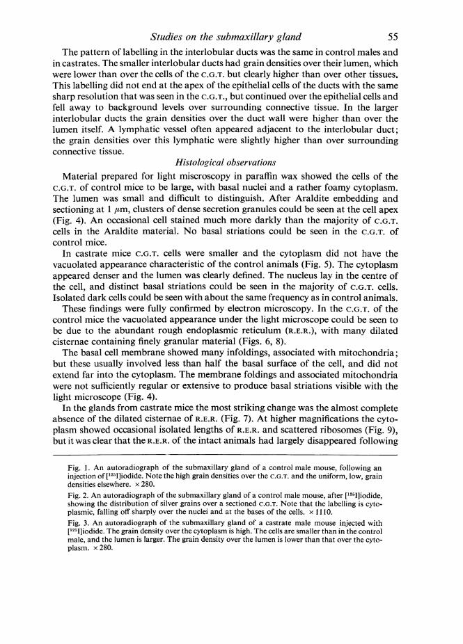
J. Anat. (1971), 109, 1, pp. 51-62 51With 9 figuresPrinted in Great Britain
The effects of castration on theultrastructure and the iodide-concentrating ability of
mouse submaxillary salivary glands
A. W. ROGERS AND K. BROWN-GRANT
M.R.C. Neuroendocrinology Unit, Department ofHuman Anatomy,South Parks Road, Oxford
(Accepted 29 December 1970)
INTRODUCTION
The submaxillary salivary glands of mice are immature at birth, the convolutedgranular tubules (C.G.T.), which are a striking feature of the adult gland, developing ator just before puberty (Brown-Grant & Taylor, 1963). The gland exhibits a sexualdimorphism, the C.G.T. being more prominent in the male but regressing afterthyroidectomy or castration. The histological changes occurring in the submaxillaryglands following castration have been described by Burgen & Emmelin (1961) and byShafer & Muhler (1960); ultrastructural changes have been studied by Caramia(1966a, b).
Radio-iodide is concentrated in both the submaxillary gland and in the saliva inthe mouse, in common with several other species, including man (Cohen & Myant,1959; Brown-Grant, 1961). The site of iodide concentration in the mouse is the C.G.T.(Logothetopoulos & Myant, 1956). This function of the gland also shows sexualdimorphism, the gland-blood ratios for [1311]iodide being higher in males, in which theC.G.T. form a greater proportion of the total gland, than in females (Llach, Tramez-zani & Funes, 1960; Brown-Grant & Taylor, 1963). After castration, in spite of theshrinkage and regression of the C.G.T. which results, the ability of the gland to con-centrate iodide is significantly increased, both in vivo (Llach & Tramezzani, 1962) andin vitro (Brown-Grant & Taylor, 1963).
In the present work autoradiographic studies of iodide distribution in submaxillaryglands of normal mice have been repeated using the isotope 1251. This emits aninternal conversion electron of very low energy, permitting higher resolution than waspossible in previous studies with 1311 (Logothetopoulos & Myant, 1956; Hausler,Bruns & Kutzim, 1965). Improved autoradiographic techniques (Appleton, 1964;Stumpf & Roth, 1964) have also been used to minimize diffusion artefacts. Thesestudies have been extended to the submaxillary glands of castrate mice. In addition,the changes produced in the C.G.T. by castration have been re-examined by electronmicroscopy. The ultrastructural and autoradiographic data have been correlated inan attempt to explain the anomalous effects of castration on the submaxillary glandsof mice.
4-2
A. W. ROGERS AND K. BROWN-GRANT
MATERIALS AND METHODS
The animals were adult male albino mice of the Parkes strain (about 30 g); theyreceived tap water and pelletted diet (Diet 41 B, obtained from E. Dixon andSons, Ltd., Ware) ad lib. This diet contains 117 ,ag/kg iodine. Castration was per-formed under 'Avertin' anaesthesia and animals were used 3-4 weeks later; grossatrophy of the seminal vesicles at autopsy confirmed the completeness of theoperation.The animals were injected intramuscularly with carrier-free [1251]sodium iodide
(Radiochemical Centre, Amersham). The dose varied between 20 and 50 1,c permouse. Animals were killed by bleeding from the aorta, under ether anaesthesia,2 h after injection. Fragments of submaxillary gland and blood samples were takenfor determination of gland/plasma concentration ratios for 1251, using a techniquethat has been fully described elsewhere (Brown-Grant, 1963).
Autoradiography. Fragments of submaxillary gland were rapidly frozen in iso-pentane cooled in liquid nitrogen. Sections at 4 or 5 ,m were cut from these blocks ina cryostat at -24 'C. Autoradiographs were prepared from these sections by twomethods. The first, used in the majority of the experiments, was based on that de-scribed by Appleton (1964). Microscope slides were coated with a 3-4 ,tm layer ofIlford G 5 emulsion by dipping (Rogers, 1967). When dry, these slides were frozen to-24 'C, and the cryostat sections were picked up from the knife by touching themgently with the frozen emulsion layer. The second method was based on that de-scribed by Stumpf & Roth (1964, 1969). The cryostat sections were transferred fromthe knife to a small container and freeze-dried overnight in a cryosorption apparatus(Stumpf & Roth, 1967). The sections were then mounted on emulsion layers atroom temperature.
Non-radioactive submaxillary gland was treated in the same way as a controlagainst positive chemography, which was not encountered in these experiments.However, in initial experiments control slides demonstrated very severe negativechemography. This artefact, in which latent images are lost from the emulsion duringexposure, due to chemical effects from the tissue section, was controlled by proce-dures developed as a result of experiments which have been reported elsewhere(Rogers & John, 1969).
It was found that latent image loss by negative chemography could be prevented inour experimental conditions by a combination of four precautionary steps: a deve-loper based on Metol was used (Ilford ID-l9); the emulsion layers were thoroughlydried in a desiccator before applying the frozen sections; the sections were kept underconditions of low temperature and humidity, both before and after applying them tothe emulsion, to hasten their freeze-drying; and exposure took place either at -40 'Cin a deep-freeze, or at the temperature of solid carbon dioxide. In the experimentsreported here all four precautions were taken and control slides indicated thatnegative chemography did not take place. Exposure times varied from 24 h to 33 days.Conditions of exposure and development followed closely the optimum sequencedetermined by Rogers & John (1969). After development, the sections were stainedwith Harris's haematoxylin.
Electron nmicroscopy. Tissue fragments were rapidly dissected from submaxillary
52
Studies on the submaxillary glandglands of four castrate and four control mice, and fixed in 2-5 % glutaraldehyde in0 085 M cacodylate buffer (pH 7 4). After post-fixation in 1 % osmium tetroxide, thefragments were embedded in Araldite and sectioned on an Ultratome (L.K.B.Instruments, Ltd). Pale gold sections were stained with uranyl acetate and lead citrateand examined in a Phillips electron microscope, model 200.
In addition, 1 ,am Araldite sections were cut from the same material and stained in1 00 toluidine blue in 1 % borax for examination with the light microscope.
RESULTS
Iodide concentrationThe gland/blood ratios for 1251 were determined for fragments of submaxillary
gland from four control mice (13'6, 8-2, 5 2 and 2 9) and three castrates (15 3, 8 0 and7-0).These results confirmed that iodide concentration was occurring, but the values
found showed a greater scatter than those reported by Llach & Tramezzani (1962)and by Brown-Grant & Taylor (1963). The previous experiments were carried outwith whole glands and the small fragments on which the present counts were basedprobably introduced variability in the percentage of C.G.T. present in each fragment.The mean value for the castrate mice was higher than for the controls, but owing tothe very small numbers and the wide scatter, the difference was not statisticallysignificant. The higher concentration in the glands of castrates was, however, clearlyestablished by Llach & Tramezzani (1962) and by Brown-Grant & Taylor (1963),using larger series.
In the experiment in which mouse submaxillary gland was autoradiographed byboth the Appleton and the Stumpf & Roth procedures, the distribution of silvergrains was the same by the two methods. Resolution was similar, and the qualitativeresults were comparable. In this experiment, which involved the injection of 40 ,uC1251 to each mouse, exposure times were 24-48 h. The observed grain densities by theStumpf & Roth method were uniformly higher than those by the Appleton method.Since negative chemography was excluded in both cases, it is reasonable to attributethis difference in efficiency to the lower self-absorption of the freeze-dried specimens.
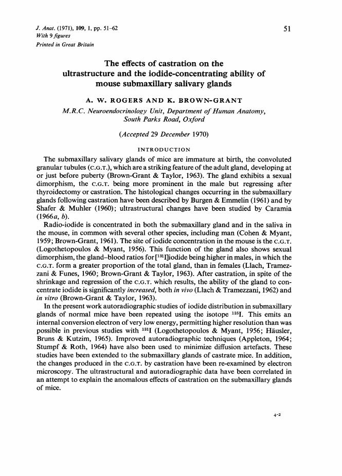
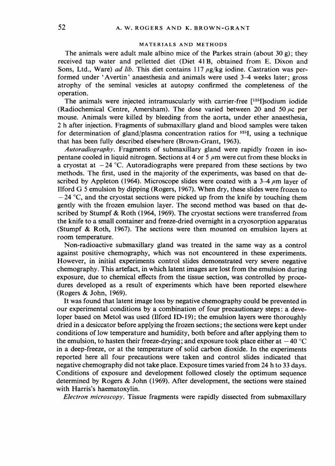
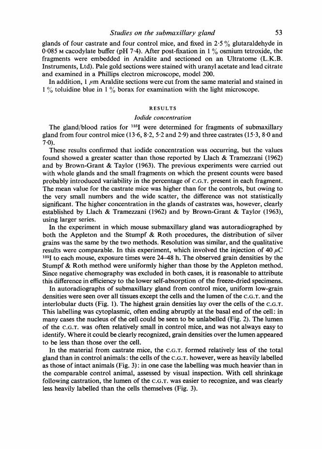
In autoradiographs of submaxillary gland from control mice, uniform low-graindensities were seen over all tissues except the cells and the lumen of the C.G.T. and theinterlobular ducts (Fig. 1). The highest grain densities lay over the cells of the C.G.T.This labelling was cytoplasmic, often ending abruptly at the basal end of the cell: inmany cases the nucleus of the cell could be seen to be unlabelled (Fig. 2). The lumenof the C.G.T. was often relatively small in control mice, and was not always easy toidentify. Where it could be clearly recognized, grain densities over the lumen appearedto be less than those over the cell.
In the material from castrate mice, the C.G.T. formed relatively less of the totalgland than in control animals: the cells of the C.G.T. however, were as heavily labelledas those of intact animals (Fig. 3): in one case the labelling was much heavier than inthe comparable control animal, assessed by visual inspection. With cell shrinkagefollowing castration, the lumen of the C.G.T. was easier to recognize, and was clearlyless heavily labelled than the cells themselves (Fig. 3).
53
54 A. W. ROGERS AND K. BROWN-GRANT'
Studies on the submaxillary glandThe pattern of labelling in the interlobular ducts was the same in control males and
in castrates. The smaller interlobular ducts had grain densities over their lumen, whichwere lower than over the cells of the C.G.T. but clearly higher than over other tissues.This labelling did not end at the apex of the epithelial cells of the ducts with the samesharp resolution that was seen in the C.G.T., but continued over the epithelial cells andfell away to background levels over surrounding connective tissue. In the largerinterlobular ducts the grain densities over the duct wall were higher than over thelumen itself. A lymphatic vessel often appeared adjacent to the interlobular duct;the grain densities over this lymphatic were slightly higher than over surroundingconnective tissue.
Histolog,ical observationsMaterial prepared for light miscroscopy in paraffin wax showed the cells of the
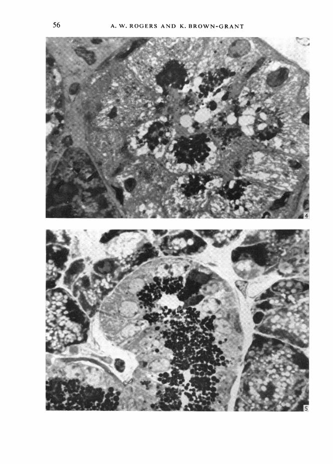
C.G.T. of control mice to be large, with basal nuclei and a rather foamy cytoplasm.The lumen was small and difficult to distinguish. After Araldite embedding andsectioning at 1 ,tm, clusters of dense secretion granules could be seen at the cell apex(Fig. 4). An occasional cell stained much more darkly than the majority of C.G.T.cells in the Araldite material. No basal striations could be seen in the C.G.T. ofcontrol mice.
In castrate mice C.G.T. cells were smaller and the cytoplasm did not have thevacuolated appearance characteristic of the control animals (Fig. 5). The cytoplasmappeared denser and the lumen was clearly defined. The nucleus lay in the centre ofthe cell, and distinct basal striations could be seen in the majority of C.G.T. cells.Isolated dark cells could be seen with about the same frequency as in control animals.
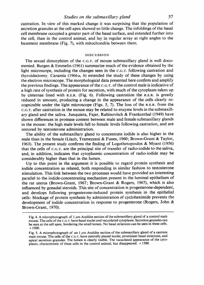
These findings were fully confirmed by electron microscopy. In the C.G.T. of thecontrol mice the vacuolated appearance under the light microscope could be seen tobe due to the abundant rough endoplasmic reticulum (R.E.R.), with many dilatedcisternae containing finely granular material (Figs. 6, 8).The basal cell membrane showed many infoldings, associated with mitochondria;
but these usually involved less than half the basal surface of the cell, and did notextend far into the cytoplasm. The membrane foldings and associated mitochondriawere not sufficiently regular or extensive to produce basal striations visible with thelight microscope (Fig. 4).
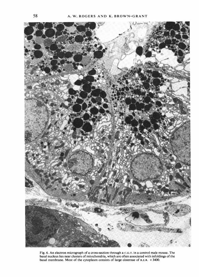
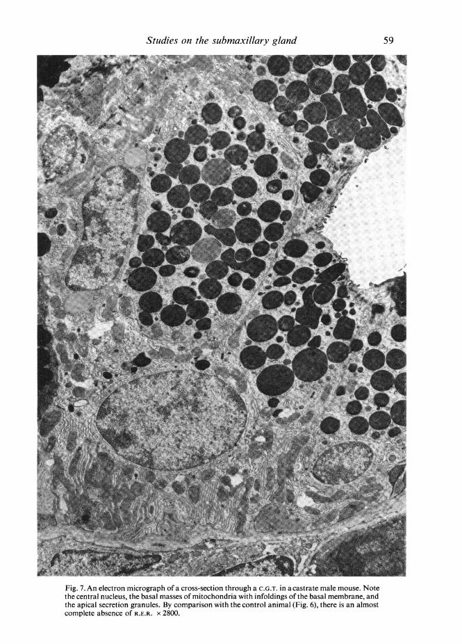
In the glands from castrate mice the most striking change was the almost completeabsence of the dilated cisternae of R.E.R. (Fig. 7). At higher magnifications the cyto-plasm showed occasional isolated lengths of R.E.R. and scattered ribosomes (Fig. 9),but it was clear that the R.E.R. of the intact animals had largely disappeared following
Fig. 1. An autoradiograph of the submaxillary gland of a control male mouse, following aninjection of [1251]iodide. Note the high grain densities over the C.G.T. and the uniform, low, graindensities elsewhere. x 280.Fig. 2. An autoradiograph of the submaxillary gland of a control male mouse, after [1251]iodide,showing the distribution of silver grains over a sectioned C.G.T. Note that the labelling is cyto-plasmic, falling off sharply over the nuclei and at the bases of the cells. x 1110.Fig. 3. An autoradiograph of the submaxillary gland of a castrate male mouse injected with[1251]iodide. The grain density over the cytoplasm is high. The cells are smaller than in the controlmale, and the lumen is larger. The grain density over the lumen is lower than that over the cyto-plasm. x 280.
55
56 A. W. ROGERS AND K. BROWN-GRANT
Studies on the submaxillary glandcastration. In view of this marked change it was surprising that the population ofsecretion granules at the cell apex showed so little change. The infoldings of the basalcell membrane occupied a greater part of the basal surface, and extended further intothe cell, than in the control animal, and lay in regular array at right angles to thebasement membrane (Fig. 7), with mitochondria between them.
DISCUSSION
The sexual dimorphism of the C.G.T. of mouse submaxillary gland is well docu-mented. Burgen & Emmelin (1961) summarize much of the evidence obtained by thelight microscope, including the changes seen in the C.G.T. following castration andthyroidectomy. Caramia (1966a, b) extended the study of these changes by usingthe electron microscope. The morphological data presented here confirm and amplifythe previous findings. The appearance of the C.G.T. of the control male is indicative ofa high rate of synthesis of protein for secretion, with much of the cytoplasm taken upby cisternae lined with R.E.R. (Fig. 6). Following castration the R.E.R. is greatlyreduced in amount, producing a change in the appearance of the cells clearly re-cognizable under the light microscope (Figs. 5, 7). The loss of the R.E.R. from theC.G.T. after castration in the mouse may be related to enzyme levels in the submaxill-ary gland and the saliva. Junquiera, Fajer, Rabinovitch & Frankenthal (1949) haveshown differences in protease content between male and female submaxillary glandsin the mouse: the high male levels fall to female levels following castration, and arerestored by testosterone administration.The ability of the submaxillary gland to concentrate iodide is also higher in the
male than in the female (Llach, Tramezzani & Funes, 1960; Brown-Grant & Taylor,1963). The present study confirms the finding of Logothetopoulos & Myant (1956)that the cells of C.G.T. are the principal site of transfer of radio-iodide to the saliva,and, in addition, indicates that cytoplasmic concentration of radio-iodide may beconsiderably higher than that in the lumen.Up to this point in the argument it is possible to regard protein synthesis and
iodide concentration as related, both responding in similar fashion to testosteronestimulation. This link between the two processes would have provided an interestingparallel to the iodide-concentrating mechanism present in the luminal epithelium ofthe rat uterus (Brown-Grant, 1967; Brown-Grant & Rogers, 1967), which is alsoinfluenced by gonadal steroids. This site of concentration is progesterone-dependent,and develops following progesterone-induced protein synthesis in the epithelialcells: blockage of protein synthesis by administration of cycloheximide prevents thedevelopment of iodide concentration in response to progesterone (Rogers, John &Brown-Grant, 1970).
Fig. 4. A microphotograph of 1 jAm Araldite section of the submaxillary gland of a control malemouse. The cells of the C.G.T. have basal nuclei and vacuolated cytoplasm. Secretion granules canbe seen at the cell apex, bordering the small lumen. No basal striations can be seen in these cells.x 1500.Fig. 5. A microphotograph of an 1 Atm Araldite section of the submaxillary gland of a castratemale mouse. The cells of the C.G.T. have centrally placed nuclei, prominent basal striations, andapical secretion granules. The lumen is clearly visible. The vacuolated appearance of the cyto-plasm, characteristic of these cells in the control animal, has disappeared. x 1500.
57
A. W. ROGERS AND K. BROWN-GRANT
A. s __
qb
4.i
I..
w}
~ ~~~O
LI....~t 4.
F,,. s-i.5A i _
+ s ~~~~~4
>t"
Fig. 6. An electron micrograph of a cross-section through a C.G.T. in a control male mouse. Thebasal nucleus lies near clusters of mitochondria, which are often associated with infoldings of thebasal membrane. Most of the cytoplasm consists of large cisternae of R.E.R. x 3400.
58
.1
IN' .
*Cw.
,& gop",I
n .' ...
_ 1r
@: 4
\ k ..
Studies on the submaxillary gland
'~~~~~~~~~~~~~~~~~~~~~~~~~~~~~~~~~~~~~~~~~~~~.. ........;
.'~~~~~~~~~~~~~~~~~r
mZ
X! ~~~~~~~~~~~~~!
Fig. 7. An electron micrograph of a cross-section through a C.G.T. in a castrate male mouse. Notethe central nucleus, the basal masses of mitochondria with infoldings of the basal membrane, andthe apical secretion granules. By comparison with the control animal (Fig. 6), there is an almostcomplete absence of R.E.R. X 2800.
59
60 A. W. ROGERS AND K. BROWN-GRANT
Studies on the submaxillary glandThe changes that occur in the mouse submaxillary gland following castration,
however, are not consistent with this simple but attractive hypothesis. Llach &Tramezzani (1962) showed that castration results in higher concentration of radio-iodide in the gland in vivo. They suggested that a reduced saliva flow, with retention ofsaliva rich in radio-iodide within the gland, might account for this rather puzzlingobservation, but similar findings in vitro (Brown-Grant & Taylor, 1963) cannot beexplained in this way. The autoradiographic evidence presented here does not supporttheir suggestion, either. No significant pooling of saliva in the interlobular ducts orC.G.T. was observed in castrate as compared to control males. The cells of the C.G.T.,however, were seen to be the principal labelled component of the gland, in castratesas in control mice, even though they were noticeably smaller in castrates (Fig. 3).The observations are consistent with the hypothesis that the cisternae of R.E.R., whichare such a priminent feature of the C.G.T. in the control gland, act as an indifferentconstituent of the cytoplasm so far as iodide concentration is concerned. Removal ofthe R.E.R. cisternae by castration appears to have no effect on the mechanism ofiodide transport, but effectively increases the amount of radio-iodide per unit weightof gland (Llach & Tramezzani, 1962) by eliminating a diluting factor.There remains no readily identifiable structure or organelle associated with the
phenomenon of iodide concentration. The infoldings of basal membrane with closelyassociated mitochondria, which are such a prominent feature of C.G.T. cells in thecastrate, are known to be present in other sites where ion transport is active. Theseinfoldings are also present in the gland of the control male (Fig. 6), though compress-ed and distorted by the cisternae of R.E.R. It is tempting to assign to this membranesystem the function of active transport of iodide, in the cells of the C.G.T. of themouse.
SUMMARY
Previous studies have shown that the submaxillary glands of castrated male miceconcentrate inorganic iodide to a higher level than those of control males. Glandsfrom both groups of mice have been autoradiographed following an injection of[1251]-iodide, using a technique that permits the localization of diffusible material. Inboth groups, iodide was concentrated in the cytoplasm of cells of the convolutedgranular tubules (C.G.T.). Lower levels of concentration were also seen in theepithelium of the interlobular ducts and in their luminal contents.
Cells of the C.G.T. of normal males contained abundant rough endoplasmicreticulum (R.E.R.) with many dilated cisternae, and apical secretion granules. In-foldings of the basal cell membrane, with many associated mitochondria, were seen.Following castration, cells of the C.G.T. were smaller: the R.E.R. largely disappeared.The infoldings of the basal cell membrane, with their associated mitochondria,formed a conspicuous array visible as basal striations under the light microscope.
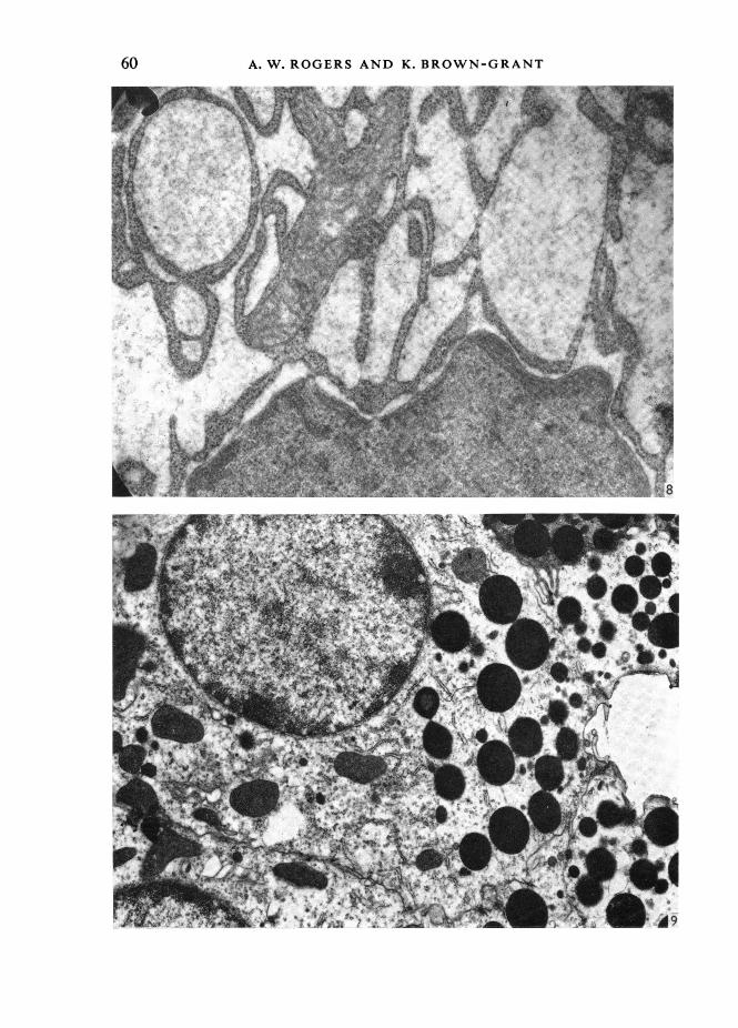
Fig. 8. An electron micrograph of the cisternae of R.E.R. in the cytoplasm of C.G.T. cells in the con-trol male mouse, showing the finely granular material filling them. x 11 500.Fig. 9. An electron micrograph of the apical two-thirds of a cell of the C.G.T. from a castrate malemouse. A few short lengths of R.E.R. are present, and many free ribosomes. The dilated cisternaeof R.E.R. characteristic of the control male are absent. x 30000.
61
A. W. ROGERS AND K. BROWN-GRANT
These findings are consistent with the hypothesis that active transport of iodide isa function of the cell membrane, which produces a high concentration of this ion inthe cytoplasm. Castration eliminates from the cells the cisternae of R.E.R., reducingthe volume of the C.G.T. and of the whole gland, without reducing the volume of thecytoplasmic compartment within which the iodide ion is concentrated.
REFERENCES
APPLETON, T. C. (1964). Autoradiography of soluble labelled compounds. Ji R. microsc. Soc. 83, 277-281.BROWN-GRANT, K. (1961). Extra-thyroidal iodide concentrating mechanism. Physiol. Rev. 41, 189-213.BROWN-GRANT, K. (1963). Failure to demonstrate a concentration of iodide by the submandibular gland
of the rat. J. Physiol., Lond. 165, 519-527.BROWN-GRANT, K. (1967) A quantitative study of the effects of progesterone and related steroids on the
uterus: plasma concentration ratio for radioactive iodide in the rat. J. Endocr. 38, 145-161.BROWN-GRANT, K. & ROGERS, A. W. (1967). The sites of concentration of radio-iodide in oviduct and
uterus of the ovarietomized rat, under influence of progesterone. J. Anat. 101, 622-623.BROWN-GRANT, K. & TAYLOR, W. (1963). The relation between structure and the concentration of iodideby the submandibular glands of mice and hamsters. J. Physiol., Lond. 165, 508-518
BURGEN, A. S. V. & EMMELIN, N. G. (1961). Physiology of the Salivary Glands. London: Arnold.CARAMIA, F. (1966a). IUltratr4wetreof mouse submaxillary gland. I. Sexual differences. J. Ultrastruct.
Res. 16, 505-523.CARAMIA, F. (1966b). Ultrastructure of mouse submaxillary gland. 2. Effect of castration in the male.
J. Ultrastruct. Res. 16, 524-536.COHEN, B. & MYANT, N. B. (1959). Concentration of salivary iodide: a comparative study. J. Physiol.,
Lond. 145, 595-610.HAUSLER, G., BRUNS, W. & KUTZIM, N. (1965). Autoradiographische Untersuchungen zur Wirkungs-
weise von Perchlorat und Methylmerkaptoimidazol auf den Jodstoffwechsel. Nuclearmedizin 4, 317-325.
JUNQUIERA, L. C., FAJER, A., RABINOVITCH, M. & FRANKENTHAL, L. (1949). Biochemical and histochemicalobservations on the sexual dimorphism of mice submaxillary glands. J. cell comp. Physiol. 34, 129-158.
LLACH, J. L. & TRAMEZZANI, J. H. (1962). The action of gonads on the concentration of radio-iodine bythe submaxillary gland of C3H mice. Revue can. Biol. 21, 23-31.
LLACH, J. L., TRAMEZZANI, J. H. & CORDERO FUNES, J. R. (1960). A sexual difference in the concentrationof iodine-131 by the submaxillary gland of mice. Nature, Lond. 188, 1204-1205.
LOGOTHETOPOULOS, J. H. & MYANT, N. B. (1956). Concentration of radio-iodide and 35S-thiocyanate bythe salivary glands. J. Physiol., Lond. 134, 189-194.
ROGERS, A. W. (1967). Techniques of Autoradiography. Amsterdam: Elsevier.ROGERS, A. W. & JOHN, P. N. (1969). Latent image stability in autoradiographs of diffusible substances.
In Autoradiography of Diffusible Substances (Ed. L. J. Roth and W. E. Stumpf). New York: AcademicPress.
ROGERS, A. W., JOHN, P. N. & BROWN-GRANT, K. (1970). Early effects of progesterone on the uterus ofthe ovariectomized rat. J. Anat. 106, 182-183.
SHAFER, W. G. & MUHLER, J. C. (1960). Endocrine influences upon the salivary glands. Ann. N.Y. Acad.Sci. 85, 215-227.
STUMPF, W. E. & ROTH, L. J. (1964). Vacuum freeze-drying of frozen sections for dry-mounting, high-resolution autoradiography. Stain Technol. 39, 219-223.
STUMPF, W. E. & ROTH, L. J. (1967). Freeze-drying of small tissue samples and thin frozen sections below-60 'C. A simple method of cryosorption pumping. J. Histochem. Cytochem. 15, 243-251.
STUMPF, W. E. & ROTH, L. J. (1969). Autoradiography using dry-mounted, freeze-dried sections. InAutoradiography of Diffusible Substances (Ed. L. J. Roth and W. E. Stumpf). New York: AcademicPress.
62