uncoupling protein 2 (ucp2) and p53 expression in invasive...
TRANSCRIPT

565
The Korean Journal of Pathology 2010; 44: 565-70DOI: 10.4132/KoreanJPathol.2010.44.6.565
Background : Uncoupling protein 2 (UCP2) is a recently identified mitochondrial inner mem-brane anion carrier and a negative regulator of reactive oxygen species production. In thisstudy, we evaluated the characteristics and relationships of UCP2 and p53 expression in breastcancer tissues. Methods : Tissue microarray slides from 107 cases of invasive ductal carci-noma of the breast were constructed, UCP2 and p53 immunohistochemical staining was con-ducted, and clinicopathological correlations were investigated. Results : UCP2 expression ininvasive ductal carcinoma was high in 53 cases (49.5%), while p53 expression in invasiveductal carcinoma was high in 37 cases (34.6%). UCP2 expression was correlated significantlywith histological grade (p = 0.038) and mitotic count (p = 0.050). UCP2 expression was cor-related significantly with p53 expression in invasive ductal carcinoma of the breast (p = 0.045).UCP2 expression (p = 0.8308) and p53 expression (p = 0.3292) showed no significant differ-ence for the overall survival rate in patients with invasive ductal carcinoma. Conclusions :UCP2 expression in invasive ductal carcinoma increased proportionally with histological gradeand mitotic count. High UCP2 expression in invasive ductal carcinoma was observed in con-junction with high p53 expression.
Key Words : Carcinoma, ductal, breast; Mitochondrial uncoupling protein 2; Tumor suppressorprotein p53; Reactive oxygen species
Kyu Yeoun Won1∙Gou Young KimYoun Wha Kim1∙Sung-Jig LimJeong Yoon Song2
565
Uncoupling Protein 2 (UCP2) and p53 Expression in Invasive
Ductal Carcinoma of Breast
565 565
Corresponding AuthorJeong Yoon Song, M.D.Department of Surgery, East-West Neo Medical Center, Kyung Hee University College of Medicine,149 Sangil-dong, Gangdong-gu, Seoul 134-727, KoreaTel: +82-2-440-6137Fax: +82-2-440-6137E-mail: [email protected]
*This work was supported by Kyung Hee UniversityResearch Fund in 2007 (KHU-20070704).
Department of Pathology, East-West NeoMedical Center, 1Kyung Hee MedicalCenter,2Department of Surgery, East-West Medical Center,Kyung Hee University College ofMedicine, Seoul, Korea
Received : January 15, 2010Accepted : August 6, 2010
Reactive oxygen species (ROS) contribute to the develop-ment of cancer. It has been suggested that ROS suppress apop-tosis and promote proliferation, invasiveness, and metastasis.1
ROS and cellular oxidant stress have long been associated withcancer. Transformed cells appear to generate more ROS thannormal cells.2 ROS also promote further genomic instabilityand stimulate signaling pathways associated with cellular growthand proliferation.3 Uncoupling protein 2 (UCP2) is a recentlyidentified mitochondrial inner membrane anion carrier, whichhas emerged as a negative regulator of ROS.4 UCP1 is expressedexclusively in brown adipose tissue and is a key molecule inthermogenesis.5 UCP2 is expressed in a variety of tissues, in-cluding the brain, lung, spleen, kidneys, liver, adipose tissues,and heart.6,7 The human UCP2 gene has been mapped to chro-
mosome 11q13.8 UCP2 functions in a variety of cell types as asensor of mitochondrial oxidative stress and may be activatedby superoxide or subsequently formed lipid peroxidation prod-ucts.9,10 The ability of cancer cells to regulate ROS levels con-tributes greatly to autonomous growth, the evasion of apoptosis,and other hallmark characteristics of adaptation.11 Thus, UCP2activity, a negative regulator of ROS, should be related to can-cer development or progression. Actually, Horimoto et al.12
reported that UCP2 expression is correlated with neoplasticchanges in human colon cancer. They also suggested that UCP2expression may increase in colon cancer as a component of tumoradaptation.12 UCP2 expression may facilitate the adaptation ofcancer cells to oxidative stress;3 however, UCP2 expression andits correlation with clinicopathological factors or outcomes in
566 Kyu Yeoun Won∙Gou Young Kim∙Youn Wha Kim, et al.
human breast cancers have not yet been investigated.p53, a widely-studied tumor suppressor gene, has also recently
been implicated in energy metabolism regulation, and UCP2inhibits apoptosis of colon cancer cells by interfering with theROS-mediated phosphorylation of p53 within the transactivat-ing domain.3
In this study, we investigated the characteristics and interrela-tionships of UCP2 and p53 expression in invasive ductal carcino-ma of the breast via immunohistochemical analysis in relation tosurvival and other clinicopathological variables.
MATERIALS AND METHODS
Patients and tissue samples
Tissue samples from 107 cases of invasive ductal carcinomawere utilized. Forty-six cases of normal breast tissues from pa-
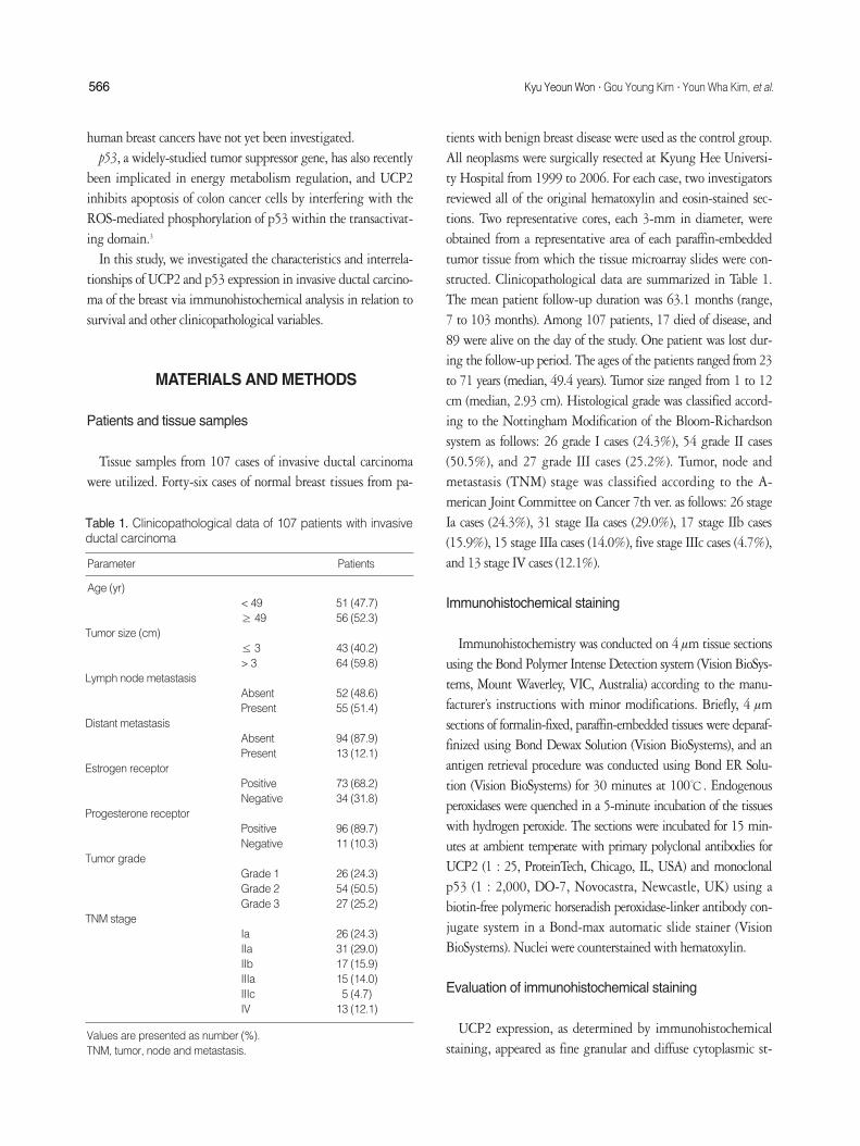
tients with benign breast disease were used as the control group.All neoplasms were surgically resected at Kyung Hee Universi-ty Hospital from 1999 to 2006. For each case, two investigatorsreviewed all of the original hematoxylin and eosin-stained sec-tions. Two representative cores, each 3-mm in diameter, wereobtained from a representative area of each paraffin-embeddedtumor tissue from which the tissue microarray slides were con-structed. Clinicopathological data are summarized in Table 1.The mean patient follow-up duration was 63.1 months (range,7 to 103 months). Among 107 patients, 17 died of disease, and89 were alive on the day of the study. One patient was lost dur-ing the follow-up period. The ages of the patients ranged from 23to 71 years (median, 49.4 years). Tumor size ranged from 1 to 12cm (median, 2.93 cm). Histological grade was classified accord-ing to the Nottingham Modification of the Bloom-Richardsonsystem as follows: 26 grade I cases (24.3%), 54 grade II cases(50.5%), and 27 grade III cases (25.2%). Tumor, node andmetastasis (TNM) stage was classified according to the A-merican Joint Committee on Cancer 7th ver. as follows: 26 stageIa cases (24.3%), 31 stage IIa cases (29.0%), 17 stage IIb cases(15.9%), 15 stage IIIa cases (14.0%), five stage IIIc cases (4.7%),and 13 stage IV cases (12.1%).
Immunohistochemical staining
Immunohistochemistry was conducted on 4 mm tissue sectionsusing the Bond Polymer Intense Detection system (Vision BioSys-tems, Mount Waverley, VIC, Australia) according to the manu-facturer’s instructions with minor modifications. Briefly, 4 mmsections of formalin-fixed, paraffin-embedded tissues were deparaf-finized using Bond Dewax Solution (Vision BioSystems), and anantigen retrieval procedure was conducted using Bond ER Solu-tion (Vision BioSystems) for 30 minutes at 100℃. Endogenousperoxidases were quenched in a 5-minute incubation of the tissueswith hydrogen peroxide. The sections were incubated for 15 min-utes at ambient temperate with primary polyclonal antibodies forUCP2 (1 : 25, ProteinTech, Chicago, IL, USA) and monoclonalp53 (1 : 2,000, DO-7, Novocastra, Newcastle, UK) using abiotin-free polymeric horseradish peroxidase-linker antibody con-jugate system in a Bond-max automatic slide stainer (VisionBioSystems). Nuclei were counterstained with hematoxylin.
Evaluation of immunohistochemical staining
UCP2 expression, as determined by immunohistochemicalstaining, appeared as fine granular and diffuse cytoplasmic st-
Parameter Patients
Age (yr)< 49 51 (47.7)≥ 49 56 (52.3)
Tumor size (cm)≤ 3 43 (40.2)> 3 64 (59.8)
Lymph node metastasis Absent 52 (48.6)Present 55 (51.4)
Distant metastasis Absent 94 (87.9)Present 13 (12.1)
Estrogen receptorPositive 73 (68.2)Negative 34 (31.8)
Progesterone receptorPositive 96 (89.7)Negative 11 (10.3)
Tumor gradeGrade 1 26 (24.3)Grade 2 54 (50.5)Grade 3 27 (25.2)
TNM stageIa 26 (24.3)IIa 31 (29.0)IIb 17 (15.9)IIIa 15 (14.0)IIIc 5 (4.7)IV 13 (12.1)
Values are presented as number (%).TNM, tumor, node and metastasis.
Table 1. Clinicopathological data of 107 patients with invasiveductal carcinoma

UCP2 Expression in Invasive Ductal Carcinoma of Breast 567
aining. Immunohistochemical staining for UCP2 was evaluat-ed based on intensity and proportion. Scattered macrophageswere the positive controls for UCP2.12 Intensity and proportionscores were as follows: 0 (negative), 1 (focal weak), 2 (diffuseweak), and 3 (diffuse strong).12 We regarded strong intensity as
indicative of high expression. p53 expression showed nuclearstaining. p53 expression was categorized as low expression (<10% of tumor cells) and high expression (≥ 10% of tumor cells).Using a receiver operating characteristic curve analysis, we con-sidered 10% as a cut-off value, because the best cutoff score thatyielded the highest p53 expression area under the curve was10%. All slides were independently evaluated by two investi-gators blinded to both the patient’s identities and the clinicaloutcomes.
Statistical analysis
The Pearson’s chi-squared test was used to evaluate the asso-ciation between UCP2 and p53 expression and several clinico-pathological variables. The Kaplan-Meier method was utilizedto determine the probability of survival, and the data were ana-lyzed via the log-rank test. Overall survival was defined as sur-vival from the date of surgery to the date of death due to can-cer. A p-value of < 0.05 was considered significant. Fig. 1. Normal breast glandular epithelium is negative or weakly
focal positive for uncoupling protein 2 expression.
Fig. 2. (A) Uncoupling protein 2 (UCP2) expression in invasiveductal carcinomas shows focally weak granular cytoplasmic stain-ing. (B) UCP2 expression in invasive ductal carcinomas shows dif-fusely weak granular cytoplasmic staining. (C) UCP2 expression ininvasive ductal carcinomas shows diffusely strong granular cyto-plasmic staining.
A B
C

RESULTS
The 46 cases of normal breast glandular epithelium evi-denced negative or weak focally positive UCP2 expression (Fig.1). Myoepithelial cells and scattered macrophages showedstrong UCP2 expression. UCP2 expression in invasive ductalcarcinoma cells was negative (n = 8, 7.5%), focally weak (n =21, 19.6%), diffusely weak (n = 25, 23.4%), and diffusely strong(n = 53, 49.5%) (Fig. 2). Negative, focally weak, and diffuselyweak expressions were classified as low expression and diffuselystrong as high expression. UCP2 expression in invasive ductalcarcinoma cells was classified as “low expression” in 54 cases(50.5%), and “high expression” in 53 cases (49.5%). p53 ex-
568 Kyu Yeoun Won∙Gou Young Kim∙Youn Wha Kim, et al.
Values are presented as number (%). *Significantly different by the chi-squared test.HPF, high power field; TNM, tumor, node and metastasis.
UCP2 expression p53 expression
Low High p-value Low High p-value
Age (yr)< 49 29 (27.1) 22 (20.5) 0.142 33 (30.8) 18 (16.8) 0.522≥ 49 25 (23.4) 31 (29.0) 37 (34.6) 19 (17.8)
Histological gradeGrade I 16 (15.0) 10 (9.3) 0.038* 22 (20.6) 4 (3.7) 0.001*Grade II 30 (28.0) 24 (22.4) 38 (35.5) 16 (15.0)Grade III 8 (7.5) 19 (17.8) 10 (9.3) 17 (15.9)
Tubule formation> 10% of the tumor 18 (16.8) 10 (9.3) 0.089 19 (17.8) 9 (8.4) 0.752≤ 10% of the tumor 36 (33.6) 43 (40.2) 51 (47.7) 28 (26.2)
Nuclear pleomorphismMinimal to moderate 38 (35.5) 33 (30.8) 0.375 53 (49.5) 18 (16.8) 0.005*Marked 16 (15.0) 20 (18.7) 17 (15.6) 19 (17.8)
Mitotic count≤ 10/HPFs 47 (43.9) 38 (35.5) 0.050* 62 (57.9) 23 (21.5) 0.001*> 11/10HPFs 7 (6.5) 15 (14.0) 8 (7.5) 14 (13.1)
Lymph node metastasisAbsent 26 (24.3) 26 (24.3) 0.925 37 (34.6) 15 (14.0) 0.225Present 28 (26.2) 27 (25.2) 33 (30.8) 22 (20.6)
Estrogen receptorPositive 40 (37.4) 33 (30.8) 0.190 52 (48.6) 21 (19.6) 0.064Negative 14 (13.1) 20 (18.7) 18 (16.8) 16 (15.0)
Progesterone receptorPositive 50 (46.7) 46 (43.0) 0.323 63 (58.9) 33 (30.8) 0.895Negative 4 (3.7) 7 (6.5) 7 (6.5) 4 (3.7)
Distant metastasisAbsent 49 (45.8) 45 (42.1) 0.356 64 (59.8) 30 (28.0) 0.119Present 5 (4.7) 8 (7.5) 6 (5.6) 7 (6.5)
Tumor size (cm)< 3 18 (16.8) 25 (23.4) 0.103 30 (28.0) 13 (12.1) 0.286≥ 3 36 (33.6) 28 (26.2) 40 (37.4) 24 (22.4)
TNM Stage Ia-IIa 29 (27.1) 28 (26.2) 0.928 42 (39.3) 15 (14.0) 0.055IIb-IV 25 (23.4) 25 (23.4) 28 (26.2) 22 (20.6)
Table 2. Correlation between uncoupling protein 2 (UCP2), p53 expression, and clinicopathological variables in 107 invasive ductalcarcinomas
Variables
Fig. 3. p53 expression in invasive ductal carcinomas showsstrong nuclear staining.

pression in invasive ductal carcinoma was classified as “low ex-pression” in 70 cases (65.4%), and “high expression” in 37 cases(34.6%) (Fig. 3). As shown in Table 2, UCP2 expression wascorrelated significantly with histological grade (p = 0.038), andmitotic count (p = 0.050). p53 expression was correlated sig-nificantly with histological grade (p = 0.001), nuclear pleo-morphism (p = 0.005), and mitotic count (p = 0.001). UCP2expression was correlated significantly with p53 expression ininvasive ductal carcinoma of the breast (p = 0.045) (Table 3).Using the Kaplan-Meier method, lymph node metastasis (p =0.0017), estrogen receptor status (p = 0.0173), distant metas-tasis (p < 0.00001), and TNM stage (p < 0.00001) were iden-tified as significant prognostic factors. However, UCP2 expres-sion (p = 0.8308) and p53 expression (p = 0.3292) showed nosignificant difference for the overall survival rate in patients withinvasive ductal carcinoma (Table 4).
DISCUSSION
UCP2 is a negative regulator of ROS and acts as a sensor ofmitochondrial oxidative stress.9 A variety of malignant tumors,including thyroid tumors, lymphomas, and colon cancer over-express UCP2.12-14 Recently, UCP2 overexpression was revealedin some breast cancer cell lines.15 Increased UCP2 expressionhas also been observed in human colon cancer cells, and has beencorrelated with the degree of neoplastic change.12 It has beensuggested that UCP2 acts as an adaptive mechanism to reduceoxidative stress in colonic tumor tissues.12 Collins et al.16 observedthat UCP2 overexpression in HepG2 human hepatoma cellslowers intracellular ROS levels and attenuates apoptosis inducedby a variety of challenges. To date, UCP2 expression in humanbreast cancer tissue has not been investigated.
In the present study, UCP2 expression in invasive ductal car-cinoma of the breast was stronger than in normal breast glandu-lar epithelium. In Horimoto’s colon cancer study, UCP2 over-expression was suggested to protect a wide array of cell types
from apoptosis and that the cytoprotective effect of UCP2 islikely based on a reduction in mitochondrial ROS generation.Tumor cells may use UCP2 as a metabolic adaptation to avoidROS-mediated apoptosis.12 Our results also support that UCP2overexpression is an adaptive response limiting ROS in breastcancer cells.15
Second, UCP2 overexpression was correlated significantlywith higher histological grade and mitotic counts in invasiveductal carcinoma. Histological grade and mitotic counts arereflective of the aggressiveness of a malignancy. These findingssuggest that more aggressive invasive ductal carcinoma cellsexpress more UCP2, which is believed to be an adaptive mech-anism for reducing ROS production.12 Changes in cancer cellmetabolism are frequently associated with more aggressive tu-mor growth and drug resistance, thereby resulting in a worseprognosis.17 One of the purposes of metabolic switches in can-cer cells is to reduce ROS generation.12 Thus, UCP2, as a nega-tive ROS regulator, might be involved in changes in cancer cellmetabolism associated with tumor aggressiveness. As a result,oxidative stress in breast cancer may contribute to induce a moremalignant transformation.
Derdak et al.3 observed that UCP2-overexpressing colon can-cer cells perform an active role in promoting cancer cell sur-vival through antiapoptotic effects of p53. We determinedwhether UCP2 expression in invasive ductal carcinoma wascorrelated with p53 expression, which is associated with apop-tosis. As a result, a positive correlation between UCP2 and p53expression was observed in invasive ductal carcinoma. Based onthese findings, we suggest that the function of UCP2 in inva-sive ductal carcinoma may be derived from a modulation of
UCP2 Expression in Invasive Ductal Carcinoma of Breast 569
Variables Overall survival rate (p-value)
Age 0.2165Tumor size (> 3 cm vs ≤ 3 cm) 0.9527Lymph node metastasis 0.0017*Histological grade (grade I, II vs grade III) 0.1107Tubule formation (> 10% vs ≤ 10%) 0.7436Nuclear pleomorphism 0.3591Mitosis 0.1932Estrogen receptor 0.0173*Progesterone receptor 0.4206Distant metastasis < 0.00001*TNM stage < 0.00001*UCP2 expression 0.8308p53 expression 0.3292
*Significantly different by Kaplan-Meier test.TNM, tumor, node and metastasis; UCP2, uncoupling protein 2.
Table 4. Analysis of 13 clinicopathological variables for overallsurvival rate in 107 invasive ductal carcinomas
UCP2 expression
High Low p-value
p53 expression High 23 (21.5) 14 (13.1) 0.045*Low 30 (28.0) 40 (37.4)
Values are presented as number (%). *Significantly different by the chi-squared test.
Table 3. Correlation of uncoupling protein 2 (UCP2) and p53expression in 107 invasive ductal carcinomas
p53 function in accordance with previous results.3 However,further studies into the relationship between UCP2-overex-pressed breast cancer and antiapoptotic effects will be required.
Human colon cancer cells that overexpress UCP2 inhibit ROSaccumulation and apoptosis after exposure to chemotherapeuticagents.3 It has also been suggested that a link may exist betweenUCP2 and the molecular mechanisms of chemoresistance, andthat UCP2 may be a molecular target of great usefulness in no-vel treatment strategies.
In conclusion, UCP2 expression was stronger in invasive du-ctal carcinoma cells than in normal breast glandular epitheli-um. Additionally, UCP2 expression in invasive ductal carcino-ma increased proportionally with histological grade and mitot-ic count. Our results assume that UCP2 overexpression may bean adaptive response, which limits ROS in breast cancer cells,and that oxidative stress contributes to induce a more malignanttransformation in invasive ductal carcinoma of the breast.
REFERENCES
1. Halliwell B. Oxidative stress and cancer: have we moved forward?
Biochem J 2007; 401: 1-11.
2. Schumacker PT. Reactive oxygen species in cancer cells: live by the
sword, die by the sword. Cancer Cell 2006; 10: 175-6.
3. Derdak Z, Mark NM, Beldi G, Robson SC, Wands JR, Baffy G. The
mitochondrial uncoupling protein-2 promotes chemoresistance in
cancer cells. Cancer Res 2008; 68: 2813-9.
4. Arsenijevic D, Onuma H, Pecqueur C, et al. Disruption of the un-
coupling protein-2 gene in mice reveals a role in immunity and reac-
tive oxygen species production. Nat Genet 2000; 26: 435-9.
5. Ricquier D, Bouillaud F. The uncoupling protein homologues: UCP1,
UCP2, UCP3, StUCP and AtUCP. Biochem J 2000; 345 Pt 2: 161-79.
6. Fleury C, Neverova M, Collins S, et al. Uncoupling protein-2: a novel
gene linked to obesity and hyperinsulinemia. Nat Genet 1997; 15:
269-72.
7. Gimeno RE, Dembski M, Weng X, et al. Cloning and characterization
of an uncoupling protein homolog: a potential molecular mediator
of human thermogenesis. Diabetes 1997; 46: 900-6.
8. Bouchard C, Pe@russe L, Chagnon YC, Warden C, Ricquier D. Linkage
between markers in the vicinity of the uncoupling protein 2 gene and
resting metabolic rate in humans. Hum Mol Genet 1997; 6: 1887-9.
9. Echtay KS, Roussel D, St-Pierre J, et al. Superoxide activates mito-
chondrial uncoupling proteins. Nature 2002; 415: 96-9.
10. Krauss S, Zhang CY, Scorrano L, et al. Superoxide-mediated activa-
tion of uncoupling protein 2 causes pancreatic beta cell dysfunction.
J Clin Invest 2003; 112: 1831-42.
11. Hanahan D, Weinberg RA. The hallmarks of cancer. Cell 2000; 100:
57-70.
12. Horimoto M, Resnick MB, Konkin TA, Routhier J, Wands JR, Baffy
G. Expression of uncoupling protein-2 in human colon cancer. Clin
Cancer Res 2004; 10(18 Pt 1): 6203-7.
13. Savagner F, Franc B, Guyetant S, Rodien P, Reynier P, Malthiery Y.
Defective mitochondrial ATP synthesis in oxyphilic thyroid tumors.
J Clin Endocrinol Metab 2001; 86: 4920-5.
14. Harper ME, Antoniou A, Villalobos-Menuey E, et al. Characteriza-
tion of a novel metabolic strategy used by drug-resistant tumor cells.
FASEB J 2002; 16: 1550-7.
15. Fine EJ, Miller A, Quadros EV, Sequeira JM, Feinman RD. Acetoac-
etate reduces growth and ATP concentration in cancer cell lines which
over-express uncoupling protein 2. Cancer Cell Int 2009; 9: 14.
16. Collins P, Jones C, Choudhury S, Damelin L, Hodgson H. Increased
expression of uncoupling protein 2 in HepG2 cells attenuates oxida-
tive damage and apoptosis. Liver Int 2005; 25: 880-7.
17. Cuezva JM, Krajewska M, de Heredia ML, et al. The bioenergetic si-
gnature of cancer: a marker of tumor progression. Cancer Res 2002;
62: 6674-81.
570 Kyu Yeoun Won∙Gou Young Kim∙Youn Wha Kim, et al.