understanding retroperitoneal anatomy for lateral …ssrr-journal.jp/pdf/001030107.pdf107 review...
TRANSCRIPT
107
REVIEW ARTICLE SPINE SURGERY AND RELATED RESEARCH
Understanding Retroperitoneal Anatomy for LateralApproach Spine Surgery
Tokumi Kanemura1), Kotaro Satake1), Hiroaki Nakashima1), Naoki Segi1), Jun Ouchida1), Hidetoshi Yamaguchi2) and
Shiro Imagama2)
1) Department of Orthopedic Surgery, Spine Center, Konan Kosei Hospital, Aichi, Japan2) Department of Orthopedic Surgery, Nagoya University Graduate School of Medicine, Aichi, Japan
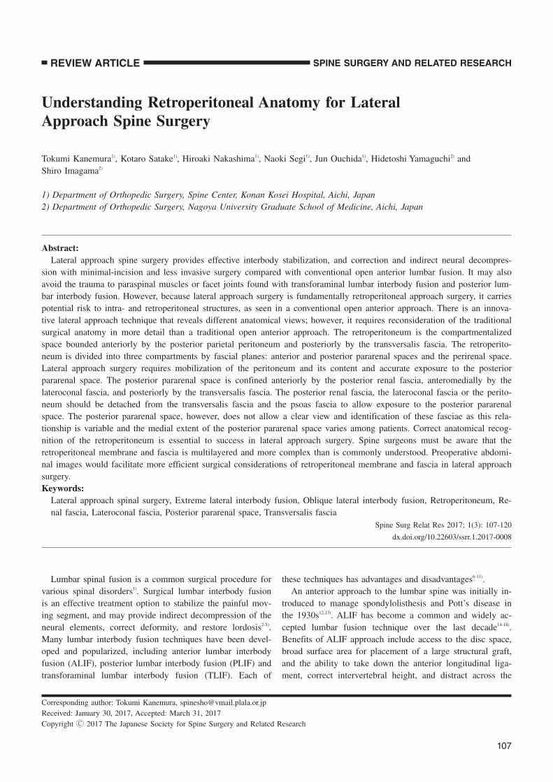
Abstract:Lateral approach spine surgery provides effective interbody stabilization, and correction and indirect neural decompres-
sion with minimal-incision and less invasive surgery compared with conventional open anterior lumbar fusion. It may also
avoid the trauma to paraspinal muscles or facet joints found with transforaminal lumbar interbody fusion and posterior lum-
bar interbody fusion. However, because lateral approach surgery is fundamentally retroperitoneal approach surgery, it carries
potential risk to intra- and retroperitoneal structures, as seen in a conventional open anterior approach. There is an innova-
tive lateral approach technique that reveals different anatomical views; however, it requires reconsideration of the traditional
surgical anatomy in more detail than a traditional open anterior approach. The retroperitoneum is the compartmentalized
space bounded anteriorly by the posterior parietal peritoneum and posteriorly by the transversalis fascia. The retroperito-
neum is divided into three compartments by fascial planes: anterior and posterior pararenal spaces and the perirenal space.
Lateral approach surgery requires mobilization of the peritoneum and its content and accurate exposure to the posterior
pararenal space. The posterior pararenal space is confined anteriorly by the posterior renal fascia, anteromedially by the
lateroconal fascia, and posteriorly by the transversalis fascia. The posterior renal fascia, the lateroconal fascia or the perito-
neum should be detached from the transversalis fascia and the psoas fascia to allow exposure to the posterior pararenal
space. The posterior pararenal space, however, does not allow a clear view and identification of these fasciae as this rela-
tionship is variable and the medial extent of the posterior pararenal space varies among patients. Correct anatomical recog-
nition of the retroperitoneum is essential to success in lateral approach surgery. Spine surgeons must be aware that the
retroperitoneal membrane and fascia is multilayered and more complex than is commonly understood. Preoperative abdomi-
nal images would facilitate more efficient surgical considerations of retroperitoneal membrane and fascia in lateral approach
surgery.
Keywords:Lateral approach spinal surgery, Extreme lateral interbody fusion, Oblique lateral interbody fusion, Retroperitoneum, Re-
nal fascia, Lateroconal fascia, Posterior pararenal space, Transversalis fascia
Spine Surg Relat Res 2017; 1(3): 107-120
dx.doi.org/10.22603/ssrr.1.2017-0008
Lumbar spinal fusion is a common surgical procedure for
various spinal disorders1). Surgical lumbar interbody fusion
is an effective treatment option to stabilize the painful mov-
ing segment, and may provide indirect decompression of the
neural elements, correct deformity, and restore lordosis2-5).
Many lumbar interbody fusion techniques have been devel-
oped and popularized, including anterior lumbar interbody
fusion (ALIF), posterior lumbar interbody fusion (PLIF) and
transforaminal lumbar interbody fusion (TLIF). Each of
these techniques has advantages and disadvantages6-11).
An anterior approach to the lumbar spine was initially in-
troduced to manage spondylolisthesis and Pott’s disease in
the 1930s12,13). ALIF has become a common and widely ac-
cepted lumbar fusion technique over the last decade14-18).
Benefits of ALIF approach include access to the disc space,
broad surface area for placement of a large structural graft,
and the ability to take down the anterior longitudinal liga-
ment, correct intervertebral height, and distract across the
Corresponding author: Tokumi Kanemura, [email protected]
Received: January 30, 2017, Accepted: March 31, 2017
Copyright Ⓒ 2017 The Japanese Society for Spine Surgery and Related Research
Spine Surg Relat Res 2017; 1(3): 107-120 dx.doi.org/10.22603/ssrr.1.2017-0008
108
disc space to create lordosis19,20). Posterior fixation, including
transpedicular screw or translaminar screw supplemented
ALIF, significantly improves segmental flexibility and fixa-
tion stiffness19,21-23). However, ALIF with posterior instru-
mented fusion requires two separate approaches (anterior/
posterior), which prolongs operative time in a single day or
necessitates staging of procedures24). In addition, ALIF has
potential approach-related serious risks and complications,
including vascular injury, ureteral damage, ileus, and retro-
grade ejaculation in males25-32). Anterior approaches may also
involve unfamiliar anatomy for spine surgeons, potentially
placing the patient at risk for approach-related complica-
tions; therefore, many spine surgeons prefer the assistance of
an “access surgeon” to perform the exposure33).
PLIF has evolved since the initial description of the tech-
nique34), with the development of additional autologous and
synthetic bone grafting options, more advanced methods of
spinal segmental fusion techniques, innovative implants, and
the pedicle screw fixation. PLIF is a traditional posterior
lumbar approach that many spine surgeons are very familiar
with and well-trained in performing. The posterior exposure
provides excellent visualization of the nerve roots and al-
lows for neural decompression while maintaining posterior
support structures. Furthermore, PLIF also allows for a 360-
degree fusion through a single approach2,3,8,10,11,35,36). Tradi-
tional open TLIF was first described in 198237). The main
concerns with the PLIF are the extent of neural retraction
required, potential nerve root injury, dural tears, and
epidural fibrosis. TLIF avoids these complications by pro-
viding direct, unilateral access to the intervertebral foraminal
space, and reducing direct dissection and surgical trauma to
spinal muscles and structural integrity20,38-42). Traditional open
PLIF and TLIF have advanced to become mini-open or
minimally invasive approaches43).
The posterior approach in PLIF and TLIF, however, is as-
sociated with potentials risks and complications such as sig-
nificant iatrogenic paraspinal muscle damage, inadvertent
durotomy, and retraction injury of nerve roots causing fibro-
sis and chronic radiculopathy. It may also be relatively more
difficult to correct coronal imbalance and restore lordosis
with PLIF/TLIF than ALIF. Furthermore, endplate prepara-
tion may be difficult compared to anterior fusion ap-
proaches20,38,44-47).
More recently, the lateral trans-psoas approach, termed
extreme, direct or lateral lumbar interbody fusion (XLIFⓇ,
Nuvasive; DLIFⓇ, Medtronic; LLIF), has gained popularity.
Initially developed in the late 1990s by Luiz Pimenta as a
lateral endoscopic trans-psoas retroperitoneal approach48), it
was first published in the literature in 2006 by Ozgur et
al.49). Since then, LLIF has gained exponential acceptance as
a minimally invasive option for thoracolumbar fusions.
Compared with traditional open anterior access, LLIF allows
a less invasive approach corridor and avoidance of great ves-
sels48-50). Oblique lateral interbody fusion (OLIF, Med-
tronicⓇ), another lateral approach surgery, was subsequently
introduced as an alternative procedure to a lateral trans-
psoas approach. The OLIF procedure allows for psoas-
preserving access via an anterior oblique retroperitoneal ap-
proach51).
These lateral procedures have advantages over the mini-
mally invasive technique, including direct visualization with
novel special instruments and light equipment, excellent disc
space preparation, and the ability to place larger interbody
cages; therefore, resisting subsidence and creating a biome-
chanically superior environment for bone healing. LLIF also
allows disc height and alignment restoration, and decom-
pression of the nerve roots by powerful indirect decompres-
sion through the ligamentotaxis created with intact anterior
and posterior longitudinal ligament, as these ligaments has a
key role in spinal alignment and stabilization49,52-54). Further-
more, the LLIF approach, including lateral release and liga-
mentotaxis by disc height restoration, is an excellent option
for sagittal and coronal deformity correction, especially in
lumbar degenerative scoliosis with laterolisthesis55-57). Numer-
ous lateral procedures have been performed worldwide, and
variations and technical refinements have been proposed.
The procedure has also been extended to a wider range of
indications, now including primary and revision surgery for
degenerative problems, adult deformity, trauma, and thoracic
disc herniation52,58-67).
The most recent advance in lateral approach surgery has
received attention for its application to other spinal problems
that were not indicated for original LLIF procedures. A lat-
eral approach corpectomy and vertebral reconstruction with
a vertebral cage comprising rectangular footplates (X-
Core2Ⓡ, Nuvasive), which may provide better subsidence re-
sistance, is an effective alternative to conventional ap-
proaches for the treatment of trauma and tumor68-71). Another
advanced lateral approach technique is anterior column re-
alignment (ACRⓇ, Nuvasive), which provides a solution for
sagittal imbalance. ACR involves a deliberate release of the
anterior longitudinal ligament (ALL) and placement of hy-
perlordotic interbody cages at either 20 or 30 degrees of lor-
dosis, which are then fixed to the vertebral body with one or
two screws. ACR results in greater segmental correction than
achieved with LLIF alone, successfully restores lumbar lor-
dosis in patients with adult spinal deformity with sagittal
imbalance72-76).
However, potential LLIF-related risks include injury to
the psoas muscle and the lumbar plexus, and to the nerves
that lie within it77-80). Therefore, the procedure requires the
use of real-time electromyographic (EMG) monitoring81).
Fundamentally, lateral approach surgery including LLIF, lat-
eral corpectomy, and ACR is retroperitoneal approach sur-
gery. An anterior retroperitoneal approach uses the latent
space between the back and psoas muscles and the perito-
neal content. As this approach has the potential risk for in-
traperitoneal or retroperitoneal structures (including viscera
and vessels), surgical exposure requires extensive mobiliza-
tion of the great vessels and the peritoneal content. Tradi-
tional ALIF method using a wide-open anterior approach,
have largely been abandoned because of significant compli-

dx.doi.org/10.22603/ssrr.1.2017-0008 Spine Surg Relat Res 2017; 1(3): 107-120
109
Table 1. The Extraperitoneal Spaces.
Portion of the
abdominal wallArea
Rertoperitoneal space
=Retroperitoneum
Posteriorly
Laterally
Between the parietal peritoneum of the posterior
abdominal wall and transversalis fascia
Preperitoneal space
(Properitoneal space)
Anteriorly Between the parietal peritoneum of the anterior
abdominal wall and the transversalis fascia.
Subperitoneal pelvic space Caudally Under the peritoneum
Figure 1. Extraperitoneal space and organs.
TF=transversalis fascia; RF=renal fascia; LCF=lateroconal fascia; PS=psoas muscle; QL=quadratus
lumborum muscle; P=pancreas; K=kidney; AC=ascending colon; DC=descending colon;
D=duodenum; A=aorta; V=vena cava.
Intraperitoneal Space
ParietalPeritoneum
TF
K KPS PS
DCAC
LCF
Anterior RF
Posterior RF
Retroperitoneum
APSPRSPPS
Preperitoneal space
AV
PDD
QL QL
cation rates and the high incidence of pain and abdominal
wall herniation after surgery. A lateral approach with spe-
cialized instruments should be developed as a less invasive
alternative to conventional open ALIF to avoid such com-
plaints. However, one of present author’s major concerns is
the limited visualization of the retroperitoneal space and
minimized working space. Limited visualization does not
warn the surgeon of the presence of viscera and vessels,
even at close quarters. Minimized working space makes it
more difficult to control vascular or visceral injury if it oc-
curs. Many studies have described complications of lateral
approach surgery concerning vascular or visceral in-
jury73,76,82-86), with some complications being catastrophic87-89).
As the lateral approach requires mobilization of the perito-
neum for exposure to the retroperitoneal space and subse-
quent lateral access, misunderstanding about the peritoneum
and inadequate exposure to the retroperitoneal space in-
creases the risk of injury to the peritoneum and its content.
To avoid or manage such complications, surgeons should be
aware of potential complications associated with the anterior
approach and recognize the importance of a comprehensive
understanding of the anatomy of the retroperitoneal space
and its content. This article reviews the clinically relevant
anatomy of the abdominal retroperitoneal spaces (the retrop-
eritoneum) to facilitate the safe and reliable exposure to the
retroperitoneal space for lateral approach surgery.
Extraperitoneal space
The extraperitoneal space is the space between the parie-
tal peritoneum and the investing fascia of the muscles. It cir-
cumferentially surrounds the abdominal cavity; posteriorly
and laterally (retroperitoneal space), anteriorly (preperitoneal
space), and caudally (subperitoneal pelvic space) (Table 1)
(Fig. 1). In the posterior abdominal wall, the extraperitoneal
space is called the retroperitoneum, which is confined to the
posterior and lateral portion of the abdominal and pelvic
wall. The investing fascia of the diaphragm, quadratus lum-
borum muscle, and the transversus muscle (with or without
psoas muscle) is called transversalis fascia (TF). The extrap-
eritoneal space in the anterior abdominal wall is called the
preperitoneal space. In the pelvis, the extraperitoneal space
is called the subperitoneal pelvic space90).
The retroperitoneum
The retroperitoneum is the compartmentalized space
bounded anteriorly by the posterior parietal peritoneum and
posteriorly by the TF. It extends from the diaphragm superi-
orly to the pelvic brim inferiorly.
Spine Surg Relat Res 2017; 1(3): 107-120 dx.doi.org/10.22603/ssrr.1.2017-0008
110
Table 2. Three Compartments of Retroperitoneum.
Boundary Organs
Anterior pararenal space APS Anterior: Posterior parietal peritoneum
Posterior: Anterior renal fascia
Lateral: Lateroconal fascia
Ascending/descending colon
Duodenal loop
Pancreas
Perirenal space PRS Anterior: Anterior renal fascia (Gerota fascia)
Posterior: Posterior renal fascia (Zuckerkandl fascia)
Kidney
Renal vessel
Adrenal glands
Renal pelvis
Proximal ureter
Posterior pararenal space PPS Anterior: Posterior renal fascia
Anteromedial: Lateroconal fascia
Posterior: Transversalis fascia
Medial: Psoas fascia
Superior: Subdiaphragmatic layer
Inferior: Open to the pelvis
Fat tissue
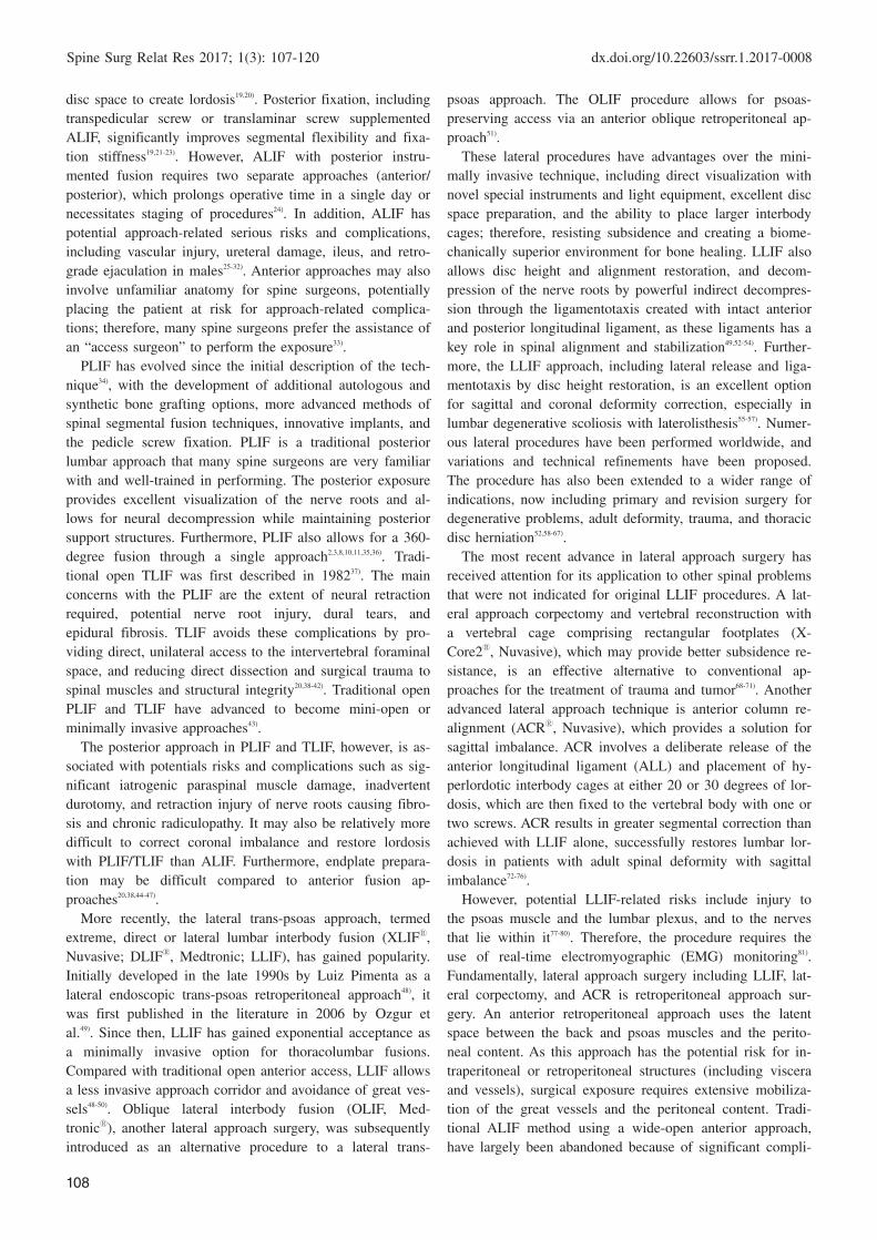
Figure 2. The three extraperitoneal compartments.
Striped areas=anterior pararenal space; stippled areas=perirenal space; cross-hatched areas=posterior
pararenal space; TF=transversalis fascia; P=pancreas; K=kidney; AC=ascending colon: DC=descending
colon; D=duodenum; A=aorta; V=vena cava.
Meyers MA, Charnsangavej C, Oliphant M. Meyers’ dynamic radiology of the abdomen: normal
and pathologic anatomy. New York: Springer; 2011. xviii, 419 p. p 116; with permission.
K DCACAV
PDD
K
Peritoneum TF
a b
Liver
K
AC
Peritoneum
TF
Adrenal
Psoas
Duodenum
Three compartments anatomy of the retroperitoneum
The retroperitoneum is divided by fascial planes into pre-
cisely three individual compartments: the anterior and poste-
rior pararenal spaces and the perirenal space91-96) (Fig. 2) (Ta-
ble 2).
Anterior pararenal space (APS)
The APS is confined anteriorly by the posterior parietal
peritoneum, and posteriorly the anterior renal fascia (RF).
Significantly, it is confined laterally by the lateroconal fascia
(LCF). It contains the ascending and descending colon, the
duodenal loop, and the pancreas. Ventrally, the APS is ana-
tomically continuous with the roots of the small bowel mes-
entery and transverse mesocolon97,98).
Perirenal space (PRS)
The PRS has the shape of an inverted cone extending
from the diaphragmatic fascia to the iliac fossa. It is con-
fined by the anterior RF (Gerota fascia99)) and posterior RF
(Zuckerkandl fascia100)). The PRS contains the kidneys, renal
vessels, adrenal glands, renal pelvis, proximal ureters, and
its investing fat (perirenal fat).
There is some controversy regarding the medial and infe-
rior extents of the PRS. Historically, it was assumed that the
PRS generally had no continuity across the midline92,96). Me-
dially, the posterior fascial layer fuses with the psoas or
quadratus lumborum fascia101), and the anterior RF blends

dx.doi.org/10.22603/ssrr.1.2017-0008 Spine Surg Relat Res 2017; 1(3): 107-120
111
Table 3. Retroperitoneal Fasciae (Ab-
breviations Used in This Article).
RF Renal fascia (anterior, posterior)
LCF Lateroconal fascia
TF Transversalis fascia
FF Fusion fascia
TLF Thoracolumbar fascia
into the dense mass of connective tissue surrounding the
great vessels at the root of the mesentery and behind the
pancreas and duodenum102). However, in vivo cases and ca-
daveric injection studies suggest there may be some commu-
nication across the midline below the level of the renal hi-
lum103,104). In addition, there is controversy regarding the
patency and caudal extent of the PRS. Previously, it was
suggested that the PRS is closed inferiorly by the fusion of
RF. However, in vivo cases and cadaveric injection studies
demonstrated that the cone-like shape of PRS is open at its
inferior extent in the extraperitoneal pelvis103,105) (Fig. 2b).
At the level of the iliac crest, below the cone of RF, the
anterior and posterior pararenal spaces are in potential com-
munication. At this same level, the LCF disappears as a dis-
tinct boundary, and the APS communicates laterally with the
preperitoneal fat of the flank stripe. Superiorly, posterior
pararenal fat continues as a thin subdiaphragmatic layer of
extraperitoneal fat91,92,96).
Posterior pararenal space (PPS)
The PPS is confined anteriorly by the posterior RF, poste-
riorly by the TF, and medially by the psoas muscle. It con-
tinues laterally external to the LCF as the preperitoneal fat
of the abdominal wall. Inferiorly, the PPS is open to the pel-
vis106). As opposed to the other two extraperitoneal spaces,
the PPS contains no organs, and almost always only con-
tains fat. Furthermore, its most notable feature is that it con-
tinues uninterrupted external to the LCF as preperitoneal fat
on the abdominal wall. It is important to recognize that it is
posterior pararenal fat, as it courses laterally external to the
LCF and deep to the TF105). The space is open laterally to-
ward the flank and inferiorly toward the pelvis. Bilaterally,
they are potentially in communication only via the preperi-
toneal fat of the anterior abdominal wall deep to the TF.
Interfascial planes
This traditional three-compartmental anatomy does not
provide a complete explanation for the spread of fluid col-
lections or tumors in the retroperitoneum. It is now believed
that the retroperitoneal fasciae are multilaminated structures
with potentially expandable interfascial planes107). These
planes are represented by the retromesenteric, retrorenal,
lateroconal, and combined interfascial planes. Knowledge of
the anatomy and interconnections of these interfascial planes
can facilitate understanding of the extent and pathways of
spread of retroperitoneal disease108).
Membrane and fasciae
Fascia is fundamental to understanding retroperitoneal
anatomy, but is often misunderstood in surgical practice.
Definitions of fascia vary between texts and between coun-
tries109). The retroperitoneal fascial planes are multilaminated
rather than composed of single membranes. The fasciae are
lamina of connective tissue approximately 2 mm thick that
form the partitions between retroperitoneum compart-
ments110). A clear concept and definition of fascia is impor-
tant when approaching the retroperitoneum (Table 3).
Peritoneum
The peritoneum is a thin, translucent, serous membrane.
The peritoneum that lines the abdominal wall is called the
parietal peritoneum, whereas the peritoneum that covers a
viscus or an organ is visceral peritoneum. Both types of
peritoneum consist of a single layer of simple low-cuboidal
epithelium called a mesothelium. The peritoneal cavity is the
potential space between the parietal peritoneum, which lines
the abdominal wall, and the visceral peritoneum, which en-
velopes the abdominal organs111).
Retroperitoneal fascial development
Derived from the mesoderm, the primitive mesenchyme
differentiates to form subcutaneous, body, and retroperito-
neal layers. The retroperitoneal layer forms three strata in
late fetal development: the outer, intermediate, and inner
strata112).
Some orthopedic surgeons interpret the term fascia to al-
ways mean the membrane capsuling the myotome. However,
this is not true. The retroperitoneal fasciae are not related to
the fasciae of the dorsal myotomes. The abdominopelvic
fasciae evolve from a continuous layer of retroperitoneal
connective tissue. The outer stratum covers the epimysium
of the abdominal wall muscles, forms the abdominal and
pelvic fascia, and becomes the TF. The intermediate stratum
forms the fascia that encloses the urinary tract, and the inner
stratum is the connective tissue associated with the coelomic
epithelium (peritoneum) itself. This becomes the fascia in-
volved with the intestinal tract113). These embryologic strata
categorize the retroperitoneal fasciae, which compartmental-
ize the spaces within the retroperitoneum114).
Renal fascia (RF)
The RF is a dense, collagenous, elastic connective tissue
sheath that envelops the kidney and perirenal fat. The poste-
rior RF was first described by Zuckerkandl100) and the ante-
rior RF by Gerota99), but both fasciae have since been named
Gerota’s fascia115). Its two layers fuse behind the ascending
or descending colon to form the LCF, which continues
around the flank to blend with the peritoneal reflection to
form the paracolic gutter. The posterior RF fuses with the
psoas or quadratus lumborum fascia at the level of the renal
hilum. Further down, it withdraws toward the quadratus
lumborum muscle, and fuses with the posterolateral margin

Spine Surg Relat Res 2017; 1(3): 107-120 dx.doi.org/10.22603/ssrr.1.2017-0008
112
Figure 3. Schema of the retropenitoneal fasciae and spaces.
RF=renal fascia; PS=psoas muscle; QL=quadratus lumborum
muscle
PS
QL
Perirenal Space
Intraperitoneal Space
Figure 4. An 82-year-old woman with retrorenal colon.
Axial computed tomography images show the descending colon wrapping around the left kidney
(a), and extending posteriorly to the quadratus lumborum muscle and medially to the psoas muscles
at L3/4 level (b).
K=kidney; DC=descending colon; PS=psoas muscle; QL=quadratus lumborum muscle.
K
DC
PS
DC
DC
QL
a b
of the psoas muscle at the level of the inferior apex of the
cone105,116,117).
Dissection studies have shown the posterior RF is divided
into two laminae at a variable point from the kidney117). The
thinner anterior leaf extends anteriorly, continuous with the
anterior RF. The thicker posterior lamina continues the LCF.
The potential space between the two laminae is anatomically
continuous with the anterior pararenal space117) (Fig. 3).
Lateroconal fascia (LCF)
Although spine surgeons may not be familiar with this
fascia, the LCF was first described in early the 1900s101,118,119).
The anterior and posterior RF merge laterally on each side
behind the ascending and descending colon to form the
LCF, separating the APS and PPS and continuing antero-
laterally deep to the TF102,120,121). This space is occupied by a
flat, capsule-like body of fat, similar to the renal adipose
capsule in the perirenal space. The term “flank pad” is used
for the flat fatty mass between the LCF and TF120). However,
variations in this fascia in adult specimens have been inves-
tigated. The site of blending of the LCF with the RF varies
between patients, as well as from side-to-side and from
cephalad to caudad, and ranges from a location anterior to
one posterior to the kidney121). LCF variations may explain
the uncommon occurrence of the retrorenal colon122-124) (Fig.
4). Abundant perirenal or pararenal fat is much more com-
mon in men than in women, and a lack of this adipose tis-
sue may contribute to the colon lying lateral to, or even be-
hind, the kidney122). Caution about retrorenal colon may have
practical applications when an invasive retroperitoneal pro-
cedure is planned125).
Furthermore, the origin of the LCF remains unknown. A
recent study of human fetuses at two different developmen-
tal stages reported that the LCF did not appear to be a pri-
mary structure such as the RF, but a result of secondary me-
chanical stress due to fatty tissue developing earlier along
the TF than in the perirenal space125).
Fusional fasciae (FF)
The FF behind the right and left retroperitonealized meso-
colon are called the right and left retrocolic fasciae of
Toldt101,126). This construct describes a retroperitoneal fascia
formed by the fusion of an embryonic mesentery with em-
bryonic retro-peritoneum (Fig. 5). The FF is created during
embryogenesis when the inner stratum forms a multilayer
dx.doi.org/10.22603/ssrr.1.2017-0008 Spine Surg Relat Res 2017; 1(3): 107-120
113
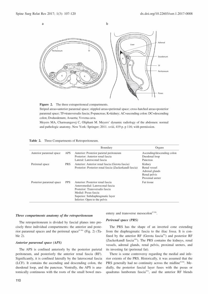
Figure 5. Origin of fusion fascia.
(a) Diagrammatic drawing of transverse section of a 12-week embryo. The colon has returned to the
abdomen and is suspended by the dorsal mesentery. DC=descending colon; S=small bowel;
A=aorta; I=inferior vena cava; K=kidney. (b) Fusion of the descending mesocolon with posterior
parietal peritoneum (dashed lines). Note the subperitoneal region of the mesentery is preserved after
fusion allowing continuity of the subperitoneal space.
Meyers MA, Charnsangavej C, Oliphant M. Meyers’ dynamic radiology of the abdomen: normal
and pathologic anatomy. New York: Springer; 2011. xviii, 419 p. 17; with permission.
Descendingmesocolon
Root of smallbowel mesentery
a b
Figure 6. Compartments and fasciae of the lower retroperito-
neum.
A transverse section at approximately the L5 vertebral level,
showing the location and relationship of the pericolic space.
Standring S. Posterior abdominal wall and retroperitoneum. For-
ty-first edition. New York: Elsevier Limited; 2016. Gray’s anato-
my: the anatomical basis of clinical practice; Chapter 62, p.
1083-1097.e2; with permission.
Externaloblique
Internal obliqueDescendingcolon
Transversusabdominis
Peritoneum
Gonadal vessels
Retroperitoneal ‘space’
Ureter
Anterior and posteriorlayers of the lowerextension of theperirenal fascia
Obturatornerve
Femoral nerveIliacusIlium
Iliac fascia
Retrocolic/mesocolic
fascia
L5 vertebra Psoas fascia
Lateral femoralcutaneous nerve
Genitofemoral nerve
Psoasmajor
CommonIliac vessel
FF with the primary dorsal peritoneum during the rotation
and posterior attachment of the gastrointestinal viscera113). FF
consists of thin (0.1-0.6 mm) connective tissue layers, some-
times bilaminar with a separate looser stratum105).
Clinically, the white line of Toldt is a significant land-
mark for digestive surgery because it can be developed to
mobilize the colon after incising along the line of Toldt. It is
an avascular layer that allows a dissection plane to be devel-
oped, and limits the spread of disease. The FF is also clini-
cally significant for spine surgeons because it immobilizes
the ascending or descending colon to the lateral or posterior
aspect of the abdominal cavity, which lies anterior to the an-
terior RF or LCF (Fig. 6)127). Therefore, recognition of the
ascending or descending colon position should always be
considered in strategic retroperitoneal procedures.
Transversalis fascia (TF)
The outer stratum forms the TF, which lies deep to the
transversus abdominis muscle and superficial to the preperi-
toneal fat and peritoneum. The TF is posterior to the kidney,
and anterior to the fascia surrounding the quadratus lumbo-
rum. Some texts include the fascia of the psoas muscle with
the TF128). The TF may fuse medially with the posterior lam-
ina of the posterior RF. This fusion creates the medial
boundary of the posterior pararenal space112,114).
Thoracolumbar fascia (TLF)
The TLF is a girdling structure comprising a complex ar-
rangement of several aponeurotic and multiple fascial layers
Spine Surg Relat Res 2017; 1(3): 107-120 dx.doi.org/10.22603/ssrr.1.2017-0008
114
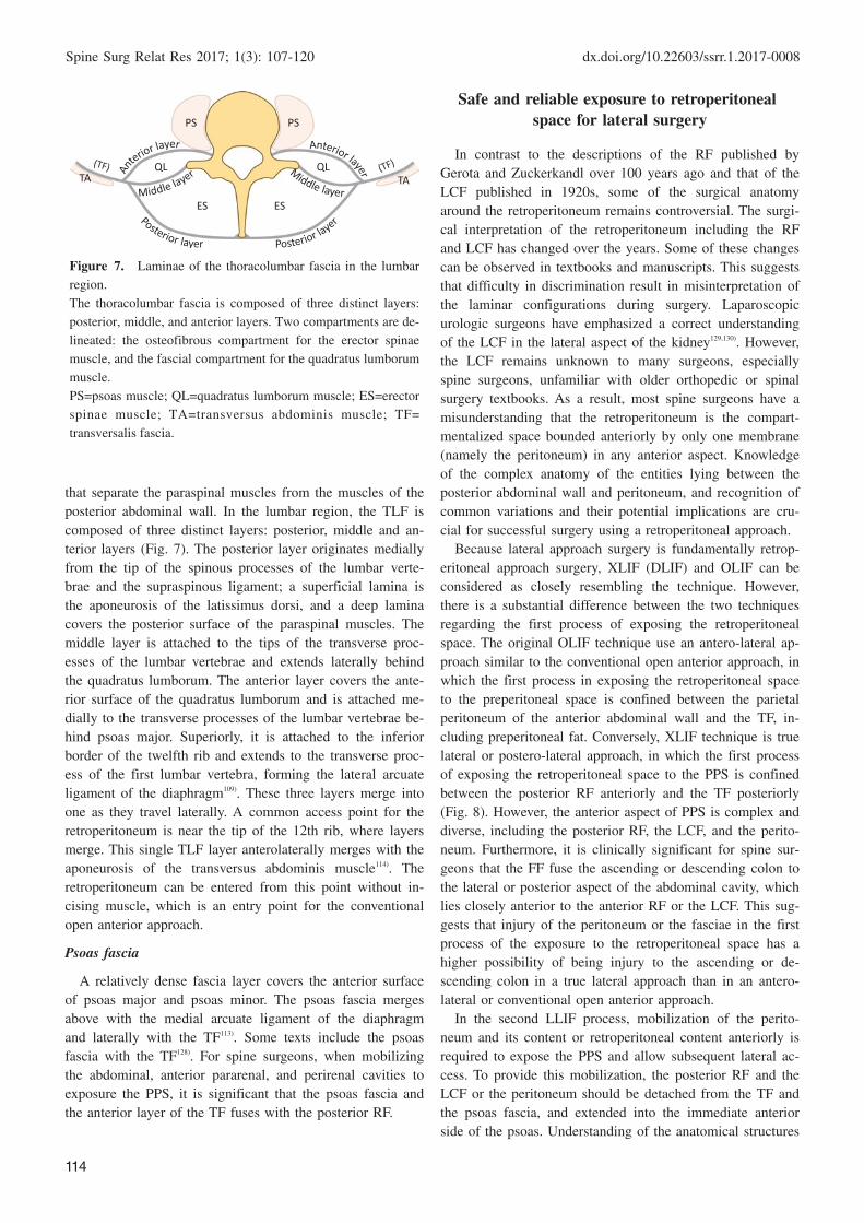
Figure 7. Laminae of the thoracolumbar fascia in the lumbar
region.
The thoracolumbar fascia is composed of three distinct layers:
posterior, middle, and anterior layers. Two compartments are de-
lineated: the osteofibrous compartment for the erector spinae
muscle, and the fascial compartment for the quadratus lumborum
muscle.
PS=psoas muscle; QL=quadratus lumborum muscle; ES=erector
spinae muscle; TA=transversus abdominis muscle; TF=
transversalis fascia.
PS
QL
PS
QLTA TA
ES ES
that separate the paraspinal muscles from the muscles of the
posterior abdominal wall. In the lumbar region, the TLF is
composed of three distinct layers: posterior, middle and an-
terior layers (Fig. 7). The posterior layer originates medially
from the tip of the spinous processes of the lumbar verte-
brae and the supraspinous ligament; a superficial lamina is
the aponeurosis of the latissimus dorsi, and a deep lamina
covers the posterior surface of the paraspinal muscles. The
middle layer is attached to the tips of the transverse proc-
esses of the lumbar vertebrae and extends laterally behind
the quadratus lumborum. The anterior layer covers the ante-
rior surface of the quadratus lumborum and is attached me-
dially to the transverse processes of the lumbar vertebrae be-
hind psoas major. Superiorly, it is attached to the inferior
border of the twelfth rib and extends to the transverse proc-
ess of the first lumbar vertebra, forming the lateral arcuate
ligament of the diaphragm109). These three layers merge into
one as they travel laterally. A common access point for the
retroperitoneum is near the tip of the 12th rib, where layers
merge. This single TLF layer anterolaterally merges with the
aponeurosis of the transversus abdominis muscle114). The
retroperitoneum can be entered from this point without in-
cising muscle, which is an entry point for the conventional
open anterior approach.
Psoas fascia
A relatively dense fascia layer covers the anterior surface
of psoas major and psoas minor. The psoas fascia merges
above with the medial arcuate ligament of the diaphragm
and laterally with the TF113). Some texts include the psoas
fascia with the TF128). For spine surgeons, when mobilizing
the abdominal, anterior pararenal, and perirenal cavities to
exposure the PPS, it is significant that the psoas fascia and
the anterior layer of the TF fuses with the posterior RF.
Safe and reliable exposure to retroperitonealspace for lateral surgery
In contrast to the descriptions of the RF published by
Gerota and Zuckerkandl over 100 years ago and that of the
LCF published in 1920s, some of the surgical anatomy
around the retroperitoneum remains controversial. The surgi-
cal interpretation of the retroperitoneum including the RF
and LCF has changed over the years. Some of these changes
can be observed in textbooks and manuscripts. This suggests
that difficulty in discrimination result in misinterpretation of
the laminar configurations during surgery. Laparoscopic
urologic surgeons have emphasized a correct understanding
of the LCF in the lateral aspect of the kidney129,130). However,
the LCF remains unknown to many surgeons, especially
spine surgeons, unfamiliar with older orthopedic or spinal
surgery textbooks. As a result, most spine surgeons have a
misunderstanding that the retroperitoneum is the compart-
mentalized space bounded anteriorly by only one membrane
(namely the peritoneum) in any anterior aspect. Knowledge
of the complex anatomy of the entities lying between the
posterior abdominal wall and peritoneum, and recognition of
common variations and their potential implications are cru-
cial for successful surgery using a retroperitoneal approach.
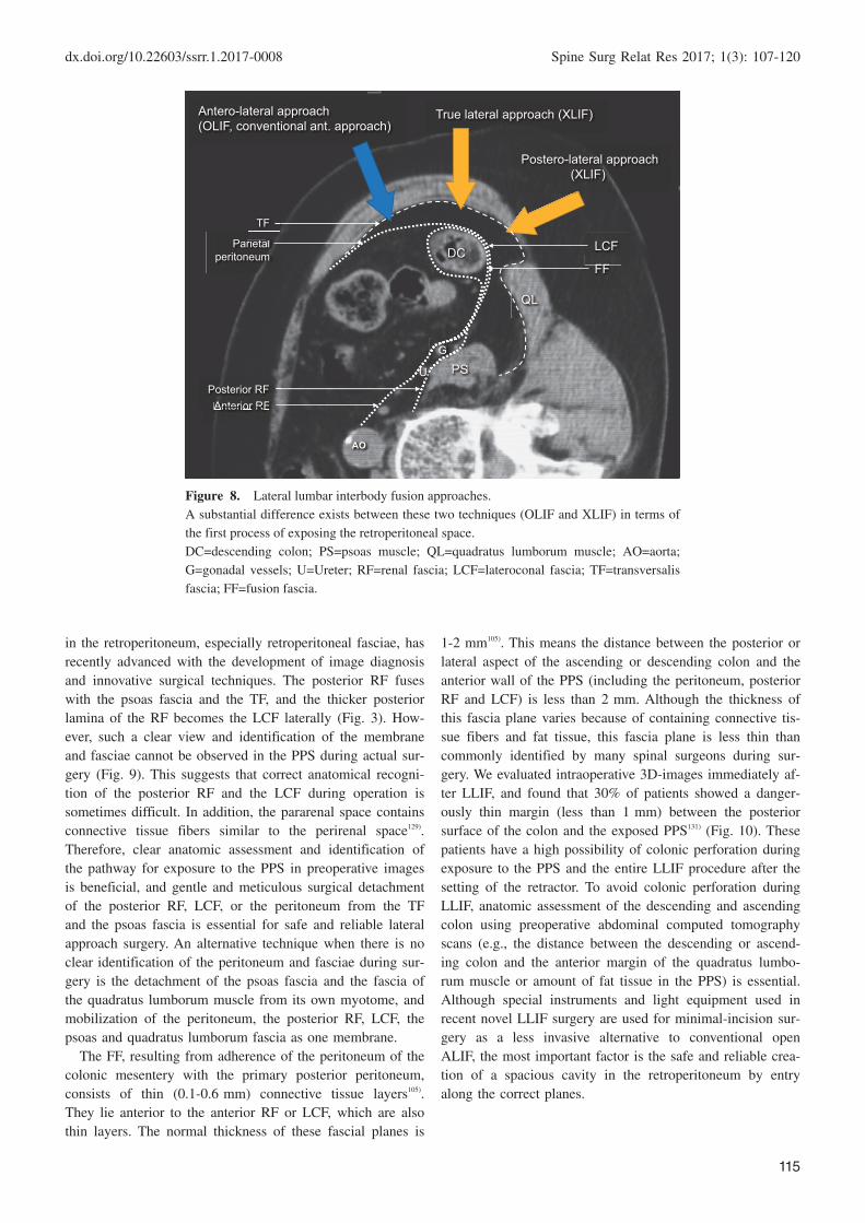
Because lateral approach surgery is fundamentally retrop-
eritoneal approach surgery, XLIF (DLIF) and OLIF can be
considered as closely resembling the technique. However,
there is a substantial difference between the two techniques
regarding the first process of exposing the retroperitoneal
space. The original OLIF technique use an antero-lateral ap-
proach similar to the conventional open anterior approach, in
which the first process in exposing the retroperitoneal space
to the preperitoneal space is confined between the parietal
peritoneum of the anterior abdominal wall and the TF, in-
cluding preperitoneal fat. Conversely, XLIF technique is true
lateral or postero-lateral approach, in which the first process
of exposing the retroperitoneal space to the PPS is confined
between the posterior RF anteriorly and the TF posteriorly
(Fig. 8). However, the anterior aspect of PPS is complex and
diverse, including the posterior RF, the LCF, and the perito-
neum. Furthermore, it is clinically significant for spine sur-
geons that the FF fuse the ascending or descending colon to
the lateral or posterior aspect of the abdominal cavity, which
lies closely anterior to the anterior RF or the LCF. This sug-
gests that injury of the peritoneum or the fasciae in the first
process of the exposure to the retroperitoneal space has a
higher possibility of being injury to the ascending or de-
scending colon in a true lateral approach than in an antero-
lateral or conventional open anterior approach.
In the second LLIF process, mobilization of the perito-
neum and its content or retroperitoneal content anteriorly is
required to expose the PPS and allow subsequent lateral ac-
cess. To provide this mobilization, the posterior RF and the
LCF or the peritoneum should be detached from the TF and
the psoas fascia, and extended into the immediate anterior
side of the psoas. Understanding of the anatomical structures
dx.doi.org/10.22603/ssrr.1.2017-0008 Spine Surg Relat Res 2017; 1(3): 107-120
115
Figure 8. Lateral lumbar interbody fusion approaches.
A substantial difference exists between these two techniques (OLIF and XLIF) in terms of
the first process of exposing the retroperitoneal space.
DC=descending colon; PS=psoas muscle; QL=quadratus lumborum muscle; AO=aorta;
G=gonadal vessels; U=Ureter; RF=renal fascia; LCF=lateroconal fascia; TF=transversalis
fascia; FF=fusion fascia.
Parietalperitoneum
LCF
Antero-lateral approach(OLIF, conventional ant. approach)
True lateral approach (XLIF)
Postero-lateral approach(XLIF)
DC
QL
PS
AO
FF
i t l
TF
U PG
Anterior RFAnterior RFPosterior RF
in the retroperitoneum, especially retroperitoneal fasciae, has
recently advanced with the development of image diagnosis
and innovative surgical techniques. The posterior RF fuses
with the psoas fascia and the TF, and the thicker posterior
lamina of the RF becomes the LCF laterally (Fig. 3). How-
ever, such a clear view and identification of the membrane
and fasciae cannot be observed in the PPS during actual sur-
gery (Fig. 9). This suggests that correct anatomical recogni-
tion of the posterior RF and the LCF during operation is
sometimes difficult. In addition, the pararenal space contains
connective tissue fibers similar to the perirenal space129).
Therefore, clear anatomic assessment and identification of
the pathway for exposure to the PPS in preoperative images
is beneficial, and gentle and meticulous surgical detachment
of the posterior RF, LCF, or the peritoneum from the TF
and the psoas fascia is essential for safe and reliable lateral
approach surgery. An alternative technique when there is no
clear identification of the peritoneum and fasciae during sur-
gery is the detachment of the psoas fascia and the fascia of
the quadratus lumborum muscle from its own myotome, and
mobilization of the peritoneum, the posterior RF, LCF, the
psoas and quadratus lumborum fascia as one membrane.
The FF, resulting from adherence of the peritoneum of the
colonic mesentery with the primary posterior peritoneum,
consists of thin (0.1-0.6 mm) connective tissue layers105).
They lie anterior to the anterior RF or LCF, which are also
thin layers. The normal thickness of these fascial planes is
1-2 mm105). This means the distance between the posterior or
lateral aspect of the ascending or descending colon and the
anterior wall of the PPS (including the peritoneum, posterior
RF and LCF) is less than 2 mm. Although the thickness of
this fascia plane varies because of containing connective tis-
sue fibers and fat tissue, this fascia plane is less thin than
commonly identified by many spinal surgeons during sur-
gery. We evaluated intraoperative 3D-images immediately af-
ter LLIF, and found that 30% of patients showed a danger-
ously thin margin (less than 1 mm) between the posterior
surface of the colon and the exposed PPS131) (Fig. 10). These
patients have a high possibility of colonic perforation during
exposure to the PPS and the entire LLIF procedure after the
setting of the retractor. To avoid colonic perforation during
LLIF, anatomic assessment of the descending and ascending
colon using preoperative abdominal computed tomography
scans (e.g., the distance between the descending or ascend-
ing colon and the anterior margin of the quadratus lumbo-
rum muscle or amount of fat tissue in the PPS) is essential.
Although special instruments and light equipment used in
recent novel LLIF surgery are used for minimal-incision sur-
gery as a less invasive alternative to conventional open
ALIF, the most important factor is the safe and reliable crea-
tion of a spacious cavity in the retroperitoneum by entry
along the correct planes.

Spine Surg Relat Res 2017; 1(3): 107-120 dx.doi.org/10.22603/ssrr.1.2017-0008
116
Figure 9. An anatomic cross-section below midlevel of the left
kidney.
The anatomic cross-section shows the termination of the posteri-
or renal fascia in relationship to the fascia of the quadratus lum-
borum muscle (arrow). A clear view and identification of these
fasciae could not be observed at the posterior pararenal space be-
cause this relationship is variable and the medial extent of the
posterior pararenal space varies from patient to patient.
K=kidney; PM=psoas muscle; C=descending colon; ARF=anterior
renal fascia; LCF=lateroconal fascia; PRF=posterior renal fascia;
1=anterior pararenal space; 2=perirenal space; 3=posterior parare-
nal space.
Meyers MA, Charnsangavej C, Oliphant M. Meyers’ dynamic
radiology of the abdomen: normal and pathologic anatomy. New
York: Springer; 2011. xviii, 419 p. 124; with permission.
Figure 10. Intraoperative three-dimensional (3D) images immediately after lateral lumbar inter-
body fusion (LLIF).
The intraoperative 3D-images in a prone position immediately after LLIF (L4/5) showed a danger-
ously thin margin (less than 1 mm, white arrow) between the posterior or lateral surface of the colon
and the developed posterior pararenal space.
DC=descending colon; dPPS=developed posterior pararenal space by LLIF procedure; PS=psoas
muscle.
DC
dPPS
PS
L4/5 LLIF
In conclusion
Lateral approach spine surgery can provide effective inter-
body stabilization and correction, and indirect neural decom-
pression with minimal-incision and less invasive surgery
compared with conventional open ALIF. It may also avert
the trauma to paraspinal muscles or facet joints found in
TLIF and PLIF. However, because lateral approach surgery
is fundamentally retroperitoneal approach surgery, there is
potential risk to intra- and retroperitoneal structures (includ-
ing viscera and vessels) as seen with a conventional open
anterior approach. Minimal-incision and less invasive lateral
surgery may be a trade-off with the limited visualization of
the retroperitoneal space and minimized working space. An
innovative lateral approach technique has demonstrated dif-
ferent anatomical views, but requires reconsideration of the
traditional surgical anatomy in more detail than a traditional
open anterior approach. Correct anatomical recognition for
the retroperitoneum is essential to success in lateral ap-
proach surgery. It must be clear to the spine surgeon that the
retroperitoneal membrane and fascia are more multilayered
and complex than commonly understood. Therefore, preop-
erative abdominal images will support more efficient surgi-
cal consideration about the retroperitoneal membrane and
fascia in lateral approach surgery. Such anatomical knowl-
edge is also useful in a conventional open approach.
Conflicts of Interest: Tokumi Kanemura has served as a
consultant to NuVasive and Medtronic. Other authors de-
clare that there are no conflicts of interest.
Sources of funding: none
dx.doi.org/10.22603/ssrr.1.2017-0008 Spine Surg Relat Res 2017; 1(3): 107-120
117
References1. Fraser RD. Interbody, posterior, and combined lumbar fusions.
Spine (Phila Pa 1976). 1995;20(24 Suppl):167s-77s. eng.
2. McAfee PC. Interbody fusion cages in reconstructive operations
on the spine. J Bone Joint Surg Am. 1999;81(6):859-80.
3. Zdeblick TA, Phillips FM. Interbody cage devices. Spine (Phila
Pa 1976). 2003;28(15 Suppl):S2-7.
4. Li J, Dumonski ML, Liu Q, et al. A multicenter study to evalu-
ate the safety and efficacy of a stand-alone anterior carbon I/F
Cage for anterior lumbar interbody fusion: two-year results from
a Food and Drug Administration investigational device exemp-
tion clinical trial. Spine (Phila Pa 1976). 2010;35(26):E1564-70.
5. Winder MJ, Gambhir S. Comparison of ALIF vs. XLIF for L4/5
interbody fusion: pros, cons, and literature review. J Spine Surg.
2016;2(1):2-8. eng.
6. Cloward RB. Posterior lumbar interbody fusion updated. Clin
Orthop Relat Res. 1985(193):16-9.
7. Penta M, Fraser RD. Anterior lumbar interbody fusion. A mini-
mum 10-year follow-up. Spine (Phila Pa 1976). 1997;22(20):
2429-34.
8. Harms JG, Jeszenszky D. Die posteriore, lumbale, interkor-
porelle Fusion in unilateraler transforaminaler Technik. Oper Or-
thop Traumatol. 1998;10(2):90-102. ger.
9. Kang BU, Choi WC, Lee SH, et al. An analysis of general
surgery-related complications in a series of 412 minilaparotomic
anterior lumbosacral procedures. J Neurosurg Spine. 2009;10(1):
60-5. eng.
10. Brantigan JW, Steffee AD, Lewis ML, et al. Lumbar interbody
fusion using the Brantigan I/F cage for posterior lumbar inter-
body fusion and the variable pedicle screw placement system:
two-year results from a Food and Drug Administration investiga-
tional device exemption clinical trial. Spine (Phila Pa 1976).
2000;25(11):1437-46.
11. Christensen FB, Hansen ES, Eiskjaer SP, et al. Circumferential
lumbar spinal fusion with Brantigan cage versus posterolateral
fusion with titanium Cotrel-Dubousset instrumentation: a pro-
spective, randomized clinical study of 146 patients. Spine (Phila
Pa 1976). 2002;27(23):2674-83.
12. Capener N. Spondylolisthesis. Br J Surg. 1932;19(75):374-86.
13. Ito H, Tsuchiya J, Asami G. A new radical operation for Pott’s
disease. TheJ Bone Joint Surg Am. 1934;16(3):499.
14. Stauffer RN, Coventry MB. Anterior interbody lumbar spine fu-
sion. Analysis of Mayo Clinic series. J Bone Joint Surg Am.
1972;54(4):756-68.
15. Chow SP, Leong JC, Ma A, et al. Anterior spinal fusion or de-
ranged lumbar intervertebral disc. Spine (Phila Pa 1976). 1980;5
(5):452-8.
16. Fujimaki A, Crock HV, Bedbrook GM. The results of 150 ante-
rior lumbar interbody fusion operations performed by two sur-
geons in Australia. Clin Orthop Relat Res. 1982(165):164-7.
17. O’Brien JP, Dawson MH, Heard CW, et al. Simultaneous com-
bined anterior and posterior fusion. A surgical solution for failed
spinal surgery with a brief review of the first 150 patients. Clin
Orthop Relat Res. 1986(203):191-5.
18. Kozak JA, Heilman AE, O’Brien JP. Anterior lumbar fusion op-
tions. Technique and graft materials. Clin Orthop Relat Res.
1994(300):45-51.
19. Pavlov PW, Meijers H, van Limbeek J, et al. Good outcome and
restoration of lordosis after anterior lumbar interbody fusion
with additional posterior fixation. Spine (Phila Pa 1976). 2004;
29(17):1893-9;discussion 900.
20. Hsieh PC, Koski TR, O’Shaughnessy BA, et al. Anterior lumbar
interbody fusion in comparison with transforaminal lumbar in-
terbody fusion: implications for the restoration of foraminal
height, local disc angle, lumbar lordosis, and sagittal balance. J
Neurosurg Spine. 2007;7(4):379-86.
21. Gerber M, Crawford NR, Chamberlain RH, et al. Biomechanical
assessment of anterior lumbar interbody fusion with an anterior
lumbosacral fixation screw-plate: comparison to stand-alone an-
terior lumbar interbody fusion and anterior lumbar interbody fu-
sion with pedicle screws in an unstable human cadaver model.
Spine (Phila Pa 1976). 2006;31(7):762-8.
22. Liljenqvist U, O’Brien JP, Renton P. Simultaneous combined an-
terior and posterior lumbar fusion with femoral cortical allograft.
Eur Spine J. 1998;7(2):125-31.
23. Zou X, Li H, Teng X, et al. Pedicle screw fixation enhances an-
terior lumbar interbody fusion with porous tantalum cages: an
experimental study in pigs. Spine (Phila Pa 1976). 2005;30(14):
E392-9.
24. Fritzell P, Hagg O, Wessberg P, et al. Chronic low back pain and
fusion: a comparison of three surgical techniques: a prospective
multicenter randomized study from the Swedish lumbar spine
study group. Spine (Phila Pa 1976). 2002;27(11):1131-41.
25. Baker JK, Reardon PR, Reardon MJ, et al. Vascular injury in
anterior lumbar surgery. Spine (Phila Pa 1976). 1993;18(15):
2227-30.
26. Faciszewski T, Winter RB, Lonstein JE, et al. The surgical and
medical perioperative complications of anterior spinal fusion
surgery in the thoracic and lumbar spine in adults. A review of
1223 procedures. Spine (Phila Pa 1976). 1995;20(14):1592-9.
27. Nourian AA, Cunningham CM, Bagheri A, et al. Effect of
Anatomic Variability and Level of Approach on Perioperative
Vascular Complications With Anterior Lumbar Interbody Fusion.
Spine (Phila Pa 1976). 2016;41(2):E73-7.
28. Quraishi NA, Konig M, Booker SJ, et al. Access related compli-
cations in anterior lumbar surgery performed by spinal surgeons.
Eur Spine J. 2013;22 Suppl 1:S16-20.
29. Rajaraman V, Vingan R, Roth P, et al. Visceral and vascular
complications resulting from anterior lumbar interbody fusion. J
Neurosurg. 1999;91(1 Suppl):60-4. eng.
30. Sasso RC, Best NM, Mummaneni PV, et al. Analysis of opera-
tive complications in a series of 471 anterior lumbar interbody
fusion procedures. Spine (Phila Pa 1976). 2005;30(6):670-4.
31. Sasso RC, Kenneth Burkus J, LeHuec JC. Retrograde ejacula-
tion after anterior lumbar interbody fusion: transperitoneal ver-
sus retroperitoneal exposure. Spine (Phila Pa 1976). 2003;28
(10):1023-6.
32. Schizas C, Foko’o N, Matter M, et al. Lymphocoele: a rare and
little known complication of anterior lumbar surgery. Eur Spine
J. 2009;18 Suppl 2:228-31.
33. Phan KMD, Xu J, Scherman DBB, et al. Anterior lumbar inter-
body fusion (ALIF) with and without an “Access Surgeon”: A
systematic review and meta-analysis. Spine (Phila Pa 1976).
2017.
34. Briggs H, Milligan PR. Chip fusion of the low back following
exploration of the spinal canal. J Bone Joint Surg Am. 1944;26
(1):125-30.
35. Mobbs RJ, Phan K, Malham G, et al. Lumbar interbody fusion:
techniques, indications and comparison of interbody fusion op-
tions including PLIF, TLIF, MI-TLIF, OLIF/ATP, LLIF and
ALIF. J Spine Surg. 2015;1(1):2-18. eng.
36. Brantigan JW, Neidre A, Toohey JS. The Lumbar I/F Cage for
posterior lumbar interbody fusion with the variable screw place-
ment system: 10-year results of a Food and Drug Administration
Spine Surg Relat Res 2017; 1(3): 107-120 dx.doi.org/10.22603/ssrr.1.2017-0008
118
clinical trial. Spine J. 2004;4(6):681-8.
37. Harms J, Rolinger H. [A one-stager procedure in operative treat-
ment of spondylolistheses: dorsal traction-reposition and anterior
fusion (author’s transl)]. Z Orthop Ihre Grenzgeb. 1982;120(3):
343-7. ger.
38. Humphreys SC, Hodges SD, Patwardhan AG, et al. Comparison
of posterior and transforaminal approaches to lumbar interbody
fusion. Spine (Phila Pa 1976). 2001;26(5):567-71.
39. Lowe TG, Tahernia AD, O’Brien MF, et al. Unilateral transfo-
raminal posterior lumbar interbody fusion (TLIF): indications,
technique, and 2-year results. J Spinal Disord Tech. 2002;15(1):
31-8.
40. Moskowitz A. Transforaminal lumbar interbody fusion. Orthop
Clin North Am. 2002;33(2):359-66.
41. Phan K, Thayaparan GK, Mobbs RJ. Anterior lumbar interbody
fusion versus transforaminal lumbar interbody fusion--systematic
review and meta-analysis. Br J Neurosurg. 2015;29(5):705-11.
42. Rosenberg WS, Mummaneni PV. Transforaminal lumbar inter-
body fusion: technique, complications, and early results. Neuro-
surgery. 2001;48(3):569-74;discussion 74-5.
43. Foley KT, Holly LT, Schwender JD. Minimally invasive lumbar
fusion. Spine (Phila Pa 1976). 2003;28(15 Suppl):S26-35.
44. McAfee PC, DeVine JG, Chaput CD, et al. The indications for
interbody fusion cages in the treatment of spondylolisthesis:
analysis of 120 cases. Spine (Phila Pa 1976). 2005;30(6 Suppl):
S60-5.
45. Okuda S, Miyauchi A, Oda T, et al. Surgical complications of
posterior lumbar interbody fusion with total facetectomy in 251
patients. J Neurosurg Spine. 2006;4(4):304-9.
46. Park Y, Ha JW. Comparison of one-level posterior lumbar inter-
body fusion performed with a minimally invasive approach or a
traditional open approach. Spine (Phila Pa 1976). 2007;32(5):
537-43.
47. Salerni AA. A minimally invasive approach for posterior lumbar
interbody fusion. Neurosurgical focus. 2002;13(6):e6.
48. Pimenta L. Lateral endoscopic transpsoas retroperitoneal ap-
proach for lumbar spine surgery.. VIII Brazilian Spine Society
Meeting. 2001.
49. Ozgur BM, Aryan HE, Pimenta L, et al. Extreme Lateral Inter-
body Fusion (XLIF): a novel surgical technique for anterior
lumbar interbody fusion. Spine J. 2006;6(4):435-43.
50. Berjano P, Lamartina C. Minimally invasive lateral transpsoas
approach with advanced neurophysiologic monitoring for lumbar
interbody fusion. Eur Spine J. 2011;20(9):1584-6.
51. Fujibayashi S, Hynes RA, Otsuki B, et al. Effect of indirect
neural decompression through oblique lateral interbody fusion
for degenerative lumbar disease. Spine (Phila Pa 1976). 2015;40
(3):E175-82.
52. Elowitz EH, Yanni DS, Chwajol M, et al. Evaluation of indirect
decompression of the lumbar spinal canal following minimally
invasive lateral transpsoas interbody fusion: radiographic and
outcome analysis. Minim Invasive Neurosurg. 2011;54(5-6):201-
6.
53. Oliveira L, Marchi L, Coutinho E, et al. A radiographic assess-
ment of the ability of the extreme lateral interbody fusion proce-
dure to indirectly decompress the neural elements. Spine (Phila
Pa 1976). 2010;35(26 Suppl):S331-7.
54. Youssef JA, McAfee PC, Patty CA, et al. Minimally invasive
surgery: lateral approach interbody fusion: results and review.
Spine (Phila Pa 1976). 2010;35(26 Suppl):S302-11.
55. Isaacs RE, Hyde J, Goodrich JA, et al. A prospective, nonran-
domized, multicenter evaluation of extreme lateral interbody fu-
sion for the treatment of adult degenerative scoliosis: periopera-
tive outcomes and complications. Spine (Phila Pa 1976). 2010;
35(26 Suppl):S322-30.
56. Acosta FL, Liu J, Slimack N, et al. Changes in coronal and sag-
ittal plane alignment following minimally invasive direct lateral
interbody fusion for the treatment of degenerative lumbar dis-
ease in adults: a radiographic study. J Neurosurg Spine. 2011;15
(1):92-6.
57. Berjano P, Lamartina C. Far lateral approaches (XLIF) in adult
scoliosis. Eur Spine J. 2013;22 Suppl 2:S242-53.
58. Castellvi AE, Nienke TW, Marulanda GA, et al. Indirect decom-
pression of lumbar stenosis with transpsoas interbody cages and
percutaneous posterior instrumentation. Clin Orthop Relat Res.
2014;472(6):1784-91.
59. Sato J, Ohtori S, Orita S, et al. Radiographic evaluation of indi-
rect decompression of mini-open anterior retroperitoneal lumbar
interbody fusion: oblique lateral interbody fusion for degener-
ated lumbar spondylolisthesis. Eur Spine J. 2015. eng.
60. Phillips FM, Isaacs RE, Rodgers WB, et al. Adult degenerative
scoliosis treated with XLIF: clinical and radiographical results of
a prospective multicenter study with 24-month follow-up. Spine
(Phila Pa 1976). 2013;38(21):1853-61.
61. Baghdadi YM, Larson AN, Dekutoski MB, et al. Sagittal bal-
ance and spinopelvic parameters after lateral lumbar interbody
fusion for degenerative scoliosis: a case-control study. Spine
(Phila Pa 1976). 2014;39(3):E166-73.
62. Strom RG, Bae J, Mizutani J, et al. Lateral interbody fusion
combined with open posterior surgery for adult spinal deformity.
J Neurosurg Spine. 2016:1-9.
63. Theologis AA, Mundis GM, Jr., Nguyen S, et al. Utility of mul-
tilevel lateral interbody fusion of the thoracolumbar coronal
curve apex in adult deformity surgery in combination with open
posterior instrumentation and L5-S1 interbody fusion: a case-
matched evaluation of 32 patients. J Neurosurg Spine. 2016:1-
12. eng.
64. Alimi M, Hofstetter CP, Cong GT, et al. Radiological and clini-
cal outcomes following extreme lateral interbody fusion. J Neu-
rosurg Spine. 2014;20(6):623-35. eng.
65. Formica M, Berjano P, Cavagnaro L, et al. Extreme lateral ap-
proach to the spine in degenerative and post traumatic lumbar
diseases: selection process, results and complications. Eur Spine
J. 2014;23 Suppl 6:684-92. eng.
66. Tohmeh AG, Khorsand D, Watson B, et al. Radiographical and
clinical evaluation of extreme lateral interbody fusion: effects of
cage size and instrumentation type with a minimum of 1-year
follow-up. Spine (Phila Pa 1976). 2014;39(26):E1582-91. eng.
67. Serak J, Vanni S, Levi AD. The extreme lateral approach for
treatment of thoracic and lumbar vertebral body metastases. J
Neurosurg Sci. 2015. eng.
68. Smith WD, Dakwar E, Le TV, et al. Minimally invasive surgery
for traumatic spinal pathologies: a mini-open, lateral approach in
the thoracic and lumbar spine. Spine (Phila Pa 1976). 2010;35
(26 Suppl):S338-46.
69. Uribe JS, Dakwar E, Le TV, et al. Minimally invasive surgery
treatment for thoracic spine tumor removal: a mini-open, lateral
approach. Spine (Phila Pa 1976). 2010;35(26 Suppl):S347-54.
70. Pekmezci M, McDonald E, Kennedy A, et al. Can a novel rec-
tangular footplate provide higher resistance to subsidence than
circular footplates? An ex vivo biomechanical study. Spine
(Phila Pa 1976). 2012;37(19):E1177-81.
71. Theologis AA, Tabaraee E, Toogood P, et al. Anterior corpec-
tomy via the mini-open, extreme lateral, transpsoas approach
dx.doi.org/10.22603/ssrr.1.2017-0008 Spine Surg Relat Res 2017; 1(3): 107-120
119
combined with short-segment posterior fixation for single-level
traumatic lumbar burst fractures: analysis of health-related qual-
ity of life outcomes and patient satisfaction. J Neurosurg Spine.
2016;24(1):60-8. eng.
72. Uribe JS, Smith DA, Dakwar E, et al. Lordosis restoration after
anterior longitudinal ligament release and placement of lateral
hyperlordotic interbody cages during the minimally invasive lat-
eral transpsoas approach: a radiographic study in cadavers. J
Neurosurg Spine. 2012;17(5):476-85.
73. Berjano P, Cecchinato R, Sinigaglia A, et al. Anterior column
realignment from a lateral approach for the treatment of severe
sagittal imbalance: a retrospective radiographic study. Eur Spine
J. 2015;24 Suppl 3:433-8. eng.
74. Pimenta L, Fortti F, Oliveira L, et al. Anterior column realign-
ment following lateral interbody fusion for sagittal deformity
correction. Eur J Orthop Surg Traumatol. 2015;25 Suppl 1:S29-
33. eng.
75. Turner JD, Akbarnia BA, Eastlack RK, et al. Radiographic out-
comes of anterior column realignment for adult sagittal plane
deformity: a multicenter analysis. Eur Spine J. 2015;24 Suppl 3:
427-32.
76. Saigal R, Mundis GM, Jr., Eastlack R, et al. Anterior Column
Realignment (ACR) in Adult Sagittal Deformity Correction:
Technique and Review of the Literature. Spine (Phila Pa 1976).
2016;41 Suppl 8:S66-73.
77. Kepler CK, Bogner EA, Herzog RJ, et al. Anatomy of the psoas
muscle and lumbar plexus with respect to the surgical approach
for lateral transpsoas interbody fusion. Eur Spine J. 2011;20(4):
550-6.
78. Le TV, Burkett CJ, Deukmedjian AR, et al. Postoperative lumbar
plexus injury after lumbar retroperitoneal transpsoas minimally
invasive lateral interbody fusion. Spine (Phila Pa 1976). 2013;38
(1):E13-20. eng.
79. Gammal ID, Spivak JM, Bendo JA. Systematic Review of Thigh
Symptoms after Lateral Transpsoas Interbody Fusion for Adult
Patients with Degenerative Lumbar Spine Disease. Int J Spine
Surg. 2015;9:62. eng.
80. Beckman JM, Vincent B, Park MS, et al. Contralateral psoas he-
matoma after minimally invasive, lateral retroperitoneal
transpsoas lumbar interbody fusion: a multicenter review of
3950 lumbar levels. J Neurosurg Spine. 2017;26(1):50-4. eng.
81. Tohmeh AG, Rodgers WB, Peterson MD. Dynamically evoked,
discrete-threshold electromyography in the extreme lateral inter-
body fusion approach. J Neurosurg Spine. 2011;14(1):31-7.
82. Tormenti MJ, Maserati MB, Bonfield CM, et al. Complications
and radiographic correction in adult scoliosis following com-
bined transpsoas extreme lateral interbody fusion and posterior
pedicle screw instrumentation. Neurosurg Focus. 2010;28(3):E7.
83. Uribe JS, Deukmedjian AR. Visceral, vascular, and wound com-
plications following over 13,000 lateral interbody fusions: a sur-
vey study and literature review. Eur Spine J. 2015;24 Suppl 3:
386-96.
84. Malham GM, Ellis NJ, Parker RM, et al. Clinical outcome and
fusion rates after the first 30 extreme lateral interbody fusions.
Scientific World J. 2012;2012:246989.
85. Akbarnia BA, Mundis GM, Jr., Moazzaz P, et al. Anterior col-
umn realignment (ACR) for focal kyphotic spinal deformity us-
ing a lateral transpsoas approach and ALL release. J Spinal Dis-
ord Tech. 2014;27(1):29-39.
86. Murray G, Beckman J, Bach K, et al. Complications and neuro-
logical deficits following minimally invasive anterior column re-
lease for adult spinal deformity: a retrospective study. Eur Spine
J. 2015;24 Suppl 3:397-404.
87. Aichmair A, Fantini GA, Garvin S, et al. Aortic perforation dur-
ing lateral lumbar interbody fusion. J Spinal Disord Tech. 2015;
28(2):71-5. Eng.
88. Assina R, Majmundar NJ, Herschman Y, et al. First report of
major vascular injury due to lateral transpsoas approach leading
to fatality. J Neurosurg Spine. 2014;21(5):794-8.
89. Balsano M, Carlucci S, Ose M, et al. A case report of a rare
complication of bowel perforation in extreme lateral interbody
fusion. Eur Spine J. 2015;24 Suppl 3:405-8.
90. Mirilas P, Skandalakis JE. Surgical anatomy of the retroperito-
neal spaces part II: the architecture of the retroperitoneal space.
Am Surg. 2010;76(1):33-42. eng.
91. Meyers MA. Acute extrapitoneal infection. Seminars in roent-
genology. 1973;8(4):445-64. eng.
92. Meyers MA. Radiological features of the spread and localization
of extraperitoneal gas and their relationship to its source. An
anatomical approach. Radiology. 1974;111(1):17-26. eng.
93. Meyers MA. Uriniferous perirenal pseudocyst: new observations.
Radiology. 1975;117(3 Pt 1):539-45. eng.
94. Meyers MA, Goodman KJ. Pathways of extrapelvic spread of
disease: Anatomic-radiologic correlation. Am J Roentgenol Ra-
dium Ther Nucl Med. 1975;125(4):900-9. eng.
95. Meyers MA, Whalen JP, Evans JA. Diagnosis of perirenal and
subcapsular masses. Anatomic-radiologic correlation. Am J
Roentgenol Radium Ther Nucl Med. 1974;121(3):523-38. eng.
96. Meyers MA, Whalen JP, Peelle K, et al. Radiologic features of
extraperitoneal effusions. An anatomic approach. Radiology.
1972;104(2):249-57.
97. Meyers MA, Oliphant M, Berne AS, et al. The peritoneal liga-
ments and mesenteries: pathways of intraabdominal spread of
disease. Radiology. 1987;163(3):593-604.
98. Oliphant M, Berne AS, Meyers MA. Spread of disease via the
subperitoneal space: the small bowel mesentery. Abdom Imag-
ing. 1993;18(2):109-16.
99. Gerota D. Beitraege zur Kenntnis des Befestigungsapparates der
Niere. Arch Anat Entwicklungsgesch. Leipzig. 1895:265-86.
100. Zuckerkandl E. Beitrage zur Anatomie des Menschlichen Kor-
pers. Ueber den Fixationsapparat der Nieren. Med Jahr. 1883;13:
59-67.
101. Congdon ED, Edson JN. The cone of renal fascia in the adult
white male. Anat Rec. 1941;80(3):289-313.
102. Mitchell GA. The renal fascia. Br J Surg. 1950;37(147):257-66.
103. Lim JH, Kim B, Auh YH. Anatomical communications of the
perirenal space. Br J Radiol. 1998;71(844):450-6. eng.
104. Mindell HJ, Mastromatteo JF, Dickey KW, et al. Anatomic com-
munications between the three retroperitoneal spaces: determina-
tion by CT-guided injections of contrast material in cadavers.
Am J Roentgenol. 1995;164(5):1173-8.
105. Meyers MA, Charnsangavej C, Oliphant M. Meyers’ dynamic
radiology of the abdomen: normal and pathologic anatomy. New
York: Springer; 2011.xviii,419p.p
106. Dodds WJ, Darweesh RM, Lawson TL, et al. The retroperitoneal
spaces revisited. AJR Am J Roentgenol. 1986;147(6):1155-61.
eng.
107. Molmenti EP, Balfe DM, Kanterman RY, et al. Anatomy of the
retroperitoneum: observations of the distribution of pathologic
fluid collections. Radiology. 1996;200(1):95-103.
108. Goenka AH, Shah SN, Remer EM. Imaging of the retroperito-
neum. Radiologic clinics of North America. 2012;50(2):333-55,
vii. eng.
109. Willard FH, Vleeming A, Schuenke MD, et al. The thoracolum-

Spine Surg Relat Res 2017; 1(3): 107-120 dx.doi.org/10.22603/ssrr.1.2017-0008
120
bar fascia: anatomy, function and clinical considerations. J Anat.
2012;221(6):507-36.
110. Gore RM, Balfe DM, Aizenstein RI, et al. The great escape: in-
terfascial decompression planes of the retroperitoneum. AJR Am
J Roentgenol. 2000;175(2):363-70.
111. Tirkes T, Sandrasegaran K, Patel AA, et al. Peritoneal and
retroperitoneal anatomy and its relevance for cross-sectional im-
aging. Radiographics: a review publication of the Radiological
Society of North America, Inc. 2012;32(2):437-51. eng.
112. MacLennan GT, Hinman F. Posterolateral and Posterior Body
Wall: Hinman’s atlas of urosurgical anatomy. Philadelphia:
Elsevier/Saunders; 2012.p
113. Palmer DA, Moinzadeh A. Surgical, Radiographic, and Endo-
scopic Anatomy of the Retroperitoneum. 11th ed./editor-in-chief,
Alan J. Wein; [editors], Louis R. Kavoussi, Alan W. Partin,
Craig A. Peters ed.: Elsevier; 2016. Campbell-Walsh urology;
p.764-83
114. Chesbrough RM, Burkhard TK, Martinez AJ, et al. Gerota ver-
sus Zuckerkandl: the renal fascia revisited. Radiology. 1989;173
(3):845-6.
115. Feldberg MAM. Computed tomography of the retroperitoneum:
an anatomical and pathological atlas with emphasis on the fas-
cial planes: Series in radiology. Martinus Nijhoff Publishers Dis-
tributors for the United States and Canada, Kluwer Boston;
1983.xiv,190p.p
116. Raptopoulos V, Kleinman PK, Marks S, Jr., et al. Renal fascial
pathway: posterior extension of pancreatic effusions within the
anterior pararenal space. Radiology. 1986;158(2):367-74. eng.
117. Hinman F. The principles and practice of urology. Philadelphia
London,: W.B. Saunders Company; 1935.1111p.p
118. Southam AH. The Fixation of the Kidney. QJM: INT J MED
Journal. 1923;os-16(64):283-308.
119. Hinman F. Atlas of urosurgical anatomy. W.B. Saunders; 1993.
xvi,553p.p
120. Marks SC, Jr., Raptopoulos V, Kleinman P, et al. The anatomical
basis for retrorenal extensions of pancreatic effusions: the role
of the renal fasciae. Surgical and radiologic anatomy: SRA.
1986;8(2):89-97. eng.
121. Hadar H, Gadoth N. Positional relations of colon and kidney de-
termined by perirenal fat. AJR Am J Roentgenol. 1984;143(4):
773-6.
122. Hopper KD, Sherman JL, Luethke JM, et al. The retrorenal co-
lon in the supine and prone patient. Radiology. 1987;162(2):443-
6.
123. Sherman JL, Hopper KD, Greene AJ, et al. The retrorenal colon
on computed tomography: a normal variant. J Comput Assist
Tomogr. 1985;9(2):339-41.
124. Matsubara A, Murakami G, Niikura H, et al. Development of
the human retroperitoneal fasciae. Cells Tissues Organs. 2009;
190(5):286-96.
125. Toldt C. Lehrbuch der gewebelehre, mit vorzugsweiser berücksi-
chtigung des menschlichen körpers. Stuttgart,: F. Enke; 1877.
xii,655,1p.p
126. Standring S. Posterior abdominal wall and retroperitoneum.
Forty-first edition. ed. New York: Elsevier Limited; 2016. Gray’s
anatomy: the anatomical basis of clinical practice; p.1083-97
127. Coffin A, Boulay-Coletta I, Sebbag-Sfez D, et al. Radioanatomy
of the retroperitoneal space. Diagn Interv Imaging. 2015;96(2):
171-86. eng.
128. Tobin CE. The renal fascia and its relation to the transversalis
fascia. Anat Rec. 1944;89(3):295-311.
129. Kato H, Kiyokawa H, Inoue H, et al. Anatomical reconsidera-
tion to renal area: lessons learned from radical nephrectomy or
adrenalectomy through a minimal incision over the 12th rib. Int
J Urol. 2004;11(9):709-13. eng.
130. Takahashi R, Furubayashi N, Nakamura M, et al. Surgical con-
siderations of the renal fascia and the retroperitoneal space
around the kidney. J Bodyw Mov Ther. 2012;16(3):392-6. eng.
131. Kanemura T, Satake K, Yamaguchi H, et al.: Possibility of colo-
nic perforation during extreme lateral interbody fusion: Distance
between descending or ascending colon and lateral approach
Pathway. 9th Annual SOLAS Meeting. 2016:16-7.
Spine Surgery and Related Research is an Open Access article distributed under
the Creative Commons Attribution‐NonCommercial‐NoDerivatives 4.0 Inter-
national License. To view the details of this license, please visit (https://creative-
commons.org/licenses/by‐nc‐nd/4.0/).