understanding tumor ecosystems by single-cell sequencing
TRANSCRIPT

REVIEW Open Access
Understanding tumor ecosystems bysingle-cell sequencing: promises andlimitationsXianwen Ren*, Boxi Kang and Zemin Zhang*
Abstract
Cellular heterogeneity within and across tumors hasbeen a major obstacle in understanding and treatingcancer, and the complex heterogeneity is masked ifbulk tumor tissues are used for analysis. The advent ofrapidly developing single-cell sequencing technologies,which include methods related to single-cell genome,epigenome, transcriptome, and multi-omics sequencing,have been applied to cancer research and led to excitingnew findings in the fields of cancer evolution, metastasis,resistance to therapy, and tumor microenvironment.In this review, we discuss recent advances andlimitations of these new technologies and theirpotential applications in cancer studies.
IntroductionA single cell is the ultimate unit of life activity, in whichgenetic mechanisms and the cellular environment inter-play with each other and shape the formation and func-tion of such complex structures as tissues and organs.Dissecting the composition and characterizing the inter-action, dynamics, and function at the single-cell resolutionare crucial for fully understanding the biology of almostall life phenomena, under both normal and diseased con-ditions. Cancer, a disease caused by somatic mutationsconferring uncontrolled proliferation and invasiveness,could in particular benefit from advances in single-cellanalysis. During oncogenesis, different populations of can-cer cells that are genetically heterogeneous emerge, evolve,and interact with cells in the tumor microenvironment,which leads to host metabolism hijacking, immune eva-sion, metastasis to other body parts, and eventual
mortality. Cancer cells can also manifest resistance to vari-ous therapeutic drugs through cellular heterogeneity andplasticity. Cancer is increasingly viewed as a ‘tumor eco-system’, a community in which tumor cells cooperate withother tumor cells and host cells in their microenviron-ment, and can also adapt and evolve to changing condi-tions [1–5].Detailed understanding of tumor ecosystems at
single-cell resolution has been limited for technologicalreasons. Conventional genomic, transcriptomic, and epi-genomic sequencing protocols require microgram-levelinput materials, and so cancer-related genomic studieswere largely limited to bulk tumor sequencing, which doesnot address intratumor heterogeneity and complexity. Theadvent of single-cell sequencing technologies [6–8] hasshifted cancer research to a new paradigm and revolution-ized our understanding of cancer evolution [7–22], tumorheterogeneity [23–46], and the tumor microenvironment[47–59]. Development of single-cell sequencing technolo-gies and the applications in cancer research have been as-tonishing in the past decade, but many challenges stillexist and much remains to be explored. Single-cell cancergenomic studies have been reviewed previously [60–63].In this review, we summarize recent progress and limita-tions in cancer sample single-cell sequencing with a focuson the dissection of tumor ecosystems.
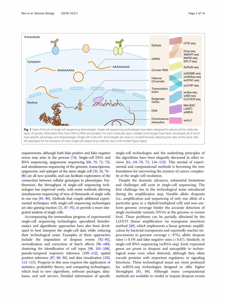
Overview of single-cell sequencing and analysisSingle-cell sequencing technologies have improved consider-ably from the initial proof-of-principle studies [6–8]. Modifi-cation of the underlying molecular biology and chemistry ofsingle-cell library preparation has provided diverse ap-proaches to obtain and amplify single-cell nucleic acids forsubsequent high-throughput sequencing [64–72] (Fig. 1).Because an individual cancer cell typically contains only ∼6–12 pg of DNA and 10–50 pg of total RNA (depending onthe cell types and status) [73], amplification is essential forsingle-cell library preparation to fulfill the sequencing input
* Correspondence: [email protected]; [email protected] Advanced Innovation Centre for Genomics, Peking-Tsinghua Centrefor Life Sciences, Biomedical Pioneering Innovation Center (BIOPIC), Schoolof Life Sciences, Peking University, Beijing 100871, China
© The Author(s). 2018 Open Access This article is distributed under the terms of the Creative Commons Attribution 4.0International License (http://creativecommons.org/licenses/by/4.0/), which permits unrestricted use, distribution, andreproduction in any medium, provided you give appropriate credit to the original author(s) and the source, provide a link tothe Creative Commons license, and indicate if changes were made. The Creative Commons Public Domain Dedication waiver(http://creativecommons.org/publicdomain/zero/1.0/) applies to the data made available in this article, unless otherwise stated.
Ren et al. Genome Biology (2018) 19:211 https://doi.org/10.1186/s13059-018-1593-z
requirements, although both false positive and false negativeerrors may arise in the process [74]. Single-cell DNA andRNA sequencing, epigenomic sequencing [68, 70, 72, 75],and simultaneous sequencing of the genome, transcriptome,epigenome, and epitopes of the same single cell [32, 35, 76–80] are all now possible, and can facilitate exploration of theconnection between cellular genotypes to phenotypes. Fur-thermore, the throughput of single-cell sequencing tech-nologies has improved vastly, with some methods allowingsimultaneous sequencing of tens of thousands of single cellsin one run [81–86]. Methods that couple additional experi-mental techniques with single-cell sequencing technologiesare also gaining traction [21, 87–91], to provide a more inte-grated analysis of single cells.Accompanying the tremendous progress of experimental
single-cell sequencing technologies, specialized bioinfor-matics and algorithmic approaches have also been devel-oped to best interpret the single-cell data while reducingtheir technological noise. Examples of these approachesinclude the imputation of dropout events [92–95],normalization and correction of batch effects [96–100],clustering for identification of cell types [98, 101–108],pseudo-temporal trajectory inference [109–112], spatialposition inference [87, 88, 90], and data visualization [102,113–115]. Progress in this area requires the application ofstatistics, probability theory, and computing technologies,which lead to new algorithms, software packages, data-bases, and web servers. Detailed information of specific
single-cell technologies and the underlying principles ofthe algorithms have been elegantly discussed in other re-views [61, 64–70, 72, 116–123]. This myriad of experi-mental and computational methods is becoming the newfoundation for uncovering the mystery of cancer complex-ity at the single-cell resolution.Despite the dramatic advances, substantial limitations
and challenges still exist in single-cell sequencing. Thefirst challenge lies in the technological noise introducedduring the amplification step. Notable allelic dropouts(i.e., amplification and sequencing of only one allele of aparticular gene in a diploid/multiploid cell) and non-uni-form genome coverage hinder the accurate detection ofsingle nucleotide variants (SNVs) at the genome or exomelevel. These problems can be partially alleviated by theLIANTI (linear amplification via transposon insertion)method [40], which implements a linear genomic amplifi-cation by bacterial transposons and reportedly reaches im-provements in genome coverage (~ 97%), allelic dropoutrates (< 0.19) and false negative rates (< 0.47). Similarly, insingle-cell RNA sequencing (scRNA-seq), lowly expressedgenes are prone to dropout and susceptible to techno-logical noise even when detected, although they oftenencode proteins with important regulatory or signalingfunctions. These technological issues are more profoundfor scRNA-seq technologies designed to offer higherthroughput [81, 84]. Although many computationalmethods are available to model or impute dropout events
Extracellular
AAAAAAAAAAAAAAAAAA
Epitope
mRNA
Circular RNA
Transcriptionfactor
Histone/Chromatin
Chromosomeinteraction
Epigeneticmarks
DNA
CITE-seq
Drop-seqSMART-seqMARS-seqSPLiT-seq
SUPeR-seq
scCHIP-seq
scHi-C
scAba-seqscBS-seqCLEVER-seq
MALBACLIANTIscMDA
scNOME-seqscDNAse-seqscATAC-seq
Cytoplasm
Nucleus
Fig. 1 State of the art of single-cell sequencing technologies. Single-cell sequencing technologies have been designed for almost all the molecularlayers of genetic information flow from DNA to RNA and proteins. For each molecular layer, multiple technologies have been developed, all of whichhave specific advantages and disadvantages. Single-cell multi-omic technologies are close to comprehensively depicting the state of the same cells.We apologize for the exclusion of many single-cell sequencing methods due to the limited figure space
Ren et al. Genome Biology (2018) 19:211 Page 2 of 14

[92, 94, 95], their performances vary and may introduceartificial biases. Much effort is needed to fully address thischallenge.The second challenge is that only a small fraction of
cells from bulk tissues can be sequenced. Bulk tissues con-sist of millions of cells, but present studies can often onlysequence hundreds to thousands of single cells because oftechnological and economic limitations [9–11, 20, 25,124–126]. To what extent the sequenced cells representthe distribution of cells in the entire tissue of interest isnot clear. A plausible solution to address this challengewould be to further improve the throughput of cellularcaptures, e.g., MARS-seq [82] and SPLiT-seq [86], or al-ternatively to combine bulk and single-cell sequencing to-gether and then conduct deconvolution analysis [127].Deconvolution analysis for bulk RNA-seq data usescell-type signature genes as inputs [128–130], which canbe substituted by single-cell sequencing results, althoughcritical computational challenges still exist, such as collin-earity among single cells. If marker genes for known celltypes are orthogonal to each other, the proportions of eachcell type in a bulk sample can be reliably estimated. How-ever, collinearity of gene expression exists widely amongsingle cells, which complicates the deconvolution process.At present, successful deconvolution of bulk RNA-seqdata based on scRNA-seq-defined signatures has been re-ported only in cases where orthogonal molecular signa-tures and fine cluster structures are well balanced [131].The wide usage of scRNA-seq based deconvolution willhinge upon the availability of comprehensive single-cellclusters and the development of general methods forselecting orthogonal signatures for each cell type.Spatial information of single cells in the tissue is often
lost during the isolation step and thus single-cell sequen-cing data typically do not show how cells are organized toimplement the concerted function within a tissue of inter-est. Many new techniques have been developed to keep orrestore the spatial information of sequenced single cellssuch as fluorescence in situ hybridization (FISH), single-molecule fluorescence in situ hybridization (smFISH),laser capture microdissection, laser scanning microscopy,including two-photon laser scanning microscopy, andfluorescence in situ sequencing [21, 30, 87–91, 132–143].However, at present all of these techniques have inherentlimitations and only apply to specific spatial architecture.For example, while FISH-based technologies can map thespatial distribution of a set of selected genes upon whichthe spatial information of single cells subject to RNA-seqcan be reconstructed via probabilistic inference, themethods are limited to two dimensions and the inferenceis primarily dependent on the availability of marker genesthat can properly discriminate the spatial characteristicswith sufficient resolutions. Other conditions for validmarker genes include accurate and robust estimation of
their expression levels, but this requirement can be greatlycompromised by inherent dropout in scRNA-seq proto-cols. Accurate restoration of single cell spatial positionsvia FISH-based inference also requires replicable tissuesfor parallel FISH and scRNA-seq, which can be only ap-proximately fulfilled on model organisms. For human can-cers, however, such requirements usually cannot be metand spatial-recording methods have thus been proposed.With laser capture microdissection, single cells are ob-tained simultaneously when their spatial information is re-corded. However, the cellular throughput of such methodsis extremely limited due to operation difficulties, and thebiological interpretation of the recorded spatial positionsare confined because adjacent cells cannot be properlydissected for scRNA-seq, whereas sequenced cells areoften distantly distributed. Low molecular throughput isalso problematic with these recently developed in situ se-quencing methods. Typically, only tens or hundreds ofknown genes can be in situ labeled or sequenced, far fromthe requirement of fully understanding the molecularlandscapes of single cells of interest. Furthermore, the rep-licability of such complicated experiments also imposesbarriers for their practical applications to human samples.Because single-cell sequencing captures individual cells
at a particular time point, other factors such as cell cycleand functional state must be considered. By contrast,these factors are often ignored in bulk sequencing dueto the average effect. Cell cycle phases can be discernedby phase-specific expression analysis [144–146], but celltypes and cell states can be hard to distinguish. Some-times even cancer cells cannot be easily distinguishedfrom normal cells, although inferred DNA copy numbersare often used for this purpose [22, 47, 51]. More robustmethods are needed for cell type determination in silico.Compared to traditional bulk sequencing technologies,
which characterize samples via a gene-by-sample matrix,single-cell sequencing adds a cellular layer between genesand samples, which results in a gene-by-cell-by-sampledata structure. Addition of the cellular dimension allowssimultaneous characterization of samples at both the mo-lecular and cellular level. However, bioinformatics and al-gorithmic methods for single-cell sequencing data analysisare generally developed for gene-by-cell data, which essen-tially have the same structure with the gene-by-samplematrices. Although methods exploiting the cellular dimen-sion for phenotype classification have been proposed[147], tools sufficiently employing all the molecular, cellu-lar, and sample information of the new data structure arestill needed.Given the maturation of single-cell sequencing tech-
nologies, especially scRNA-seq, the scale of datasets ofone study soon increases from hundreds to tens of thou-sands and even millions of cells. For large programs, e.g.,the Human Cell Atlas project [148], the volume of data
Ren et al. Genome Biology (2018) 19:211 Page 3 of 14

demands more robust computer hardware and software.Although a few down-sampling or convolution-basedmethods have been proposed to manage large-scalescRNA-seq data for clustering and differential expressionanalysis [149–151], efficient and effective algorithms areof pressing need to circumvent these difficulties.
Complexity of tumor ecosystemsCancer is known for its heterogeneity, at the inter- andintra-tumor levels [152]. Within a tumor, differentspatial sites have different composition of cancer cellclones (Fig. 2), which results in spatial heterogeneity[152]. As cancer cells evolve, temporal variations alsoarise during the course of cancer genesis and progres-sion, causing temporal heterogeneity [152]. In additionto cancer cells, tumors are also infiltrated with stromal,immune, and other cell types. The diversity of these cellsforms the basis of the heterogeneity of the tumor micro-environments [1, 4, 153]. The complex and dynamic na-ture of cancer heterogeneity within tumors is analogousto ecosystems. Thorough understanding of the compos-ition, interactions, dynamics, and operating principles oftumor ecosystems is key to understanding cancer evolu-tion and the emergence of drug resistance. Multi-region
sampling coupled with bulk sequencing is a plausible ap-proach to investigating intra-tumor heterogeneity on thegenome scale [36, 154, 155]. However, although this ap-proach reveals intra-tumor heterogeneity, it cannot directlydissect the cellular composition of tumors. Computationaldeconvolution techniques could help infer the cellular com-position of tumors, but such analyses are limited to a fewknown cell types [128–130]. Single-cell sequencing repre-sents a quantum technological leap, as it allows the mostprecise dissection of the complex architecture of tumorswhile capturing rare cell types. Here, we review recentprogress on understanding tumor ecosystems usingsingle-cell sequencing technologies (Table 1).
Decomposition of clonal and sub-clonal tumorstructureEarly success of single-cell sequencing applications incancer research came from the studies of clonal andsub-clonal structure of primary tumors. DNA-basedsingle-cell sequencing has been applied to breast [7, 20,21, 26, 156, 157], kidney [158], bladder [159], and colontumors [39, 160, 161], glioblastoma [162], andhematological malignancies such as acute myeloidleukemia and acute lymphoblastic leukemia [11, 33,
Stromal cells
SS
Tumor boundary
Tumor vasculature
Normal cells
Myeloid cells
Lymphoid cells
Cancer cellsStromal cells
Low tumor purityHigh tumor purity
Fig. 2 Spatial heterogeneity of tumors. A tumor is a complex ecosystem composed of various cell types which show heterogeneous spatialdistributions. The cell types within a tumor generally contain cancer cell clones, normal cells that have not been transformed, stromal cells, immunecells, and endothelial cells. Because of the spatial heterogeneity, bulk sequencing from a specific specimen will produce an average signal ofthousands of cells with unknown composition, which forms a hidden confounding factor that interferes with the interpretations of cancer researchand diagnosis. Single-cell sequencing inherently has the power to dissect the cellular composition of tissues, providing a powerful tool to advancecancer studies
Ren et al. Genome Biology (2018) 19:211 Page 4 of 14
163–165]. These studies demonstrated the existence ofcommon mutations among different cancer cell clonesin individual cancer patients, which provided evidencefor the origin of common cancerous cells and subse-quent clonal evolution. Meanwhile, the application ofscRNA-seq in glioma [22, 51, 166] demonstrated thatcell differentiation of neural stem cells also contributesto tumor heterogeneity, thus supporting a cancer stemcell model. Notably, a recent study of intra-tumor diver-sification of colorectal cancers [42] integrated single-celltechnologies and tumor organoid culture to show thatcancer cells had several times more somatic mutationsthan normal cells. The authors of this study also ob-served that most of the mutations occurred during thefinal dominant clonal expansion, contributed by muta-tional processes absent from normal controls. Inaddition to canonical mutations, transcriptomic alter-ations and DNA methylation were cell-autonomous,stable, and followed the phylogenetic tree of each cancer.The study by Roerink et al. [42] provided a paradigm ofcancer evolution by characterizing clonal and sub-clonaltumor structures, and indicated the potential dynamicsof cancer progression. These findings exemplify theunique power of single-cell sequencing to characterizethe diversity of cancer cells, resulting in different evolu-tionary models between cancers. In particular, single-celldata challenged the cancer stem cell model by showingthat continued proliferation and clonal expansionformed the majority of tumor cells. Furthermore,scRNA-seq data supported the cancer stem cell modelby demonstrating the contribution of cell differentiationto tumor heterogeneity. Copy number alternations(CNAs) and point mutations of cancer cells were subjectto different evolutionary modes, with the former prefer-ring punctuated evolution and the latter preferring
gradual accumulation. Outstanding disparities need tobe resolved before consistent models of cancer genesisand evolution can be applied to a wide range of cancers.Studies with larger sample size and higher molecularand cellular resolution are needed to reconcile variouscancer evolution models. Sequencing analysis ofsingle-cell-derived organoids could provide a templatefor investigating cancer evolution, but this should be ex-tended to larger samples and other cancer types.
Monitoring cancer progress through characterizationof circulating tumor cellsCirculating tumor cells (CTCs) are extremely rare inblood (1 in 106), with only tens of cells captured from atypical blood draw [60]. The application of bulk sequen-cing to such limited input material for genomic explor-ation is difficult, hindering the analysis of cancer cellmigration via blood. Single-cell sequencing has trans-formed the ability to characterize CTCs and has been usedto identify metastatic potential of CTCs in cancer metasta-sis models, to monitor abnormal signaling pathways fordrug-resistance prediction. By characterizing mutationprofiles of CTCs, their tissue sources can be matched tothe positions of primary and metastatic tumors [13, 16,24, 167, 168]. This type of analysis holds great potential inearly cancer detection and real-time monitoring of diseaseprogression with or without treatment. Furthermore, theorigin and destination of CTCs could be further exploredto reveal the dissemination conditions of specific tumors.The application of DNA-based single-cell sequencing toCTCs in colon cancer [161], melanoma [169], lung cancer[170], and prostate cancer [171, 172] revealed that thecopy number profiles of CTCs are highly similar to pri-mary and metastatic tumors but point mutation profilesshow much greater variations, consistent with punctuated
Table 1 Recent progress of cancer studies based on single-cell sequencing
Technology Study topic References
Single-cell DNA-seq Heterogeneity of cancer clones [18, 33, 40, 45, 157]
Single-cell DNA-seq Mutation profiles of CTCs [16, 43]
Single-cell RNA-seq Expression patterns of cancer cells upon treatment [19, 29, 46, 165]
Single-cell RNA-seq Expression heterogeneity and dynamics of cancer cells [22, 37, 38, 41, 44]
Single-cell RNA-seq Expression patterns of CTCs [31]
Single-cell RNA-seq Heterogeneity of tumor microenvironment [47–52, 55–59]
Single-cell RNA-seq, mass cytometry Heterogeneity of tumor microenvironment [53, 54]
Single-cell DNA-seq and RNA-seq Integrated analysis of cancer cells [20, 166]
Single-cell epigenomics Epigenomics of cancer cells [187]
Single-cell multi-omics Multi-omics analyses of the same cancer cells [32]
Single-cell-derived organoids Diversification of cancer cells [42]
Spatial single-cell sequencing Spatial heterogeneity and metastasis of cancer cells [21, 30]
Single-cell DNA-seq Amplification methods [7, 8, 39, 40, 172, 206–208]
Ren et al. Genome Biology (2018) 19:211 Page 5 of 14

evolution of CNAs and gradual evolution of point muta-tions observed within tumors. A recent integrative analysisof colon, breast, gastric, and prostate cancers bysingle-cell DNA sequencing compared the mutation pro-files between primary tumor cells and CTCs, and revealedconvergent evolution of CNAs from primary cancer tis-sues to CTCs [16]. Remarkably, CNAs affecting the onco-gene MYC and the tumor suppressor gene PTEN wereobserved only in a minor proportion of primary tumorcells but were present in all CTCs spanning multiple can-cer types. These observations suggest that the potential ofprimary tumor cells to transit into CTCs are quite uneven,or otherwise strong selection pressure exists upon CTCsduring the metastasis process. To resolve the detailed mo-lecular mechanisms involved in the generation of CTCs inprimary tumors to colonization in metastasis sites, it willbe important to temporally trace the variations of CTCsduring cancer progression from primary tumors to metas-tasis in both a research and clinical setting. Furthermore,scRNA-seq has been used in the study of CTCs in melan-oma [173], breast [167], pancreatic [126, 174], andprostate cancers [31], revealing specific transcriptional sig-natures of CTCs relative to their primary and metastatictumors. Extracellular matrix proteins were specificallyexpressed by CTCs, and plakoglobin appeared to be a keyregulator of CTC clusters with survival advantages distinctfrom individual CTCs. Furthermore, abnormal signalingpathways for drug resistance prediction can be monitoredusing scRNA-seq of CTCs, as illustrated by the Miyamotoet al. study [31], in which scRNA-Seq profiling of 77 CTCsfrom 13 prostate cancer patients revealed extensive het-erogeneity of the androgen receptor gene at both expres-sion and splicing levels. Activation of non-canonical Wntsignaling was observed in the retrospective study of CTCsfrom patients treated with an androgen receptor inhibitor,indicating the potential resistance to therapy. Despite en-viable progress, CTC studies remain limited by difficultiesin the detection and enrichment of CTCs from blood.How to effectively obtain insight into the generation, pro-gress, metastasis, and response to therapies of the entiretumor through the characterization of CTCs is still an elu-sive question.
Interrogating the genesis and evolution of therapyresistanceChemotherapy and targeted therapies have been import-ant weapons to combat cancers, but drug resistance iscommon for most tumors. Due to the complexity ofcancer drug resistance, the underlying mechanisms re-main poorly understood for most human cancers, whichhampers the development of new approaches to over-come drug resistance. An important question to addressis whether drug resistance arises from rare pre-existingsubclones with drug-resistant phenotypes prior to
treatment (intrinsic resistance) or, alternatively, is ac-quired through induction of new mutations conferringdrug-resistance (acquired resistance). Acquired versusintrinsic resistance has been studied for decades in bac-teria, which are single-cell systems [175], but remainselusive in most human cancers. Single-cell sequencingcan be used to resolve tumor heterogeneity, reconstructthe evolutionary trajectories of cancer cells, and identifyrare subclones, and has therefore been a promisingmethod to address drug resistance [19, 25, 29, 47, 165].The recent study by Kim et al. [20] of triple-negativebreast cancers treated with neoadjuvant chemotherapyemployed both single-cell DNA- and RNA-sequencingto resolve the genesis and evolution of drug-resistantclones. Using DNA data from 900 cells and RNA datafrom 6862 cells, CNAs in drug-resistant subclones werefound to be pre-existing and adaptively selected whiletheir expression profiles were acquired through transcrip-tional reprogramming in response to chemotherapy.These results suggest a model of drug-resistance acquisi-tion involving both intrinsic and acquired modes of evolu-tion. According to the newly proposed model, drugresistance-associated CNAs are acquired in rare tumorclones during several short evolutionary bursts at theearliest stages of tumor progression and then subject togradual evolution. Following anti-tumor therapies, the se-lective pressure will result in two fates for tumor cells:clonal extinction and persistence, during which thepre-existing rare drug-resistant tumor clones will persistand become the major clones. The transcriptional pro-grams of the persisting clones will converge on a few com-mon pathways associated with the therapy-resistancephenotypes. Both genomic mutations and transcriptionalreprogramming could be relevant in understanding ther-apy resistance as they might exert different modes of evo-lution for changes at individual levels. It remains unclearhow different mechanisms coordinate with one another;therefore, more powerful technologies, such as single-cellmulti-omics, are needed to address these questions.
Dissecting the tumor microenvironment tounderstand cancer immune evasion and metastasisThe tumor microenvironment represents all compo-nents of a solid tumor that are not cancer cells. Besidesthe genetic and non-genetic heterogeneity among tumorclones, heterogeneity among tumor-infiltrating stromaland immune cells in the microenvironment also playsvital roles in tumor growth, angiogenesis, immune eva-sion, metastasis, and responses to various therapies.With bulk DNA sequencing, the genomes of these cellsin the microenvironment are indistinguishable fromthose of normal tissues and thus often interfere with thedetection of tumor CNAs and point mutations by alter-ing tumor purity. With bulk RNA sequencing, the
Ren et al. Genome Biology (2018) 19:211 Page 6 of 14
mRNAs of these cells are intermingled with those oftumor cells, which makes it difficult to untangle the ex-pression signals by tumor cells from those by micro-environment cells. The variable compositions of tumormicroenvironment often become ‘dark matter’ that con-founds subsequent analyses. Although pathway analysismay indicate major types of infiltrated cells, the resultsare not sufficiently detailed to provide insights into theunderlying mechanisms of tumor phenotypes. Computa-tional deconvolution analysis can infer tumor-infiltratingcell types based on tumor bulk RNA-seq data [128–130].However, these algorithms are limited by the availabilityof gene signatures specific to individual cell types andthe collinearity among gene signature profiles.The majority of these limitations are overcome by
single-cell sequencing. With scRNA-seq, the immunelandscapes of melanoma [47], glioblastoma [176], breast[52, 55, 56], head and neck [48], colorectal [50], liver[49], kidney, [54, 58] and lung [53, 57, 59] cancers havebeen depicted at unprecedented resolution. New im-mune cell subtypes with distinct functions or states havebeen identified, and genes specifically expressed in rareimmune cells have been linked to tumor immune eva-sion. For example, results from a recent single cell studyof lung cancers by 10X Genomics [59] revealed thattumor-enriched B cells can be further grouped into sixclusters, of which two follicular B cell clusters are char-acterized by the high expression of CD20, CXCR4, andHLA-DRs. By contrast, two plasma B-cell clusters ex-press immunoglobulin gamma and the remaining twomucosa-associated lymphoid tissue-derived B-cell clus-ters have immunoglobulins A and M and JCHAIN assignature molecules. Subtypes of macrophages were alsodepicted by mass cytometry [53]. In particular, T cells,which specifically recognize tumor neoantigens and killcancer cells in a targeted way, have been in the spotlightof single cell interrogation of several cancer types [49,55, 57]. Tissue-resident T-cell subsets are found in liver,lung, and breast tumors, with lower T-cell exhaustionlevels associated with better prognosis [49, 55, 57]. Im-munotherapies that reinvigorate cytotoxic T cells via im-mune checkpoint blockade or adoptively transferneoantigen-specific T cells are therapeutically effective inmultiple cancer types [177]. Specific T-cell clusters withsuppressive functions in treatment-naïve tumors andT-cell clusters that respond to immunotherapies havebeen identified [47, 49, 178, 179]. Signature genes ofthese T-cell clusters, e.g., LAYN identified in exhaustedCD8+ T cells and regulatory T cells of liver cancer, canprovide attractive biomarkers to predict patient re-sponses to cancer immunotherapies and potentiallyserve as new candidate targets for further investigation.Nevertheless, accompanying these great achievements,single-cell studies of tumor microenvironment are
limited in their depictions of spatial, temporal, and inter-active characteristics among cancer and immune cells.Besides the immune cells themselves, cancer-associated
fibroblasts (CAFs) also play crucial roles in cancer im-mune evasion and metastasis. Heterogeneity of CAFs invarious cancer types via scRNA-seq has been shown inseveral studies [47, 48, 50, 59]. In lung cancer studies by10X Genomics [59], five distinct types of tumor-residentfibroblasts were identified that expressed unique reper-toires of collagens and other extracellular matrix mole-cules. In colorectal cancers profiled by SMART-seq2 [50],two distinct subtypes of CAFs were identified, one ofwhich was enriched for epithelial–mesenchymal transition(EMT)-related genes, which is consistent with resultsfrom the lung cancer study [59]. The heterogeneity ofCAFs of these cancer types was consistent with resultsfrom earlier studies in metastatic melanoma and head andneck cancer, in which the potential functions of CAF sub-clusters were indicated [47, 48]. Interestingly, a specificsubcluster of CAFs that exclusively expressed multiplecomplement factors, including C1S, C1R, C3, C4A, CFB,and C1NH (SERPING1), correlated with T-cell infiltrationbased on data analysis from the Cancer Genome Atlasproject [47]. Although the correlation cannot imply caus-ality, the cellular and molecular mechanisms of T-cell re-cruitment by CAFs should be studied. Furthermore,certain CAFs observed in a head and neck cancersingle-cell study were found to co-localize with malignantcells highly expressing a p-EMT (partial EMT) geneprogram that is correlated with metastasis [48]. Theco-localization was supported by numerous ligand–recep-tor interactions between CAFs and the corresponding ma-lignant cells, thus providing new clues for the underlyingmechanisms of tumor invasion. The dynamic nature ofCAF gene expression certainly deserves furtherexploration.
Outlook of single-cell sequencing in cancer researchSingle-cell epigenomic technologies are maturing andsteadily making their way to cancer research [15, 68, 72,180–190] (Fig. 3). These technologies provide variousmeans to explore DNA methylation status, chromosomeaccessibility, protein binding, and high-order chromosomeconformations. As single-cell epigenomic technologies de-pict the molecular layers connecting the genome and itsfunctional outputs, the adaptation of single-cell epige-nomic technologies to cancer research would greatly ad-vance the understanding of regulatory mechanisms ofcancer cell phenotypes and provide new therapeutic tar-gets to combat cancers [191]. New insights may also in-clude mechanisms of cancer cell mutagenesis asepigenomics plays key roles in chromosome stability anddynamics [192]. Single-cell epigenomic technologies mayalso help investigate the regulatory mechanisms that shape
Ren et al. Genome Biology (2018) 19:211 Page 7 of 14

tumor-infiltrating cells, and thus help in advancing thedevelopment of therapies that target the tumormicroenvironment.Despite its exciting prospects, single-cell sequencing still
faces notable technical challenges that limit the release ofits full power in cancer research and clinical applications.For example, the single layer–omics technology generallyonly gives a snapshot of the state of tested cells. Thoroughunderstanding of the functions of individual cells often re-quires comprehensive molecular information that coversall layers from the nucleus to extracellular matrix, and in-cludes genomes, epigenomes, chromosome confirmation,transcriptomes, proteomes, metabolomes, and interac-tomes (Fig. 3). Comprehensive information is importantfor cancer studies because of the great genomic and phon-emic heterogeneity of cancer cells. Single-cell multi-omicstechnologies [32, 76–79, 124, 187, 193] have proved feas-ible but these methods are still in the infant phase of de-velopment, limited by low coverage, throughput, andautomation levels. Wide application of such technologiesin cancer research and clinics requires more effort toconquer the aforementioned challenges. CITE-seq hasbeen used to simultaneously profile mRNA levels and theabundance of a set of selected proteins of cancer samples[80]. Furthermore, SUPeR-seq allows simultaneous meas-uring of linear and circular RNA levels within the samesingle cancer cell and associated cells [124], and G&T-seqprovides both genomic and transcriptomic information ofa given cell [76]. scTrio-seq has been used to obtain epige-nomic, genomic, and transcriptomic information of thesame cancer cell [32].Future challenges will include circumventing the loss
of spatial information of tested single cells during the
dissociation step. Tumor ecosystems are highly orga-nized and dynamic; therefore, the spatial positions ofvarious cancer cells and the tumor microenvironmentcells and their interactions may play pivotal roles duringcancer progression, metastasis, immune evasion, and thedevelopment of therapeutic resistance (Fig. 3). Integra-tion of imaging techniques with single-cell sequencinghave made meaningful progress in this area. By record-ing the spatial information of single cells or important‘anchor genes’ via FISH, smFISH, immunohistochemis-try, laser capture microdissection, laser scanning micros-copy, or in situ sequencing, the spatial structure ofsingle cells can be experimentally recorded or computa-tionally reconstructed [21, 87–91, 132, 138, 143, 149],thereby shedding light on the spatial heterogeneity oftumor ecosystems. The recently developed NICHE-seqtechnology [89] allows isolation of immune cells in aspecifically prescribed niche of model animals forsingle-cell sequencing, which provides a powerful tool toexplore tumor immunology in animal models. However,the wider application of NICHE-seq to clinical sampleswill take time, because two-photon laser scanningmicroscopy requires the targeted cells to be optically la-beled, which at present is only possible on modelanimals. ProximID maps the cellular interaction networkof tissues and could be used for spatial position mappingto cellular physical networks [194] to show how can-cer cells interplay with the tumor microenvironment(Fig. 3). ProximID dissects tissues into doublets or tripletsto capture the physical interactions among cells and deter-mines cellular identities via scRNA-seq. ProximID pro-vides great promise for cellular interaction and spatialposition mapping, as shown by the recently proposed
AA
AA
AA
AA
A
AAAAAAAAA
(c) Cellular interaction mapping
(d) Single-cell epigenetics(a) Spatial single-cell sequencing
(b) Single-cell multi-omics
Fig. 3 Potential applications of single-cell sequencing technologies in cancer research. a Spatial single-cell sequencing. Integration of single-cellsequencing technologies with spatial information of cells to analyze the spatial architecture of tumors. This technique is not yet widely used butis important for cancer biology and treatment. b Single-cell multi-omics. Interrogation of the cellular interaction network within tumors by single-cell sequencing. The very recent development of ProximID, which maps physical cellular interaction networks via single-cell RNA-seq without priorknowledge of component cell types, has proved the principles of single-cell multi-omics [194] and provides great promise for cancer research. cCellular interaction mapping. Application of single-cell multi-omics techniques to resolve both the somatic mutations and gene expression, which willallow the investigation of immunogenicity of single cancer cells. d Single-cell epigenetics. Techniques to resolve the heterogeneity of cancer cells andtumor-infiltrating immune cells, which may provide new insights into the regulatory mechanisms within tumors and new drug targets to modulatetumor progression
Ren et al. Genome Biology (2018) 19:211 Page 8 of 14

paired-cell sequencing method that adopts a similar strat-egy [195]; however, cellular throughput is still modest atpresent. A newer version of ProximID parallels the micro-dissection of doublets and triplets with single cell identitydetermination, and improves the throughput at the ex-pense of accuracy of cell identity assignment. Overall, cre-ative technological advances in the basic research fieldhave recently emerged in quick succession. Despite obvi-ous pros and cons, they provide exciting new tools to in-terrogate human cancers at the single-cell level.Furthermore, the development of new computational
and analytical tools is often lagging behind correspondingexperimental methods. The new single-cell sequencingdata, with added new dimensions or features, often violatethe analytical assumptions of bulk sequencing studies,which makes existing analytical tools obsolete or under-powered. For example, the data structure of single-cell se-quencing of cancers requires the application of tensors todepict the gene-by-cell-by-sample relationships, whereasthe bulk sequencing data can be sufficiently encapsulatedby gene-by-sample matrices. Analytical tools currentlyavailable are generally designed for matrix-based datastructure. Reduction of dimensionality from tensors tomatrices is currently needed to use the availablebioinformatics tools to analyze either gene-by-cell,gene-by-sample, or cell-by-sample relationships. Tools forsimultaneous analysis of gene–cell–sample relationshipsare urgently needed. The ever-increasing data size ofsingle-cell sequencing studies also requires more robustcomputational powers. Down-sampling is often applied toreduce data size so that the dataset can be analyzed.Computational algorithms that can handle largesingle-cell sequencing datasets while simultaneously main-taining similar analytical performance are needed. Thespatial single-cell RNA sequencing technique also gener-ates unprecedented data type, for which two new algo-rithms have been proposed recently [196, 197], allowinganalysis of the spatial variance of cancers. Computationaldevelopment specifically for single-cell data will likely bethe field to watch in the next few years, because there aremany unresolved yet important issues. It is hoped thatbioinformatics of single-cell analysis will catch up with therapid technology development and the ever-expanding ap-petite for new data in the cancer research field.
Potential applications of single-cell sequencing inthe clinicSingle-cell technologies use limited input materials toresolve tumor heterogeneity and so have great potentialin the cancer clinic for diagnosis, prognosis, early detec-tion, risk assessment, progress monitoring, and therapyresponse prediction. Single cancerous cells can be iso-lated from blood samples in early stages of cancer gen-esis [161, 170, 172], which enables early detection and
assessment of cancers [198, 199]. If a set of known drivermutations are observed independently in multiple singlecancer cells, clonal expansion of cancerous cells is in-ferred. Additional diagnostic tests are then combined tovalidate the inference, and further monitoring or treat-ments may be needed. For diagnosed cancer patients,single-cell sequencing can reveal clonal and subclonal in-formation of their tumor lesions with respect to their gen-omic and transcriptomic characteristics, upon whichclinicians can determine the most suitable therapies [200].With longitudinal sampling of CTCs or DTCs (dissemi-nated tumor cells), single-cell sequencing also allows themonitoring of patient responses to the prescribed therap-ies [31, 171, 201]. The resulting genomic and transcrip-tomic information can be used to examine the selectivepressure of drugs to various cancer clones and alert theemergence or expansion of drug-resistance cancer clones[20]. The non-invasive nature of CTC or DTC isolationalso greatly reduces the inherent risks of core biopsy dir-ectly at the tumor site. Single-cell sequencing data poten-tially provide metrics beyond conventional genomicmutation data or gene expression data for prognosis ana-lysis. For example, various indices for tumor heterogeneitycould be designed to predict responses to therapies, prob-ability of metastasis, disease-free periods, and overall sur-vival [147, 202–205].
ConclusionsSince its inception, single-cell sequencing has revolution-ized cancer research. The pioneering studies have coveredthe development and applications of single-cell DNA andRNA sequencing to address a wide range of topics such asintra-tumor heterogeneity of primary tumors, roles ofCTCs and DTCs during metastasis, evolution of therapyresistance, and the characteristics of tumor microenviron-ments. Many novel biological insights have been obtained,and the revolution is just starting. Improvement of exist-ing single-cell sequencing technologies, emergence of newtechniques, and the integration of single-cell sequencingwith other experimental protocols have provided powerfultoolsets to understand many of the remaining mysteries ofcancers. Single-cell epigenomics, multi-omics, and spatialsingle-cell sequencing technologies are some of the majordirections of single-cell sequencing technologies that willbring the second wave of revolutions of cancer research.
AbbreviationsCAF: Cancer-associated fibroblast; CNA: Copy number alteration;CTC: Circulating tumor cells; DTC: Disseminated tumor cells; EMT: Epithelial–mesenchymal transition; FISH: Fluorescence in situ hybridization; scRNA-seq: Single-cell RNA sequencing; sm-FISH: Single molecule fluorescence insitu hybridization; SNV: Single nucleotide variant
FundingThis project was supported by grants from the Beijing Advanced InnovationCentre for Genomics at Peking University, Key Technologies R&D Program
Ren et al. Genome Biology (2018) 19:211 Page 9 of 14
(2016YFC0900100), and the National Natural Science Foundation of China(81573022, 31530036, 91742203).
Authors’ contributionsXR and ZZ conceived and wrote the manuscript. BK designed and made thefigures. All authors read and approved the final manuscript.
Competing interestsThe authors declare that they have no competing interests.
Publisher’s NoteSpringer Nature remains neutral with regard to jurisdictional claims in publishedmaps and institutional affiliations.
References1. Valkenburg KC, de Groot AE, Pienta KJ. Targeting the tumour stroma to
improve cancer therapy. Nat Rev Clin Oncol. 2018;15:366–81.2. Amend SR, Roy S, Brown JS, Pienta KJ. Ecological paradigms to understand
the dynamics of metastasis. Cancer Lett. 2016;380:237–42.3. Merlo LMF, Pepper JW, Reid BJ, Maley CC. Cancer as an evolutionary and
ecological process. Nat Rev Cancer. 2006;6:924–35.4. Maley CC, Aktipis A, Graham TA, Sottoriva A, Boddy AM, Janiszewska M, et
al. Classifying the evolutionary and ecological features of neoplasms. NatRev Cancer. 2017;17:605–19.
5. Greaves M, Maley CC. Clonal evolution in cancer. Nature. 2012;481:306–13.6. Tang F, Barbacioru C, Wang Y, Nordman E, Lee C, Xu N, et al. mRNA-Seq
whole-transcriptome analysis of a single cell. Nat Methods. 2009;6:377–82.7. Navin N, Kendall J, Troge J, Andrews P, Rodgers L, McIndoo J, et al. Tumour
evolution inferred by single-cell sequencing. Nature. 2011;472:90–4.8. Baslan T, Kendall J, Rodgers L, Cox H, Riggs M, Stepansky A, et al. Genome-
wide copy number analysis of single cells. Nat Protocols. 2012;7:1024–41.9. Lu S, Zong C, Fan W, Yang M, Li J, Chapman AR, et al. Probing meiotic
recombination and aneuploidy of single sperm cells by whole-genomesequencing. Science. 2012;338:1627–30.
10. Hou Y, Song L, Zhu P, Zhang B, Tao Y, Xu X, et al. Single-cell exomesequencing and monoclonal evolution of a JAK2-negativemyeloproliferative neoplasm. Cell. 2012;148:873–85.
11. Hughes AE, Magrini V, Demeter R, Miller CA, Fulton R, Fulton LL, et al. Clonalarchitecture of secondary acute myeloid leukemia defined by single-cellsequencing. PLoS Genet. 2014;10:e1004462.
12. Landau DA, Clement K, Ziller MJ, Boyle P, Fan J, Gu H, et al. Locallydisordered methylation forms the basis of intratumor methylome variationin chronic lymphocytic leukemia. Cancer Cell. 2014;26:813–25.
13. Demeulemeester J, Kumar P, Moller EK, Nord S, Wedge DC, Peterson A, etal. Tracing the origin of disseminated tumor cells in breast cancer usingsingle-cell sequencing. Genome Biol. 2016;17:250.
14. Jahn K, Kuipers J, Beerenwinkel N. Tree inference for single-cell data.Genome Biol. 2016;17:86.
15. Corces MR, Buenrostro JD, Wu B, Greenside PG, Chan SM, Koenig JL, et al.Lineage-specific and single-cell chromatin accessibility charts humanhematopoiesis and leukemia evolution. Nat Genetics. 2016;48:1193–203.
16. Gao Y, Ni X, Guo H, Su Z, Ba Y, Tong Z, et al. Single-cell sequencingdeciphers a convergent evolution of copy number alterations from primaryto circulating tumor cells. Genome Res. 2017;27:1312–22.
17. Kuipers J, Jahn K, Raphael BJ, Beerenwinkel N. Single-cell sequencing datareveal widespread recurrence and loss of mutational hits in the life historiesof tumors. Genome Res. 2017;27:1885–94.
18. Wu H, Zhang XY, Hu Z, Hou Q, Zhang H, Li Y, et al. Evolution andheterogeneity of non-hereditary colorectal cancer revealed by single-cellexome sequencing. Oncogene. 2017;36:2857–67.
19. Brady SW, McQuerry JA, Qiao Y, Piccolo SR, Shrestha G, Jenkins DF, et al.Combating subclonal evolution of resistant cancer phenotypes. NatCommun. 2017;8:1231.
20. Kim C, Gao R, Sei E, Brandt R, Hartman J, Hatschek T, et al. Chemoresistanceevolution in triple-negative breast cancer delineated by single-cellsequencing. Cell. 2018;173:879–93 e13.
21. Casasent AK, Schalck A, Gao R, Sei E, Long A, Pangburn W, et al. Multiclonalinvasion in breast tumors identified by topographic single cell sequencing.Cell. 2018;172:205–17 e212.
22. Tirosh I, Venteicher AS, Hebert C, Escalante LE, Patel AP, Yizhak K, et al.Single-cell RNA-seq supports a developmental hierarchy in humanoligodendroglioma. Nature. 2016;539:309–13.
23. Dalerba P, Kalisky T, Sahoo D, Rajendran PS, Rothenberg ME, Leyrat AA, et al.Single-cell dissection of transcriptional heterogeneity in human colontumors. Nat Biotechnol. 2011;29:1120–7.
24. Powell AA, Talasaz AH, Zhang H, Coram MA, Reddy A, Deng G, et al. Singlecell profiling of circulating tumor cells: transcriptional heterogeneity anddiversity from breast cancer cell lines. Plos One. 2012;7:e33788.
25. Lee MC, Lopez-Diaz FJ, Khan SY, Tariq MA, Dayn Y, Vaske CJ, et al. Single-cellanalyses of transcriptional heterogeneity during drug tolerance transition incancer cells by RNA sequencing. Proc Natl Acad Sci U S A. 2014;111:E4726–35.
26. Cleary AS, Leonard TL, Gestl SA, Gunther EJ. Tumour cell heterogeneitymaintained by cooperating subclones in Wnt-driven mammary cancers.Nature. 2014;508:113–7.
27. Patel AP, Tirosh I, Trombetta JJ, Shalek AK, Gillespie SM, Wakimoto H, et al.Single-cell RNA-seq highlights intratumoral heterogeneity in primaryglioblastoma. Science. 2014;344:1396–401.
28. Pollen AA, Nowakowski TJ, Shuga J, Wang X, Leyrat AA, Lui JH, et al. Low-coverage single-cell mRNA sequencing reveals cellular heterogeneity andactivated signaling pathways in developing cerebral cortex. Nat Biotechnol.2014;32:1053–8.
29. Kim KT, Lee HW, Lee HO, Kim SC, Seo YJ, Chung W, et al. Single-cell mRNAsequencing identifies subclonal heterogeneity in anti-cancer drug responsesof lung adenocarcinoma cells. Genome Biol. 2015;16:127.
30. Janiszewska M, Liu L, Almendro V, Kuang Y, Paweletz C, Sakr RA, et al. In situsingle-cell analysis identifies heterogeneity for PIK3CA mutation and HER2amplification in HER2-positive breast cancer. Nat Genet. 2015;47:1212–9.
31. Miyamoto DT, Zheng Y, Wittner BS, Lee RJ, Zhu H, Broderick KT, et al. RNA-Seq of single prostate CTCs implicates noncanonical Wnt signaling inantiandrogen resistance. Science. 2015;349:1351–6.
32. Hou Y, Guo H, Cao C, Li X, Hu B, Zhu P, et al. Single-cell triple omicssequencing reveals genetic, epigenetic, and transcriptomic heterogeneity inhepatocellular carcinomas. Cell Res. 2016;26:304–19.
33. Bakker B, Taudt A, Belderbos ME, Porubsky D, Spierings DC, de Jong TV, etal. Single-cell sequencing reveals karyotype heterogeneity in murine andhuman malignancies. Genome Biol. 2016;17:115.
34. Mann KM, Newberg JY, Black MA, Jones DJ, Amaya-Manzanares F, Guzman-Rojas L, et al. Analyzing tumor heterogeneity and driver genes in singlemyeloid leukemia cells with SBCapSeq. Nat Biotechnol. 2016;34:962–72.
35. Angermueller C, Clark SJ, Lee HJ, Macaulay IC, Teng MJ, Hu TX, et al. Parallelsingle-cell sequencing links transcriptional and epigenetic heterogeneity.Nat Methods. 2016;13:229–32.
36. Liu M, Liu Y, Di J, Su Z, Yang H, Jiang B, et al. Multi-region and single-cellsequencing reveal variable genomic heterogeneity in rectal cancer. BMCCancer. 2017;17:787.
37. Gao R, Kim C, Sei E, Foukakis T, Crosetto N, Chan LK, et al. Nanogrid single-nucleus RNA sequencing reveals phenotypic diversity in breast cancer. NatCommun. 2017;8:228.
38. Giustacchini A, Thongjuea S, Barkas N, Woll PS, Povinelli BJ, Booth CAG, etal. Single-cell transcriptomics uncovers distinct molecular signatures of stemcells in chronic myeloid leukemia. Nat Med. 2017;23:692–702.
39. Zong C, Lu S, Chapman AR, Xie XS. Genome-wide detection of single-nucleotide and copy-number variations of a single human cell. Science.2012;338:1622–6.
40. Chen C, Xing D, Tan L, Li H, Zhou G, Huang L, et al. Single-cell whole-genome analyses by linear amplification via transposon insertion (LIANTI).Science. 2017;356:189–94.
41. Pastushenko I, Brisebarre A, Sifrim A, Fioramonti M, Revenco T, Boumahdi S,et al. Identification of the tumour transition states occurring during EMT.Nature. 2018;556:463–8.
42. Roerink SF, Sasaki N, Lee-Six H, Young MD, Alexandrov LB, Behjati S, et al.Intra-tumour diversification in colorectal cancer at the single-cell level.Nature. 2018;556:457–62.
43. Carter L, Rothwell DG, Mesquita B, Smowton C, Leong HS, Fernandez-Gutierrez F, et al. Molecular analysis of circulating tumor cells identifiesdistinct copy-number profiles in patients with chemosensitive andchemorefractory small-cell lung cancer. Nat Med. 2017;23:114–9.
44. Lawson DA, Bhakta NR, Kessenbrock K, Prummel KD, Yu Y, Takai K, et al.Single-cell analysis reveals a stem-cell program in human metastatic breastcancer cells. Nature. 2015;526:131–5.
Ren et al. Genome Biology (2018) 19:211 Page 10 of 14

45. Martelotto LG, Baslan T, Kendall J, Geyer FC, Burke KA, Spraggon L, et al.Whole-genome single-cell copy number profiling from formalin-fixedparaffin-embedded samples. Nat Med. 2017;23:376–85.
46. Suzuki A, Matsushima K, Makinoshima H, Sugano S, Kohno T, Tsuchihara K,et al. Single-cell analysis of lung adenocarcinoma cell lines reveals diverseexpression patterns of individual cells invoked by a molecular target drugtreatment. Genome Biol. 2015;16:66.
47. Tirosh I, Izar B, Prakadan SM, Wadsworth MH 2nd, Treacy D, Trombetta JJ, etal. Dissecting the multicellular ecosystem of metastatic melanoma bysingle-cell RNA-seq. Science. 2016;352:189–96.
48. Puram SV, Tirosh I, Parikh AS, Patel AP, Yizhak K, Gillespie S, et al. Single-celltranscriptomic analysis of primary and metastatic tumor ecosystems in headand neck cancer. Cell. 2017;171:1611–24 e1624.
49. Zheng C, Zheng L, Yoo JK, Guo H, Zhang Y, Guo X, et al. Landscape ofinfiltrating t cells in liver cancer revealed by single-cell sequencing. Cell.2017;169:1342–56 e1316.
50. Li H, Courtois ET, Sengupta D, Tan Y, Chen KH, Goh JJL, et al. Referencecomponent analysis of single-cell transcriptomes elucidates cellularheterogeneity in human colorectal tumors. Nat Genet. 2017;49:708–18.
51. Venteicher AS, Tirosh I, Hebert C, Yizhak K, Neftel C, Filbin MG, et al. Decouplinggenetics, lineages, and microenvironment in IDH-mutant gliomas by single-cellRNA-seq. Science. 2017;355. https://doi.org/10.1126/science.aai8478.
52. Chung W, Eum HH, Lee H-O, Lee K-M, Lee H-B, Kim K-T, et al. Single-cellRNA-seq enables comprehensive tumour and immune cell profiling inprimary breast cancer. Nat Commun. 2017;8:15081.
53. Lavin Y, Kobayashi S, Leader A, Amir El-Ad D, Elefant N, Bigenwald C, et al.Innate immune landscape in early lung adenocarcinoma by paired single-cell analyses. Cell. 2017;169:750–65.
54. Chevrier S, Levine JH, Zanotelli VRT, Silina K, Schulz D, Bacac M, et al. Animmune atlas of clear cell renal cell carcinoma. Cell. 2017;169:736–49 e718.
55. Savas P, Virassamy B, Ye C, Salim A, Mintoff CP, Caramia F, et al. Single-cellprofiling of breast cancer T cells reveals a tissue-resident memory subsetassociated with improved prognosis. Nat Med. 2018;24:986–93.
56. Azizi E, Carr AJ, Plitas G, Cornish AE, Konopacki C, Prabhakaran S, et al.Single-cell map of diverse immune phenotypes in the breast tumormicroenvironment. Cell. 2018;173:1293–308.
57. Guo X, Zhang Y, Zheng L, Zheng C, Song J, Zhang Q, Kang B, Liu Z, Jin L,Xing R, et al. Global characterization of T cells in non-small-cell lung cancerby single-cell sequencing. Nat Med. 2018;174:1293–308.
58. Young MD, Mitchell TJ, Vieira Braga FA, Tran MGB, Stewart BJ, Ferdinand JR,et al. Single-cell transcriptomes from human kidneys reveal the cellularidentity of renal tumors. Science. 2018;361:594–9.
59. Lambrechts D, Wauters E, Boeckx B, Aibar S, Nittner D, Burton O, et al.Phenotype molding of stromal cells in the lung tumor microenvironment.Nat Med. 2018;24:1277–89.
60. Navin NE. The first five years of single-cell cancer genomics and beyond.Genome Res. 2015;25:1499–507.
61. Saadatpour A, Lai S, Guo G, Yuan GC. Single-cell analysis in cancergenomics. Trends Genet. 2015;31:576–86.
62. Tsoucas D, Yuan GC. Recent progress in single-cell cancer genomics. CurrOpin Genet Dev. 2017;42:22–32.
63. Müller S, Diaz A. Single-cell mRNA sequencing in cancer research:integrating the genomic fingerprint. Front Genet. 2017;8:73.
64. Shapiro E, Biezuner T, Linnarsson S. Single-cell sequencing-based technologieswill revolutionize whole-organism science. Nat Rev Genet. 2013;14:618–30.
65. Gawad C, Koh W, Quake SR. Single-cell genome sequencing: current stateof the science. Nat Rev Genet. 2016;17:175–88.
66. Wu AR, Wang J, Streets AM, Huang Y. Single-cell transcriptional analysis.Annu Rev Anal Chem (Palo Alto Calif). 2017;10:439–62.
67. Stubbington MJT, Rozenblatt-Rosen O, Regev A, Teichmann SA. Single-celltranscriptomics to explore the immune system in health and disease.Science. 2017;358:58–63.
68. Wen L, Tang F. Single cell epigenome sequencing technologies. MolAspects Med. 2018;59:62–9.
69. Potter SS. Single-cell RNA sequencing for the study of development,physiology and disease. Nat Rev Nephrol. 2018;14:479–92.
70. Schwartzman O, Tanay A. Single-cell epigenomics: techniques andemerging applications. Nat Rev Genet. 2015;16:716–26.
71. Svensson V, Natarajan KN, Ly L-H, Miragaia RJ, Labalette C, Macaulay IC, etal. Power analysis of single-cell RNA-sequencing experiments. Nat Methods.2017;14:381–7.
72. Kelsey G, Stegle O, Reik W. Single-cell epigenomics: recording the past andpredicting the future. Science. 2017;358:69–75.
73. Livesey FJ. Strategies for microarray analysis of limiting amounts of RNA.Brief Funct Genomic Proteomic. 2003;2:31–6.
74. Navin NE. Cancer genomics: one cell at a time. Genome Biol. 2014;15:452.75. Clark SJ, Lee HJ, Smallwood SA, Kelsey G, Reik W. Single-cell epigenomics:
powerful new methods for understanding gene regulation and cell identity.Genome Biol. 2016;17:72.
76. Macaulay IC, Haerty W, Kumar P, Li YI, Hu TX, Teng MJ, et al. G&T-seq:parallel sequencing of single-cell genomes and transcriptomes. NatMethods. 2015;12:519–22.
77. Guo F, Li L, Li J, Wu X, Hu B, Zhu P, et al. Single-cell multi-omics sequencingof mouse early embryos and embryonic stem cells. Cell Res. 2017;27:967–88.
78. Clark SJ, Argelaguet R, Kapourani C-A, Stubbs TM, Lee HJ, Alda-CatalinasC, et al. scNMT-seq enables joint profiling of chromatin accessibilityDNA methylation and transcription in single cells. Nat Commun.2018;9:781.
79. Hu Y, Huang K, An Q, Du G, Hu G, Xue J, et al. Simultaneous profiling oftranscriptome and DNA methylome from a single cell. Genome Biol.2016;17:88.
80. Stoeckius M, Hafemeister C, Stephenson W, Houck-Loomis B, ChattopadhyayPK, Swerdlow H, et al. Simultaneous epitope and transcriptomemeasurement in single cells. Nat Methods. 2017;14:865–8.
81. Zheng GXY, Terry JM, Belgrader P, Ryvkin P, Bent ZW, Wilson R, et al.Massively parallel digital transcriptional profiling of single cells. NatCommun. 2017;8:14049.
82. Jaitin DA, Kenigsberg E, Keren-Shaul H, Elefant N, Paul F, Zaretsky I, et al.Massively parallel single-cell RNA-seq for marker-free decomposition oftissues into cell types. Science. 2014;343:776–9.
83. Gole J, Gore A, Richards A, Chiu Y-J, Fung H-L, Bushman D, et al. Massivelyparallel polymerase cloning and genome sequencing of single cells usingnanoliter microwells. Nat Biotechnol. 2013;31:1126–32.
84. Macosko EZ, Basu A, Satija R, Nemesh J, Shekhar K, Goldman M, et al. Highlyparallel genome-wide expression profiling of individual cells using nanoliterdroplets. Cell. 2015;161:1202–14.
85. Rotem A, Ram O, Shoresh N, Sperling RA, Schnall-Levin M, Zhang H, et al.High-throughput single-cell labeling (Hi-SCL) for RNA-seq using drop-basedmicrofluidics. PLoS One. 2015;10:e0116328.
86. Rosenberg AB, Roco CM, Muscat RA, Kuchina A, Sample P, Yao Z, et al.Single-cell profiling of the developing mouse brain and spinal cord withsplit-pool barcoding. Science. 2018;360:176–82.
87. Achim K, Pettit JB, Saraiva LR, Gavriouchkina D, Larsson T, Arendt D, et al.High-throughput spatial mapping of single-cell RNA-seq data to tissue oforigin. Nat Biotechnol. 2015;33:503–9.
88. Halpern KB, Shenhav R, Matcovitch-Natan O, Tóth B, Lemze D, Golan M, etal. Single-cell spatial reconstruction reveals global division of labour in themammalian liver. Nature. 2017;542:352–6.
89. Medaglia C, Giladi A, Stoler-Barak L, De Giovanni M, Salame TM, Biram A, etal. Spatial reconstruction of immune niches by combining photoactivatablereporters and scRNA-seq. Science. 2017;358:1622–6.
90. Satija R, Farrell JA, Gennert D, Schier AF, Regev A. Spatial reconstruction ofsingle-cell gene expression data. Nat Biotechnol. 2015;33:495–502.
91. Chen J, Suo S, Tam PP, Han JJ, Peng G, Jing N. Spatial transcriptomic analysis ofcryosectioned tissue samples with Geo-seq. Nat Protoc. 2017;12:566–80.
92. Lin P, Troup M, Ho JWK. CIDR: ultrafast and accurate clustering throughimputation for single-cell RNA-seq data. Genome Biol. 2017;18:59.
93. Pierson E, Yau C. ZIFA: dimensionality reduction for zero-inflated single-cellgene expression analysis. Genome Biol. 2015;16:241.
94. Li WV, Li JJ. An accurate and robust imputation method scImpute forsingle-cell RNA-seq data. Nat Commun. 2018;9:997.
95. Gong W, Kwak IY, Pota P, Koyano-Nakagawa N, Garry DJ. DrImpute:imputing dropout events in single cell RNA sequencing data. BMCBioinformatics. 2018;19:220.
96. Bacher R, Chu L-F, Leng N, Gasch AP, Thomson JA, Stewart RM, et al. SCnorm:robust normalization of single-cell RNA-seq data. Nat Methods. 2017;14:584–6.
97. McCarthy DJ, Campbell KR, Lun ATL, Wills QF. Scater: pre-processing, qualitycontrol, normalization and visualization of single-cell RNA-seq data in R.Bioinformatics. 2017;33:1179–86.
98. Butler A, Hoffman P, Smibert P, Papalexi E, Satija R. Integrating single-celltranscriptomic data across different conditions, technologies, and species.Nat Biotechnol. 2018;36:411–20.
Ren et al. Genome Biology (2018) 19:211 Page 11 of 14

99. Haghverdi L, Lun ATL, Morgan MD, Marioni JC. Batch effects in single-cellRNA-sequencing data are corrected by matching mutual nearest neighbors.Nat Biotechnol. 2018;36:421–7.
100. Vallejos CA, Risso D, Scialdone A, Dudoit S, Marioni JC. Normalizing single-cell RNA sequencing data: challenges and opportunities. Nat Methods. 2017;14:565–71.
101. Kiselev VY, Kirschner K, Schaub MT, Andrews T, Yiu A, Chandra T, et al. SC3:consensus clustering of single-cell RNA-seq data. Nat Methods. 2017;14:483–6.
102. Wang B, Zhu J, Pierson E, Ramazzotti D, Batzoglou S. Visualization andanalysis of single-cell RNA-seq data by kernel-based similarity learning. NatMethods. 2017;14:414–6.
103. Žurauskienė J, Yau C. pcaReduce: hierarchical clustering of single celltranscriptional profiles. BMC Bioinformatics. 2016;17:140.
104. Grun D, Lyubimova A, Kester L, Wiebrands K, Basak O, Sasaki N, et al. Single-cell messenger RNA sequencing reveals rare intestinal cell types. Nature.2015;525:251–5.
105. Tsoucas D, Yuan G-C. GiniClust2: a cluster-aware, weighted ensembleclustering method for cell-type detection. Genome Biol. 2018;19:58.
106. Jiang L, Chen H, Pinello L, Yuan GC. GiniClust: detecting rare cell types fromsingle-cell gene expression data with Gini index. Genome Biol. 2016;17:144.
107. Xu C, Su Z. Identification of cell types from single-cell transcriptomes usinga novel clustering method. Bioinformatics. 2015;31:1974–80.
108. Jiang H, Sohn LL, Huang H, Chen L. Single cell clustering based on cell-pairdifferentiability correlation and variance analysis. Bioinformatics. 2018;34:3684–94.
109. Setty M, Tadmor MD, Reich-Zeliger S, Angel O, Salame TM, Kathail P, et al.Wishbone identifies bifurcating developmental trajectories from single-celldata. Nat Biotechnol. 2016;34:637–45.
110. Trapnell C, Cacchiarelli D, Grimsby J, Pokharel P, Li S, Morse M, et al. Thedynamics and regulators of cell fate decisions are revealed bypseudotemporal ordering of single cells. Nat Biotechnol. 2014;32:381–6.
111. Teschendorff AE, Enver T. Single-cell entropy for accurate estimation ofdifferentiation potency from a cell's transcriptome. Nat Commun. 2017;8:15599.
112. Ji Z, Ji H. TSCAN: pseudo-time reconstruction and evaluation in single-cellRNA-seq analysis. Nucleic Acids Res. 2016;44:e117.
113. El-ad DA, Davis KL, Tadmor MD, Simonds EF, Levine JH, Bendall SC, et al.viSNE enables visualization of high dimensional single-cell data and revealsphenotypic heterogeneity of leukemia. Nat Biotechnol. 2013;31:545–52.
114. Weinreb C, Wolock S, Klein AM. SPRING: a kinetic interface for visualizinghigh dimensional single-cell expression data. Bioinformatics. 2018;34:1246–8.
115. Ding J, Condon A, Shah SP. Interpretable dimensionality reduction of single celltranscriptome data with deep generative models. Nat Commun. 2018;9:2002.
116. Rostom R, Svensson V, Teichmann SA, Kar G. Computational approaches forinterpreting scRNA-seq data. FEBS Lett. 2017;591:2213–25.
117. Wen L, Tang F. Single-cell sequencing in stem cell biology. Genome Biol.2016;17:71.
118. Poirion OB, Zhu X, Ching T, Garmire L. Single-cell transcriptomicsbioinformatics and computational challenges. Front Genet. 2016;7:163.
119. Wagner A, Regev A, Yosef N. Revealing the vectors of cellular identity withsingle-cell genomics. Nat Biotechnol. 2016;34:1145–60.
120. Huang L, Ma F, Chapman A, Lu S, Xie XS. Single-cell whole-genomeamplification and sequencing: methodology and applications. Annu RevGenomics Hum Genet. 2015;16:79–102.
121. Stegle O, Teichmann SA, Marioni JC. Computational and analyticalchallenges in single-cell transcriptomics. Nat Rev Genet. 2015;16:133–45.
122. de Vargas RL, Claassen M. Computational and experimental single cellbiology techniques for the definition of cell type heterogeneity, interplayand intracellular dynamics. Curr Opin Biotechnol. 2015;34:9–15.
123. Grun D, van Oudenaarden A. Design and analysis of single-cell sequencingexperiments. Cell. 2015;163:799–810.
124. Fan X, Zhang X, Wu X, Guo H, Hu Y, Tang F, et al. Single-cell RNA-seqtranscriptome analysis of linear and circular RNAs in mouse preimplantationembryos. Genome Biol. 2015;16:148.
125. Bose S, Wan Z, Carr A, Rizvi AH, Vieira G, Pe'er D, et al. Scalable microfluidicsfor single-cell RNA printing and sequencing. Genome Biol. 2015;16:120.
126. Yu M, Ting DT, Stott SL, Wittner BS, Ozsolak F, Paul S, et al. RNA sequencingof pancreatic circulating tumour cells implicates WNT signalling inmetastasis. Nature. 2012;487:510–3.
127. Salehi S, Steif A, Roth A, Aparicio S, Bouchard-Cote A, Shah SP. ddClone:joint statistical inference of clonal populations from single cell and bulktumour sequencing data. Genome Biol. 2017;18:44.
128. Newman AM, Liu CL, Green MR, Gentles AJ, Feng W, Xu Y, et al. Robustenumeration of cell subsets from tissue expression profiles. Nat Methods.2015;12:453–7.
129. Li B, Severson E, Pignon J-C, Zhao H, Li T, Novak J, et al. Comprehensiveanalyses of tumor immunity: implications for cancer immunotherapy.Genome Biol. 2016;17:174.
130. Racle J, de Jonge K, Baumgaertner P, Speiser DE, Gfeller D. Simultaneousenumeration of cancer and immune cell types from bulk tumor geneexpression data. eLife. 2017;6:e26476.
131. Schelker M, Feau S, Du J, Ranu N, Klipp E, MacBeath G, et al. Estimation ofimmune cell content in tumour tissue using single-cell RNA-seq data. NatCommun. 2017;8:2032.
132. Shah S, Lubeck E, Zhou W, Cai L. In situ transcription profiling of single cellsreveals spatial organization of cells in the mouse hippocampus. Neuron.2016;92:342–57.
133. Stahl PL, Salmen F, Vickovic S, Lundmark A, Navarro JF, Magnusson J, et al.Visualization and analysis of gene expression in tissue sections by spatialtranscriptomics. Science. 2016;353:78–82.
134. Wahlby C. The quest for multiplexed spatially resolved transcriptionalprofiling. Nat Methods. 2016;13:623–4.
135. Chen KH, Boettiger AN, Moffitt JR, Wang S, Zhuang X. Spatially resolved,highly multiplexed RNA profiling in single cells. Science. 2015;348:aaa6090.
136. Crosetto N, Bienko M, van Oudenaarden A. Spatially resolvedtranscriptomics and beyond. Nat Rev Genet. 2015;16:57–66.
137. Lee JH, Daugharthy ER, Scheiman J, Kalhor R, Yang JL, Ferrante TC, et al.Highly multiplexed subcellular RNA sequencing in situ. Science. 2014;343:1360–3.
138. Lovatt D, Ruble BK, Lee J, Dueck H, Kim TK, Fisher S, et al. Transcriptome invivo analysis (TIVA) of spatially defined single cells in live tissue. NatMethods. 2014;11:190–6.
139. Lee JH, Daugharthy ER, Scheiman J, Kalhor R, Ferrante TC, Terry R, et al.Fluorescent in situ sequencing (FISSEQ) of RNA for gene expressionprofiling in intact cells and tissues. Nat Protoc. 2015;10:442–58.
140. Lubeck E, Coskun AF, Zhiyentayev T, Ahmad M, Cai L. Single-cell in situ RNAprofiling by sequential hybridization. Nat Methods. 2014;11:360–1.
141. Ke R, Mignardi M, Pacureanu A, Svedlund J, Botling J, Wahlby C, et al. In situsequencing for RNA analysis in preserved tissue and cells. Nat Methods.2013;10:857–60.
142. Larsson C, Grundberg I, Soderberg O, Nilsson M. In situ detection andgenotyping of individual mRNA molecules. Nat Methods. 2010;7:395–7.
143. Wen L, Tang F. Reconstructing complex tissues from single-cell analyses.Cell. 2014;157:771–3.
144. Liu Z, Lou H, Xie K, Wang H, Chen N, Aparicio OM, et al. Reconstructing cellcycle pseudo time-series via single-cell transcriptome data. Nat Commun.2017;8:22.
145. Scialdone A, Natarajan KN, Saraiva LR, Proserpio V, Teichmann SA, Stegle O,et al. Computational assignment of cell-cycle stage from single-celltranscriptome data. Methods. 2015;85:54–61.
146. Leng N, Chu L-F, Barry C, Li Y, Choi J, Li X, et al. Oscope identifies oscillatorygenes in unsynchronized single-cell RNA-seq experiments. Nat Methods.2015;12:947–50.
147. Sima C, Hua J, Bittner ML, Kim S, Dougherty ER. Phenotype classificationusing moment features of single-cell data. Cancer Inform. 2018;17:1176935118771701.
148. Rozenblatt-Rosen O, Stubbington MJT, Regev A, Teichmann SA. The humancell atlas: from vision to reality. Nature. 2017;550:451–3.
149. Iacono G, Mereu E, Guillaumet-Adkins A, Corominas R, Cusco I, Rodriguez-Esteban G, et al. bigSCale: an analytical framework for big-scale single-celldata. Genome Res. 2018;28:878–90.
150. Wolf FA, Angerer P, Theis FJ. SCANPY: large-scale single-cell gene expressiondata analysis. Genome Biol. 2018;19:15.
151. Sinha D, Kumar A, Kumar H. Bandyopadhyay S. Sengupta D. dropClust: efficientclustering of ultra-large scRNA-seq data. Nucleic Acids Res. 2018;46:e36.
152. Andor N, Graham TA, Jansen M, Xia LC, Aktipis CA, Petritsch C, et al. Pan-cancer analysis of the extent and consequences of intratumorheterogeneity. Nat Med. 2016;22:105–13.
153. Maman S, Witz IP. A history of exploring cancer in context. Nat Rev Cancer.2018;18:359–76.
154. Gerlinger M, Rowan AJ, Horswell S, Larkin J, Endesfelder D, Gronroos E, et al.Intratumor heterogeneity and branched evolution revealed by multiregionsequencing. N Engl J Med. 2012;366:883–92.
Ren et al. Genome Biology (2018) 19:211 Page 12 of 14

155. Zhang J, Fujimoto J, Zhang J, Wedge DC, Song X, Zhang J, et al. Intratumorheterogeneity in localized lung adenocarcinomas delineated by multiregionsequencing. Science. 2014;346:256–9.
156. Wang Y, Waters J, Leung ML, Unruh A, Roh W, Shi X, et al. Clonal evolutionin breast cancer revealed by single nucleus genome sequencing. Nature.2014;512:155–60.
157. Eirew P, Steif A, Khattra J, Ha G, Yap D, Farahani H, et al. Dynamics ofgenomic clones in breast cancer patient xenografts at single-cell resolution.Nature. 2015;518:422–6.
158. Xu X, Hou Y, Yin X, Bao L, Tang A, Song L, et al. Single-cell exomesequencing reveals single-nucleotide mutation characteristics of a kidneytumor. Cell. 2012;148:886–95.
159. Li Y, Xu X, Song L, Hou Y, Li Z, Tsang S, et al. Single-cell sequencing analysischaracterizes common and cell-lineage-specific mutations in a muscle-invasive bladder cancer. Gigascience. 2012;1:12.
160. Yu C, Yu J, Yao X, Wu WKK, Lu Y, Tang S, et al. Discovery of biclonal originand a novel oncogene SLC12A5 in colon cancer by single-cell sequencing.Cell Res. 2014;24:701–12.
161. Heitzer E, Auer M, Gasch C, Pichler M, Ulz P, Hoffmann EM, et al. Complextumor genomes inferred from single circulating tumor cells by array-CGHand next-generation sequencing. Cancer Res. 2013;73:2965–75.
162. Francis JM, Zhang C-Z, Maire CL, Jung J, Manzo VE, Adalsteinsson VA, et al.EGFR variant heterogeneity in glioblastoma resolved through single-nucleussequencing. Cancer Discov. 2014;4:956–71.
163. Jan M, Snyder TM, Corces-Zimmerman MR, Vyas P, Weissman IL, Quake SR,et al. Clonal evolution of preleukemic hematopoietic stem cells precedeshuman acute myeloid leukemia. Sci Transl Med. 2012;4:149ra118.
164. Gawad C, Koh W, Quake SR. Dissecting the clonal origins of childhoodacute lymphoblastic leukemia by single-cell genomics. Proc Natl Acad Sci US A. 2014;111:17947–52.
165. Ebinger S, Ozdemir EZ, Ziegenhain C, Tiedt S, Castro Alves C, Grunert M, etal. Characterization of rare, dormant, and therapy-resistant cells in acutelymphoblastic leukemia. Cancer Cell. 2016;30:849–62.
166. Müller S, Liu SJ, Di Lullo E, Malatesta M, Pollen AA, Nowakowski TJ, et al.Single-cell sequencing maps gene expression to mutational phylogenies inPDGF- and EGF-driven gliomas. Mol Syst Biol. 2016;12:889.
167. Aceto N, Bardia A, Miyamoto DT, Donaldson MC, Wittner BS, Spencer JA, etal. Circulating tumor cell clusters are oligoclonal precursors of breast cancermetastasis. Cell. 2014;158:1110–22.
168. Li Y, Wu S, Bai F. Molecular characterization of circulating tumor cells-frombench to bedside. Semin Cell Dev Biol. 2018;75:88–97.
169. Ruiz C, Li J, Luttgen MS, Kolatkar A, Kendall JT, Flores E, et al. Limitedgenomic heterogeneity of circulating melanoma cells in advanced stagepatients. Phys Biol. 2015;12:016008.
170. Ni X, Zhuo M, Su Z, Duan J, Gao Y, Wang Z, et al. Reproducible copynumber variation patterns among single circulating tumor cells of lungcancer patients. Proc Natl Acad Sci U S A. 2013;110:21083–8.
171. Dago AE, Stepansky A, Carlsson A, Luttgen M, Kendall J, Baslan T, et al.Rapid phenotypic and genomic change in response to therapeutic pressurein prostate cancer inferred by high content analysis of single circulatingtumor cells. PLoS One. 2014;9:e101777.
172. Lohr JG, Adalsteinsson VA, Cibulskis K, Choudhury AD, Rosenberg M, Cruz-Gordillo P, et al. Whole-exome sequencing of circulating tumor cellsprovides a window into metastatic prostate cancer. Nat Biotechnol. 2014;32:479–84.
173. Ramskold D, Luo SJ, Wang YC, Li R, Deng QL, Faridani OR, et al. Full-lengthmRNA-Seq from single-cell levels of RNA and individual circulating tumorcells. Nat Biotechnol. 2012;30:777–82.
174. Ting DT, Wittner BS, Ligorio M, Jordan NV, Shah AM, Miyamoto DT, et al.Single-cell RNA sequencing identifies extracellular matrix gene expressionby pancreatic circulating tumor cells. Cell Rep. 2014;8:1905–18.
175. Luria SE, Delbruck M. Mutations of bacteria from virus sensitivity to virusresistance. Genetics. 1943;28:491–511.
176. Darmanis S, Sloan SA, Croote D, Mignardi M, Chernikova S, Samghababi P,et al. Single-cell RNA-seq analysis of infiltrating neoplastic cells at themigrating front of human glioblastoma. Cell Rep. 2017;21:1399–410.
177. Sharma P, Hu-Lieskovan S, Wargo JA, Ribas A. Primary, adaptive, andacquired resistance to cancer immunotherapy. Cell. 2017;168:707–23.
178. Wei SC, Levine JH, Cogdill AP, Zhao Y, Anang N-AAS, Andrews MC, et al.Distinct cellular mechanisms underlie anti-CTLA-4 and anti-PD-1 checkpointblockade. Cell. 2017;170:1120–33 e1117.
179. Zappasodi R, Budhu S, Hellmann MD, Postow MA, Senbabaoglu Y, Manne S,et al. Non-conventional inhibitory CD4+Foxp3–PD-1hi T Cells as a biomarkerof immune checkpoint blockade activity. Cancer Cell. 2018;33:1017–32 e7.
180. Stelzer Y, Shivalila CS, Soldner F, Markoulaki S, Jaenisch R. Tracing dynamicchanges of DNA methylation at single-cell resolution. Cell. 2015;163:218–29.
181. Zhu C, Gao Y, Guo H, Xia B, Song J, Wu X, et al. Single-cell 5-formylcytosinelandscapes of mammalian early embryos and ESCs at single-base resolution.Cell Stem Cell. 2017;20:720–31 e5.
182. Guo H, Zhu P, Guo F, Li X, Wu X, Fan X, et al. Profiling DNA methylomelandscapes of mammalian cells with single-cell reduced-representationbisulfite sequencing. Nat Protoc. 2015;10:645–59.
183. Stevens TJ, Lando D, Basu S, Atkinson LP, Cao Y, Lee SF, et al. 3D structuresof individual mammalian genomes studied by single-cell Hi-C. Nature. 2017;544:59–64.
184. Flyamer IM, Gassler J, Imakaev M, Brandao HB, Ulianov SV, Abdennur N, etal. Single-nucleus Hi-C reveals unique chromatin reorganization at oocyte-to-zygote transition. Nature. 2017;544:110–4.
185. Ramani V, Deng X, Qiu R, Gunderson KL, Steemers FJ, Disteche CM, et al.Massively multiplex single-cell Hi-C. Nat Methods. 2017;14:263–6.
186. Nagano T, Lubling Y, Stevens TJ, Schoenfelder S, Yaffe E, Dean W, et al.Single-cell Hi-C reveals cell-to-cell variability in chromosome structure.Nature. 2013;502:59–64.
187. Satpathy AT, Saligrama N, Buenrostro JD, Wei Y, Wu B, Rubin AJ, et al.Transcript-indexed ATAC-seq for precision immune profiling. Nat Med. 2018;24:580–90.
188. Cusanovich DA, Daza R, Adey A, Pliner HA, Christiansen L, Gunderson KL, etal. Multiplex single cell profiling of chromatin accessibility by combinatorialcellular indexing. Science. 2015;348:910–4.
189. Buenrostro JD, Wu B, Litzenburger UM, Ruff D, Gonzales ML, Snyder MP, etal. Single-cell chromatin accessibility reveals principles of regulatoryvariation. Nature. 2015;523:486–90.
190. Rotem A, Ram O, Shoresh N, Sperling RA, Goren A, Weitz DA, et al. Single-cell ChIP-seq reveals cell subpopulations defined by chromatin state. NatBiotechnol. 2015;33:1165–72.
191. Wang L, Leite de Oliveira R, Huijberts S, Bosdriesz E, Pencheva N, Brunen D,et al. An acquired vulnerability of drug-resistant melanoma with therapeuticpotential. Cell. 2018;173:1413–25.
192. Friedman N, Rando OJ. Epigenomics and the structure of the livinggenome. Genome Res. 2015;25:1482–90.
193. Dey SS, Kester L, Spanjaard B, Bienko M, van Oudenaarden A. Integratedgenome and transcriptome sequencing of the same cell. Nat Biotechnol.2015;33:285–9.
194. Boisset J-C, Vivié J, Grün D, Muraro MJ, Lyubimova A, van Oudenaarden A.Mapping the physical network of cellular interactions. Nat Methods. 2018;15:547–53.
195. Halpern KB, Shenhav R, Massalha H, Toth B, Egozi A, Massasa EE, et al.Paired-cell sequencing enables spatial gene expression mapping of liverendothelial cells. Nat Biotechnol. 2018;36:962–70.
196. Edsgärd D, Johnsson P, Sandberg R. Identification of spatial expressiontrends in single-cell gene expression data. Nat Methods. 2018;15:339–42.
197. Svensson V, Teichmann SA, Stegle O. SpatialDE: identification of spatiallyvariable genes. Nat Methods. 2018;15:343–6.
198. Russell MR, D'Amato A, Graham C, Crosbie EJ, Gentry-Maharaj A, Ryan A, etal. Novel risk models for early detection and screening of ovarian cancer.Oncotarget. 2017;8:785–97.
199. Bowtell DD, Boehm S, Ahmed AA, Aspuria P-J, Bast RC Jr, Beral V, et al.Rethinking ovarian cancer II: reducing mortality from high-grade serousovarian cancer. Nat Rev Cancer. 2015;15:668–79.
200. Baudino TA. Targeted cancer therapy: the next generation of cancertreatment. Curr Drug Discov Technol. 2015;12:3–20.
201. Scher HI, Lu D, Schreiber NA, Louw J, Graf RP, Vargas HA, et al. Associationof AR-V7 on circulating tumor cells as a treatment-specific biomarker withoutcomes and survival in castration-resistant prostate cancer. JAMA Oncol.2016;2:1441–9.
202. Burrell RA, McGranahan N, Bartek J, Swanton C. The causes andconsequences of genetic heterogeneity in cancer evolution. Nature. 2013;501:338–45.
203. Murugaesu N, Chew SK, Swanton C. Adapting clinical paradigms to thechallenges of cancer clonal evolution. Am J Pathol. 2013;182:1962–71.
204. Almendro V, Cheng Y-K, Randles A, Itzkovitz S, Marusyk A, Ametller E, et al.Inference of tumor evolution during chemotherapy by computational
Ren et al. Genome Biology (2018) 19:211 Page 13 of 14

modeling and in situ analysis of genetic and phenotypic cellular diversity.Cell Rep. 2014;6:514–27.
205. Sadanandam A, Lyssiotis CA, Homicsko K, Collisson EA, Gibb WJ,Wullschleger S, et al. A colorectal cancer classification system that associatescellular phenotype and responses to therapy. Nat Med. 2013;19:619–25.
206. Baslan T, Kendall J, Ward B, Cox H, Leotta A, Rodgers L, et al. Optimizingsparse sequencing of single cells for highly multiplex copy numberprofiling. Genome Res. 2015;25:714–24.
207. Dean FB, Hosono S, Fang LH, Wu XH, Faruqi AF, Bray-Ward P, et al.Comprehensive human genome amplification using multiple displacementamplification. Proc Natl Acad Sci U S A. 2002;99:5261–6.
208. Leung K, Klaus A, Lin BK, Laks E, Biele J, Lai D, et al. Robust high-performancenanoliter-volume single-cell multiple displacement amplification on planarsubstrates. Proc Natl Acad Sci U S A. 2016;113:8484–9.
Ren et al. Genome Biology (2018) 19:211 Page 14 of 14