understanding your cerebral aneurysm, diagnosis and treatment
TRANSCRIPT

Understanding Your Cerebral Aneurysm, Diagnosis and Treatment OptionsA Patient Information Guide for CODMAN ENTERPRISE® Vascular Reconstruction Device and Delivery System
www.depuy.com
©Codman & Shurtleff, Inc. 2011. All rights reserved.
Codman & Shurtleff, Inc.325 Paramount DriveRaynham, MA 02767USATel: +1 (800) 225-0460
Codman Neurovascular is a business unit of Codman & Shurtleff, Inc.
ENT-05-001 5/11 ADDB/UM
Attach Label
Attach Label
CoDMAN ENtErPrIsE Vascular reconstruction Deviceand Delivery system
Codman & shurtleff, Inc.325 Paramount Drive
raynham, MA 02767-0350, UsA1-800-225-0460 / 1-508-828-3000
stent Implant Card®

INSTRUCTIONS: Please carry this card at all times and show it to any medical personnel who may be treating you.
Patient Name:
Date of Implant:
site of Implant:
Implanting Physician:
Hospital:
Address:
telephone #:
Primary Physician:
telephone #:
Magnetic Resonance Imaging (MRI) Informationthe CoDMAN ENtErPrIsE Vascular reconstruction Device (VrD) and Delivery system has been shown to be Mr Conditional in MrI systems operating under the following conditions: static magnetic field of 3-tesla or less, spatial gradient field of 720-Gauss/cm or less, and a maximum Mr system reported whole-body-averaged specific absorption rate (sAr) of 3-W/kg for 15 minutes of scanning. Before you have an MrI scan, or for questions regarding your CoDMAN ENtErPrIsE VrD, please contact your physician or Codman & shurtleff, Inc. at 1-800-225-0460 / 1-508-828-3000.
VrD and Delivery system Implant Card and/or
®
Patient Education Guide, please visit www.depuy.comto access the CoDMAN ENtErPrIsE

325 Paramount DriveRaynham, MA 02767
800-225-0460
CODMAN ENTERPRISE®
Vascular Reconstruction Device and Delivery System
©Codman & Shurtleff, Inc. 2011. All rights reserved.

2
Introduction .........................................................................................4
What Is a Cerebral Aneurysm? ...........................................................5
What Are the Risk Factors for an Aneurysm? ...................................6
What Are the Symptoms of an Aneurysm? .......................................6
Are All Aneurysms the Same? ............................................................7
What Is an Aneurysm Rupture? ..........................................................8 What Are the Symptoms of a Cerebral Aneurysm? ..........................9
What Is a Wide-Neck Aneurysm? ........................................................9
How Is an Aneurysm Diagnosed? ...................................................... 10
If One Aneurysm Forms, Will Others Form? ....................................... 11
What Are the Symptoms of an Unruptured Aneurysm? ...................... 11
What Treatments Are Available? ...................................................... 11
Medical Therapy ................................................................................ 12
Neurosurgery .................................................................................... 12
Neurovascular Intervention ............................................................... 12
Coil Embolization Procedure .............................................................. 13
Table of Contents
Humanitarian Device (USA ONLY)
The CODMAN ENTERPRISE® Vascular Reconstruction Device and Delivery System is authorized by Federal Law for use with embolic coils for the treatment of wide-neck, intracranial, saccular or fusiform aneurysms arising from a parent vessel with a diameter of ≥ 2.5mm and ≤ 4mm. Wide-neck is defined as having a neck width ≥ 4mm or a dome to neck ratio < 2. The effectiveness of this device for this use has not been demonstrated.

3
Coil Embolization Using an Intracranial Stent ................................. 14
What Is the CODMAN ENTERPRISE® Vascular Reconstruction Device and Delivery System? ................................. 15
What Are the Potential Risks and Benefits of the Intracranial Stenting Procedure? ......................................................... 15
In-Stent Restenosis and Other Potential Complications ....................... 16
How Is the Intracranial Stenting Procedure with the CODMAN ENTERPRISE Stent Performed? ........................................ 18
Step-by-Step Procedure ..................................................................... 18
What Happens after the Procedure? .................................................. 19
Taking Your Medications ...................................................................20
What Follow-up Is Required after Treating Your Aneurysm? ...............20
MRI Testing .......................................................................................20
Summary ............................................................................................ 21
Glossary of Terms ..............................................................................22

4
Introduction
If you or a member of your family has been diagnosed with a cerebral or brain aneurysm, you may have questions about the condition and its treatment, especially if your doctor has recommended a neurovascular intervention using coils or a stent in combination with coils. This booklet will answer some common questions. Please read this booklet and discuss any questions with your physician. Please note that throughout this guide you will see different types of aneurysms located in various places. Treatment techniques may vary depending on your physician.
Diagram 1 – Blood vessels of the brain Diagram 2 – Blood vessels of the brain
Coil – An implantable medical device that has long strands of very thin, coiled wire that look like guitar strings but are flexible like telephone cords that facilitate clot formation within an aneurysm.
Neurovascular intervention – A minimally invasive procedure involving the cerebral vascular system where contrast dye is injected into the arteries in the brain via a catheter. Different types of medical devices may be used to treat any abnormalities.

5
What Is a Cerebral Aneurysm?
An aneurysm is a weak spot in the wall of a blood vessel that stretches or balloons out, forming a thin-walled bubble or sac. Aneurysms can form in blood vessels anywhere in the body. Cerebral aneurysms form in blood vessels of the brain. An aneurysm may become so weak that it ruptures and bleeds, similar to a balloon bursting.
Aneurysm –A weak spot in the wall of a blood vessel that stretches or balloons out, forming a thin-walled bubble or sac.
Cerebral – Having to do with the brain.
Stent – A specially designed, expandable metal tube that is inserted into a vessel. A stent acts as a scaffold to provide structure for a vessel. In a wide neck aneurysm, a stent is placed across the opening or neck of the aneurysm to secure the placement of coils and to maintain blood flow through the artery in which the stent is placed.
Diagram 3 – Healthy blood vessels Diagram 4 – Cerebral aneurysm

6
What Are the Risk Factors for an Aneurysm?
Aneurysms most commonly occur in people ages 35 to 60 and are more likely to occur in women. Aneurysms can develop due to several reasons, some examples are: smoking or because of infections, use of drugs that damage the blood vessels of the brain (such as amphetamines or cocaine), or an injury to the head. In rare cases, aneurysms are caused by other blood vessel diseases, for example, a disease called fibromuscular dysplasia. Also in some cases, a tendency to form aneurysms runs in families.
What Are the Symptoms of an Aneurysm?
A small, unruptured aneurysm usually does not cause any symptoms. Larger aneurysms may begin to put pressure on nearby structures, resulting in localized pain or headaches. As the aneurysm enlarges, it can begin to put enough pressure on the brain or nearby nerves that the patient experiences vision problems, arm or leg numbness, weakness, memory problems, speech problems, or seizures.
Amphetamines – A central nervous system stimulant that increases energy and decreases appetite; used to treat narcolepsy and some forms of depression. Cocaine – A substance extracted from the leaves of the coca plant that may act as a powerful short-acting stimulant that speeds up the activity of some brain chemicals. Its effects may include euphoria, restlessness, excitement, or a feeling of well-being.
Fibromuscular dysplasia – Fibromuscular dysplasia, commonly called FMD, is a disease that causes one or more arteries in the body to have abnormal cell development in the artery wall. As a result, areas of narrowing, called stenosis, may occur. If enough narrowing causes a decrease in blood flow through the artery, an aneurysm may result.

7
Are All Aneurysms the Same?
Aneurysms can be different sizes. • Aneurysmslessthan10mmareconsideredsmall.• Aneurysms10mmto20mmarelargeaneurysms.• Aneurysmslargerthan20mmarecalledgiantaneurysms.
Aneurysms also differ in shape. Some examples are:• Saccular(likeasack)withanarrowneck(alsocalledberryaneurysms,
because they look like a berry growing from the side or branch of a blood vessel; the narrow aneurysm neck looks like the stem of the berry).
• Saccularwithawideneck.Inawide-neckaneurysm,theneckisatleast4mm wide, or at least half as wide as the distance from the neck opening to the top or dome of the aneurysm.
•Fusiform(spindle-shaped),withoutadistinctneck.
Diagram 5 – Saccular aneurysm
Diagram 7 – Fusiform aneurysm
Diagram 6 – Wide-neck saccular aneurysm
Finally, aneurysms can be in different locations in the brain. Most develop on the major arteries deep within the center of the brain, either slightly toward the front near the eyes (anterior circulation) or slightly toward the back of the head (posterior circulation). Some people have multiple aneurysms in different places.
Aneurysm size, shape, and location affect how likely it is that the aneurysm will rupture and bleed. Aneurysms are usually less likely to bleed if they are small and uniform in size.

8
What Is an Aneurysm Rupture?
You may hear the terms ruptured or unruptured when referring to aneurysms. A rupture happens when the thin wall of an aneurysm tears open, similar to a balloon bursting, which allows blood to spill out into surrounding areas. Bleeding like this is called a hemorrhage.• Whenbloodfromacerebralaneurysmspillsdirectlyintothebrain,
this is called a hemorrhagic stroke. Symptoms of this serious condition can include arm or leg weakness or paralysis, problems speaking or understanding speech, vision problems, or seizures.
• Followingahemorrhagicstroke,thereisariskofpermanentdamagetothe brain or death, though some people experience only mild effects. If a ruptured aneurysm is not treated, however, there is a substantial risk another bleed may occur.
When an aneurysm bleeds, there is a risk of permanent neurological problems. Some people experience mild effects. If the ruptured aneurysm is not treated, there is a substantial risk that another bleed may occur.
Diagram 9 – A ruptured aneurysm Diagram 8 – Cerebral circulation: Anterior - front view, Posterior - back view
Hemorrhagic stroke – When blood from a cerebral aneurysm spills directly into the brain.
Rupture – Tearing of a tissue.

9
What Are the Symptoms of a Cerebral Aneurysm?
A small, unruptured aneurysm (one that has not torn open) usually does not cause any symptoms.
Larger unruptured aneurysms, as they stretch, may begin to put pressure on parts of the brain or nearby nerves. This pressure can cause localized pain or headaches. Also, depending on where the aneurysm is and what parts of the brain it presses on, the person may start to have vision problems, arm or leg numbness, weakness, memory problems, speech problems or seizures.
If an aneurysm ruptures, the person usually experiences a sudden, very severe headache, often described by survivors as “the worst headache of my life!” The headache may be accompanied by nausea, vomiting, stiffness in the neck, blurred or double vision, sensitivity to light, or loss of sensation.
Diagram 11 – In a wide-neck aneurysm, the neck (opening) is at least 4 mm wide, or the neck is at least twice as wide as the dome (top).
Diagram 10 – Hemorrhagic stroke
Some of the risk factors that may cause an aneurysm to rupture are:• LargeAneurysm• HighBloodPressure• CigaretteSmoking• HeavyAlcoholConsumption• FamilyHistory• DrugAbuse
What Is a Wide-Neck Aneurysm?
A wide-neck aneurysm is defined as having a neck width (opening at the base of the aneurysm) of at least 4 mm, or a neck at least twice as wide as the height of the aneurysm dome (top of the aneurysm).

10
How Is an Aneurysm Diagnosed?An imaging test called CTA (computed tomographic angiography) is used to diagnose a cerebral aneurysm. This test shows the blood vessels in the brain. The patient lies on a table that slides into a CT scanner, shaped like a large ring. A dye is injected to make the blood vessels show up clearly on an x-ray. A series of x-rays are taken to look for abnormalities, such as an aneurysm, in the blood vessels.
Diagram 12 – CT angiogram showing an aneurysm
In a second test, called MRA (magnetic resonance angiography), patients are placed on a table that slides into a magnetic resonance scanner, and the blood vessels are imaged to detect a cerebral aneurysm. Both of these screening tests are useful to detect most cerebral aneurysms larger than 3-5 mm (about 3/16 inch).
Computed tomographic angiography – A diagnostic test that uses x-rays taken from many angles to produce cross-sectional images of a part of the body.
Magnetic resonance angiography – A procedure in which radio waves and magnetic fields are used to generate computer images of the body’s internal tissues.
Diagram 13 – Magnetic resonance angiography
The most reliable test is called a diagnostic cerebral angiogram. This test allows the doctor to look at the blood vessels of the brain and blood flow. In this test, the patient lies on an X-ray table. A small tube (catheter) is inserted through a blood vessel in the leg and guided into each of the blood vessels in the neck that go to the brain. In order for the vessels to show up clearly on the x-ray, contrast dye is injected through the catheter before x-ray pictures are taken. Because the dye is injected through a catheter, this test is slightly more invasive and less comfortable. However, it is the most reliable

11
way to detect all types and sizes of cerebral aneurysms. Before any treatment is considered, a diagnostic cerebral angiogram is usually performed in order to fully map a plan for therapy.
If One Aneurysm Forms, Will Others Form?The presence of one aneurysm is associated with a 15-20 percent chance of having at least one or multiple other aneurysms.
What Are the Symptoms of an Unruptured Aneurysm?Smaller aneurysms usually have no symptoms. As an aneurysm enlarges, however, it can produce headaches or localized pain. If an aneurysm gets very large, it may produce pressure on the normal brain tissue or adjacent nerves. This pressure can cause difficulty with vision, numbness or weakness of an arm or leg, difficulty with memory or speech, or seizures.
What Treatments Are Available?
Currently, there are three main treatment options for cerebral aneurysms: medication, neurosurgery, or neurovascular intervention. The treatment recommended for each patient depends on many factors, such as the aneurysm’s size, shape and location, whether it has ruptured or not, and the patient’s individual situation.
Diagram 14 – An angiogram showing an aneurysm
Contrast dye (x-ray dye)– A substance that is opaque to x-rays, used to permit visualization of internal body structures.
Diagnostic cerebral angiogram – A test used to diagnose abnormalities with the blood vessels of the brain. It is a also used to determine if an aneurysm is present. This test involves guiding a small tube (catheter) from the leg blood vessels into the blood vessels of the neck and injecting contrast (dye) to see the blood flow.
12
Medical TherapyNot all aneurysms require invasive treatment. If an aneurysm is small, unruptured, and not causing symptoms, the doctor may instead prescribe medications to control risk factors such as high blood pressure. Regular checkups are necessary to monitor blood pressure and other medical conditions. Regular imaging tests will show if the aneurysm begins to grow or change.
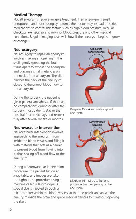
NeurosurgeryNeurosurgery to repair an aneurysm involves making an opening in the skull, gently spreading the brain tissue apart to expose the aneurysm, and placing a small metal clip on the neck of the aneurysm. The clip pinches the neck of the aneurysm closed to disconnect blood flow to the aneurysm.
During the surgery, the patient is given general anesthesia. If there are no complications during or after the surgery, most patients stay in the hospital four to six days and recover fully after several weeks or months.
Neurovascular InterventionNeurovascular intervention involves approaching the aneurysm from inside the blood vessels and filling it with material that acts as a barrier to prevent blood from flowing into it, thus sealing off blood flow to the aneurysm.
During a neurovascular intervention procedure, the patient lies on an x-ray table, and images are taken throughout the procedure using a machine called a fluoroscope. A special dye is injected through a microcatheter within the blood vessels so that the physician can see the aneurysm inside the brain and guide medical devices to it without opening the skull.
Diagram 15 – A surgically clipped aneurysm
Diagram 16 – Microcatheter is positioned in the opening of the aneurysm

13
Coil Embolization ProcedureThis is a type of neurovascular intervention procedure. There are a few different types of material used to fill an aneurysm. The most common are coils – long strands of very thin, coiled wire that look like guitar strings but are flexible like telephone cords.
In a coil embolization procedure, the physician packs several of these coils into the aneurysm one by one until it is full. The coils will remain inside the aneurysm, and a clot or embolus will form around them, making it difficult for any more blood to enter the aneurysm. Because the body’s natural response to the coils creates an embolus, the procedure is called coil embolization.
In this procedure, the physician first makes a small incision or puncture in the patient’s inner thigh and inserts a tube into a large blood vessel in the leg. This tube is a catheter sheath introducer. Then, a thin guidewire is inserted through the catheter sheath introducer. Because the guidewire is metal, the physician can see it on the x-ray screen (fluoroscopy) to guide it through the blood vessels up to the brain and to the aneurysm itself.
If there are no side effects or complications during or after the procedure, most patients stay in the hospital one or two days and recover fully after about a week.
Catheter sheath introducer – A hollow tube placed in a blood vessel and used to aid in the insertion of additional catheters or devices.
Embolization – Blocking a blood vessel or aneurysm so that blood can no longer flow into it.
Embolus – A clot or other plug that may obstruct circulation.
Fluoroscopy – An x-ray procedure in which x-rays are transmitted through the body onto a fluorescent screen; beneficial in observing the movement of joints or organ systems (e.g., the movement of material through the arterial system.
Guidewire – A flexible wire over which other devices, such as catheters, are guided to their target site.
Microcatheter – Small, thin, flexible tubes threaded into vessels to allow injection of contrast into specific areas or for the insertion of medical devices. Micro – a small catheter.

14
Over this guidewire, the physician threads a couple of long, thin tubes called catheters. A guiding catheter is threaded up to the base of the skull, and a smaller microcatheter runs from there through the blood vessels inside the brain to the aneurysm itself.
Then, the guidewire is removed. The physician threads the coils into the microcatheter one at a time and pushes them all the way up into the aneurysm. More coils are packed in until the aneurysm is completely full.
Diagram 17 – An aneurysm packed with coils
Guiding catheter – A catheter positioned in a blood vessel to allow for the passage of other devices through the catheter to a target site.
Coil Embolization Using an Intracranial Stent
Some types of aneurysms are difficult to treat with coils alone. For example, a wide-neck aneurysm may have such a large opening that the coils might not stay inside; they could fall back into the blood vessel and block or partly block the blood flow.
In these cases, the physician may first insert a stent inside the blood vessel where the aneurysm is located. A stent is a small, wire mesh tube that looks like a tiny roll of chain-link fence. In a coil embolization procedure for a wide-neck aneurysm, the stent is placed across the aneurysm neck and extends past the opening on both sides. This will help support the blood vessel.
The physician then inserts the coils as described above, threading them between the wires of the stent and into the aneurysm.
The stent acts as a small scaffold to hold the coils inside the aneurysm so they will not fall back into the vessel.

15
What Is the CODMAN ENTERPRISE Vascular Reconstruction Device and Delivery System?
The CODMAN ENTERPRISE Vascular Reconstruction Device and Delivery System is a type of intracranial stent. It is made of a flexible metal material called nitinol and is self-expanding.
When used in a procedure, the stent is squeezed into a very narrow tube that is part of its “delivery system” so that it will fit through the catheters.
When it is positioned precisely at the target site, the physician gently pushes the stent out of the tube, and it expands to its original shape, pressing firmly against the inner wall of the artery.
What Are the Potential Risks and Benefits of the Intracranial Stenting Procedure?Some of the potential adverse events or complications that may be associated with intracranial stenting include:• Ruptureandbleedingoftheaneurysmorbloodvessel• Aneurysmrecanalization• Death• Allergicreactionsordrugreactions• Irregularheartrhythm• Emergencyneurosurgery
The effectiveness of the CODMAN ENTERPRISE Vascular Reconstruction Device and Delivery System for use in treating wide-neck cerebral aneurysms has not been demonstrated.
Aneurysm recanalization – A previously treated aneurysm that refills with some blood and may require re-treatment.
Nitinol – A type of metal that “remembers” its shape and will return to that shape after being deformed.
Diagram 18 – An expanded CODMAN ENTERPRISE Vascular Reconstruction Device and Delivery System

16
In-Stent Restenosis and Other Potential ComplicationsStenosis refers to narrowing or blockage in a blood vessel. When a stent is implanted in a blood vessel, the lining of the vessel is injured. The body initiates a natural healing response to repair this injury. Although the healing response is important, in some cases, it is exaggerated. This exaggerated response can lead to the accumulation of scar tissue within the stent, narrowing or blocking the blood vessel. This is called in-stent restenosis. In-stent restenosis can lead to a lack of blood flow and may result in damage to the brain.
A similar potential problem is stent thrombosis, or formation of a blood clot (thrombus) within the stent. Stent thrombosis can occur soon after stent implantation (acute stent thrombosis) or after some time (delayed stent thrombosis). Stent thrombosis can block blood flow through the vessel, potentially leading to ischemic stroke.
Stent migration (movement of the stent from its original precise position) also may occur. Other potential complications are re-opening of the aneurysm, puncture-site related complications, or blockage of side vessels by the stent.
Potential long-term complications of intracranial stents are unknown.
Ischemic stroke – Lack of blood flow in the brain, blood vessels, or major arteries leading to the brain may result in loss of consciousness, paralysis, or other symptoms depending on the extent of brain damage.
In-stent restenosis – A re-narrowing or blockage of an artery within a stent.
Stenosis – Narrowing of a blood vessel.
Thrombosis – Formation, development or presence of a thrombus.
Thrombus – An aggregation of blood frequently causing obstruction.

17
Diagram 19 – During a neuroradiologic procedure, a catheter is placed into an artery and then guided up into your brain.

18
Step-by-Step Procedure: • Asmallopeningismadeinthe
inner thigh area. A short, narrow tube, called a catheter sheath introducer, is inserted into the artery of the leg through the small puncture site. A guidewire is placed through a longer, narrower tube, called a guiding catheter. The guiding catheter is passed through the catheter sheath introducer through the leg, neck, and into the brain and is placed at the base of the skull. Then a microcatheter is placed through the guiding catheter to the brain.
• X-raydyeisinjectedthroughthemicrocatheter to allow the doctor to see the blood vessels of the brain on an x-ray machine called a fluoroscope.
• Withx-rayguidance,thedoctornavigates the microcatheter into the blood vessel that contains the aneurysm.
•Thestent,mountedonadeliverywire, is introduced into the microcatheter. The stent is pushed through the microcatheter into the aneurysm site. The stent is positioned across the neck of the aneurysm and the microcatheter is gently pulled back which causes the stent to be exposed and deployed against the vessel walls. The stent should remain open against the vessels walls at the site it was placed.
Diagram 20 – First the stent is placed across the neck of the aneurysm.
Diagram 21 – Then the coils are placed through the microcatheter which is placed through the stent cells (open areas) into the aneurysm. Physicians may use different techniques to place catheters inside the brain to access the aneurysm. In diagram 20, the microcatheter is placed through the middle cerebral artery. In diagram 21, the microcatheter is placed from a different vessel, the internal carotid artery.
Diagram 22 – The stent remains implanted across the neck of the aneurysm and the coils remain inside the aneurysm.
How Is the Intracranial Stenting Procedure with the CODMAN ENTERPRISE Stent Performed?

19
• Thestentdeliverysystemisremoved.Aguidewireandmicrocatheterarethen introduced through the guiding catheter and navigated through the open areas within the stent into the aneurysm, to facilitate the placement of coils.
• Aftertheprocedureiscomplete,thestentandthecoilswillremainimplanted inside the patient and typically most other products will be removed. The patient will then follow physicians orders after the procedure.
What Happens after the Procedure? After the procedure, you may be moved to a special care unit where nurses will be able to monitor your heart rhythm and blood pressure very closely. The catheter sheath introducer may be removed at this time, and pressure will be applied to the puncture site until bleeding stops. If the catheters were inserted through your leg, you may be instructed to lie flat and not bend your leg for several hours. The nurses will monitor your incision for changes to ensure it is healing properly. WARNING: If you see any blood or feel warmth at the puncture site, tell your nurse immediately.
Once you return to your room, you may be able to eat and drink and your family may visit, depending on your doctor’s orders. Eat foods that are light until you are able to sit upright. Drink all of the fluids offered to you, because they will help to flush the x-ray dye through your kidneys and out of your body. Your doctor will advise you when you can get out of bed and walk.
Many people go home the day after the procedure. The amount of time that you stay in the hospital depends on your doctor’s discharge orders. These orders will be based on several factors, including any difficulties you may have experienced during the procedure and how well the puncture site is healing.
Diagram 23 – Photo of a typical angiographic suite where diagnostic and interventional neurovascular procedures are performed.

20
Taking Your Medications• Afteryouleavethehospital,youmaybeinstructedtotakebloodthinning
medications (also called anti-platelet or anti-coagulant medications). Depending on what medications your doctor prescribes, you may need to have follow-up blood tests to monitor the effects of the medication on your blood. These can be done at your local hospital laboratory or primary care doctor’s office.
• CAUTION: It is very important that you take your medications exactly as prescribed, because they are intended to prevent the potential complications described earlier. Be sure not to miss any doses.
• WARNING: Call your doctor if you feel that you cannot tolerate your medications; if you develop any side effects such as bleeding, upset stomach, or rash; or if you have any questions.
What Follow-up Is Required after Treating Your Aneurysm?After your aneurysm is treated, you will need to visit your doctor as recommended. Your physician may ask you to return to the hospital or clinic to have a CT scan, MRA, or diagnostic cerebral angiogram to see how your aneurysm is responding to treatment.
MRI Testing – WARNING Before you have an MRI scan, or for questions about the coils or stent, you can provide the information below for details specific to each product. Additionally, questions can be directed to Codman Neurovascular. Call800-225-0460 and follow prompts to access “Product Complaints.”
TRUFILL DCS ORBIT® COILThrough non-clinical testing, the TRUFILL DCS ORBIT coil has been shown to be MRI Conditional at field strengths of 3-Tesla or less, SAR of 2.0 W/kg for 20 minutes of MRI.
CODMAN ENTERPRISE Vascular Reconstruction Device and Delivery SystemThe CODMAN ENTERPRISE Vascular Reconstruction Device and Delivery System has been shown to be MRI Conditional in MRI systems operating under the following conditions: static magnetic field of 3-Tesla or less, spatial gradient field of 720-Gauss/cm or less, and a maximum MRI system reported whole-body-averaged specific absorption rate (SAR) of 3-W/kg for 15 minutes of scanning.
Tesla – A unit measure of magnetic strength.

21
Summary
You have an important role to play to ensure that your procedure will be successful. Review this booklet, cooperate with your physician, and follow through with your responsibilities as part of the patient / medical team. If you have any questions or concerns, please contact your physician to discuss them.

22
Amphetamines – A central nervous system stimulant that increases energy and decreases appetite; used to treat narcolepsy and some forms of depression.
Aneurysm – A weak spot in the wall of a blood vessel that stretches or balloons out, forming a thin-walled bubble or sac.
Aneurysm recanalization – A previously treated aneurysm that refills with some blood and may require re-treatment.
Angiogram – A test used to visualize vessel abnormalities by injecting a special (contrast) dye into the vessels via a catheter. This allows the doctor to see on x-ray where the vessel abnormality is located. Catheter sheath introducer – A hollow tube placed in a blood vessel and used to aid in the insertion of additional catheters or devices. Cerebral – Having to do with the brain. Cocaine – A substance extracted from the leaves of the coca plant that may act as a powerful short-acting stimulant that speeds up the activity of some brain chemicals. Its effects may include euphoria, restlessness, excitement, or a feeling of well-being. Coil – An implantable medical device that has long strands of very thin, coiled wire that look like guitar strings but are flexible like telephone cords and facilitate clot formation within an aneurysm. Computed tomographic angiography – A diagnostic test that uses x-rays taken from many angles to produce cross-sectional images of a part of the body.
Contrast dye (x-ray dye) – A substance that is opaque to x-rays, used to permit visualization of internal body structures.
Diagnostic cerebral angiogram – A test used to diagnose abnormalities with the blood vessels of the brain. It is a also used to determine if an aneurysm is present. This test involves guiding a small tube (catheter) from the leg blood vessels into the blood vessels of the neck and injecting contrast (dye) to see the blood flow. Embolic – Having to do with an embolus, which is a blood clot or other foreign material that causes blockage in a vessel.
Glossary of Terms

23
Embolization – Blocking a blood vessel or aneurysm so that blood can no longer flow into it.
Embolus – A clot or other plug that may obstruct circulation. Fibromuscular dysplasia – Fibromuscular dysplasia, commonly called FMD, is a disease that causes one or more arteries in the body to have abnormal cell development in the artery wall. As a result, areas of narrowing, called stenosis, may occur. If enough narrowing causes a decrease in blood flow through the artery, an aneurysm may result.
Fluoroscopy – An x-ray procedure in which x-rays are transmitted through the body onto a fluorescent screen; beneficial in observing the movement of joints or organ systems (e.g., the movement of material through the arterial system).
Guidewire – A flexible wire over which other devices, such as catheters, are guided to their target site.
Guiding catheter – A catheter positioned in a blood vessel to allow for the passage of other devices through the catheter to a target site. Hemorrhage – Loss of blood from damaged blood vessels.
Hemorrhagic stroke – When blood from a cerebral aneurysm spills directly into the brain.
In-stent restenosis – A re-narrowing or blockage of an artery within a stent. Interventional neuroradiology / INR also known as endovascular surgery – A medical specialty that addresses problems of the cerebral vascular system using minimally invasive or endovascular techniques. This usually includes vascular procedures that are intracranial (within the skull) and extracranial (outside the skull, but above the heart).
Ischemic stroke – Lack of blood flow in the brain, blood vessels, or major arteries leading to the brain may result in loss of consciousness, paralysis, or other symptoms depending on the extent of brain damage. Magnetic resonance angiography – A procedure in which radio waves and magnetic fields are used to generate computer images of the body’s internal tissues.

Microcatheter – Small, thin, flexible tubes threaded into vessels to allow injection of contrast into specific areas or for the insertion of medical devices.
Micro – a small catheter.
Narcolepsy – A sleep disorder consisting of recurring episodes of sleep during the day and often disrupted sleep at night. Neurology – The medical science that deals with the nervous system and disorders affecting it.
Neurovascular intervention – A minimally invasive procedure involving the cerebral vascular system where contrast dye is injected into the arteries in the brain via a catheter. Different types of medical devices may be used to treat any abnormalities. Nitinol – A type of metal that “remembers” its shape and will return to that shape after being deformed.
Parent artery – The artery from which a given artery (the branch) originates.
Radiopaque – An object that blocks x-rays so that it creates an outline of the structure being looked at on x-ray film. Rupture – Tearing of a tissue. Stenosis – Narrowing of a blood vessel. Stent or Vascular Reconstruction Device – A specially designed, expandable metal tube that is inserted into a vessel. A stent acts as a scaffold to provide structure for a vessel. In a wide neck aneurysm, a stent is placed across the opening or neck of the aneurysm to secure the placement of coils and to maintain blood flow through the artery in which the stent is placed.
Tesla – A unit measure of magnetic strength.
Thrombosis – Formation, development or presence of a thrombus.
Thrombus – An aggregation of blood frequently causing obstruction.
Vascular Reconstruction Device – See stent.
24