unionicola (wolcottatax) arcuata · pdf fileunionicola (wolcottatax) arcuata ... fourth...
TRANSCRIPT
Unionicola (Wolcottatax) arcuata (Wolcott 1898) Plates 191-193 in Vidrine (1996a)
Synonomy--
• Atax arcuata Wolcott 1898, Wolcott 1899
• Atax arcuatus Wolcott 1898 in Piersig 1901
• Unionicola arcuata (Wolcott 1898) in Marshall 1927 and 1933b, Mitchell 1954, Crowell 1961, and Habeeb 1967
• Unionicola (Parasitatax) arcuata (Wolcott 1898) in Viets and Plate 1954, Vidrine 1980a, 1986c, and 1986e
• Unionicola (Wolcottatax) arcuata (Wolcott 1898) in Vidrine 1992b, Vidrine 1996a-e, Vidrine et al. 2008, and Edwards and Vidrine 2013.
Museum type number(s) and location-- Marshall Collection, Field Museum of Natural History, Chicago, Illinois. Type locality and host-- Pyganodon grandis in Michigan. Etymology-- Named for its arcuate tarsi on posterior walking legs. Diagnosis-- Character states of the subgenus; dorsum apparently lacking a dorsal plate; coxal plates abbreviate and with distinct borders; pedipalp Ta with 2, distinct clawlets; genital fields with numerous acetabula (ca. 33-38); all legs with small, bifid tarsal claws with the dorsal prong shorter than the ventral prong; fourth walking leg with Ta distinctly arcuate (see key figure on page 9); ratio of leg IV tarsus versus tibia ca. 0.60-0.70 (Tuzovskij and Semenchenko (2015) report specimens with ratios ca. 0.85-0.90).
• Male (22 specimens)-- Length including capitulum 1200 (1000-1400) (all measurements in micrometers (µm)); length of posterior coxal group 333 (270-400); dorsal lengths of pedipalp segments: Ti 144 (120-180); Ta 75 (60-90); dorsal lengths of leg segments: leg I: TFe 177 (140-220); Ge 229 (160-300); Ti 214 (160-280); Ta 137 (110-170); leg IV: TFe 321 (240-430); Ge 424 (310-580); Ti 578 (440-780); Ta 379 (320-470).
• Female (19 specimens)-- Length including capitulum 1600 (1300-1900); length of posterior coxal group 320 (260-400); dorsal lengths of pedipalp segments: Ti 143 (120-170); Ta 73 (65-90); dorsal lengths of leg segments: leg I: TFe 180 (150-240); Ge 235 (180-310);

Ti 226 (170-290); Ta 138 (110-170); leg IV: TFe 326 (260-420); Ge 429 (320-570); Ti 574 (450-780); Ta 391 (320-480).
Notes-- Unionicola arcuata is generally a parasite of members of the mussel subfamily Anodontinae. The specimens vary greatly in size, but relative leg segment ratios are consistent enough to prevent ready separation into subgroups based solely upon size. Apparently, U. arcuata in North America forms a Rassenkreis, since varied size morphs are found in specific taxa of hosts—with new studies on species in Asia, it is apparent that there are several species involved in my North American lots—an Artenkreis. It is also highly likely that cryptic species are involved as the mites are known from a rather large number of host genera. Unionicola arcuata appears to one of the most common mites found in Asian mussels. However, I suspect that these are not the same species as the North American populations, but rather represent another Artenkreis. I will present some illustrations of Asian U. arcuata in my discussion of U. arcuatoides, but many are available (Wen and Zhu 1999; Tuzovskij and Semenchenko 2015). Wen and other authors have studied populations of U. arcuata using molecular analysis—there they demonstrated 2 key discoveries: 1) subgenus Wolcottatax is indeed different from subgenus Unionicola; 2) populations of U. arcuata from different host populations are different species (Wu et al. 2011). The variation within populations of North American U. arcuata is sufficient to cause much discussion. I have prepared graphs of measurements of the fourth walking leg segments, especially the tibia and tarsus in order to demonstrate the variation, but this is just more than I want to discuss in this work. My resolution to this is to provide photos of 4 varieties of this mites in new plates 1-4 along with Wolcott’s original drawing. The plates contain the tarsi of the fourth walking legs—they are all similar and different from Asian forms for the most part. However, if you look at the details, e.g., the placement of the dorsal setae and the ventral setae along the length of the tarsus, the differences are sufficient to give one pause. As such, I think that we are dealing with several species, but the differences are too vague to use at present to justify the separation of the mites, and I continue to treat the North American group as a single, diverse species. Of course, I expect that DNA analysis would separate these species and more; but this is not available at this time. We will see what happens with efforts to separate species using DNA analysis when we discuss the subgenus Unionicola. I
am leaving the discussion of the Asian U. arcuata to my discussion of U. arcuatoides. I am hoping that that will reduce confusion! Adults, sometimes as many as 100 mites per host, oviposit in the demibranch blades (gills) of freshwater mussels (Vidrine 1980a). This species is commonly found in lotic waters, and this species apparently does not co-occur with mites belonging to the subgenus Unionicola; however, it does commonly co-occur with members of the subgenus Dimockatax. Thus, anodontine mussels in North America commonly have mites, but 2 interesting communities tend to occur: 1) a population of members of the subgenus Unionicola, often with many mites present; 2) a community with members of the subgenus Dimockatax, usually a male and one or 2 females, and members of the subgenus Wolcottatax, often with many mites present. The 3 kinds of mites are closely related and formerly were all considered to be members of a single subgenus. The population and community structures that I have observed and others have studied, included the ‘harem’ nature of populations of U. formosa in mussels by Ron Dimock. Much work on behavior and evolution of the group could be done. Hosts in North America (I have prepared a discussion of Asian specimens in the Hosts section of U. arcuatoides):
• Alasmidonta heterodon (Lea): Prolasmidonta heterodon (Lea), Ashuelot River, New Hampshire (Vidrine, 1980a); Massachusetts.
• Alasmidonta marginata Say: Margaritana marginata Say, Michigan and Pennsylvania (Wolcott, 1899); Decurambis marginata Say in Viets and Plate 1954; Michigan (Kelly, 1899); and Arkansas (Vidrine, 1980a).
• Alasmidonta undulata (Say): Rhode Island (Vidrine, 1980a); Canada (Vidrine 1986c).
• Alasmidonta viridis (Rafinesque): Margaritana deltoides Lea, Michigan? (Wolcott, 1899); Pressodonta calceola Lea in Viets and Plate 1954; Alasmidonta calceola (Lea), Douglass Lake, Wisconsin (Marshall 1927 and 1933b); Michigan (Vidrine, 1980a); Canada (Vidrine 1986c).
• Alasmidonta varicosa (Lamarck): Canada (Vidrine 1986c).
• Anodontoides ferussacianus (Lea): Anodonta subcylindracea Lea, Michigan (Wolcott, 1899) (and in Viets and Plate 1954); Douglas Lake, Wisconsin (Marshall, 1927); Canada (Vidrine 1986c).
• Anodontoides radiatus (Conrad): Alabama (Vidrine 1980a); Strophitus subvexus (= S. radiatus), Louisiana and Mississippi, (Vidrine, 1980a).
• Lampsilis radiata (Gmelin): Unio luteolus Lamarck, Michigan (Wolcott, 1899) (= Ligumia fasciata Rafinesque in Viets and Plate 1954); Vidrine 1980a.
• Lasmigona compressa (Lea): Canada (Vidrine 1986c).
• Lasmigona costata (Rafinesque): Margaritana rugosa Barnes, Michigan (Wolcott, 1899) (and in Viets and Plate 1954); Vidrine 1980a.
• Pyganodon fragilis (Lamarck): Anodonta fragilis, Michigan (Wolcott, 1899) Anodonta marginata Say in Viets and Plate 1954; Anodonta cataracta (Say) Vidrine, 1980a.
• Pyganodon grandis (Say): Anodonta footiana Lea, Michigan (Wolcott, 1899) (=Pyganodon grandis footiana Lea in Viets and Plate 1954); Anodonta grandis Say in Vidrine 1980a, 1989a.
• Strophitus undulatus (Say): Anodonta edentula Say, Michigan (Wolcott 1899) (and in Viets and Plate 1954); Canada (Vidrine 1986c).

Figures of North American U. arcuata (from Vidrine 1996):
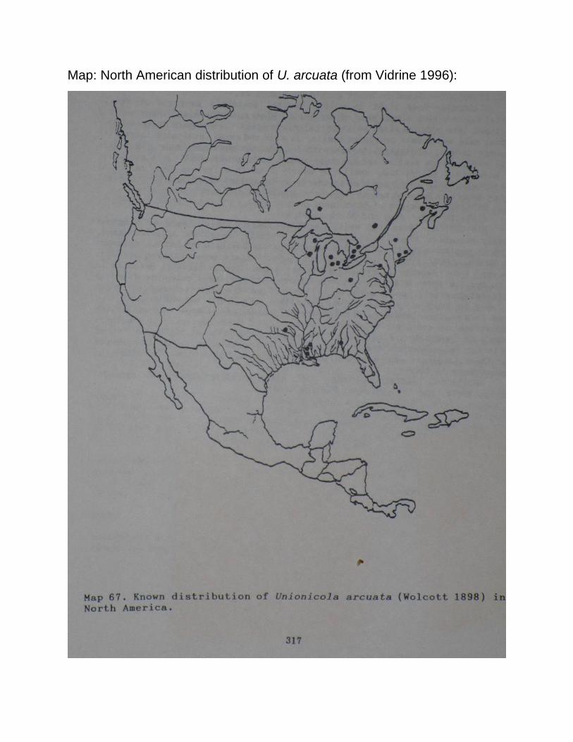
Map: North American distribution of U. arcuata (from Vidrine 1996):

Plate 191: Top left: male venter. Top right: tarsus of leg IV. Middle left:
male posterior venter. Middle upper right: male venter. Middle lower right:
male genital field. Bottom left: composite photo of walking legs of male.
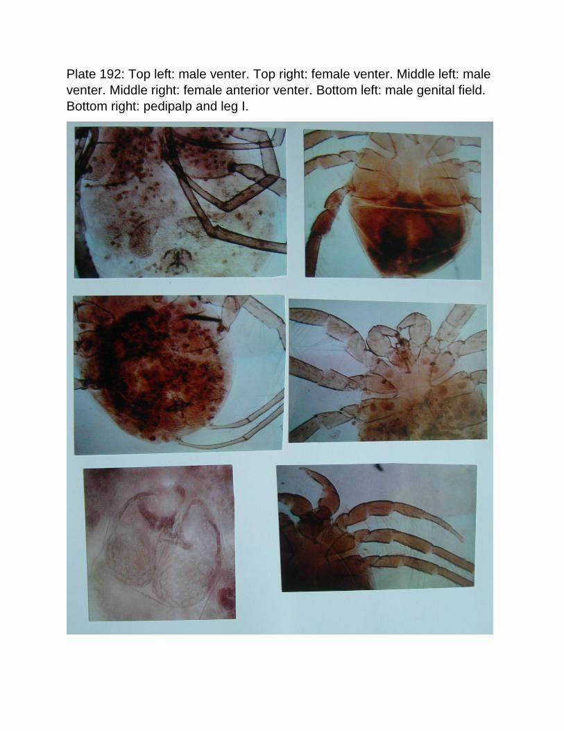
Plate 192: Top left: male venter. Top right: female venter. Middle left: male
venter. Middle right: female anterior venter. Bottom left: male genital field.
Bottom right: pedipalp and leg I.
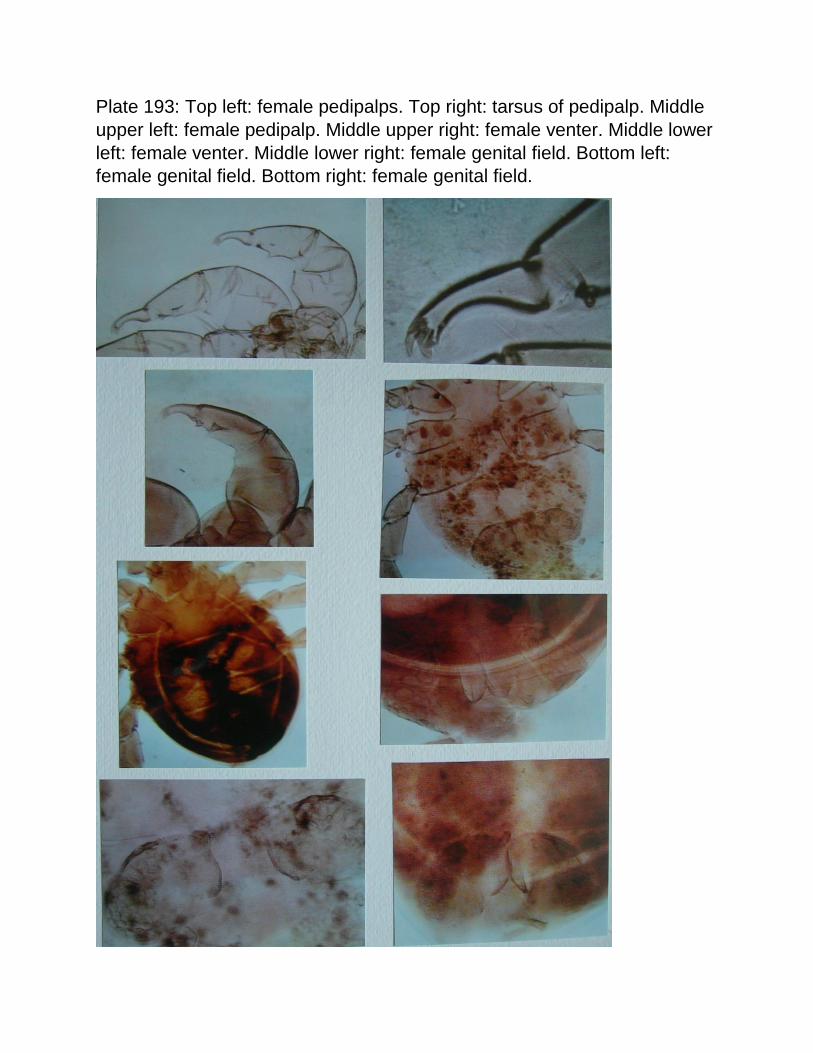
Plate 193: Top left: female pedipalps. Top right: tarsus of pedipalp. Middle
upper left: female pedipalp. Middle upper right: female venter. Middle lower
left: female venter. Middle lower right: female genital field. Bottom left:
female genital field. Bottom right: female genital field.
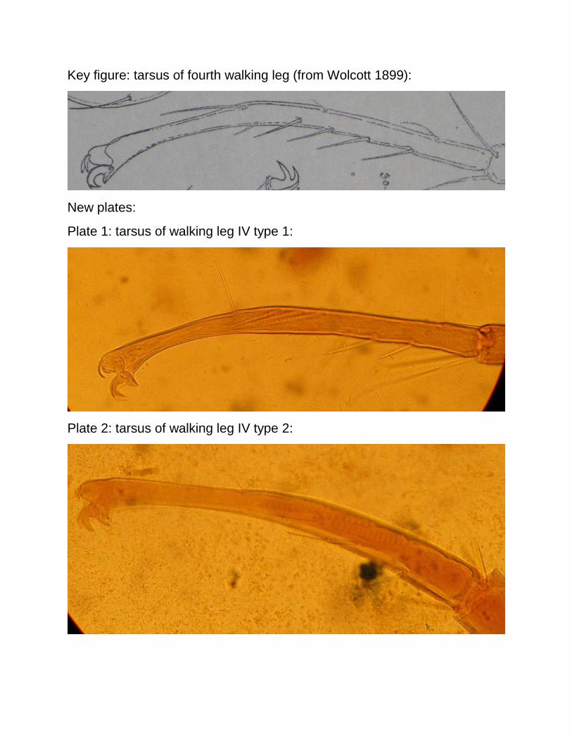
Key figure: tarsus of fourth walking leg (from Wolcott 1899):
New plates:
Plate 1: tarsus of walking leg IV type 1:
Plate 2: tarsus of walking leg IV type 2:
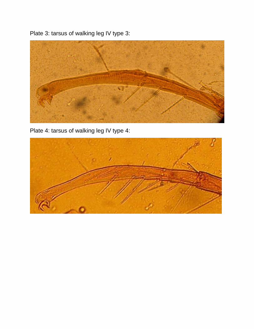
Plate 3: tarsus of walking leg IV type 3:
Plate 4: tarsus of walking leg IV type 4:
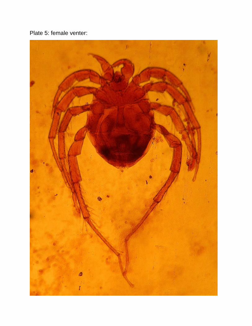
Plate 5: female venter:
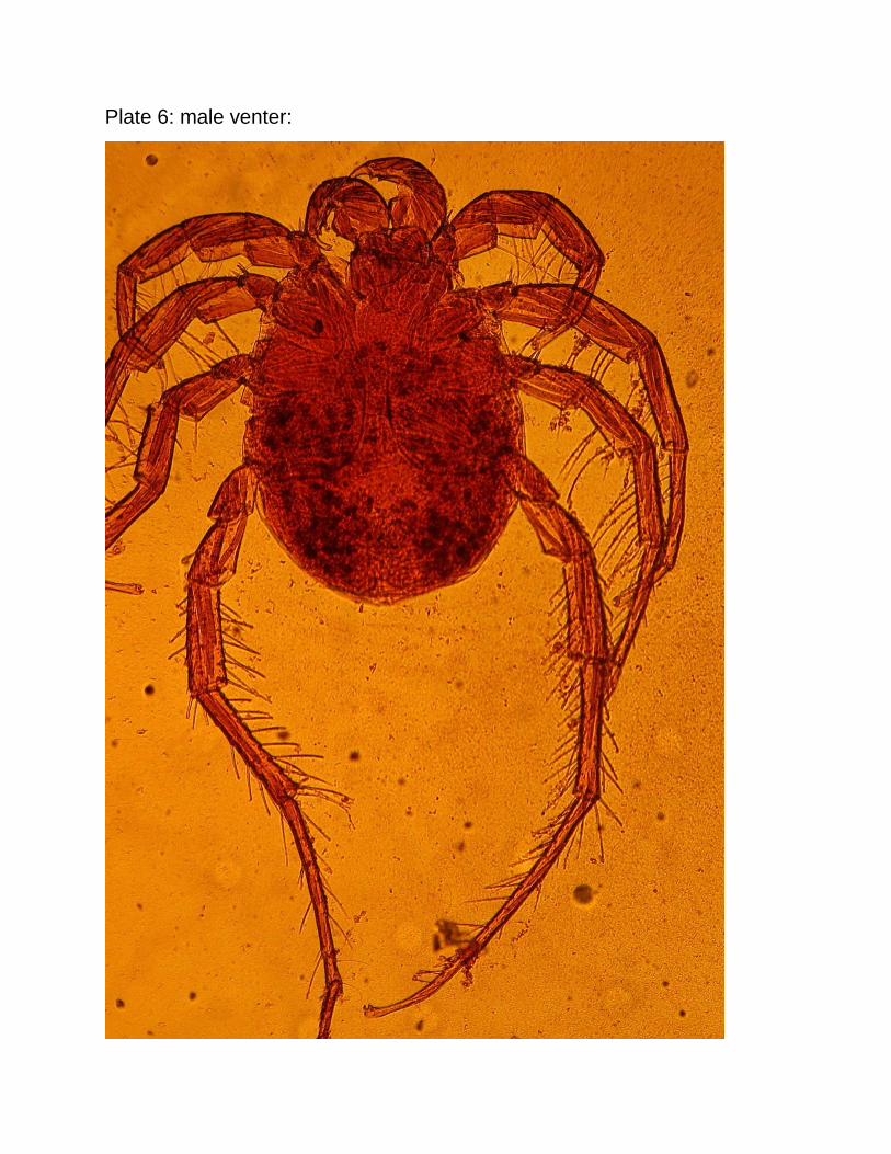
Plate 6: male venter:
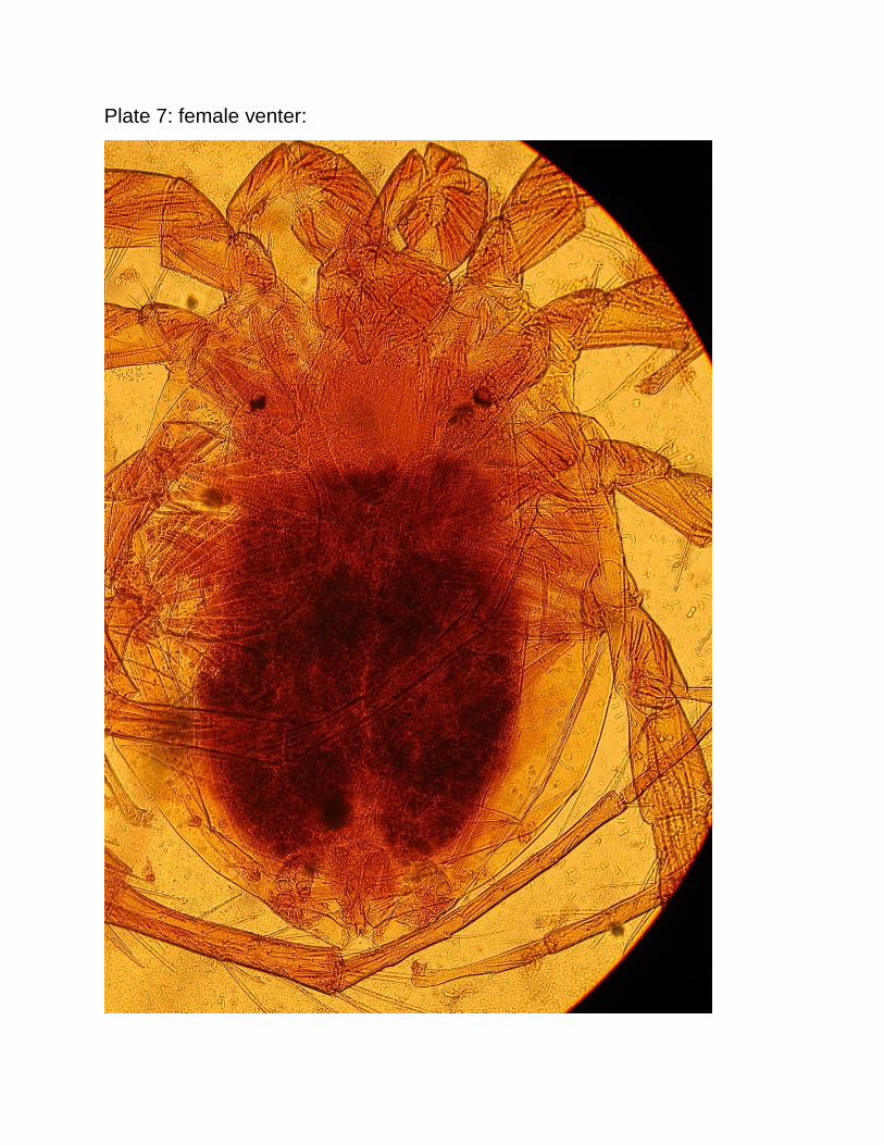
Plate 7: female venter:
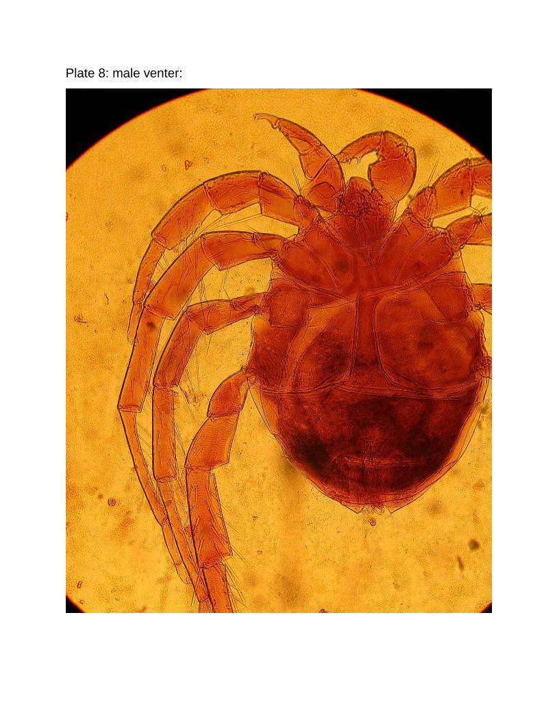
Plate 8: male venter:
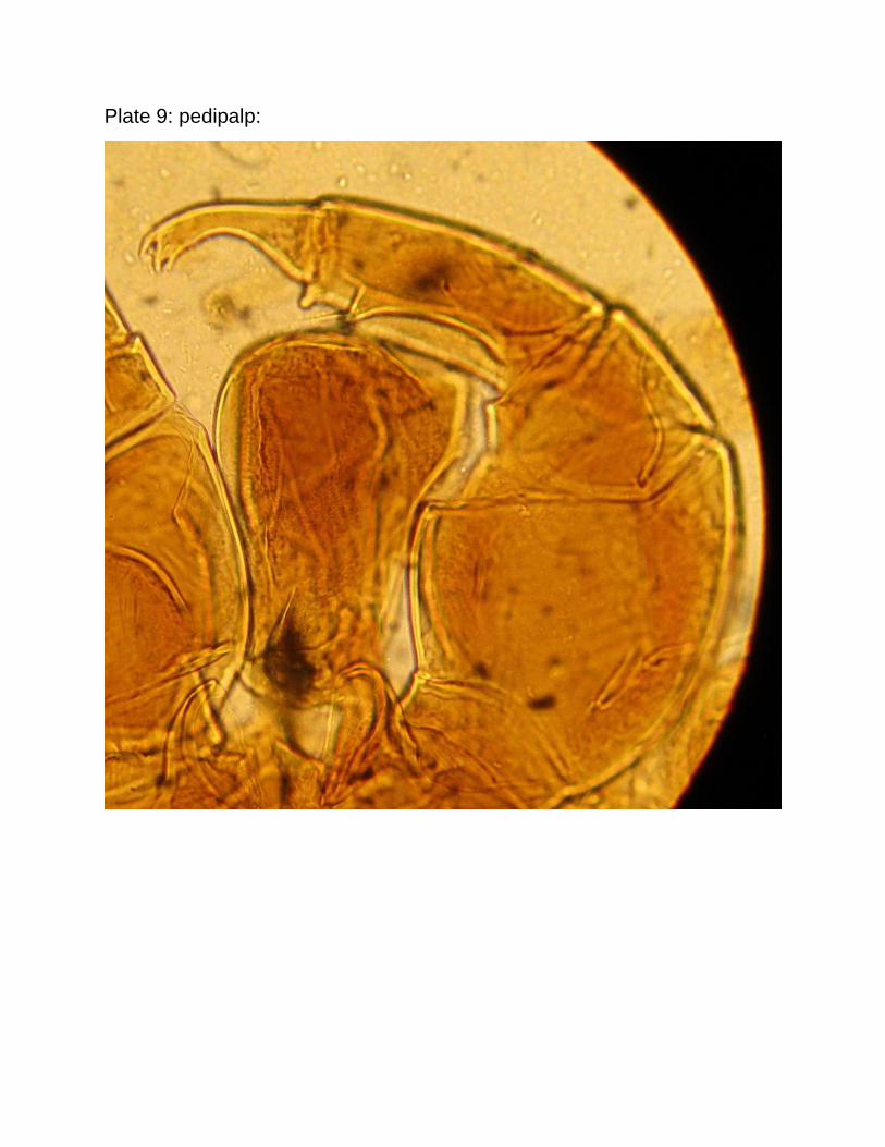
Plate 9: pedipalp:
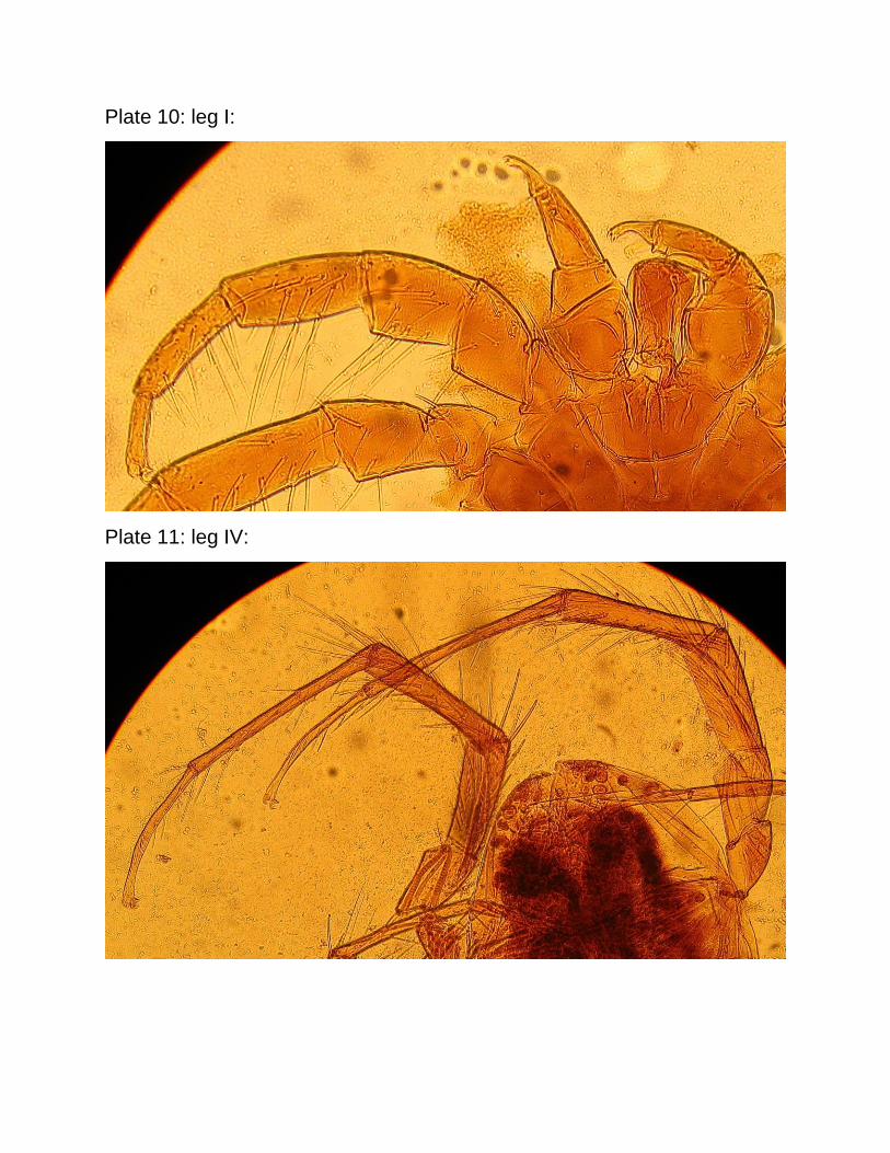
Plate 10: leg I:
Plate 11: leg IV: