unit 6: digestive systemthe respiratory and digestive passages meet in the pharynx. they separate...
TRANSCRIPT

Unit 6: Digestive System

Learning Objectives:
• Know the sequence of digestive track and its associated organs.
• Know the various digestive enzymes and its releasing organs.
• Know the various hormones and chemicals associated with digestion.
• Understand the importance of each enzyme and its releasing points.

Digestion Overview
•Animals obtain energy by breaking food molecules into smaller pieces.
• The basic fuel molecules are amino acids, lipids and sugars.
•Digestion is the chemical breakdown of large food molecules into smaller molecules that can be used by cells.

The processing of food has four basic steps:
i. Ingestionii. Digestioniii. Absorptioniv. Elimination

IngestionFood enters the digestive tract through the mouth.

DigestionDigestion occurs in two different ways
i. Mechanical digestion - breaking food into smaller pieces ex: Chewing
ii. Chemical digestion - breaking food into smaller molecules
ex: Enzymes

SalivaWhen chewing food, our mouth will secret saliva through salivary gland. Glands are tissue that secret chemicals.
Food is moistened and lubricated in the mouth.
Contains the enzyme salivary amylase to begin the breakdown of starch.

Food passes to the stomach through the esophagus.
Mucous lubricates and helps hold
the chewed food together in a
clump called a bolus.

The respiratory and digestive passages meet in the
pharynx. They separate posterior to the pharynx to form
the esophagus (leading to the
stomach) and the trachea
(leading to the lungs). When
swallowing, the epiglottis
automatically moves down to
block the passage to the trachea
so that food does not end up in the lungs.


The bolus is moved to the stomach by peristalsis (rhythmic muscle contractions).

A muscle called the cardiac sphincter controls the
movement of food from the esophagus to the stomach.
This muscle prevents food in the stomach from reentering
the esophagus.
Sometimes acid
does splash up into
the esophagus
causing what we call heartburn.
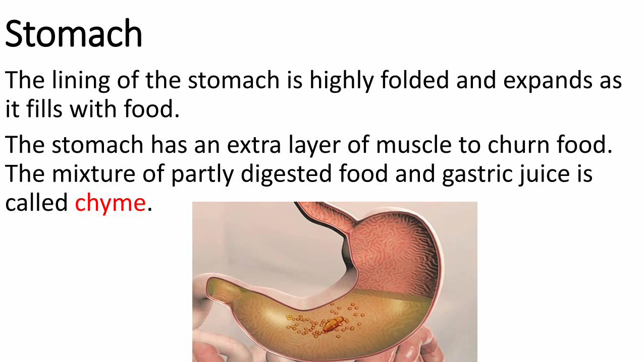
StomachThe lining of the stomach is highly folded and expands as it fills with food.
The stomach has an extra layer of muscle to churn food. The mixture of partly digested food and gastric juice is called chyme.
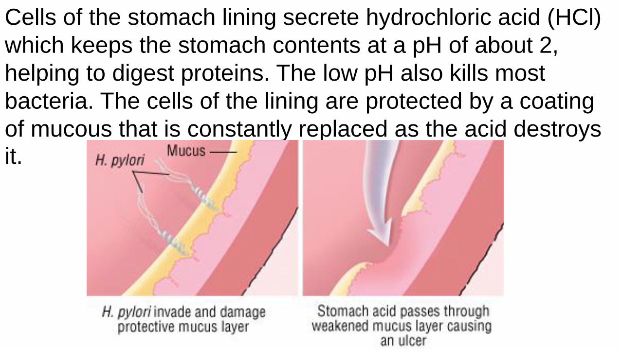
Cells of the stomach lining secrete hydrochloric acid (HCl)
which keeps the stomach contents at a pH of about 2,
helping to digest proteins. The low pH also kills most
bacteria. The cells of the lining are protected by a coating
of mucous that is constantly replaced as the acid destroys it.

Stomach Ulcer

They also secrete pepsinogen, a protein digesting enzyme. Inactive pepsinogen is cleaved to form pepsin - the active form of the enzyme. This is important to protect the cells thatproduce pepsin from being digested themselves. Pepsin is most active in a pH of 2.

Pepsin cuts proteins into short pieces of polypeptides. Note: still in polymer form


There is no digestion of carbohydrates or fats in the stomach.
What happened to salivary amylase?
Very little absorption occurs in the stomach, with some
exceptions such as water, aspirin and alcohol. All other
absorption occurs in the intestine.

A muscle called the Pyloric Sphincter controls the
movement of food from the stomach to the small intestine.
This muscle slowly squirt out pinches of chyme to avoid
flooding the small
Intestine with foodParticles.

Small IntestineSmall intestine can be divided into three sections:
Duodenum, jejunum, and ilium.
The majority of digestive
Enzymes are released
Into duodenum.
The small intestine is where most digestion occurs.

In the duodenum, chyme, pancreatic enzymes, and bile from the
liver and gallbladder are mixed.

The presence of food in the duodenum also triggers the
release of various hormones
(1) The intestine must be protected from the acid contained
in gastric juice. When chyme enters the small intestine, the
cells of the duodenum release the hormone secretin. This
hormone stimulates the pancreas to produce sodium
bicarbonate (NaHCO3-), which neutralizes the acidic chyme
and shuts off pepsin. It also stimulates the liver to secrete
bile.

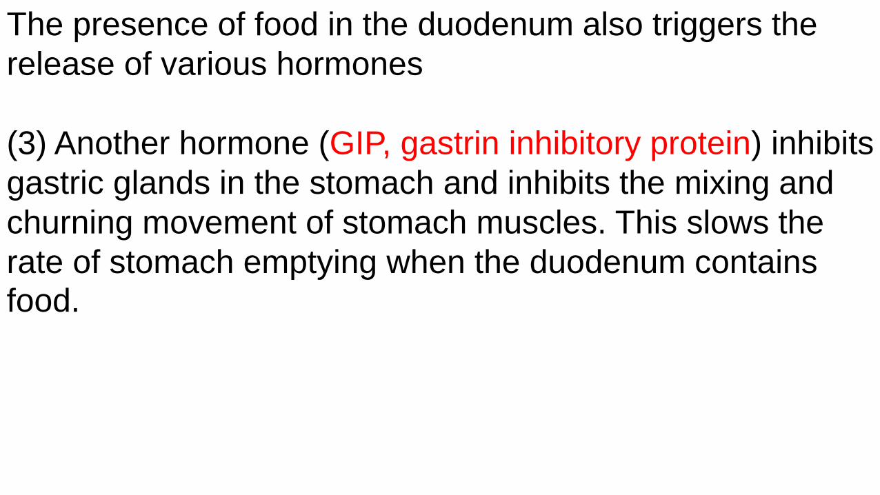
The presence of food in the duodenum also triggers the
release of various hormones
(2) Another hormone (CCK, cholecystokinin), stimulates
the gallbladder to release bile and the pancreas to produce
pancreatic enzymes.
The presence of food in the duodenum also triggers the
release of various hormones
(3) Another hormone (GIP, gastrin inhibitory protein) inhibits
gastric glands in the stomach and inhibits the mixing and
churning movement of stomach muscles. This slows the
rate of stomach emptying when the duodenum contains food.

The pancreas secretes enzymes, bicarbonate and hormones.
Secretions of the pancreas reach the duodenum via the pancreatic duct. The fluid containsi. The protein digesting enzyme trypsin.ii. The starch digesting enzyme pancreatic amylase.iii. The fat digesting enzyme lipase.iv. The nucleic acid digesting enzyme nuclease.

Why so many enzymes to digest protein?

The pancreas also produces hormones that regulate levels
of sugar in the blood. These hormones include:
Insulin: Released to turn glucose into glycogen.Glucagon: Released to turn glycogen into glucose.

a) Does the above graph represent positive or negative
feedback?

b) Explain the hormonal response when the i) blood glucose concentration is high. ii) blood glucose concentration is low.
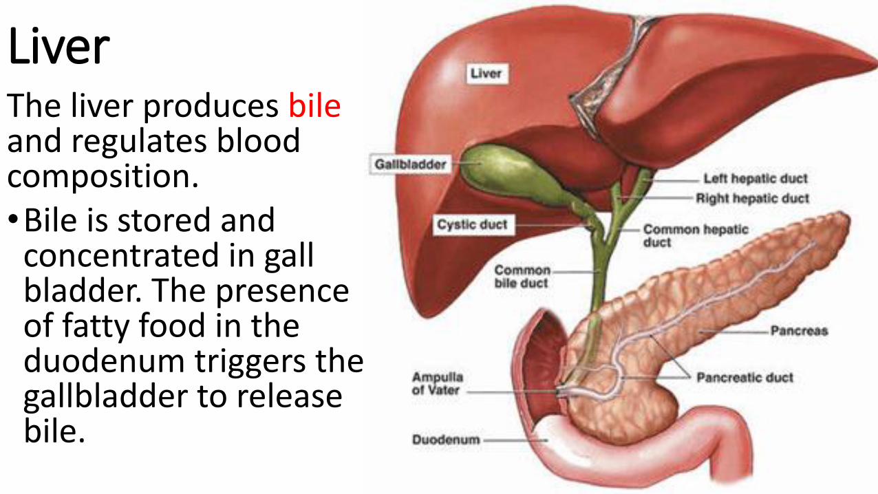
LiverThe liver produces bileand regulates blood composition.•Bile is stored and concentrated in gall bladder. The presence of fatty food in the duodenum triggers the gallbladder to release bile.

Bile travels through the common bile duct to the
duodenum.
Bile salts are soluble in both lipids and water. This enables
them to break apart fat droplets in chyme to create smaller
droplets. This increases the surface area for lipase to work on and increases the speed of their digestion.

Cholesterol can bind bile together causing crystals to form.
If these crystals become large enough they can block the
common bile duct.
If the common bile becomes blocked, bile salts can
accumulation in the skin and produce a yellow color in a condition called jaundice.

Another role of the liver is to remove toxins from the blood.
The liver absorbs or chemically modifies toxic substances to prevent them remaining in circulation.
• Ammonia produced by the digestion of proteins is converted to a less toxic compound (urea) by the liver. Urea is removed from the blood by the kidneys and eliminated in urine.
• Alcohol and drugs are metabolized by liver cells into less harmful compounds. Other toxins, pesticides, and carcinogens are also detoxified.

Digestive enzymes secreted by the cells of the small intestine digest lactose, sucrose and other sugars. Some adults lose the ability to produce lactase, resulting in the condition called lactose intolerance.

Absorption of food in the intestine.• The walls of the small intestine are covered with small
projections called villi. These increase the surface area of the small intestine to increase absorption.
• The villi themselves are covered with many tiny projections called microvilli, which increase the surface area still further.

The molecules resulting
from the digestion of
proteins and
carbohydrates are
absorbed by cells of the
intestinal lining.
The villi contain
capillaries, tiny blood
vessels which allows
efficient transfer of these
molecules to the blood.

The products of fat
digestion are absorbed
through the villi into the
lymphatic system.
Lacteal is the lymphatic
system extension that
absorbs fat and lipid.

The large intestine concentrates solids by reabsorbing water.It has no digestive function, but functions to absorb water. If water is not absorbed, as can happen during certain bacterial infections, diarrhea can result, causing dehydration and saltloss.
The large intestine (along with some absorbed by the small intestine) recovers about 90% of the water that enters the digestive system. Remember that this is very important for a terrestrial vertebrate.

Bacteria (mostly E. coli) live and reproduce in the colon. Anaerobic digestion (fermentation) of material by these bacteria produces gas in the colon. They also produce vitamins for the host, including vitamin K some B vitamins. Bacteria are lost when feces is eliminated, making exposure to feces dangerous.

Fiber (cellulose) tends to fill up the colon and cause it to be emptied. Low fiber diets result in slower passage of food through colon and have been linked to colon cancer.
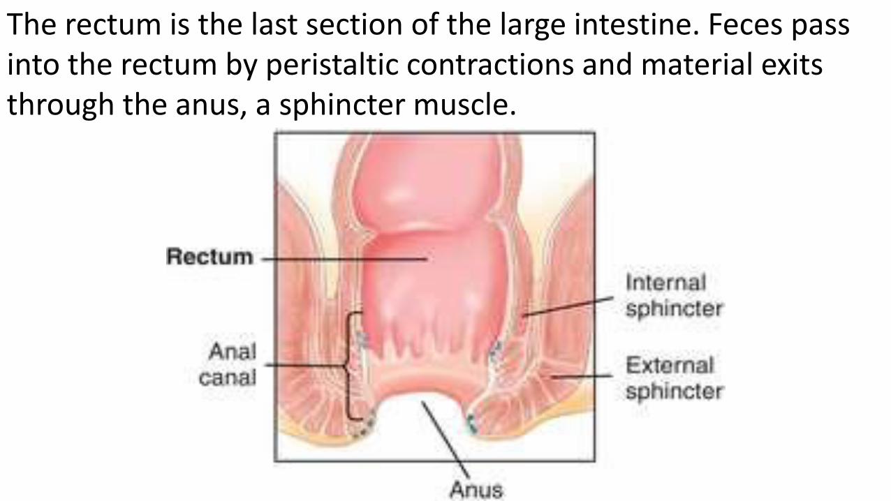
The rectum is the last section of the large intestine. Feces pass into the rectum by peristaltic contractions and material exits through the anus, a sphincter muscle.


Review Questions:Label the parts on the
diagram and give one
function for each during the
digestion of a protein.


In the following reaction, product X could be a(n)
A. peptide. B. fatty acid. C. nucleotide. D. amino acid.
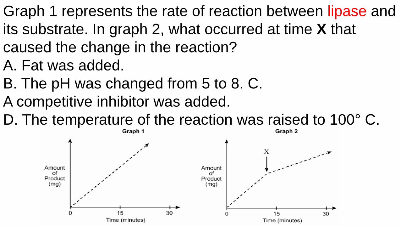
Graph 1 represents the rate of reaction between lipase and
its substrate. In graph 2, what occurred at time X that
caused the change in the reaction?
A. Fat was added.
B. The pH was changed from 5 to 8. C.
A competitive inhibitor was added.
D. The temperature of the reaction was raised to 100° C.

a) The diagrams illustrate a reaction that occurs in the small
intestine. Give the specific name for each of the following.
- Molecule X:
- Molecule Y:
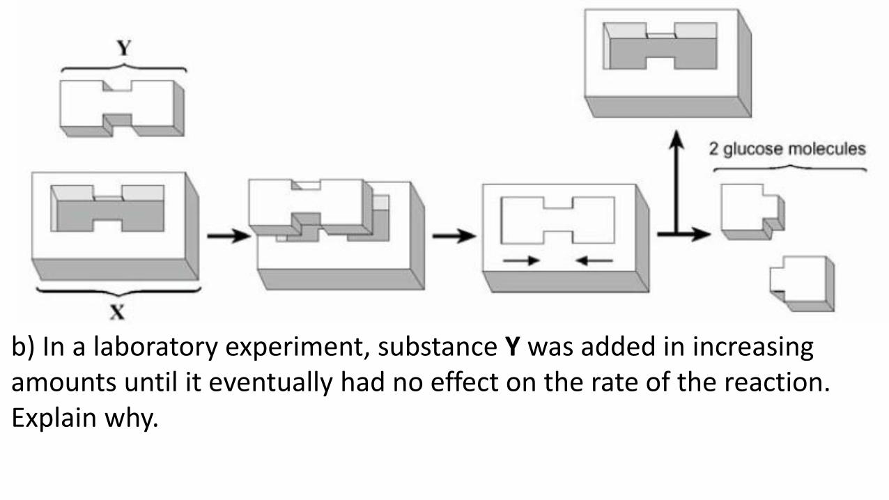
b) In a laboratory experiment, substance Y was added in increasing amounts until it eventually had no effect on the rate of the reaction. Explain why.

c) A solution containing lead ions was added to the reaction. How will the addition of this solution affect the reaction? Explain why.