united states of america - archive-it
TRANSCRIPT
This t1 or car: from t semia Drug L P8pres
UNITED STATES OF AMERICA . .
FOOD AND DRUG ADMINISTRATION
CENTER FOR BIOLOGICS EVALUATION AND RESEARCH
+ + + + +
VACCINES AND RELATED BIOLOGICAL
PRODUCTS ADVISORY COMMITTEE
+ + + + +
MEETING
WEDNESDAY,
MAY 16, 2001
Ballroom, Holiday Inn Gaithersburg, 2 Montgomery
Village Avenue, Gaithersburg, Maryland, at 9:00 a.m.,
Dr. Robert S. Daum, Acting Chair, presiding.
PRESENT:
ROBERT S. DAUM, M.D., Acting Chair
C. ESTUARDO AGUILAR-CORDOVA, M.D., Ph.D.
DONALD BLAIR, Ph.D.
JOHN COFFIN, Ph.D.
JAMES COOK, M.D.
MICHAEL DECKER, M.D.
PAMELA S. DIAZ, M.D., Member
NEAL R. GROSS
(202) 234-4433
COURT REPORTERS AND TRANSCRIBERS 1323 RHODE ISLAND AVE., NW. WASHINGTON, D.C. 20005-3701 www.nealrgross.com

2
.PRESENT (Continued) i
ALEX J. VANDER EB, Ph.D.
WALTER L. FAGGETT, M.D., Member
BARbARA LOE FISHER, Member-
JUDITH D. GOLDBERG,.Sc.D., member
DIANE E. GRIFFIN, M.D., Ph.D., Member
STEPHEN HUGHES, Ph.D.
SAMUEL L. KATZ, M.D., Member
KWANG SIK KIM, M.D., Member
STEVE KOHL, M.MD., Member
,PAMELA McINNES, D.D.S., Msc.(Dent.)
PHILIP MINOR, Ph.D.
LAWRENCE MOULTON, Ph.D.
MARTIN MYERS, M.D.
SUZETTE PRIOLA, Ph.D.
DAVID S. STEPHENS, M.D., Member
SIDNEY WOLFE, M.D.
NANCY CHERRY, Executive Secretary
NEAL R. GROSS
(202) 234433
COURT REPORTERS AND TRANSCRIBERS 1323 RHODE ISLAND AVE., N.W. WASHINGTON, DC 20005-3701 www.nealrgross.com

C-O-N-T-E-N-T-S
Conflict of Interest Statement . . . . . . . . . 4
Introductions . . . . . . . . . . . . . . . . . 4
Introduction to the Session on Designer Cell Substrate, Dr.AndrewLewis . . . . ...16
Designer Cell Substrates for Vaccine Development: Concepts and Issues, Dr. Steve Hughes . . . . . . . . . . . . . 35
Adenovirus Biology as Related to Development and Use of Adenovirus Vectors, Dr. Estuardo Aguilar-Cordova . . . . . . . . . 54
Adenovirus Transformation of Human Cells and.the Development of 293 and PER.CG for the Manufacture of Defective Adenovirus Vectors, Dr.AlexvanderEb . . . . . . . . . .
Adenovirus Transformed Cell Tumorigenicity and Transformed Cell-Host Interaction that Determine their Tumor Forming Capability, Dr. James Cook . . . . . . . . . . . . .
Quantitative Assessment of the Risks of Residual DNA, Dr. Keith Peden . . . . .
Introduction to Adventitious Agent Issues, Dr. Philip Krause, . . . . . . . . . . .
Transmissible Spongiform Encephalopathy Agents as an Issue in the Use of Neoplastic Cell Substrates, Dr. Sue Priola . . . . . . .
- Adventitious Agent Testing of Neoplastic Cell Substrates, Dr. Philip Krause . . . . .
Review of OVRR-CBER Issues with the Use of Adenovirus-vectored Vaccines and their Complementing Designer Cell Substrates, Dr. Hana Golding . . . . . . . . . . . .
Committee Discussion . . . . . . . . . . . . . 239
NEAL R. GROSS COURT REPORTERS AND TRANSCRIBERS
1323 RHODE ISLAND AVE., N.W.
. 77
100
131
161
168
196
228
(202) 234-4433 WASHINGTON, D.C. 200053701 ww.nealrgross.com
1
4
P-R-O-C-E-E-D-I-N-G-S
2 (9:Ol a.m.1
.3
4
5
6
ACTING CHAIRMAN DAUM: Good morning. We
will begin our session with turning the floor over to
Nancy Cherry, who will read the.conflict of interest
statement.
7
8
9
MS. CHERRY: First of all, I'd like to
welcome you all to this meeting, and then I will read
the statement.
10
11
12
13
14
15
16
The following announcement addresses
conflict of interest issues associated with this
Session 2 of the Vaccines and Related Biological
Products Advisory Committee meeting on May 16th, 2001.
This open session is focused on discussion
on adventitious agent testing, tumorigenicitytesting,
and issues related to residual cell substrate DNA of
17
18
novel and neoplastic cell substrates used to
manufacture viral vaccines.
19
20
No temporary voting members have been
appointed for this session.
21 To determine if any conflicts of interest
22 existed, the agency reviewed the submitted agenda and
23
24
25
all financial interests .reported by the meeting
participants. As a result of this review, the
following disclosures are being made regarding the
NEAL R. GROSS
(202) 234-4433
COURT REPORTERS AND TRANSCRIBERS 1323 RHODE ISLAND AVE., N.W. WASHINGTON, D.C. 200053701 www.nealrgross.com
1
2
3
4
5
6
7
8
9
10
11
12
13
14
15
16
17
18
18
20
21
22
23
24
25
discussion May 16th.
5
Drs. Griffin, Aguilar-Cordova, and Ketner have
each been granted a waiver in accordance with 18 USC
208(b) (3), which permits them to participate fully in
the discussions.
Also, in accordance with Section 2635.502
of the Standards of Conduct, Drs. Coffin and Moulton
have been granted appearance determinations which
permit them to participate fully in the discussions.
Drs. Daum, Goldberg, Griffin, Kim,
Stephens, Blair, Priola, Hughes, Cook, McInnes, and
Minor have associations with firms that could be or
appear to be affected by the Committee discussions.
However, in accordance with 18 USC 208 and with the
section I referenced above of the Standards of
Conduct, it has been determined that none of these
associations is sufficient to warrant the need for a
waiver, for a written appearance determination or for
exclusion.
The agency has determined that the
services of Dr. van der Eb as a non-voting guest are
essential. Dr. van der Eb has reported that he
received a consulting fee for scientific advice on
Crucell's human cell line.
In addition, the agency has determined
NEAL R. GROSS
(202) 234-4433
COURT REPORTERS AND TRANSCRIBERS 1323 RHODE ISLAND AVE., N.W. WASHINGTON, D.C. 200053701 wvvw.nealrgross.com
1
2
3
4
ii
6
7
8
9
10
11
12
13
14
15
16
17
18
19
20
21
22
23
24
that the services of Dr. Michael Decker as a non-
voting guest from industry are also essential. Dr.
Decker is employed by Aventis Pasteur as the Vice
President of Medical and Scientific Affairs. He
reported a financial interest in a firm that could be
affected by the committee discussion.
In addition, Dr. Decker's employer has
associations with university researchers and with
major vaccine manufacturers.
In the event that the discussions involve
specific products or firms not on the agenda and for
which FDA's participants have a financial interest,
the participantsare reminded of the need to exclude
themselves from the discussions. Their recusals will
be noted for the public record.
With respect to all other meeting
participants, we ask in the interest of fairness that
you state your name and affiliation and-any current or
previous financial.involvement with any firm whose
products you wish to comment on.
Copies of all waivers and appearance
determinations addressed in this announcement are
available by written request under the Freedom of
Information Act.
25 And I do have one other announcement. The
6
NEAL R. GROSS
(202) 2344433
COURT REPORTERS AND TRANSCRIBERS 1323 RHODE ISLAND AVE., N.W. WASHINGTON, D.C. 20005-3701 www.nealrgross.com
1
2
3
4
5
6
7
8
9
10
11
12
13
14
15
16
17
18
19
20
21
22
23
24
25
7
Committee management specialists that did so muchwork
to put this meeting together are, I guess, both
sitting out at the front desk now. Denise Royster is
being assisted today by Rosanna Harvey, and if you
have any problems, please see them.
ACTING CHAIRMAN DAUM: Thank you very
much, Nancy.
There's a peculiar microphone feedback in
the room that seems to be resonating around when
anyone is speaking. It sounds like someone
whispering, and I realize after a while that it's me
and it's my echo going around. We had it when Dr.
Patriarca was speaking last time also.
Can you give it a thought? Maybe I'm just
sitting at the funnel here.
PARTICIPANT: Are you hearing it now?
ACTING CHAIRMAN DAUM: When I speak I am.
Also, cell phones, beepers, all the things
you can't use on airplanes, please don't use them here
either. Different reason. They really distract the
tone of the discussion and the Committee
deliberations, and I'd very much be grateful if
everybody now thought about whether they have a beeper
or cell phone that could ring and disrupt the,
Committee.
NEAL R. GROSS
(202) 2344433
COURT REPORTERS AND TRANSCRIBERS 1323 RHODE ISLAND AVE., N.W. WASHINGTON, D.C. 20005-3701 www.nealrgross.com

1 I would like to take a few minutes to go
2 around the table and have people introduce themselves
3 this morning, and I would like to ask that there be a
4 slight discrimination in the process, unless the way
5
6
7
we usually do it, and that is .we'll start with Dr.
Griffin and come down as far as Ms. Fisher, which are
the standard Committee members, and then I'm going to
8
9
10.
ask everybody else, starting with Dr. Myers and
working our way around, to not only say who they are
and what their affiliation is, but sort of explain how
11 that affiliation gets them here in one sentence or
12 two. Why are they consulting to our Committee for in
13
14
15
general or for this particular issue.
I think that would be helpful in terms of
orienting everyone toward the discussion. So, Dr.
16 Griffin, would you start us off, please?
17 DR. GRIFFIN: So I am Diane Griffin from
18 Johns Hopkins. I'm the chair of the Molecular
19 Microbiology and Immunology Department in the School __
20 '.i of Public Health, and I'm going to explain a little
21 ~-bit about myself.
22 I'm interested in the pathogenesis of
23 viral infections.
24 ACTING cwmm~~ DAD-M: Perfect.
25 DR. STEPHENS: I'm David Stephens from
NEAL R. GROSS
(202) 234-4433
COURT REPORTERS AND TRANSCRIBERS 1323 RHODE ISLAND AVE., N.W. WASHINGTON, D.C. 20005-3701 www.nealrgross.com
9
1 Emory University, Director of the Division of
2 Infectious Diseases. I'm a bacteriologist, not a
3 virologist. So 1'11 pass to the next person.
4 ACTING CHAIRMAN DAUM: Committee members
5 need to be less explicit in this regard.
6 (Laughter.)
7 ACTING CHAIRMAN DAUM: This is not a total
8 expose, but rather an opportunity for the Committee to
9 understand why the consultants that are here today, in
10 fact, are.
11 Dr. Goldberg.
12 DR. GOLDBERG: Hi. Judy Goldberg. I'm
13 the Director of Biostatistics,at New York University,
14 School of Medicine.
15 DR. KATZ : I'm Sam Katz, a pediatric
16 infectious disease person from Duke who's spent most
17 of his career studying vaccines.
18
19
20
21
DR. DIAZ: I'm Pamela Diaz, pediatric
infectious disease person and the Director of
Infectious Diseases for the Chicago Department of
Health.
22
23
DR. KOHL: I'm Steve Kohl, pediatric
infectious diseases and at the Argonne Health Science
24
25
University, with an expertise in viral immunology.
DR. KIM: I'm Kwang Sik Kim. I'm head of
NEAL R. GROSS
(202) 2344433
COURT REPORTERS AND TRANSCRIBERS 1323 RHODE ISLAND AVE., N.W. WASHINGTON, D.C. 20005-3701 www.nealrgross.com
1
2
3
4
5
6
7
8
9
10
11
12
13
14
15
16
17
18
19
20
21
22
23
24
25
10
-pediatric infectious diseases at Johns Hopkins School.
My work has been primarily on the pathogenesis of
infectious diseases, primarilyonbacterialinfections
in 'pediatrics.
MS. FISHER: Barbara.Loe Fisher, President
of the National Vaccine Information Center, a
nonprofit organization that's concerned about vaccine
safety.
DR. MYERS: I'm Martin Myers. I'm the
Director of the National Vaccine Program Office.
Background: pediatrician in infectious diseases
interested in pathophysiology, particularly animal
models of Herpes,viral infections; former Chairman of
Pediatrics.
MS. McINNES: I'm Pamela McInnes, Deputy
Director, Division of Microbiology and Infectious
Diseases, National Institute of Allergy and Infectious
Diseases. NIAID is, of course, an important funder
through public money, expenditure on basic, applied,
and clinical research in infectious diseases.
DR. VAN DER EB: I am Alex van der Eb,
emeritus professor at the University of Leiden, with
expertise in viral transformation and cancer in
general. I'm still active in the lab and scientific
advisor to Crucell, a member of. the Scientific
NEAL R. GROSS
(202) 234-4433
COURT REPORTERS AND TRANSCRIBERS 1323 RHODE ISLAND AVE., N.W. WASHINGTON, D.C. 20005-3701 www.nealrgross.com
1
2
3
4
5
6
7
8
9
10
11
12
13
14
15
16
17
18
19
.20
21
22
23
24
25
. . Advisory Committee.
11
DR. DECKER: I'm Dr. Michael Decker. I'm
a member of the Departments of Preventive Medicine and
Infectious Diseases at Vanderbilt University, where
for, oh, ten or 15 years I've been actually involved
in, clinical research and vaccines. Recently I've
joined Aventis, Pasteur as Vice President for
Scientific and Medical Affairs, and I'm here because
through a typical federal process, I am the vaccine
industry representative to VerPAC.
DR. AGUILAR-CORDOVA: I'm Estuardo
Aguilar. I'm with the Harvard Gene Therapy
Initiative, and I've been asked to come here primarily
because of my work in antiviral vectors and their use
in gene therapy applications.
DR. COFFIN: John Coffin. I'm a professor
in the Department of Molecular Biology and
Microbiology at Tufts University and also part-time
Director of the NCI's HIV Drug Resistance Program and -- also part-time cranberry grower. And I'm here, I
guess, because my research over quite a number of
years has been engaged in understanding how
retroviruses work and how they transform cells and
issues related to that.
DR. COOK: I'm Jim Cook. I’m Chief of
NEAL R. GROSS
(202) 234-4433
COURT REPORTERS AND TRANSCRIBERS 1323 RHODE ISLAND AVE., N.W. WASHINGTON, D.C. 200053701 www.nealrgross.com
1
2
3
4
12
> Infectious Disease at the Univers,ity of Illinois, and
my research interest is adenoviral early gene
expression, especially ElA and how it affects the
cell's response to the inflammatory response in host..
5
6
DR. BLAIR: I'm Don.Blair. I'm Chief of
the Oncogene Mechanism Section of the Center for
7
8
9
Cancer Research at the NC1 and have a long history of
interest in DNAbiological activity andtumorigenesis.
DR. MOULTON: Larry Moulton. I'm a
10
11
12
13
biostatistician at Johns Hopkins University, and I
spend the majority of my time working on vaccine
safety and vaccine efficacy studies.
DR. KETNER: I'm Gary Ketner from the
14
15
Department of Molecular Microbiology at the Johns
Hopkins University now Bloomberg School of Public
16 Health, and I'm an adenovirus geneticist.
17
18
19
DR. MINOR: I'm Philip Minor. I'm from
the National Institute of Biological Standards and
Control in the United Kingdom. We're concerned with
20 quality control and quality issues and regulation of
21
22
viral vaccines, and we also get involved in viral
contamination, issues of biological products.
23
24
DR. WOLFE: I'm Sid Wolfe. I'm a general
internist by clinical training, and since leaving NIH
25 30 years ago, I've spent most of my time at the Public
NEAL R. GROSS
(202) 234-4433
COURT REPORTERS AND TRANSCRIBERS 1323 RHODE ISLAND AVE., N.W. WASHINGTON, D.C. 200053701 www.nealrgross.com
13
1
2
3
4
5
6
7
8
9
10
11
12
13
14
15
-.Citizens Health Research Group in activities that
relate to the FDA, drugs, biologics, and I think I'm
here because we've worked closely, sometimes in an
antagonist way, but closely with the FDA for 30 years
to try and sort through problems.
This is certainly one of the most
interesting and important issues that's come at least
to my attention, and I'm glad to be asked to
participate.
DR. PRIOLA: I'm Sue Priola from the Rocky
Mountain Laboratories, which is an off, off, off
campus branch of National Institutes of Health, and
I'm here to provide information about infectivity TSE
infection, and tissue culture cells and the risks
involved.
16
17
DR. HUGHES: I'm Steve Hughes. I'm from
the HIV Drug Resistance Program of the NCI, and I have
18 a longstanding interest in retroviruses.and retroviral
19 vectors.
20
21
22
ACTING CHAIRMAN DAUM: And I'm Robert -.. G_ Daum. I'm from -- I'm with parainfluenza virus
infection.
23 (Laughter.)
24
25
ACTING CHAIRMAN DAUM: ~'rn from the
University of Chicago. I'm head of the Section of
NEAL R. GROSS
(202) 2344433
COURT REPORTERS AND TRANSCRIBERS 1323 RHODE ISLAND AVE., N.W. WASHINGTON, D.C. 20005-3701 www.nealrgross.com
1
2
3
4
5
6
7
8
9
10
11
12
13
14
15
16
17
18
19
20
21
22
23
24
25
14
.PediatriC Infectious Diseases there. My interests
include antimicrobially induced stress in Gram
positive bacteria, and that's my day job, and my
closet research concerns clinical evaluation of
vaccines and strategies for improving immunization
rates in inner city children.
And so with that, I welcome everybody,
members and guests, to our meeting. We have obviously
a very distinguished panel of consultants today to
help us with these important issues.
And at this point I'd like to move on with
the body of the meeting and call on Dr. Andrew Lewis
from the FDA, who will introduce us to this session on
so-called designer cell substrates.
While Dr. Lewis is walking up to the
podium, could the FDA folks tell us who they are also
and just in the same kind of brief, USA Todav format?
DR. PEDEN: Yes, my name is Keith Peden.
I'm in the Division of Viral Products in the Office of
Vaccines at CBER. We're involved in the regulation of
vaccines, and as a nighttime job we do some research
on HIV.
DR. KRAUSE: Phil Krause in the Laboratory
of DNA Viruses. I'm interested in viral latency and
in viral detection.
NEAL R. GROSS
(202) 234-4433
COURT REPORTERS AND TRANSCRIBERS 1323 RHODE ISLAND AVE., N.W. WASHINGTON, DC. 200053701 vww.nealrgross.com
1 DR. GOLDING: I'm Hana Golding. I'm the
2
3
Chief of the Laboratory of Retrovirus Research in
Division of Viral Product. I'm very much involved in
4 regulation of HIV vaccine, and my scientific world
5
6
has been focused on HIV cell entry and HIV vaccine
development..
7 ACTING CHAIRMAN DAUM: Thank you very
8
9
10
11
12
13
14
15
kindly.
DR. GRIFFIN: I am Diane Griffin from
Johns Hopkins.
DR. STEPHENS: I'm David Stephens from
Emory University.
DR. GOLDBERG: Judy Goldberg from New York
University.
DR. KATZ: Sam Katz from Duke University.
16
17
DR. DIAZ: Pamela Diaz, Chicago Department
of Health.
18
19
20
21
DR. KOHL: Steve Kohl, Argonne Health
Science University.
DR. KIM: Kwang Sik Kim, Johns Hopkins
School of Medicine.
22 MS. FISHER: Barbara Loe Fisher, National
23 Vaccine Information Center:
24 DR. MYERS: MartinMyers, National Vaccine
25 Program Office.
15
NEAL R. GROSS
(202) 234-4433
COURT REPORTERS AND TRANSCRIBERS 1323 RHODE ISLAND AVE., N.W. WASHINGTON, D.C. 200053701 www.nealrgross.com
1
2
16
DR. COFFIN: John Coffin, Tufts University
and sometimes NCI.
3
4
DR. COOK: Jim Cook, University of
Illinois.
5
6
DR. BLAIR: Don Blair, NCI.
DR. MOULTON: Larry Moulton, Johns Hopkins
7 University.
18
9
10
11
DR. KETNER: Gary Ketner, Johns Hopkins.
DR. MINOR: Philip Minor from the National
Institute of Biological Standards in the U.K.
DR. WOLFE: Sid Wolfe, Public Citizens
12 Health Research Group.
13 DR. HUGHES: Steve Hughes, NCI.
14 ACTING CHAIRMANDAUM: And I'mRobert Daum
15 from the University of Chicago.
16 DR. LEWIS: And by way of introduction,
17 I'm Andrew Lewis, as it says on this slide. Maybe we
18 need to cut the lights down a bit. Can people see
19 this better now?
20 I'm the Chief of the Laboratory of DNA
21
22
23
Viruses, Division of Viral Products. I came to the
FDA about a little over five years ago, having spent
basically a 30-year career at the National Institutes
24 of Health studying adenoviruses and adenovirus
25 transformed cells.
NEAL R. GROSS
(202) 234-4433
COURT REPORTERS AND TRANSCRIBERS 1323 RHODE ISLAND AVE., N.W. WASHINGTON, DC. 200053701 ww.xnealrgross.com
1
2
3
4
5
6
7
8
9
10
11
12
13
14
15
16
Several of the topics for discussion today
have evolved from studies of viral oncology, using in
vitro tissue culture systems in studies of neoplastic
development in vivo using) animal models.
To understand the terminology that's
evolved from these fields that will be used by some of
17 the speakers today, I've defined in this slide what we
18
19
mean when we say we need neoplastic cells, cell
transformation, cell line tumorigenicity and viral
20
21
22
oncogenicity.
Neoplastic cells is, for our discussion
today, used in its broadest sense to include
23
24
is
17
My role in introducing today's session is
twofold. The first is to review the status of the
Office of Vaccines' approach to the use of neoplastic
cell substrates for viral vaccine development and,
second, to introduce the topic of designer cell
substrates and the issues associated with their use
for vaccine manufacture.
Is this better? Keith, could you see
about focusing this slide? Is that better?
Okay. Thank you.
spontaneously transformed cells, virus transformed
cells or other types of immortalized cell lines that
may be either tumorigenic or non-tumorigenic.
NEAL R. GROSS
(202) 234-4433
COURT REPORTERS AND TRANSCRIBERS 1323 RHODE ISLAND AVE., N.W. WASHINGTON, D.C. 20005-3701 www.nealrgross.com
1
2
3
4
5
6
7
8
9
10
11
12
13
14
15
16
17
18
19
20
21
22
23
24
25
18
Transformation is a process by which
normal cells are changed by viral or cellular
oncogenes or spontaneous events to become immortal
neoplktic cells.
Tumorigenicity if. the ability of
neoplastic cells growing in tissue culture to multiply
and develop into tumors when injected into animals,
and oncogenicity is the ability of a virus or viral or
cellular genes to convert the cells of an injected
animal into tumor cells.
Now, the use of neoplastic cells for
vaccine manufacture has been discouraged since 1954.
A number of factors are contributing to the need to
reconsider neoplastic cell substrates for vaccine
development, and those factors that are related to the
discussion today.are presented in this slide.
First, cell lines capable of complimenting
the growth of defective viral vectors used as antigen
delivery systems and hence of vaccines.
Second is the development of virtual
vectored HIV vaccines.
Finally, progress in understanding
carcinogenesis and detecting adventitious agents, and
the successful experience with highly purified
biologicals that are actually derives from tumor
NEAL R. GROSS
(202) 2344433
COURT REPORTERS AND TRANSCRIBERS 1323 RHODE ISLAND AVE., N.W. WASHINGTON, D.C. 20005-3701 www.nealrgross.com
19
1 . cells.
2
3
-4
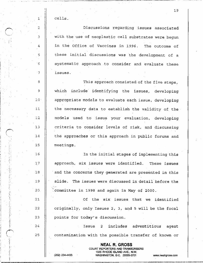
Discussions regarding issues associated
with the use of neoplastic cell substrates were begun
in the Office of Vaccines in 1996. The outcome of
5
6
7
these initial discussions was 'the development of a
systematic approach to consider and evaluate these
issues.
8
9
10
11
12
13
14
15
This approach consisted of the five steps,
which include identifying the issues, developing
appropriate models to evaluate each issue, developing
the necessary data to establish the validity of the
models used to issue your evaluation, developing
criteria to consider levels of risk, and discussing
the approaches or this approach in public forums and
meetings.
16
17
18
19
20
In the initial stages of implementing this
approach, six issues were identified. These issues
and the concerns they generated are presented in this
slide. The issues were discussed in detail before the
Icommittee in 1998 and again in May of 2000.
21
22
Of the six issues that we identified
originally, only Issues 2, 3, and 5 will be the focal
23 points for today's discussion.
24 Issue 2 includes adventitious agent
25 contamination with‘the possible transfer of known or
NEAL R. GROSS
(202) 234-4433
COURT REPORTERS AND TRANSCRIBERS 1323 RHODE ISLAND AVE., N.W. WASHINGTON, D.C. 200053701 www.nealrgross.com
1
2
3
4
5
6
7
8
9
10
11
12
13
14
15
16
17
18
19
20
21
22
23
24
25
20
-unknown viruses. For purposes of today's discussion,
we will include agents of transmissible spongiform
encephalopathy under the category of adventitious
agents.
Issue 3 includes residual cell substrate
DNA contamination with the possible transfer of
activated oncogenic and/or infectious genetic
information.
And Issue 5 includes viral-viral and
viral-cellular interactions with the possibility of
transfer of novel or recombinant viruses, and for the
issues that we will be dealing with today, this
includes replication competent adenoviruses.
Now, to manage the model and risk
assessment aspects of the Office of Vaccines'
approach, what we're calling a defined risk evaluation
was developed. The basic aspect of this evaluation
includes assessing quantitative where' possible the
riskposed by the issues, establishing the probability
of a worst case scenario for plausible issues, using
available data to evaluate plausible risk individually
and cumulatively, and using cumulative data to assess
the relative risk of the product.
The concept and implementation of the
.defined risk evaluation will be presented in more
NEAL R. GROSS COURT REPORTERS AND TRANSkRiBERS
1323 RHODE ISLAND AVE., N.W. (202) 2344433 WASHINGTON, D.C. 200053701 www.nealrgross.com

1
2
3
4
5
6
7
8
9
10
11
12
13
14
15
16
17
18
19
20
21
22
23
24
25
21
detail by Drs. Peden and Krause when they discuss
residual substrate DNA and with adventitious agent
issues later this morning and this afternoon.
To implementthe public discussion stage
of the CBER approach, our plan'was presented to the
Advisory Committee in November of 1998. During this
meeting, the Committee recommended that we develop the
plan into a draft document and present the plan for
discussion at an international workshop on cell
substrates.
This recommendation was implemented over
the next nine months and culminated in a workshop on
neoplastic cell substrates that was held in Rockville,
Maryland, in September of 1999.
Additional discussions at the Office of
Vaccine followed this meeting and the public
discussion of neoplastic cell substrates was continued
at the May Advisory Committee last year.
Now, to briefly summarize the substance of
the Office of Vaccine's presentations at the May 2000
Advisory Committee meeting, neoplastic cell substrates
were divided into five categories. Category 1
included human cells used for vaccine manufacture that
are transformed by known mechanisms. Since there are
no cell lines like this, hypothetical examples include
NEAL R. GROSS
(202) 234-4433
COURT REPORTERS AND TRANfkRIBERS 1323 RHODE ISLAND AVE., N.W. WASHINGTON, D.C. 20005-3701 www.nealrgross.com
1
2
3
4
5
6
7
8
9
10
11
12
13
14
15
16
17
18
19
20
21
22
23
24
25
22
the dipldid WI-38 and MRC-5 cell strains that are
immortalized by human telomerase gene.
Category 2 includes early passage human
diploid cells transformed by known mechanisms.
Examples include the 293 cells and PER.CG cells that
are going to be the focal point of our discussion
today.
Category 3 through 5 represent non-human
primate cells transformed spontaneously. These
include VERO cells, CV-1 cells and BSC-1 cells. All
cell lines that are derived from tumors of any
species, and those cells lines that are not covered by
Categories 1 through 4.
Examples of these types of cells in
Categories 3 through 5 include HeLa cells and the HUT-
78 cells, which is used to propagate HIV virus.
Now, these categories were developedbased
on estimations of the difficulties in managing the
regulatory issues associated with different types of _' -cells. Possible management approaches were presented
for each category.
However, today time doesn't permit me to
review the variety of issues and approaches that were
raised by cells in each of these categories. This
information is available in the transcripts of the May
NEAL R. GROSS
(202) 234-4433
COURT REPORTERS AND TRANSCRIBERS 1323 RHODE ISLAND AVE., N.W. WASHINGTON, D.C. 20005-3701 www.nealrgross.com

1
-2
3
4
5
6
7
8
9
10
11
12
13
14
15
16
17
18
19
20
21
22
23
24
25
23
2000 meeting, which are present on the CBER web site.
Of these five categories, only Categories
1 and 2 as examples of designer cell substrates are
going-to be discussed today.
And as I mentioned, the subject of today's
meeting is to consider issues associated with designer
cellsubstrates which fall into Categories 1 and 2, as
you just saw. For today's discussions, we're defining
designer cell substrates as normal human cells.
They're neoplastically transformed by a known viral or
cellular oncogenes or by immortalizing cellular genes.
Because it's now possible to engineer or
design all types of mammalian cells to express desired
traits, this definition may need to be altered in the
future. In the next talk, Dr. Steve Hughes will
present in more detail the development of designer
Gel1 substrates and the issues associated with their
use.
Like the factors that are stimulating the
need to use all types of neoplastic cells and
substrates for vaccine development, there are a number
of factors behind the need to develop and use designer
cell substrates for vaccine development. These
factors include the development of cells to complement
the replication of bioengineered viral vectors,
NEAL R. GROSS COURT REPORTERS AND TRANSCRIBERS
(202) 234-4433 1323 RHODE ISlAND AVE., N.W. WASHINGTON, D.C. 200053701 wvdw.nealrgross.com
1
2
.- I increasing experience with viral vectors in gene
therapy and the production of biologically active
3 proteins, and hence the development of vaccines and
4 the development of HIV vaccines.
5
6
I should like to point out the development
and use of bioengineered defective viral vectors to
7 serve as vaccines by delivering immunizing antigens
8
9
10
requires the use of cells containing the missing
copies of the defective viral genes to assist the
growth of the defective vector.
11
12
13
In the third talk this morning, Dr.
Aguilar will have much more to say about viral vectors
and especially adenovirus vectors as vaccine delivery
14
15
16
17
18
systems.
'The designer cell substrates we'll be
considering today include 293 cells, which are human
embryonic kidney cells transformed by restriction
enzyme flea fragment of the Adenovirus 5 genome.
19 Frank Graham described this cell line in 1977.
20 PER.C.6 cells, which are human embryonic
21 retinal cells that are transformed by a clone fragment
22 of the Adenovirus 5 genome, these cells were described
23
24
by Frits Fallaux in 1998.
Because there's beenverylittle'published
25 on PER.CG cells and a considerable amount of
24
NEAL R. GROSS
(202) 234-4433
COURT REPORTERS AND TRANSkRIBERS 1323 RHODE ISLAND AVE., N.W. WASHINGTON, D.C. 20005-3701 www.nealrgross.com
25
1
2
3
'information has accumulated on 293 cells since they
became available in 1977, much of our discussion today
will focus on 293 cells.
4
5
6
7
8
9
10
11
12
13
14
The talk by Dr. Alex van der Eb later this
morning will discuss the origins and the
characteristics of these cell lines.
The regulatory issues associated with the
use of designer cell substrates are similar to the
issues associated with the use of other types of
neoplastic cell substrates. These issues include
tumorigenicity and the ability of cells on tumors in
animals, residual cell substrate DNA contamination,
and the possible contamination with adventitious
agents.
15
16
17
18
19
Andin contrast, the cells are transformed
spontaneously, are derived from mammalian tumors that
arise in animals or humans. Designer cells have the
perceived advantage of starting with cells that are
known to be normal and are neoplastically transformed
20 by a known mechanism.
21
22
23
From a regulatory perspective, this type
of information provides an additional level of
assurance that unknown factors which might be present
24 in the cell substrate of less certain origin are not
25 available to enhance any risk to vaccine recipients.
NEAL R. GROSS
(202) 234-4433
COURT REPORTERS AND TRANiCRIBERS 1323 RHODE ISLAND AVE., N.W. WASHINGTON, D.C. 200053701 www.nealrgross.com
26
1
2
3
4
.- The issue that tops the list of concerns
with the use of designer cell substrates and
neoplastic cell substrates, in particular, is their
tumorigenicity, which is their potential to grow into
5 tumors when injected into rodents.
6 For many years assays of tUItIOrigeniCity
7 have been used to discriminate between cells that are
8 suitable for vaccine development and those that are
9 not.
10 The risk believed to be associated with
11 the capacity to produce tumors in animals are noted in
12
13
14
15
16
this slide. Tumorigenicity has been perceived to be
a trait associated with high risk, and due to the
possibility of transferring cell components, either
DNA or proteins or possibly viruses, with oncogenic
activity to vaccine recipients.
17 However, proteins from tumor cells are
18 unable to sustain neoplastic development, and they're
19
20
unable to transform cells. This leaves cell DNA and _ . oncogenic viruses as the risk factors associated with
21 cell substrates that are tumorigenic.
22 In order for the Committee to appreciate
23 what we mean when we talk about tumorigenicity of
24 adenovirus transformed human cells, studies on the
25 tumorigenicity of 293 cells are presented in the next
NEAL R. GROSS
(202) 234-4433
COURT REPORTERS AND TRANS’CRIBERS 1323 RHODE ISLAND AVE., N.W. WASHINGTON, D.C. 2000,5-3701 www.nealrgross.com

27
1 slide, and they're compared with A-549 cells, a cell
2 line that was established from a human lung tumor.
3 I have to apologize for the transfer of
4 our information by computer to the people making the
5 slides because I became Lew is rather than Lewis, and
6 the mouse obviously suffered a discrepancy as well.
7 But in this slide, what we're looking at
8 are a series of tumorigenicity assays, one done by
9 Frank Graham and two done by myself. In the original
10 description of the 293 cell line, Graham reported that
11 the cells weaklytumorigenic, andtheyproduced tumors
12 in only three of 20 animals inoculated with -- and I
13 think this may be hard to see -- but that's ten
14 million cells per mouse.
15 We repeated this experiment ten years
16 later, did a little more detailed inoculations and the
17 animals inoculated with 100 million cells per mouse,
18 ten million cells per mouse, and a.million cells per
19
20
mouse, and we basically discovered or found, got the ';&:_r: .,. '$Z ,,,.! -'>;same results that Frank Graham got in that of the . _,
21 number of cells required to produce tumors in mice was
22 somewhere in the range of ten million cells.
23
24.
The way that these data are reported is in
terms of the TPD-50 value, which is tumor producing
25 dose at a 50 percent endpoint. That's the number of
NEAL R. GROSS
(202) 234433
COURT REPORTERS AND TRANkRIBERS 1323 RHODE ISLAND AVE., N.W. WASHINGTON, DC. 200053701 www.nealrgross.com
1
2
3
4
5
6
7
cells that's required to Produce tumors in 50 percent
of the mice, and these numbers are basically
comparable.
However, when you compare these to A-549
cells, which is the cells derived from human tumors,
it only takes about 1,000 cells to produce tumors for
50 percent of the mice.
8
9
10
Therefore, the A-549 cells are about 1,000
to 10,000-fold more efficient in inducing tumors in
animals than are the 293 cells.
11
12
13
Dr. Jim Cook is going to have a lot more
to say about tumorigenicity of adenovirus transformed
cells later this morning.
14
15
The potential risk associated with
residual cell substrate DNA in vaccines prepared in
16
17
18
19
designer cells represents another concern. DNA from
neoplastic cells can contain activated oncogenes,
viral oncogenes, the genomes of oncogenic viruses,
latent viruses, as well as retrovirus proviruses. -* -
20 Clone cellularoncogenes caninducetumors
21
22
in rodents, and.DNA from oncogenic viruses and cloned
viral oncogenes can also induce tumors in rodents.
23
24
Latent viral genomes in retrovirus proviruses
sequestered in cell DNA can be infectious.
25 Duetothese observations, the possibility
NEAL R. GROSS
(202) 234433
COURT REPORTERS AND TRANSCRIBERS 1323 RHODE ISLAND AVE., N.W. WASHINGTON, D.C. 20005-3701 www.nealrgross.com
1 must be considered that residual DNA from designer
2 cell substrates could transfer either neoplastic
3 activity or infectious virus genomes to vaccine
4 recipients.
5 The talk by Dr. Peden later this morning
6 is going to cover in detail the issues associated with
7 the use of residual DNA.
8
9
10
The third concern associated with the use
of designer cell substrates is the possibility of
adventitious agent contamination. All cell substrates
11
12
13
14
15
16
17
18
are subjected to possible contamination with
adventitious agents. Due to their laboratory origins,
the designer cell substrates might represent a risk of
adventitious agent contamination because they're
neoplastically transformed and may be tumorigenic.
Designer cell substrates might represent a risk of
contamination with unknown, possibly latent oncogenic
agents.
19
20
21
Dr. Krause in his talk this afternoon will
address the issues specifically associated with
evaluating designer cell substrates for adventitious
22 agents.
23
24
I'd like to conclude my talk by saying
that today we are facing a transition. By considering
25 the issues associated with the use of Adenovirus 5
29
NEAL R. GROSS
(202) 234-4433
COURT REPORTERS AND TRANSkRlBERS 1323 RHODE ISLAND AVE., N.W. WASHINGTON, D.C. 200053701 www.nealrgross.com
1
2
3
4
5
6
7
8
9
10
11
12
13
14
15
16
17
18
19
20
21
22
23
24
25
30
-transformed cells, such as 293 cells, we're confronted
with the first of the truly novel neoplastic cell
substrates that we've discussed 'with the Committee
over the past three years.
As these cells fall.into the category of
being tumorigenic, they represent a transition from
the previous way of thinking about cell substrates
that goes back over four decades to future ways of
thinking about cell substrates.
As with most of these types of situations,
this transition presents risks that must be
confronted. However, this transition also presents
the possibility of future rewards. Those rewards will
come from the ability to maximize the benefits that
can be obtained by the application of molecular
technology to the development of safe and effective
vaccines.
The challenge facing us today is to
objectively review the data. that's available on these
types of cells, determine what these data tell us
about their potential to produce safe and effective
vaccines.
I think that's the end of the slides. To
assist CBER and the Committee in this review, We've
invited those individuals who have introduced
NEAL R. GROSS COURT REPORTERS AND TRANSCRIBERS
1323 RHOdE ISLAND AVE., N.W. (202) 2344433 WASHINGTON, D.C. 20005-3701 wwv.nealrgross.com
1
2
3
4
5
6
7
8
9
10
11
12
13
14
15
16
17
18
19
20
21
22
23
24
25
31
themselves to you whose work qualifies them as experts
with sufficient experience with defective viral
complementing cell systems and the issues they raise
to review the relevant data before the Committee, to
answer Committee questions, and to offer their
opinions regarding the issues .that need to be
addressed.
Before they begin to speak, I'd like to
just take this opportunity to thank them for the time
that they have used to assist the Office of Vaccines,
the Committee, and the public in these discussions.
This concludes my talk. I'd be happy to
try to answer any questions.
ACTING CHAIRMAN DAD-M: .Thank you very
much, Dr. Lewis. That provides a useful setting for
us to continue hearing about this issue.
It also reminds some of us that it's time
for our annual visual screening test.
(Laughter.)
ACTING CHAIRMAN DAUM: We do have the
opportunity run behind a little bit here in terms of
scheduling if there are Committee questions.
Alternatively, we can get some more information on the
table and then initiate discussion.
Is there Committee input? Dr. Goldberg,
-NEAL R. GROSS
(202) 234-4433
COURT REPORTERS AND TRANSCRIBERS 1323 RHODE ISLAND AVE., N.W. WASHINGTON, D.C. 20005-3701 www.nealrgross.com

32
1 and then Dr. Griffin.
2 DR. GOLDBERG: Yeah, just on your table of
3
4
5
6
tumorigenicity where YOU show the rates of
tumorigenicity in the 293 cells in the nude mice, can
you give me some feel for how you feel that you can
distinguish these levels?
7 For example, you have -- I can't see. I'm
8
9
sorry -- you don't observe any tumors in four nude
mice at ten to the sixth in one experiment and in
10 another experiment you observe four of four.
11
12
13
And you know, any calculations I do would
suggest that you really with four animals can't
distinguish.
14
15
16
So can you give me some feel for what
other information you're bringing to bear on this to
make the distinctions about what the TPD-50 is?
17
18
19
DR. LEWIS: I guess I'm having a little
bit of a hard time hearing what you were saying.
You're trying to understand how we calculate the TPD-
20 5'0?
21 DR. GOLDBERG: No, I think I do know how
22 you do that, but my concern or my question really is:
23 how do you feel that based on these experiments with
24 four mice at each of these dose levels that you can
25 really estimate the TPD-50 with any certainty to make
NEAL R. GROSS
(202) 234-4433
COURT REPORTERS AND TRANSCRIBERS 1323 RHODE ISLAND AVE., N.W. WASHINGTON, DC. 200053701 www.nealrgross.com
1
2
3
4
5
6
7
8
9
10
11
12
13
14
15
16
17
18
19
20
21
22
23
24
25
.a distinction between --
,..., 33
DR. LEWIS: Okay.
DR. GOLDBERG: -- ten to the three and ten
to the sixth, for example?
DR. LEWIS: Basically, the data that was
used to do this came from a series of titrations that
we did on Adenovirus 12 transformed mouse cells.
These assays were repeated ten times, and each time
they were done in four mice, but nude mice are
expensive, and each time they were done the standard
deviation of those assays was about plus or minus .6
of a log. Okay?
So based on the information that we
obtained with that, we are reasonably confident that
this represents an accurate way of reflecting this
type of information.
The data on the 549 cells and many of the
293 cells were repeated at least twice, and the
numbers are basically the same.
ACTING CHAIRMAN DAUM: Dr. Griffin.
DR. GRIFFIN: Well, I guess I was being
puzzled by the same table. And maybe I just missed
this, and I got a little clued in what you just said.
The difference between the 293 cells and
the A-549 cells is one is Ad. 5 transformed and the
NEAL R. GROSS
(202) 234-4433
COURT REPORTERS AND TRANSCRIBERS 1323 RHODE ISLAND AVE., N.W. WASHINGTON, D.C. 20005-3701 wdw.nealrgross.com
1
2
3
4
5
6
7
8
9
10
11
12
13
14
15
16
17
18
34
.-.other is Ad. 12 transformed?
DR. LEWIS: No.
DR. GRIFFIN: And they have different --
DR. LEWIS: No. A-549 cells are a cell
line was established from a human. I believe it's an
oat cell (phonetic) carcinoma. Okay? And they were
established directly from the human tumor in the
tissue culture. They are not virus transformed. They
are a cell line that developed from a human tumor that
developed in nature, a spontaneous tumor in the human.
DR. GRIFFIN: So the point to be made from
this is that cells differ in how likely they are --
DR. LEWIS: Well, yes, that's one point.
The second point is that it takes a large number of
Adenovirus 5 transformed cells to produce tumors.
This is true both in adenovirus transformed mouse
cells, Adenovirus 5 transformed hamster cells, as well
as adenovirus transformed human cells.
19
20
21
They fall into a categorythat,most people
-would define as weakly tumorigenic, and this is a
characteristic of Adenovirus 5 transformed cells.
22
23
24
DR. GRIFFIN: Thank you.
ACTING CHAIR& DAUM: Okay. Thank you.
I'd like to move on then at this point --
25 thank you very much, Dr. Lewis -- to Dr. Steve Hughes'
NEAL R. GROSS
(202) 234-4433
COURT REPORTERS AND TRANSCRIBERS 1323 RHODE ISLAND AVE., N.W. WASHINGTON. D.C. 200053701 www.nealrgross.com
1
2
,3
4
5
6
7
8
9
10
11
12
13
14
15
16
17
18
19
20
21
22
23
24
25
35
presentation, entitled "Designer Cell Substrates for
Vaccine Development: Concepts and Issues.1f
Dr. Hughes.
DR. HUGHES: This is somewhat smudged. I
may challenge people's optical state once again.
Thank you.'
Since this subject has been so ably
introduced by Andy, I'll try and go through this
quickly.
Basically the question to consider, of
course, is how designer cell substrates, in fact,
differ from other permanent cell lines with
transformed cells, and basically in the past,
spontaneous transformation has been used to establish
cell lines, and that simply means you take cells from
an animal usually or an embryo and passage them in
culture, and it's a particular characteristic of
rodent cells that after some period of passage the
cells undergo some sort of change,'which we still
don't understand clearly, that alters both their
ability to grow permanently in culture and alters some
of their physical and biological properties.
The other way that cells have been
immortalized or immortal cells have been derived is as
was just mentioned, from tumors taken from either
NEAL R. GROSS COURT REPORTERS AND TRANSCRIBERS
1323 RHODE ISLAND AVE., N.W. (202) 234-4433 WASHINGTON, D.C. 200053701 www.nealrgross.com
1
2
3
4
5
6
7
8
9
10
11
12
13
14
15
16
17
18
19
20
21
22
23
24
25
36
..humanS or animals, and in some cases, these can be
established directly in culture, and in some cases
these tumor cells are then serially passaged in
animals, and both of these methods have been used to
establish a number of types of cells lines that basic
researchers, such as myself, use routinely in the
laboratory.
And it's very convenient, but it has a
particular disadvantage, and that is that in neither
case do we have any clear notion of what changes have
taken place in these cells, what it is about these
c,ells that differentiates them from the normal cells
that don't have the properties of either being
transformed or growing forever in culture.
And so one of the things that I think
makes everyone a little nervous about these types of
cells is not necessarily that they have something
specific wrong with them, but, in fact, the very fact
that we don't know what it is that has changed them.
.We don't know how they differ from the normal cells
that everyone feels reasonably comfortable with.
And as an alternative to that kind of
idea, what's meant, as Andy has just told you, by a
designer cell substrate that differentiates it from
these two types of cells is that one now can take
NEAL R. GROSS COURT REPORTERS AND TRANSCRIBERS
1323 RHODE ISLAND AVE., N.W. (202) 234-4433 WASHINGTON, DC. 20005-3701 www.nealrgross.com
3
4
6
8
16
18
23
24
25
.
1
2
37
specific -- which I have not spelled correctly --
specific DNA segments either derived from virus,
derived from cells, that can change the growth
properties of normal cells, and in so doing, we now
have something in which we understand what agent it is
that is causing the cells to behave differently.
And that gives us some particular handle
and some particular feeling that we have at least some
idea of what's going on.
This does not, of course, eliminate all
the worries that one might have. There are issues.
One of the issues is that there is the question of
whether this specific DNA that when it's added
actually has some sort of risk associated with it.
Of course if this DNA segment is capable
of causing the cells to grow forever in culture, it
may have oncogenic potential, and in fact, as you've /
just heard discussed by Andy, there is some reason to
think that in the case of the adeno early region that
there is some oncogenic potential of that.
SO you really would worry about carrying
the DNA if you're using a vaccine preparation with the
vaccine material that you're going to use.
So there is actually still a question of
the degree to which this is a serious concern, and
NEAL R. GROSS
(202) 234-4433
COURT REPORTERS AND TRANSCRIBERS 1323 RHODE ISLAND AVE., N.W. WASHINGTON, DC. 200053701 wiw.nealrgross.com
1
2
3
4
5
6
7
8
9
10
11
12
13
14
15
16
17
18
19
20
21
22
23
24
25
38
you'll hear more about that later today. I would also
be pleased to tell you one of the things that was
discussed the last time this group met was that there
should be in a sense a collaboration between the NC1
and the FDA to -try and get .a more quantitative
assessment of what the risk is in terms of using
defined amounts of defined oncogenic DNA segments.
I'm pleased to say that that interaction
has reached the point where it's funded and that there
will be some quantitative studies to try and establish
exactly what the risk is at least from some defined
DNA segments.
As Andy has also mentioned, there is the
issue of adventitious agents, that is to say, that any
cell, whether it's a cell that is permanent in
culture, whether it's a norma. diploid fibroblast can
be infected with virus, can have other agents
associated with it.
And in both of these cases, as. I just
tried to allude in the DNA, in part the question here
is both understanding what sorts of things pose risks
and, secondly, trying to understand how it is we can
determine what.agents, in particular for adventitious
agents, what adventitious agents might be present.
And so what I've tried to say is that the
NEAL R. GROSS
(202) 234-4433
COURT REPORTERS AND TRANSCRIBERS 1323 RHODE ISLAND AVE., N.W. WASHINGTON, D.C. 200053701 www.nealrgross.com

1
2
3
_-. _ .*_/--* *= ..vx~17-.- ._ll_ _ ~--~-.~- ._x_~I.__.-“II-“. ---------. --- -~-
39
_ issue, I think, from the DNA is at least in part one
of risk assessment, and I think that there actually
are reasonable ways of defining what the risks are.
4
5
6
7
8
9
10
11
One can take, particularly if one knows
what DNA segments one is dealing with; one can take
those DNA segments; one can take defined amounts .of
those DNA segments; one can inject them into animal
models, and one can define the oncogenicity, and based
on that, one can get some reasonable measure, some
idea of what it is that we're facing in terms of the
risk.
12
13
14
15
16
17
And in the case of adventitious agents,
one of the nice things about modern molecular biology
and biotechnology is that we now have much better ways
of looking for at least nucleic acid bearing agents,
and you'll hear, I think, a little later today in
considerably more detail than I intend to discuss the
18 sorts of things that are under consideration as ways
19 of doing this.
20
21
22
23
So the question then becomes given that we
.have these tools and given that we have these
problems, what sorts of things should we do. How
should we go about trying to be as safe as possible?
24
25
And I think one of the things, and 1,think
it's going to come up in considerably more detail, is
NEAL R. GROSS
(202) 234-4433
COURT REPORTERS AND TRANSCRIBERS 1323 RHODE ISLAND AVE., N.W. WASHINGTON, D.C. 20005-3701 www.nealrgross.com

1
2
10
11
12
13
14
15
16
17
18
19
20
21
22
23
24
25
40
_ culture history. You would like to know where the
cells have come from. You would like to know where
they have spent time. It's sort of like worrying
about where your teenage children go at night.
A.nd there are a cou.ple of things that I
think are worth discussing, although probably only
briefly, that might not be sort of immediately obvious
if one just thinks about passage or culture history,
and it's been alluded to, I believe, earlier that, in
fact, the source of the serum and what might be in the
serum turns out to be a substantial consideration.
And this is true for both agents like BSE
and, of course, for viruses as well. And I think
there's another issue that I don't believe has been
discussed in any particular detail, but actually I
think does matter not even so much for designer cell
substrates, but for substrates that are derived in a
sense. directly from tumor material, and that's the
idea that it's one of the traditional methods for
deriving particularly cell lines from human tumors, is
to passage the cells in mice.
And there's a particular consideration
which is one which makes 'for one of John Coffin's
favorite stories, that suggests that there is an
element of risk here that people don't always
NEAL R. GROSS
(202) 234-4433
COURT REPORTERS AND TRANSCRIBERS 1323 RHODE ISLAND AVE., N.W. WASHINGTON, D.C. 2000.53701 www.nealrgross.com
(
1
2
3
4 endogenous retroviruses preferentially replicate in
5
9
10
11
12
13
14
15
16
17
18
19
20
21
22
23
24
25
. consider.
Mice, of course, contain several families
of endogenous retroviruses, and some of these
cells derived from mice, and some actually replicate
preferentially in non-rodent cells.
And one of the things that happens when,
for example, human cells are passaged through nude
mice is that that provides a wonderful opportunity
actually for xenotropic viruses -- these are the
viruses that 1ik.e to replicate in non-rodent cells --
to actually infect <he human cells.
And it's quite possible actually in this
kind of culture history to add an adventitious agent
that one would really not normally think would be one
you'd have to look for in a cell derived from a human
or a primate.
SO these sorts of considerations, I think,
are very important and certainly I think we have to
give substantial consideration to having a defined
culture history not so much because it will
necessarily rule out all possibility of adventitious
agents, but we can understand if we know the culture
history what sorts of adventitious agents we should
look for. 9
(202) 234-4433
NEAL R. GROSS COURT REPORTERS AND TRANSCRIBERS
1323 RHODE ISLAND AVE., N.W. WASHINGTON, DC. 2000.5-3701 w.nealrgross.com
1
2
3
4
5
6
7
8
9
10
11
12
13
14
15
16
17
18
19
20
21
22
23
24
25
42
And one of the problems in searching for
adventitious agents with, for example, the nucleic
acid technology that will be used for many viruses is
you only find the things you look for, and if you know
what to look for, it makes your,job much easier.
And the final thing that I think is
perhaps the most challenging part.of the problem, the
part of the problem that I'm not sure I have a
particularly good idea yet how to resolve, is the
issue of the stability of the genotype or the
phenotype of the cells.
And the reason this is a consideration
actually goes back to the idea that I introduced the
talk with that, in fact, you can derive cells, cell
lines by simply passage in culture; that, in fact,
there is such a thing as spontaneous transformation.
AAnd of course, not only is there
spontaneous transformation, but upon passage the
properties of the cells in culture upon prolonged
passage can change. They don't have to change, but
change can occur.
Now, that means that, in fact, the
phenotype, for sure, and. probably the underlying
genotype has altered during the passage of the cells.
Cell lines do change upon prolonged
NEAL R. GROSS COURT REPORTERS AND TRANSjZRlBERS
1323 RHODE ISLAND AVE., N.W. (202) 234-4433 WASHINGTON, D.C. 200053701 www.nealrgross.com

1
2
3
4
5
6
7
8
9
10
11
12
13
14
15
16
17
18
19
20
21
22
23
24
25
43
.passage in cell culture, and so the question then
arises if that is true, how do we gain confidence that
the properties of the cells after some period in
culture, in fact, match the properties of the cells
with which we began. That is to say if we're quite
confident that we've made a designer cell line that
has the desirable properties and has only the changes
we put in and then we passage it for a long time,
given that the cell lines can change, how do we know
that the cell line hasn't changed?
And that seems to me to be one of the
substantial requests that we need to consider, and of
course, one of the old standards of tissue culture
people is simply to use cells that have been passaged
a relatively small number of times, and that, of
course, because these changes appear to be
spontaneous, some sort of genetic accident, by using
low passage cells, the chances that some change has
taken place seems to be better. The hossibility that
there's a change seems to be less.
But the final thing that I think we ought
to at least begin to think about is the idea that we
might at least in some cases give consideration to
using some sort of regulatable system to drive the
expression of the gene that causes the cell to change
NEAL R. GROSS
(202) 2344433
COURT REPORTERS AND TRANSCRIBERS 1323 RHODE ISLAND AVE., N.W. WASHINGTON, D.C. 20005-3701 www.nealrgross.com
1
2
3
4
5
6
7
8
9
13
14
15
16
17
18
19
20
21
22
23
24
25
.its properties.
44
And if we imagine, for example, that we
have some sort of promoter that has a switch on it
that we can turn on and off so that we can turn on and
off the gene that we're interested in that.is causing
the cells to be transformed, then if that is the cell
-- I'm,sorry -- that is, if the gene we've added is,
in fact, the agent that changes the properties of the
cell, if we switch that gene off, then the cells'
properties ought to fall back to that of the starting
cell, which was not permanent or transformed.
And I mean, it may be that I'm throwing
this out as an idea, not as a solution. You may not
want to use necessarily an inducible promoter, but the
idea that I think is central here is somehow to find
a way to regulate the expression of the gene you're
interested in, whether it's some sort of dominant
negative effect either at the protein level, at the
nucleic acid level or the inducible promoter.
The idea that we want to, I think, think
about is can we.validate, can we determine after some
passage that the agent that we think is changing the
properties of the cell is,. in fact, the responsible
agent -- ~'rn sorry -- the gene, the designer gene that
we've added, or have there been some additional
NEAL,R. GROSS
(202) 2344433
COURT REPORTERS AND TRANSCRIBERS 1323 RHODE ISLAND AVE., N.W. WASHINGTON, D.C. 200053701 www.nealrgross.com
1
2
3
4
5
6
7
8
9
10
11
12
13
14
15
16
17
18
19
20
21
22
23
24
25
45
changes in the genotype and phenotype that are
influencing the behavior of the cell.
And I think giving some consideration to
that idea will be important, and I think at that point
I'll stop, and if there are ques.tions I'd be happy to
answer them.
ACTING CHAIRMAN DAUM: Okay. I'd like the
questions at this time to be focused mainly on Dr.
Hughes' presentation. There will be plenty of time
for more general discussion later.
DR. COFFIN: Steve, there's an issue you
didn't raise actually that comes up particularly when
one is considering these cells for growth of viral
vaccines, and that is the potential of the cells to
actually contribute genes to the vaccine virus itself
by some sort of recombination and the consequences of
that, and I think that's an issue that could arise,
particularly if the retroviruses of a cell line has
picked up an endogenous xenotropic virus or with the
early genes of adenovirus.
DR. HUGHES: I deliberately, as i'm sure
you're aware, avoided that issue both because I think
there will be consideration'of the recombination issue
by others later and because I believe that the issues
are somewhat different for adenovirus, which I think
NEAL R, GROSS
(202) 2344433
COURT REPORTERS AND TRANSCRIBERS 1323 RHODE ISLAND AVE., N.W. WASHINGTON, D.C. 20005-3701 www.nealrgross.com
1
2
3
4
5
6
7
8
9
10
11
12
13
14
15
16
17
18
19
20
21
22
23
24
25
46
-we're supposed to be focused on today, and
retroviruses.
And I think the issue of recombination and
the mechanics of recombination particularly as they
pertain to retroviruses are a bit beyond the scope of
the discussion we have here. So that was a deliberate
omission.
But I certainly think that as Dr. Coffin
points out that the issue that he raises is a real
one, and that we should give very careful
consideration to issues of not only what the viruses
can do to cells but, in fact, in some more complicated
sense what the cells or things in the cells can do to
the viruses.
ACTING CHAIRMAN DAUM: Thank you, Dr.
Coffin and Dr. Hughes.
Dr. Kim.
DR. KIM: Are there any designer cell
substrates on the horizon or on the radar screen that
,are shown not to be oncogenic or less likely to be
oncogenic?
that.
DR. HUGHES: I'm not qualified to answer
ACTING CHAIRMAN DAUM: Would you like to
try, in looking for answers to this question?
NEAL R. GROSS
(202) 234-4433
COURT REPORTERS AND TRANSCRIBERS 1323 RHODE ISLAND AVE., N.W. WASHINGTON, D.C. 20005-3701 www.nealfgross.com
47
1 .-
2
(Ltiughter;)
ACTING CHAIRMAN DAUM: Sorry, but I do
3
4
recognize you are number two in line.
Dr. Aguilar-Cordova.
5
6
7
8
DR. AGUILAR-CORDOVA: Yes. You talk about
the transformation of cells and for a cell to become
tumorigenic there's some old data showing a series of
events. So if one has only one agent, wouldn't that
9
10
11
just make ten minus one and the oncogenic event may
still be there on that genotype?
DR. HUGHES: I'm not quite sure I
12 precisely understand your question, but it certainly
13 is the case that we now believe that for most tumors
14
15
16
17
multiple genetic changes are needed, but many of the
things that we regard as tumorigenic, whether they're
chemical agents or viral agents, and these studies
have been confirmed by genetic manipulation of mice,
18
19
20
that anything we do that moves us one step closer to
the required number, be it two, three, five, whatever
it is, if we add any one thing to the list of changes,
21 if we make any of the changes, that by doing that, by
making the change, you do bring the cell closer to a
23
24
25
transformed phenotype, and that you can show, for
example, in mice by the p53 knockout mice, which have
only a single change, get spontaneous tumors at a very
NEAL R. GROSS
(202) 234-4433
COURT REPORTERS AND TRANSCRIBERS 1323 RHODE ISLAND AVE., N.W. WASHINGTON, D.C. 200053701 www.nealrgross.com

1
2
3
4
5
6
7
8
9
10
11
12
13
14
15
16
17
18
19
20
21
22
23
24
25
48
. . high rate because one layer of safety has been
removed.
And I think that's' the sort of thing that
we would be nervous about, and of course, some of the
cells may have more than one change so that you could
actually from the DNA, depending on what the cell
substrate was, actually deliver more than one of the
things necessary to drive a cell in an animal or a
human towards the transformed phenotype.
So the single one is not good, and some of
the cells may have had more than one. So I think it
makes us feel better that there are more thanone, but
I don't think it means that things were perfectly
safe.
ACTING CHAIRMAN DAUM: Dr. Minor, please.
Then Dr. Kohl.
DR. MINOR: The tumorigenicity assays done
in rodents for very, very good technical reasons
clearly, but is it possible that there are actually
species effects; that if you took the immune response
out of things, that you would find a different
tumorigenicity ranking in a different species?
I mean, how relevant are the rodents do
you think to a human situation?
DR. HUGHES: I think,the answer is -- and
NEAL R. GROSS
(202) 2344433
COURT REPORTERS AND TRANSCRIBERS 1323 RHODE ISLAND AVE., N.W. WASHINGTON, D.C. 200053701 wvw.nealrgross.com
1
2
3
4
5
6
7
8
9
10
11
12
13
14
15
16
17
1%
19
20
21
22
23
24
25
49
.this is by definition a speculation because the
experiments, in general, can only be done in rodents.
So you have to sort of extrapolate.
But there is enough good data, I think,
from.chemical carcinogenesis to make one believe that,
in fact, there are very strong species effects in some
cases.
And I think that's a concern, but I think
the choice in some sense experimentally is between
doing the experiments in rodents, in which you have
the worry that it may not perfectly reflect what
happens in humans, and not doing the .experiment at
all.
And while I have some reservations of the
exact sort you mention, in terms of worrying about
doing the experiments in rodents and applying it to
humans, I certainly would rather have rodent data and
try and worry about the extrapolation than have no
data at all.
ACTING CHAIRMAN DAUM: Thank you.
Dr. Kohl and then Dr. Myers.
DR. KOHL: That was my question.
ACTING CHAIRMAN DAUM: Dr. Myers, please.
DR. MYERS: I guess I have two questions.
On the confidence of the stability of the genome,
NEAL R. GROSS COURT REPORTERS AND TRANSCRIBERS
1323 RHODE ISLAND AVE., N.W. (202) 234-1433 WASHINGTON, D.C. 200053701 vwv.nealrgross.com
,.
1 would you be more confident if the designer segment
2 were excised and the tumorigenicity was lost?
3
4
5
And the second question is related to
that, and that is could you tell us about the
relevance of the tumorigenicity.limited to nude-nude
6 mice?
7
8
9
10
11
12
13
14
15
16
DR. HUGHES: I don't think it's probably
technically feasible to excise the segment, but I
think there are ways of setting up the experiment so
that you can interfere with the expression.
DR. MYERS: Knock it out?
DR. HUGHES: Knockout technology is
probably not the easiest, but the point I'm really
trying to get at is what I think you want to look at
is not necessarily the precise technology or
necessarily even to limit yourself to a precise
17
18
technology, but to be able to somehow develop either
a technology or technologies that will allow you to
19 ask the question that, in a sense, you're posing.
20
21
22
23
If you then interfere with or obliterate
'the expression of the thing you think is driving the
cells towards this permanent tumorigenic phenotype,
does that, in fact, change'the behavior 'of the cells
24 as you would.expect?
25 And if you can do that, I think you're
NEAL R. GROSS
(202) 2344433
COURT REPORTERS AND TRANSCRIBERS 1323 RHODE ISLAND AVE., N.W. WASHINGTON, O.C. 200053701 www.nealrgross.com
.il - _ - . .
2
3
4
5
6
7
8
9
10
11
12
13
14
15
,16
17
DR. BLAIR: Well, I guess the question is,
if I understand it, is in an immunocompromised animal
does the fact that the cell is tumorigenic have any
relevance to normal situations, and I guess, you know,
the argument would be that at least by demonstrating.
the tumorigenicity in the nude, you've shown the
potential, and the failure to be tumorigenic in an
immunocompetent system presumably arises from the
immune response which could at some stage, at some
mechanism be lost or be modified.
18 So I think, you know, the demonstration of
19
.20
21
22
23
24
the tumorigenicity in a immunocompromised system is
important because it does demonstrate that there is
that potential, as opposed to no potential or what
presumably is no potential at all.
ACTING CHAIRMAN DAUM: Thank you for
clarifying, Dr. Blair.
25 Dr. Lewis, then Dr. Aguilar-Cordova.
NEAL R. GROSS
-quite confident that there's nothing else going on or
nothing substantial going on. I don't know that I --
I think actually in terms of responding to the nude
mouse. question I'd feel more comfortable probably
deferring to my colleague Don Blair, who I think is
much more of an expert on tumorigenicity in nude mice
than I am.
(202) 234433
COURT REPORTERS AND TRANSCRIBERS 1323 RHODE ISLAND AVE., ti.W. WASHINGTON, DC. 20005-3701 wwf.nealrgross.com
1
6
7
8
9
10
11
12
13 But so far nobody has proposed one of
14
15
16
those as a designer cell substrate for our attention.
ACTING CHAIRMAN DAUM: Thank you, Dr.
Lewis.
17 DR. AGUILAR-CORDOVA: Myquestionactually
18 follows very well on that, and it's a follow-up on
19 what I started to say.
20 So if it's a series of events, would a
21 particular event, whether it be the telomerase, the
22 SV40 T antigen or myc, be oncogenic depending on the
23 -background of the cell that it hits so that they're
24 complementing oncogenes?
25 And I guess that begs the question to
52
DR:LEWIS: Just in response to Dr. Kim's
question about immortalized cells that are not
tumorigenic, the only system that we're aware of in
which.that occurs by immortalizing cells with the
human telomerase gene data suggests that it takes
three different genes, telomerase, a, and SV40 to
transform a normal cell to a cell that is, in fact,
tumorigenic.
You can immortalize cells with hTERT with
a telomerase gene, and those cells, as far as .I
understand right now from what we're aware of in the
literature, are not tumorigenic.
NEAL R. GROSS
(202) 234-4433
COURT REPORTERS AND TRANSCRIBERS 1323 RHODE ISLAND AVE., N.W. WASHINGTON, D.C. 20005-3701 ww.nealrgross.com

1
2
3
4
5
53
whether any cell, tumorigenic or normal, is any safer
depending on what the target cell is. It would appear
normal if it only has an activated end myc., for
example. It wouldn't appear tumorigenic, nor
immortalized perhaps.
6
7
8
9
10
11
12
13
DR. HUGHES: Those of us who have spent
many, many hours, days peering through microscopes at
cells that had single oncogenes delivered by, for
example, retroviruses that give very high levels of
expression could in many cases see the effects either
by change in morphology, change in growth pattern,
change -in behavior of adding a single gene.
So I think there are certainly reasons to
14
15
16
17
18
19
20
believe that even if it takes multiple changes to
produce the Frank phenotypic tumorigenic phenotype in
vivo, that the adding of individual oncogenes one at
a time or in some cases removing or ablating tumor
suppressor genes actually does substantially change
the properties of the cells, even if it's not .- sufficient necessarily to drive the cell to its full
21 Frank transformed phenotype.
22 So I actually believe it is important to
23 in some sense keep track, and I also think, as I tried
24 to say earlier, that because these things are, in
25 fact, additive as far as we know in humans, that
NEAL R. GROSS
(202) 234-4433
COURT REPORTERS AND TRANSCRIBERS 1323 RHODE ISLAND AVE., N.W. WASHINGTON, D.C. 200053701 www.nealrgross.com

1
2
3
4
5
6
7
8
9
-providing one or Wo steps in the direction of the
oncogenicity is something we'd like to avoid, I think.
One could argue in many cases that
chemicalcarcinogenesis provides exactly one or two of
the changes and the long duration that you see to the
development of the tumor after the initial exposure to
the chemical insult actually represents the fact that
the chemical may have changed only one or two things,
and the rest must occur spontaneously later.
10
11
12
But that still enhances the risk
substantially, and I think those are things that we
need to worry about.
13 ACTING CHAIRMAN DAUM: Thank you very
14 much, Dr. Hughes.
15
16
17
18
I think we'll move on, if you would. That
I was a very helpful presentation, to Dr. Aguilar-
Cordova -- I hope I'm not butchering your name -- who
will tell us about adenovirus biology'as related to
19 development and use of adenovirus vectors.
20 DR. AGUILAR-CORDOVA: Can you hear me?
21 Yeah. So I'm just going to give a
22
23
general, generic background on the adenoviruses so
that we can use this for further discussion and their
24 use as vectors.
25 So adenoviruses were identified in the
NEAL R. GROSS
(202) 234-4433
COURT REPORTERS AND TRANS‘CRIBERS 1323 RHODE ISLAND AVE., N.W. WASHINGTON, D.C. 20005-3701 www.nealrgross.com
1
2
3
4
5
6
7
8
9
55
early '50s from an adenoid tumor, from an adenoid
tissue, thus the name adenoviruses, and it's
associated with some fairly common illnesses in the
lay group referred as the "common cold," some eye
inflammations, et cetera.
It's composed of a linear, double stranded
DNA encapsidated in a protein shell; hasno envelope,
and there are many different types of adenoviruses in
nature.
10
11
12
13
They are primarilyclassifiedbasedonthe
organism of origin, and so there are two major groups:
Mastadenoviruses and the Aviadenoviruses, those that
come from mammals and those that come from birds.
14
15
16
17
18
19
20
21
22
23
Hopefully nobody else has slides.
(Laughter.)
DR. AGUILAR-CORDOVA: The further
characterization is in the antigenicity of .the
terminal knob in the fiber protein and hexon epitopes
and, thus, the serotype, and you'll hear about
Serotype 2, Serotype 5, the most commonly used, and
there are many other different serotypes, and also by
hemagglutination, binding of the fiber protein to red
blood cells.
24 And it turns out that some groups of
.25 adenoviruses have more tumorigenic ability in rodent
NEAL R. GROSS
(202) 234-4433
COURT REPORTERS AND TRANSCRIBERS 1323 RHODE ISLAND AVE., N.W. WASHINGTON, D.C. 200053701 www.nealrgross.com
1
2
3
4
5
6
7
8
9
10
11
12
13
14
15
16
17
18
19
20
21
22
23
24
25
56
. cells. None have been shown, as you will hear
probably later on, to be tumorigenic in humans.
The virus is icosahedral. It has 240
hexons, I2 for each of 20 triangular phases and 12
pentons, and as you can see, .it has these little
fibers. The fibers and the pentons and the hexons are
what constitute the serotype of the virus.
Inside that icosahedralproteinbasethere
is a double stranded genome flanked by two terminal
repeats and some proteins that go with that genome,
and here's the list of them.
Primarily the terminal protein is
important to keep the stability of the genome and to
condense it.
The gene structure is linear as well, but
it does express from both strands. What you've heard
here is ElA and ElB. The El region, these are
critical for expression of other genes' of the virus,
and thus the majority of the vectors that are used in
adenoviruses are vectors in whi,chthese two genes have
been deleted and replaced by the gene of interest.
There are two origins of replication. In
the ITRs, inverted terminal repeats, transcription
units include five early genes, the ElA and ElB that
I just mentioned, the E2 region, the E4 region, and
NEAL R. GROSS
(202) 2344433
COURT REPORTERS AND TRANS’CRISERS 1323 RHODE ISLAND AVE., N.W. WASHINGTON, D.C. 200053701 www.nealrgross.com
.
57
1
2
'6
7
8
9
10
11 in part by binding to some of the cellular genes that
12 regulate cell cycle and expansion, like p53 and
13 retinal blastomagy (phonetic).
14 E2 has three proteins. It's involved in
15
16
17
18
virus reproduction, DNA replication, in particular.
E3, there are four proteins. It's believed that this
is protection from viral infection, and thus it down
regulates the ability of the cell that.has the virus
19 to be immunogenic.
20 One of the down functions is that it down
21 regulates the expression of MHC Class 1, and thus the
22 host can't recognize the other proteins going on.
23 And the E4, there are at least four
24
25
the E3 region. 1'11 talk a little bit more about
these. There are two delayed early and one major late
transcript that includes late one through five.
The ElA, there are two proteins, and this
'is host activation, and what 'this really does is
activate the transcription and induce hosts to enter
S phase, and that activates a transcription of most of
the other adenoviral genes;
ElB is also two proteins, and it induces
cell growth, and we know no what ElA and ElB do this
proteins, and it has miscellaneous activities, such as
regulation of transcription, MRNA transport, and DNA
NEAL R. GROSS
(202) 234-4433
COURT REPORTERS AND TRANSCRIBERS 1323 RHODE ISLAND AVE., N.W. WASHINGTON, D.C. 20005-3701 wvw.nealrgross.com
1
2
3
4
5
6
7
8
9
10
11
12
13
14
replication of the virus.
The late genes, there are five of them.
They're mostly involved in the structure of the virus
itself and the stabilization of that core.
The viral life cycle and replication is
really two faces. There are early events, which occur
in the first six hours after infection, and that
includes absorption, penetration of the virus into the
cell, disassembly of the virion core, and
transcription and translation of the early genes.
One that begins, the late events by
definition start, and that's in the next 18 hours or
so, and that is when there is the construction of new
virions.
15
16
17
18
19
20
.-
And we have approximately ten to 50,000.
Traditionally it was said there's about 10,000 virions
per cell. We.know now that there are -- when we
produce them in the laboratory, we can produce a lot
more than that. So wild type probably also does more
than that.
21
22
23
Now, the next step, of course, is that's
the virus. How does one use the virus to optimize
gene transmission and, therefore, its potential use in
24 gene therapy?
25 And there are two key factors of
NEAL R. GROSS
58
(202) 234-4433
COURT REPORTERS AND TRANSCRIBERS 1323 RHODE ISLAND AVE., N.W. WASHINGTON, D.C. 200013701 www.nealrgross.com
1 adenoviruses that'actudliy have'allowed us to use this
2 variance as good, effective vectors. One is that we
3 can package up to 105 percent of its capacity. So
4 when we take out the ELA or the ElB genes, that gives
5 a little bit of space, and then you have actually a
6 five percent wiggle. room there.
7 And the other very important issue is that
8 one can manipulate the virus in a circular form. That
9 is, the ITRs can be circularized in a plasmid-like
10 structure, and one can clone and change contents that
11 way.
12 SO given those two factors, we can then
13 manipulate the virus easily and make it an efficient
14 vector.
15 This obviously you will not be able to
16 read, but it is to show that there are many different
17 viral vector vehicles that one can use. The most
18 common ones are retroviruses, adenoviruses, and
19 associated viruses and Herpes viruses, and they all ._
20 have pluses and minuses depending on the use that one ..'
21 will have for them.
22 Retroviruses are often used as an
23 advantage, which is that it will enter the cell
24
25
efficient, integrate so there will be stable
expression for long terms, and that one has no viral
NEAL R. GROSS COURT REPORTERS AND TRANSCRIBERS
(202) 2344433 1323 RHODE ISLAND AVE., N.W. WASHINGTON, D.C. 20005370T www.nealrgross.com
1 .. genes in the most common of these vectors.
2 And the disadvantages is that they're hard
8
9
10
11
12
13
14
expresser; that the viral genes are often in the
vector so that it's immunogenic, and it gets disposed
of by the host fairly quickly.
In the case of vaccines, this may be a
15 very useful disadvantage. So it may not be a
16 disadvantage.
17 The same as for the other vector types,
18
19
20
21
there are advantages and disadvantages depending on
what one needs to use them for. --
A_ So typically on first generation vectors
what one does is put the gene of interest instead of
22 the ElA/ElB region. The E3 region is often deleted to
23 create a little bit more space. The E3 region is
24
25 vitro expansion.
60
to.produce. They have a limited insert size, and that
they may integrate randomly and thus cause mutation.
On the flip side for adenoviruses, the
advantages often listed are that it enters cells
sufficiently, produces high expression of therapeutic
gene, the transgene of interest; does not integrate
into the host chromosome, and thus the disadvantages
that are often related that it's not a long-term
totally for all apparent reasons irrelevant for in
NEAL R. GROSS COURT REPORTERS AND TRANSCRIBERS
(202) 234-4433 1323 RHODE ISLAND AVE., N.W. WASHINGTON, DC. 200053701 www.nealrgross.com
1 But as you can see, there would be a lot
more of the viral genes that are still within the
3 vector,.and even though there is no ElA, there is some
4 leakage and some expression of those viral genes after
5 transduction.
6 So typically then one would create this in
7 the laboratory, clone whatever gene of interest one
8 wants, use that plasma to transfect a packaging cell.
9 In this case we're talking about 293 cells or PER.CG
10 cells. These are cells that express the ElA/ElB
11
12
constitutively. So they can entrance, complement the
deficiency of this vector, and then one caa produce a
13. lot of those virions in the laboratory.
14 And theoretically once that virion then
15 gets used to infect the target cell, it will not make
16 anymore virions because it will not have this ElA/ElB
17 region.
18 In that type of vector, the virus has
19 approximately 8 kb of space for foreign DNA, as I
20 mentioned, its replication deficiency. One can 1 21 product them in very high titers in the laboratory,
22 often close to ten to the. 13th viral particles per
23
24
milliliter. It affects a variety.of tissues. It goes
through a receptacle CAR for coxsackievirua adenovirus
25 receptor, and it's fairly prevalent throughout nature,
NEAL R. GROSS.
(202) 234-4433
COURT REPORTERS AND TRANSCRIBERS 1323 RHODE ISLAND AVE., N.W. WASHINGTON, D.C. 200053701 www.nealrgross.com
61
62
1
2
3
4
5
6
7
8
9
10
11
12
13
14
15
16
17
18
19
20
21
_ and then it also uses some integrants.
And it can get into nonreplicating
tissues, such as antigen presenting cells, and it can
have high levels of transgene expression.
Now, there have been many different
evolutions of the types of adenoviralvectors that are
currently in use. What I've described to you so far
is what is referred to as the first generation.
That's El minus, and it can be E3 positive or minus.
The secondgenerationvectors or so-called
second generation is El minus as well, and then they
had an additional mutation either in the E2 gene or E4
gene, and again, E3 positive or negative.
They haven't been quite referred to as any
generation. I just called it generation X here. They
are the ones that are ElA positive, ElB minus, and
then the E3 region that's again positive or minus, and
various farther generations like X.1 here is ElA and
EIB with a conditional promoter so that they replicate
-only specific tissue types in which that promoter is .
'active.
22 And the final generation at least so far
23
24
is X.2, which are helper 'dependent, and these are
closer to what a retroviral vector would be like in as
25 far as viral gene content. Everything has been
NEAL R. GROSS
(202) 234-4433
COURT REPORTERS AND TRANSCRIBERS 1323 RHODE ISLAND AVE., N.W. WASHINGTON, D.C. 200053701 www.nealrgross.com
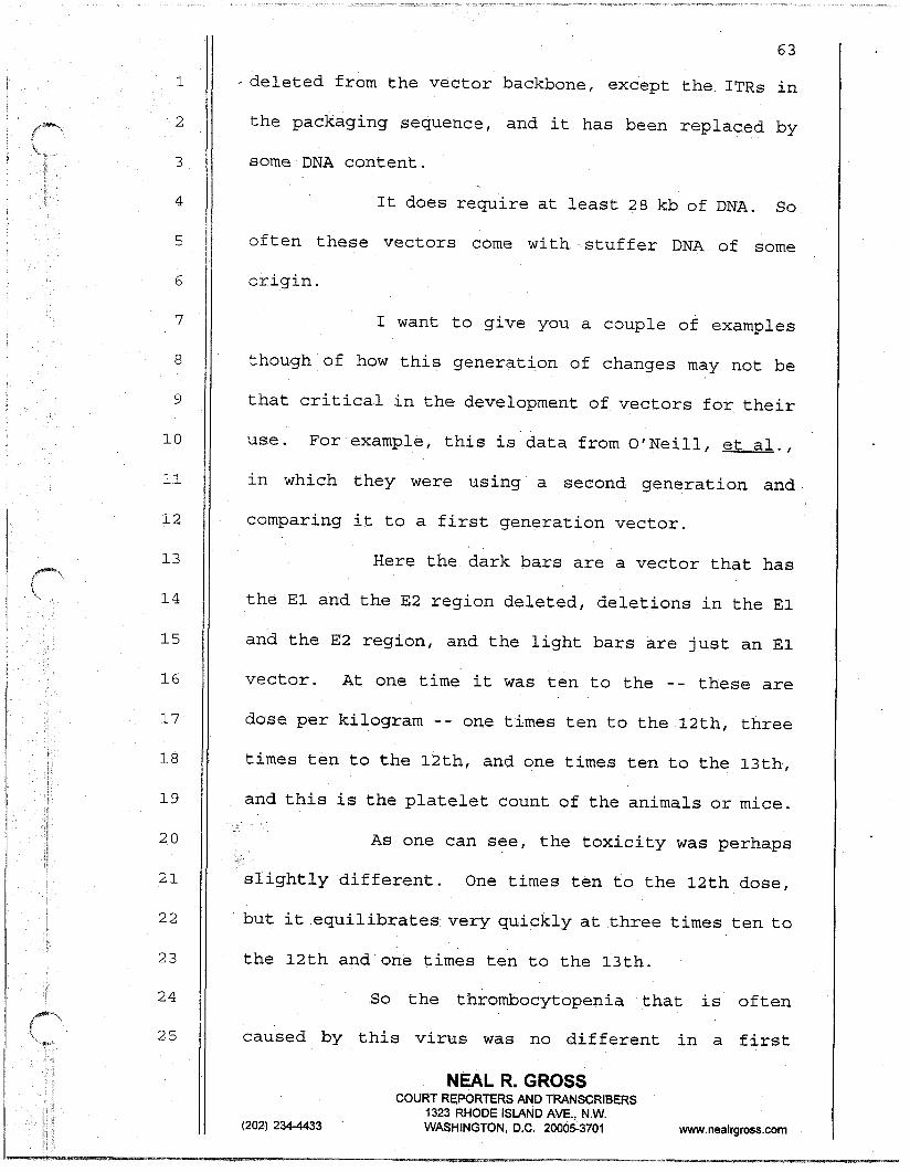
63
1 ~ deleted from the vector backbone, except the ITRs in
'2 the packaging sequence, and it has been replaced by
3 some DNA content.
4 It does require at least 28 kb of DNA. So
5 often these vectors come with .stuffer DNA of some
6 origin.
7 I want to give you a couple of examples
8 though.of how this generation of changes may not be
9 that critical in the development of vectors for their
10 use. For example, this is data from O'Neill, et al.,
11 in which they were using a second generation and
12 comparing it to a first generation vector.
13 Here the dark bars are a vector that has
14 the El and the E2 region deleted, deletions in the El
15 and the E2 region, and the light bars are just an El
16 vector. At one time it was ten to the -- these are
17
18
dose per kilogram -- one times ten to the 12th, three
times ten to the 12th, and one times ten to the 13th,
19 and this is the platelet count of the animals or mice.
20
21
.* As one can see,
j,-. the toxicity was perhaps .,
slightly different. One times ten to the 12th dose,
22 but it equilibrates very quickly at three times ten to
23 the 12th and one times ten to the 13th.
24 So the thrombocytopenia that is often
25 caused by this virus was no different in a first
NEAL R. GROSS
(202) 2344433
COURT REPORTERS AND TRANSCRIBERS 1323 RHODE ISLAND AVE., N.W. WASHINGTON, D.C. 200053701 www.nealrgross.com
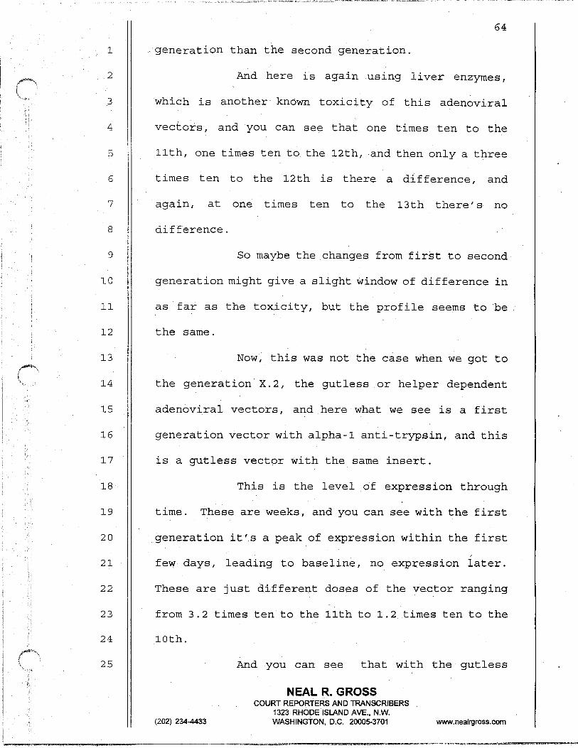
64
1 generation than the second generation.
2 And here is again using liver enzymes,
3 which is another known toxicity of this adenoviral
4 vectors, and you can see that, one times ten to the
5 llth, one times ten to the 12th,.and then only a three
6
7
8
times ten to the 12th is there a difference, and
again, at one times ten to the 13th there's no
difference.
9
10
11
So maybe the changes from first to second
generation might give a slight window of difference in
as far as the toxicity, but the profile seems to be
12 the same.
13 Now, this was not the case when we got to
14
15
16
17
the generation X.2, the gutless or helper dependent
adenoviral vectors, and here what we see is a first
generation vector with alpha-l anti-trypsin, and this
is a gutless vector with the same insert.
18 This is the level of expression through
19
20
time. These are weeks, and you can see with the first
generation it's a peak of expression within the first
21
22
23
24
few days, leading to baseline, no expression iater.
These are just different doses of the vector ranging
from 3.2 times ten to the lith to 1.2 times ten to the
10th.
25 And you can see that with the gutless
NEAL R. GROSS
(202) 234-4433
COURT REPORTERS AND TRANSCRIBERS 1323 RHODE ISLAND AVE., N.W. WASHINGTON, D.C. 200053701 www.nealrgross.com
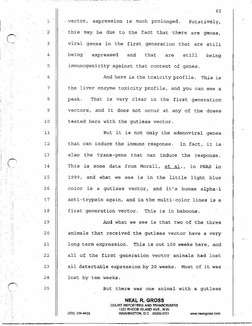
1
2
3
4
5
6
7
8
9
10
11
12
13
14
15
,I6
17
18
19
20
21
22
23
24
25
vector, expression is much prolonged. Putatively,
this may be due to the fact that there are genes,
viral genes in the first generation that are still
being. expressed and that are still being
immunogenicity against that content of genes.
And here is the toxicity profile. This is
the liver enzyme toxicity profile, and you can see a
peak. That is very clear in the first generation
vectors', and it does not occur at any of the doses
tested here with the gutless vector.
But it is not only the adenoviral genes
that can induce the immune response. In fact, it is-
also the trans-.gene that can induce the response.
This is some data from Morall, et al., in PNAS in
1999, and what we see is in the little light blue
color is a gutless vector, and it's human alpha-l
anti-trypsin again, and in the multi-color lines is a
first generation vector. This is in baboons.
And what we see is that two of the three
animals that received the gutless veator have a very
long term expression. This is out 100 weeks here, and
all of the first generation vector animals had lost
all detectable expression by 20 weeks. Most of it was
lost by ten weeks.
But there was one animal with a gutless
NEAL R. GROSS
(202) 234-4433
COURT REPORTERS AND TRANSCRIBERS 1323 RHODE ISLAND AVE., N.W. WASHINGTON, D.C. 200053701 vdww.nealrgross.com
66
1 that actually lost all etipression by ten weeks, and
2 that animal, in fact, had developed an immune response
3 to the alpha-l anti-trypsin.
4
5
So these vectors can be used to generate
an immune response to the coded.gene. Of course, in
6
7
8
addition to the variables that I just spoke about,
there's variables for the analyses of the vectors, and
I will just go very, very brief couple of slides on
9
10
11
this mostly to give you a sense of what is not known
rather than what is known. I will not go through all
of the safety testing of quality control that's
12 normally done for all viral agents.
13
14
15
16
17
And in order to do that, I want to just
give you an idea of how these vectors are typically
quantified,' and so we have the quantity of particles
that we can hetect easily, but. then we also need to
know of those particles how many of them are
18 functional and can transduce the gene of interest into
19 the target cells.
20
21
Often what we do is we just layer a soup
.fully of particles on top of some target cells. The
22
23
problem with this is that these particles, depending
on how deep this soup is,. how long the assay gets
24 allowed to progress, and many other variables, most of
25 these particles will never reach one of the target
NEAL R. GROSS
(202) 234-4433
COURT REPORTERS AND TRANSCRIBERS 1323 RHODE ISLAND AVE., N.iM WASHINGTON, D.C. 20005-3701 www.nealrgross.com
cells. SO may not be detected, and whether they get
detected one time or another is quite variable.
4 preparation to six laboratories, and we had a two log
5 differential in the determined titer from those
6 laboratories, all experienced with adenoviruses and
7 also how they get handled may be very specific. This
8 is just a bit of data from a paper by Neiber Hoffman,
9 al., et and you see here you can't tell the distance,
10 but there's a seven log differential on, the vector
11 that was shipped just across town into a clinical
12 setting as an experiment for .shipment only from a
13 vector that was shipped on dry ice and a vector that
14 was not shipped on dry ice.
15
16 was the CO, that was seeping in, dropping the pH, and
17 there tias a seven log differential that could be lost
18 in a very short period of time.
19
20 I've shown that due to their biology they can be
21 converted into efficient gene transfer vehicles.
22 They're not inherently dangerous, even as wild type
23 viruses.
24
25 toxicity profiles. Additional safety of the second
67
So much so that we sent out an identical
So it turns out in that particular case it
So in conclusion, adenoviruses, I think,
Not alladenoviralvectors have equivalent
NEAL R. GROSS
(202) 234-4433
COURT REPORTERS AND TRAN$CRIBERS 1323 RHODE ISLAND AVE., N.W. WASHINGTON, D.C. 20005-3701 www.nealrgross.com

1
2
3
4
5
6
7
8
9
10
11
12
13
14
15
16
17
18
19
20
21
22
23
24
25
68
,.generation vectors may be transient and only in a
small, temporal stage.
And this is actually very clear because
standardization of the dose specifications is
necessary, and I'm told there is a very standard way
of determining the potency and the quantity of these
entities. It is very difficult to analyze the data as
a whole, and thus, assurance of clinical potency.
And just as a last mention, there is
currently a working group that is developing a
standard of wild type adenoviruses so that all of
these things can be compared and quantified.
That's all I have.
much.
ACTING CHAIRMAN DADM: Thank you very
That was extremely helpful. It raises
many questions for us to consider.
Dr. Coffin and then Dr. Kohl and then Dr.
Stephens, Dr. Katz.
: DR. COFFIN: ', You said something early on
'regarding the difference between adenoviral vectors
and retroviral vectors that I think has something of
the status of an urban legend without ever actually
having been subject to a real test, and that is the
idea that retrovirus vectors might have some greater
NEAL R. GROSS
(202) 2344433
COURT REPORTERS AND TRAN!Z$RIBERS 1323 RHODE ISLAND AVE., N.W. WASHINGTON, D.C. 20005-3701 www.nealrgross.com

1
2
3
-danger because of the property of integrating their
DNA into a cell, in the cell DNA, whereas adenoviruses
don't do that.
4
5
6
7
8
9
10
11
That's actually not true, of course.
Adenoviruses do, of course, -- adenovirus DNA does get
integrated after infection at some low event, and I
believe that the -- I don't know exactly what the
numbers are and I don't know if anybody knows exactly
what the numbers are, but it's quite likely that you
make up that entire difference in efficiency in
integration by the difference in the doses of
12 adenoviral vectors versus retroviral vectors that you
13
14
15
16
17
give, and in fact, the probability of the integration
of a fragment of adenovirus DNA may be equally high as
a probability of retrovirus DNA integration after
administration of these as gene therapy or vaccine
vehicles.
18 Can you comment on that?
19 DR. AGUILAR-CORDOVA: The fact is that
20
21
22
they do integrate occasionally. It's not their
standard method of operation, but they do integrate
occasionally. I didn't mean to imply that they were
23 more dangerous or not. I was just reading off a list
24 of often stated events.
25 The one difference would be that they do
69
NEAL R. GROSS
(202) 234-4433
COURT REPORTERS AND TRANSCRIBERS 1323 RHODE ISLAND AVE., N.W. WASHINGTON, D.C. 20005-3701 wwdf.nealrgross.com
1 .no carry a promoter enhancer in there, in the LTR. So
2
3
at least~the promotion enhancement effect ,of the
integration would be different.
4 DR. COFFIN: But there must be one
5
6
7
somewhere in the vector or it wouldn't be any good as
a vector, and often more than one, of course, if --
DR. AGUILAR-CORDOVA: Often, yes, and so
8
9
10
how they compare as far as integration I can't
comment. I don't know that there is any data. There
is data showing that they do integrate on occasion,
11
12
yes, and they persist.
ACTING CHAIRMAN DAUM: Dr. Kohl, please.
13
14
15
DR. KOHL: Two questions.
Thanks for your talk. It was enjoyable
and elucidating.
16 You mentioned the concept of leakage.
17 Could you elaborate that a little further, and can you
18 get enough leakage from a gene that supposedly is
19 deleted that you can get a competent virus injected
20 into the host?
21
22
23
24
DR. AGUILAR-CORDOVA: So the leakage that
I was speaking about was from the genes that were not
deleted. Obviously the one that's deleted can't leak
on there.
25 However, there is the potential for
70
NEAL R. GROSS
(202) 234-4433
COURT REPORTERS AND TRANSCRIBERS 1323 RHODE ISLAND AVE., N.W. WASHINGTON, D.C. 200053701 www.nealrgross.com
1
2
3
4
5
6
7
8
9
10
11
12
13
14
15
16
17
18
19
20
21
22
23
24
25
71
- recombination with the gene inside the packaging cell
and thus leading to a replication competent virus. I
believe that that will be spoken to as far as why the
PER-C6 cells were developed, and the difference
between them and the 293 cells..
DR. KOHL: Can you elaborate a little bit
more on the toxicities of the adenoviralvector? What
causes that? What's the mechanism of the toxicity?
The platelets, the liver function transmission and
other toxicities as well?
DR. AGUILAR-CORDOVA: Yes. So what we
know are what the toxicities are, and particularly, of
course, as you've heard, there was an incident in'the
University of Pennsylvania where a young man died due
to a large dose of an adenoviral vector directly
injected into their hepatic artery.
In that case what was seen was a DIC like
syndrome with upper respiratory distress, and what we
have seen in many animal models is an elevated liver
enzyme content, often transient and recoverable, and
not just animal models. Many Phase 1 studies have
seen the same thing.
Thrombocytopenia, believed but not shown
to be caused due to endothelial cell damage and
leakage because it is consumptive thrombocytopenia.
NEAL R. GROSS
(202) 234-4433
COURT REPORTERS AND TRANSCRIBERS 1323 RHODE ISLAND AVE., N.W. WASHINGTON, D.C. 200053701 ww-wnealrgrossxom
72
1
2
3
DR. KOHL: Who does the mechanism?
DR. AGUILAR-CORDOVA: The mechanism is
probably cytopathic effects of the vector and
4 potentially an immunological response to the original
5 infection.
6
7
ACTING CHAIRMAN DAUM: I have Dr.
Stephens, Dr. Katz, Dr. van der Eb.
8 DR. STEPHENS: In this discussion, there's
9
10
11
some at least in my mind confusion about the issues of
gene therapy vectors versus vaccine vectors, and I'd
like you to kind of clarify that issue for us, if you
12 would.
13
14
15
16
17
18
More specifically, the questionrelatesto
the E3 sequence and whether you think that should be
in or out of the vaccine delivery vector.
DR. AGUILAR-CORDOVA: Sure. So not just
in this discussion is there a confusion of vaccine or
gene therapy. I think as a member of the RAC we've
19
20
'21
22
had that discussion a lot, too, what should be
accepted or not, and many of the gene therapy
applications especially in cancer are, in fact,
vaccines. We're trying to vaccinate against cancer.
23 And Dr. Ginsberg started working with an
24
25
E3 region in early '8Os, I believe, or before, and
he's big prop.onent of leaving the E3 region in when
NEAL R. GROSS
(202) 234-4433
COURT REPORTERS AND TRANSCRIBERS 1323 RHODE ISLAND AVE., N.W. WASHINGTON, D.C. 200053701 www.nealrgross.com
1
2
3
4
5
6
7
8
9
10
11
12
13
14
15
16
17
18
19
20
21
22
23
24
25
73
one wants to create an immune response because in that ..
situation, it is possible that the adenovirus itself
is an adjuvant to whatever one wants to create an
immune response against.
The flip side of t.hat is that in some
studies in my laboratory and others, when transducing
a gene that one wants to create a CTL response
against, for example, with an adenoviral vector, we
tend to get an awful lot of CTL against adenowirus and
very little to none against the gene of interest.
So it could go either way.
ACTING CHAIRMAN DAUM: Thank you.
Dr. Katz, please.
DR. KATZ : What is the receptor for
adenovirus and what cells express the receptor?
DR. AGUILAR-CORDOVA: The known receptor,
and there are, probably others, the known receptor is
a module called CAR, coxsackie adenovirus receptor,
and its distribution is fairly ubiquitous. Epithelial
'7 cells are especially high expressers, and it also uses L some integrants as co-receptors.
ACTING CHAIRMAN DAUM: Dr. van der Eb.
DR. VAN DER EB: I'd like to come back to
the issue of leakage, leakage that you mentioned.
Even a deleted or undeleted vectors are supposed not
NEAL’ R. GROSS
(202) 2344433
COURT REPORTERS AND TRANSCRIBERS 1323 RHODE ISLAND AVE., N.W. WASHINGTON, D.C. 200053701 www.nealrgross.com
1
2
3
4
5
74
_ to express the rest of the viral genome that is still
present in the vector. That is because the EIA gene
is the master switch that directs the expression of
the rest of the viral genome.
Now, it's known already for a rather long
6
7
8
time that leakage may occur and expressionof the rest
of the viral genome occurs when very high
multiplicities of infection are used.
9
10
11
For reason then this creates a kind of
ElA-like activity in the cell and leads to expression
of the rest of the viral genome with consequent
12 reaction of the host cell immunological reaction of
13 the host.
14
15
16
17
18
19
DR. AGUILAR-CORDOVA: Right. So in order
for efficient transcription of the other genes, the El
region is necessary. However, it seems like there is
some gene expression, although maybe not at sufficient
levels to produce virions and to create the El.
But given the data, there seems to be some
20 1- expression of the other viral genes even in its
21 absence.
22
23
DR. VAN DER EB: Is the data of Tom Shenk
from long ago?
24
25
(202) 2344433
DR. AGUILAR-CORDOVA: Right.
ACTING CHAIRMANDAUM: Two last questions,
NEAL R. GROSS COURT REPORTERS AND TRANSCRIBERS
1323 RHODE ISLAND AVE., N.W. WASHINGTON, D.C. 200053701 www.nealrgross.com

1
2
3
4
5
6
7
8
9
10
11
12
13
14
15
16
17
18
19
20
21
22
23
24
25
75
~ please.
DR. COOK: I'd just like to respond to the
question about E3 in or out. E3's job from the virus'
point of view is to shut down Class 1 expression or
travel to the surface through Golgi mechanisms. So
theoretically if that were uniformly true and you were
trying to make a vaccine and it required expression of
that peptide in the cell in which E3 was co-expressed,
it would be a good idea maybe not to have E3 present.
The truth is when YOU infect with
adenovirus in normal human cells or cells that don't
express ElA, that phenomenon is very late in
infection. It doesn't happen until probably 48 to'72
hours after infection with that virus.
So chances are the peptide expression
could occur, depending on what the kinetics are. If
the cell co-expresses ElA, the E3 effect is much
greater. So it might depend on how. you rate the
system, but theoretically, at least, one would have to
consider whether E3 is downregulating Class 1
expression on the surface and whether that alters
antigen presentation or peptide expression on the
surface of the gene of interest.
ACTING CHAIRMAN DAUM: Thank you, Dr.
Cook.
NEAL R. GROSS.
(202) 234-4433
COURT REPORTERS AND TRANSCRIBERS 1323 RHODE ISLAND AVE., N.W. WASHINGTON, D.C. 200053701 www.nealrgross.com

1
2
3
4
5
6
7
8 I-
9
10
11
12
13
14
15
16
17
18
19
. . Dr. Blair.
DR. BLAIR: Yeah. This comes out of my
retroviral background, but is there any evidence that
there's an encapsidation of cell DNA/RNA protein in
the virus as it's assembled?
DR. AGUILAR-CORDOVA: Not that I know of.
Not at the level that we've seen in retroviruses
certainly with the RNAs, especially of viral-like
proteins or viral-like particles and so on.
But certainly there's the possibility of
that, and certainly there's the possibility of
recombination events, non-specific recombination
events that would package random pieces of DNA.
ACTING CHAIRMAN DAUM: Thank you very
much, Dr. Aguilar-Cordova.
And we will shorten the 20-minute
scheduled break to a 15-minute break. I have 10:40.
We'll reassemble at lo:55 and continue with Dr. van
der Eb.
20
21
(Whereupon, the foregoing matter went off
the record at IO:39 a.m. and went back on
22 the record at lo:58 a.m.)
23
24
ACTING CHAIR.P@N DAUM: Would everybody
please settle down as quickly as they can?
25 ACTING CHAIRMAN DAUM: We're ready to
NEAL R. GROSS
76
(202) 234-4433
COURT REPORTERS AND TRANSCRIBERS 1323 RHODE ISLAND AVE., N.W. WASHINGTON, D.C. 20005-3701 www.nealrgross.com

1
2
3
4
5
6
7
8
9
10
11
12
13
14
15
16
17
18
19
20
21
22
23
24
25
continue with the open session.
We will now call on Dr. Alex van der Eb to
tell us about adenovirus transformation of human cells
and the development of 293 and PER.CG cells for the
manufacture of defective adenovirus vaccines.
Welcome, Dr. van der Eb.
DR. VAN DER EB: Thank you.
So what I would like to do is to describe
to you how and why we have made two different cell
lines, adenovirus transformed human embryo cell lines
which are called 293 and PER.CG. Both cell lines were
made in my lab, and also the cells, the starting
material, was prepared by myself at the University of
Leiden. The 293 cell was made by Frank Graham in 1973
from human embryonic kidney cells that were made from
fetal tissue one year ago by myself one year before
that, so that was in 19 -- probably in 1972, whereas
the PER.CG cell was made by Ron Bout and Frits Fallaux
in 1995 from an embryonic retina cultures that were
made from fetal tissue by me ten years before that, in
1985.
This just shows you again the adenovirus
genome and you have seen it already. The interest in
this virus was due to the fact that the viruses can
transform cells in tissue culture. In fact, all human
NEAL R. GROSS
(202) 234-4433
COURT REPORTERS AND TRANSCRIBERS 1323 RHODE ISLAND AVE., N.W. WASHINGTON, DC. 2000!5-3701 www.nealrgross.com

6
8
16
18
23
24
78
adenoviruses or almost all can transform cells in
tissue culture, and also that certain types of
adenoviruses can induce tumors in experimental
animals.
The transforming region -- oh, I hope it
survived -- the transforming region is associated with
the left-most about ten percent of the genome that
harbors the El region.
We became interested in transforming -- in
the question whether human cells could be transformed,
and therefore, I will tell you how we got the
transformed human cells, and it all started actually
in 1972 when Frank Graham in my lab developed the
calcium phosphate DNA transfection technique, which
made it possible in the first place to make infectious
virus with intact viral DNA.
If you transfect the intact viral DNA of
Adenovirus Type 5 into permissive human cells you get
infectious virus, but it also turned out -- do you
have a pointer here? There is no pointer? Okay.
Thank you.
And it turns out that not only it was
possible to get infectious virus bytransfecting human
cells with the intact viral DNA, but also purified DNA
proved capable of transforming cultured rodent cells,
NEAL R. GROSS
(202) 234-4433
COURT REPORTERS AND TRANSCRIBERS 1323 RHODE ISLAND AVE., N.W. WASHINGTON, D.C. 200053701 www.nealrgross.com
1
2
3
4
5
6
7
8
9
10
11
12
13
14
15
.but human cells could not be transformed.
And the reason is that these human cells
got destroyed by the viralytic (phonetic) reaction.
If the DNA was sheared, however, up to 3 mega delta
Daltons, up to three million Daltons, it turned out
that the transforming potential of rodent cells still
remained intact, indicating that-only a portion of the
viral genome, a rather small proportion of the viral
genome is necessary for transformation.
As I said, purified Adenovirus 5 DNA
transfected itit0 permissive human cells yields
effective virus, but human cells could not be
transformed, everywhere interested in transforming
human cells by adenoviruses just in order to find out
whether that is possible.
16
17.
18
19
20
21
But we found some evidence that permissive
human cells could be perhaps transformed from the fact
that semi-permissive rodent cell cultures could be
transformed if the DNA of adenovirus was sheared into.
smaller pieces, and these were Syrian hamster kidney
cells.
22 The transforming activity, and this was
23 done with detailed shearing studies at that time;
24
25
there were no restriction enzymes. There was no DNA
cloning at that time; that the transforming activity
NEAL R. GROSS
79
(202) 234-4433
COURT REPORTERS AND TRANSCRIBERS 1323 RHODE ISLAND AVE., N.W. WASHINGTON, D.C. 200053701 www.nealrgross.com
80
1
2
3
4
. associated with the 11 left percent, left-most 11
percent of the adenovirus genome, and this all affect
the basis of the transformation by adenovirus of the
humancells with fragments of adenoviruses.
5
6
7
.8
9
10
11
So the reason why we wanted to transform
human cells is just to answer the question whether
human cells can be transformed at all by adenoviruses,
human adenovirus, and if so, which part of the
adenovirus DNA is required to transform cells? Is
that the same area that is also needed for
transformation of rodent cell or is it less or is it
12 more?
13
14
15
16
17
18
19
20
And then can we simply develop a model to
study transformation of human cells? And that was at
that time important because although there was no
evidence that human adenoviruses have anything to do
with cancer in humans, it was still an open issue, an
open question, and in fact,, it is at this moment
still, although clearly there is no evidence that
human adenoviruses have anything to do with cancer in
21 men.
22 So the method that we followed was take
23 human embryonic kidney cultures. Why kidney cultures?
24 And that is mainly because of the fact that the rodent
25 system, the rodent model that we used were always baby
NEAL R. GROSS
(202) 234-4433
COURT REPORTERS AND TRANSCRIBERS 1323 RHODE, ISIANQ AVE., N.W. WASHINGTON, DC. 20005-3701 www.nealrgross.com

1
2
3
4
5
6
7
8
9
10
11
12
13
.- rat kidney or baby mouse kidney or baby hamster
kidney. The kidney cells were very suitable for these
transformation studies with sheared adenovirus DNA.
When we transfected these human embryonic
kidney cultures with sheared purified Adenovirus 5 DNA
with the calcium phosphate technique using carrier
DNA of salmon sperms. So this was not restriction
enzyme fragments. They were just not yet usable.
One year later we had resection enzyme
available to make pure DNA, but the first and also the
293 cell was made with sheared Adenovirus 5 DNA, then
simply scored for transformed colonies as we did with
the rodent cultures.
14
15
16
17
18
19
20
21
22
So the kidney material, the fetal kidney
material was as follows. The kidney of the fetus was,
with an unknown family history, was obtained in 1972
probably. The precise date is not known anymore.
The fetus, as far as I can remember was
completely normal. Nothing was wrong. The reasons
for the abortion were unknown to me. I probably knew
it at that time, but it got lost, all this
information.
23
24
The kidneys of the fetus were then
isolated and the kidney cells were isolated in the SO-
25 called still air cabinet. There were no laminar flow
NEAL R. GROSS
(202) 234-4433
COURT REPORTERi AND TRANSCRIBERS 1323 RHODE ISLAND AVE., N.W. WASHINGTON, D.C. 200013701 w.nealrgros.s.com
81
1
2
3
4
5
6
7
8
9
10
11
12
82
.-hoods at that time, and this, is simply a still air
cabinet that was also used all over for tissue culture
and worked quite well. There was W lights in it just
to sterilize it, and that was all.
So as we did also. for the rat kidney
cells, the surrounding membranes were removed as
completely as possible, and the kidneys were then
minced with scissors, trypsinized, and the cells that
were recovered after removing the trypsin were
cultured in medium containing bovine serum, calf
serum. That is what we know.
And this caif serum was obtained not from
13
14
15
a commercial source. We either got, it from somebody
else, from another lab, or we made it ourselves from
blood, calif blood.
16
17
18
19
20
Rodent, monkey, and other human cell
cultures took place in the same general area at that
time. So there was one cell culture room, and there
all of the experiments, all the cell 'culture work was
-being done. . .
21
22
There was also experiments 'with viruses,
but that was in a separate virus cultured unit, and we
23 used in addition to Adenovirus 5 whole viruses, also
24
25
the oncogenic Adenovirus 12, as well as SV40 and
possibly also already Herpes virus, but maybe Herpes
NEAL R. GROSS
(202) 234-4433
COURT REPORTERS AND TRANSCRIBERS 1323 RHODE ISLAND AVE., N.W. WASHINGTON, D.C. 200053701 www.neairgross.com
i
83
1
2
3
4
5
6
7
8
9
10
_ virus was not yet used at that time:
So the method was DNA from wild type
Adenovirus 5, was isolated from virions. So we had to
prepare the DNA by first growing and purifying the
virions, and the DNA was then fragmented by shearing
in this case through a 22 gauge needle up to about
eight million Daltons. There was no cloning strategy
at that time, and the DNA fragments were transfected
as I already indicated with salmon sperm DNA with the
calcium technique.
11
12
13
14
15
16
17
18
19
20
21
The results were rather disappointing. In
the first experiment of quite a number of dishes there.
were not a single transformed colony. So we repeated
it. Again, no transformed colony.
However, after many other experiments, we
found finally one transformed colonywhichwas visible
in the cultures, and that colony appeared 33 days
after transfection was seen, 33. days after
transfection. .--
This colony, this single colony was picked
and established and became the 293 cell.
22 There were two colonies here mentioned,
23 and that is because one, the second colony, was only
24 seen after the cells at the end of the experiment, the
25 cultures were fixed and stained, and one other colony
NEAL R. GROSS
(202) 2344433
COURT REPORTERS AND TRANSCRlBiRS 1323 RHODE ISLAND AVE., N.W. WASHINGTON, D.C. 200053701 www.nealrgross.com

1
2
3
4
5
6
7
8
9
10
11
12
13
14
15
16
17
18
19
20
21
22
23
24
25
84
was seen at the edge of a dish which we had missed.
So the single transformed colony which
would give rise to 293 was very difficult to expand.
The cells hardly grew at all, and after five months in
culture, it was possible to free.ze down the number of
ampules. Only three ampules, passage four was that.
And at that time the cells started to grow
faster, but still relatively poorly and a doubling
time was at least a week or more than a week.
So it appeared from these experiments that
human cells are resistant to transformation by
Adenovirus 5. Although these cells replicate
extremely well in replicating Adenovirus 5, they
cannot be transformed with the same DNA that also
transforms rodent cells quite efficiently.
So up to this moment, it's still unclear
why human cells are resistant to transformation by
adenoviruses. One possibility is that the 293 cell
came out of a cell line that had some kind of a
mutation so that it became permissive to
transformation.
Another possibility is that since this is
primary human embryonic kidney which consists, of
course, of many different cell types, that there is
one -- that there are very few cells in the whole
NEAL R. GROSS
(202) 2344433
COURT REPORTERS AND TRANSCRIBERS 1323 RHODE ISLAND AVE., N.W. WASHINGTON, D.C. 200053701 www.nealrgross.com
1
2
85
T culture which are permissive to transformation, and
that this particular cell came from one of those
3 transformation prone cells.
4
5
6
7
8
We will never know probably, but one
possibility is that the 29? cell is actually a neural
cell that was present in that culture. We don't know,
and this is something that occurred to me when I was
traveling here ,to Gaithersburg, and so that 'is a
9
10
possibility that canprobablybe testedbecause neural
cells appear to be more prone to transformation by
11 adenovirus.
12
13
14
15
16
'17
We also tried human diploid skin
fibroblasts for transformation, never any positive
result. We also have tried human embryonic lung
cells. No positive transformation.
Anyway, around page 13, the cells went
into crisis, the same type of crisis that is also seen
18 when SP40 transformation is followed in human diploid
19 fibroblasts, for example. They always go in crisis.
20
21
22
23
24
25
This crisis lasted nearly three months.
During that time, the cells remained on the dish or
gradually started to die, some of them at least. So
you have to defeat the cultures for a long time.
Nothing happens. There is no cell division.
And then the culture started to recover
NEAL R. GROSS
(202) 23-44433
COURT REPORTERS AND TRANSCRIBERS 1323 RHODE ISLAND AVE.. N.W. WASHINGTON, D.C. 20005-3701 www.nealrgross.cofn
1
2
3
4
5
6
,7
8
9
10
11
12
13
14
15
16
17
18
i9
20
21
22
23
24
25
for some reason not in the same way as usually in the
case of SV40, but apparently cells all over the plate
begin to grow, whatever that means. We don't know
what happens in this crisis base.
After crisis, when telomerase is
activated, apparentlywhenthe cells start recovering,
the cells were subcultured and the growth rate
increased significantly. Severalambules were shipped
then by Frank Graham to McMaster in Canada, where he
went to Anestilles (phonetic) in 1974, and the data
have been published in several papers.
I would like to show here also the part of
the adenovirus genome present in the 293 cells. It is
not completely sharp, but it doesn't really matter,
and this is the left-most 4,041 nucleotides. There is
also some E4 region present in these cells, which is
not expressed, however. So this is in the 293 cells.
Now, in addition to basic research,
adenovirus DNA also became interesting, Adenoviruses
became interesting as factors for gene therapy. So
this occurred in the '80s when people started to think
about gene therapy, introducing genes into cells,
first retroviral vectors, 'and also later adenoviral
vectors.
And adenoviral vectors, in contrast to
NEAL R. GROSS
(202) 234-433
COURT REPORTERS AND TRANSCRIBERS 1323 RHODE ISLAND AVE., N.W. WASHINGTON, D.C. 20005-3701 www.nealrgross.com

1
2
3
4
5
6
7
8
9
10
11
12
13
14
15
a7
._ retroviral vectors, in fact, have as have been already
said today a deletion in the El gene and in the place
of the El gene, you can clone the gene of interest,
and the reason that El deleted adenovirus was chosen
was, in fact, the present or the availability of the
293 cells, which turned out to be very suitable
packaging cell line for these first generation viral
vectors because they expressed the El genes.
so El deleted recombinant adenovirus
vectors were being used more and more for gene
transfer purposes. Adenovirus vectors, you've heard
it, are quite suitable for similar reasons. They have
certain disadvantages also compared to retroviral
vectors, for example, but they certainly have a number
of important advantages.
16
17
18
19
20
21
22
23
The cells to grow the replication
deficient El deleted adenovirus vectors were also
available, and those were the 293 cells, and in fact,
already in 1994 the first clinical study with an El ._ deleted adenovirus vector was done, was made by
Crystal in '94, which probably was the, if I remember
correctly, the CFTR gene. That was the first clinical
trial gene in an adenovirus vector.
24 So there was a new use now for the 293
25 cells. Two, nine, three for quite some period of time
NEAL R. GROSS
(202) 2344433
COURT REPORTERS AND TRAN~CRBERS 1323 RHODE ISLAND AVE., N.W. WASHINGTON, D.C. 200053701 www.nealrgross.com
1 '. was the only packaging cell line available for growth
2 of adenovirus vectors.
3
4
El deleted vectors for gene transfer
became common practice. More and more groups started
5
6
7
a
9
10
11
12
13
to use adenoviruses as a factor; but there were also
limitations to the available packaging cell line, 293.
It became apparent at that time, and that is that due
to recombination between El sequences from the 293
cells into the El deleted vector could occur, and this
gave rise to formation of replication competent
adenovirus, RCA, and it turned out to be very
difficult to produce large batches of RCA free vector,
and of course, the RCA is almost identical to the
14 vector with the gene of interest, and therefore cannot
15
16
17
ia
19
20
be physically separated from the vector.
So it was clear that 293 was not really
the ideal vector for gene therapy used, and therefore,
we decided around 19 -- oh, this first, just to show
you here, here is the recombinant vector in which the
-El gene is deleted, and instead of it, the gene of ',
21 interest can be inserted here.
22
23
24
25
And here are the 293 cells with a
proportion of the adenovirus genome integrated with
the El gene and P9, protein 9, and there turns out to
be quite a considerable overlap at both sides between
NEAL R. GROSS
(202) 234-4433
COURT REPORTERS AND TRANSCRIBERS 1323 RHODE ISLAND AVE., N.W. WASHINGTON, D.C. 200053701 www.nealrgross.com
1
2
3
4
89
the recombinant adenovirus vector, first generation
vectors of both sides of the El region.
And be recombination you can get back RCA,
which.is essentially, again, the wild type adenovirus.
5
6
7
8
9
10
11
12
So the combination of the El gene in the
vector yields wild type virus, which is capable of
replicating. This could cause toxicity, high
concentrations perhaps of virus in places where you
don't want it. It could give rise to uncontrolled
dissemination not only of the wild type virus, but
also of the recombinant factor that replicates
together with it in the same cell.
13
14
15
16
17
18
19
It could theoretically also yield new
virus strains in the case of capsid modified vector
where the capsid is modified in such a way, for
example, that it can. attach to other receptors and
other cells. So in that case when that becomes
replication competent you can say that this is a'
partially new virus that you have created.
20
21
22
Also, it could yield, give rise to
replication deficient El containing viruses in the
case of multiplydelete'dvectors, for example, vectors
23 that in addition to deletion of El also deleted in E2,
24
25
E2A, for example, when El is reinserted into the
vector, it will still be replication deficient because
NEAL R. GROSS
(202) 234-4433
COURT REPORTERS AND TRANSCRIBERS 1323 RHODE ISLAND AVE., N.W. WASHINGTON, D.C. 20005-3701 vww.nealrgross.com
1
2
3
4
5
6
7
8
9
10
11
12
13
14
15
16
17
ia
19
20
21
22
23
24
25
90
the E2A gene is absent, but this vector, in theory, is
immortalizing or transformation competent virus that
you have created.
So in 1995, Brahm Bout (phonetic), Frits
Fallaux, Brahm Bout from IntroGene and Frits Fallaux
from our university Gene Therapy Group, decided that
we should try and make a new helper cell line and
matching factor in such a way that there is no
sequence overlap between the factor and the advanced
sequences in the cell line.
And indeed, in order to make a new system
that allowed pharmaceutical production of adenovirus
vectors, three of RCA. It should also meet
pharmaceutical standards. If you start all over again
you can just as well try to do that, and it could be
the basis for the manufacture of multiply deleted
Adenovirus 5 vectors also.
So we choose the human embryonic retina
cells at that time. Why not kidney cells? Simply
because these cells were so resistant to adenovirus
transformation that we didn't think it would be
worthwhile to try it all.
Human embryonic retina was chosen because
Gallimore had shown not long before that that human
embryonic retina was permissive to transformation,
NEAL R. GROSS
(202) 234-4433
COURT REPORTERS AND TRAN?CRIBERS 1323 RHODE ISLAND AVE., N.W. WASHINGTON, D.C. 200013701 www.nealrgross.com
1 .- could be transformed by adenoviruses, Adenovirus 5 and
2
3
4
12, and that was, again, based on some other studies
in animals, and therefore, we decided to take human
embryonic retina cells.
5 So they can be transformed by Adenovirus
6 5, and also at least in some of the cases that we have
7 studied, there is no real crisis. So the cells become
8
9
10
il
transformed and then go on to become immortal without
a real crisis in which the whole culture stops the
fighting.
12
Transformation is still a rather low
efficiency, but anyway, there is transformation, and
13 it is reproducible.
14
15
16
17
18
19
20
So I isolated retina from a'fetus, from a
healthy fetus as far as could be seen, of 18 weeks
old. There was nothing special with a family history
or the pregnancy was completely normal up to the 18
weeks, and it turned out to be a socially indicated
abortus', abortus provocatus, and that was simply
because the woman wanted to get rid of the fetus.
21 We got this. There was permission, et
22
23
cetera, and that was, however, was in 1985, ten years
before this.
24
25
This shows that the cells were isolatedin
October I as, Leiden University in my lab. They were
NEAL R. GROSS
(202) 234433
COURT REPORTERS AND TRANSkRIBERS 1323 RHODE ISLAND AVE., N.W. WASHINGTON, D.C. 20005-3701 www.nealrgross.com
1
2
3
4
5
6
7
8
9
10
11
12
13
14
15
16
17
18
19
20
21
22
23
24
25
92
then isolated inseparate cell culture area, which
contained a laminar air flow cabinet, and that was we
did it in the cell culture area of the three different
cell culture rooms that we had available at that-time.
That was only devoted to diploid cell cultures, human
cell cultures.
The cell culture media were, of course,
from certified supplies. At that time already '85, I
should say, the cells were frozen,' stored in liquid
nitrogen, and in 1995 one of these files was thawed
for the generation of the PER.CG cells.
We used defined El, identifiers El DNA
construct, in order to eliminate sequence homology
between the cells and the vectors, and that would
allow RCA contamination free El deleted vector
production.
The El genes in the PER.CG cells were
regulated not by the ElA promoter, but by the PGK
promoter, and the whole thing was all sequenced and.
functionally characterized. I can show you if you are
interested the data on expression of the viral genes.
Transfection was carried out at Leiden
University. There was no carrier DNA used. In '95
was this. The transfection yielded a number of
different colonies after about 18 days, and one of
NEAL R. GROSS i
(202) 234-4433
COURT REPORTERS AND TRANSCRIBERS 1323 RHODE ISLAND AVE., N.W. WASHINGTON, DC. 200053701 www.nealrgross.com
1
2
3
4
93
several of those were isolated. One of them, Clone 6,
finally was established and gave rise to PER.CG, and
Clone 6 was chosen because it gave the highest yield
of viruses and also had rather high expression of ElA
5 and ElB gene products.
6
7
8
These cells did not go through crisis, but
it is possible that in some case a crisis appears, as
I remember from experiments of Phil Gallimore, that
9
10
11
12
13
14
15
sometimes a short crisis may be observed.
So after the transformation event at the
university in Leiden and after the colonies were
picked, everything was transferred to IntroGene, which
was close by also in Leiden, in fact, in the same
building, and the whole documentation control was done
by them.
16
17
18
In the dedicated cell culture area defined
materials were used, of course. Cell banks were laid
down at IntroGene, passage 29, 33, and 36.
J-9
20
This shows you the information. The
recombinant identifiers itself it shown here. The P9
21
22
gene is still present at the right hand and, in fact,
of El, and here is the area where El is deleted and
23
24
25
the gene of interest is inserted.
Then the PER.CG cell was transformed by an
El gene construct with a PGK promoter and a polyacid
NEAL R. GROSS
(202) 234-4433
COURT REPORTERS AND TRANSCRIBERS 1323 RHODE ISLAND AVE., N.W. WASHINGTON, D.C. 200053701 wwv.nealrgross.com
1
2
3
4
5
6
7
8
9
10
11
12
13
14
15
16
17
18
19
20
21
22
23
24
25
94
.. of Hepatitis B virus, I believe.
And here is no sequence homology and real
RCA has never been observed in many, many different
experiments.
And this is the final slide just showing,
you some comparisons between 293 and PER.CG. Again,
I remind you that both cell lines were made in my lab
for different reasons.
The objective, as I indicated, is for 293
-- was basic research, and we have done many different
transformation studies after that, not transformation
studies, but gene expression studies with human
embryonic kidney cells in the years following that up
to now, I would say.
PER.CG was made just for pharmaceutical
manufacturing of adenovirus vectors. As to RCA free,
PER.CG is RCA free. Two, nine, three is not.
The history documentation of the cell line
has been carried out completely for PER.CG and was not
done at that time for 293. We -had no donor
information on 293 or what was available got lost, and
this is available for PER.CG. Containment at that
time was a little primitive perhaps and was now done
in a laminar flow cabinet.
The serum sources were of noncommercial
NEAL R. GROSS COURT REPORTERS AND TRANt%RlBERS
(202) 2344433 1323 RHODE ISLAND AVE.: N.W. WASHINGTON, D.C. 20005-3701 www.nealrgross.com
1
2
3
4
5
6
7
8
9
10
11
12
13
14
15
16
17
18
19
20
21
22
23
24
25
95
.. use. Probably I have made it myself. Certified
samples -- supplies were now used for serum and
medium, et cetera.
Crisis free history just means that 293
had a crisis,' was not crisis free at the long crisis,
and these cells, PER.CG had no crisis for some reason.
Andthenpharmaceuticalindustrystandard.
I realize that this sounds a bit commercial, but
PER.CG were made for that particular purpose. Also,
as far as I know, more than 50 different companies
have taken license for PER.CG. 1
Two, nine, three was not in the same way
characterized, is in the public domain, whereas PER.CG
is licensed.
So I think I'm at the end if somebody
wants to see the data again of virus production and so
on, but I don't think that's very important.
ACTING CHAIRMAN DAUM: Thank you very
much, Dr. van der Eb.
We'll take a couple of questions. Dr.
Decker, then Ms. Fisher. Dr. Kohl.
DR. DECKER: Did you say that adenovirus
is not capable human diploid cell transformer because
of cytolysis? The human cell --
DR. VAN DER EB: Yeah, yeah. Well, yes.
NEAL R. GROSS
(202) 234-4433
COURT REPORTERS AND TRANSCRIBERS 1323 RHODE ISLAND AVE., N.W. WASHINGTON, D.C. 20005-3701 www.nealrgross.com
1
2
'3
4
5
6
7
8
9
10
11
12
13
14
15
16
What I said is that you -- if you take intact DNA or
a virus and you put it on human diploid.cells, the
types that we have used is the embryonic kidney, the
skin, embryonic lung. Then you see lytic reaction,
and that will just wipe out a whole culture area.
DR. DECKER: Does that imply that an
attenuated and human diploid cell adapted might then
adapting it that way so you didn't get the cytolysis
might unmask a transformation capability?
DR. VAN DER EB: Theoretically that is a
possibility. I don't believe it because the three
different types of diploid human cells that we have
tested were so resistent to transformation just by
DNA, also by fragments of the DNA, resection fragments
of the DNA. We did it later also, but I don't believe
that that is a big issue.
17
18
19
20
What I think is that there might be other
tissues, cells and tissues in the human body that can
be transformed, for example, retina cells. It could
be that neural cells are also transformed.
21 It is known that Adenovirus Type 12 is
22
23
more clearly -- much more efficient in transforming
neural cells of neural origin than Adenovirus 5, but
24 this is five that we are talking about here.
25 ACTING CHAIRMAN DAUM: Thank you.
NEAL R. GROSS
96
(202) 2344433
COURT REPORTERS AND TRANSCRIBERS 1323 RHODE ISLAND AVE., N.W. WASHINGTON, D.C. 200053701 www.nealrgross.com
1
2
3
97
Ms. Fisher, then Dr. Kohl and Dr. Minor.
MS. FISHER: From your chart you said that
there was no cell crisis with the --
4 DR. VAN DER EB: No.
5
6
7
8
9
10
11
12
13
14
15
16
17
1‘8
19
20
21
22
MS. FISHER: -- use .of PER.CG, but before
that you said that there was a short crisis observed.
DR. VAN DER EB: There have been, I
believe, a short type of crisis observed by Gallimore
in some cases, but he just described that the cells
slowed down a little bit, and during one or two weeks
did not seem to grow and then took off again.
So you can perhaps not say that that is a
crisis. I don't know. That could be a crisis, but
the whole culture just took off again and continued.
ACTING CHAIRMAN DAUM: Dr. Kohl, then Dr.
Minor, please.
DR. KOHL: Regarding the possibility of
prion transmittable diseases, can you tell us more
specifically about the fetal calf history of PER.CG,
especially back in '85?
You said it was from certified sources,
but I'd like --
23
24
25
DR. VAN DER EB: Yeah.
DR. KOHL: -- to know more about that,
number one.
NEAL R. GROSS
(202) 2344433
COURT REPORTERS AND TRANSCRIBERS 1323 RHODE ISLAND AVE., N.W. WASHINGTON, D.C. 20005-3701 www.nealrgross.com
1 DR. VAN DER EB: Yeah.
2
3
4
DR. KOHL: And, number'two, can you tell
us about the neurological history of the mother and
the father of the fetus?
5
6
7
8
9
10
11
12
DR. VAN DER EB: I can, yes. As to the.
source of the serum, we were able to trace back that
-- let me see where I have it -- that the serum was
obtained in August of 1985 from -- it was Flow, Flow
serum, and it was not exactly stated where the serum
came from in this particular case, but the Flow serum
samples that we got in the years before and afterwards
were all from North American sources at that time.
13
14
15
Also we had sometimes GIBCO, also North
American sources. It was .certainly not European
source.
16
17
18
,I9
20
21
Yes, we got these cells. These serum
samples were selected by the University of Rotterdam
for growth of diploid cells very carefully, and they
usually get something like seven different samples
either from Flow or GIBCO or both, and they test which
one is the best for cloning of human diploid cells.
22
23
And if they select one, the batch is-large
enough so that they can have enough for half a year,
24 and we had the other half.
25 ACTING CHAIRMAN DAUM: Thank you.
NEAL R. GROSS
(202) 2344433
COURT REPORTERS AND TRANSCRIBERS 1323 RHODE ISLAND AVE., N.W. WASHINGTON. D.C. 200053701
98
www.nealrgross.com
1
2
3
4
5
6
7
8
9
10
11
12
13
14
15
16
17
18
19
20
21
22
23
24
25
99
Dr. Minor, please.
DR. VAN DER EB: Oh, prion. No, you had
another question?
DR. KOHL: The neurological histories of
the mother and the father.
DR. VAN DER EB: Both the mother and the
father. The mother was completely normal. That I
know and had -- there was nothing wrong with the
mother. She had at least two children afterwards in
the same hospital in Leiden, which were completely
healthy.
The father was not known, not to the
hospital anymore, what was written down, and unknown
father, and that was, in fact, the reason why the
abortion was requested.
ACTING CHAIRMAN DAUM: Dr. Minor.
DR. MINOR: You may have said this and I
missed it. Is there anything know about the copy
number of El in the PER.CG and the site of integration
of the DNA? In other words, is El really all there is
to it?
DR. VAN DER EB: Yeah.
DR. MINOR: Or'is it where it's actually
put in the --
DR. VAN DER EB: No, El is the only thing
NEAL R. GROSS
(202) 234-4433
COURT REPORTERS AND TRANSCRIBERS 1323 RHODE ISLAND AVE., N.W. WASHINGTON, D.C. 200053701 www.nealrgross.com
1
2
3
4
5
6
7
8
9
10
11
12
13
14
15
16
17
ia
19
20
21
22
23
24
25
100
. . that's present that are in PER.C6. I can't exactly
'remember. Maybe somebody in the audience can correct
me. I think about six or seven copies in it, which
are all located close to each other. So they may be
in that kind of tandem repeat that you often see after
transfection with calcium phosphate, and it's only --
that is, I think, only one side on one chromosome is
integrated, nothing else.
ACTING CHAIRMAN DAUM: Thank you.
I think at this point we're going to thank
Dr. van der Eb very much for another informative
presentation, and ask Dr. Cook to tell us about
adenovirus transformed cell tumorigenicity and
transformed cell host interactions that determine
their tumor forming capacity.
DR. COOK: So what I'd like to do is focus
on. tumor development per se to start with in
experimental tumorigenicity models and.how that might
relate to the question at hand and then talk a little
hit about the ability of the ElA gene of this El
-region of Adeno. 5 to sensitize cells in which it's
expressed to immunological injury. That's been our
area of interest and I think it to some extent
explains the lack of tumorigenicity of these cells in
immunocompetent animals.
NEAL R. GROSS
(202) 2344433
COURT REPORTERS AND TRANSCRIBERS 1323 RHODE ISLAND AVE., N.W. WASHINGTON, D.C. 200053701 www.nealrgross.com