universidade de sÃo paulo faculdade de odontologia … · À faculdade de odontologia de bauru da...
TRANSCRIPT
UNIVERSIDADE DE SÃO PAULO
FACULDADE DE ODONTOLOGIA DE BAURU
FERNANDA FURTADO PIRAS
Cement space of all-ceramic crowns
Espaço de cimentação de coroas totais cerâmicas
BAURU
2019

FERNANDA FURTADO PIRAS
Cement space of all-ceramic crowns
Espaço de cimentação de coroas totais cerâmicas
Tese constituída por artigos apresentada a Faculdade de Odontologia de Bauru da Universidade de São Paulo para obtenção do título de Doutora em Ciências no Programa de Ciências Odontológicas Aplicadas, na área de concentração de Prótese Dentária. Orientador: Prof. Dr. José Henrique Rubo
BAURU
2019
Piras, Fernanda Furtado Cement space of all-ceramic crowns / Fernanda Furtado Piras. – Bauru, 2019. 77p. : il. ; 31cm. Tese (Doutorado) – Faculdade de Odontologia de Bauru. Universidade de São Paulo Orientador: Prof. Dr. José Henrique Rubo
Autorizo, exclusivamente para fins acadêmicos e científicos, a reprodução total ou parcial desta dissertação/tese, por processos fotocopiadores e outros meios eletrônicos. Assinatura: Data:

FOLHA DE APROVAÇÃO

DEDICATÓRIA
Dedico essa tese a todos que de alguma forma me deram força e coragem para
continuar meu caminho, aos que participaram do dia-a-dia de minha luta, me dando
força e razões para seguir em frente. Sou uma pessoa muito abençoada, pela família
que tenho, nela construí meus principais alicerces: educação, dignidade e respeito.
A Deus,
Obrigada por guiar meus passos durante todo esse caminho, por me fortalecer e
amparar durante os momentos difíceis. Ele me fez ver um caminho, quando já não
enxergava mais. Obrigada por atender minhas necessidades, nunca deixei de
acreditar que estaria ao meu lado. Sou infinitamente grata por tudo que sou e tenho.
Á minha amada família,
Meus pais Delvair Joaquim Piras e Meire Furtado Piras,
Vocês acreditaram em mim antes mesmo que eu acreditasse. Sempre me
proporcionaram tudo para que fosse possível chegar até aqui. Sou eternamente grata
pelo amor, carinho, educação e força que me deram sempre. Amo muito vocês!
Pai,
Você soube ser onipresente quando mais precisei. Com você aprendi muito, mesmo
quando as palavras não puderam ser ditas. Preparou nós três para tudo, parecia já
prever que caminharíamos separados em breve. Obrigada por ser o melhor pai onde
quer que for.
Mãe,
à você em especial, dedico este título. Com você aprendi a ser rocha, num chão de
areia movediça. Meu melhor exemplo de mulher, uma mãe pra ninguém botar defeito,
mas que Deus durante sua caminhada a escolheu no papel de pai também. Peço a
Deus que me permita dar a alguém o amor e os ensinamentos de mãe que você nos
deu. Não tenha dúvidas, foi por você que consegui!

Minha irmã, Marina Furtado Piras,
Ter você é saber que a caminhada nunca será sozinha, o que traz paz e tranquilidade
para meu coração. Um exemplo de mulher mesmo tão menina, a vida não mediu os
obstáculos na sua trajetória, mas você sabiamente soube atravessá-los de forma
admirável. Amo você! No final, seremos nós com nossas diferenças, semelhanças-
não só físicas, mas num amor incondicional.
Minhas filhas de quatro patas,
Kellinha e Catarina, não há como explicar cada olhar, “lambidinha”, latido ou miado.
Vocês souberam dar amor, enquanto pediam carinho. Não consigo imaginar como
seria sem vocês.
Meu namorado, agora noivo, Leandro Rahal Mestrener,
Pela paciência, carinho e companheirismo incondicional. Você me amparou e ajudou
em todos os obstáculos, o que certamente tornou o caminho mais prazeroso e menos
árduo. Obrigada por doar parte do seu tempo para me ajudar nessa etapa, não tenho
como agradecer tudo que fez pela gente até agora. Você acreditou em mim quando
eu mesma já tinha desistido, certamente não foi por acaso que você surgiu na minha
vida num dos momentos mais difíceis. Eu te amo.


AGRADECIMENTOS ESPECIAIS
O meu profundo agradecimento as pessoas que, desde o início do
doutoramento, contribuíram para a concretização dessa tese. Sem a contribuição de
cada um de vocês não seria possível.
Ao meu orientador, Prof. Dr. José Henrique Rubo, sou muito grata por todo o
aprendizado nesses 6 anos de orientação. A princípio minha admiração vinha pelo
docente que era, a maneira como tratava os alunos, a paz e a simplicidade naquilo
que ensinava. No entanto, numa situação parecida com a de hoje, véspera de minha
defesa de Mestrado, somente um “anjo”, envolto em sua paz de espírito e
tranquilidade podia me amparar naquele momento que “acidentalmente” minha vida
viria a passar. Eis que ali estava o senhor, um grande amigo, como poucos são nessas
horas. Desse dia em diante, entendi que não caberia outro orientador na minha
trajetória. Meu profundo agradecimento!
Aos professores do departamento de Prótese Dentária da FOB-USP:
Prof. Dr. Luiz Fernando Pegoraro, um dos nomes mais consagrados da
Prótese Dentária brasileira quanto orgulho tive de aprender diretamente com o senhor,
obrigada por todo conhecimento transmitido. Cada “psicologia reversa” foi um
incentivo a estudar mais e mais. Sem contar com as aulas práticas na clínica de pós,
quando pudemos conviver mais frequentemente. Sem dúvidas é o meu espelho na
docência.
Prof. Dr. Estevam Augusto Bonfante, meu coorientador por consideração e
de coração. Com certeza, meu maior exemplo durante a pós-graduação, a sabedoria
e o conhecimento que tem é invejável, além de instigante. Um prodígio da
Odontologia. Durante toda minha pós-graduação esteve presente transmitindo
conhecimentos e conselhos, sempre com muito carinho. Parte da minha tese dedico
ao senhor, não tenho palavras para agradecê-lo. Professor, sinta-se responsável por
ter me estimulado a chegar até aqui!

Prof. Dr. Pedro César Garcia de Oliveira, além do excelente professor que é,
suas características de bondade e acolhimento se destacam. Sinto-me privilegiada
por ter sido sua aluna. O amor e o carinho com tudo e com todos é admirável e
reconfortante. Em especial, devo muito ao senhor, um grande amigo que me acolheu
de braços abertos para chorar meu choro e enxugar minhas lágrimas quando mais
precisei. Poucos foram aqueles que se importaram, mas o senhor nunca esqueceu da
minha dor, estava sempre ali para me amparar. Sem mais, o meu muito obrigada por
tudo!
Prof. Dr. Gerson Bonfante, mesmo durante nossa não tão longa convivência,
se fez marcante como professor. Observar o seu atendimento clínico e sua orientação
com os alunos de graduação foram enriquecedores no meu processo de aprendizado.
É admirável e inspirador a maneira como exerce a docência. Sempre tão gentil,
carinhoso, prestativo e educado comigo em tudo que precisei. Além disso, seu bom
humor e suas conversas, mostram o quanto é uma pessoa simples e que se coloca
disponível e acessível ao aluno. Sou admiradora do trabalho que faz e da pessoa que
é.
Prof. Dr. Renato de Freitas, meu eterno mestre, aquele que abriu as portas
da sua casa (Via Oral), acreditou em mim e pouco tempo depois me deu a
oportunidade de iniciar a minha carreira na docência num dos seus cursos. Sempre
bem humorado e com um sorriso no rosto me ensinou muito mais que Odontologia,
me mostrou que é preciso amar o que faz. Se não põe amor no que faz, não vai pra
frente. Não é mesmo, Professor?! Não tenho palavras para agradecer o grande amigo
que é e tudo que sempre fez para me ajudar nessa caminhada. Devo muito ao senhor,
meu muito obrigada!
Às queridas Profa. Dra. Ana Lucia Pompeia Fraga de Almeida e Profa. Dra.
Karin Hermana Neppelenbroek agradeço pelo carinho, ensinamentos e apoio em
toda trajetória. Vocês são exemplos de mulher, esposa, mãe e Professora. Sou muito
grata!

Aos Professores: Dr. Accácio Lins do Valle, Dr. Paulo César Conti, Dr.
Carlos dos Reis Pereira de Araújo, Dra. Lucimar Falavinha Vieira, Dra. Simone
Soares, Dr. Vinicius Carvalho Porto, Dr. Wellington Cardoso Bonachela obrigada
pelos ensinamentos, oportunidade e amizade transmitidos.
À Faculdade de Odontologia de Bauru da Universidade de São Paulo, na
pessoa de seu Diretor, Prof. Dr. Carlos Ferreira dos Santos. A infraestrutura da FOB
é incrível e o ensino fornecido é de alta qualidade. Agradeço a essa instituição, por ter
me acolhido e viabilizado minha capacitação profissional na docência. Sou muito grata
por tudo que aqui vivi e aprendi. Além de conhecimento, levo bons amigos e muita
saudade!

AGRADECIMENTOS
Agradeço também as pessoas que contribuíram direta ou indiretamente para a
concretização dessa tese.
Aos funcionários do departamento de Prótese dentária da FOB-USP: Déborah
Andrea Riêra Blasca, obrigada pelo carinho, paciência e prontidão em ajudar com as
burocracias sempre que precisei. Cleide Vital Martins, sempre com um sorriso no
rosto estava disposta a ajudar com muita boa vontade. Marcelo Henrique Giatti de
Souza, Reivanildo Francisco Viana obrigada pela disponibilidade e por toda ajuda
ao longo desses anos na pós graduação.
Aos funcionários da pós-graduação da FOB-USP, em especial à Fatima
Cassador Carvalho e Leila Regina da Silva Yerga Sanchez, pela atenção e auxílio
durante a resolução de questões burocráticas. Agradeço também a auxiliar de
consultório da clínica de pós-graduação, Hebe de Freitas Pereira e Cleusa
Gonçalves Leite, estava sempre disposta a ajudar esbanjando bom humor e amor no
que faz.
Aos meus amigos de turma do Doutorado da Reabilitação Oral, Ilana Ramalho,
Verena Cunha, Thereza Pacheco, Andréa Procópio, Vinícius Marques, Gustavo
Andrade, Patrick Alves, Oscar Toala, Michelly Lima, pelo companheirismo,
amizade, ajuda, carinho, por tudo que passamos juntos. Espero que todos tenham um
belo caminho profissional e que estas amizades não sejam esquecidas. Sem dúvidas,
formamos uma bela família!
Ao querido amigo, Vinícius Rizzo Marques, meu parceiro de cursos e projetos.
Sou muito grata pelas inúmeras oportunidades ofertadas. Sem dúvida um amigo-
irmão que carrego para minha vida. Admiro a competência com que realiza todas as
atividades que lhe são atribuídas. Gratidão!

À querida amiga, Ilana Ramalho, que desde o começo me ajudou muito nas
clínicas e nos seminários. Na reta final dessa caminhada, sua ajuda foi fundamental
para que eu pudesse estar aqui. Além de aprender muito com a profissional que é,
sem dúvida é um ser humano admirável. Sem contar que, nossas aventuras juntas
rendem um bom livro de comédia, uma dupla “inusitada” pra jamais esquecer. Que
você voe cada vez mais alto, porque pra você o céu é o limite. Estarei sempre aqui
para aplaudi-la, sou sua fã. Muito obrigada por tudo!
À minha querida amiga, Mércia Cunha, longe ou perto nós estaremos sempre
juntas. Obrigada por todo apoio na minha caminhada, mas principalmente agradeço
pelo exemplo de pessoa que é. Somos muito parecidas não só na história de vida,
mas na determinação, por isso vamos em frente, juntas somos mais fortes! Conte
comigo sempre.
Ao amigo Guilherme Oliveira, pela amizade e carinho de sempre. Agradeço
pelas risadas, choros, conselhos, broncas, mas principalmente por estar ao meu lado
mesmo longe.
Aos meus veteranos da pós graduação em Reabilitação Oral, Vinícius
Bianco, Leonardo Marques, Max Dória, Vitor Guarçoni, Luana Mendonça, Lívia
Aguiar, Adriana Braga, Laís Pires, Vinícius Fardin, Juliana Hotta além da
amizade, por toda ajuda nesse percurso, sem dúvidas levo vocês comigo para
sempre.
Em especial o amigo, Vitor Guarçoni de Paula, pela paciência e sempre
disposição em ajudar às inúmeras vezes que precisei. Mesmo longe sei que posso
contar com você, meu muito obrigada!
Aos amigos mais “novos” da pós graduação em Reabilitação Oral, Henrique
Quevedo, Ernesto Benalcázar, Adolfo Lopes, Milena Marques e Samira Strelhow
pelo companheirismo e aprendizado compartilhado. Vocês são feras!

Às minhas parceiras de pesquisa, Fernanda Ferruzzi e Brunna Ferrairo,
desde o início estivemos juntas nesse projeto. Sem dúvida, grande parte dessa
pesquisa não teria saído sem a ajuda de vocês. Muito obrigada pela parceria, a qual
com certeza levarei comigo para quem sabe futuros trabalhos juntas. Meu carinho e
admiração por vocês.
Aos meus cunhados, Vinícius Rahal Mestrener e Ana Carolina Storari, meus
sogros Sandra Rahal Mestrener e Jair Mestrener, agradeço pela amizade e ajuda
em diversos momentos. Obrigada pelo apoio, momentos de descontração e carinho
sempre.
Aos meus amigos “irmãos”, Vitor Oshiro, Claire Gauch e Fábio Lopes, que
estiveram disponíveis para meus desabafos, mas também torciam e comemoravam
junto comigo minhas conquistas, mesmo que muitas vezes à distância. Obrigada pelos
conselhos, broncas, momentos de descontração e por serem exatamente o que a
palavra AMIGO significa. Não posso imaginar minha vida sem vocês! Amo vocês!
Ao queridíssimo amigo, Márcio Jardim, um “pai” adotivo - como o próprio
costuma se referir, que Deus me deu. AMIGO como poucos! Obrigada por enxugar
minhas lágrimas e por compartilhar meu choro, mas principalmente, sou muito grata
pelos bons e incontáveis momentos de descontração e gargalhadas. Com você
aprendi a leveza de se viver a vida, semear amor para colher um dia. Você é um
exemplo de pai, dentista e amigo. Sou muito grata pela amizade e carinho sempre.
Aos meus colegas da Unimar, Rogério Buchain, Daniela Buchain, Silvia
Padovan, Miriam Magro, William Saranholi, Bruna Trazzi, Bruna Ferraz, Bruna
Andrade, Fabiane Toledo, Rachel Eleutério, Eliana Mazuqueli, Paulo Novais e
Gilberto Garutti obrigada pelo carinho com o qual me acolheram nessa universidade,
pelos momentos de descontração e “desespero” vividos juntos. Em especial agradeço
a coordenadora do curso de Odontologia da Unimar, Beatriz Flávia Trazzi, pela ajuda
nos momentos em que mais precisei para que pudesse concluir meu Doutorado,
assim como em tantas outras ocasiões. Sem dúvida a família Unimar deve muito a
você e me incluo nessa. Gratidão!

Às parceiras de trabalho, Franciny Ionta e Lívia Comar, sem dúvida ganhei
duas AMIGAS mesmo! Juntas, em tão pouco tempo, já enfrentamos muita coisa.
Admiro as duas não só pelas professoras, profissionais, mas sem dúvida pelo exemplo
de grandes mulheres que são. Estaremos juntas, sempre! Muito obrigada por tudo.
Aos meus amigos de consultório, Jarrier e Jucilene Peraçoli, Fábio Avelar e
equipe, Eduardo e Marta Rezende, agradeço pelo incentivo, conhecimento
compartilhado, amizade e parceria de sempre. Com vocês aprendi o real significado
de equipe, e que nada se constrói sozinho.
Ao Prof. Dr. Heitor Marques Honório, agradeço por sempre se mostrar
disponível para ajudar, quer seja na realização de análises estatísticas em horários
imprevistos, ou mesmo no compartilhamento de idéias. Agradeço também por me
inspirar para sempre querer montar aulas didáticas “show”. Admiro muito seu trabalho.
Á Profa. Dra. Ana Flávia Sanches Borges, por permitir a utilização dos
equipamentos necessários do laboratório para a realização desse estudo e pela
sempre disposição em ajudar e ensinar. Não tenho palavras para agradecê-la.
Em especial, ao técnico de laboratório Alcides Urias da Costa, uma pessoa
com coração sem igual. Sempre bem humorado estava disposto a ajudar e facilitar o
nosso trabalho. Muito obrigada por tudo!
Aos Professores do departamento de Endodontia, Prof. Dr. Marco Antônio
Hungaro Duarte e Prof. Dr. Murilo Alcalde, pela disponibilização e auxílio no manejo
do microtomógrafo, bem como do esteromiscroscópio. Sou muito grata.


À agência financiadora de pesquisa CAPES, agradeço pela concessão de
bolsa de doutorado durante meu doutorado.
À agência financiadoras de pesquisa FAPESP, agradeço pela concessão do
auxílio pesquisa, por meio do processo 2013/10021-5.
À banca avaliadora, agradeço aos professores por aceitarem o convite para
participar da banca avaliadora dessa tese, e por contribuírem para o enriquecimento
da mesma.
Agradeço a todos que de alguma forma contribuíram para a realização deste
trabalho, ou que me impulsionaram de alguma forma para conquistar esse título.


“Você nunca sabe a força que tem. Até que
a única alternativa é ser forte”.
Johnny Depp

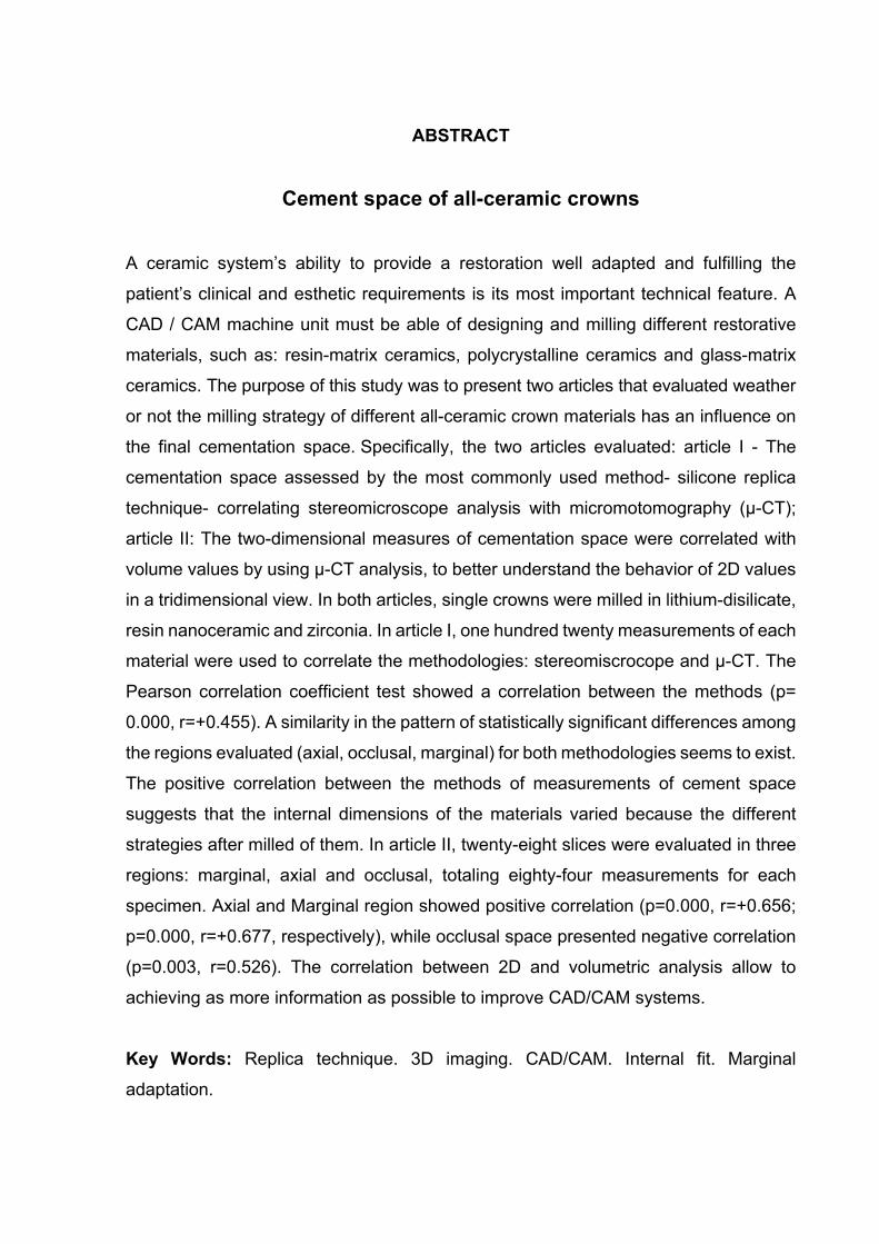
ABSTRACT
Cement space of all-ceramic crowns
A ceramic system’s ability to provide a restoration well adapted and fulfilling the
patient’s clinical and esthetic requirements is its most important technical feature. A
CAD / CAM machine unit must be able of designing and milling different restorative
materials, such as: resin-matrix ceramics, polycrystalline ceramics and glass-matrix
ceramics. The purpose of this study was to present two articles that evaluated weather
or not the milling strategy of different all-ceramic crown materials has an influence on
the final cementation space. Specifically, the two articles evaluated: article I - The
cementation space assessed by the most commonly used method- silicone replica
technique- correlating stereomicroscope analysis with micromotomography (µ-CT);
article II: The two-dimensional measures of cementation space were correlated with
volume values by using µ-CT analysis, to better understand the behavior of 2D values
in a tridimensional view. In both articles, single crowns were milled in lithium-disilicate,
resin nanoceramic and zirconia. In article I, one hundred twenty measurements of each
material were used to correlate the methodologies: stereomiscrocope and µ-CT. The
Pearson correlation coefficient test showed a correlation between the methods (p=
0.000, r=+0.455). A similarity in the pattern of statistically significant differences among
the regions evaluated (axial, occlusal, marginal) for both methodologies seems to exist.
The positive correlation between the methods of measurements of cement space
suggests that the internal dimensions of the materials varied because the different
strategies after milled of them. In article II, twenty-eight slices were evaluated in three
regions: marginal, axial and occlusal, totaling eighty-four measurements for each
specimen. Axial and Marginal region showed positive correlation (p=0.000, r=+0.656;
p=0.000, r=+0.677, respectively), while occlusal space presented negative correlation
(p=0.003, r=0.526). The correlation between 2D and volumetric analysis allow to
achieving as more information as possible to improve CAD/CAM systems.
Key Words: Replica technique. 3D imaging. CAD/CAM. Internal fit. Marginal
adaptation.

RESUMO
Espaço de cimentação de coroas totais cerâmicas
As principais características dos sistemas cerâmicos é a capacidade de abranger
estética e boa adaptação clínica. A tecnologia CAD/CAM é capaz de projetar e fresar
diferentes materiais, como: cerâmicas de matriz resinosa, policristalinas, e ainda,
cerâmicas de matriz vítrea. O objetivo deste trabalho é apresentar dois artigos que
avaliam se a estratégia de fresagem de diferentes materiais cerâmicos influenciam no
espaço final de cimentação. No primeiro artigo, especificamente, o espaço de
cimentação foi avaliado pela metodologia mais utilizada, réplica de silicona. A película
de silicona foi mensurada através da estereomicroscopia e microtomografia
computadorizada (µ-CT) e as metodologias foram correlacionadas. No segundo
artigo, através da metodologia de µ-CT, as medidas bidimensionais do espaço de
cimentação foram correlacionadas com o volume obtido, a fim de compreender melhor
o comportamento dos valores 2D numa visão tridimensional. Em ambos artigos,
coroas unitárias foram fresadas em dissilicato de lítio, resina nanocerâmica e zircônia.
No artigo I, cento e vinte medidas foram utilizadas para correlacionar as metodologias
empregadas, estereomicroscopia e µ-CT. O teste de correlação de Pearson
apresentou correlação entre os métodos (p= 0.000, r=+0.455). Uma similaridade no
padrão de diferenças estatisticamente significantes nas regiões avaliadas (axial,
oclusal, marginal) parece existir entre as metodologias. A correlação positiva entre os
métodos de mensuração do espaço de cimentação sugerem que as dimensões
internas variam por conta dos diferentes processos que as coroas são submetidas
após a fresagem. No artigo II, oitenta e oito fatias obtidas pelo µ-CT foram avaliadas
em três regiões: marginal, axial e oclusal, totalizando oitenta e quatro medidas por
espécime. A correlação positiva entre os métodos de mensuração do espaço de
cimentação sugerem que as dimensões internas variam por conta dos diferentes
processos que as coroas são submetidas após a fresagem. Artigo II, oitenta e oito
fatias foram avaliadas em três regiões: marginal, axial e oclusal, totalizando oitenta e
quatro medidas por espécime. Houve correlação positiva entre as regiões axial e
marginal (p=0.000, r=+0.656; p=0.000, r=+0.677, respectivamente) e negativa na
oclusal (p=0.003, r= -0.526). A correlação entre as

avaliações bi e tridimensionais permitem obter mais informações possíveis para
aprimorar o sistema CAD/CAM.
Palavras-chave: Adaptação. Silicona. Microtomografia por Raio-X. Cerâmicas.


TABLE OF CONTENTS
1 INTRODUCTION ............................................................................................ 19
3 DISCUSSION ................................................................................................. 61
4 CONCLUSIONS ............................................................................................. 69
REFERENCES ............................................................................................... 73


Introduction

1 Introduction
19
1 Introduction
The ceramic crowns are the first choice when the goal is to achieve natural
appearance with the best aesthetic result associated with the biocompatibility
(CONRAD; SEONG; PESUN, 2007). The success of these restorations is influenced
bythe resistance to fracture, aesthetic result and the marginal fit (GARDNER, 1982).
Biologically, the misfit can contribute to the development of caries lesions
(KARLSSON, 1986), plaque accumulation (SALTZBERG et al., 1976) and cement
dissolution COOPER et al., 1971).
It is known that predefined cement space can influence in ceramic crown fit
(CONTREPOIS et al., 2013). In CAD/CAM systems, the cement space was set through
the software interface. Reviewed studies showed the impact of different setting in
internal and marginal crown fit (NAKAMURA et al., 2003; 2005; IWAI et al., 2008;
HMAIDOUCH; NEUMANN; MUELLER, 2011). The small cementation space may
cause premature contacts between internal surface of the crown and the abutment
tooth (NAKAMURA et al., 2003), as also a greater width layer of cement in occlusal
region, thus widening the marginal gap (HMAIDOUCH; NEUMANN; MUELLER, 2011).
Thicker cement contributes to polymerization shrinkage (tensile stress in the intaglio
surface of crowns), and this factor could cause debonding of ceramic restorations
(MAY et al., 2012).
A CAD / CAM machine unit must be able of designing and milling different
restorative materials, such as resin-matrix ceramics, polycrystalline ceramics and
glass-matrix ceramics (GRACIS et al., 2015). Consequently, different designing and
milling strategies must be used to obtain the best result of each material. Biomaterials
available for CAD/CAM have their own features regarding mechanical properties,
chemical properties and machinability (LUTHARDT et al., 2004). Considering that
different material properties can result in different wear of the milling tools, it would be
relevant if suppliers could provide additional information regarding the lifespan of
1 Introduction
20
milling tools based on the amount of wasted material, since the fit of crowns may be
affected by the abovementioned factors (LEBON et al., 2016).
Lithium disilicate, for example, consists of a glass matrix ceramic that presents
fracture strength, color and translucency similar to enamel, whichundergoes through a
crystallization process, whereby the milled restoration is brought to a furnace at 840°C
for 25 min to ensure that material properties reach a flexural strength of 360 MPa (±60
MPa) and fracture toughness between 2 e 2.5 MPa*m0.5 (WIEDHAHM, 2007). After
crystallization, a shrinkage of 0.3% is expected, which implies in the production of a
slightly larger restoration soon after milling (REICH et al., 2011).
A resin of nanoceramic material (Lava Ultimate, 3M Oral Care) was introduced
in an attempt to combine the advantages of a ceramic material and the ease of milling
the composite resins. This material is intended to better absorb masticatory forces,
reducing restoration internal stresses, providing wear resistance combined with a
highly polished surface, color stability and accurate and well adapted margins
(AWADA; NATHANSON, 2015).
Another material, the zirconia (Y-TZP), a polycrystalline ceramic, presents
relevant properties for a restorative material such as high flexural strength (900 a 1200
Mpa), fracture toughness (9 MPa*m0.5), hardness (13,5 GPa) and wear resistance
(REKOW; ZHANG; THOMPSON, 2007; VAGKOPOULOU et al., 2009). The
advantages of faster production, low cost and ease of machining pre-sintered zirconia
blocks have led to this system gaining popularity. However, high temperature sintering
is required after the milling to achieve the final hardness and strength. The final
sintering is followed by a shrinkage of 25% in volume (DENRY; KELLY, 2008), which
means that a larger restoration must be milled for compensation, which may eventually
influence the fit (COLPANI; BORBA; DELLA BONA, 2013).
Although there are several methods available for quantification of crown misfit
and cement internal space, the replica technique has been validated and used in
several studies because is a non-destructive technics, such in vivo and in vitro
researches, with accuracy and reliable (LAURENT et al., 2008; REICH et al., 2011)
ability to reproduce the thickness of the cement pellicle (RAHMÉ et al., 2008). A light-
body elastomeric impression material is used to simulate the thickness of the cement

1 Introduction
21
space, which is then measured either two-dimensionally using an optical microscope,
scanning electron microscope (SEM) or three-dimensionally, by means of
microcomputed tomography analysis (µ-CT) (SHAMSEDDINE et al., 2016).
Acceptable values of marginal and internal space of prosthetic restorations are
known and have been reported to be around 100 and 150µm (HUANG et al., 2015),
but information about volume is lacking. It may be clinically more relevant to have a
three-dimensional view of the internal space to better understand the misfit of the
crowns. In this context, microcomputed tomography (µ-CT) is a nondestructive method
could be useful to quantify and qualify the cement space in the buccolingual and
mesiodistal direction of crowns and to measure the average internal space by means
of three-dimensional reconstruction (de PAULA SILVEIRA et al., 2017).
Based on the thickness of the cement layer should be as uniform and as thin as
possible (DE JAGER et al., 2004), the overall aim of this study was to investigate if the
CAD could transfer the cement spaces for each material considering their process after
milled and the same stl. (the standard file type used) parameter. Each article had
specifically evaluated:
According to the literature, the thickness of the cement layer should be as
uniform and as thin as possible (DE JAGER et al., 2004). Thus, the general objective
of this study was to investigate if the CAD could transfer the cement spaces for each
specific material, considering the process after milling and the same stl. parameter.
The present thesis will be presented in two separate articles, which had specific
objectives:
• Article I: The aim of this study was to evaluate the cement space by µ-CT
analysis, compared to the optical analysis of the silicone replica,
since the latter is the most commonly used method.
• Article II: The aim of this study was to correlate two-dimensional
measurements with volume values by µ-CT analysis.


Discussion

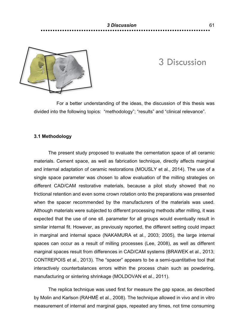
3 Discussion
61
3 Discussion
For a better understanding of the ideas, the discussion of this thesis was
divided into the following topics: “methodology”; “results” and “clinical relevance”.
3.1 Methodology
The present study proposed to evaluate the cementation space of all ceramic
materials. Cement space, as well as fabrication technique, directly affects marginal
and internal adaptation of ceramic restorations (MOUSLY et al., 2014). The use of a
single space parameter was chosen to allow evaluation of the milling strategies on
different CAD/CAM restorative materials, because a pilot study showed that no
frictional retention and even some crown rotation onto the preparations was presented
when the spacer recommended by the manufacturers of the materials was used.
Although materials were subjected to different processing methods after milling, it was
expected that the use of one stl. parameter for all groups would eventually result in
similar internal fit. However, as previously reported, the different setting could impact
in marginal and internal space (NAKAMURA et al., 2003; 2005), the large internal
spaces can occur as a result of milling processes (Lee, 2008), as well as different
marginal spaces result from differences in CAD/CAM systems (BRAWEK et al., 2013;
CONTREPOIS et al., 2013). The “spacer” appears to be a semi-quantitative tool that
interactively counterbalances errors within the process chain such as powdering,
manufacturing or sintering shrinkage (MOLDOVAN et al., 2011).
The replica technique was used first for measure the gap space, as described
by Molin and Karlson (RAHMÉ et al., 2008). The technique allowed in vivo and in vitro
measurement of internal and marginal gaps, repeated any times, not time consuming

3 Discussion
62
and relatively not expensive (LAURENT et al., 2008). When using silicone replica, the
space can be evaluated from a thin section of silicone pellicle (REICH et al., 2005). It
is possible to observe marginal and internal space in one (1D), two (2D) and three
dimensional (3D) (OKA et al., 2016), depends on the technology used, such as the
optical microscopy, scanning electron microscopy, optical comparator screen and
microcomputer tomography (µ-CT) (SHEMBESH et al., 2016).
Despite these advantages, certain difficulties can arise on measuring the
thickness of the pellicle (LAURENT et al., 2008), the mixing, dispensing, handling of
the silicone for the cement replica (DAHL; RØNOLD; DAHL, 2017). However, the
replica technique combined with optical microscopy has been shown to be a practical
and established method (LAURENT et al., 2008), although there is the difficulty in
identifying reference points (CONTREPOIS et al., 2013) and limited number of slices
to measure.
The last technique used was microcomputer tomography (µ-CT), which allows
evaluating 2D and 3D the cement space, could provide great number of measurements
sites (CONTREPOIS et al., 2013). This is noninvasive and nondestructive
methodology (KIM et al., 2016), whose major disadvantage is the formation of radiation
artifacts caused by the different radiation coefficient of the materials during the volume
evaluation (BORBA et al., 2011), also this technique is considered expensive and time-
consuming (RUNGRUANGANUNT; KELLY; ADAMS, 2010).
Yet among the different methods to determine the fit accuracy of prosthetic
restorations, no single method has proven to be superior, suggesting that the
combination of at least two measurement methods may be useful to verify and validate
the cementation space characterization (NAWAFLEH et al., 2013). Owing to this, in
article 1, the cement space was evaluated two-dimensionally by two methods,
stereomicroscope and µ-CT, aiming to correlate the measurements. Previous studies
reported that the optical microscopic replica reading showed high accuracy
measurements for under 15 µm thicknesses, whereas above 100 µm the accuracy
decreases. In contrast, the µ-CT was capable to resolve thicknesses to approximately
10 µm (RUNGRUANGANUNT; KELLY; ADAMS, 2010).
3 Discussion
63
As previous showed, the defined space was not exact, as expected. The cement
constitutes a certain thickness, which result in crown intaglio surface contact with the
prepared tooth, consequently increasing marginal space (PRUDENTE et al., 2017;
YILDRIM et al., 2017). Although, the acceptable marginal and internal values may be
known (MCLEAN; VON FRAUNHOFER, 1971), the exact volume of that space is not
(YILDRIM et al., 2017). More than accuracy value, 3D measurement allowed qualify
the cement space (YILDRIM et al., 2017). Due to this, in second article, 2D
measurements of cement space of CAD/CAM crowns was compared with their internal
volume, trying to establish a correlation between the two measurements, aiming to
achieve as more information as possible to improve CAD/CAM systems. Considering
that in the first article, the correlation between in 2D analysis by stereomicroscope and
µ-CT showed the possibility to apply the µ-CT in two-dimensionally for all-ceramic
crowns.
3.2 Results
In this study, the CAD/CAM system used a 2-step workflow. In Step-1, large
diameter burs reduce the block up to 0.3 mm larger than the final dimensions of the
restoration. In Step-2, finer grit burs refine the milling process. There is limited
information in the literature regarding the wear of burs as a result of material properties,
what certainly would help better understand the results of cement space obtained. In
the system used, material hardness did not seem to affect the milling speed and
regardless of milled material, the replacement of burs is indicated by the software
(CHAVALI et al., 2017).
The classes of restorative materials differ considerably with respect to their
mechanical, chemical and machinability properties (LUTHARDT et al., 2004).
However, our study question was whether the CAD-CAM system used would
reproduce the cement space from the .stl to the milled restorations, yet adjusting post
processing alterations during crystallization or sintering. Most ceramic materials seem
to present clinically acceptable marginal and internal adaptation values
3 Discussion
64
(CONTREPOIS et al., 2013). Gaps of up to 150 µm are commonly considered as being
clinically tolerable (BOENING et al., 2000).
The resin nanoceramic presented the lowest values of marginal and axial
discrepancy, as previously reported (de PAULA SILVEIRA, et al., 2017,) probably
because no dimensional alterations are expected after milling in an already processed
block. In this study, the negative correlation showed between volume and occlusal
space, could suggest that limited cement space may lead to incomplete crown seating
and eventually in increased marginal gap (HMAIDOUCH; NEUMANN; MUELLER,
2011). The lowest volume, but the larger occlusal space turns more difficult to seat the
crown, because less cement flows to the axial and marginal regions (HMAIDOUCH;
NEUMANN; MUELLER, 2011; MAI; LEE; LEE, 2017). The manufacturer is no longer
indicated this material as a crown material, by due to debonding issues that occurred
at higher rates than commonly expected. This finding may be due to the low modulus
of elasticity of the material and resiliency that results in deformation during occlusal
contacts. Also, the high content of polycrystalline material zirconia at the nanoscale
may hinder long-term adhesion at the intaglio surface.
Lithium disilicate presented the highest value of occlusal space, in accordance
with literature studies (NAN et al., 2017; SHAMSSEDINE et al., 2016). This can be the
result of the crystallization process (0.3% of shrinkage) or because of burs of reduced
diameter used for refinement (REICH et al., 2011; SHAMSSEDINE et al., 2016). The
poor occlusal fit of glass-ceramic restorations may result in an increase in the risk of
fractures because of the reduced support and stabilization of the ceramic through
adhesion to the tooth substrate (ZELTNER et al., 2017), the clinical performance of
bonded restorations might decrease due to the polymerization shrinkage (MAY et al.,
2012). Conversely, thinner cement space associated to loading application onto the
surface of the crown could lead to tensile fracture of the ceramic at the cementation
surface, due to the low modulus of resin cements (4-6GPa) compared with glass-
ceramic (70GPa) (SILVA et al., 2008).
On the other hand, the smallest value of occlusal space and the greater volume
was presented by Y-TZP, the negative correlation between them allows to understand
that the insufficient space for the luting agent in occlusal space, may hinder the seating
3 Discussion
65
of the crown eventually leading to higher risk of marginal leakage (QUINTAS et al.,
2004). Zirconia showed the greater marginal value between the materials studied.
Although cement space values observed are within acceptable ranges, debonding of
Y-TZP prostheses remain an issue likely associated with its polycrystalline structure
and difficulties in bonding, but also on preparation and milling parameters that should
not be overlooked.
3.3 Clinical relevance
An inadequate marginal fit can lead known biological and mechanical issues,
such as plaque retention, washout of the luting agent, caries, pulpal inflammation,
periodontal disease (CONTREPOIS et al., 2013) and loss the restoration retention. As
possible consequences of an internal misfit, no frictional retention and even some
crown rotation onto the preparations resulting in reduced fracture toughness (REICH
et al., 2013) and sometimes debonding of the all-ceramic crowns.
Although, this study concerned about the same .stl parameter and CAD/CAM
system to milled all the material used, the results showed the “spacer’ appears to be a
semi-quantitative tool that interactively counterbalances errors within the process chain
such as powdering, manufacturing or sintering shrinkage (MOLDOVAN et al., 2011).
This tool requires more studies to better comprise and, consequently, improve all
digital restorations.
The article 1 showed the correlation between optical and µ-CT. The
measurements of the scanned pellicles may be more reliable when thinner sections
are under measurement, and the stereomicroscope allow limited number of measures,
while µ-CT allow two and three-dimensional analysis. In addition to, in article 2 the
correlation between 2D and volumetric measures of cement space allows
understanding the main issues related to the investigated all-ceramic systems.

Conclusion

4 Conclusions
69
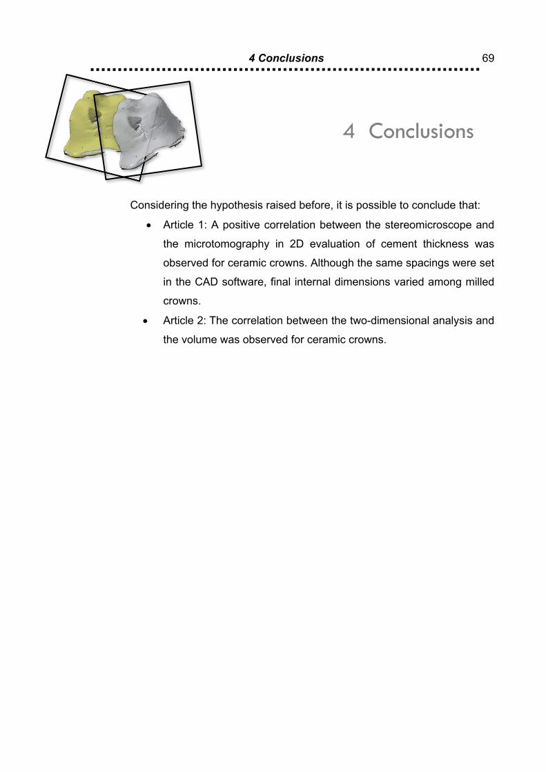
4 Conclusions
Considering the hypothesis raised before, it is possible to conclude that:
• Article 1: A positive correlation between the stereomicroscope and
the microtomography in 2D evaluation of cement thickness was
observed for ceramic crowns. Although the same spacings were set
in the CAD software, final internal dimensions varied among milled
crowns.
• Article 2: The correlation between the two-dimensional analysis and
the volume was observed for ceramic crowns.


References

References
73
REFERENCES
Awada A, Nathanson D. Mechanical properties of resin-ceramic CAD/CAM restorative materials. J Prosthet Dent. 2015 Oct;114(4):587-93
Boening KW, Wolf BH, Schmidt AE, Kastner K, Walter MH. Clinical fit of Procera AllCeram crowns. J Prosthet Dent 2000;84:419–424.
Borba M, Cesar PF, Griggs JA, Della Bona Á. Adaptation of all-ceramic fixed partial dentures. Dent Mater 2011;27:1119-26.
Brawek PK, Wolfart S, Endres L, Kirsten A, Reich S. The clinical accuracy of single crowns exclusively fabricated by digital workflow-the comparison of two systems. Clin Oral Invest 2013;17:2119-25.
Chavali R, Nejat AH, Lawson NC. Machinability of CAD-CAM materials. J Prosthet Dent. 2017;118:194-199.
Colpani JT, Borba M, Della Bona A. Evaluation of marginal and internal fit of ceramic crown copings. Dent Mater. 2013 Feb;29(2):174-80.
Conrad HJ, Seong WJ, Pesun IJ. Current ceramic materials and systems with clinical recommendations: a systematic review.J Prosthet Dent 2007;98:389-404.
Contrepois M, Soenen A, Bartala M, Laviole O. Marginal adaptation of ceramic crowns: a systematic review. J Prosthet Dent. 2013;110:447-454.
Cooper TM, Christensen GJ, Laswell HR, Baxter R. Effect of venting on cast gold full crowns. J Prosthet Dent 1971;26:621-6.
Dahl BE, Rønold HJ, Dahl JE. Internal fit of single crowns produced by CAD-CAM and lost-wax metal casting technique assessed by the triple-scan protocol. J Prosthet Dent. 2017;117:400-404.
De Jager N, Pallav P, Feilzer AJ. The apparent increase of the Young’s modulus in thin cement layers. Dent Mater 2004;20(5):457–62.

References
74
de Paula Silveira, AC, Chaves, SB, Hilgert, LA, Ribeiro AP. Marginal and internal fit of CAD-CAM fabricated composite resin and ceramic crowns scanned by 2 intraoral cameras. J Prosth Dent . 2017;117(3):386-392.
Denry I, Kelly JR. State of the art of zirconia for dental applications. Dent Mater. 2008 Mar;24(3):299-307.
Gardner FM. Margins of complete crownsdliterature review. J Prosthet Dent 1982;48:396-400.
Gracis S, Thompson VP, Ferencz JL, Silva NR, Bonfante EA. A new classification system for all-ceramic and ceramic-like restorative materials. Int J Prosthodont. 2015 May-Jun;28(3):227-35.
Hmaidouch R, Neumann P, Mueller WD. Influence of preparation form, luting space setting and cement type on the marginal and internal fit of CAD/CAM crown cop- ings. Int J Comput Dent 2011;14:219-26.
Huang Z, Zhang L, Zhu J, Zhao Y, Zhang X. Clinical marginal and internal fit of crowns fabricated using different CAD/CAM technologies. J Prosthodont 2015;24:291-5.
Iwai T, Komine F, Kobayashi K, Saito A, Matsumura H. Influence of convergence angle and cement space on adaptation of zirconium dioxide ceramic copings. Acta Odontol Scand 2008;66:214-8.
Karlsson S. A clinical evaluation of fixed bridges, 10 years following insertion. J Oral Rehabil 1986;13:423-32.
Kim JH, Jeong JH, Lee JH, Cho HW. Fit of lithium disilicate crowns fabricated from conventional and digital impressions assessed with micro-CT. J Prosthet Dent 2016;116:551-7.
Laurent M, Scheer P, Dejou J, Laborde G. Clinical evaluation of marginal fit of cast crown-validation of the silicone replica method. J Oral Rehabil 2008;35:116-22.
Lebon N, Tapie L, Duret F, Attal JP. Understanding dental CAD/CAM for restorations - dental milling machines from a mechanical engineering view- point. Part A: chairside milling machines. Int J Comput Dent 2016;19:45-62.
Luthardt R, Holzhuter M, Rudolph H, Herold V, Walter M. CAD/CAM-machining effects on Y-TZP zirconia. Dent Mater. 2004 Sep;20(7):655-62.
References
75
May LG, Kelly JR, Bottino MA, Hill T. Effects of cement thickness and bonding on the failure loads of CAD/CAM ceramic crowns: multi-physics FEA modeling and monotonic testing. Dent Mater 2012;28:e99-109.
McLean JW, von Fraunhofer JA. The estimation of cement film thickness by an in vivo technique. Br Dent J 1971;131:107–111.
Moldovan O, Luthardt RG, Corcodel N, Rudolph H. Three-dimensional fit of CAD/CAM-made zirconia copings. Dent Mater 2011;27:1273–1278.
Mously HA, Finkelman M, Zandparsa R, Hirayama H. Marginal and internal adapta- tion of ceramic crown restorations fabricated with CAD/CAM technology and the heat- press technique. J Prosthet Dent 2014;112:249-256.
Nakamura T, Dei N, Kojima T, Wakabayashi K. Marginal and internal fit of Cerec 3 CAD/CAM all-ceramic crowns. Int J Prosthodont 2003;16:244-8.
Nakamura T, Tanaka H, Kinuta S, Akao T, Okamoto K, Wakabayashi K, et al. In vitro study on marginal and internal fit of CAD/ CAM all-ceramic crowns. Dent Mater J 2005;24:456-9.
Nam SJ, Yoon MJ, Kim WH, Ryu GJ, Bang MK, Huh JB. Marginal and Internal Fit of Conventional Metal-Ceramic and Lithium Disilicate CAD/CAM Crowns. Int J Prosthodont 2015;28:519-21.
Nawafleh NA, Mack F, Evans J, Mackay J, Hatamleh MM. Accuracy and reliability of methods to measure marginal adaptation of crowns and FDPs: a literature review. J Prosthodont 2013;22:419-428.
Oka Y, Sasaki J, Wakabayashi K, Nakano Y, Okamura SY, Nakamura T, Imazato S3, Yatani H. Fabrication of a radiopaque fit-testing material to evaluate the three-dimensional accuracy of dental prostheses. Dent Mater 2016;32:921-928.
Prudente MS, Davi LR, Nabbout KO, Prado CJ, Pereira LM, Zancopé K, Neves FD. Influence of scanner, powder application, and adjustments on CAD-CAM crown misfit. J Prosthet Dent 2018;119:377-383.
Quintas AF, Oliveira F, Bottino MA. Vertical marginal discrepancy of ceramic copings with different ceramic materials, finish lines, and luting agents: an in vitro evaluation. J Prosthet Dent 2004;92:250–257.

References
76
Rahmé HY, Tehini GE, Adib SM, Ardo AS, Rifai KT. In vitro evaluation of the “replica technique” in the measurement of the fit of Procera crowns. J Contemp Dent Pract 2008;9:25-32.
Reich S, Uhlen S, Gozdowski S, Lohbauer U. Measurement of cement thickness under lithium disilicate crowns using an impression material technique. Clin Oral Investig. 2011Aug;15(4):521-6.
Reich S, Vollborn T, Mehl A, Zimmermann M. Intraoral optical impression system: an overview. Int J Comput Dent 2013;16: 143–162.
Reich S, Wichmann M, Nkenke E, Proeschel P. Clinical fit of all-ceramic three-unit fixed partial dentures, generated with three different CAD/CAM systems. Eur J Oral Sci 2005;113(2):174–9.
Rekow D, Zhang Y, Thompson V. Can material properties predict survival of all-ceramic posterior crowns? Compend Contin Educ Dent. 2007 Jul;28(7):362-8.
Rungruanganunt P, Kelly JR, Adams DJ. Two imaging techniques for 3D quantification of pre-cementation space for CAD/CAM crowns. J Dent. 2010;38:995-1000.
Saltzberg DS, Ceravolo FJ, Holstein F, Groom G, Gottsegen R. Scanning electron microscope study of the junction between restorations and gingival cavosurface margins. J Prosthet Dent 1976;36:517-22.
Shamseddine L, Mortada R, Rifai K, Chidiac JJ. Marginal and internal fit of pressed ceramic crowns made from conventional and computer-aided design and computer-aided manufacturing wax patterns: An in vitro comparison. J Prosthet Dent. 2016;116:242-8.
Shembesh M, Ali A, Finkelman M, Weber HP, Zandparsa R. An in vitro comparison of the marginal adaptation accuracy of CAD-CAM restorations using different impression systems. J Prosthodont. 2017;26:581-586.
Silva NR, de Souza GM, Coelho PG, Stappert CF, Clark EA, Rekow ED et al. Effect of water storage time and composite cement thickness on fatigue of a glass-ceramic trilayer system. J Biomed Mater Res B Appl Biomater 2008;84:117-23.
Vagkopoulou T, Koutayas SO, Koidis P, Strub JR. Zirconia in dentistry: Part 1. Discovering the nature of an upcoming bioceramic. Eur J Esthet Dent. 2009 Summer;4(2):130-51.
References
77
Wiedhahn K. From blue to white: new high-strength material for Cerec—IPS e.max CAD LT. Int J Comput Dent 2007;10:79–91.
Yildrim G, Uzun IH, Keles A. Evaluation of marginal and internal adaptation of hybrid and nanoceramic systems with microcomputed tomography: An in vitro study. J Prosthet Dent. 2017;118:200-207.
Zeltner M, Sailer I, Mühlemann S, Özcan M, Hämmerle CH, Benic GI. Randomized controlled within-subject evaluation of digital and conventional workflows for the fabrication of lithium disilicate single crowns. Part III: marginal and internal fit. J Prosthet Dent 2017;117:354-362.