universiti putra malaysia molecular …psasir.upm.edu.my/8717/1/fsmb_1997_2_a.pdfuniversiti putra...
TRANSCRIPT
UNIVERSITI PUTRA MALAYSIA
MOLECULAR CHARACTERIZATION OF Salmonella enteritidis ISOLATES BY PULSED FIELD GEL ELECTROPHORESIS
AND PLASMID PROFILING
LOKE CHUI FUNG
FSMB 1997 2
MOLECULAR CHARACTERIZATION OF Salmonella enteritidis ISOLATES BY PULSED FIELD GEL ELECTROPHORESIS
AND PLASMID PROFILING
By
LOKE CHUI FUNG
Thesis Submitted in Fulfilment of the Requirements for the
Degree of Master of Science in the Faculty of
Food Science and Biotechnology, University Putra Malaysia.
April 1997

Specially dedicated to
my beloved family
and friends ....... .
ACKNOWLEDGEMENTS
The author wishes to thank her family members for all their love,
encouragement, guidance and for always being there for her.
The author extends her deepest gratitude to the chairman of the
supervisory committee, Dr. Raha Abdul Rahim, for her encouraging advice,
suggestions, guidance, never-ending patience, her kindness and her willingness
to help. The author also wishes to thank the members of the supervisory
committee, Assoc. Prof. Dr. Gulam Rusul, Assoc. Prof. Dr. Kbatijah
YusotJ and Dr. Son Radu for their advice, supervision, suggestions and
guidance.
A special note of thanks is also extended to her best friend, Min Min,
for all her help, patience, generosity and appreciated company. Special thanks
are also conveyed to all the members of the Microbiology Laboratory:
Endang, Sahilah, Kak Mai, Zainuri, Dayang and Dr. Jorgen Leisner for
all their kind help, efforts and time spent on her that is very much appreciated.
Lastly, the author wishes to express her appreciation to her friends for
their help and supports during the run of the project.
iii
TABLE OF CONTENTS
Page
ACKNOWLEDGEMENTS LIST OF TABLES
1ll
Vll
ix x
Xl
xiii
LIST OF FIGURES LIST OF PLATES ABSTRACT ABSTRAK
CHAPTER
I
IT
ill
GENERAL INTRODUCTION Objectives
LITERATURE REVIEW
1 3
4 Introduction 4 General Description of Salmonella 5 Classification of Salmonella 6 Salmonellosis in Animals and Humans 1 1
Human Salmonellosis 12 Avian Salmonellosis 16
Pathogenesis and Toxicology of Salmonella 1 8 Salmonella Typing System 1 9
Serotyping 20 Biotyping 2 1 Phage Typing 22 Chemotyping 23
Antibiotic Susceptibility of Salmonella 24 Application of Molecular Techniques as Epidemiological lli� n
Plasmid Content Analysis of Salmonella ententuns 28 Restriction Fragment Length Polymorphism in Pulsed Field Gel Electrophoresis 3 1
ANTIMICROBIAL SUSCEPTIBILITY AND PLASMID PROFILING OF SAIMONELLA ENTERI11DIS ISOLATES Introduction Materials and Methods
Bacterial Strains, Media and Propagation Antibiotic Susceptibility Testing
IV
35 35 37 37 38
N
v
VI
Small Scale Isolation of Plasmid DNA 38 Results 41
Resistance to Antimicrobial Agents 41 Comparison of Antimicrobial Resistance Patterns between Salmonella ententzdrs Isolates from Human and Poultry Sources 45 Plasmid Analysis of Salmonella ententzdrs Isolates 48
Discussion 53
OPTIMIZATION OF THE RAMPED PULSE TIME AND THE TOTAL RUN TIME FOR PULSED FIELD GEL ELECTROPHORESIS ANALYSIS Introduction Materials and Methods
Results
Preparation of Chromosomal DNA for Pulsed Field Gel Electrophoresis (pFGE) Analysis Restriction Endonucleases Digestion and Pulsed Field Gel Electrophoresis Analysis Size Markers for Pulsed Field Gels Restriction Endonucleases
Chromosomal DNA Patterns of Salmonella enterztzdrs Isolates
Discussion
ANALYSIS OF INTACT CHROMOSOMAL DNA OF SAIMONELLA EN1ERll1DIS BY PULSED FIELD GEL ELECTROPHORESIS Introduction Materials and Methods
Pulsed Field Gel Electrophoresis Analysis Data Analysis of Pulsed Field Gel Electrophoresis
Results Restriction Endonucleases Digestion of Intact
62 62 63
63
64 66 66 67
67 75
82 82 83 83 84 86
Chromosomal DNA 86 Comparison of the Restriction Pulsed Field Gel Electrophoresis Patterns between Human and Poultry Isolates 94
Discussion 100
GENERAL DISCUSSION 110
v

VII CONCLUSIONS . . . '" ., .. , ... , .. , '" ... '" ....... ,. ... . ... 114
REFERENCES . . . . . . . . . . . . . . . . . . . . . . . . . . . . . . . . . . . . . . . . . . . . . . . . . . . . . . . 115
VITAE
vi
LIST OF TABLES
Table Page
1 Antigenic Schema for Salmonella 8
2 Food Poisoning in MalaysIa, 1994 &1995 15
3 Antigenic Relationships among Members of Enterobactenaceae 22
4 Zone Diameter Interpretive Standards 39
5 Comparison of Antimicrobial Resistance of Salmonella ententuils Isolates from Human and Poultry Sources 43
6 Susceptibility Testing of Resistance to Multiple Antimicrobial Agents among Salmonella ententuils Isolates from Human and Poultry 44
7 Antimicrobial Resistance Patterns in Salmonella ententuils 46
8 Comparison of Antibiotics Resistance Profile Groups of Salmonella ententuils Isolates from Human and Poultry 47
9 Plasmid Profiles Classes of Salmonella ententldzs Isolates from Poultry and Human Sources 49
10 Number of Plasmid DNA Bands Harbored by Salmonella ententldziIsolates from Poultry and Human 52
11 Plasmid Patterns in Salmonella ententldls 52
12 Plasmid Profiles and Antimicrobial Resistance Patterns of Salmonella ententldzs Isolates from Human and Poultry 58
13 Electrophoretic Conditions for Each Restriction Endonucleases used in Pulsed Field Gel Electrophoresis 84
14 Criteria for Interpreting Pulsed Field Gel Electrophoresis Patterns 85
15 FValues of Each PFGE Patterns for Salmonella ententldzs Isolates 88
V1l
Table Page
16 Comparison ofPFGE Patterns in Salmonella enteritidis
Isolates . . . . . . . . . . . . . . . . . . . . . . . . . . . . . . . . . . . . . . . . . . . . . . . . . . . . . . . . . 98
17 Characteristics of Salmonella enteritidis Isolates from Poultry and Human Sources . ....... . . ..... . . . . ... . . . ..... . . . . 107
viii
LIST OF FIGURES
Figure Page
1 A Schematic Representative of Plasmid Patterns Detected in the Isolates of Salmonella ententuiIs 50
2 A Schematic Representative of XbaI-Pulsed Field Gel Electrophoresis Fingerprints 87
3 A Schematic Representative of SpeI-Pulsed Field Gel Electrophoresis Fingerprints 93
4 A Schematic Representative of Pvull-, M/uI-, Sall-, EcoRI-
and HmdllI-Pulsed Field Gel Electrophoresis Fingerprints 95
5 Possible Genetic Events in the Bacterial Genome 103
IX
LIST OF PLATES
Plates Page
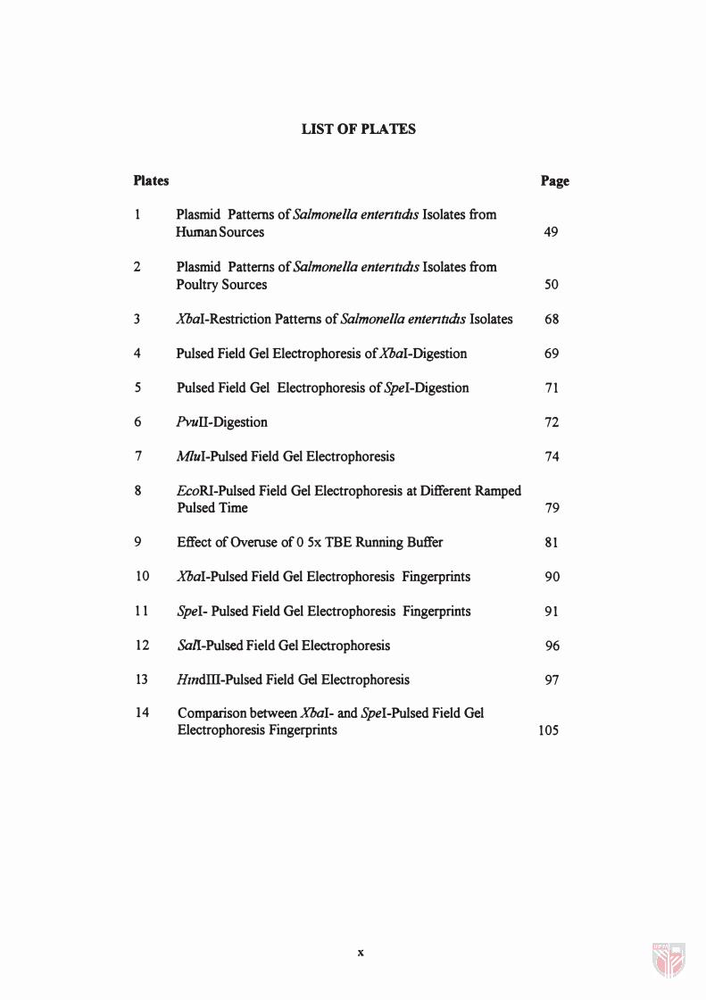
1 Plasmid Patterns of Salmonella ententldzs Isolates from Human Sources 49
2 Plasmid Patterns of Salmonella ententldzs Isolates from Poultry Sources 50
3 XbaI-Restriction Patterns of Salmonella ententldzs Isolates 68
4 Pulsed Field Gel Electrophoresis of XbaI-Digestion 69
5 Pulsed Field Gel Electrophoresis of SpeI-Digestion 71
6 PvuTI-Digestion 72
7 MluI-Pulsed Field Gel Electrophoresis 74
8 EcoRI-Pulsed Field Gel Electrophoresis at Different Ramped Pulsed Time 79
9 Effect of Overuse of 0 5x TBE Running Buffer 81
10 XbaI-Pulsed Field Gel Electrophoresis Fingerprints 90
11 SpeI- Pulsed Field Gel Electrophoresis Fingerprints 91
12 Sall-Pulsed Field Gel Electrophoresis 96
13 HmdID-Pulsed Field Gel Electrophoresis 97
14 Comparison betweenXbaI- and SpeI-Pulsed Field Gel Electrophoresis Fingerprints 105
x
Abstract of thesis submitted to the Senate of Universiti Pertanian Malaysia in fulfilment of the requirements for the degree of Master of Science.
MOLECULAR CHARACTERIZATION OF SALMONELLA
ENTERITIDIS ISOLATES BY PULSED FIELD GEL ELECTROPHORESIS AND PLASMID PROFILING
By
LOKE CHUIFUNG
APRIL 1997
Chairman: Dr. Raha Abdul Rahim
Faculty: Food Science and Biotechnology
Seventy Salmonella enteritidis isolates from poultry and 48 isolates
from human sources were analyzed for their restriction polymorphism patterns
generated by pulsed field gel electrophoresis (pFGE), plasmid profiles and
antimicrobial susceptibility patterns. In the present study, PFGE analysis
following digestion with two low-frequency-cleavage restriction
endonucleases, XbaI (5'-TCTAGA-3') and SpeI (5'-ACTAGT-3') generated
nine and five distinct fingerprints respectively with F values ranging from 0.06
to 0.97. Digestion with high-frequency-cleavage restriction endonucleases,
M1uI (5'-ACGCGT-3') and Pvun (5'-CAGCTG-3') revealed less
polymorphism in their PFGE patterns with more than 95% of the S. enteritidis
isolates belonging to a single fingerprint. PFGE restriction analysis with Sail
(5'-GTCGAC-3'), EcoRI (5'-GAATTC-3') and HindIII (5'-AAGCTT-3')
xi
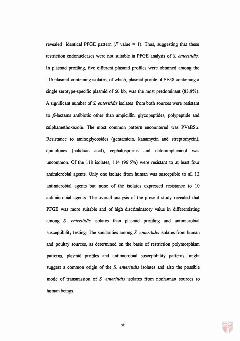
revealed identical PFGE pattern (F value = 1). Thus, suggesting that these
restriction endonucleases were not suitable in PFGE analysis of S. enteritidis.
In plasmid profiling, five different plasmid profiles were obtained among the
1 16 plasmid-containing isolates, of which, plasmid profile of SE38 containing a
single serotype-specific plasmid of 60 kb, was the most predominant (83.8%).
A significant number of S. enteritidis isolates from both sources were resistant
to p-Iactams antibiotic other than ampicillin, glycopeptides, polypeptide and
sulphamethoxazole. The most common pattern encountered was PVaBSu.
Resistance to aminoglycosides (gentamicin, kanamycin and streptomycin),
quinoiones (nalidixic acid), cephalosporins and chloramphenicol was
uncommon. Of the 1 18 isolates, 1 14 (96.5%) were resistant to at least four
antimicrobial agents. Only one isolate from human was susceptible to all 12
antimicrobial agents but none of the isolates expressed resistance to 10
antimicrobial agents. The overall analysis of the present study revealed that
PFGE was more suitable and of high discriminatory value in differentiating
among S. enteritidis isolates than plasmid profiling and antimicrobial
susceptibility testing. The similarities among S. enteritidis isolates from human
and poultry sources, as determined on the basis of restriction polymorphism
patterns, plasmid profiles and antimicrobial susceptibility patterns, might
suggest a common origin of the S. enteritidis isolates and also the possible
mode of transmission of S. enteritidis isolates from nonhuman sources to
human beings.
xii
Abstrak tesis yang dikemukakan kepada Senat, Universiti Pertanian Malaysia untuk memenuhi keperluan bagi mendapat Ijazah Master Sains.
PENCIRIAN MOLEKUL BAGI ISOLAT-ISOLAT SALMONELLA
ENTERITIDIS DENGAN ELEKTROFORESIS GEL ' PULSED FIELD' DAN PEMPROFAnAN PLASNUD
Oleh
LOKE CHUI FUNG
APRil., 1997
Pengerusi : Dr. Raha Abdul Rahim
Fakulti: Sains Makanan dan Bioteknologi
Tujuh puluh Salmonella enteritidis isolat daripada ayam dan 48
daripada sumber manusia telah dianalisis untuk corak polimorfisma penghad
yang dihasilkan oleh elektroforesis gel "pulsed field" (PFGE), profail plasmid
dan corak kepekaan antimikrob. Dalam kajian ini, analisis PFG� yang diikuti
dengan penghadaman dengan dua enzim penghad pemotongan-frekuensi-
rendah, XbaI (5'-TCTAGA-3') dan SpeI (5'-ACTAGT-3') masing-masing
menghasilkan sembilan dan lima «fingerprint" dengan nilai F dari 0.06 hingga
0.97. Penghadaman dengan enzim penghad pemotongan-frekuensi-tinggi, MluI
(5'-ACGCGT-3') dan PvuII (5'-CAGCTG-3') menunjukkan kurang
polimorfisma dalam corak PFGEnya dengan lebih daripada 95% isolat
S. enteritidis dari satu '1ingerprint" tunggal. Analisis penghad PFGE dengan
SaII (5'-GTCGAC-3'), EcoRI (5'-GAATTC-3') dan HindIIJ. (5'-AAGCTT-3')
xiii
menunjukkan corak PFGE yang sarna (nilai F=l). Maka, dapat dicadangkan
bahawa enzim penghad ini tidak sesuai dalarn analisis PFGE untuk S.enteritidis.
Dalarn pemprofailan plasmid, lima profail plasmid berlainan telah diperolehi dari
116 isolat yang mengandungi plasmid, yang mana, profail plasmid SE38 yang
mengandungi satu plasmid spesifik-serotaip tunggal 60 kb adalah paling
pradominan (83.8%). Satu bilangan isolat S. enteritidis yang signifikan dari
kedua-dua sumber adalah resistan terhadap antibiotik �-laktarn melainkan
ampisilin, glikopeptida, polipeptida dan sulfametoksazol. Corak yang paling
kerap ditemui ialah PVaBSu. Ketahanan terhadap aminoglikosida (gentamisin,
kanamisin dan streptomisin), kuinolon (asid nalidisik), sefalosporin dan
kloramfenikol adalah jarang. Dari 118 isolat, 114 (96.5%) adalah resistan
terhadap sekurang-kurangnya empat agen antimikrob. Hanya satu isolat dari
manusia peka terhadap kesemua 12 agen antimikrob tetapi tiada isolat
menunjukkan ketahanan terhadap 10 agen antimikrob. Analisis keseluruhan
kajian ini menunjukkan bahawa PFGE adalah lebih sesuai dan mempunyai nilai
pemisahan yang lebih tinggi dalarn membezakan antara isolat S. enteritidis jika
dibandingkan dengan pemprofailan plasmid dan ujian kepekaan antibiotik,
Persamaan antara isolat S. enteritidis dari sumber manusia dan ayam,
sepertimana yang ditentukan berdasarkan corak polimorfisma penghad, profail
plasmid dan corak kepekaan antimikrob, mungkin mencadangkan satu punca
yang sarna bagi S. enteritidis dan juga kemungkinan transmisi isolat
S. enteritidis dari sumber bukan manusia kepada manusia.
xiv
CHAPTER I
GENERAL INTRODUCTION
Salmonellosis is often considered to be the most frequently occurring
infectious disease caused by the members of the bacterial genus Salmonella,
which belong to the family Enterobacteriaceae. The emergence of
salmonellosis as a world problem has been dealt with elsewhere since 1950's
(Oye, 1964). Till now, an estimated annual incidence of 1.3 billion cases and 3
million deaths are reported (Thong et al. , 1995). The salmonellae are widely
distributed in nature, with man and animals being their primary reservoirs.
Because of the ubiquitous distribution of salmonellae in the environment and
the broad host adaptability of over 2100 serotypes of this genus (Calnek et. al.,
1991), the control of salmonellosis has become an extremely complex problem.
The complexity of the problem and the certainty that there is strain diversity
within each serotype has necessitated the development of special methods of
strain identification for epidemiological purposes.
Salmonella enterica serovar Enteritidis or S. enteritidis is one of the
subspecies which causes gastroenteritis, a nontyphoidal salmonellosis, with a
worldwide distribution in both humans and animals. An increasing number of
outbreaks of gastroenteritis has been reported since 1960. It has been
suggested that many sporadic cases of salmonellosis may actually be part of the
1
2
unrecognized outbreaks which escape from detection because of the lack of
efficient epidemiological markers. In both community and nosocomial
outbreaks, bacterial epidemic strains have often been defined by using methods
such as serotyping, biotyping and antimicrobial resistance patterns. However,
these phenotypic determinations have not always been successful in
differentiating S. enteritidis strains.
Recently, DNA fingerprinting based on the detection by restriction
fragment length polymorphism (RFLP) of chromosomal DNA has been used
increasingly to improve the identification of foodborne pathogens and also to
differentiate strains below the level of serotyping. These molecular techniques
include plasmid profiling, ribotyping, peR-based amplification and the RFLP
by using pulsed field gel electrophoresis (pFGE). The recent development of
PFGE has provided another approach for obtaining molecular fingerprint which
may be useful in epidemiological studies. PFGE has successfully been applied
to perform comparative chromosomal DNA analysis of several pathogens for
epidemiological investigation and is believed to possess a discriminating
capacity greater than those of ribotyping and other probe-based RFLPs
methods (Kuhn et aI., 1995; Liebisch and Schwarz, 1996; Thong et aI., 1996).
The DNA digested patterns produced by restriction endonucleases, such as
SpeJ, AvrIl and XbaI, in PFGE, have revealed clear differences between the
bacterial strains (Thong et al., 1995). It is suggested that the DNA digested
pattern produced by restriction endonucleases may provide a sensitive means of
3
SpeI, AvrIl and .xDaI, in PFGE� have revealed clear differences between the
bacterial strains (Thong et aI. , 1995). It is suggested that the DNA digested
pattern produced by restriction endonucleases may provide a sensitive means of
differentiating individual strains of Salmonella. Here� we evaluate three
epidemiological methods of subtyping S. enteritidis isolates from both poultry
and human sources by using plasmid profiles, restriction analysis of
chromosomal DNA by PFGE, and antimicrobial susceptibility patterns.
Objectives
The objectives of the present study are to compare the antibiotic
susceptibility patterns and the plasmid profiles among S. enteritidis isolates, to
compare the polymorphisms of chromosomal DNA by PFGE, to distinguish
S. enteritidis isolates exhibiting the same plasmid profile by PFGE fingerprint,
to establish the possible link between the markers used in the study, and to
clarify the possible mode of transmission of pathogenic isolates of S. enteritidis
from nonhuman sources to human beings.
CBAPTERll
LITERATURE REVIEW
Introduction
Salmonellosis is a foodbome disease caused by the members of the
genus Salmonella. The latter was named after an American bacteriologist and
veterinarian, Daniel E. Salmon, in 1900. This genus of organism consists of
only one species, S. enterica (Ewing, 1986) and is composed of more than
2000 serotypes, which also include the group previously classified as Arizona
hinshawii (Siebeling et a/., 1984). All strains of Salmonella may be presumed
to be pathogenic to human and often animals. However, a few serotypes of
Salmonella appear to be host-specific, these include S. typhi, which causes
typhoid fever in human, S. pullorum and S. gallinarum, which cause pul10rurn
disease and fowl typhoid respectively in poultry. Most serotypes of Salmonella
can cause gastrointestinal disease when ingested by human. In the past decades,
there has been an increased incidence of gastrointestinal infections caused by
S.enteritidis (Kirby, 1985; Goldberg and Rubun, 1988; Rodrique et. al. , 1 990;
Phillips and George, 1994) and now S. enteritidis has become the most
predominant serotype in many countries including Malaysia.
4
General Description of StdmoneUa
S. typhi was the first member of the Salmonella to be recognized as a
pathogen. It was first seen in 1880 by Eberth and isolated by Gaftky in 1884
(Burrows, 1959). Later, other Salmonella species associated with the onset of
diseases were isolated. Salmon and Smith isolated S. choleraesuis in 1885.
S.enteritidis was isolated by Gaertner in 1888 and in 1892, Loeffler isolated
S.typhimurium (D'Aoust, 1989).
The genus Salmonella is composed of motile bacteria that conform to
the definition of the family Enterobacteriaceae and the tnbe Salmonellae. The
family Enterobacteriaceae, named by Rahn in 1937, and now are described as
intestinal. bacteria, possess the following characteristics (Buchanan and
Gibbons, 1974).
"Small Gram-negative rods; motile· by peritrichous flagella or nonmotile. Capsulated or non-capsulated. Not spore-fonning; not acid-fast. Aerobic and facultatively anaerobic. Grow readily on meat extract media but some members have special growth requirements. Chemoorganotropic; metabolism respiratory and fermentative. Acid is produced from the fermentation of glucose, other carbohydrates and alcohol; usually aerogenic but anaerogemc groups and mutants may occur. Catalase positive with the exception of one serotype of Shigella; oxidase negative. Nitrates are reduced to nitrites except by some strains of Erwinia. G+C content of DNA: 39-59 moles %."
According to the eighth edition of Bergey's Manual of Detenninative
Bacteriology (Holt et aI., 1984), the genus Salmonella is defined as follows:
6
''Rods, usually motile by peritrichous flagella.; non-motile mutants may occur and one type (S. gallinarum and S. pullorum) is always nonmotile. Colonies are generally 2-4 mm in diameter but certain types (S.abortus-equi, S. typhi-suis and S. abortus-suis) produce colonies of about 1 tnm. Most strains will grow on defined media without special growth factors and they can use citrate as carbon source. Most strains are aerogenic but S. typhi, an important exception, never produces gas."
Salmonella spp. generally grow well at 35-37°C and are susceptible to
temperature of lower than 5°C or higher than 49°C (Bryan et al. , 1979).
However, Salmonella can still promote survival even after exposure to a
freezing temperature. The growth of Salmonella is generally inhIbited in the
presence of 3-4% sodium chloride. Study by Alford and Palumbo (1969) have
revealed that most Salmonella were inhibited in the presence of 2-8% sodium
chloride where their total cell yield decreased and the lag phase of their growth
curve increased. Generally, Salmonella can grow well at pH between 6.5 and
7.5, however, growth of Salmonella at pH value as low as 4 was reported
(Chung and Goepfert, 1970).
Oassification of Salmonella
The bacterial nomenclature of the genus Salmonella has been in the
state of change for decades and becomes exceptionally confusing and
controversial in many countries because the naming of Salmonella has not been
co-ordinated to the international agreement. Many serotypes of Salmonella
7
which were found to be somewhat host specific or adapted, were named either
for the disease caused or for the animal involved, as if they represented a
distinct species within the genus Salmonella. Specific epithets such as S. typhi,
S. paratyphi, S. paratyphi A, S. paratyphi B and S. paratyphi C, were named
according to a human disease. Whilst, S. choleraesuis, S. pullorum and
S.abortus ovis were associated with the affected animal species. In addition,
there are some serotypes named after a geographical location, such as
S.newport, S. panama, S. florida and S. indiana.
According to the Kauffmann-White Scheme (1986), the genus
Salmonella is subdivided into five serologically defined subgenera I - V, where
the identification of Salmonella is based on the detection of the specific
antigenic components present. It was known that there were two distinct types
of antigens present on the Salmonella cell surface. The somatic antigen (0 =
ohne Hauch) is a heat stable, polysaccharide associated with the body of the
cell. It is the antigen first detennined in Salmonella serology using the slide
agglutination technique to group the organism. In the Kauffinan-White scheme,
the Salmonella were grouped into different serogroups, namely serogroup A,
B, Ct, Cz, C3, D1, E1, Ez, E3, 14 F,G1, Gz, H, 1, K, L, N. The assignment of
Salmonella retied on the antigen specificities and also the occurrence of certain
specificities. For example, serogroup A contains 0 antigen 2, C1 contains 0
antigen 7, Cz contains 0 antigen 8 (Table 1). The somatic antigens of
Salmonella are comprised of lipid-polysaccharide-polypeptide complexes
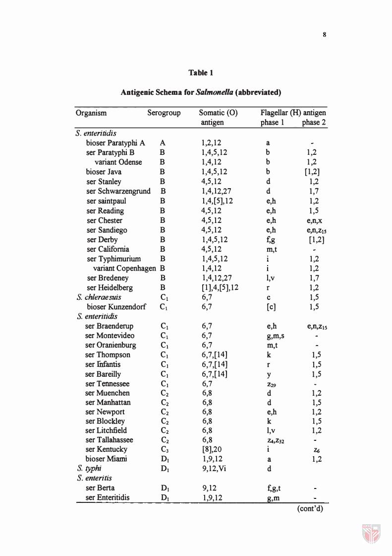
8
Table 1
Antigenic Schema for Salmonella (abbreviated)
Organism Serogroup Somatic (0) Flagellar (H) antigen antigen phase 1 phase 2
S. enteritidis bioser Paratyphi A A 1,2,12 a ser Paratyphi B B 1,4,5,12 b 1,2
variant Odense B 1,4,12 b 1,2 bioser Java B 1,4,5,12 b [1,2] ser Stanley B 4,5,12 d 1,2 ser Schwarzengrund B 1,4,12,27 d 1,7 ser saintpaul B 1,4,[5],12 e,h 1,2 serReading B 4,5,12 e,h 1,5 ser Chester B 4,5,12 e,h e,n,x
ser Sandiego B 4,5,12 e,h e,n,ZlS serOerby B 1,4,5,12 fog [1,2] ser California B 4,5,12 m,t ser Typhimuriurn B 1,4,5,12 1,2
variant Copenhagen B 1,4,12 1,2 ser Bredeney B 1,4,12,27 l,v 1,7 ser Heidelberg B [1],4,[5],12 r 1,2
S. chleraesuis C1 6,7 c 1,5 bioser Kunzendorf C1 6,7 [c] 1,5
S. enteritidis ser Braenderup C1 6,7 e,h e,n,Z15 ser Montevideo C1 6,7 g,m,s ser Oranienburg C1 6,7 m,t ser Thompson C1 6,7,[14] k 1,5 ser fnfantis C1 6,7,[14] r 1,5 ser Bareilly C1 6,7,[14] Y 1,5 ser Tennessee C1 6,7 Z29
ser Muenchen C2 6,8 d 1,2 ser Manhattan C2 6,8 d 1,5 serNewport C2 6,8 e,h 1,2 ser Blockley C2 6,8 k 1,5 ser Litchfield C2 6,8 l,v 1,2 ser T allabassee C2 6,8 Z4,Z32 ser Kentucky C3 [8],20 Z6 bioser Miami 01 1,9,12 a 1,2
S. typhi 01 9,12,Vi d S. enteritis
ser Berta 01 9,12 f,g,t ser Enteritidis 01 1,9,12 g,m
(cont'd)
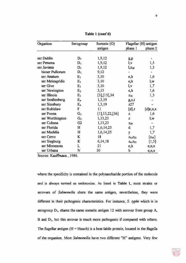
9
Table 1 (cont'd)
Organism Serogroup Somatic (0) Flagellar (H) antigen antigen phase 1 phase 2
ser Dublin Dl 1�9�12 �p ser Penama DJ 1�9)2 I�v 1,5 ser Javiana Dl 1�9,12 1,z28 1,5
bioser Pullorum DJ 9,12 ser Anatum El 3�10 e,h 1,6 ser Meleagridis El 3,10 e,h �w ser Give El 3�10 I�v 1,7 ser Newington E2 3,15 e,h 1,6 ser Illinois E3 [31�[15],34 ZIO 1,5 ser Senftenberg E4 1,3,19 g,s,t ser Simsbury E4 1�3,19 z27 ser Rubislaw F 11 [d],r [d]e,n,x serPoona G1 [1 ],13,22,[36] Z 1,6 ser Worthington G2 1,13,23 Z I,w ser Cubana G2 1,13,23 Z29 ser Florida H 1,6,14,25 d 1,7 ser Madelia H 1,6,14,25 Y 1,7 ser Cerro K 18 Z4,Z23 [Z4S] ser Siegburg K 6�14,18 z.s,z23 [1,5] ser Minnesota L 21 e,h e,n,x ser Urbana N 30 b e,n,x
Source: Kauffmann , 1986.
where the specificity is contained in the polysaccharide portion of the molecule
and is always tenned as endotoxlns. As listed in Table 1, most strains or
serovars of Salmonella share the same antigen, nevertheless, they were
different in their pathogenic characteristics. For instance, S. typhi which is in
serogroup Dt, shares the same somatic antigen 12 with serovar from group A,
B and DI, but this serovar is much more pathogenic if compared with others.
The flagellar antigen (H = Hauch) is a heat-labile protein, located in the flagella
of the organism. Most Salmonella have two different "H" antigens. Very few
]0
such as S. enteritidis Moser Gallinarum and Pullorum, have none as they are
non-motile. Others possess three or even four "If' antigens. Occasionally, a
third type antigen called the Vi antigen, is present and always associated with
the virulent strains. Vi antigen, a capsular antigen, is a heat-labile, envelope
antigen, surrounding the ceU wall which masks the somatic antigen rendering
the organism resistant to the action of 0 sera. Normally, agglutination of
Salmonella cells occurs when suspended in sera containing antibodies against
either 0, H or Vi antigen. Actually, this property was found to be useful in
identifying and characterizing Salmonella serotypes, and now more than 2000
serologically distinct types of Salmonella have been descnbed.
As a matter of expediency in taxonomy, Edwards and Ewing (1972)
recommended the use of a three species concept in Salmonella nomenclature
which recognized the species S. typhi, S. choleraesuis and S. enteritidis, where
the latter includes all strains except for the two species mentioned formerly. As
an example, S. typhimurium as formerly known, was named as S. enteritidis ser
Typhimurium, while S. pullorum, which is an aberrant strain, was named as
S.enteritidis bioser Pullorum. However, this classification scheme has not been
adopted to the Bergey's Manual of Determinative Bacteriology.
According to Le Minor et al. (1986), the genus Salmonella, based on
the DNA relatedness, consists of a single species, S. choleraesuis, and possibly
as many as seven subspecies, namely S. choleraesuis subsp. Choleraesuis,