universiti putra malaysia specular reflection …psasir.upm.edu.my/id/eprint/65263/1/fsktm 2015 48...
TRANSCRIPT

UNIVERSITI PUTRA MALAYSIA
SPECULAR REFLECTION REMOVAL AND BLOODLESS VESSEL SEGMENTATION FOR 3-D HEART MODEL RECONSTRUCTION
FROM SINGLE VIEW IMAGES
AQEEL ABDULLAH AHMED AL-SURMI
FSKTM 2015 48

SPECULAR REFLECTION REMOVAL AND BLOODLESS VESSEL
SEGMENTATION FOR 3-D HEART MODEL RECONSTRUCTION
FROM SINGLE VIEW IMAGES
Thesis Submitted to the School of Graduate Studies, Universiti Putra
Malaysia, in Fulfilment of the Requirements for the Degree of Doctor of
Philosophy
February 2015
By
AQEEL ABDULLAH AHMED AL-SURMI

i
COPYRIGHT
All material contained within the thesis, including without limitation text, logos, icons,
photographs and all other artwork, is copyright material of Universiti Putra Malaysia
unless otherwise stated. Use may be made of any material contained within the thesis for
non-commercial purposes from the copyright holder. Commercial use of material may only
be made with the express, prior, written permission of Universiti Putra Malaysia.
Copyright © Universiti Putra Malaysia

ii
DEDICATIONS
This thesis is dedicated to my mother who taught me to use what I have learned to help
people, it is also dedicated to my father who taught me that if I make a wish and work
hard, it would come true.
To whom taught me to be brave and patient.
To my brothers, my wife, and my wonderful Kids.
To my supervisor and entire committee.
Finally, To All whom I love.
i
Abstract of thesis presented to the Senate of Universiti Putra Malaysia in fulfilment of
the requirement for the degree of Doctor of Philosophy
SPECULAR REFLECTION REMOVAL AND BLOODLESS VESSEL
SEGMENTATION FOR 3-D HEART MODEL RECONSTRUCTION FROM
SINGLE VIEW IMAGES
By
AQEEL ABDULLAH AHMED AL-SURMI
February 2015
Chairperson: Associate Prof. Rahmita Wirza O.K. Rahmat, PhD
Faculty: Computer Science and Information Technology
Three Dimensional (3D) human heart model is attracting attention for its role in medical
images for education and clinical purposes. Analysing 2D images to obtain meaningful
information requires a certain level of expertise. Moreover, it is time consuming and
requires special devices to obtain aforementioned images. In contrary, a 3D model
conveys much more information. 3D human heart model reconstruction from medical
imaging devices requires several input images, while reconstruction from a single view
image is challenging due to the colour property of the heart image, light reflections, and
its featureless surface.
Lights and illumination condition of the operating room cause specular reflections on the
wet heart surface that result in noises forming of the reconstruction process. Image-based
technique is used for the proposed human heart surface reconstruction. It is important the
reflection is eliminated to allow for proper 3D reconstruction and avoid imperfect final
output. Specular reflections detection and correction process examine the surface
properties. This was implemented as a first step to detect reflections using the standard
deviation of RGB colour channel and the maximum value of blue channel to establish
colour, devoid of specularities. The result shows the accurate and efficient performance
of the specularities removing process with 88.7% similarity with the ground truth.
Realistic 3D heart model reconstruction was developed based on extraction of pixel
information from digital images to allow novice surgeons to reduce the time for cardiac
surgery training and enhancing their perception of the Operating Theatre (OT). Cardiac
medical imaging devices such as Magnetic Resonance Imaging (MRI), Computed
Tomography (CT) images, or Echocardiography provide cardiac information. However,
these images from medical modalities are not adequate, to precisely simulate the real
environment and to be used in the training simulator for cardiac surgery. The propose
method exploits and develops techniques based on analysing real coloured images taken
during cardiac surgery in order to obtain meaningful information of the heart anatomical
structures.
ii
Another issue is the different human heart surface vessels. The most important vessel
region is the bloodless, lack of blood, vessels. Surgeon faces some difficulties in locating
the bloodless vessel region during surgery. The thesis suggests a technique of identifying
the vessels’ Region of Interest (ROI) to avoid surgical injuries by examining an enhanced
input image. The proposed method locates vessels’ ROI by using Decorrelation Stretch
technique. This Decorrelation Stretch can clearly enhance the heart’s surface image.
Through this enhancement, the surgeon become enables effectively identifying the
vessels ROI to perform the surgery from textured and coloured surface images. In
addition, after enhancement and segmentation of the vessels ROI, a 3D reconstruction of
this ROI takes place and then visualize it over the 3D heart model.
Experiments for each phase in the research framework were qualitatively and
quantitatively evaluated. Two hundred and thirteen real human heart images are the
dataset collected during cardiac surgery using a digital camera. The experimental results
of the proposed methods were compared with manual hand-labelling ground truth data.
The cost reduction of false positive and false negative of specular detection and
correction processes of the proposed method was less than 24% compared to other
methods. In addition, the efficient results of Root Mean Square Error (RMSE) to measure
the correctness of the z-axis values to reconstruction of the 3D model accurately
compared to other method. Finally, the 94.42% accuracy rate of the proposed vessels
segmentation method using RGB colour space achieved is comparable to other colour
spaces. Experimental results show that there is significant efficiency and robustness
compared to existing state of the art methods.
iii
Abstrak tesis yang dikemukakan oleh Senat Universiti Putra Malaysia sebagai
memenuhi keperluan untuk ijazah Doktor Falsafah
PENYINGKIRAN REFLEKSI SPEKULAR DAN PENSEGMENAN PEMBULUH
DARAH TANPA DARAH BAGI PEMBINAAN SEMULA 3-D MODEL
JANTUNG DARIPADA IMEJ PANDANGAN TUNGGAL
Oleh
AQEEL ABDULLAH AHMED AL-SURMI
Februari 2015
Pengerusi: Profesor Madya Rahmita Wirza O.K. Rahmat, PhD
Fakulti: Sains Komputer dan Teknologi Maklumat
Model jantung manusia tiga dimensi (3D) menarik perhatian kerana peranannya dalam
imej perubatan untuk tujuan pendidikan dan klinikal. Proses menganalisa imej 2D untuk
mendapatkan maklumat yang signifikan memerlukan tahap kepakaran yang tertentu.
Selain itu, proses untuk mendapatkan imej tersebut turut memakan masa dan
memerlukan alatan khas. Sebaliknya, model 3D boleh memberikan banyak maklumat.
Pembinaan semula model jantung manusia 3D daripada peranti pengimejan perubatan
memerlukan beberapa input imej, dan pembinaan semula daripada imej sudut pandang
tunggal merupakan proses yang mencabar disebabkan oleh ciri warna imej jantung,
pantulan cahaya dan permukaan tanpa sifat imej berkenaan.
Lampu dan keadaan pencahayaan di dalam dewan bedah memberikan pantulan spekular
pada permukaan jantung yang lembab telah menyebabkan hingar dalam proses
pembinaan semula. Teknik berasaskan imej telah dicadangkan untuk pembinaan semula
permukaan jantung manusia. Oleh itu, pantulan ini perlu disingkirkan bagi membolehkan
pembinaan semula model 3D yang tepat. Teknik pengesanan pantulan spekular dan
pembetulan dengan mengenalpasti sumber cahaya dan ciri-ciri permukaan sebenar juga
dicadangkan. Analisis statistik antara sisihan piawai bagi saluran warna RGB dan nilai
maksimum saluran biru telah dijalankan untuk mewujudkan warna tanpa pantulan
spekular. Hasil keputusan menunjukkan teknik ini telah mencapai prestasi ketepatan dan
kecekapan iaitu 88.7% bersamaan dengan data 'ground truth'.
Pembinaan semula model jantung 3D yang realistik telah dibangunkan berdasarkan
pengekstrakan maklumat daripada piksel imej digital bagi meningkatkan persepsi
mereka terhadap persekitaran sebenar dan mampu mengurangkan masa untuk latihan
pembedahan jantung. Peranti pengimejan perubatan jantung seperti Pengimejan
Resonans Magnetik (MRI), Tomografi Berkomputer (CT), atau Ekokardiografi
menyediakan maklumat mengenai jantung. Walau bagaimanapun, imej daripada peranti
ini tidak mencukupi dan kurang tepat untuk diguna pakai bagi tujuan latihan pembedahan
jantung melalui peranti simulasi. Kaedah yang dicadangkan adalah dengan
iv
membangunkan teknik berasaskan analisis imej berwarna sebenar yang diambil semasa
pembedahan jantung untuk mendapatkan maklumat struktur anatomi jantung yang tepat.
Selain itu, setiap pembuluh darah permukaan jantung manusia adalah berbeza dimana
rantau pembuluh darah tanpa darah adalah kawasan paling penting. Pakar bedah
menghadapi kesukaran dalam mengenalpasti kawasan pembuluh darah tanpa darah
semasa proses pembedahan. Teknik mengenal pasti 'Region of Interest' (ROI) pembuluh
darah untuk mengelakkan kecederaan semasa proses pembedahan turut diperkenalkan
dengan memeriksa input imej tertingkat. Penyelesaiannya adalah mengenal pasti ROI
pembuluh darah dengan menggunakan teknik regangan nyahkolerasi. Teknik ini jelas
boleh meningkatkan imej permukaan jantung. Melalui peningkatan ini, pakar bedah
mampu mengenalpasti ROI pembuluh darah secara efektif untuk melakukan
pembedahan daripada imej bertekstur dan permukaan yang berwarna. Di samping itu,
selepas peningkatan dan pensegmenan ROI pembuluh darah, pembangunan semula ROI
3D ini dilakukan dan kemudian digabungkan dengan model jantung 3D.
Eksperimen bagi setiap fasa dalam kerangka penyelidikan telah dijalankan dan dianalisa
secara kualitatif dan kuantitatif. Sebanyak 213 imej jantung manusia yang sebenar telah
dikumpulkan sebagai dataset diambil menggunakan kamera digital semasa sesi
pembedahan jantung. Keputusan eksperimen kaedah yang dicadangkan telah
dibandingkan dengan data 'ground truth' iaitu pelabelan tangan secara manual. Teknik
pengesanan spekular dan proses pembetulan telah menunjukkan kos positif palsu dan
negatif palsu berkurangan 24% berbanding dengan kaedah lain. Selain itu, keputusan
Ralat Punca Min Kuasa Dua (RMSE) untuk mengukur ketepatan nilai-nilai paksi Z untuk
pembinaan semula model 3D juga lebih efisyen berbanding dengan kaedah lain. Akhir
sekali, kaedah pensegmenan pembuluh darah yang menggunakan ruang warna RGB
telah mencapai kadar ketepatan 94.42% berbanding dengan penggunaan ruang warna
lain. Keputusan eksperimen semua rangka kerja di atas menunjukkan kaedah ini
berkesan dan tegap berbanding dengan kaedah sedia ada.

v
ACKNOWLEDGEMENTS
Foremost of all, all thanks to Almighty Allah who is the source of my strength and my
life. I thank Allah for his immense grace and blessing every stage of my entire life. Peace
and blessings of Allah be upon our Prophet Muhammad Sallallahu Alaihi Wasallam,
who was sent for mercy to the world.
I owe tremendous debts of gratitude to the following:
My supervisor Associated Prof. Dr. Rahmita Wirza O.K. Rahmat, who has
subsequently served as both teacher and advocate during the entire process, and
taught me so much and was a source of genuine inspiration to me. She believed in
me, encouraged me greatly, and provided guidance in every step in my research.
Dr. Rahmita, I am grateful to her for her patience, motivation, enthusiasm, and
immense knowledge. Her encouragement and help made me feel confident to
overcome every difficulty I encountered in all time of the research and writing of
this thesis. What I really learned from her, however, is her attitude to work and life
- always aiming for excellence.
My committee members Associated Prof. Dr. Fatimah binti Khalid, Prof. Ramlan
Mahmod, and Prof. Mohd Zamrin Dimon, who have been endless sources of
wisdom, enthusiasm, and inspiration. I wish to thank my committee members those
willingly shared their knowledge and research skills which enable me to accomplish
my thesis.
My department colleagues and fellow students, and extend my gratitude to the
Faculty of Computer Science and Information Technology for always being so
helpful and friendly. School of Graduate Studies, Library, and Universiti Putra
Malaysia, for providing an excellent research environment.
Sincere appreciation and gratitude are extended to many people who have assisted
and encouraged me along the way. People here are genuinely nice and want to help
you out and I am glad to have interacted with many. If I have forgotten anyone, I
apologize.
In addition, appreciation and gratitude goes to the staff of the PPUKM (Pusat
Perubatan Universiti Kebangsaan Malaysia) and CTC-UiTM (Clinical Training
Centre Universiti Teknologi MARA) for the warm welcome and cooperation
during data collection.
I also thank my friend (you know who you are!) for providing support, advice,
guidance, and friendship that I needed throughout my time here, an extremely nice
and helpful person.
My parents for nurturing, encouragement and their willingness to allow me to take
things apart, while knowing that I might not succeed in putting them back together.
Also, my brother Ibrahim whose belongings I so often dismantled.

vi
I certify that a Thesis Examination Committee has met on 5 February 2015 to conduct
the final examination of Aqeel Abdullah Ahmed Al-Surmi on his thesis entitled
“Specular Reflection Removal and Bloodless Vessel Segmentation for 3-D Heart Model
Reconstruction from Single View Images” in accordance with the Universities and
University Colleges Act 1971 and the Constitution of the Universiti Putra Malaysia
[P.U.(A) 106] 15 March 1998. The Committee recommends that the student be awarded
the Doctor of Philosophy.
Members of the Thesis Examination Committee were as follows:
Abdul Azim b Abd Ghani, PhD
Professor
Faculty of Computer Science and Information Technology
Universiti Putra Malaysia
(Chairman)
M. Iqbal bin Saripan, PhD
Professor
Faculty of Engineering
Universiti Putra Malaysia
(Internal Examiner)
Shyamala a/p C. Doraisamy, PhD
Associate Professor
Faculty of Computer Science and Information Technology
Universiti Putra Malaysia
(Internal Examiner)
Arcot Sowmya, PhD
Professor
University of New South Wales
Australia
(External Examiner)
ZULKARNAIN ZAINAL, PhD
Professor and Deputy Dean
School of Graduate Studies
Universiti Putra Malaysia
Date:
vii
This thesis was submitted to the Senate of Universiti Putra Malaysia and has been
accepted as fulfilment of the requirement for the degree of Doctor of Philosophy. The
members of the Supervisory Committee were as follows:
Rahmita Wirza O.K. Rahmat, PhD
Associate Professor
Faculty of Computer Science and Information Technology
Universiti Putra Malaysia
(Chairman)
Fatimah binti Khalid, PhD
Associate Professor
Faculty of Computer Science and Information Technology
Universiti Putra Malaysia
(Member)
Ramlan Mahmod, PhD
Professor
Faculty of Computer Science and Information Technology
Universiti Putra Malaysia
(Member)
Mohd Zamrin Dimon, PhD
Professor
Faculty of Medicine Universiti Kebangsaan Malaysia
Universiti Technology Mara Malaysia
(Member)
BUJANG BIN KIM HUAT, PhD
Professor and Dean
School of Graduate Studies
Universiti Putra Malaysia
Date:

viii
Declaration by graduate student
I hereby confirm that:
this thesis is my original work;
quotations, illustrations and citations have been duly referenced;
this thesis has not been submitted previously or concurrently for any other degree
at any other institutions;
intellectual property from the thesis and copyright of thesis are fully-owned by
Universiti Putra Malaysia, as according to the Universiti Putra Malaysia (Research)
Rules 2012;
written permission must be obtained from supervisor and the office of Deputy Vice-
Chancellor (Research and Innovation) before thesis is published (in the form of
written, printed or in electronic form) including books, journals, modules,
proceedings, popular writings, seminar papers, manuscripts, posters, reports,
lecture notes, learning modules or any other materials as stated in the Universiti
Putra Malaysia (Research) Rules 2012;
there is no plagiarism or data falsification/fabrication in the thesis, and scholarly
integrity is upheld as according to the Universiti Putra Malaysia (Graduate Studies)
Rules 2003 (Revision 2012-2013) and the Universiti Putra Malaysia (Research)
Rules 2012. The thesis has undergone plagiarism detection software.
Signature: Date:
Name and Matric No.:
ix
Declaration by Members of Supervisory Committee
This is to confirm that:
the research conducted and the writing of this thesis was under our supervision;
supervision responsibilities as stated in the Universiti Putra Malaysia (Graduate
Studies) Rules 2003 (Revision 2012-2013) are adhered to.
Signature:
Name of Chairman of
Supervisory Committee:
Signature:
Name of Member of
Supervisory Committee:
Signature:
Name of Member of
Supervisory
Committee:
Signature:
Name of Member of
Supervisory Committee:
x
TABLE OF CONTENTS
Page
ABSTRACT i
ABSTRAK iii
ACKNOWLEDGEMENTS v
APPROVAL vi
DECLARATION viii
LIST OF TABLES xiii
LIST OF FIGURES xiv
LIST OF ABBREVIATIONS xvii
CHAPTER
1 INTRODUCTION 1 1.1. Background 1
1.1.1. 3D Model of the Human Heart 1
1.1.2. Lighting Effect on 3D Modelling 2
1.2. Motivation and Importance of the Research 2
1.3. Research Problem 4
1.4. Research Significance 6
1.5. Research Objectives 7
1.6. Research Scope 7
1.7. Thesis Organization 7
2 LITERATURE REVIEW 9 2.1. Introduction 9
2.2. Specular Reflections Detection and Correction 10
2.2.1. Specular Reflections Detection 10
2.2.2. Specular Reflections Correction 13
2.3. 3D Reconstruction from Multiple Images 14
2.4. 3D Reconstruction from a Single Image 18
2.5. Heart Surface Vessels Segmentation 19
2.6. Summary 25
3 RESEARCH METHODOLOGY 26 3.1. Research Overview 26
3.2. Flowchart of the Research 28
3.3. Data Acquisition 30
3.3.1. Preparing for Open Heart Surgery (Cardiac 30
Surgery)
3.3.2. Procedure for Real Human Heart Data 31
3.4. Pre-processing of Acquired Data 32
3.5. Specular Reflections Detection and Correction 32
3.5.1. Reflection Detection Process 33
3.5.2. Reflection Correction Process 34
3.6. Realistic 3D Heart Surface Model Reconstruction from a 35
Single Image
3.7. Vessel Segmentation and 3D Visualization 35
xi
3.8. Implementation 36
3.9. Summary 37
4 SPECULAR REFLECTIONS DETECTION AND 38 CORRECTION
4.1. Introduction 38
4.2. Specular Reflections Detection 39 4.2.1. Threshold Value Estimation 39 4.2.1.1. Algorithm of Specular Reflection 40 Detection
4.3. Specular Reflection Correction 42 4.3.1. Algorithm of Specular Reflection Correction 42
4.4. Implementation 44
4.5. Experiments, Results and Discussions 46 4.5.1. Experiments in Specular Reflections Detection 46 Process
4.5.2. Experiments in Specular Reflections Correction 48 Process
4.5.3. Results and Validation 48
4.6. Advantage and Limitation 51
4.7. Summary 52
5 RECONSTRUCTION OF REALISTIC 3-D HEART MODEL 53 FROM A SINGLE IMAGE
5.1. Introduction 53 5.2. Materials and Methods 56 5.2.1. 3D Data Files 56 5.2.2. Algorithm of Realistic 3D Reconstruction 58 5.3. Implementation 59 5.4. Experiments, Results and Discussion 61 5.4.1. 3D Model Surface Smoothing Experiment 69 5.5. Advantage and Limitation 73
5.6. Summary 73
6 3D VESSEL SEGMENTATION AND RECONSTRUCTION 75
6.1. Introduction 75
6.2. Heart Surface Vessel Segmentation 78
6.3. 3D Vessel Reconstruction 79
6.4. Vessel Curve Fitting and 3D Visualization 79
6.5. Implementation 79
6.6. Experiment, Results and Discussion 82 6.6.1. Discussion with Respect to Different Types of 82 Colour Spaces
6.6.2. Discussion with Respect to Vessel Segmentation 87 Methods for Medical Images
6.6.3. Discussion with Respect to Other Segmentation 90 Methods
6.7. Advantage and Limitation 92
6.8. Summary 92

xii
7 CONCLUSION AND FUTURE WORK 94
7.1. Concluding Remarks 94
7.1.1. Specular Reflection Detection and Correction 94 7.1.2. Realistic Three Dimensional Reconstruction from a 94
Single Image
7.1.3. 3D Vessel Segmentation and Reconstruction 95
7.2. Research Contributions 96
7.3. Recommendations for Future Work 97
REFERENCES 98
APPENDICES 116 APPENDIX A 116
APPENDIX B 118
APPENDIX C 128
APPENDIX D 132
BIODATA OF STUDENT 133
LIST OF PUBLICATIONS 134

xiii
LIST OF TABLES
Table Page
1.1 Pathways training to obtain cardiothoracic surgery board 4
certification in US [5]
4.1 A Confusion matrix for positive and negative tuples 50
4.2 Quantitative evaluation of the proposed method compared to the 50
Inpainting algorithm [41] and to the Eight neighbour pixels
algorithm [190]
5.1 Result of RMSE test using fifteen human heart images 68
5.2 Sample of participant response to the 3D models of the fruits 72
surface
5.3 Correct response for each participant 72
xiv
LIST OF FIGURES
Figure Page
1.1 3D surface of the heart (original image courtesy of Google 2
biodigital human)
1.2 Human heart images used in traditional learning system 3
1.3 Novice surgeon watching expert during cardiac surgery. Image 5
taken in UKMMC
1.4 3D modelling procedure for the human heart using images from 6
medical imaging device. (a) A human heart slices images. (b)
Segmented border of the heart slices. (c) Reconstructed 3D model
[15]
2.1 Block diagram showing the sequence of the topics reviewed in this 9
thesis
2.2 Object surface illuminated by a light source and imaged through a 14
polarization filter [28]
2.3 Structured lighting detection using (a) Mono camera. (b) Stereo 17
camera. (c) 3D reconstructed surface [71]
3.1 Research framework of the thesis 27
3.2 Original human heart image with specularities 28
3.3 Flowchart of the research 29
3.4 Simple heart anatomy. Left Atrium (LA), Right Atrium (RA), Left 30
Ventricle (LV), and Right Ventricle (RV)
3.5 The initial phase of a surgical procedure, images taken in the 31
operating theatre in PPUKM
3.6 Camera position. Images taken in operating theatre in PPUKM 32
3.7 Different operation theatre lighting sources 33
3.8 Example of the specular regions detection process 34
3.9 Example of the specular regions correction process 34
3.10 Result of 3D human heart model from single image 35
3.11 Process of segmentation and visualization of the vessels over the 36
human heart surface
4.1 Original human heart image with specularities, this image taken 39
during cardiac surgery in PPUKM
4.2 Pseudo code of specular reflections detection process 40
4.3 Specular reflections detection process. (a) Original Human heart 41
image. (b) Mask image of the specular reflection pixels. (c) Final
detection of the specularities (black)
4.4 Several lighting sources in the operation theatre 42 4.5 Pseudo code of specular reflections correction process 43
4.6 L-inverse ( Г ) shape 43
4.7 Specular reflections correction process. (a) Original human heart 44
image. (b) Masking of the specular reflection pixels. (c) Specular
correction by proposed Г method
4.8 The GUI design for specular detection and correction algorithms 46 4.9 Several surgical operation theatre lighting sources 47
xv
4.10 Specular reflection cover the human heart. (a) Original heart 47
image. (b) detection of specularities (black)
4.11 Results of mean filter with different window sizes 48 4.12 Specular reflections detection and correction results 49
4.13 Specular correction output images. (a) Original heart images. (b) 51
Proposed Г method. (c) Inpainting algorithm [41]. (d) Eight
neighbour pixels [190]
5.1 Interface of cardiac surgery training software using authoring tools 54
http://www.abc.net.au/science/lcs/heart.htm
5.2 R3DHH reconstruction framework 56 5.3 Pseudo code of data extraction from human heart images 57
5.4 The GUI design for R3DHH algorithm 60
5.5 Snapshots of 3D human heart model result windows 61
5.6 Examples of specularities effect on 3D reconstruction process. (a) 62
Original heart image. (b) 3D Model with specular reflections. (c)
3D model without specular reflections
5.7 Examples of 3D reconstruction using R3DHH. (a) Original heart 63
images. (b) and (c) Different view of 3D reconstructed models
5.8 3D shapes similarity comparison framework 66
5.9 Calculation pseudo code of 3D shapes similarity comparison 66
experiment
5.10 3D similarity result of SD and VR for z from Pixel intensity 67
comparing to z from 3D Paraboloid standard equation
5.11 Result of RMSE for fifteen human heart images 68 5.12 Sharp edges appear on the surface of the R3DHH model. (a) 69
Original heart image. (b) Full 3D heart model. (c) Skip some data
for demonstration view only
5.13 Bezier curve result of R3DHH model surface 70
5.14 Slide example in questionnaire 71
6.1 Human blood vessels [213] 75
6.2 Human heart surface images. A different view shows the human 76
heart surface vessels
6.3 Vessel segmentation and visualization over the R3DHH surface 78
model
6.4 The GUI design for R3DHH algorithm with the vessels 80
segmentation and reconstruction process
6.5 Human heart image. (a) Before cardiac surgery. (b) After cardiac 81
surgery in the vessel ROI
6.6 Result of vessels visualization over the R3DHH surface model. (a) 81
Original heart images. (b) R3DHH with original vessel colour. (c)
R3DHH with white vessel colour (demonstration only)
6.7 The GUI design for colour spaces transformation and vessel 84
segmentation
6.8 Results of vessels segmentation using different colour spaces 86
6.9 Accuracy results for vessel segmentations using different colour 86
spaces
6.10 Human heart vessel segmentation result using Frangi algorithm 88
[235]

xvi
6.11 Human heart vessels segmentation result using ARIA algorithm 89
[237]
6.12 Human heart vessels segmentation result using several general 91
segmentation algorithms
6.13 Human heart surface vessel magnified part of veins using Adobe 92
Photoshop CS4 eyedropper tool
xvii
LIST OF ABBREVIATIONS
2D Two Dimensional
3D Three Dimensional
4D Four Dimensional
CABG Coronary Artery Bypass Graft
CSG Constructive Solid Geometry
CT Computed Tomography
CTA Computed Tomography Angiography
CTC-UiTM Clinical Training Centre Universiti Teknologi MARA
CVD Cardiovascular Diseases
DOP Degree of Polarization
DSFM Deformable Shape-from-Motion
EGC Extruded Generalized Cylinders FN False Negative
FP False Positive
GC Generalized Cylinders
GS Greyscale
GUI Graphical User Interface
HSI Hue, Saturation, and Intensity HSV Hue, Saturation and Value
LA Left Atrium
LV Left Ventricle
MOH Ministry of Health
MRA Magnetic Resonance Angiogram MRI Magnetic Resonance Imaging
NURBS Non-Uniform Rational Basis Splines
OT Operating Theatre
PCMRI Phase Contrast Magnetic Resonance Imaging
PET Positron Emission Tomography
PPUKM Pusat Perubatan Universiti Kebangsaan Malaysia
R3 Real Coordinate Space with Three Dimension R3DHH Reality Three Dimensional Human Heart
RA Right Atrium
RCSS Ramphal Cardiac Surgery Simulator
RGB Red, Green and Blue
RMSE Root Mean Square Error
ROI Region of Interest
RV Right Ventricle
SD Standard Deviation
SFM Shape-from-Motion
SPECT Single Positron Emission Tomography
SR Specular Reflection
TN True Negative
TP True Positive
UKMMC Universiti Kebangsaan Malaysia Medical Center
VB6 Visual Basic 6.0
VR Variance
YUV Luminance Y and Chrominance UV
CHAPTER 1
INTRODUCTION
This chapter presents a brief background about the 3D model in the computer graphics
along with related concerns in medical images particularly three dimensional reality
model from the human heart images. This is followed by the motivation of the research
interest in a realistic 3D heart model reconstruction and vessel segmentations to identify
the region of interest for surgical operation. This chapter will next give the details of the
research problems, research significance, objectives, and the scope of this research.
Finally, this chapter concludes with the organization of the thesis.
1.1. Background
There are increasing needs for realistic 3D models in several fields such as augmented
reality, virtual environments, and especially in the medical field. Moreover, in computer
graphics and computer vision fields, 3D modelling techniques remains one of the active
technologies that many researchers focus on. In computer, a 2D image with depth
perception is described as 3D. The 3D models give a feeling of the reality of the object
in the scene.
This section presents a brief background about 3D heart model from different graphics
software, tools, or imaging devices. Lighting effect on 3D modelling is also discussed.
This section also highlights the need of realistic 3D heart model for cardiac surgery
training. Furthermore, the techniques that have been used to acquire the human heart
images for the purpose of obtaining a realistic 3D heart model are highlighted.
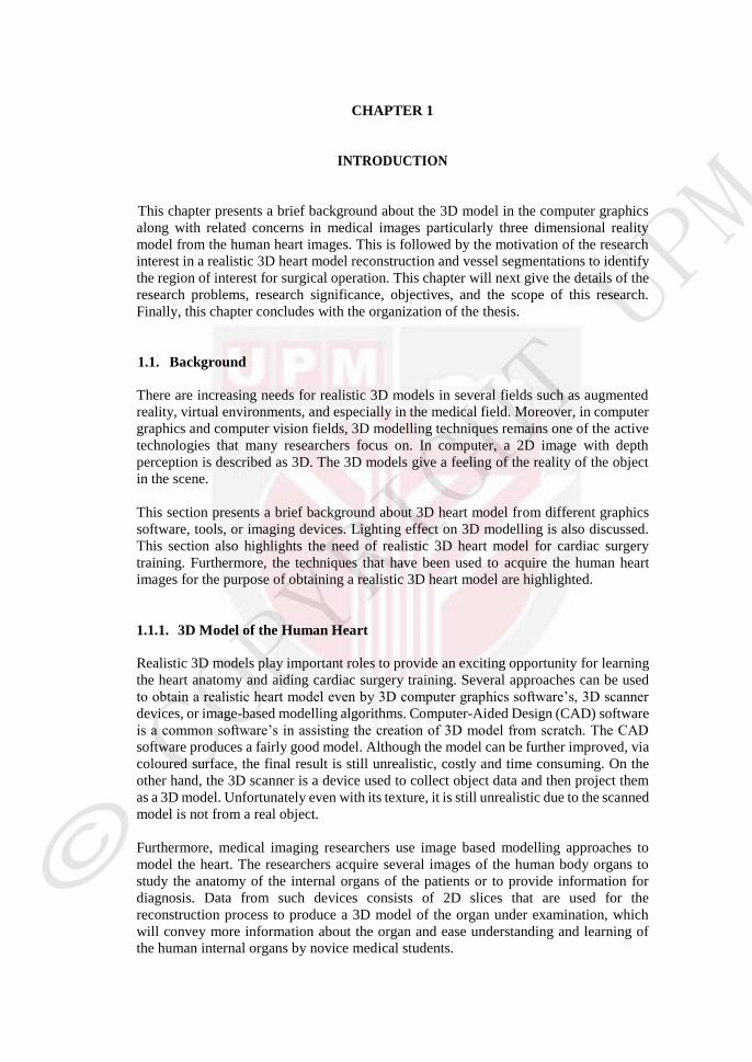
1.1.1. 3D Model of the Human Heart
Realistic 3D models play important roles to provide an exciting opportunity for learning
the heart anatomy and aiding cardiac surgery training. Several approaches can be used
to obtain a realistic heart model even by 3D computer graphics software’s, 3D scanner
devices, or image-based modelling algorithms. Computer-Aided Design (CAD) software
is a common software’s in assisting the creation of 3D model from scratch. The CAD
software produces a fairly good model. Although the model can be further improved, via
coloured surface, the final result is still unrealistic, costly and time consuming. On the
other hand, the 3D scanner is a device used to collect object data and then project them
as a 3D model. Unfortunately even with its texture, it is still unrealistic due to the scanned
model is not from a real object.
Furthermore, medical imaging researchers use image based modelling approaches to
model the heart. The researchers acquire several images of the human body organs to
study the anatomy of the internal organs of the patients or to provide information for
diagnosis. Data from such devices consists of 2D slices that are used for the
reconstruction process to produce a 3D model of the organ under examination, which
will convey more information about the organ and ease understanding and learning of
the human internal organs by novice medical students.

2
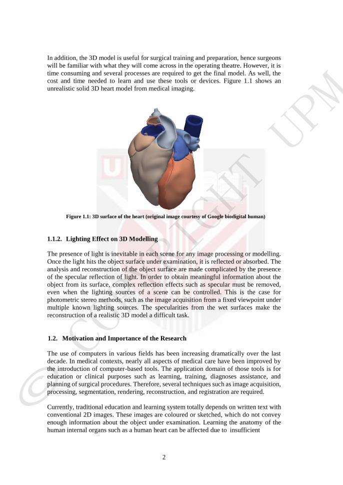
In addition, the 3D model is useful for surgical training and preparation, hence surgeons
will be familiar with what they will come across in the operating theatre. However, it is
time consuming and several processes are required to get the final model. As well, the
cost and time needed to learn and use these tools or devices. Figure 1.1 shows an
unrealistic solid 3D heart model from medical imaging.
Figure 1.1: 3D surface of the heart (original image courtesy of Google biodigital human)
1.1.2. Lighting Effect on 3D Modelling
The presence of light is inevitable in each scene for any image processing or modelling.
Once the light hits the object surface under examination, it is reflected or absorbed. The
analysis and reconstruction of the object surface are made complicated by the presence
of the specular reflection of light. In order to obtain meaningful information about the
object from its surface, complex reflection effects such as specular must be removed,
even when the lighting sources of a scene can be controlled. This is the case for
photometric stereo methods, such as the image acquisition from a fixed viewpoint under
multiple known lighting sources. The specularities from the wet surfaces make the
reconstruction of a realistic 3D model a difficult task.
1.2. Motivation and Importance of the Research
The use of computers in various fields has been increasing dramatically over the last
decade. In medical contexts, nearly all aspects of medical care have been improved by
the introduction of computer-based tools. The application domain of those tools is for
education or clinical purposes such as learning, training, diagnoses assistance, and
planning of surgical procedures. Therefore, several techniques such as image acquisition,
processing, segmentation, rendering, reconstruction, and registration are required.
Currently, traditional education and learning system totally depends on written text with
conventional 2D images. These images are coloured or sketched, which do not convey
enough information about the object under examination. Learning the anatomy of the
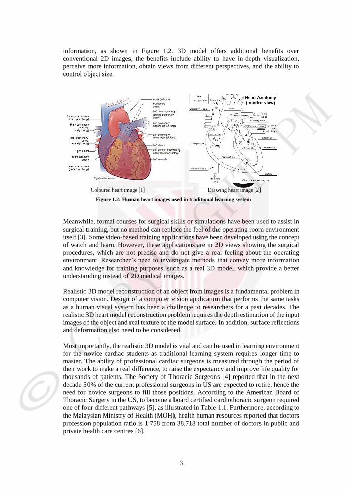
human internal organs such as a human heart can be affected due to insufficient
3
U
information, as shown in Figure 1.2. 3D model offers additional benefits over
conventional 2D images, the benefits include ability to have in-depth visualization,
perceive more information, obtain views from different perspectives, and the ability to
control object size.
Coloured heart image [1] Drawing heart image [2]
Figure 1.2: Human heart images used in traditional learning system
Meanwhile, formal courses for surgical skills or simulations have been used to assist in
surgical training, but no method can replace the feel of the operating room environment
itself [3]. Some video-based training applications have been developed using the concept
of watch and learn. However, these applications are in 2D views showing the surgical
procedures, which are not precise and do not give a real feeling about the operating
environment. Researcher’s need to investigate methods that convey more information
and knowledge for training purposes, such as a real 3D model, which provide a better
understanding instead of 2D medical images.
Realistic 3D model reconstruction of an object from images is a fundamental problem in
computer vision. Design of a computer vision application that performs the same tasks
as a human visual system has been a challenge to researchers for a past decades. The
realistic 3D heart model reconstruction problem requires the depth estimation of the input
images of the object and real texture of the model surface. In addition, surface reflections
and deformation also need to be considered.
Most importantly, the realistic 3D model is vital and can be used in learning environment
for the novice cardiac students as traditional learning system requires longer time to
master. The ability of professional cardiac surgeons is measured through the period of
their work to make a real difference, to raise the expectancy and improve life quality for
thousands of patients. The Society of Thoracic Surgeons [4] reported that in the next
decade 50% of the current professional surgeons in US are expected to retire, hence the
need for novice surgeons to fill those positions. According to the American Board of
Thoracic Surgery in the US, to become a board certified cardiothoracic surgeon required
one of four different pathways [5], as illustrated in Table 1.1. Furthermore, according to
the Malaysian Ministry of Health (MOH), health human resources reported that doctors
profession population ratio is 1:758 from 38,718 total number of doctors in public and
private health care centres [6].
4
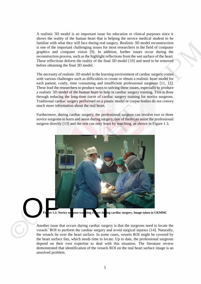
Table 1.1: Pathways training to obtain cardiothoracic surgery board certification in US [5]
Pathway Total
length of training
Components Duration of each
components
Board certification
Classical
7-8 years
General surgery
residency 5 years
General surgery
(optional)
Thoracic surgery fellowship
2-3 years Thoracic surgery
Fast-track
(4+3)
7 years
General surgery residency
4 years General surgery
(optional)
Thoracic surgery fellowship
3 years Thoracic surgery
Integrated
6 years Integrated
cardiothoracic surgery residency
6 years
Thoracic surgery
Vascular
+
Thoracic
7-8 years
Integrated vascular
surgery residency 5 years Vascular surgery
Thoracic surgery fellowship
2-3 years Thoracic surgery
Moreover, this statistic clearly states that the demand for novice surgeons is increasing.
However, the long-time training of cardiac surgery will lead to delay in surgical
operations while the number of the heart disease patients' is continuously increasing.
These motivated to develop a realistic human heart 3D model, which can reduce the
training and learning time of cardiac surgery for novice surgeons.
1.3. Research Problem
Medical applications make extensive use of high quality 3D models. Several 3D models
are manually created, with the help of the available modelling tools. However, it is costly
and time consuming to learn how to use these tools [7]. An interesting and easier
alternative is modelling from images. Modelling of medical images to produce a 3D
model of human internal organs is done via image sequences acquired from medical
imaging devices.
The vast development of imaging acquisition devices in recent years led to an increase
of the dimensionality of these images to 3D and Four Dimensional (4D) based on a scan
of multiple 2D slices in one axial direction. To generate a more intuitive representation
of the obtained images, a 3D model is reconstructed from segmented 2D slices [8]. The
3D model is useful for learning the internal human organ anatomy as well as training for
cardiac surgery. In addition, a 3D model helps reduce learning time. The input images
are acquired as a stack of parallel slices and collectively represent a 3D model of the
heart. Then a several analysis and processing are required to these slices such as, camera
calibration, noise removal, filtering, features extraction and matching, and the depth
determination. Finally, the model reconstruction were done. However, a reconstructed
model of medical imaging such as Computed Tomography Angiography (CTA) requires
several processes that are costly and time consuming in terms of data acquisition and
data storage, and has the potential to produce unrealistic 3D solid model.
5
OPYRIG
A realistic 3D model is an important issue for education or clinical purposes since it
shows the reality of the human heart that is helping the novice medical student to be
familiar with what they will face during real surgery. Realistic 3D model reconstruction
is one of the important challenging issues for most researchers in the field of computer
graphics and computer vision [9]. In addition, further issues occur during the
reconstruction process, such as the highlight reflections from the wet surface of the heart.
These reflections deform the reality of the final 3D model [10] and need to be removed
before obtaining the final 3D model.
The necessity of realistic 3D model in the learning environment of cardiac surgery comes
with various challenges such as difficulties to create or obtain a realistic heart model for
each patient, costly, time consuming and insufficient professional surgeons [11, 12].
These lead the researchers to produce ways to solving these issues, especially to produce
a realistic 3D model of the human heart to help in cardiac surgery training. This is done
through reducing the long-time curve of cardiac surgery training for novice surgeons.
Traditional cardiac surgery performed on a plastic model or corpse bodies do not convey
much more information about the real heart.
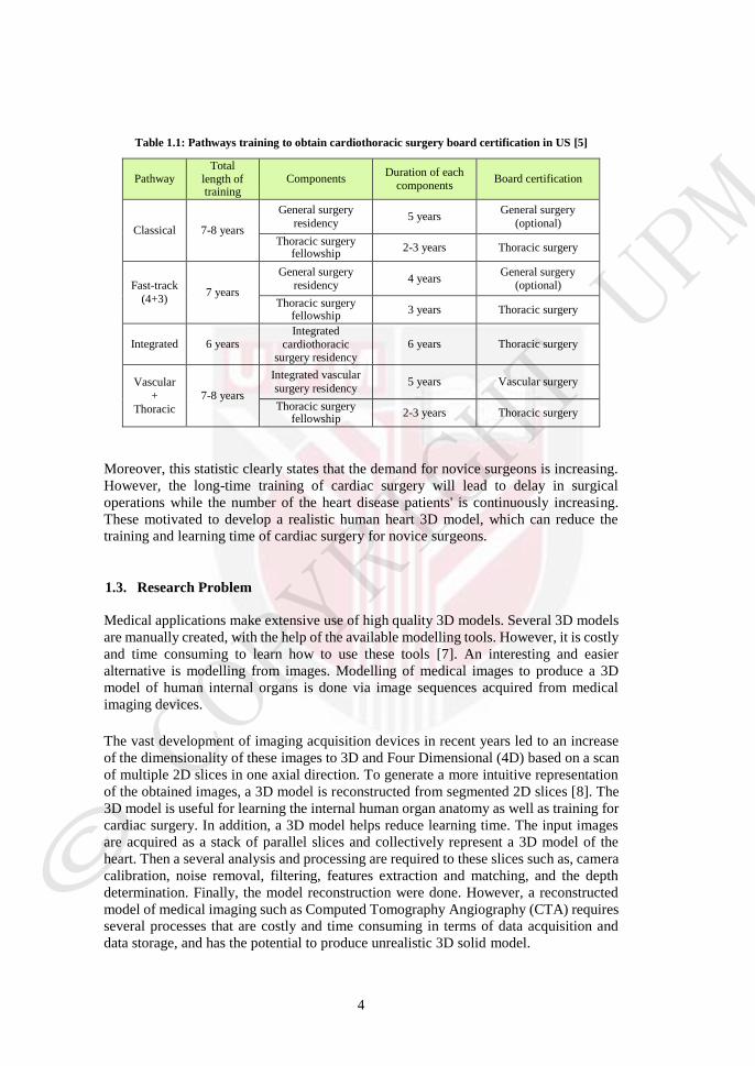
Furthermore, during cardiac surgery, the professional surgeon can involve two or three
novice surgeons to learn and assist during surgery, one of them can assist the professional
surgeon directly [13] and the rest can only learn by watching, as shown in Figure 1.3.
Figure 1.3: Novice surgeon watching expert during cardiac surgery. Image taken in UKMMC
Another issue that occurs during cardiac surgery is that the surgeons need to locate the
vessels’ ROI to perform the cardiac surgery and avoid surgical injuries [14]. Naturally,
the vessels lie over the heart surface. In some cases, vessels ROI might be covered by
the heart surface fats, which needs time to locate. Up to date, the professional surgeons
depend on their own expertise to deal with this situation. The literature review
demonstrated that identification of the vessels ROI on the real heart surface image is an
unsolved problem.
6
O RI
Meanwhile, detection and correction of the specular reflections that occur on the input
heart surface image caused by the lighting sources of the OT are important. Providing a
realistic 3D heart model from a single image, as well as achieving better identification
of the vessels ROI by enhancing the input data can help surgeons during the cardiac
surgery to locate the vessels where to perform the surgery.
1.4. Research Significance
The significance of the algorithms in computer vision is on automatic and realistic 3D
model reconstruction of the object with minimum or no user interaction. Such an
algorithm is capable of realistic reconstruction and it has immense applications in
modelling education, industry, medical, virtual and augmented reality.
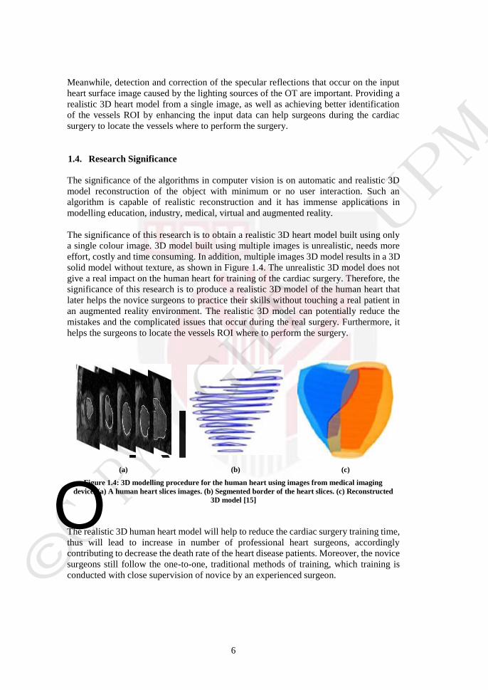
The significance of this research is to obtain a realistic 3D heart model built using only
a single colour image. 3D model built using multiple images is unrealistic, needs more
effort, costly and time consuming. In addition, multiple images 3D model results in a 3D
solid model without texture, as shown in Figure 1.4. The unrealistic 3D model does not
give a real impact on the human heart for training of the cardiac surgery. Therefore, the
significance of this research is to produce a realistic 3D model of the human heart that
later helps the novice surgeons to practice their skills without touching a real patient in
an augmented reality environment. The realistic 3D model can potentially reduce the
mistakes and the complicated issues that occur during the real surgery. Furthermore, it
helps the surgeons to locate the vessels ROI where to perform the surgery.
(a) (b) (c)
Figure 1.4: 3D modelling procedure for the human heart using images from medical imaging
device. (a) A human heart slices images. (b) Segmented border of the heart slices. (c) Reconstructed
3D model [15]
The realistic 3D human heart model will help to reduce the cardiac surgery training time,
thus will lead to increase in number of professional heart surgeons, accordingly
contributing to decrease the death rate of the heart disease patients. Moreover, the novice
surgeons still follow the one-to-one, traditional methods of training, which training is
conducted with close supervision of novice by an experienced surgeon.
7
1.5. Research Objectives
The main objective of this research is to reconstruct a realistic 3D model of the human
heart. This model can be used in a cardiac surgery learning exercise for novice medical
students. The training using realistic 3D model adds a new perspective to the traditional
approach to review heart anatomy to perform the cardiac surgery. Vessel ROI has to be
identified in relation to some features. In addition, surgeons must decide which vessel to
perform the surgery, keeping in mind the functional consequences of these actions.
Finally, it provides an opportunity to integrate training concepts to practice surgery
before performing a real procedure on a patient.
Hence, to achieve those objectives, the following sub-objectives will be considered:
1. To propose new algorithms for specular reflection detection and correction from
human heart single view image datasets based on the L-inverse shape that is
accurate and fast specularities detection and correction.
2. To propose a realistic 3D human heart model reconstructed from a single view
image using a fast and robust approach based on intensity value that shows the
closeness of the 3D model. Subsequently, smooth the 3D surface model using a
Bezier curve approximation.
3. To introduce a new vessels segmentation approach by identifying and accurately
locating the correct vessel ROI to avoid surgery injuries and perform the surgery
based on RGB colour space.
1.6. Research Scope
This thesis focuses on realistic 3D model reconstruction of a real human heart rather than
the computer graphics creation of artificial models using graphics software, tools, or
imaging devices. This realistic 3D model is to be used in cardiac surgery training
environment. Furthermore, the thesis identifies the vessels ROI where to perform the
surgery by using patient heart images. The input data will be taken during an open-heart
surgery. However, for the purpose of this research, the focus is limited to using only a
digital camera to acquire the input data, the acquiring data are images or videos whereas
the video will be converted to still images. Chosen images will be used as an input in this
research, i.e. images without obstacles, such as surgical tools, surgeon's head or hands.
1.7. Thesis Organization
A brief background and motivation of the research are presented in this chapter, as well
as the research problem, significance, objectives, and research scope. Furthermore, the
rest of this thesis is organized as follows.
Chapter 2 introduces a review of state of the art literature of specular reflection detection
methods prior to inpainting methods to correct the specularities. Then, it presents a 3D
reconstruction method from multiple and single images. Furthermore, reviews about the
existing vessel segmentation techniques are presented.
8
Chapter 3 describes the overall research framework. In addition, this chapter explains
about the data acquisition, pre-processing of data, and brief details for the rest of the
research chapters.
Chapter 4 presents a specular reflections algorithm, which is detected by a proposed
threshold value using colour information. Then, an inpainting algorithm is proposed to
correct the detected specular pixel using L-inverse surrounding pixels. Hence, the input
image is corrected and will be used to obtain realistic 3D heart surface model.
Chapter 5 describes the obtaining of the realistic 3D model algorithm of the real human
heart images that have been corrected from the specular reflections algorithm. It assumes
the pixel intensity value used as a third axis for 3D reconstruction. The three axes are
then estimated for each pixel by using positions and intensities. Moreover, the surface
model is then smoothed by applying the Bezier curve approximation technique.
In chapter 6, according to different characteristics of the vessels, hybrid algorithm is
applied to segment the surface vessels of the human heart image. It combines a vessel
segmentation and 3D vessel model reconstruction to create a realistic 3D model of the
human heart surface. In this chapter, an approximation technique to the extracted surface
vessels using a Bezier curve was perform. The segmented vessels are correctly guiding
the surgeons where to perform the surgery.
For validation of each proposed method, several experiments are conducted on a digital
data and presented at the end of each chapter along with their results and discussions.
Finally, a conclusion of the whole thesis, limitations and potential future work is given
in chapter 7.
9
REFERENCES
[1] N. NHLBI. (2011, 15-11-2013). Anatomy of the Heart. Available:
http://www.nhlbi.nih.gov/health/health-topics/topics/hhw/anatomy#
[2] Admin. (2011, 15-11-2013). Human Body Anatomy. Available:
http://www.eht.k12.nj.us/~elwellc/heart%20diagram%20answers_files/image0
01.gif
[3] I. Kron. Surgical mentorship. J Thorac Cardiovasc Surg, vol. 142, pp. 489-492,
2011.
[4] The-Society-of-Thoracic-Surgeons. Become a CT Surgeon. STS-2008.
[5] V. Tchantchaleishvili, S. Mokashi, T. Rajab, F. Chen, and J. Schmitto.
Comparison of cardiothoracic surgery training in usa and germany. 2010.
[6] M. Ministry of Health. (2013, 2-2-2014). Health Human Resources. Available:
http://www.moh.gov.my/english.php/pages/view/405
[7] L. Brutto and P. Meli. Computer vision tools for 3D modelling in archaeology.
International Journal of Heritage in the Digital Era, vol. 1, pp. 1-6, 2012.
[8] N. Toussaint, M. Sermesant, C. Stoeck, S. Kozerke, and P. Batchelor. In vivo
Human 3D Cardiac Fibre Architecture: Reconstruction Using Curvilinear
Interpolation of Diffusion Tensor Images. Medical Image Computing and
Computer-Assisted Intervention – MICCAI10. vol. 6361, ed: Springer Berlin
Heidelberg, 2010, pp. 418-425.
[9] S. Se and P. Jasiobedzki. Photo-realistic 3D model reconstruction. IEEE
International Conference on Robotics and Automation, ICRA06 Proceedings.,
pp. 3076-3082, 2006.
[10] J. Gu, T. Kobayashi, M. Gupta, and S. K. Nayar. Multiplexed illumination for
scene recovery in the presence of global illumination. IEEE International
Conference on Computer Vision (ICCV11), pp. 691-698, 2011.
[11] R. Jonas. Comprehensive surgical management of congenital heart disease:
CRC Press, 2004.
[12] J. D. Beard, J. Marriott, H. Purdie, and J. Crossley. Assessing the surgical skills
of trainees in the operating theatre: a prospective observational study of the
methodology. Health technology assessment (Winchester, England), vol. 15,
pp. i-xxi, 2011.
[13] T. Henry. (2009, 25-2-2014). Shortage of cardiothoracic surgeons expexted.
Available:
http://www.amednews.com/article/20090814/profession/308149998/8/

99
[14] J. W. Cannon and M. A. Peck. Vascular injuries in the young. Perspectives in
vascular surgery and endovascular therapy, 2011.
[15] D. Tang, C. Yang, T. Geva, and J. Pedro. Patient-specific MRI-based 3D FSI
RV/LV/patch models for pulmonary valve replacement surgery and patch
optimization. Journal of biomechanical engineering, vol. 130, p. 041010, 2008.
[16] T. Kanade and K. Ikeuchi. Introduction to the special issue on physical
modeling in computer vision. IEEE Transactions on Pattern Analysis and
Machine Intelligence, vol. 13, pp. 609-610, 1991.
[17] S. Ullman. On visual detection of light sources. Biological Cybernetics, vol. 21,
pp. 205-211, 1976.
[18] K. Forbus. Light source effects. 22nd Annual Technical Symposium, pp. 50-55,
1979.
[19] E. U. Schirmibeck, I. Nagy, H. Mayer, A. Knoll, R. Lange, and R.
Bauemschmitt. Automatic coronary artery detection on in situ heart images.
Computers in Cardiology, pp. 785-788, 2004.
[20] T. Johannes. Motion Compensation in Minimally Invasive Robotic Surgery.
Ph.D. dissertation, Technische UniversitÄat MÄunchen, Germany, 2003.
[21] M. Groger, W. Sepp, and G. Hirzinger. Structure driven substitution of specular
reflections for realtime heart surface tracking. IEEE International Conference
on Image Processing, ICIP05, pp. II-1066-9, 2005.
[22] G. Brelstaff and A. Blake. Detecting specular reflections using Lambertian
constraints. Second International Conference on Computer Vision, pp. 297-302,
1988.
[23] S. Shafer. Using color to separate reflection components. Color Research &
Application, vol. 10, pp. 210-218, 1985.
[24] R. Gershon, A. Jepson, and J. Tsotsos. The Use of Color in Highlight
Identification. IJCAI, pp. 752-754, 1987.
[25] G. Klinker, S. Shafer, and T. Kanade. Using a color reflection model to separate
highlights from object color. Proceeding. 1st International Conference
Computer Vision ICCV, pp. 145-150, 1987.
[26] L. Wolff. Polarization-based material classification from specular reflection.
IEEE Transactions on Pattern Analysis and Machine Intelligence, vol. 12, pp.
1059-1071, 1990.
[27] L. Wolff and T. Boult. Constraining object features using a polarization
reflectance model. IEEE Transactions on Pattern Analysis and Machine
Intelligence, vol. 13, pp. 635-657, 1991.

100
[28] S. Nayar, X. Fang, and T. Boult. Removal of specularities using color and
polarization. IEEE Computer Society Conference on Computer Vision and
Pattern Recognition, Proceedings CVPR '93., pp. 583-590, 1993.
[29] S. Nayar, X. Fang, and T. Boult. Separation of Reflection Components Using
Color and Polarization. Int. J. Comput. Vision, vol. 21, pp. 163-186, 1997.
[30] R. Bajcsy, S. Lee, and A. Leonardis. Color image segmentation with detection
of highlights and local illumination induced by inter-reflections. Pattern
Recognition. Proceedings, 10th International Conference, pp. 785-790 vol.1,
1990.
[31] R. Bajcsy, S. Lee, and A. Leonardis. Detection of diffuse and specular interface
reflections and inter-reflections by color image segmentation. International
Journal of Computer Vision, vol. 17, pp. 241-272, 1996.
[32] S. Lin, Y. Li, S. B. Kang, X. Tong, and H.-Y. Shum. Diffuse-Specular
Separation and Depth Recovery from Image Sequences. presented at the
Proceedings of the 7th European Conference on Computer Vision-Part III,
2002.
[33] K.-K. Ma and J. Wang. Color distance histogram: a novel descriptor for color
image segmentation. 7th International Conference on Control, Automation,
Robotics and Vision. ICARCV02. , pp. 1228-1232, 2002.
[34] J. Park. Detection of specular highlights in color images using a new color space
transformation. IEEE International Conference on Robotics and Biomimetics,
ROBIO04., pp. 737-741, 2004.
[35] G. Zimmerman-Moreno and H. Greenspan. Automatic detection of specular
reflections in uterine cervix images. pp. 61446E-61446E, 2006.
[36] J. Oh, S. Hwang, J. Lee, W. Tavanapong, J. Wong, and d. Groen. Informative
frame classification for endoscopy video. Medical Image Analysis, vol. 11, pp.
110-127, 2007.
[37] T. Stehle. Removal of Specular Reflections in Endoscopic Images. Acta
Polytechnica: Journal of Advanced Engineering, vol. 46, pp. 32-36, 2006.
[38] V. Bochko and Y. Miyake. Highlight Removal in Endoscope Images.
Conference on Colour in Graphics, Imaging, and Vision, pp. 167-171, 2006.
[39] C. Xu, X. Ye, Y. Wu, and S. Zhang. Highlight Detection and Removal Based
on Chromaticity. Image Analysis and Recognition. vol. 3656, ed: Springer
Berlin Heidelberg, 2005, pp. 199-206.
[40] H. Greenspan, S. Gordon, G. Zimmerman, S. Lotenberg, J. Jeronimo, S. Antani,
and R. Long. Automatic Detection of Anatomical Landmarks in Uterine Cervix
Images. IEEE Transactions on Medical Imaging, vol. 28, pp. 454-468, 2009.
101
[41] M. Arnold, A. Ghosh, S. Ameling, and G. Lacey. Automatic segmentation and
inpainting of specular highlights for endoscopic imaging. J. Image Video
Process., pp. 1-12, 2010.
[42] A. Das, A. Kar, and D. Bhattacharyya. Elimination of specular reflection and
identification of ROI: The first step in automated detection of Cervical Cancer
using Digital Colposcopy. IEEE International Conference on Imaging Systems
and Techniques (IST11), pp. 237-241, 2011.
[43] D. Gajpal, A. Biswas, and R. Potdar. Reconstruction of Arteries in Heart Image
Affected by Specular Reflection. International Journal of Emerging Technology
and Advanced Engineering, vol. 2, pp. 259-263, 2012.
[44] H. Ghodrati, J. Dehghani, and H. Danyali. A new accurate noise-removing
approach for non-cooperative iris recognition. Signal, Image and Video
Processing, vol. 8, pp. 1-10, 2014.
[45] H. Freeman. On the Encoding of Arbitrary Geometric Configurations. IRE
Transactions on Electronic Computers, vol. EC-10, pp. 260-268, 1961.
[46] F. Kuhl and C. Giardina. Elliptic Fourier features of a closed contour. Computer
Graphics and Image Processing, vol. 18, pp. 236-258, 1982.
[47] S. Tchoulack, J. Pierre Langlois, and F. Cheriet. A video stream processor for
real-time detection and correction of specular reflections in endoscopic images.
Circuits and Systems and TAISA Conference., pp. 49-52, 2008.
[48] F. Vogt, D. Paulus, and H. Niemann. Highlight substitution in light fields.
International Conference on Image Processing, pp. I-637-I-640 vol.1, 2002.
[49] M. Gröger, W. Sepp, T. Ortmaier, and G. Hirzinger. Reconstruction of Image
Structure in Presence of Specular Reflections. presented at the Proceedings of
the 23rd DAGM-Symposium on Pattern Recognition, 2001.
[50] E. Mariano, Dueire Lins, G. Pereira e Silva, J. Fan, P. Majewicz, and M. Thielo.
Correcting Specular Noise in Multiple Images of Photographed Documents.
International Conference on Document Analysis and Recognition (ICDAR11),
pp. 915-919, 2011.
[51] C. Wang, S. Kamata, and L. Ma. A Fast Multi-View Based Specular Removal
Approach for Pill Extraction. ICIP, 2013.
[52] R. T. Tan and K. Ikeuchi. Separating Reflection Components of Textured
Surfaces Using a Single Image. IEEE Trans. Pattern Anal. Mach. Intell., vol.
27, pp. 178-193, 2005.
[53] D. Stoyanov and Y. Guang Zhong. Removing specular reflection components
for robotic assisted laparoscopic surgery. IEEE International Conference on
Image Processing, ICIP., pp. III-632-5, 2005.
102
[54] M. Bertalmio, G. Sapiro, V. Caselles, and C. Ballester. Image inpainting.
presented at the Proceedings of the 27th annual conference on Computer
graphics and interactive techniques, 2000.
[55] K. Garden and R. Robb. 3-D reconstruction of the heart from few projections:
A practical implementation of the McKinnon-Bates algorithm. IEEE
Transactions on Medical Imaging, vol. 5, pp. 233-239, 1986.
[56] D. King, M. Harrison, A. Gopal, R. Martin, and A. DeMaria. Improved
reproducibility of left atrial and left ventricular measurements by guided three-
dimensional echocardiography. Journal of the American College of Cardiology,
vol. 20, pp. 1238-1245, 1992.
[57] M. Handschumacher, J. Lethor, S. Siu, D. Mele, M. Rivera, M. Picard, A.
Weyman, and R. Levine. A new integrated system for three-dimensional
echocardiographic reconstruction: development and validation for ventricular
volume with application in human subjects. Journal of the American College of
Cardiology, vol. 21, pp. 743-753, 1993.
[58] A. Franke, F. Flachskampf, H. Kühl, H. Klues, F. Job, M. Merx, W. Krebs, and
P. Hanrath. 3-dimensional reconstruction of multiplanar transesophageal
echocardiography images: a methodologic report with case examples.
Zeitschrift für Kardiologie, vol. 84, p. 633, 1995.
[59] N. Pandian, J. Roelandt, N. Nanda, L. Sugeng, Q. L. CAO, J. Azevedo, S.
Schwartz, M. Vannan, A. Ludomirski, and G. Marx. Dynamic Three-
Dimensional Echocardiography. Echocardiography, vol. 11, pp. 237-259, 1994.
[60] J. Pilet, V. Lepetit, and P. Fua. Fast non-rigid surface detection, registration and
realistic augmentation. International Journal of Computer Vision, vol. 76, pp.
109-122, 2008.
[61] D. Pizarro and A. Bartoli. Feature-based deformable surface detection with self-
occlusion reasoning. International Journal of Computer Vision, vol. 97, pp. 54-
70, 2012.
[62] R. Garg, A. Roussos, and L. Agapito. Robust trajectory-space TV-L1 optical
flow for non-rigid sequences. Energy Minimization Methods in Computer
Vision and Pattern Recognition, pp. 300-314, 2011.
[63] R. Hartley and A. Zisserman. Multiple view geometry in computer vision:
Cambridge university press, 2003.
[64] L Higgins and H. Christopher. A computer algorithm for reconstructing a scene
from two projections. Readings in Computer Vision: Issues, Problems,
Principles, and Paradigms, MA Fischler and O. Firschein, eds, pp. 61-62, 1987.
[65] O. Faugeras, Q. Luong, and S. Maybank. Camera self-calibration: Theory and
experiments. Computer Vision—ECCV'92, pp. 321-334, 1992.
103
[66] J. Xiao, J. Chai, and T. Kanade. A closed-form solution to non-rigid shape and
motion recovery. International Journal of Computer Vision, vol. 67, pp. 233-
246, 2006.
[67] Y. Dai, H. Li, and M. He. A simple prior-free method for non-rigid structure-
from-motion factorization. IEEE Conference on Computer Vision and Pattern
Recognition (CVPR12), pp. 2018-2025, 2012.
[68] C. Tomasi and T. Kanade. Shape and motion from image streams under
orthography: a factorization method. International Journal of Computer Vision,
vol. 9, pp. 137-154, 1992.
[69] B. Bascle and A. Blake. Separability of pose and expression in facial tracking
and animation. Sixth International Conference on Computer Vision, pp. 323-
328, 1998.
[70] I. Akhter, Y. Sheikh, S. Khan, and T. Kanade. Nonrigid structure from motion
in trajectory space. Advances in neural information processing systems, pp. 41-
48, 2009.
[71] Maier-Hein, P. Mountney, A. Bartoli, H. Elhawary, D. Elson, A. Groch, A.
Kolb, M. Rodrigues, J. Sorger, and S. Speidel. Optical techniques for 3D surface
reconstruction in computer-assisted laparoscopic surgery. Medical Image
Analysis, vol. 17, pp. 974-996, 2013.
[72] J. Geng. Rainbow three-dimensional camera: new concept of high-speed three-
dimensional vision systems. Optical Engineering, vol. 35, pp. 376-383, 1996.
[73] P. Huang, Q. Hu, F. Jin, and F. Chiang. Color-encoded digital fringe projection
technique for high-speed three-dimensional surface contouring. Optical
Engineering, vol. 38, pp. 1065-1071, 1999.
[74] C. Guan, L. Hassebrook, and D. Lau. Composite structured light pattern for
three-dimensional video. Optics Express, vol. 11, pp. 406-417, 2003.
[75] S. Rusinkiewicz, O. Hall-Holt, and M. Levoy. Real-time 3D model acquisition.
ACM Transactions on Graphics (TOG), pp. 438-446, 2002.
[76] P. S. Huang, C. Zhang, and F.-P. Chiang. High-speed 3-D shape measurement
based on digital fringe projection. Optical Engineering, vol. 42, pp. 163-168,
2003.
[77] J. Salvi, J. Pages, and J. Batlle. Pattern codification strategies in structured light
systems. Pattern Recognition, vol. 37, pp. 827-849, 2004.
[78] C. Albitar, P. Graebling, and C. Doignon. Robust Structured Light Coding for
3D Reconstruction. ICCV, pp. 1-6, 2007.
[79] S. Gorthi and P. Rastogi. Fringe projection techniques: whither we are? Optics
and lasers in engineering, vol. 48, pp. 133-140, 2010.
104
[80] X. Maurice, P. Graebling, and C. Doignon. Epipolar based structured light
pattern design for 3-d reconstruction of moving surfaces. IEEE International
Conference on Robotics and Automation (ICRA11), pp. 5301-5308, 2011.
[81] C. Lorenz and N. Krahnstöver. Generation of point-based 3D statistical shape
models for anatomical objects. Computer Vision and Image Understanding, vol.
77, pp. 175-191, 2000.
[82] A. Frangi, D. Rueckert, J. Schnabel, and W. Niessen. Automatic construction
of multiple-object three-dimensional statistical shape models: Application to
cardiac modeling. IEEE Transactions on Medical Imaging, vol. 21, pp. 1151-
1166, 2002.
[83] R. Davies, C. Twining, T. Cootes, J. Waterton, and C. Taylor. A minimum
description length approach to statistical shape modeling. IEEE Transactions on
Medical Imaging, vol. 21, pp. 525-537, 2002.
[84] S. Mitchell, J. Bosch, B. Lelieveldt, R. van der Geest, J. Reiber, and M. Sonka.
3-D active appearance models: segmentation of cardiac MR and ultrasound
images. IEEE Transactions on Medical Imaging, vol. 21, pp. 1167-1178, 2002.
[85] J. Lötjönen, S. Kivistö, J. Koikkalainen, D. Smutek, and K. Lauerma. Statistical
shape model of atria, ventricles and epicardium from short-and long-axis MR
images. Medical Image Analysis, vol. 8, pp. 371-386, 2004.
[86] K. Ikeuchi and B. K. Horn. Numerical shape from shading and occluding
boundaries. Artificial intelligence, vol. 17, pp. 141-184, 1981.
[87] R. Zhang, P. Tsai, J. Cryer, and M. Shah. Shape-from-shading: a survey. IEEE
Transactions on Pattern Analysis and Machine Intelligence, vol. 21, pp. 690-
706, 1999.
[88] Pierre Gurdjos, F. Courteille, A. Crouzil, J. Durou, and I. TCI. Towards shape
from shading under realistic photographic conditions. 2004.
[89] J. Durou, M. Falcone, and M. Sagona. Numerical methods for shape-from-
shading: A new survey with benchmarks. Computer Vision and Image
Understanding, vol. 109, pp. 22-43, 2008.
[90] G. Wang, W. Su, and Y. Song. A new shape from shading approach for specular
surfaces. Artificial Intelligence and Computational Intelligence, ed: Springer,
2011, pp. 71-78.
[91] B. Super and A. Bovik. Shape from texture using local spectral moments. IEEE
Transactions on Pattern Analysis and Machine Intelligence, vol. 17, pp. 333-
343, 1995.
[92] T. Vetter. Synthesis of novel views from a single face image. International
Journal of Computer Vision, vol. 28, pp. 103-116, 1998.
105
[93] T. Hassner and R. Basri. Example based 3D reconstruction from single 2D
images. Conference on Computer Vision and Pattern Recognition Workshop,
CVPRW'06., pp. 15-15, 2006.
[94] D. Hoiem, A. Efros, and M. Hebert. Automatic photo pop-up. ACM
Transactions on Graphics (TOG), pp. 577-584, 2005.
[95] A. Saxena, M. Sun, and A. Ng. Make3d: Learning 3d scene structure from a
single still image. IEEE Transactions on Pattern Analysis and Machine
Intelligence, vol. 31, pp. 824-840, 2009.
[96] B. Liu, S. Gould, and D. Koller. Single image depth estimation from predicted
semantic labels. IEEE Conference on Computer Vision and Pattern Recognition
(CVPR10), pp. 1253-1260, 2010.
[97] A. François and G. Medioni. Interactive 3D model extraction from a single
image. Image and Vision Computing, vol. 19, pp. 317-328, 2001.
[98] D. Terzopoulos, A. Witkin, and M. Kass. Symmetry-seeking models and 3D
object reconstruction. International Journal of Computer Vision, vol. 1, pp. 211-
221, 1988.
[99] L. Cohen and I. Cohen. Finite-element methods for active contour models and
balloons for 2-D and 3-D images. IEEE Transactions on Pattern Analysis and
Machine Intelligence, vol. 15, pp. 1131-1147, 1993.
[100] L. Zhang, G. Dugas-Phocion, J.-S. Samson, and S. Seitz. Single-view modelling
of free-form scenes. The Journal of Visualization and Computer Animation,
vol. 13, pp. 225-235, 2002.
[101] J. Duchon. Splines minimizing rotation-invariant semi-norms in Sobolev
spaces. Constructive theory of functions of several variables, ed: Springer,
1977, pp. 85-100.
[102] M. Prasad, A. Zisserman, and A. Fitzgibbon. Fast and controllable 3D
modelling from silhouettes. Eurographics, Short Papers, pp. 9-12, 2005.
[103] M. Prasad and A. Fitzgibbon. Single view reconstruction of curved surfaces.
pp. 1345-1354, 2006.
[104] R. Kikinis, L. Gleason, F. Jolesz, R. Taylor, S. Lavallee, and G. Burdea.
Surgical planning using computer-assisted three-dimensional reconstructions.
Computer Integrated Surgery, vol. 8, pp. 147-154, 1996.
[105] M. Clarkson, D. Rueckert, A. King, P. Edwards, D. Hill, and D. Hawkes.
Registration of video images to tomographic images by optimising mutual
information using texture mapping. pp. 579-588, 1999.
[106] S. Hakamata, D. Miyoshi, T. Nakaguchi, N. Tsumura, and Y. Miyake.
Reconstruction of 3D organ image using endoscope with Magneto-position-
106
sensor. IEIC Technical Report (Institute of Electronics, Information and
Communication Engineers), vol. 106, pp. 13-18, 2006.
[107] H. Fuchs, M. Livingston, R. Raskar, K. Keller, J. Crawford, P. Rademacher, S.
Drake, and A. Meyer. Augmented reality visualization for laparoscopic surgery.
Medical Image Computing and Computer-Assisted Interventation—Miccai’98,
ed: Springer, 1998, pp. 934-943.
[108] A. Iyengar, H. Sugimoto, D. Smith, and M. Sacks. Dynamic in vitro
quantification of bioprosthetic heart valve leaflet motion using structured light
projection. Annals of Biomedical Engineering, vol. 29, pp. 963-973, 2001.
[109] M. Hayashibe, N. Suzuki, and Y. Nakamura. Laser-scan endoscope system for
intraoperative geometry acquisition and surgical robot safety management.
Medical Image Analysis, vol. 10, pp. 509-519, 2006.
[110] F. Mourgues, F. Devemay, and E. Coste-Maniere. 3D reconstruction of the
operating field for image overlay in 3D-endoscopic surgery. pp. 191-192, 2001.
[111] D. Burschka, M. Li, R. Taylor, and G. Hager. Scale-invariant registration of
monocular stereo images to 3d surface models. pp. 2581-2586, 2004.
[112] C. Wu and Y. Sun. 3D Reconstruction of Human Inner Structure from Video
by Using Geometric Constraints. 2007.
[113] T. Okatani and K. Deguchi. Shape Reconstruction from an Endoscope Image
by Shape from Shading Technique for a Point Light Source at the Projection
Center. Computer Vision and Image Understanding, vol. 66, pp. 119-131, 1997.
[114] S. Yeung, H. Tsui, and A. Yim. Global shape from shading for an endoscope
image. pp. 318-327, 1999.
[115] A. Tankus, N. Sochen, and Y. Yeshurun. Reconstruction of medical images by
perspective shape-from-shading. pp. 778-781, 2004.
[116] D. Hong, W. Tavanapong, J. Wong, J. Oh, and P. Groen. 3D reconstruction of
colon segments from colonoscopy images. pp. 53-60, 2009.
[117] D. Filev and R. Yager. A generalized defuzzification method via BAD
distributions. International Journal of Intelligent Systems, vol. 6, pp. 687-697,
1991.
[118] R. Ohlander, K. Price, and D. R. Reddy. Picture segmentation using a recursive
region splitting method. Computer Graphics and Image Processing, vol. 8, pp.
313-333, 1978.
[119] X. Lin and S. Chen. Color image segmentation using modified HSI system for
road following. pp. 1998-2003, 1991.

107
[120] L. Bonsiepen and W. Coy. Stable segmentation using color information.
Computer Analysis of Images and Patterns, ed. R. Klette, Proc. of CAIP, vol.
91, pp. 77-84, 1991.
[121] R. Taylor and P. Lewis. Colour image segmentation using boundary relaxation.
pp. 721-724, 1992.
[122] I. Ismaili and D. Gillies. Colour image segmentation using regression analysis
in RGB space. Machine Graphics & Vision, vol. 3, pp. 373-384, 1994.
[123] T. Westman, D. Harwood, T. Laitnen, and M. Pietikanen. Color segmentation
by hierarchical connected components analysis with image enhancement by
symmetric neighborhood filters. pp. 796-802, 1990.
[124] F. Meyer. Color image segmentation. pp. 303-306, 1992.
[125] T. Vlachos and A. Constantinides. A graph-theoretic approach to colour image
segmentation and contour classification. pp. 298-302, 1992.
[126] C.-L. Huang, T.-Y. Cheng, and C.-C. Chen. Color images' segmentation using
scale space filter and Markov random field. Pattern Recognition, vol. 25, pp.
1217-1229, 1992.
[127] D. Tseng and C. Chang. Color segmentation using perceptual attributes. pp.
228-231, 1992.
[128] M. Yachida and S. Tsuji. Application of color information to visual perception.
Pattern Recognition, vol. 3, pp. 307-323, 1971.
[129] D. Shen, G. Chen, Y. Zheng, E. Blasch, and K. Pham. Game theoretic approach
to similarity-based image segmentation. pp. 813708-813708, 2011.
[130] D. Shen, E. Blasch, K. Pham, and G. Chen. A clustering game based framework
for image segmentation. pp. 818-823, 2012.
[131] T. Huntsberger and M. Descalzi. Color edge detection. Pattern Recognition
Letters, vol. 3, pp. 205-209, 1985.
[132] M. S. Yang. A survey of fuzzy clustering. Mathematical and Computer
modelling, vol. 18, pp. 1-16, 1993.
[133] R. Harikumar, V. Kumar, G. Karthick, K. Chand, and N. Kumar. Hierarchical
Clustering Algorithm for Intensity Based Cluster Merging and Edge Detection
in Medical Images. pp. 323-337, 2013.
[134] R. Young. Simulation of human retinal function with the Gaussian derivative
model. USA, 1986, pp. 564-569.
[135] S. Lanser. Kantenorientierte Farbsegmentation im CIE-Lab Raum. pp. 639-646,
1993.
108
[136] S. Lanser and W. Eckstein. A modification of Deriche's approach to edge
detection. pp. 633-637, 1992.
[137] G. Klinker, S. Shafer, and T. Kanade. Image segmentation and reflection
analysis through color. pp. 229-244, 1988.
[138] G. Klinker, S. Shafer, and T. Kanade. A physical approach to color image
understanding. International Journal of Computer Vision, vol. 4, pp. 7-38, 1990.
[139] R. Bajcsy, S. Lee, and A. Leonardis. Image segmentation with detection of
highlights and inter-reflections using color. 1989.
[140] R. K. Bajcsy, S. W. Lee, and A. Leonardis. Color image segmentation and color
constancy. pp. 245-255, 1990.
[141] W. Higgins, W. Spyra, and E. Ritman. Automatic extraction of the arterial tree
from 3-D angiograms. pp. 563-564, 1989.
[142] N. Niki, Y. Kawata, H. Satoh, and T. Kumazaki. 3D imaging of blood vessels
using x-ray rotational angiographic system. IEEE Conference Record Nuclear
Science Symposium and Medical Imaging Conference. , San Francisco, CA, pp.
1873-1877, 1993.
[143] A. Klein, F. Lee, and A. Amini. Quantitative coronary angiography with
deformable spline models. IEEE Transactions on Medical Imaging, vol. 16, pp.
468-482, 1997.
[144] M. Molina, G. Prause, P. Radeva, and M. Sonka. 3D catheter path
reconstruction from biplane angiograms. pp. 504-512, 1998.
[145] D. Guo and P. Richardson. Automatic vessel extraction from angiogram
images. Computers in Cardiology., Cleveland, OH, pp. 441-444, 1998.
[146] Y. Sato, S. Nakajima, N. Shiraga, H. Atsumi, S. Yoshida, T. Koller, G. Gerig,
and R. Kikinis. Three-dimensional multi-scale line filter for segmentation and
visualization of curvilinear structures in medical images. Medical Image
Analysis, vol. 2, pp. 143-168, 1998.
[147] A. Sarwal and A. Dhawan. 3-d reconstruction of coronary arteries. Engineering
in Medicine and Biology Society, Engineering Advances: New Opportunities
for Biomedical Engineers. Proceedings of the 16th Annual International
Conference of the IEEE, Baltimore, MD, pp. 504-505, 1994.
[148] M. Chwialkowski, Y. Ibrahim, H. Li, and R. Peshock. A method for fully
automated quantitative analysis of arterial flow using flow-sensitized MR
images. Computerized Medical Imaging and Graphics, vol. 20, pp. 365-378,
1996.
[149] T. Tozaki, Y. Kawata, N. Niki, H. Ohmatsu, and N. Moriyama. 3-D
visualization of blood vessels and tumor using thin slice CT images. IEEE
109
Conference Record Nuclear Science Symposium and Medical Imaging
Conference, pp. 1470-1474, 1995.
[150] Y. Kawata, N. Niki, and T. Kumazaki. An approach for detecting blood vessel
diseases from cone-beam CT image. International Conference on Image
Processing, Proceedings., pp. 500-503, 1995.
[151] E. Sorantin, C. Halmai, B. Erdohelyi, K. Palágyi, L. Nyúl, K. Ollé, B. Geiger,
F. Lindbichler, G. Friedrich, and K. Kiesler. Spiral-CT-based assessment of
tracheal stenoses using 3-D-skeletonization. IEEE Transactions on Medical
Imaging, vol. 21, pp. 263-273, 2002.
[152] J. Udupa and P. Saha. Fuzzy connectedness and image segmentation.
Proceedings of the IEEE, vol. 91, pp. 1649-1669, 2003.
[153] E. Bullitt and S. Aylward. Analysis of time-varying images using 3D vascular
models. Applied Imagery Pattern Recognition Workshop, pp. 9-14, 2001.
[154] R. Poli and G. Valli. An algorithm for real-time vessel enhancement and
detection. Computer methods and programs in Biomedicine, vol. 52, pp. 1-22,
1997.
[155] J. Canny. Finding edges and lines in images. Massachusetts Inst. of Tech.
Report, vol. 1, 1983.
[156] W. Hart, M. Goldbaum, B. Cote, and P. Kube. Automated measurement of
retinal vascular tortuosity. Proceedings of the AMIA Annual Fall Symposium,
p. 459, 1997.
[157] S. Wood, G. Qu, and L. Roloff. Detection and labeling of retinal vessels for
longitudinal studies. International Conference on Image Processing,
Proceedings., pp. 164-167, 1995.
[158] Q. Chen, K. Stock, P. Prasad, and H. Hatabu. Fast magnetic resonance imaging
techniques. European journal of radiology, vol. 29, pp. 90-100, 1999.
[159] J. Chen, Y. Sato, and S. Tamura. Orientation space filtering for multiple
orientation line segmentation. IEEE Transactions on Pattern Analysis and
Machine Intelligence, vol. 22, pp. 417-429, 2000.
[160] M. Kass, A. Witkin, and D. Terzopoulos. Snakes: Active contour models.
International Journal of Computer Vision, vol. 1, pp. 321-331, 1988.
[161] D. Rueckert, P. Burger, S. Forbat, R. Mohiaddin, and Z. Yang. Automatic
tracking of the aorta in cardiovascular MR images using deformable models.
IEEE Transactions on Medical Imaging, vol. 16, pp. 581-590, 1997.
[162] R. Toledo, X. Orriols, X. Binefa, P. Radeva, J. Vitria, and J. Villanueva.
Tracking elongated structures using statistical snakes. IEEE Conference on
Computer Vision and Pattern Recognition, Proceedings., pp. 157-162, 2000.
110
[163] F. Quek and C. Kirbas. Simulated wave propagation and traceback in vascular
extraction. International Workshop on Medical Imaging and Augmented
Reality, Proceedings., pp. 229-234, 2001.
[164] R. van der Weide, C. Bakker, and M. Viergever. Localization of intravascular
devices with paramagnetic markers in MR images. IEEE Transactions on
Medical Imaging, vol. 20, pp. 1061-1071, 2001.
[165] R. Chan, W. C. Karl, and R. Lees. A new model-based technique for enhanced
small-vessel measurements in X-ray cine-angiograms. IEEE Transactions on
Medical Imaging, vol. 19, pp. 243-255, 2000.
[166] A. Puig. Cerebral blood vessels modeling. Polytechnical Univ. Catalunya,
Barcelona, Spain, Tech. Rep, 1998.
[167] T. Binford. Visual perception by computer. IEEE conference on Systems and
Control, p. 262, 1971.
[168] G. Agin and T. Binford. Computer description of curved objects. IEEE
Transactions on Computers, vol. 100, pp. 439-449, 1976.
[169] M. Zerroug and R. Nevatia. Quasi-invariant properties and 3-D shape recovery
of non-straight, non-constant generalized cylinders. IEEE Computer Society
Conference on Computer Vision and Pattern Recognition, Proceedings
CVPR'93., pp. 96-103, 1993.
[170] T. O'Donnell, T. Boult, X. Fang, and A. Gupta. The extruded generalized
cylinder: A deformable model for object recovery. IEEE Computer Society
Conference on Computer Vision and Pattern Recognition, Proceedings
CVPR'94., pp. 174-181, 1994.
[171] T. Kayikcioglu and S. Mitra. Unique determination of shape and area of
coronary arterial cross-section from biplane angiograms. Fifth Annual IEEE
Symposium on Computer-Based Medical Systems, Proceedings, pp. 596-603,
1992.
[172] T. Kayikcioglu and S. Mitra. A new method for estimating dimensions and 3-d
reconstruction of coronary arterial trees from biplane angiograms. IEEE
Symposium on Computer-Based Medical Systems, Proceedings. , pp. 153-158,
1993.
[173] J. Fessler and A. Macovski. Object-based 3-D reconstruction of arterial trees
from magnetic resonance angiograms. IEEE Transactions on Medical Imaging,
vol. 10, pp. 25-39, 1991.
[174] C. Smets, G. Verbeeck, P. Suetens, and A. Oosterlinck. A knowledge-based
system for the delineation of blood vessels on subtraction angiograms. Pattern
Recognition Letters, vol. 8, pp. 113-121, 1988.
111
[175] S. Stansfield. ANGY: A rule-based expert system for automatic segmentation
of coronary vessels from digital subtracted angiograms. IEEE Transactions on
Pattern Analysis and Machine Intelligence, pp. 188-199, 1986.
[176] V. Bombardier, M. Jaulent, A. Bubel, and J. Bremont. Cooperation of two fuzzy
segmentation operators for digital substract angiograms analysts. Proceedings
of the Sixth IEEE International Conference on Fuzzy Systems, pp. 1057-1062,
1997.
[177] D. Kottke and Y. Sun. Segmentation of coronary arteriograms by iterative
ternary classification. IEEE Transactions on Biomedical Engineering, vol. 37,
pp. 778-785, 1990.
[178] W. Grimson, T. Lozano-Perez, N. Noble, and S. White. An automatic tube
inspection system that finds cylinders in range data. IEEE Computer Society
Conference on Computer Vision and Pattern Recognition, Proceedings
CVPR'93, pp. 446-452, 1993.
[179] B. Thirion, B. Bascle, V. Ramesh, and N. Navab. Fusion of color, shading and
boundary information for factory pipe segmentation. IEEE Conference on
Computer Vision and Pattern Recognition, Proceedings., pp. 349-356, 2000.
[180] Q. Huang and G. Stockman. Generalized tube model: Recognizing 3d elongated
objects from 2d intensity images. IEEE Computer Society Conference on
Computer Vision and Pattern Recognition, Proceedings CVPR'93, pp. 104-109,
1993.
[181] W. Zhang, H. Ling, S. Prummer, K. Zhou, M. Ostermeier, and D. Comaniciu.
Coronary Tree Extraction Using Motion Layer Separation. Medical Image
Computing and Computer-Assisted Intervention – MICCAI09. vol. 5761, ed:
Springer Berlin Heidelberg, 2009, pp. 116-123.
[182] B. Sarel and M. Irani. Separating Transparent Layers through Layer
Information Exchange. Computer Vision - ECCV04. vol. 3024, T. Pajdla and J.
Matas, Eds., ed: Springer Berlin Heidelberg, 2004, pp. 328-341.
[183] Y. Zhu, Y. Tsin, H. Sundar, and F. Sauer. Image-based respiratory motion
compensation for fluoroscopic coronary roadmapping. Medical Image
Computing and Computer-Assisted Intervention–MICCAI10, ed: Springer,
2010, pp. 287-294.
[184] Y. Rouchdy and L. Cohen. A geodesic voting method for the segmentation of
tubular tree and centerlines. IEEE International Symposium on Biomedical
Imaging: From Nano to Macro, pp. 979-983, 2011.
[185] L. Cohen and Y. Rouchdy. Geodesic voting methods: overview, extensions and
application to blood vessel segmentation. 2013.
[186] S. Bidhult, M. Carlsson, K. Steding-Ehrenborg, H. Arheden, and E. Heiberg. A
new method for vessel segmentation based on a priori input from medical
112
expertise in cine phase-contrast Magnetic Resonance Imaging. Journal of
Cardiovascular Magnetic Resonance, vol. 16, p. P355, 2014.
[187] S. Lee and R. Bajcsy. Detection of specularity using color and multiple views.
Computer Vision — ECCV'92. vol. 588, G. Sandini, Ed., ed: Springer Berlin
Heidelberg, 1992, pp. 99-114.
[188] T. Gevers and H. Stokman. Classifying color transitions into shadow-geometry,
illumination, highlight or material edges. International Conference on Image
Processing, Proceedings., pp. 521-524, 2000.
[189] J. Han, M. Kamber, and J. Pei. Data Mining: Concepts and Techniques, 3rd
Edition ed.: Morgan Kaufmann Publishers, 2011.
[190] S. Muthukumar, N. Dr.Krishnan, P. Pasupathi, and S. Deepa. Analysis of Image
Inpainting Techniques with Exemplar, Poisson, Successive Elimination and 8
Pixel Neighborhood Methods. International Journal of Computer Applications,
2010.
[191] L. Changine-Science. (2003, 1-5-2011). Virtual Open Heart Surgery. Available:
http://www.abc.net.au/science/lcs/heart.htm
[192] R. Morris and D. Pybus. "Orpheus" cardiopulmonary bypass simulation system.
The Journal of extra-corporeal technology, vol. 39, p. 228, 2007.
[193] D. Raasch, J. Austin, and R. Tallman. Self-priming hemodynamic reservoir and
inline flow meter for a cardiopulmonary bypass simulation. The Journal of
extra-corporeal technology, vol. 42, pp. 145-149, 2010.
[194] P. Ramphal, D. Coore, M. Craven, N. Forbes, S. Newman, A. Coye, S. Little,
and B. Silvera. A high fidelity tissue-based cardiac surgical simulator. European
journal of cardio-thoracic surgery, vol. 27, pp. 910-916, 2005.
[195] M. Halmann, S. Sideman, U. Dinnar, H. Azhari, and R. Beyar. Microcomputer-
based system for 3D reconstruction, analysis and 3D animation of the heart and
its coronary arteries. Computers in Cardiology, Proceedings., pp. 29-31, 1989.
[196] J. W. Peifer, N. F. Ezquerra, C. D. Cooke, R. Mullick, L. Klein, M. E. Hyche,
and E. V. Garcia. Visualization of multimodality cardiac imagery. Biomedical
Engineering, IEEE Transactions on, vol. 37, pp. 744-756, 1990.
[197] M. Kuwahara and S. Eiho. 3-D heart image reconstructed from MRI data. 9th
International Conference on Pattern Recognition, pp. 1198-1201 vol.2, 1988.
[198] A. Young, P. Hunter, and B. Smaill. Epicardial surface estimation from
coronary angiograms. Comput. Vision Graph. Image Process., vol. 47, pp. 111-
127, 1989.
113
[199] K. Hoffmann, C. Metz, and Y. Chen. Determination of 3D imaging geometry
and object configurations from two biplane views: An enhancement of the
Metz–Fencil technique. Medical Physics, vol. 22, p. 1219, 1995.
[200] S. Chen and C. Metz. Improved determination of biplane imaging geometry
from two projection images and its application to three-dimensional
reconstruction of coronary arterial trees. Medical Physics, vol. 24, p. 633, 1997.
[201] A. Al-Surmi. 2D to 3D Image Converter. Master Degree, Univirsiti Putra
Malaysia, 2008.
[202] A. Saxena, S. Chung, and A. Ng. 3-d depth reconstruction from a single still
image. International Journal of Computer Vision, vol. 76, pp. 53-69, 2008.
[203] G. Wyszecki and W. Stiles. Color science. Wiley, New York, 1982.
[204] T. Patrick and J. Craig. Visual Basic 2005 Cookbook: Solutions for VB 2005
Programmers: O'Reilly Media, 2006.
[205] W. Pratt. Digital Image Processing: PIKS Scientific Inside, 4th ed.: Wiley-
Interscience, 2007.
[206] D. Evans and P. Carpenter. Errors involved in radiological heart volume
determination by the ellipsoid-approximation technique. British Heart Journal,
vol. 27, p. 429, 1965.
[207] H. Gotoda. 3D shape comparison using multiview images. Natl. Inst. Inform. J,
vol. 7, pp. 19-25, 2003.
[208] R. Ohbuchi, T. Otagiri, M. Ibato, and T. Takei. Shape-similarity search of three-
dimensional models using parameterized statistics. 10th Pacific Conference on
Computer Graphics and Applications, Proceedings., pp. 265-274, 2002.
[209] M. Kazhdan, T. Funkhouser, and S. Rusinkiewicz. Rotation invariant spherical
harmonic representation of 3D shape descriptors. Proceedings of the
Eurographics/ACM SIGGRAPH symposium on Geometry processing, pp. 156-
164, 2003.
[210] A. Elinson, D. Nau, and W. Regli. Feature-based similarity assessment of solid
models. Proceedings of the fourth ACM symposium on Solid modeling and
applications, pp. 297-310, 1997.
[211] P. Krsek, G. Lukács, and R. Martin. Algorithms for computing curvatures from
range data. the Mathematics of Surfaces VIII, pp. 1-16, 1998.
[212] R. Samuel. 3D Computer Graphics: A Mathematical Introduction with
OpenGL: Cambridge University Press, UK, 2003.
[213] A. Maton. Human Biology and Health: Pearson Prentice Hall, 1993.
114
[214] M. G. GROUP. (2006, 24-11-2013). ACROBAT-i Positioner System.
Available: http://www.maquet.com/int/product/ACROBAT-i-Positioner-
System?parentNodeId=hae1lgnr&tab=Overview
[215] R. Pohle and K. Toennies. Segmentation of medical images using adaptive
region growing. Proc. SPIE Medical Imaging, pp. 1337-1346, 2001.
[216] S. Eiho, H. Sekiguchi, N. Sugimoto, T. Hanakawa, and S. Urayama. Branch-
based region growing method for blood vessel segmentation. Proceedings of
International Society for Photogrammetry and Remote Sensing Congress, pp.
796-801, 2004.
[217] J. Kittler, J. Illingworth, and J. Föglein. Threshold selection based on a simple
image statistic. Computer Vision, Graphics, and Image Processing, vol. 30, pp.
125-147, 1985.
[218] E. Davies. Computer and Machine Vision, Fourth Edition: Theory, Algorithms,
Practicalities, Fourth ed.: Elsevier, 2012.
[219] S. Galic and S. Loncaric. Spatio-temporal image segmentation using optical
flow and clustering algorithm. Proceedings of the First International Workshop
on Image and Signal Processing and Analysis, IWISPA, pp. 63-68, 2000.
[220] D. Ilea, O. Ghita, K. Robinson, R. Sadleir, M. Lynch, D. Brennan, and P.
Whelan. Identification of body fat tissues in MRI data. 2004.
[221] R. Gonzalez and R. Woods. Digital Image Processing, 3rd ed.: Prentice Hall,
2008.
[222] K. Haris, S. Efstratiadis, N. Maglaveras, and A. Katsaggelos. Hybrid image
segmentation using watersheds and fast region merging. IEEE Transactions on
Image Processing, vol. 7, pp. 1684-1699, 1998.
[223] P. Felkel, R. Wegenkittl, and A. Kanitsar. Vessel tracking in peripheral CTA
datasets-an overview. Spring Conference on Computer Graphics, Budmerice,
pp. 232-239, 2001.
[224] J. Suri, K. Liu, L. Reden, and S. Laxminarayan. A review on MR vascular image
processing algorithms: acquisition and prefiltering: part I. IEEE transactions on
information technology in biomedicine: a publication of the IEEE Engineering
in Medicine and Biology Society, vol. 6, p. 324, 2002.
[225] J. S. Suri, K. Liu, L. Reden, and S. Laxminarayan. A review on MR vascular
image processing: skeleton versus nonskeleton approaches: part II. IEEE
transactions on information technology in biomedicine: a publication of the
IEEE Engineering in Medicine and Biology Society, vol. 6, pp. 338-350, 2002.
[226] K. Bühler, P. Felkel, and A. La Cruz. Geometric methods for vessel
visualization and quantification—a survey: Springer Berlin Heidelberg, 2004.
115
[227] C. Kirbas and F. Quek. A review of vessel extraction techniques and algorithms.
ACM Computing Surveys (CSUR), vol. 36, pp. 81-121, 2004.
[228] D. Lesage, E. D. Angelini, I. Bloch, and G. Funka-Lea. A review of 3D vessel
lumen segmentation techniques: Models, features and extraction schemes.
Medical Image Analysis, vol. 13, pp. 819-845, 2009.
[229] R. Alley. Algorithm Theoretical Basis Document for: Decorrelation Stretch.
1999.
[230] S. Patil. Reconfigurable hardware for color space conversion: ProQuest, 2008.
[231] K. Jack. Video Demystified: A Handbook for the Digital Engineer: Elsevier,
2005.
[232] M. Sima, S. Vassiliadis, S. Cotofana, and v. Eijndhoven. Color space
conversion for MPEG decoding on FPGA-augmented trimedia processor. IEEE
International Conference on Application-Specific Systems, Architectures, and
Processors, Proceedings., pp. 250-259, 2003.
[233] D. Travis. Effective color displays: Theory and practice: Academic press
London, 1991.
[234] A. Ford and A. Roberts. Colour space conversions. Westminster University, p.
31, 1998.
[235] A. Frangi, W. Niessen, and M. Viergever. Three-dimensional modeling for
functional analysis of cardiac images, a review. IEEE Transactions on Medical
Imaging, vol. 20, pp. 2-5, 2001.
[236] C. Petitjean and J. Dacher. A review of segmentation methods in short axis
cardiac MR images. Medical Image Analysis, vol. 15, pp. 169-184, 2011.
[237] P. Bankhead, N. Scholfield, G. McGeown, and T. Curtis. Fast retinal vessel
detection and measurement using wavelets and edge location refinement. PloS
one, vol. 7, p. e32435, 2012.
[238] E. Vermes, H. Childs, I. Carbone, P. Barckow, and M. Friedrich. Auto-
Threshold quantification of late gadolinium enhancement in patients with acute
heart disease. Journal of Magnetic Resonance Imaging, vol. 37, pp. 382-390,
2013.