university of dundee parkin–phosphoubiquitin complex
TRANSCRIPT

University of Dundee
Parkin–phosphoubiquitin complex reveals cryptic ubiquitin-binding site required forRBR ligase activityKumar, Atul; Chaugule, Viduth K.; Condos, Tara E. C.; Barber, Kathryn R.; Johnson, Clare;Toth, Rachel; Sundaramoorthy, Ramasubramanian; Knebel, Axel; Shaw, Gary S.; Walden,HelenPublished in:Nature Structural & Molecular Biology
DOI:10.1038/nsmb.3400
Publication date:2017
Document VersionPeer reviewed version
Link to publication in Discovery Research Portal
Citation for published version (APA):Kumar, A., Chaugule, V. K., Condos, T. E. C., Barber, K. R., Johnson, C., Toth, R., ... Walden, H. (2017).Parkin–phosphoubiquitin complex reveals cryptic ubiquitin-binding site required for RBR ligase activity. NatureStructural & Molecular Biology, 24(5), 475-483. DOI: 10.1038/nsmb.3400
General rightsCopyright and moral rights for the publications made accessible in Discovery Research Portal are retained by the authors and/or othercopyright owners and it is a condition of accessing publications that users recognise and abide by the legal requirements associated withthese rights.
• Users may download and print one copy of any publication from Discovery Research Portal for the purpose of private study or research. • You may not further distribute the material or use it for any profit-making activity or commercial gain. • You may freely distribute the URL identifying the publication in the public portal.
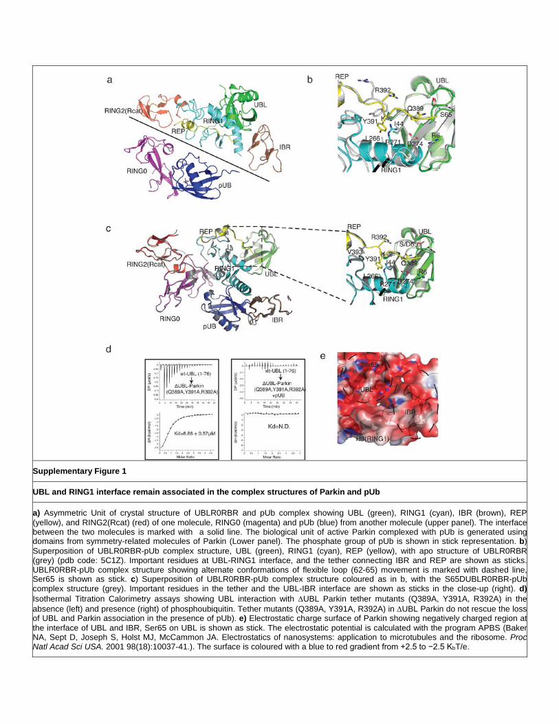
Supplementary Figure 1
UBL and RING1 interface remain associated in the complex structures of Parkin and pUb
a) Asymmetric Unit of crystal structure of UBLR0RBR and pUb complex showing UBL (green), RING1 (cyan), IBR (brown), REP (yellow), and RING2(Rcat) (red) of one molecule, RING0 (magenta) and pUb (blue) from another molecule (upper panel). The interface between the two molecules is marked with a solid line. The biological unit of active Parkin complexed with pUb is generated using domains from symmetry-related molecules of Parkin (Lower panel). The phosphate group of pUb is shown in stick representation. b) Superposition of UBLR0RBR-pUb complex structure, UBL (green), RING1 (cyan), REP (yellow), with apo structure of UBLR0RBR (grey) (pdb code: 5C1Z). Important residues at UBL-RING1 interface, and the tether connecting IBR and REP are shown as sticks. UBLR0RBR-pUb complex structure showing alternate conformations of flexible loop (62-65) movement is marked with dashed line, Ser65 is shown as stick. c) Superposition of UBLR0RBR-pUb complex structure coloured as in b, with the S65DUBLR0RBR-pUb complex structure (grey). Important residues in the tether and the UBL-IBR interface are shown as sticks in the close-up (right). d) Isothermal Titration Calorimetry assays showing UBL interaction with ∆UBL Parkin tether mutants (Q389A, Y391A, R392A) in the absence (left) and presence (right) of phosphoubiquitin. Tether mutants (Q389A, Y391A, R392A) in ∆UBL Parkin do not rescue the loss of UBL and Parkin association in the presence of pUb). e) Electrostatic charge surface of Parkin showing negatively charged region at the interface of UBL and IBR, Ser65 on UBL is shown as stick. The electrostatic potential is calculated with the program APBS (Baker NA, Sept D, Joseph S, Holst MJ, McCammon JA. Electrostatics of nanosystems: application to microtubules and the ribosome. Proc Natl Acad Sci USA. 2001 98(18):10037-41.). The surface is coloured with a blue to red gradient from +2.5 to −2.5 KbT/e.
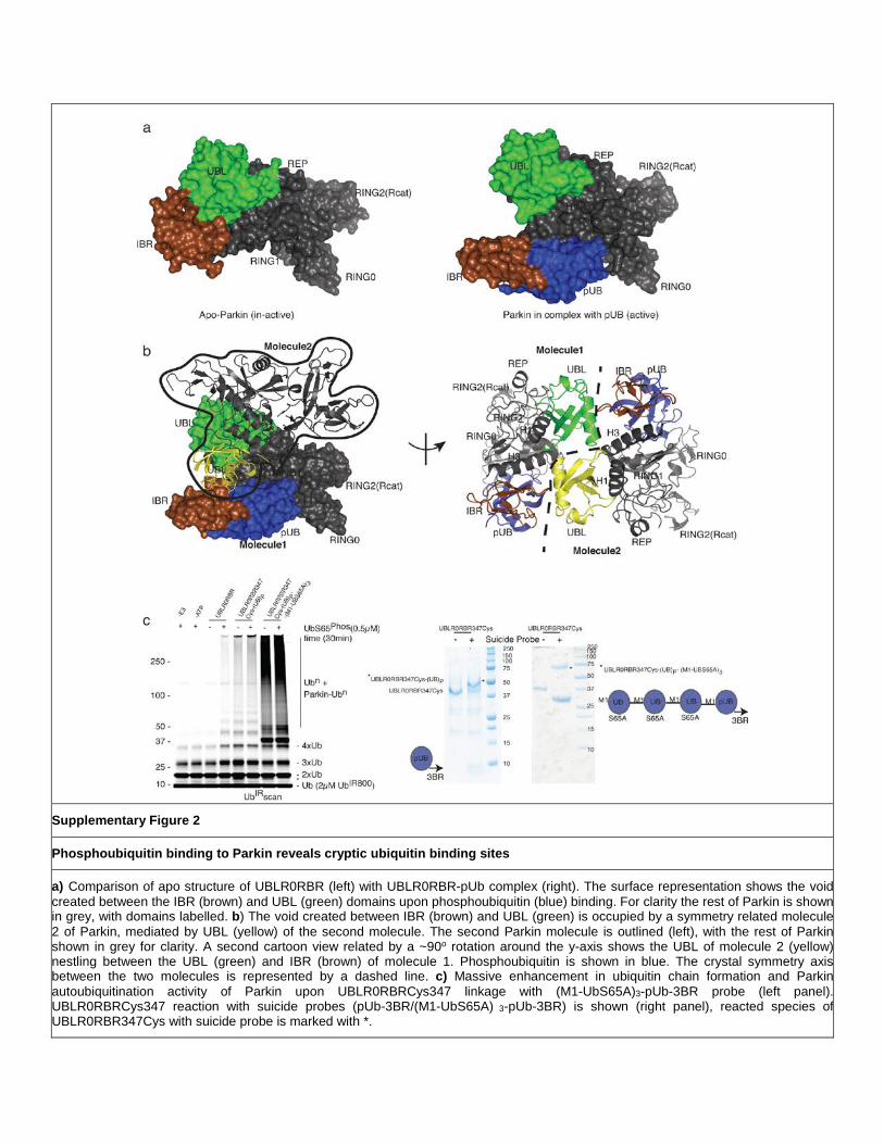
Supplementary Figure 2
Phosphoubiquitin binding to Parkin reveals cryptic ubiquitin binding sites
a) Comparison of apo structure of UBLR0RBR (left) with UBLR0RBR-pUb complex (right). The surface representation shows the void created between the IBR (brown) and UBL (green) domains upon phosphoubiquitin (blue) binding. For clarity the rest of Parkin is shown in grey, with domains labelled. b) The void created between IBR (brown) and UBL (green) is occupied by a symmetry related molecule 2 of Parkin, mediated by UBL (yellow) of the second molecule. The second Parkin molecule is outlined (left), with the rest of Parkin shown in grey for clarity. A second cartoon view related by a ~90o rotation around the y-axis shows the UBL of molecule 2 (yellow) nestling between the UBL (green) and IBR (brown) of molecule 1. Phosphoubiquitin is shown in blue. The crystal symmetry axis between the two molecules is represented by a dashed line. c) Massive enhancement in ubiquitin chain formation and Parkin autoubiquitination activity of Parkin upon UBLR0RBRCys347 linkage with (M1-UbS65A)3-pUb-3BR probe (left panel). UBLR0RBRCys347 reaction with suicide probes (pUb-3BR/(M1-UbS65A) 3-pUb-3BR) is shown (right panel), reacted species of UBLR0RBR347Cys with suicide probe is marked with *.
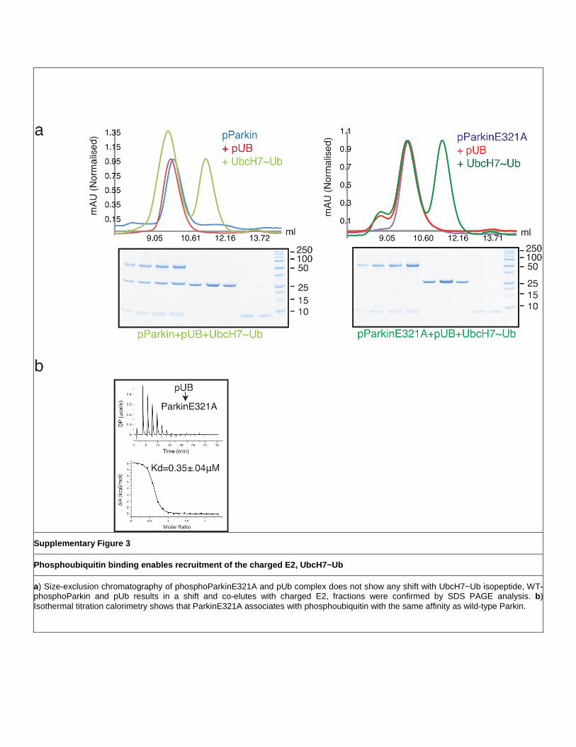
Supplementary Figure 3
Phosphoubiquitin binding enables recruitment of the charged E2, UbcH7~Ub
a) Size-exclusion chromatography of phosphoParkinE321A and pUb complex does not show any shift with UbcH7~Ub isopeptide, WT-phosphoParkin and pUb results in a shift and co-elutes with charged E2, fractions were confirmed by SDS PAGE analysis. b) Isothermal titration calorimetry shows that ParkinE321A associates with phosphoubiquitin with the same affinity as wild-type Parkin.

Supplementary Figure 4
Model of phosphoubiquitin-bound Parkin in complex with donor ubiquitin
a) The UblR0RBR structure in complex with phosphoubiquitin, coloured as in Figure 1a with all domains labelled, is modelled with a ubiquitin moiety in the H3-IBR binding site. The model is based on a combination of the crystallographically related UBL domain, the HOIP RBR/E2~Ub structure, and the chemical shift perturbations showing the ubiquitin binding site on Parkin. An arrow indicates the route from the tail of the ubiquitin molecule to the catalytic cysteine in the RING2(Rcat) domain. b) Ubiquitination assay following the ubiquitination of Miro1 in the presence of increasing concentrations of inactive UblR0RBR Parkin, or UblR0RBR covalently linked to phosphoubiquitin. Activity is monitored following fluorescently labelled ubiquitin, and a coomassie stained gel shows input levels.
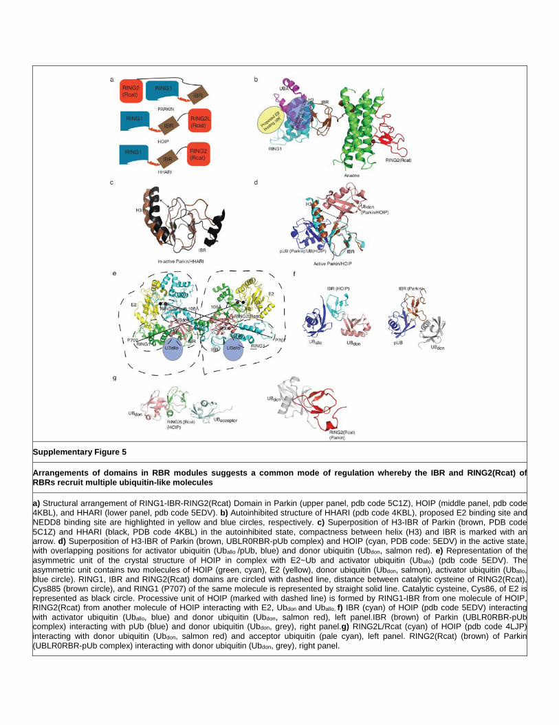
Supplementary Figure 5
Arrangements of domains in RBR modules suggests a common mode of regulation whereby the IBR and RING2(Rcat) of RBRs recruit multiple ubiquitin-like molecules
a) Structural arrangement of RING1-IBR-RING2(Rcat) Domain in Parkin (upper panel, pdb code 5C1Z), HOIP (middle panel, pdb code 4KBL), and HHARI (lower panel, pdb code 5EDV). b) Autoinhibited structure of HHARI (pdb code 4KBL), proposed E2 binding site and NEDD8 binding site are highlighted in yellow and blue circles, respectively. c) Superposition of H3-IBR of Parkin (brown, PDB code 5C1Z) and HHARI (black, PDB code 4KBL) in the autoinhibited state, compactness between helix (H3) and IBR is marked with an arrow. d) Superposition of H3-IBR of Parkin (brown, UBLR0RBR-pUb complex) and HOIP (cyan, PDB code: 5EDV) in the active state, with overlapping positions for activator ubiquitin (Uballo /pUb, blue) and donor ubiquitin (Ubdon, salmon red). e) Representation of the asymmetric unit of the crystal structure of HOIP in complex with E2~Ub and activator ubiquitin (Uballo) (pdb code 5EDV). The asymmetric unit contains two molecules of HOIP (green, cyan), E2 (yellow), donor ubiquitin (Ubdon, salmon), activator ubiquitin (Uballo, blue circle). RING1, IBR and RING2(Rcat) domains are circled with dashed line, distance between catalytic cysteine of RING2(Rcat), Cys885 (brown circle), and RING1 (P707) of the same molecule is represented by straight solid line. Catalytic cysteine, Cys86, of E2 is represented as black circle. Processive unit of HOIP (marked with dashed line) is formed by RING1-IBR from one molecule of HOIP, RING2(Rcat) from another molecule of HOIP interacting with E2, Ubdon and Uballo, f) IBR (cyan) of HOIP (pdb code 5EDV) interacting with activator ubiquitin (Uballo, blue) and donor ubiquitin (Ubdon, salmon red), left panel.IBR (brown) of Parkin (UBLR0RBR-pUb complex) interacting with pUb (blue) and donor ubiquitin (Ubdon, grey), right panel.g) RING2L/Rcat (cyan) of HOIP (pdb code 4LJP) interacting with donor ubiquitin (Ubdon, salmon red) and acceptor ubiquitin (pale cyan), left panel. RING2(Rcat) (brown) of Parkin (UBLR0RBR-pUb complex) interacting with donor ubiquitin (Ubdon, grey), right panel.
