university of groningen copper stress affects iron ... · global changes of iron homeostasis are...
TRANSCRIPT

University of Groningen
Copper Stress Affects Iron Homeostasis by Destabilizing Iron-Sulfur Cluster Formation inBacillus subtilisChillappagari, Shashi; Seubert, Andreas; Trip, Hein; Kuipers, Oscar; Marahiel, Mohamed A.;Miethke, MarcusPublished in:Journal of Bacteriology
DOI:10.1128/JB.00058-10
IMPORTANT NOTE: You are advised to consult the publisher's version (publisher's PDF) if you wish to cite fromit. Please check the document version below.
Document VersionPublisher's PDF, also known as Version of record
Publication date:2010
Link to publication in University of Groningen/UMCG research database
Citation for published version (APA):Chillappagari, S., Seubert, A., Trip, H., Kuipers, O. P., Marahiel, M. A., & Miethke, M. (2010). CopperStress Affects Iron Homeostasis by Destabilizing Iron-Sulfur Cluster Formation in Bacillus subtilis. Journalof Bacteriology, 192(10), 2512 - 2524. DOI: 10.1128/JB.00058-10
CopyrightOther than for strictly personal use, it is not permitted to download or to forward/distribute the text or part of it without the consent of theauthor(s) and/or copyright holder(s), unless the work is under an open content license (like Creative Commons).
Take-down policyIf you believe that this document breaches copyright please contact us providing details, and we will remove access to the work immediatelyand investigate your claim.
Downloaded from the University of Groningen/UMCG research database (Pure): http://www.rug.nl/research/portal. For technical reasons thenumber of authors shown on this cover page is limited to 10 maximum.
Download date: 13-10-2018

JOURNAL OF BACTERIOLOGY, May 2010, p. 2512–2524 Vol. 192, No. 100021-9193/10/$12.00 doi:10.1128/JB.00058-10Copyright © 2010, American Society for Microbiology. All Rights Reserved.
Copper Stress Affects Iron Homeostasis by Destabilizing Iron-SulfurCluster Formation in Bacillus subtilis£
Shashi Chillappagari,1 Andreas Seubert,1 Hein Trip,2 Oscar P. Kuipers,2
Mohamed A. Marahiel,1 and Marcus Miethke1*
Department of Chemistry-Biochemistry, Philipps University Marburg, Hans-Meerwein-Str., D-35032 Marburg, Germany,1 andMolecular Genetics Group, Groningen Biomolecular Sciences and Biotechnology Institute, University of Groningen,
Kerklaan 30, 9751 NN Haren, Netherlands2
Received 19 January 2010/Accepted 2 March 2010
Copper and iron are essential elements for cellular growth. Although bacteria have to overcome limitationsof these metals by affine and selective uptake, excessive amounts of both metals are toxic for the cells. Here weinvestigated the influences of copper stress on iron homeostasis in Bacillus subtilis, and we present evidencethat copper excess leads to imbalances of intracellular iron metabolism by disturbing assembly of iron-sulfurcofactors. Connections between copper and iron homeostasis were initially observed in microarray studiesshowing upregulation of Fur-dependent genes under conditions of copper excess. This effect was found to berelieved in a csoR mutant showing constitutive copper efflux. In contrast, stronger Fur-dependent geneinduction was found in a copper efflux-deficient copA mutant. A significant induction of the PerR regulon wasnot observed under copper stress, indicating that oxidative stress did not play a major role under theseconditions. Intracellular iron and copper quantification revealed that the total iron content was stable duringdifferent states of copper excess or efflux and hence that global iron limitation did not account for copper-dependent Fur derepression. Strikingly, the microarray data for copper stress revealed a broad effect on theexpression of genes coding for iron-sulfur cluster biogenesis (suf genes) and associated pathways such ascysteine biosynthesis and genes coding for iron-sulfur cluster proteins. Since these effects suggested aninteraction of copper and iron-sulfur cluster maturation, a mutant with a conditional mutation of sufU,encoding the essential iron-sulfur scaffold protein in B. subtilis, was assayed for copper sensitivity, and itsgrowth was found to be highly susceptible to copper stress. Further, different intracellular levels of SufU werefound to influence the strength of Fur-dependent gene expression. By investigating the influence of copper oncluster-loaded SufU in vitro, Cu(I) was found to destabilize the scaffolded cluster at submicromolar concen-trations. Thus, by interfering with iron-sulfur cluster formation, copper stress leads to enhanced expression ofcluster scaffold and target proteins as well as iron and sulfur acquisition pathways, suggesting a possiblefeedback strategy to reestablish cluster biogenesis.
Copper and iron are essential elements employed in path-ways that are conserved in all kingdoms of life. In eukaryotes,several interdependent connections between copper and ironhomeostasis have been described previously (21). For example,high-affinity iron uptake in Saccharomyces cerevisiae is medi-ated by multicopper-dependent Fet3p and Fet5p convertingFe(II) into Fe(III) (54, 55), and uptake of copper is associatedwith ferric reductase activity of Fre1p and Fre2p, convertingCu(II) into Cu(I) (16, 22). Further, copper starvation down-regulates respiratory functions to preserve iron and copper forother cellular processes (60). In mammals, iron transport fromthe lumen into the blood circulation is coupled with oxidationof Fe(II) to Fe(III) by the multicopper-ferroxidases hephaestinand ceruloplasmin (45, 62). Fet3p and ceruloplasmin are alsoinvolved in copper oxidation to prevent accumulation of theprooxidant Cu(I) (56).
In contrast, investigation of the relationships between ironand copper homeostasis in bacteria has only recently started.
While global effects of copper stress on the transcriptome levelhave been described for model bacteria such as Escherichia coli
and Bacillus subtilis (28, 41), detailed aspects of such connec-tions have been studied so far mainly in E. coli. The multicop-per oxidase CueO, a laccase-like enzyme present in theperiplasm, which was suggested as the site of elevated oxidiz-able copper levels (35), is responsible for the oxidation ofCu(I) and also the siderophore enterobactin, which is a poten-tial reductant for Cu(II) (17). Catecholate-containing ligandssuch as enterobactin, the major high-affinity iron scavenger inenteric bacteria (49), are able to reduce Cu(II) into Cu(I),which in turn may further oxidize compounds via formation ofreactive oxygen species (ROS) (27). CueO, which is expressedin the presence of copper, oxidizes enterobactin, its cat-echolate precursors, and its degradation products, leading tothe formation of 2-carboxymuconate if 2,3-dihydroxybenzoateis the substrate (18). Another connection between copper andiron homeostasis is the bacterial iron storage protein Dps (58).It protects E. coli cells from copper stress, but this is notdependent on presumed functions such as DNA binding orcopper storage.
Recently, investigations of the general toxicity effects of cop-per revealed that Cu(I), the predominant intracellular species(35), destabilizes iron-sulfur cofactors that are weakly bound to
* Corresponding author. Mailing address: Department of Chemis-try-Biochemistry, Philipps University Marburg, Hans-Meerwein-Str.,D-35032 Marburg, Germany. Phone: 49 6421 2825794. Fax: 49 64212822191. E-mail: [email protected].
£ Published ahead of print on 16 March 2010.
2512
at U
niv
ers
ity o
f Gro
nin
gen J
uly
13, 2
010
jb.A
SM
.OR
G -
DO
WN
LO
AD
ED
FR
OM
dehydratases of primary metabolism (34). Dihydroxy-acid de-hydratase (IlvD) in the common branched-chain amino acidsynthesis pathway and isopropylmalate dehydratase (LeuC) inthe leucine-specific branch, as well as fumarase A (FumA) and6-phosphogluconate dehydratase (Edd), were found to be af-fected in vivo, and fumarase A was also found to be affectedduring studies in vitro. In this context, it was also shown that anE. coli sufABCDSE mutant was more susceptible to copperstress, indicating that the E. coli SUF system for iron-sulfurcluster assembly contributes to copper resistance.
Much less is known about associations of copper and ironpathways in Gram-positive bacteria. In the soil-dwelling modelbacterium B. subtilis, these pathways have been separately in-vestigated in recent studies. B. subtilis employs bacillibactin asan endogenous high-affinity iron scavenger and has furtheruptake capacities for hydroxamate siderophores and ele-mental iron (38, 44). The bacillibactin pathway comprisesthe dhbACEBF genes for bacillibactin synthesis (37), ymfE (re-named ymfD upon genome resequencing), encoding a majorfacilitator superfamily transporter for bacillibactin export (6,39), feuABC-yusV, coding for an ABC-type transporter thatmediates Fe-bacillibactin uptake, and besA, which permits in-tracellular hydrolysis of the siderophore trilactone backbone(38, 44, 47). Except for the bacillibactin exporter gene, thesegenes are part of the Fur regulon, which responds to intracel-lular iron limitation (3). Other systems of iron homeostasisinclude iron storage proteins, also called “miniferritins,” whichare encoded by dps and mrgA (8, 9), as well as the recentlydescribed SUF-type system for iron-sulfur cluster maturation(1). The B. subtilis SUF system is encoded by the sufCDSUB
gene cluster, and sufU was found to be the major scaffold proteinused for cluster assembly and transfer to target proteins.
On the other hand, B. subtilis copper homeostasis is con-trolled mainly by the global regulator CsoR, which targets bothcopper efflux and influx (10, 53). The copper efflux operoncopZA codes for the CopZ copper chaperone and the CopAefflux pump, which act together (5), while the gene ycnJ codesfor a copper uptake system, which is further negatively regu-lated by YcnK (10). Another copper chaperone, YpmQ, wasfound to be essential for cytochrome maturation (36). YhdQ
(CueR), a MerR-type regulator, was found to bind to thecopZA promoter as well, but the physiological relevance of thisis uncertain (53).
The current study describes the effects of environmentalcopper excess primarily on iron homeostasis in B. subtilis. Mi-croarray studies revealed different regulatory patterns of ironhomeostasis genes depending on the intracellular copper con-tent in different strains affected in copper homeostasis. Notonly iron acquisition genes but also genes encoding iron-sulfurcluster scaffolding proteins and target enzymes of iron-sulfurcofactors were found to be affected. Direct effects observed forcopper and iron-sulfur cluster assembly on SufU and alteredexpression of iron acquisition genes depending on intracellularamounts of SufU provide evidence that iron-sulfur biogenesisis a primary target of copper toxicity and indicate that theglobal changes of iron homeostasis are driven by an enhancedrequirement for iron for iron-sulfur cluster formation undercopper stress.
MATERIALS AND METHODS
Bacterial strains, plasmids, and growth media. The bacterial strains and
plasmids used in this study are listed in Table 1. B. subtilis strains were grown in
Belitsky minimal medium (BMM) under constant shaking at 250 rpm and 37°C.
BMM was supplemented with 0.5% (wt/vol) glucose as a carbon source, 0.45 mM
glutamate, and all vital nutrients required, including 1 mM FeSO4 (57). In studies
including the sufU conditional mutant, glucose was replaced by 0.2% fructose as
a C source for all strains, and various concentrations of xylose were added to
trigger Pxyl-dependent sufU expression (1). Copper excess conditions were main-
tained by adding freshly prepared CuCl or CuSO4 at a 0.5 mM final concentra-
tion if not indicated otherwise. Fresh liquid medium for growth under different
copper conditions was inoculated from an overnight preculture that was grown at
37°C with constant shaking at 225 rpm. Special care was taken to wash the
glassware additionally with 0.1 M HCl and double-distilled water before auto-
claving. The antibiotics erythromycin (1 mg ml21), lincomycin (25 mg ml21), and
spectinomycin (100 mg ml21) were used for the selection of B. subtilis mutant
strains. E. coli strains were grown in LB medium. For selection of E. coli Top10
strains containing transformed plasmids, the antibiotic kanamycin (50 mg ml21)
was used. E. coli BL21 was used for protein overexpression. For RNA prepara-
tions, bacteria were harvested at the mid-log growth phase.
DNA manipulations and genetic techniques. DNA preparations and transfor-
mations were carried out as described previously (24, 52). Electroporation was
used for the transformation of plasmids into E. coli cells. Natural competence
obtained by high-salt/low-salt treatment was used for transformations of B. sub-
tilis ATCC 21332 during mutant constructions (29). Restriction enzymes, T4
TABLE 1. Strains and plasmids used
Strain or plasmid Genotype or description Reference or source
B. subtilis strainsATCC 21332 Wild type 11CSP100 DcsoR::erm This studyCSP104 DyhdQ::spc This studyCSP105 DcopA::spc This studyCSP106 DcsoR::erm DyhdQ::spc This studyAA04 DsufU::kan amyE::pXsufU 1
E. coli strainsTOP10 F2 mcrA D(mrr-hsdRMS-mcrBC) f80lacZDM15 DlacX74 deoR nupG
recA1 araD139 D(ara-leu)7697 galU galK rpsL (Strr) endA1 l2Invitrogen
BL21(DE3) F2 ompT gal dcm lon hsdSB(rB2 mB
2) l(DE3) Novagen
PlasmidspUS19 Donor of antibiotic resistance cassette; Spcr 7pMUTIN Donor of antibiotic resistance cassette; Ermr 59pET28a_sufU pET28a(1) containing an N-terminal His6 tag fusion of sufU 1
VOL. 192, 2010 COPPER STRESS AND IRON HOMEOSTASIS IN B. SUBTILIS 2513
at U
niv
ers
ity o
f Gro
nin
gen J
uly
13, 2
010
jb.A
SM
.OR
G -
DO
WN
LO
AD
ED
FR
OM

DNA ligase, and calf intestinal phosphatase were used according to the manu-
facturer’s instructions (New England Biolabs).
Mutant construction. B. subtilis deletion mutants were generated by introduc-
ing PCR fusions of resistance marker cassettes and chromosomal long flanking
homology regions (63). Long flanking homologous fragments were amplified
from upstream and downstream regions of the target genes using primers listed
in Table 2. The 39 ends of the resulting upstream and downstream PCR products
were designed to be complementary to the 39 ends of the fused resistance
cassette. Hybrid constructs were obtained by using the flankers as primers during
the fusion PCR. After purification, constructs were transformed into the wild-
type (WT) strain, and gene deletion mutants were selected on antibiotic plates.
All PCRs were performed with Platinum Pfx DNA polymerase (Invitrogen).
Deletions of the genes were confirmed by PCR using isolated chromosomal
DNAs of selected transformants as templates. The erythromycin resistance cas-
settes used for the construction of the DcsoR mutant was amplified from the
pMUTIN vector. To generate the DyhdQ and DcopA mutants, the spectinomycin
resistance cassette from the pUS19 vector was used.
Microarray experiments. To perform microarray experiments, ATCC 21332
WT, DcsoR, and DcsoR DyhdQ mutant strains were grown in BMM under normal
conditions (no copper added) and under copper-replete conditions (with addi-
tion of freshly prepared 0.5 mM CuCl). Cultures were harvested in the mid-log
phase for RNA extraction. The Macaloid/Roche method was used for the RNA
extraction (30). Concentrations of RNA were measured using the nanodrop
method. For reverse transcription, a solution containing 18 ml annealing mix (10
to 20 mg total RNA, 2 ml random nonamers adjusted to the final volume of 18 ml
with nuclease-free water) was incubated for 5 min at 70°C and for 10 min at 4°C
for annealing. Reverse transcription was performed by addition of this annealing
mix to a solution containing 6 ml Superscript III buffer (supplied with the reverse
transcriptase), 10 mM dithiothreitol (DTT), nucleotide master mix containing
aminoallyl-dUTP, and 300 U Superscript III reverse transcriptase (Invitrogen).
cDNA was synthesized overnight at 42°C in a total volume of 30 ml. The ami-
noallyl-modified cDNA was incubated with the CyDye N-hydroxysuccinimide
(NHS) ester (Cy3 or Cy5 monoreactive dye) at room temperature in the dark for
60 to 90 min. Labeled cDNAs were purified using Nucleo Spin Extract II col-
umns, and the dye incorporation was measured using the nanodrop method.
Equal quantities of Cy3/Cy5-incorporated cDNAs were mixed, dried via vacuum
centrifugation, and then dissolved in 5 ml H2O, followed by heating at 94°C for
2 min. The heated sample was subsequently mixed with 30 ml preheated hybrid-
izing buffer (68°C), hybridized onto microarray glass slides, and incubated over-
night at 68°C in a hybridization oven.
Microarray data analysis. Hybridized DNA arrays were read using a GenePix
autoloader 4200AL (Axon Instruments), and the data obtained were processed
with ArrayPro 4.5 (Media Cybernetics Inc., Silver Spring, MD) ArrayVision
software. Expression levels were processed and normalized (Lowess method)
with Micro-Prep software (15, 61). The ln-transformed ratios of the expression
levels were subject to a t test using the Cyber-T analysis tool (4). Three inde-
pendent measurements for each condition along with an additional dye swap
were analyzed. The results obtained were averaged, and significant expression
ratios of .1.5 for all upregulated genes and ,0.7 for all downregulated genes
were set with a general pBayes reliability of ,1023.
Dot blot analysis. B. subtilis strains were grown in BMM without (control) or
with addition of copper (0.5 mM). In studies with the sufU conditional mutant,
0.2% fructose was used as a C source, and various concentrations of xylose as
inducer were added. Inoculation of fresh medium to an initial optical density at
600 nm (OD600) of 0.05 was done with overnight precultures. Cells were har-
vested at an OD600 of 0.25, and total RNA was isolated from these cells using the
RNAZol method. RNA-to-protein ratios were determined using the nanodrop
method at 260 nm/280 nm. The RNA-to-protein ratios were above 1.65 in all
samples. Denaturing gel electrophoresis was done to test the RNA quality for
16S and 23S rRNAs. Two micrograms of RNA from each sample was subse-
quently dotted onto the nylon membrane using a dot blot apparatus and hybrid-
ized after UV cross-linking with a UTP-11-digoxigenin-labeled antisense RNA
probe. Riboprobes specific for dhbB, dhbF, feuB, feuC, and besA transcripts were
synthesized by in vitro transcription using T7 RNA polymerase and primers listed
in Table 2. After hybridization and washing, the filters were treated with a
digoxigenin-specific antibody fragment conjugated with alkaline phosphatase
(Roche) and AttoPhos (Amersham Biosciences) as an enhanced chemifluores-
cence (ECF) substrate. The hybridization signals were detected with a Storm860
fluorescence imager, and relative signal quantification was performed with
ImageQuant software.
Western blot analysis. B. subtilis WT cells were grown in BMM with 0.2%
fructose and different concentrations of copper. The sufU conditional mutant was
grown in minimal medium with 0.2% fructose and different concentrations of
xylose as an inducer of the Pxyl promoter. Cultures were harvested at an OD600
of 0.8, cells were disrupted by sonication, and cytosolic protein extracts (25 mg
total protein per lane) were separated by SDS-PAGE prior to blotting onto nylon
membranes. SufU was detected by using specific polyclonal antibodies from
rabbit and goat anti-rabbit IgG conjugated with alkaline phosphatase as a sec-
ondary antibody.
TABLE 2. Primers used to create mutants and riboprobes
Function and primer name Primer sequence (59 to 39)a
Mutant constructionCsp100-DcsoR-Us-FP ................CCACATGACGAAGCAACTTCGTACAGCsp100-DcsoR-Us-RP................GCAAGTCAGCACGAACACGAACCGCTTTTATGGTTTAATGTTTTATGTTCGTTATGCTTTTCCATCsp100-DcsoR-Ds-FP.................GTCTATTTTTAATAGTTATCTATTATTTAACGGGAGGAAATAAGGGGAACAGGCCATTTCTGAGCCsp100-DcsoR-Ds-RP................GGCTTCCCGTTTGTCACGGTTCCCsp104-DyhdQ-Us-FP................CTAAGCAGAGGGCCCCATCATACCCsp104-DyhdQ-Us-RP...............CTCTTGCCAGTCACGTTACGTTATTAGTTGAGTGCCGCTAGTTCACTGATGCGCsp104-DyhdQ-Ds-FP................CTATAAACTATTTAAATAACAGATTAAAAAAATTATAAAGCAAACTCCCGCCTGAAAAGCTCCsp104-DyhdQ-Ds-RP ...............GCAGCCCTCTTCGATGATGGAATCCsp105-DcopA-Us-FP................GCTCATGTACAACCTCAGCATCTGGCsp105-DcopA-Us-RP ...............CTCTTGCCAGTCACGTTACGTTATTAGCAACATACTCACTCCTTTATATACACCTGGCsp105-DcopA-Ds-FP................CTATAAACTATTTAAATAACAGATTAAAAAAATTATAAGGCTATATGCCGGTTTTTGTTTTTCATT
GACACCsp105-DcopA-Ds-RP ...............GCAGAACGCCGTTTTGATTGATAAAGCC
Riboprobe constructionFP RP dhbB ...............................GCGCGTTTAAGAGAACGAATCTGCRP RP dhbB ...............................TAATACGACTCACTATAGGGTTTGCTGACGTTTTTTGAACGTCTGCFP RP dhbF................................GCATTCACCATATAGCGATCGACGRP RP dhbF ...............................TAATACGACTCACTATAGGGCGCCGCTTCTTTTAACGCGTTTACFP RP feuB.................................CGGTGTTAGTATTCGGCCTTGCRP RP feuB ................................TAATACGACTCACTATAGGGGCACTGCCGGTTAGAATGATGFP RP feuC ................................GCTCTTATTCCAAGGCCAGAAGGRP RP feuC ................................TAATACGACTCACTATAGGGCTTCCGTTTTTCCACACCATGGFP RP besA ................................CCTGTGATTTATCTGCTGGATGCCRP RP besA................................TAATACGACTCACTATAGGGAGCGAATGGCCAAAGATTGTTTG
a Underlining indicates the complementary resistance cassette overlaps for mutant construction. Bold indicates T7 promoter regions in riboprobe primers.
2514 CHILLAPPAGARI ET AL. J. BACTERIOL.
at U
niv
ers
ity o
f Gro
nin
gen J
uly
13, 2
010
jb.A
SM
.OR
G -
DO
WN
LO
AD
ED
FR
OM

Determination of intracellular copper and iron concentrations. WT B. subtilis
strain ATCC 21332 and the DcsoR and DcopA mutants were grown in BMM
overnight. Fresh BMM (100 ml) either with 0.5 mM copper (excess) or without
copper (control) was inoculated with overnight cultures with a starting OD600 of
0.05, and the cells were harvested in mid-log phase. Cells were centrifuged at
18,000 3 g for 5 min, and the pellets were washed three times with buffer
containing 10 mM Tris-HCl and 1 mM EDTA (pH 7.5) and finally with MilliQ-
water to remove extracellular traces of salt. Cells were dried overnight at 85°C
and treated with suprapure nitric acid for quantitative breaking, and intracellular
metal contents were analyzed by inductively coupled plasma mass spectrometry
(ICP-MS) using an Agilent 7500ce ICP-MS.
Quantification of intracellular NAD and NADH. Estimation of nicotinamide
levels in the cells was done following published protocols (33). Briefly, the DcsoR
deletion mutant strain was grown in parallel with the WT in BMM under the
desired conditions. Cells were harvested at an OD600 of 0.6 at 4°C, and pellets
were either shock frozen in liquid nitrogen for later use or directly processed.
Cells were washed once with ice-cold 50 mM phosphate buffer (KH2PO4, pH 7.0;
adjusted with KOH) and then resuspended in 5 to 7 ml of 0.1 M Tris-HCl, pH
8.2. To differentiate between oxidized and reduced nicotinamide levels, the
suspension was divided into two equal portions of 3 ml each. The first portion
was used to extract the oxidized forms (NAD1) by adding 1.5 ml of 0.33 N HCl,
and the second portion was used to extract reduced forms (NADH) by adding 1.5
ml of 0.33 N NaOH. Both samples were incubated in a water bath maintained at
50°C for 10 min. Extracts were cooled to 0°C, and samples were neutralized by
adding 0.45 ml of 1 N NaOH to portion 1 and 0.45 ml of 1 N HCl to portion 2.
Care was taken to avoid local concentrations of acid or alkali by dropwise
addition with continuous shaking. Insoluble material was removed at 23,000 3 g
for 15 min. Further, reoxidation of NAD1 in the second portion was performed
by adding 10 ml of 146-mg/ml 2-oxoglutarate–53.3 mg of NH4Cl (pH 7) adjusted
with NaOH and 10 ml of glutamic dehydrogenase (Sigma; ;40 units/mg) and
incubating at room temperature for 15 min. Reoxidized nicotinamides were
extracted by the addition of 100 ml of 5 N HCl and incubation for 15 min at 50°C
followed by neutralization with 90 ml of 5 N NaOH. Precipitates were removed
at 23,000 3 g for 15 min. To analyze the nicotinamide levels, 2 ml of 0.1 M
pyrophosphate-semicarbazide buffer (containing 100 ml of 0.11 M sodium pyro-
phosphate, 5 ml of l M semicarbazide hydrochloride, and 5 ml of 1 M glycine, pH
8.8; adjusted with NaOH) was added to 1 ml of clarified extracts, and 10 ml
ethanol was added as a substrate. After the blank was measured at 340 nm, 10 ml
of alcohol dehydrogenase (Sigma; ;300 units/mg) was added. Reaction mixtures
were incubated at room temperature for 10 min to allow total conversion, and
final values were measured at 340 nm.
In vitro experiments with recombinant SufU. Recombinant SufU was overpro-
duced and purified as described previously (1). Briefly, cultures were induced at
an OD600 of 0.5 to 0.7 with 0.5 mM IPTG (isopropyl-b-D-thiogalactopyranoside)
for 4 h at 37°C. After being harvested, the cells were resuspended in 50 mM
HEPES–300 mM NaCl (pH 8.0) and disrupted by using a French press (Sim
Aminco) at 10,000 lb/in2. After removal of the cell debris (by centrifugation at
34,000 3 g at 4°C), the supernatant was loaded on an Ni21-nitrilotriacetic acid
(NTA) column (Qiagen). Elution was performed with a linear gradient (0 to
100%) of 50 mM HEPES, 300 mM NaCl, and 250 mM imidazole (pH 8.0) at a
flow rate of 1 ml per min over 50 min using a fast protein liquid chromatography
(FPLC) system (Amersham Pharmacia Biotech). The sample fractions were
analyzed by SDS-PAGE, and fractions with the highest protein yields were
pooled, concentrated, and dialyzed using Amicon Ultra-15 centrifugal filter units
(Millipore) with a 10,000-molecular-weight cutoff. For cluster reconstitution,
SufU (50 mM, 1 mg/ml) was reduced anaerobically (95% N2–5% H2 atmosphere)
with 5 mM DTT in 25 mM Tris-HCl–100 mM NaCl (pH 8.0) for 1 h. Ferric
ammonium citrate was then added at a stoichiometry of 4:1 (iron to protein) and
incubated for approximately 10 min until the observed color change to red was
stable. An equal amount of Li2S was added slowly, and the sample was incubated
for 15 min before unbound iron and sulfide were removed via size exclusion
chromatography over a PD-10 column equilibrated in 25 mM Tris-HCl–100 mM
NaCl (pH 8.0) containing 0.5 mM DTT. Reconstituted holo-SufU was then
titrated anaerobically with Cu(I) or Cu(II), and UV/visible absorption spectra
were recorded on a Jasco V-550 spectrophotometer.
RESULTS
Deregulation of iron uptake and amino acid metabolism
under copper stress. Microarray studies performed with theWT strain and copper efflux mutants under conditions of cop-per excess (0.5 mM copper supplementation) showed effectson both copper and iron homeostasis gene expression. Genesof the cop operon for copper efflux were upregulated about 2-to 3-fold, and genes ycnJK, involved in copper influx, weredownregulated with similar ratios in the WT under copperexcess, as expected under conditions requiring copper detoxi-fication (Table 3). Further, Fur-regulated genes for high-affin-ity iron uptake, especially via the bacillibactin pathway, were
TABLE 3. Effects of copper stress on CsoR and Fur regulation in WT, DcsoR, and DcsoR DyhdQ strains
Category and gene
Induction ratioa
Gene functionb
WT (Cu/co) DcsoR/WT (Cu)DcsoR DyhdQ/WT
(Cu)
CsoR-regulated genescopA 2.6 6.4 6.3 Copper efflux pumpcopZ NS 4.2 5.2 Copper efflux chaperoneycnJ 0.4 2.3 3.2 Copper uptake pumpycnK 0.3 1.9 4.9 Copper uptake regulator
Fur-regulated genesdhbA 1.5 0.4 0.4 2,3-Dihydro-2,3-dihydroxybenzoate dehydrogenasedhbB 2.7 0.3 0.2 IsochorismatasedhbC 1.7 0.4 0.2 Isochorismate synthasedhbE 2.2 0.3 0.2 2,3-Dihydroxybenzoate adenylationdhbF 2.3 0.3 0.1 Bacillibactin biosynthesisfeuA 2.8 0.6 0.3 Ferribacillibactin binding proteinfeuB 2.3 0.7 0.6 Ferribacillibactin uptake (integral membrane protein)feuC 1.2 0.7 0.7 Ferribacillibactin uptake (integral membrane protein)yusV 1.3 0.7 0.7 Ferribacillibactin uptake (ATPase unit)besA 2.1 0.3 0.3 Ferribacillibactin trilactone hydrolasebtr 1.6 0.8 0.9 Regulator of ferribacillibactin uptake
a Shown are mean induction ratios from four independent transcriptome comparisons. Cu, copper excess; co, control without added copper. Bold indicates ratiosshowing at least 1.5-fold upregulation, and underlining indicates ratios showing at least 1.5-fold downregulation. NS, no significant ratio based on pBayes statisticalanalysis.
b Gene functions are taken from the SubtiWiki database (13).
VOL. 192, 2010 COPPER STRESS AND IRON HOMEOSTASIS IN B. SUBTILIS 2515
at U
niv
ers
ity o
f Gro
nin
gen J
uly
13, 2
010
jb.A
SM
.OR
G -
DO
WN
LO
AD
ED
FR
OM
generally upregulated (Table 3). However, when a copper-stressed csoR knockout mutant was compared with the WTunder the same conditions, this regulatory pattern was foundto be inverted. Thus, derepression of Fur-regulated genes ap-peared to be significantly less strong in the copper-stressedDcsoR strain. Since expression of iron acquisition genes wasnot found to be altered when the transcriptome profiles of theDcsoR and WT strains were compared under normal condi-tions (data not shown), it was excluded that CsoR may have adirect regulatory effect on expression of these targets. Thus, wespeculated that this effect was based on higher copper effluxcapacities in the DcsoR strain. Indeed, the mutant showed 4- to6-fold-higher induction of copper detoxification genes copZA
than the WT under copper stress conditions (Table 3). Further,when the transcriptomes of copper-stressed WT and DcsoR
DyhdQ double mutant strains were compared, even strongerderepression rates of iron uptake genes were observed in theknockout strain, suggesting possible additive effects of CsoRand YhdQ in regulation of copper detoxification. Although thefunction of YhdQ was revised to be directly involved in tran-scriptional regulation of the B. subtilis cop operon (53), thisproposed regulator was found to bind to its promoter regionand hence could still contribute at least indirectly to localregulation. Thus, the array data indicated that copper stressinduces genes required for iron uptake, while enhanced copperdetoxification is a determinant that prevents upregulation ofthese genes.
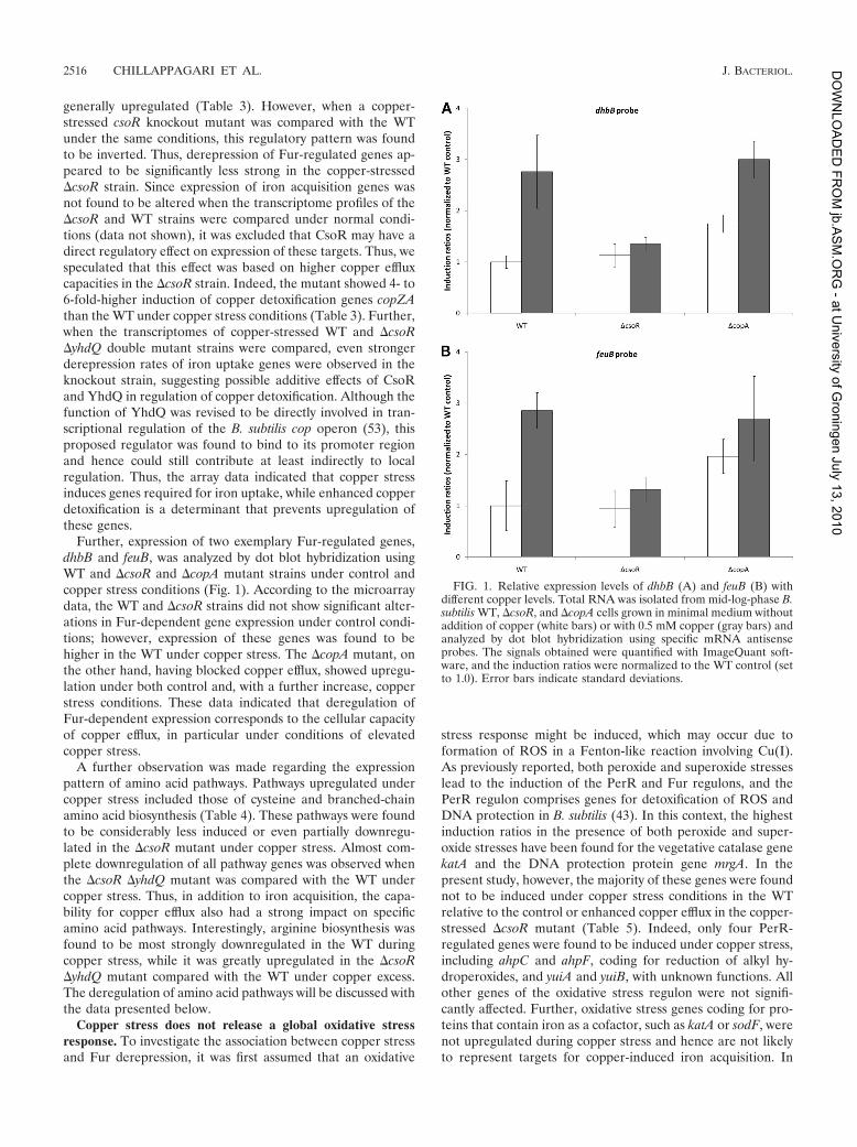
Further, expression of two exemplary Fur-regulated genes,dhbB and feuB, was analyzed by dot blot hybridization usingWT and DcsoR and DcopA mutant strains under control andcopper stress conditions (Fig. 1). According to the microarraydata, the WT and DcsoR strains did not show significant alter-ations in Fur-dependent gene expression under control condi-tions; however, expression of these genes was found to behigher in the WT under copper stress. The DcopA mutant, onthe other hand, having blocked copper efflux, showed upregu-lation under both control and, with a further increase, copperstress conditions. These data indicated that deregulation ofFur-dependent expression corresponds to the cellular capacityof copper efflux, in particular under conditions of elevatedcopper stress.
A further observation was made regarding the expressionpattern of amino acid pathways. Pathways upregulated undercopper stress included those of cysteine and branched-chainamino acid biosynthesis (Table 4). These pathways were foundto be considerably less induced or even partially downregu-lated in the DcsoR mutant under copper stress. Almost com-plete downregulation of all pathway genes was observed whenthe DcsoR DyhdQ mutant was compared with the WT undercopper stress. Thus, in addition to iron acquisition, the capa-bility for copper efflux also had a strong impact on specificamino acid pathways. Interestingly, arginine biosynthesis wasfound to be most strongly downregulated in the WT duringcopper stress, while it was greatly upregulated in the DcsoR
DyhdQ mutant compared with the WT under copper excess.The deregulation of amino acid pathways will be discussed withthe data presented below.
Copper stress does not release a global oxidative stress
response. To investigate the association between copper stressand Fur derepression, it was first assumed that an oxidative
stress response might be induced, which may occur due toformation of ROS in a Fenton-like reaction involving Cu(I).As previously reported, both peroxide and superoxide stresseslead to the induction of the PerR and Fur regulons, and thePerR regulon comprises genes for detoxification of ROS andDNA protection in B. subtilis (43). In this context, the highestinduction ratios in the presence of both peroxide and super-oxide stresses have been found for the vegetative catalase genekatA and the DNA protection protein gene mrgA. In thepresent study, however, the majority of these genes were foundnot to be induced under copper stress conditions in the WTrelative to the control or enhanced copper efflux in the copper-stressed DcsoR mutant (Table 5). Indeed, only four PerR-regulated genes were found to be induced under copper stress,including ahpC and ahpF, coding for reduction of alkyl hy-droperoxides, and yuiA and yuiB, with unknown functions. Allother genes of the oxidative stress regulon were not signifi-cantly affected. Further, oxidative stress genes coding for pro-teins that contain iron as a cofactor, such as katA or sodF, werenot upregulated during copper stress and hence are not likelyto represent targets for copper-induced iron acquisition. In
FIG. 1. Relative expression levels of dhbB (A) and feuB (B) withdifferent copper levels. Total RNA was isolated from mid-log-phase B.subtilis WT, DcsoR, and DcopA cells grown in minimal medium withoutaddition of copper (white bars) or with 0.5 mM copper (gray bars) andanalyzed by dot blot hybridization using specific mRNA antisenseprobes. The signals obtained were quantified with ImageQuant soft-ware, and the induction ratios were normalized to the WT control (setto 1.0). Error bars indicate standard deviations.
2516 CHILLAPPAGARI ET AL. J. BACTERIOL.
at U
niv
ers
ity o
f Gro
nin
gen J
uly
13, 2
010
jb.A
SM
.OR
G -
DO
WN
LO
AD
ED
FR
OM

contrast, genes under the control of the Rex (YdiH) regulator,which senses the ratio of intracellular NADH to NAD (31, 50),were significantly affected (Table 5). Under relative copperlimitation in the DcsoR mutant compared to the WT undercopper excess, genes of the cytochrome family and genes in-volved in cytochrome synthesis as well as genes encodingNADH-dependent lactate dehydrogenase and lactate per-mease were induced. Since NADH levels are expected to belinked with the copper-dependent cytochrome system, theNADH/NAD ratios in the copper-stressed WT and DcsoR
mutant strains were determined and found to be 0.9 and 3.4,respectively. Thus, copper-dependent recycling of NADH wassignificantly affected in the DcsoR mutant under copper stresscompared to the corresponding WT conditions. In the copper-stressed WT compared with the control, Rex-dependent geneswere downregulated, including cydA and cydB, coding for iron-dependent cytochrome biosynthesis. Together, these data sug-
gest that copper excess leads to significant changes in cellularenergy metabolism, which, however, are not associated withelevated levels of oxidative stress, including iron-dependentROS detoxification or increased levels of iron-dependent cy-tochrome utilization.
Copper-induced Fur derepression is related to high intra-
cellular copper content. To determine the overall intracellularlevels of copper present in the WT and the DcsoR and DcopA
mutants, ICP-MS measurements of total cell fractions werecarried out with cells cultured under different growth condi-tions. Differences in intracellular copper levels were detectedin the WT, DcsoR, and DcopA strains under normal and copperexcess conditions (Table 6). While in the WT and DcsoR mu-tant strains, copper contents were similarly low under normalconditions, DcopA cells had an about a 1,000-fold-higher cop-per content. Under conditions of copper excess, the coppercontent drastically increased in all strains, but with an 800-fold
TABLE 4. Effects of copper stress on amino acid biosynthesis genes in WT, DcsoR, and DcsoR DyhdQ strains
Category and gene
Induction ratioa
Gene functionb
WT (Cu/co) DcsoR (Cu/co)DcsoR DyhdQ/WT
(Cu)
Cysteine biosynthesiscysC 7.7 0.9 0.3 Probable adenylylsulfate kinasecysH 4.2 1.6 0.2 39-Phosphoadenylylsulfate reductasecysK 3.0 0.2 0.2 Cysteine synthetase AcysP 2.7 0.1 0.3 Sulfate permeasecysE 1.5 1.2 0.6 Serine acetyltransferasesat 6.3 0.5 0.2 Probable sulfate adenylyltransferaseylnD 6.5 0.7 0.2 Similar to uroporphyrin-III C-methyltransferaseylnE 3.5 1.2 0.4 Unknown, part of cysH operonmccA (yrhA) 3.3 0.6 1.1 O-Acetylserine-thiol-lyase (cysteine synthase)mccB (yrhB) 1.2 1.4 0.5 Cystathionine gamma-lyasecysI (yvgR) 1.6 1.4 0.7 Sulfite reductasecysJ (yvgQ) 1.7 0.4 0.7 Sulfite reductase
Leucine biosynthesisleuA 1.5 0.8 0.7 2-Isopropylmalate synthaseleuB 2.2 1.5 0.5 3-Isopropylmalate dehydrogenaseleuC 2.2 1.0 0.8 3-Isopropylmalate dehydratase (large subunit)leuD 2.2 1.2 0.5 3-Isopropylmalate dehydratase (small subunit)
Isoleucine/valine biosynthesisilvA 1.8 1.5 0.5 Threonine dehydrataseilvB 2.4 0.3 0.6 Acetolactate synthase (large subunit)ilvC 1.5 0.9 0.7 Ketol-acid reductoisomeraseilvD 2.6 0.9 0.6 Dihydroxy-acid dehydrataseilvH 2.2 0.7 0.5 Acetolactate synthase (small subunit)
Arginine biosynthesisargG 0.2 0.7 15.0 Argininosuccinate synthaseargJ 0.2 1.4 10.5 Ornithine acetyltransferase/amino acid acetyltransferaseargD 0.2 0.6 9.2 N-Acetylornithine aminotransferaseargB 0.2 0.4 7.1 N-Acetylglutamate 5-phosphotransferaseargC 0.2 2.5 6.2 N-Acetylglutamate gamma-semialdehyde dehydrogenaseargH 0.1 0.9 13.6 Argininosuccinate lyaseargF 0.1 0.3 23.5 Ornithine carbamoyltransferasecarA 0.2 0.9 13.2 Carbamoyl-phosphate transferase-arginine (subunit A)carB 0.2 0.6 14.5 Carbamoyl-phosphate transferase-arginine (subunit B)artP (yqiX) 0.1 0.1 16.8 Arginine transport (binding protein)artQ (yqiY) 0.4 0.7 6.3 Arginine transport (permease)artR (yqiZ) 0.4 1.6 5.7 Arginine transport (ATP binding protein)
a Shown are mean induction ratios from four independent transcriptome comparisons. Cu, copper excess; co, control without added copper. Bold indicates ratiosshowing at least 1.5-fold upregulation, and underlining indicates ratios showing at least 1.5-fold downregulation.
b Gene functions are taken from the SubtiWiki database (13).
VOL. 192, 2010 COPPER STRESS AND IRON HOMEOSTASIS IN B. SUBTILIS 2517
at U
niv
ers
ity o
f Gro
nin
gen J
uly
13, 2
010
jb.A
SM
.OR
G -
DO
WN
LO
AD
ED
FR
OM
difference between the WT and DcsoR strains. The differencesin intracellular copper content were found to correspond to theobserved pattern of induction of Fur-regulated genes. In con-trast, the intracellular iron content was found to be in the samerange in WT and mutant cells under normal and copper excessconditions. Thus, Fur derepression under copper stress did notdepend on changes in the total intracellular iron content. Dueto the broad differences in intracellular copper levels undernormal conditions and copper stress, it seemed unlikely that
derepression was caused by direct effects on transcriptionalregulation such as inactivation of the Fur repressor, as previ-ously suggested (41). Thus, it was assumed that copper-medi-ated induction of Fur-dependent genes was related to moreindirect changes in iron-dependent cellular processes.
Copper stress induces both iron-sulfur cluster biogenesis
and cluster target genes. One major aspect of iron homeostasisis the formation of iron-sulfur cofactors. Inspection of themicroarray data for regulation of iron-dependent pathwaysrevealed that the genes involved in biogenesis of iron-sulfurcofactors were upregulated under conditions of copper excess,including the sufU gene, encoding the major iron-sulfur clusterscaffold protein (1), and two other putative scaffold-encodinggenes, sufA and sufB (Table 7). Further, 22 of a total of 47genes currently known to code for iron-sulfur cofactor-depen-dent proteins in B. subtilis were significantly upregulated underconditions of copper excess in both the WT and the csoR
mutant. Two of the affected proteins, LeuC and YviD, wererecently shown to be targets of copper-dependent iron-sulfurcluster distortion in E. coli (34). Thus, we speculated thatcopper stress may on one hand directly influence stability ofiron-sulfur clusters bound to their target proteins but on the
TABLE 5. Effects of copper stress on PerR and Rex (YdiH) regulation in WT and DcsoR strains
Category and gene
Induction ratioa
Gene product withiron cofactor
Gene functionb
WT (Cu/co)DcsoR/WT
(Cu)
PerR-regulated geneskatA 0.8 1.2 Yes Vegetative catalase 1mrgA 0.7 1.5 Yes Metalloregulation DNA-binding stress proteinahpC 2.2 0.9 No Alkyl hydroperoxide reductase (small subunit)ahpF 2.3 0.9 No Alkyl hydroperoxide reductase (large subunit)hemA 0.8 1.2 No Glutamyl-tRNA reductasehemX 1.4 0.9 No Negative effector of the concn of HemAhemC 1.0 1.0 No Porphobilinogen deaminasehemD 1.1 1.0 No Uroporphyrinogen III cosynthasehemB 0.9 1.2 No (zinc) Delta-aminolevulinic acid dehydratasehemL 0.9 0.9 Yes Glutamate-1-semialdehyde 2,1-aminotransferasefur 1.0 1.1 Yes Transcriptional repressor of iron uptakeperR 1.1 1.3 Yes Transcriptional repressor of the peroxide reguloniseA (yoeB) 0.2 2.0 Not predicted Protection against cell envelope stressyuiA 2.1 0.9 Not predicted UnknownyuiB 3.2 0.8 Not predicted UnknownzosA (ykvW) 1.0 1.1 No (zinc) Similar to heavy metal-transporting ATPase
Other oxidative stress genessodA 1.5 1.3 No (manganese) Superoxide dismutasesodF 0.8 1.0 Yes Superoxide dismutaseyojM 1.2 0.9 No (zinc) Similar to superoxide dismutase
Rex-regulated genesldh 0.3 7.2 No L-Lactate dehydrogenaselctP 0.3 5.9 No L-Lactate permeaseywcJ 0.3 3.8 No Similar to nitrite transportercydA 0.7 2.0 Yes Cytochrome bd ubiquinol oxidase (subunit I)cydB 0.3 4.9 Yes Cytochrome bd ubiquinol oxidase (subunit II)cydC 0.6 2.4 No ABC transporter required for expression of cytochrome bd
(ATP binding protein)cydD 0.9 NS No ABC transporter required for expression of cytochrome bd
(ATP binding protein)
a Shown are mean induction ratios from four independent transcriptome comparisons. Cu, copper excess; co, control without added copper. Bold indicates ratiosshowing at least 1.5-fold upregulation, and underlining indicates ratios showing at least 1.5-fold downregulation. NS, no significant ratio based on pBayes statisticalanalysis.
b Gene functions are taken from the SubtiWiki database (13).
TABLE 6. ICP-MS measurement of total intracellular copper andiron contents under copper excess or control conditions in WT,
DcsoR, and DcopA strains
Strain
Cu content(ppm)a
Cucontentratio
(Cu/co)
Fe content(ppm)
Fecontentratio
(Cu/co)Cu co Cu co
WT 325 0.005 65,000 0.1 0.077 1.3DcsoR mutant 0.4 0.002 200 0.065 0.092 0.7DcopA mutant 892 2.0 446 0.217 0.082 2.6
a Cu, copper excess; co, control. Values are based on dry cell weights ofsamples.
2518 CHILLAPPAGARI ET AL. J. BACTERIOL.
at U
niv
ers
ity o
f Gro
nin
gen J
uly
13, 2
010
jb.A
SM
.OR
G -
DO
WN
LO
AD
ED
FR
OM
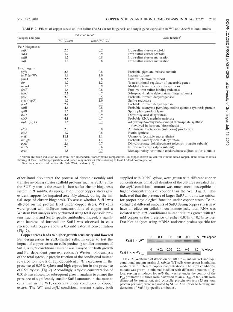
other hand also target the process of cluster assembly andtransfer involving cluster scaffold proteins such as SufU. Sincethe SUF system is the essential iron-sulfur cluster biogenesissystem in B. subtilis, its upregulation under copper stress gaveevident support for impaired assembly already during the ini-tial steps of cluster biogenesis. To assess whether SufU wasaffected on the protein level under copper stress, WT cellswere grown with different concentrations of copper and aWestern blot analysis was performed using total cytosolic pro-tein fractions and SufU-specific antibodies. Indeed, a signifi-cant increase of intracellular SufU was observed in cellsstressed with copper above a 0.3 mM external concentration(Fig. 2).
Copper stress leads to higher growth sensitivity and lowered
Fur derepression in SufU-limited cells. In order to test theimpact of copper stress on cells producing smaller amounts ofSufU, a sufU conditional mutant was assayed for both growthand Fur-dependent gene expression. A Western blot analysisof the total cytosolic protein fraction of the conditional mutantrevealed low levels of Pxyl-dependent sufU expression in thepresence of 0.05% xylose and high expression in the presenceof 0.5% xylose (Fig. 2). Accordingly, a xylose concentration of0.05% was chosen for subsequent growth analysis to ensure thepresence of significantly smaller SufU amounts in the mutantcells than in the WT, especially under conditions of copperexcess. The WT and sufU conditional mutant strains, both
supplied with 0.05% xylose, were grown with different copperconcentrations. Final cell densities of the cultures revealed thatthe sufU conditional mutant was much more susceptible tohigher concentrations of copper than the WT (Fig. 3). Thisindicated that the presence of larger SufU amounts was criticalfor proper physiological function under copper stress. To in-vestigate if different amounts of SufU during copper stress mayhave an effect on cellular iron homeostasis, total RNA wasisolated from sufU conditional mutant cultures grown with 0.5mM copper in the presence of either 0.05% or 0.5% xylose.Dot blot analyses using mRNA antisense probes specific for
TABLE 7. Effects of copper stress on iron-sulfur (Fe-S) cluster biogenesis and target gene expression in WT and DcsoR mutant strains
Category and geneInduction ratioa
Gene functionb
WT (Cu/co) DcsoR/WT (Cu)
Fe-S biogenesissufU 2.3 0.7 Iron-sulfur cluster scaffoldsufA 1.9 0.9 Iron-sulfur cluster scaffoldsufB 1.7 0.8 Iron-sulfur cluster maturationsufC 1.8 0.8 Iron-sulfur cluster maturation
Fe-S targetsysfD 2.3 0.8 Probable glycolate oxidase subunitlutB (yvfW) 1.9 1.0 Lactate oxidaseyhbA 2.6 0.8 Putative electron transportfnr 1.7 1.2 Transcriptional regulator of anaerobic genesmoaA 1.5 0.9 Molybdopterin precursor biosynthesisfadF 1.6 0.8 Putative iron-sulfur binding reductaseleuC 2.2 0.7 3-Isopropylmalate dehydratase (large subunit)yrhE 2.0 0.5 Probable formate dehydrogenasecysJ (yvgQ) 1.7 1.0 Sulfite reductaseyoaE 2.7 0.7 Probable formate dehydrogenaseskfB 4.6 0.8 Probable coenzyme pyrroloquinoline quinone synthesis proteinsplB 1.6 0.8 Spore photoproduct lyaseilvD 2.6 0.9 Dihydroxy-acid dehydrataseyfjO 4.1 0.7 Probable RNA methyltransferaseispG (yqfY) 1.6 0.7 4-Hydroxy-3-methylbut-2-en-1-yl diphosphate synthase
(involved in isoprene biosynthesis)albA 2.0 0.8 Antilisterial bacteriocin (subtilosin) productionbioB 1.9 0.8 Biotin synthaseyotD 11.1 1.1 Unknown (possible rubrerythrin)mmgE 1.5 1.1 Probable 2-methylcitrate dehydratasepyrK 2.6 0.7 Dihydroorotate dehydrogenase (electron transfer subunit)narG 2.0 0.6 Nitrate reductase (alpha subunit)qcrA 1.5 0.7 Menaquinol:cytochrome c oxidoreductase (iron-sulfur subunit)
a Shown are mean induction ratios from four independent transcriptome comparisons. Cu, copper excess; co, control without added copper. Bold indicates ratiosshowing at least 1.5-fold upregulation, and underlining indicates ratios showing at least 1.5-fold downregulation.
b Gene functions are taken from the SubtiWiki database (13).
FIG. 2. Western blot detection of SufU in B. subtilis WT and sufUconditional mutant strains. B. subtilis WT cells were grown in minimalmedium with different copper concentrations. The sufU conditionalmutant was grown in minimal medium with different amounts of xy-lose, serving as inducer for sufU that was set under the control of thePxyl promoter. Cultures were harvested at an OD600 of 0.8, cells weredisrupted by sonication, and cytosolic protein extracts (25 mg totalprotein per lane) were separated by SDS-PAGE prior to blotting anddetection of SufU by specific antibodies.
VOL. 192, 2010 COPPER STRESS AND IRON HOMEOSTASIS IN B. SUBTILIS 2519
at U
niv
ers
ity o
f Gro
nin
gen J
uly
13, 2
010
jb.A
SM
.OR
G -
DO
WN
LO
AD
ED
FR
OM

five Fur-dependent gene transcripts were performed to detectFur expression levels with smaller and larger amounts of in-tracellular SufU. The analysis revealed that mutant cells grownin the presence of 0.5% xylose showed a 2- to 3-fold-higherinduction of the tested Fur-regulated genes than those grownin the presence of 0.05% xylose (Fig. 4), indicating an associ-ation of higher intracellular SufU levels and Fur derepressionunder the tested conditions. Thus, since SufU is an importantcomponent of intracellular iron distribution via iron-sulfurcluster assembly and targeting, it was suggested that higherrequirements for iron may result not only from reduced iron-sulfur cluster stability on various target proteins but also fromimpaired stability during cluster assembly on SufU under cop-per stress.
Iron-sulfur cluster scaffolding on SufU is a direct target of
copper stress in vitro. To assess whether copper leads to im-paired cluster stability on SufU, the scaffold protein was re-combinantly produced and was loaded with iron-sulfur clusteranaerobically by in vitro reconstitution as described previously(1). The purified holo-SufU protein carried a [4Fe-4S] clusteras the major cluster species as indicated by cluster type-specificabsorption between 350 and 450 nm in the UV/visible spec-trum. holo-SufU was then titrated anaerobically with increas-ing amounts of Cu(I) or Cu(II). With increasing amounts ofCu(I), a decrease of cluster-specific absorption was observed(Fig. 5A). Even submicromolar concentrations of Cu(I) hadsignificant effects on cluster stability. Near the range ofequimolar stoichiometries of Cu(I) (10 mM) and holo-SufU(50 mM), cluster absorbance was nearly completely abolished,indicating almost complete disintegration of the protein-boundcluster. In contrast, cluster destabilization was not observedwhen titrating with Cu(II) in the same molar range (Fig. 5B).This suggested that elevated levels of intracellular Cu(I) mayhave direct effects on iron-sulfur cluster assembly and clusterstability on SufU, which in turn had an impact on severalcentral iron-sulfur-dependent cellular processes, as shown pre-
viously (1), and may further reflect the observed deregulationof gene expression in different pathways associated with ironand sulfur homeostasis.
DISCUSSION
While interdependent processes in homeostasis of iron andcopper have been shown to occur in eukaryotes, knowledgeabout such processes in bacterial systems is still limited. Here,the Gram-positive model bacterium B. subtilis was tested for itsresponse to copper stress. Generally, this leads to the inductionof detoxification mechanisms, such as the CsoR-dependentCopZA efflux system in B. subtilis (53) or the CueR-regulatedcytoplasmic efflux pump CopA and periplasmic copper-clear-ance system CueO, as well as CusRS-dependent CusCFBA, inE. coli (46). While copper in vitro leads to the initiation ofFenton-like reactions resulting in generation of ROS (20), itwas thought that release of the oxidative stress defense mightbe another response induced by copper stress. However, thetranscriptome data on copper stress in WT and copper effluxmutant strains gave another scenario of how copper stressglobally affects cellular physiology. Interestingly, pathways foracquisition of iron and sulfur components were found to beupregulated, including the Fur-dependent bacillibactin path-way and cysteine biosynthesis. These findings are in agreementwith a previous study on global metal ion stress (41). However,the lower Fur induction ratios observed in our study may bedue to the use of B. subtilis ATCC 21332 (sfp1) as the WTbackground, which is capable of high-affinity iron scavengingvia bacillibactin. Furthermore, our study showed upregulationduring copper stress of pathways that depend on the presenceof iron-sulfur cofactors, such as those for branched-chainamino acid biosynthesis. The iron-sulfur cluster enzymes in-volved in valine, isoleucine, and leucine synthesis, IlvD and
FIG. 4. Relative expression levels of Fur-regulated genes dhbB,dhbF, feuB, feuC, and besA in the B. subtilis sufU conditional mutantgrown in minimal medium under conditions of copper excess (0.5 mMcopper) in the presence of 0.05% xylose (white bars) or 0.5% xylose(gray bars). Total RNA was isolated from mid-log-phase cultures andanalyzed by dot blot hybridization using specific mRNA antisenseprobes (three hybridizations each), and the signals obtained werequantified with ImageQuant software. Means of hybridization signalswere normalized for each probe to those obtained from the 0.05%xylose conditions (set to 1.0) and were plotted with correspondingstandard deviations.
FIG. 3. Growth analysis of B. subtilis WT (white bars) and sufUconditional mutant (gray bars) strains under copper stress. Cells weregrown in minimal medium with different concentrations of copper inmicrotiter scale with three parallels for each strain and growth condi-tion. Final cell densities (OD600) were measured after 12 h of growth.Means of the parallel determinations were normalized to the celldensities of control cultures without added copper (set to 100%) andwere plotted with corresponding standard deviations.
2520 CHILLAPPAGARI ET AL. J. BACTERIOL.
at U
niv
ers
ity o
f Gro
nin
gen J
uly
13, 2
010
jb.A
SM
.OR
G -
DO
WN
LO
AD
ED
FR
OM

LeuC, were recently found to be targets of copper stress bysystematic growth feeding experiments in E. coli (34). In ad-dition to ilvD and leuC, we identified 20 other genes coding foriron-sulfur cluster-dependent proteins that were upregulatedduring copper excess conditions. Among them are genes en-coding enzymes for biotin synthesis (BioB), molybdopterinsynthesis (MoaA), pyrimidine synthesis (PyrK), and electrontransfer components such as QcrA of the respiratory chain.They might not in all cases be direct targets of copper-depen-dent iron-sulfur cluster destabilization, as most likely in casesof IlvD and LeuC, and upregulation may involve further phys-iological effects associated with their metabolic function. How-ever, since copper primarily destabilizes solvent-exposed clus-ters as found, e.g., in dehydratases (34), there is a furtherpotential layer of action associated with copper toxicity. Mat-uration of iron-sulfur cluster proteins depends not only oncluster integrity at the final target sites but also on the up-stream process of cluster biogenesis and transfer. As this study
demonstrates, cluster biogenesis is also prone to destabiliza-tion by copper excess. B. subtilis contains the SUF system foriron-sulfur cluster assembly, with SufU as a major scaffoldingprotein (1). The sufU gene was found to be induced underconditions of copper excess together with two other potentialscaffold genes, sufA and sufB, and cluster formation on recom-binant SufU was revealed to be susceptible to Cu(I). Sinceiron-sulfur clusters are transiently bound to scaffolding pro-teins and are generally solvent accessible during this state ofmaturation (32, 42), destabilization by copper at the biogenesislevel may be an effect. The data showed further that SufU in
vivo protects cells against copper toxicity, as indicated for theSUF system in E. coli (34). In this context, it is interesting tonote that E. coli, possessing both the constitutive ISC and thestress-dependent SUF cluster biogenesis systems (26), is gen-erally less susceptible to copper stress than B. subtilis. Indeed,transcriptome studies in E. coli revealed almost no significanteffects on the expression levels of Fur-regulated iron acquisi-tion genes in the presence of 0.75 mM copper-glycine in themedium, but showed effects only in the presence of 2 mMcopper-glycine, yielding expression rates similar to those ob-served in our study (28). Further studies showed that E. coli
WT strains are viable at copper concentrations of above 10mM (18, 23), whereas concentrations of above 1 to 5 mM werealready lethal for B. subtilis. In addition to the possibility thatdetoxification systems are more efficient in E. coli, lower coppertoxicity could also result from the presence of two iron-sulfurcluster biogenesis systems instead of one in B. subtilis, althoughthe SUF system was shown to be more protective not only undercopper stress but also under an excess of cobalt (34, 48).
Regarding the toxicity mechanism in the case of increasingintracellular copper content, copper may replace iron at thecluster binding site by occupying the cysteine-thiolate donorpositions, as recently suggested (34). Indeed, it is well knownthat the Cu(I) and Fe(III) ion species are highly competitive(64), and the selective influence of Cu(I), which is the predom-inant intracellular copper species (35), may further result fromits natural preference for S donor ligands, while Cu(II) prefersN donors (2). The model of metal replacement at the iron-sulfur cluster binding sites is also supported by the finding thatcobalt stress also affects the function of iron-sulfur clusterproteins, including the IscU and SufA scaffolding componentsin E. coli (48). Cobalt can occupy metal binding sites withmedium affinity among the physiological relevant trace metalsbased on the Irving-Williams series, and thus both copper andcobalt could successfully compete for thiolate donor sites ifthese metals are present in excess. Alternatively, it cannot beexcluded that redox processes are involved in cluster destabi-lization, such as electron exchanges between Cu(I) and cluster-bound Fe(III). Thus, if iron-sulfur cluster assembly is notshielded from intracellular copper either by efflux systems orby cytosolic sequestration, which is mediated by metallothio-neins and putatively also ferritins in several bacteria (51), clus-ter damage on biogenesis scaffolds can be considered a seriousconsequence if copper amounts exceed cellular efflux and stor-age capacities.
Still, the open question remains as to how copper inducesthe iron starvation conditions indicated by Fur derepression onglobal scale. Generally, several alternatives could be raised toexplain the mechanism of Fur derepression. First, copper-in-
FIG. 5. Effect of copper on stability of holo-SufU in vitro. Recom-binant SufU was reconstituted anaerobically with iron and sulfur toyield, after purification via a PD-10 size exclusion column, about 50mM holo-SufU protein carrying a [4Fe-4S] cluster with typical absorp-tion features (“shoulder”) between 350 and 450 nm. The protein wastitrated anaerobically with Cu(I) (A) or Cu(II) (B), and absorptionspectra were recorded after each titration step. Cu(I) concentrationsduring titration were 0, 0.001, 0.006, 0.016, 0.05, 0.075, 0.1, 0.2, 0.5,0.75, 1, 2, 5, and 10 mM, resulting in a continuous decrease of absorp-tion of the [4Fe-4S] shoulder. Cu(II) concentrations during titrationwere 0, 0.01, 0.1, 1, and 10 mM, which did not result in significantbleaching of cluster absorption.
VOL. 192, 2010 COPPER STRESS AND IRON HOMEOSTASIS IN B. SUBTILIS 2521
at U
niv
ers
ity o
f Gro
nin
gen J
uly
13, 2
010
jb.A
SM
.OR
G -
DO
WN
LO
AD
ED
FR
OM
duced oxidative stress may lead to oxidation of the Fur core-pressor Fe(II). However, in contrast to observations in vitro
(20), oxidative stress was not released by copper in vivo andthus is unlikely to contribute to Fur derepression. This obser-vation is in agreement with a previous study showing thatcopper stress does not catalyze oxidative DNA damage in E.
coli and actually protects cells from ROS activity (35). Second,we excluded that the Fur repressor was inactivated by replace-ment of iron by copper, since the high intracellular copperlevels found in the WT upon copper stress did not lead to fullderepression of the target genes, as found for a fur mutant inprevious studies (3), but were comparable to those found dur-ing iron limitation (,0.1 mM extracellular iron) in the bacilli-bactin-producing WT strain (40). Further, it was shown for E.
coli Fur that Cu(II) binds and converts it into a repressor in
vitro (12). Thus, if a switch from iron to copper were possiblein vivo, a strengthened repression of the Fur regulon would beexpected, which would represent the opposite effect of theobserved response. The absence of this effect at all is mostlikely due to the intracellular Cu(I) state, which prefers adifferent ligand environment than Cu(II). Extracellularly in-duced iron limitation by decreasing iron solubility in the pres-ence of high salinity (0.7 M NaCl) (25) can further be excludedas a relevant parameter for Fur derepression in this study,since the effective copper salt concentrations used were morethan 1,000-fold lower, and intracellular total iron levels werefound to be similar under both copper excess and normalconditions. Thus, since iron was sufficiently supplied to copper-
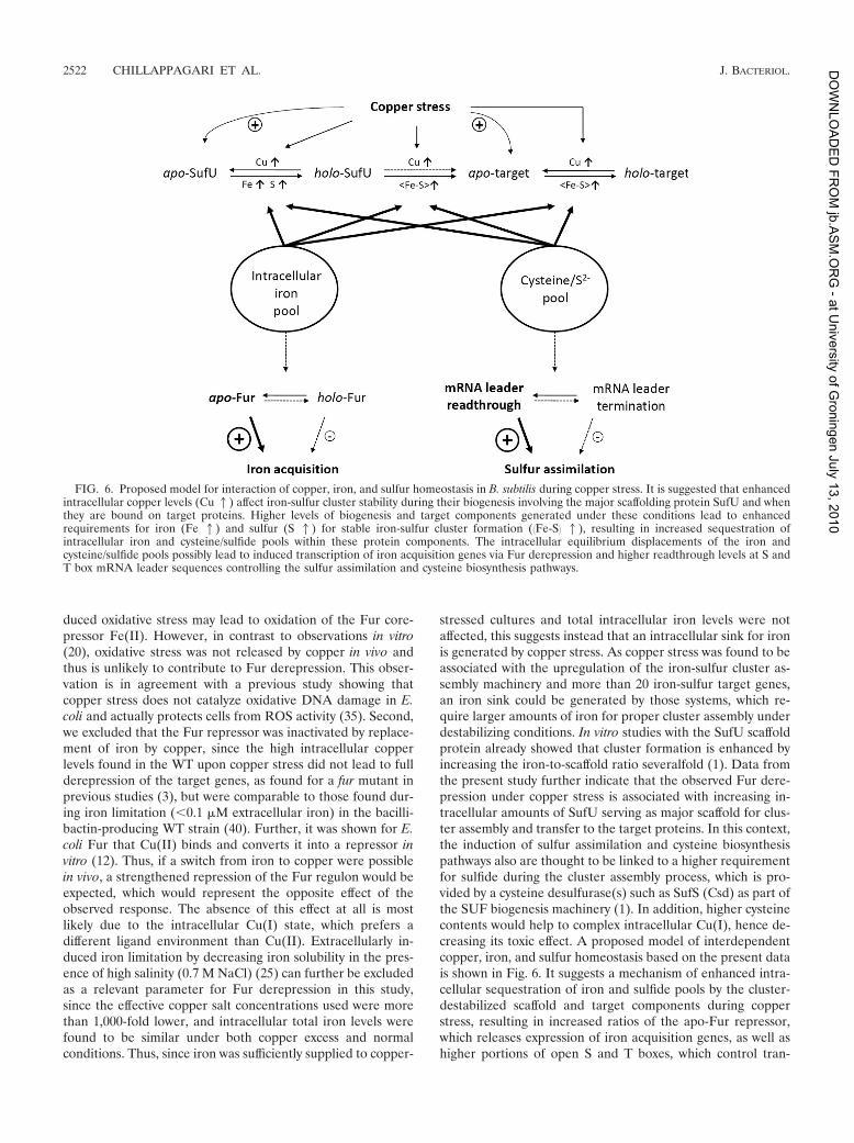
stressed cultures and total intracellular iron levels were notaffected, this suggests instead that an intracellular sink for ironis generated by copper stress. As copper stress was found to beassociated with the upregulation of the iron-sulfur cluster as-sembly machinery and more than 20 iron-sulfur target genes,an iron sink could be generated by those systems, which re-quire larger amounts of iron for proper cluster assembly underdestabilizing conditions. In vitro studies with the SufU scaffoldprotein already showed that cluster formation is enhanced byincreasing the iron-to-scaffold ratio severalfold (1). Data fromthe present study further indicate that the observed Fur dere-pression under copper stress is associated with increasing in-tracellular amounts of SufU serving as major scaffold for clus-ter assembly and transfer to the target proteins. In this context,the induction of sulfur assimilation and cysteine biosynthesispathways also are thought to be linked to a higher requirementfor sulfide during the cluster assembly process, which is pro-vided by a cysteine desulfurase(s) such as SufS (Csd) as part ofthe SUF biogenesis machinery (1). In addition, higher cysteinecontents would help to complex intracellular Cu(I), hence de-creasing its toxic effect. A proposed model of interdependentcopper, iron, and sulfur homeostasis based on the present datais shown in Fig. 6. It suggests a mechanism of enhanced intra-cellular sequestration of iron and sulfide pools by the cluster-destabilized scaffold and target components during copperstress, resulting in increased ratios of the apo-Fur repressor,which releases expression of iron acquisition genes, as well ashigher portions of open S and T boxes, which control tran-
FIG. 6. Proposed model for interaction of copper, iron, and sulfur homeostasis in B. subtilis during copper stress. It is suggested that enhancedintracellular copper levels (Cu1) affect iron-sulfur cluster stability during their biogenesis involving the major scaffolding protein SufU and whenthey are bound on target proteins. Higher levels of biogenesis and target components generated under these conditions lead to enhancedrequirements for iron (Fe 1) and sulfur (S 1) for stable iron-sulfur cluster formation (KFe-SL 1), resulting in increased sequestration ofintracellular iron and cysteine/sulfide pools within these protein components. The intracellular equilibrium displacements of the iron andcysteine/sulfide pools possibly lead to induced transcription of iron acquisition genes via Fur derepression and higher readthrough levels at S andT box mRNA leader sequences controlling the sulfur assimilation and cysteine biosynthesis pathways.
2522 CHILLAPPAGARI ET AL. J. BACTERIOL.
at U
niv
ers
ity o
f Gro
nin
gen J
uly
13, 2
010
jb.A
SM
.OR
G -
DO
WN
LO
AD
ED
FR
OM

scription termination during sulfur assimilation and cysteinebiosynthesis (19).
Interestingly, and as a complement to our findings, the op-posite effect of increased Fur repression during copper starva-tion has also been observed in Pseudomonas aeruginosa (14).Although it is presumed that this was caused by lower demandfor iron through the respiratory chain, a total knockout of allcopper-dependent terminal oxidases did not change the effectof Fur target downregulation. Thus, it is possible that alsounder copper starvation and standard conditions, Fur regula-tion is influenced through different levels of intracellular cop-per toxicity, the onset of which was observed here at lownanomolar concentrations with holo-SufU as a target in vitro.
Altogether, the upregulation of physiologically importantpathways during copper stress may be regarded as a recruitingstrategy for components and substrates required for ensuringfunctional iron-sulfur cluster biogenesis also under destabiliz-ing conditions of copper toxicity. Further investigations, espe-cially on iron-sulfur cluster formation and transfer in vivo, willbe necessary to obtain more insights into the processes ofmetal-induced iron-sulfur cluster destabilization and reconsti-tution.
ACKNOWLEDGMENTS
We thank Evert-Jan Blom (University of Groningen) for help withmicroarray studies, Erhard Bremer (University of Marburg) for helpwith chemifluorescence detection, Roland Lill (University of Marburg)for support with antibody production, Alexander G. Albrecht (Univer-sity of Marburg) for supplying purified protein, and Jurgen Knoll andRudiger Penzel (University of Marburg) for help with ICP-MS mea-surements.
We gratefully acknowledge the Deutsche Forschungsgemeinschaftand Fonds der Chemischen Industrie for financial support.
REFERENCES
1. Albrecht, A. G., D. J. A. Netz, M. Miethke, A. J. Pierik, O. Burghaus, F.
Peuckert, R. Lill, and M. A. Marahiel. 2010. SufU is an essential iron-sulfurcluster scaffold protein in Bacillus subtilis. J. Bacteriol. 192:1643–1651.
2. Andreini, C., L. Banci, I. Bertini, and A. Rosato. 2008. Occurrence of copperproteins through the three domains of life: a bioinformatic approach. J.Proteome Res. 7:209–216.
3. Baichoo, N., T. Wang, R. Ye, and J. D. Helmann. 2002. Global analysis of theBacillus subtilis Fur regulon and the iron starvation stimulon. Mol. Microbiol.45:1613–1629.
4. Baldi, P., and A. D. Long. 2001. A Bayesian framework for the analysis ofmicroarray expression data: regularized t-test and statistical inferences ofgene changes. Bioinformatics 17:509–519.
5. Banci, L., I. Bertini, S. Ciofi-Baffoni, R. Del Conte, and L. Gonnelli. 2003.Understanding copper trafficking in bacteria: interaction between the coppertransport protein CopZ and the N-terminal domain of the copper ATPaseCopA from Bacillus subtilis. Biochemistry 42:1939–1949.
6. Barbe, V., S. Cruveiller, F. Kunst, P. Lenoble, G. Meurice, A. Sekowska, D.
Vallenet, T. Wang, I. Moszer, C. Medigue, and A. Danchin. 2009. From aconsortium sequence to a unified sequence: the Bacillus subtilis 168 refer-ence genome a decade later. Microbiology 155:1758–1775.
7. Benson, A. K., and W. G. Haldenwang. 1993. Regulation of sigma B levelsand activity in Bacillus subtilis. J. Bacteriol. 175:2347–2356.
8. Bozzi, M., G. Mignogna, S. Stefanini, D. Barra, C. Longhi, P. Valenti, and E.
Chiancone. 1997. A novel non-heme iron-binding ferritin related to theDNA-binding proteins of the Dps family in Listeria innocua. J. Biol. Chem.272:3259–3265.
9. Chen, L., and J. D. Helmann. 1995. Bacillus subtilis MrgA is a Dps(PexB)homologue: evidence for metalloregulation of an oxidative-stress gene. Mol.Microbiol. 18:295–300.
10. Chillappagari, S., M. Miethke, H. Trip, O. P. Kuipers, and M. A. Marahiel.
2009. Copper acquisition is mediated by YcnJ and regulated by YcnK andCsoR in Bacillus subtilis. J. Bacteriol. 191:2362–2370.
11. Cooper, D. G., C. R. Macdonald, S. J. Duff, and N. Kosaric. 1981. Enhancedproduction of surfactin from Bacillus subtilis by continuous product removaland metal cation additions. Appl. Environ. Microbiol. 42:408–412.
12. de Lorenzo, V., S. Wee, M. Herrero, and J. B. Neilands. 1987. Operator
sequences of the aerobactin operon of plasmid ColV-K30 binding the ferricuptake regulation (fur) repressor. J. Bacteriol. 169:2624–2630.
13. Florez, L. A., S. F. Roppel, A. G. Schmeisky, C. R. Lammers, and J. Stulke.
2009. A community-curated consensual annotation that is continuously up-dated: the Bacillus subtilis centred wiki SubtiWiki. Database (Oxford) 2009:
bap012.14. Frangipani, E., V. I. Slaveykova, C. Reimmann, and D. Haas. 2008. Adap-
tation of aerobically growing Pseudomonas aeruginosa to copper starvation.J. Bacteriol. 190:6706–6717.
15. Garcia de la Nava, J., S. van Hijum, and O. Trelles. 2003. PreP: geneexpression data pre-processing. Bioinformatics 19:2328–2329.
16. Georgatsou, E., L. A. Mavrogiannis, G. S. Fragiadakis, and D. Alexandraki.
1997. The yeast Fre1p/Fre2p cupric reductases facilitate copper uptake andare regulated by the copper-modulated Mac1p activator. J. Biol. Chem.272:13786–13792.
17. Grass, G., and C. Rensing. 2001. CueO is a multi-copper oxidase that conferscopper tolerance in Escherichia coli. Biochem. Biophys. Res. Commun. 286:
902–908.18. Grass, G., K. Thakali, P. E. Klebba, D. Thieme, A. Muller, G. F. Wildner,
and C. Rensing. 2004. Linkage between catecholate siderophores and themulticopper oxidase CueO in Escherichia coli. J. Bacteriol. 186:5826–5833.
19. Grundy, F. J., and T. M. Henkin. 2002. Synthesis of serine, glycine, cysteine,and methionine, p. 245–254. In A. L. Sonenshein (ed.), Bacillus subtilis andits closest relatives. ASM Press, Washington, DC.
20. Gunther, M. R., P. M. Hanna, R. P. Mason, and M. S. Cohen. 1995. Hydroxylradical formation from cuprous ion and hydrogen peroxide: a spin-trappingstudy. Arch. Biochem. Biophys. 316:515–522.
21. Harris, E. D. 2001. Copper and iron: a landmark connection of two essentialmetals. J. Trace Elem. Exp. Med. 14:207–210.
22. Hassett, R., and D. J. Kosman. 1995. Evidence for Cu(II) reduction as acomponent of copper uptake by Saccharomyces cerevisiae. J. Biol. Chem.270:128–134.
23. Hiniker, A., J. F. Collet, and J. C. Bardwell. 2005. Copper stress causes an invivo requirement for the Escherichia coli disulfide isomerase DsbC. J. Biol.Chem. 280:33785–33791.
24. Hoch, J. A. 1991. Genetic analysis in Bacillus subtilis. Methods Enzymol.204:305–320.
25. Hoffmann, T., A. Schutz, M. Brosius, A. Volker, U. Volker, and E. Bremer.
2002. High-salinity-induced iron limitation in Bacillus subtilis. J. Bacteriol.184:718–727.
26. Johnson, D. C., D. R. Dean, A. D. Smith, and M. K. Johnson. 2005. Structure,function, and formation of biological iron-sulfur clusters. Annu. Rev. Bio-chem. 74:247–281.
27. Kamau, P., and R. B. Jordan. 2002. Kinetic study of the oxidation of catecholby aqueous copper(II). Inorg. Chem. 41:3076–3083.
28. Kershaw, C. J., N. L. Brown, C. Constantinidou, M. D. Patel, and J. L.
Hobman. 2005. The expression profile of Escherichia coli K-12 in response tominimal, optimal and excess copper concentrations. Microbiology 151:1187–1198.
29. Klein, C., C. Kaletta, N. Schnell, and K. D. Entian. 1992. Analysis of genesinvolved in biosynthesis of the lantibiotic subtilin. Appl. Environ. Microbiol.58:132–142.
30. Kuipers, O. P., M. M. Beerthuyzen, R. J. Siezen, and W. M. De Vos. 1993.Characterization of the nisin gene cluster nisABTCIPR of Lactococcus lactis.Requirement of expression of the nisA and nisI genes for development ofimmunity. Eur. J. Biochem. 216:281–291.
31. Larsson, J. T., A. Rogstam, and C. von Wachenfeldt. 2005. Coordinatedpatterns of cytochrome bd and lactate dehydrogenase expression in Bacillussubtilis. Microbiology 151:3323–3335.
32. Liu, J., N. Oganesyan, D. H. Shin, J. Jancarik, H. Yokota, R. Kim, and S. H.
Kim. 2005. Structural characterization of an iron-sulfur cluster assemblyprotein IscU in a zinc-bound form. Proteins 59:875–881.
33. London, J., and M. Knight. 1966. Concentrations of nicotinamide nucleotidecoenzymes in micro-organisms. J. Gen. Microbiol. 44:241–254.
34. Macomber, L., and J. A. Imlay. 2009. The iron-sulfur clusters of dehydrata-ses are primary intracellular targets of copper toxicity. Proc. Natl. Acad. Sci.U. S. A. 106:8344–8349.
35. Macomber, L., C. Rensing, and J. A. Imlay. 2007. Intracellular copper doesnot catalyze the formation of oxidative DNA damage in Escherichia coli. J.Bacteriol. 189:1616–1626.
36. Mattatall, N. R., J. Jazairi, and B. C. Hill. 2000. Characterization of YpmQ,an accessory protein required for the expression of cytochrome c oxidase inBacillus subtilis. J. Biol. Chem. 275:28802–28809.
37. May, J. J., T. M. Wendrich, and M. A. Marahiel. 2001. The dhb operon ofBacillus subtilis encodes the biosynthetic template for the catecholic sid-erophore 2,3-dihydroxybenzoate-glycine-threonine trimeric ester bacillibac-tin. J. Biol. Chem. 276:7209–7217.
38. Miethke, M., O. Klotz, U. Linne, J. J. May, C. L. Beckering, and M. A.
Marahiel. 2006. Ferri-bacillibactin uptake and hydrolysis in Bacillus subtilis.Mol. Microbiol. 61:1413–1427.
39. Miethke, M., S. Schmidt, and M. A. Marahiel. 2008. The major facilitatorsuperfamily-type transporter YmfE and the multidrug-efflux activator Mta
VOL. 192, 2010 COPPER STRESS AND IRON HOMEOSTASIS IN B. SUBTILIS 2523
at U
niv
ers
ity o
f Gro
nin
gen J
uly
13, 2
010
jb.A
SM
.OR
G -
DO
WN
LO
AD
ED
FR
OM
mediate bacillibactin secretion in Bacillus subtilis. J. Bacteriol. 190:5143–5152.
40. Miethke, M., H. Westers, E. J. Blom, O. P. Kuipers, and M. A. Marahiel.
2006. Iron starvation triggers the stringent response and induces amino acidbiosynthesis for bacillibactin production in Bacillus subtilis. J. Bacteriol.188:8655–8657.
41. Moore, C. M., A. Gaballa, M. Hui, R. W. Ye, and J. D. Helmann. 2005.Genetic and physiological responses of Bacillus subtilis to metal ion stress.Mol. Microbiol. 57:27–40.
42. Morimoto, K., E. Yamashita, Y. Kondou, S. J. Lee, F. Arisaka, T. Tsukihara,
and M. Nakai. 2006. The asymmetric IscA homodimer with an exposed[2Fe-2S] cluster suggests the structural basis of the Fe-S cluster biosyntheticscaffold. J. Mol. Biol. 360:117–132.
43. Mostertz, J., C. Scharf, M. Hecker, and G. Homuth. 2004. Transcriptomeand proteome analysis of Bacillus subtilis gene expression in response tosuperoxide and peroxide stress. Microbiology 150:497–512.
44. Ollinger, J., K. B. Song, H. Antelmann, M. Hecker, and J. D. Helmann. 2006.Role of the Fur regulon in iron transport in Bacillus subtilis. J. Bacteriol.188:3664–3673.
45. Osaki, S., and D. A. Johnson. 1969. Mobilization of liver iron by ferroxidase(ceruloplasmin). J. Biol. Chem. 244:5757–5758.
46. Outten, F. W., D. L. Huffman, J. A. Hale, and T. V. O’Halloran. 2001. Theindependent cue and cus systems confer copper tolerance during aerobic andanaerobic growth in Escherichia coli. J. Biol. Chem. 276:30670–30677.
47. Peuckert, F., M. Miethke, A. G. Albrecht, L. O. Essen, and M. A. Marahiel.
2009. Structural basis and stereochemistry of triscatecholate siderophorebinding by FeuA. Angew. Chem. Int. Ed. Engl. 48:7924–7927.
48. Ranquet, C., S. Ollagnier-de-Choudens, L. Loiseau, F. Barras, and M. Fon-
tecave. 2007. Cobalt stress in Escherichia coli. The effect on the iron-sulfurproteins. J. Biol. Chem. 282:30442–30451.
49. Raymond, K. N., E. A. Dertz, and S. S. Kim. 2003. Enterobactin: an arche-type for microbial iron transport. Proc. Natl. Acad. Sci. U. S. A. 100:3584–3588.
50. Reents, H., R. Munch, T. Dammeyer, D. Jahn, and E. Hartig. 2006. The Fnrregulon of Bacillus subtilis. J. Bacteriol. 188:1103–1112.
51. Robinson, N. J. 2008. A bacterial copper metallothionein. Nat. Chem. Biol.4:582–583.
52. Sambrook, J., E. F. Fritsch, and T. Maniatis. 1989. Molecular cloning: alaboratory manual, 2nd ed. Cold Spring Harbor Laboratory Press, ColdSpring Harbor, NY.
53. Smaldone, G. T., and J. D. Helmann. 2007. CsoR regulates the copper effluxoperon copZA in Bacillus subtilis. Microbiology 153:4123–4128.
54. Spizzo, T., C. Byersdorfer, S. Duesterhoeft, and D. Eide. 1997. The yeastFET5 gene encodes a FET3-related multicopper oxidase implicated in irontransport. Mol. Gen. Genet. 256:547–556.
55. Stearman, R., D. S. Yuan, Y. Yamaguchi-Iwai, R. D. Klausner, and A.
Dancis. 1996. A permease-oxidase complex involved in high-affinity ironuptake in yeast. Science 271:1552–1557.
56. Stoj, C., and D. J. Kosman. 2003. Cuprous oxidase activity of yeast Fet3p andhuman ceruloplasmin: implication for function. FEBS Lett. 554:422–426.
57. Stulke, J., R. Hanschke, and M. Hecker. 1993. Temporal activation of beta-glucanase synthesis in Bacillus subtilis is mediated by the GTP pool. J. Gen.Microbiol. 139:2041–2045.
58. Thieme, D., and G. Grass. 2010. The Dps protein of Escherichia coli isinvolved in copper homeostasis. Microbiol. Res. 165:108–115.
59. Vagner, V., E. Dervyn, and S. D. Ehrlich. 1998. A vector for systematic geneinactivation in Bacillus subtilis. Microbiology 144:3097–3104.
60. van Bakel, H., E. Strengman, C. Wijmenga, and F. C. Holstege. 2005. Geneexpression profiling and phenotype analyses of S. cerevisiae in response tochanging copper reveals six genes with new roles in copper and iron metab-olism. Physiol. Genomics 22:356–367.
61. van Hijum, S. A., J. Garcia de la Nava, O. Trelles, J. Kok, and O. P. Kuipers.
2003. MicroPreP: a cDNA microarray data pre-processing framework. Appl.Bioinformatics 2:241–244.
62. Vulpe, C. D., Y. M. Kuo, T. L. Murphy, L. Cowley, C. Askwith, N. Libina, J.
Gitschier, and G. J. Anderson. 1999. Hephaestin, a ceruloplasmin homo-logue implicated in intestinal iron transport, is defective in the sla mouse.Nat. Genet. 21:195–199.
63. Wach, A. 1996. PCR-synthesis of marker cassettes with long flanking homol-ogy regions for gene disruptions in S. cerevisiae. Yeast 12:259–265.
64. Waldron, K. J., and N. J. Robinson. 2009. How do bacterial cells ensure thatmetalloproteins get the correct metal? Nat. Rev. Microbiol. 7:25–35.
2524 CHILLAPPAGARI ET AL. J. BACTERIOL.
at U
niv
ers
ity o
f Gro
nin
gen J
uly
13, 2
010
jb.A
SM
.OR
G -
DO
WN
LO
AD
ED
FR
OM