university of groningen degenerative full thickness ... · the primary outcome measure is the...
TRANSCRIPT

University of Groningen
Degenerative full thickness rotator cuff tearsLambers Heerspink, Frederik
IMPORTANT NOTE: You are advised to consult the publisher's version (publisher's PDF) if you wish to cite fromit. Please check the document version below.
Document VersionPublisher's PDF, also known as Version of record
Publication date:2016
Link to publication in University of Groningen/UMCG research database
Citation for published version (APA):Lambers Heerspink, F. (2016). Degenerative full thickness rotator cuff tears: Towards optimal management[Groningen]: Rijksuniversiteit Groningen
CopyrightOther than for strictly personal use, it is not permitted to download or to forward/distribute the text or part of it without the consent of theauthor(s) and/or copyright holder(s), unless the work is under an open content license (like Creative Commons).
Take-down policyIf you believe that this document breaches copyright please contact us providing details, and we will remove access to the work immediatelyand investigate your claim.
Downloaded from the University of Groningen/UMCG research database (Pure): http://www.rug.nl/research/portal. For technical reasons thenumber of authors shown on this cover page is limited to 10 maximum.
Download date: 07-06-2018

2CHAPTER 2
Clinical and radiological outcome of conservative vs. surgical
treatment of atraumatic degenerative rotator cuff rupture:
design of a randomized controlled trial.
Okke Lambers HeerspinkRoy Hoogeslag
Ron DiercksPepijn van Eerden
Inge van den Akker-ScheekJos van Raay
BMC Musculoskelet Disord. 2011 Jan 26;12:25. doi: 10.1186/1471-2474-12-25.

22
2
Cha
pter
2
ABSTRACT
Background: Subacromial impingement syndrome is a frequently observed disorder in
orthopedic practice. Lasting symptoms and impairment may occur when a subsequent
atraumatic rotator cuff rupture is also present. However, degenerative ruptures of the
rotator cuff can also be observed in asymptomatic elderly individuals. Treatment of
these symptomatic degenerative ruptures may be conservative or surgical. Acceptable
results are reported for both treatment modalities. No evidence-based level-1 studies
have been conducted so far to compare these treatment modalities. The objective of
this study is to determine whether there is a difference in outcome between surgical
reconstruction and conservative treatment of a degenerative atraumatic rotator cuff
tendon rupture.
Methods/Design: A randomized controlled trial will be conducted. Patients aged
between 45 and 75 with a symptomatic atraumatic rotator cuff rupture as diagnosed
by MRI will be included. Exclusion criteria are traumatic rotator cuff rupture,
frozen shoulder and diabetes mellitus. Patients will be randomized into two groups.
Conservative treatment includes physical therapy according to a standardized
protocol, NSAIDs and, if indicated, subacromial infiltration with a local anesthetic
and corticosteroids. Surgical reconstruction is performed under general anesthesia
in combination with an interscalenus plexus block. An acromioplasty with
reconstruction of the rotator cuff tendon is performed, as described by Rockwood
et al. Measurements take place preoperatively and 6 weeks, 3 months, 6 months
and 1 year postoperatively. The primary outcome measure is the Constant score.
Secondary measures include both disease-specific and generic outcome measures,
and an economic evaluation. Additionally, one year after inclusion a second MRI will
be taken of all patients in order to determine whether extent and localization of the
rupture as well as the amount of fatty degeneration are prognostic factors.

23
2
Clinical and radiological outcom
e of conservative vs. surgical treatment of atraum
atic degenerative rotator cuff rupture: design of a random
ized controlled trial.
Discussion: Both surgical as conservative treatment of a symptomatic atraumatic
rotator cuff tendon rupture is used in current practice. There is a lack of level-1
studies comparing surgical vs. conservative treatment. This randomized controlled
trial has been designed to determine whether the surgical treatment of a degenerative
atraumatic rotator cuff tendon rupture may lead to a better functional and radiological
outcome than conservative treatment after one year of follow-up.

24
2
Cha
pter
2
BACKGROUND
Subacromial impingement syndrome (SIS) is the most frequently recorded disorder of
shoulder complaints.1,2 The etiology of SIS is multifactorial. Both extrinsic factors, like
impingement of the rotator cuff relative to the acromion, age, smoking and diabetes
mellitus, and intrinsic factors, like overhead use, repetitive microtraumata and inflammation
pathways, contribute to the development of SIS.3 This leads to a continuum of subacromial
edema and subdeltoideal bursitis, and eventually an atraumatic rupture of the rotator cuff.4
Clinically, a rotator cuff rupture is characterized by painful or impaired active abduction,
with reduced strength in abduction, external rotation and elevation. However, a degenerative
rotator cuff rupture may also be asymptomatic.5 Ten percent of the population has an
atraumatic and subclinical rotator cuff rupture in their fourth decade of life; this increases
to 50% in the sixth decade and 80% in the eight decade.6 Fifty percent of patients over 50
years of age with an asymptomatic rotator cuff rupture become symptomatic within 5 years.7
Treatment of symptomatic degenerative rotator cuff ruptures can be conservative
or surgical.8 Objectives are relieving pain and restoring shoulder function. Conservative
treatment consists of analgesic medication, such as non-steroidal anti-inflammatory drugs
(NSAIDs), subacromial infiltration of lidocaine and corticosteroids, and physical therapy. The
few existing studies that involve conservative treatment have not properly defined what this
non-surgical treatment entailed.9-13 Described success rates of conservative treatment vary
widely, from 40 to 80%.9-13 The quality of the studies that support conservative management
is generally poor. Most studies are of a low-quality level, are retrospective, and do not
represent the entire population of patients with a rotator cuff rupture. Moreover, they do not
distinguish between degenerative atraumatic and traumatic rotator cuff ruptures.9-13
Surgical treatment for rotator cuff ruptures includes subacromial bursectomy,
acromioplasty, debridement in case of partial tears, and surgical reconstruction of the rotator
cuff. A combination of procedures is often carried out, which can be performed as open
procedures or using arthroscopic techniques.14
Reported satisfactory outcome of surgical treatment of a rotator cuff rupture is 38-95%.15-20
Most studies do not distinguish between degenerative atraumatic and traumatic rotator cuff
ruptures. In some studies the satisfaction rate as reported by patients was high, yet functional
outcome was poor.15,17,19 This seems to indicate that pain is more important than range of
motion from a patient perspective. Duration of symptoms longer than 34 months, female

25
2
Clinical and radiological outcom
e of conservative vs. surgical treatment of atraum
atic degenerative rotator cuff rupture: design of a random
ized controlled trial.
gender and higher ASA scores are correlated with a poor outcome.17 Postoperative retears
occur in 11-92% of patients.21 A rotator cuff retear correlates with decreased functional
outcome.22 It is stated that the amount of fatty muscular degeneration and atrophy of the
rotator cuff is a bad prognostic factor for functional outcome.23
A recent review could not draw firm conclusions with regard to efficacy of reconstruction
of rotator cuff ruptures.8 A significant number of patients underwent surgery after nonoperative
therapy, suggesting that they may not have been satisfied with the latter. Currently there
are no level-1 randomized studies that compare surgical to non-surgical treatment. It is
therefore unclear whether surgical repair of a symptomatic atraumatic full-thickness rotator
cuff rupture of the shoulder leads to a better functional result than conservative treatment.
We designed a randomized controlled trial aiming to compare clinical, functional and
radiological outcome of surgical reconstruction versus conservative treatment of atraumatic
degenerative rotator cuff ruptures. Moreover, it will be determined whether extent and
localization of the rupture as well as the amount of fatty degeneration are prognostic factors
for the outcome of both treatments. This paper reports the study design of the COPACABANA
(Conservative vs. Operative treatment of Atraumatic rotator Cuff rupture, Anatomically and
Radiology After oNe yeAr) study.

26
2
Cha
pter
2
METHODS/DESIGN
DESIGNThe COPACABANA study is designed as a level-1 prospective randomized controlled trial.
The study design, procedures, protocols and informed consent are approved by the local
Medical Ethical Committee (registration number MZH2008-36). The trial is registered in the
Netherlands Trial Registry (NTR TC 2343). The inclusion period is planned from April 2009
to December 2011.
STUDY POPULATIONA MRI scan will be done on all patients aged between 45 and 75 with a clinically suspected
atraumatic rotator cuff rupture who were referred to the departments of orthopedic surgery
and rehabilitation of both Martini Hospital and University Medical Center Groningen, The
Netherlands. If the MRI of the affected shoulder shows — as assessed by two independent
assessors (PE, FOLH) — a full thickness rotator cuff rupture with degenerative characteristics,
the patient will be included in the study. Exclusion criteria are traumatic rotator cuff
rupture, previous surgical treatment of the shoulder, frozen shoulder, radiological and
symptomatic osteoarthritis of the glenohumeral or acromioclavicular joint, (rheumatoid)
arthritis, diabetes mellitus and cognitive disorders, neurological disease or language barriers
impairing participation. All patients must give informed consent before participating in the
COPACABANA study.
RANDOMIZATIONPatients who meet the inclusion criteria will be informed about the study. After consenting
to participate, patients are allocated to conservative or surgical treatment. One researcher
(FOLH) will perform randomization. Randomization procedure is based on opaque sealed
envelopes. Patients will be informed in accordance with the Dutch Medical Treatment
Contracts Act about the treatment, the risk of complications and the anticipated period of
recovery for both treatment groups.

27
2
Clinical and radiological outcom
e of conservative vs. surgical treatment of atraum
atic degenerative rotator cuff rupture: design of a random
ized controlled trial.
INTERVENTIONSConservative treatment consists of a subacromial steroid infiltration. The first infiltration
will be performed immediately following inclusion. Using a 21-gauge needle with the
covered syringe, the treating orthopedic surgeon injects the patient’s subacromial bursa
via the anterolateral approach applying an aseptic technique. If no relief is obtained with
the first subacromial infiltration, a second infiltration will be performed under radiological
or ultrasound guidance. The infiltration will be repeated at a maximum of three times.
Further treatment consists of analgesic medication with NSAIDs, paracetamol, and/or
tramadol. Patients will be referred to a physiotherapist that uses a standardized protocol
developed by the Department of Physical Therapy of Martini Hospital (Table I). During
the first 4 weeks passive movements will be performed to preserve glenohumeral and
scapulothoracic mobility. After 6 weeks active movements will be performed. Twelve weeks
after commencement of treatment physiotherapy will be aimed at strength regeneration.
Mobility will be further optimized. Physical therapy is continued until patients reach a full
range of motion and an improvement in strength is achieved.
Table I: Protocol for conservative treatment
Weeks 0-4
Maintain scapulothoracic mobility. Passive anteflexion/abduction 45°, exorotation 10°. Circumduction training.
Weeks 4-6
Guided active motion.
Weeks 6-12
Active motion guided by pain. Active coordination and stability training.
Weeks 12--
Start strength training. Optimize mobility, coordination and stability training

28
2
Cha
pter
2
The surgical procedure is performed by two qualified and experienced surgeons (JvR,
RD). Surgery will take place within 6 weeks after the inclusion date. If indicated, NSAIDs or
other pain medications will be prescribed in the period until surgery.
Surgery is carried out under general anesthesia, if indicated supplemented with an
interscalenus plexus block. The operation is performed in beach-chair position. The approach
applied is the anterolateral “deltoid on” approach. The coracoacromial ligament is detached
from its insertion and the subacromial bursa is excised. The anteroinferior part of the
acromion is removed, as well as osteophytes on the lower surface of the acromioclavicular
joint. The ruptured ends of the rotator cuff are mobilized and repaired. Reconstruction
depends on the type of rupture,24 augmented by using bone anchors. If repair is not possible
due to extensive degenerative changes, debridement and a biceps tenolysis at the glenoid
and tenodesis in the sulcus bicipitalis tenodesis with bone anchors will be performed.
No tendon transfers or artificial augmentation devices will be used. The deltoid muscle is
reattached to the acromion by transosseal refixation. As no differences in outcome have
been reported between open or arthroscopic repairs, we chose to use the open approach,
omitting the possible bias of a learning curve.14 Standard postoperative pain relief protocols
used in the participating hospitals will be used throughout the study.
After surgery, the patient receives a sling for six weeks combined with exercise
instructions (Table II). Patients will be referred to a physiotherapist who uses a standardized
protocol (Table II). During the first 6 weeks the emphasis is on mobility, not on strength. After
6 weeks strength development will be included as well. Physical therapy after surgery is
done until patients reach a full range of motion and an improvement in strength is achieved.

29
2
Clinical and radiological outcom
e of conservative vs. surgical treatment of atraum
atic degenerative rotator cuff rupture: design of a random
ized controlled trial.
Table II: Protocol for surgical treatment
1st day postoperatively
Active motion of elbow, wrist, hand. Passive shoulder motion up to pain threshold. Affected arm in sling for 6 weeks; remove only for personal hygiene.
3rd day postoperatively
Passive motion (including passive exorotation). Submaximal isometric contractions (<30% of voluntary isometric force). Practice ADL* and sleep postures.
2 weeks postoperatively
Outpatient control for wound, neurovascular status, etc. Control of motion and exercises. Continuation of exercises. No combined abduction-exorotation.
6 weeks postoperatively
Outpatient control. Goal: passive motion is the same as preoperative motion. Start guided active motion and expand to active motion. Remove sling. Start rotator cuff rehabilitation. No combined abduction-exorotation.
8 weeks postoperatively
Goal: range of active motion is at least 50% of preoperative.
12 weeks postoperatively
Outpatient control. Goal: active range of motion is the same as preoperative. Start combined abduction-exorotation movement.
* activities of daily living
MEASUREMENTSOutcome assessment will take place in both study groups at randomization (T0), 6 weeks
post-randomization (T1), and at 3, 6 and 12 months (T2, T3, T4) post-randomization. At
12 months follow-up a second MRI of the affected shoulder will be taken (Figure 1). The
outcome assessment will take place in an outpatient clinic setting and will focus on shoulder
function and pain. Functional outcome (primary outcome) will be determined with the
Constant Murley score. Secondary outcome measures are the Dutch Simple Shoulder Test,
the Visual Analogue Scale for pain and restriction, the Goutallier score (Table III), anatomical
localization of the rotator cuff rupture and integrity of the rotator cuff. Further, a generic,
patient-based cost-effectiveness analysis will be performed.

30
2
Cha
pter
2
FIGURE 1: Flow diagram.
SHOULDER FUNCTION AND CLINICAL SHOULDER SCOREThe Constant Murley Score will be used.25 This score system combines a shoulder function
test (65 points) with a subjective evaluation of shoulder complaints by the patient (35 points).
Additionally, patients will complete the Dutch Simple Shoulder Test; a questionnaire of 13
questions (yes/no) relating to the perception of symptoms of shoulder pain and function in
the last 24 hours.
Enrolment
12 monthsMRI
12 monthsMRI
End of study
Follow-Up
Clinical visit and MRI
Randomisation (n=108)
6 weeks postoperatively3 months postoperatively
6 weeks3 months
Surgical treatmentSurgery within 6 weeks
Conservative treatmentStart analgesic medication, subacromial infiltration
Allocation
Exclusion criteria:
• Traumatic rotator cuff rupture• Previous surgical treatment of the
shoulder• Frozen shoulder• Radiological and symptomatic
osteoarthritis of the glenohumeral or acromioclavicular joint
• (Rheumatoid) arthritis• Diabetes mellitus and cognitive
disorders• Neurological disease or language
barriers impairing participation
31
2
Clinical and radiological outcom
e of conservative vs. surgical treatment of atraum
atic degenerative rotator cuff rupture: design of a random
ized controlled trial.
PAINPain and restriction will be measured using a Visual Analogue Scale. Zero represents the
least likely pain and restriction, and 10 the most likely pain and restriction.
RADIOLOGICAL INVESTIGATIONThe Goutallier score (Table III), which classifies the amount of fatty degeneration of a muscle
and anatomical localization of the rotator cuff rupture, will be reviewed on a MRI.26,27 The
MRI will be made at inclusion and 12 months after commencement of treatment. The extent
and localization of the rupture as well as the amount of fatty degeneration and amount of
retraction is determined by two independent assessors (PE, FOLH).
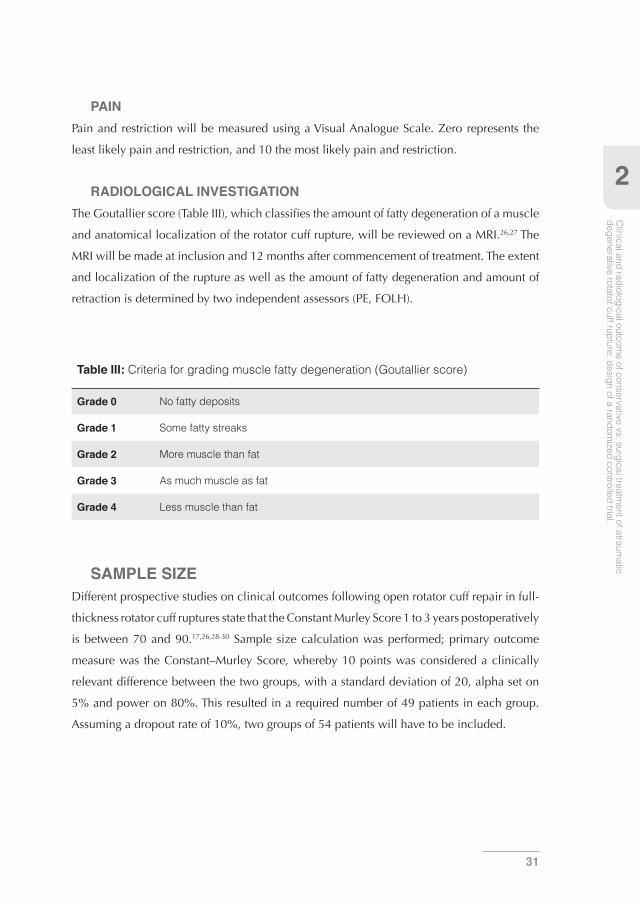
Table III: Criteria for grading muscle fatty degeneration (Goutallier score)
Grade 0 No fatty deposits
Grade 1 Some fatty streaks
Grade 2 More muscle than fat
Grade 3 As much muscle as fat
Grade 4 Less muscle than fat
SAMPLE SIZEDifferent prospective studies on clinical outcomes following open rotator cuff repair in full-
thickness rotator cuff ruptures state that the Constant Murley Score 1 to 3 years postoperatively
is between 70 and 90.17,26,28-30 Sample size calculation was performed; primary outcome
measure was the Constant–Murley Score, whereby 10 points was considered a clinically
relevant difference between the two groups, with a standard deviation of 20, alpha set on
5% and power on 80%. This resulted in a required number of 49 patients in each group.
Assuming a dropout rate of 10%, two groups of 54 patients will have to be included.

32
2
Cha
pter
2
STATISTICAL ANALYSISThe means and standard deviations of the intervention and control group will be calculated
for patient and outcome characteristics. In order to determine whether a difference exists
between the surgical and conservative groups as far as primary outcome measurement
(pain and function of the shoulder as determined with the Constant-Murley Score) over
all five measurement moments is concerned, a random effect analysis shall be carried out
which includes corrections for hospital and operating surgeon and for a number of patient
characteristics. This analysis will be repeated with the secondary outcome measurements as
dependent variables.
The prognostic value of localization and extent of the rotator cuff rupture and the degree
of fatty degeneration influencing the functional recovery after 12 months will be analyzed
using a linear regression model. Here too corrections will be made for hospital and operator
and for a number of patient characteristics.
DISCUSSION
Both conservative and surgical treatment for degenerative atraumatic rotator cuff tendon
rupture is performed in current practice. A clear distinction between indications for surgical
repair and conservative treatments cannot be made on the basis of the current evidence.
To date, there are no randomized controlled studies that compare outcome of surgical
vs. conservative treatment of atraumatic rotator cuff ruptures. The COPACABANA study is
designed to determine which treatment result in better outcome. Furthermore, it is possible
to identify prognostic factors for outcome of surgical repair of the rotator cuff.

33
2
Clinical and radiological outcom
e of conservative vs. surgical treatment of atraum
atic degenerative rotator cuff rupture: design of a random
ized controlled trial.
REFERENCES
1. Gomoll AH, Katz JN, Warner JJ, Millett PJ. Rotator cuff disorders: Recognition and management
among patients with shoulder pain. Arthritis Rheum. 2004;50(12):3751-3761.
2. van der Windt DA, Koes BW, de Jong BA, Bouter LM. Shoulder disorders in general practice:
Incidence, patient characteristics, and management. Ann Rheum Dis. 1995;54(12):959-964.
3. Nho Shane J SJ. Rotator cuff degeneration: Etiology and pathogenesis. American Journal of Sports
Medicine, The. 2008-5;36(5):987-93.
4. Neer C S CS. Impingement lesions. Clin Orthop. 1983-3(173):70-7.
5. Lehman C, Cuomo F, Kummer FJ, Zuckerman JD. The incidence of full thickness rotator cuff tears
in a large cadaveric population. Bull Hosp Jt Dis. 1995;54(1):30-31.
6. Sher JS, Uribe JW, Posada A, Murphy BJ, Zlatkin MB. Abnormal findings on magnetic resonance
images of asymptomatic shoulders. J Bone Joint Surg Am. 1995;77(1):10-15.
7. Yamaguchi K, Tetro AM, Blam O, Evanoff BA, Teefey SA, Middleton WD. Natural history
of asymptomatic rotator cuff tears: A longitudinal analysis of asymptomatic tears detected
sonographically. J Shoulder Elbow Surg. 2001;10(3):199-203.
8. Coghlan JA, Buchbinder R, Green S, Johnston RV, Bell SN. Surgery for rotator cuff disease.
Cochrane Database Syst Rev. 2008;(1):CD005619. doi(1):CD005619.
9. Dalton SE. The conservative management of rotator cuff disorders. Br J Rheumatol. 1994;33(7):663-
667.
10. Goldberg BA, Nowinski RJ, Matsen FA,3rd. Outcome of nonoperative management of full-
thickness rotator cuff tears. Clin Orthop Relat Res. 2001;(382)(382):99-107.
11. Shibata Y, Midorikawa K, Emoto G, Naito M. Clinical evaluation of sodium hyaluronate for the
treatment of patients with rotator cuff tear. J Shoulder Elbow Surg. 2001;10(3):209-216.
12. Hawkins RH, Dunlop R. Nonoperative treatment of rotator cuff tears. Clin Orthop Relat Res.
1995;(321)(321):178-188.
13. Itoi E, Tabata S. Conservative treatment of rotator cuff tears. Clin Orthop Relat Res. 1992;(275)
(275):165-173.
14. Seida JC, LeBlanc C, Schouten JR, et al. Systematic review: Nonoperative and operative treatments
for rotator cuff tears. Ann Intern Med. 2010;153(4):246-255.
15. Sonnery-Cottet B, Edwards TB, Noel E, Walch G. Rotator cuff tears in middle-aged tennis players:
Results of surgical treatment. Am J Sports Med. 2002;30(4):558-564.

34
2
Cha
pter
2
16. Worland RL, Arredondo J, Angles F, Lopez-Jimenez F. Repair of massive rotator cuff tears in
patients older than 70 years. J Shoulder Elbow Surg. 1999;8(1):26-30.
17. Lam F, Mok D. Open repair of massive rotator cuff tears in patients aged sixty-five years or over:
Is it worthwhile? J Shoulder Elbow Surg. 2004;13(5):517-521.
18. Sperling JW, Cofield RH, Schleck C. Rotator cuff repair in patients fifty years of age and younger.
J Bone Joint Surg Am. 2004;86-A(10):2212-2215.
19. Tibone JE, Elrod B, Jobe FW, et al. Surgical treatment of tears of the rotator cuff in athletes. J Bone
Joint Surg Am. 1986;68(6):887-891.
20. Diercks RL, Ham SJ, Ros JM. Results of anterior shoulder decompression surgery according to
neer for shoulder impingement syndrome; little effect on fitness for work. Ned Tijdschr Geneeskd.
1998;142(22):1266-1269.
21. Cheung EV, Silverio L, Sperling JW. Strategies in biologic augmentation of rotator cuff repair:
A review. Clin Orthop Relat Res. 2010;468(6):1476-1484.
22. Sugaya H, Maeda K, Matsuki K, Moriishi J. Repair integrity and functional outcome after
arthroscopic double-row rotator cuff repair. A prospective outcome study. J Bone Joint Surg Am.
2007;89(5):953-960.
23. Goutallier D, Postel JM, Radier C, Bernageau J, Godefroy D, Zilber S. How repaired rotator cuff
function influences constant scoring. Orthop Traumatol Surg Res. 2010;96(5):500-505.
24. Burkhart SS. The principle of margin convergence in rotator cuff repair as a means of strain
reduction at the tear margin. Ann Biomed Eng. 2004;32(1):166-170.
25. Constant CR, Murley AH. A clinical method of functional assessment of the shoulder. Clin Orthop
Relat Res. 1987;(214)(214):160-164.
26. Goutallier D, Postel JM, Gleyze P, Leguilloux P, Van Driessche S. Influence of cuff muscle fatty
degeneration on anatomic and functional outcomes after simple suture of full-thickness tears.
J Shoulder Elbow Surg. 2003;12(6):550-554.
27. Goutallier D D. Fatty muscle degeneration in cuff ruptures. pre- and postoperative evaluation by
CT scan. Clin Orthop. 1994-7(304):78-83.
28. Milano G, Grasso A, Salvatore M, Zarelli D, Deriu L, Fabbriciani C. Arthroscopic rotator cuff repair
with and without subacromial decompression: A prospective randomized study. Arthroscopy.
2007;23(1):81-88.
29. Bishop J, Klepps S, Lo IK, Bird J, Gladstone JN, Flatow EL. Cuff integrity after arthroscopic versus
open rotator cuff repair: A prospective study. J Shoulder Elbow Surg. 2006;15(3):290-299.

35
2
Clinical and radiological outcom
e of conservative vs. surgical treatment of atraum
atic degenerative rotator cuff rupture: design of a random
ized controlled trial.
30. Goutallier D, Postel JM, Van Driessche S, Godefroy D, Radier C. Tension-free cuff repairs with
excision of macroscopic tendon lesions and muscular advancement: Results in a prospective
series with limited fatty muscular degeneration. J Shoulder Elbow Surg. 2006;15(2):164-172.
