university of groningen exploring drug safety of
TRANSCRIPT

University of Groningen
Exploring drug safety of radiopharmaceuticalsSchreuder, Nanno
DOI:10.33612/diss.180382182
IMPORTANT NOTE: You are advised to consult the publisher's version (publisher's PDF) if you wish to cite fromit. Please check the document version below.
Document VersionPublisher's PDF, also known as Version of record
Publication date:2021
Link to publication in University of Groningen/UMCG research database
Citation for published version (APA):Schreuder, N. (2021). Exploring drug safety of radiopharmaceuticals. University of Groningen.https://doi.org/10.33612/diss.180382182
CopyrightOther than for strictly personal use, it is not permitted to download or to forward/distribute the text or part of it without the consent of theauthor(s) and/or copyright holder(s), unless the work is under an open content license (like Creative Commons).
The publication may also be distributed here under the terms of Article 25fa of the Dutch Copyright Act, indicated by the “Taverne” license.More information can be found on the University of Groningen website: https://www.rug.nl/library/open-access/self-archiving-pure/taverne-amendment.
Take-down policyIf you believe that this document breaches copyright please contact us providing details, and we will remove access to the work immediatelyand investigate your claim.
Downloaded from the University of Groningen/UMCG research database (Pure): http://www.rug.nl/research/portal. For technical reasons thenumber of authors shown on this cover page is limited to 10 maximum.
Download date: 18-03-2022

562641-L-bw-Schreuder562641-L-bw-Schreuder562641-L-bw-Schreuder562641-L-bw-SchreuderProcessed on: 25-8-2021Processed on: 25-8-2021Processed on: 25-8-2021Processed on: 25-8-2021 PDF page: 1PDF page: 1PDF page: 1PDF page: 1
Exploring drug safety of radiopharmaceuticals
Nanno Schreuder
2021

562641-L-bw-Schreuder562641-L-bw-Schreuder562641-L-bw-Schreuder562641-L-bw-SchreuderProcessed on: 25-8-2021Processed on: 25-8-2021Processed on: 25-8-2021Processed on: 25-8-2021 PDF page: 2PDF page: 2PDF page: 2PDF page: 2
Cover design by: Harma Makken, persoonlijkproefschrift.nl Printed by: Gildeprint, Enschede ISBN: 978-94-6419-290-2 Copyright © Nanno Schreuder, 2021 All rights reserved. No part of this thesis may be reproduced, stored in a retrieval system or transmitted in any form, by any means, without permission of the author. Printed on 100% recycled paper.

562641-L-bw-Schreuder562641-L-bw-Schreuder562641-L-bw-Schreuder562641-L-bw-SchreuderProcessed on: 25-8-2021Processed on: 25-8-2021Processed on: 25-8-2021Processed on: 25-8-2021 PDF page: 3PDF page: 3PDF page: 3PDF page: 3
Exploring drug safety of radiopharmaceuticals
PhD thesis
to obtain the degree of Doctor at the University of
Groningen on the authority of the
Rector Magnificus, Prof. C. Wijmenga and in accordance with a decision by the Doctorate Board.
This thesis will be defended in public on
Friday 15 October 2021 at 14:30 hours
by
Nanno Schreuder
born on 28 April 1972 in Bathmen

562641-L-bw-Schreuder562641-L-bw-Schreuder562641-L-bw-Schreuder562641-L-bw-SchreuderProcessed on: 25-8-2021Processed on: 25-8-2021Processed on: 25-8-2021Processed on: 25-8-2021 PDF page: 4PDF page: 4PDF page: 4PDF page: 4
Supervisors Prof. E.P. van Puijenbroek Prof. J.G.W. Kosterink Co-supervisor Dr. P.L. Jager Assessment committee Prof. R.A.J.O. Dierckx Prof. A.C.G. Egberts Prof. M.N. Lub-de Hooge

562641-L-bw-Schreuder562641-L-bw-Schreuder562641-L-bw-Schreuder562641-L-bw-SchreuderProcessed on: 25-8-2021Processed on: 25-8-2021Processed on: 25-8-2021Processed on: 25-8-2021 PDF page: 5PDF page: 5PDF page: 5PDF page: 5
Table of Contents
Chapter 1 General introduction and outline of the thesis 7
Chapter 2 Adverse events of diagnostic radiopharmaceuticals:a systematic review
27
Chapter 3 Patient-reported adverse events of radiopharmaceuticals: development and validation of a questionnaire
91
Chapter 4 Patient-reported adverse events of radiopharmaceuticals: a prospective study of 1,002 patients
115
Chapter 5 Anaphylactic reaction to [99mTc]Tc-macrosalb 141
Chapter 6 Discontinuation of metformin to prevent metformin-induced high colonic FDG uptake: is 48 hours sufficient?
149
Chapter 7 Radiopharmaceuticals in acute porphyria 167
Chapter 8 Lack of consistent dose recommendations for radiopharmaceuticals in patients with renal insufficiency: results of a systematic review
183
Chapter 9 Summary, future perspectives, and conclusions 217
Chapter 10 Nederlandse samenvatting (Summary in Dutch) 233
Appendices List of publications
Curriculum Vitae
Dankwoord (Acknowledgments in Dutch)
Supplementary material
Index of radiopharmaceuticals
249
251
253
257
297

562641-L-bw-Schreuder562641-L-bw-Schreuder562641-L-bw-Schreuder562641-L-bw-SchreuderProcessed on: 25-8-2021Processed on: 25-8-2021Processed on: 25-8-2021Processed on: 25-8-2021 PDF page: 6PDF page: 6PDF page: 6PDF page: 6

562641-L-bw-Schreuder562641-L-bw-Schreuder562641-L-bw-Schreuder562641-L-bw-SchreuderProcessed on: 25-8-2021Processed on: 25-8-2021Processed on: 25-8-2021Processed on: 25-8-2021 PDF page: 7PDF page: 7PDF page: 7PDF page: 7
Chapter 1
General introduction and outline of the
thesis
Nanno Schreuder 1, 2 1 Groningen Research Institute of Pharmacy, PharmacoTherapy, -Epidemiology & -Economics,
University of Groningen, Groningen, the Netherlands
2 GE Healthcare Radiopharmacy Zwolle, Zwolle, the Netherlands
Unpublished

562641-L-bw-Schreuder562641-L-bw-Schreuder562641-L-bw-Schreuder562641-L-bw-SchreuderProcessed on: 25-8-2021Processed on: 25-8-2021Processed on: 25-8-2021Processed on: 25-8-2021 PDF page: 8PDF page: 8PDF page: 8PDF page: 8

562641-L-bw-Schreuder562641-L-bw-Schreuder562641-L-bw-Schreuder562641-L-bw-SchreuderProcessed on: 25-8-2021Processed on: 25-8-2021Processed on: 25-8-2021Processed on: 25-8-2021 PDF page: 9PDF page: 9PDF page: 9PDF page: 9
GENERAL INTRODUCTION AND OUTLINE OF THE THESIS | 9
1 1.1 INTRODUCTION
Nuclear medicine is a medical specialty that uses radiopharmaceuticals for the diagnosis or treatment of patients. A radiopharmaceutical is a radioactive drug that consists of a tracer part linked to a radionuclide. The tracer part is a molecule or particle; it determines where the radiopharmaceutical will localise in the body. The radionuclide is an atom emitting radiation that can be detected by scanners or that delivers a radiation dose to a target area. Nuclear medicine uses the radiation the radionuclide emits to visualise or treat organs where the tracer localises. Radionuclides typically used in nuclear medicine are alpha, beta–, beta+, and gamma emitters (α, β–, β+, and γ). An alpha-particle is a helium nucleus ( 𝐻𝐻𝐻𝐻2
4 ); a beta–-particle is an electron. Both deliver a high energy over a short distance and are used for therapy in nuclear medicine. A beta+-particle, also called a positron which, upon interacting with an electron, is annihilated and gives rise to two photons or gamma rays (γ-rays). Gamma rays are a form of electromagnetic radiation, have a long range in tissue, and can be used in imaging in nuclear medicine1, 2. Most radiopharmaceuticals are used for diagnostic purposes to enable visualisation of physiological processes using imaging modalities such as planar imaging, single photon emission computed tomography (SPECT), or positron emission tomography (PET). Figure 1.1 shows a schematic presentation of SPECT and PET. Planar imaging acquires a two-dimensional image of the body or body parts. Single photon emission computed tomography uses a gamma camera (with one or two detectors), which rotates around the patient to detect gamma emission from single photon-emitting radionuclides and acquires information from multiple angles to construct a three-dimensional image. Positron emission tomography uses positron emitting radionuclides and employs multiple detectors placed in a ring around the patient to detect coincident photons originating from annihilation of a positron with an electron3.

562641-L-bw-Schreuder562641-L-bw-Schreuder562641-L-bw-Schreuder562641-L-bw-SchreuderProcessed on: 25-8-2021Processed on: 25-8-2021Processed on: 25-8-2021Processed on: 25-8-2021 PDF page: 10PDF page: 10PDF page: 10PDF page: 10
10 | EXPLORING DRUG SAFETY OF RADIOPHARMACEUTICALS
Fig. 1.1 Schematic presentation of SPECT and PET. (A) The radionuclide emits a photon that is detected by a rotating gamma camera. (B) The radionuclide emits a positron that travels in tissue for a short distance until it interacts with an electron. When an electron and positron annihilate, they produce two gamma photons to be emitted in opposite directions. Multiple gamma detectors positioned in a circle detect these photons. Adapted with permission from Berger et al4.
Today, healthcare professionals often combine SPECT and PET with computed tomography (CT), which uses X-rays. These hybrid cameras are then referred to as SPECT/CT or PET/CT and offer better imaging than SPECT or PET alone, because they have the ability to image both processes in the body as well as anatomy, which allows for accurate localization of abnormalities. Over the years, nuclear medicine imaging modalities have become essential in many areas of medicine to detect and monitor diseases1, 3, 5. An example of one of the most-used diagnostic

562641-L-bw-Schreuder562641-L-bw-Schreuder562641-L-bw-Schreuder562641-L-bw-SchreuderProcessed on: 25-8-2021Processed on: 25-8-2021Processed on: 25-8-2021Processed on: 25-8-2021 PDF page: 11PDF page: 11PDF page: 11PDF page: 11
GENERAL INTRODUCTION AND OUTLINE OF THE THESIS | 11
1 radiopharmaceuticals is [18F]fludeoxyglucose (FDG)* in combination with PET for diagnosis, staging, restaging, and assessing therapy response in oncology. Other examples of diagnostic radiopharmaceuticals using SPECT are [99mTc]Tc-tetrofosmin for myocardial perfusion imaging to detect ischaemia and myocardial infarction, and [123I]ioflupane, used in the diagnosis of Parkinson’s disease5, 8. Radiopharmaceuticals are also used in therapy; these typically contain a beta– or alfa particle-emitting radioactive element. These therapeutic radiopharmaceuticals deploy their action by delivering a radiation dose to a specific target in the body in order to damage or eradicate abnormal tissue9. An example of a therapeutic radiopharmaceutical is [223Ra]Ra-dichloride for the treatment of bone metastases in castrate-resistant prostate cancer10.
1.2 DRUG SAFETY
Drug safety is an important aspect of medical practice and has the public’s full attention. A tragedy with the drug thalidomide in the early 1960s made everyone aware that taking drugs is not without possible negative consequences. Thalidomide was marketed in the late 1950s as an anti-emetic and used to treat morning sickness in pregnant women. It proved to be a potent teratogen, causing birth defects in thousands of children11. This tragedy led to improvements in drug testing and requirements for safety evaluation. Regulators now conduct extensive safety evaluation on drugs—including radiopharmaceuticals. Drugs must show a favourable benefit-risk profile before manufacturers can market them. However, pre-marketing trials, in general, include a relatively small number of patients, have a limited follow-up time, and usually exclude specific groups of patients—such as, children, elderly, pregnant women or patients with specific underlying disorders, like renal-impairment—who might be at greater risk of adverse events. Often, information about a drug’s safety becomes available from its use in larger or specific groups of patients after it is marketed. This phase of the lifecycle of a drug is often where uncommon adverse drug reactions, drug reactions with a long time to onset,
*The names of the radiopharmaceuticals in this thesis were standardised according to the Anatomical Therapeutic Chemical classification system and the International Consensus Radiochemistry Nomenclature Guidelines6, 7.

562641-L-bw-Schreuder562641-L-bw-Schreuder562641-L-bw-Schreuder562641-L-bw-SchreuderProcessed on: 25-8-2021Processed on: 25-8-2021Processed on: 25-8-2021Processed on: 25-8-2021 PDF page: 12PDF page: 12PDF page: 12PDF page: 12
12 | EXPLORING DRUG SAFETY OF RADIOPHARMACEUTICALS
or issues related to the use of the drug—such as treatment failure, poor quality, interactions with other substances, and incorrect use of drugs—may arise. Information regarding use in a clinical setting, as provided by healthcare professionals and patients, is important to understanding more about the safety of a drug and will ultimately aid in improving the safety of a drug12, 13. In the past decades, radiopharmaceuticals have proven to be very safe14, 15. Nevertheless, safety issues with radiopharmaceuticals may occur. However, radiopharmaceutical drug safety issues differ from other drugs because of some unique aspects of radiopharmaceuticals. One such aspect is that the tracer part in the formulation is usually administered in very low mass quantities, generally in the range of micrograms, and therefore does not have a pharmacological effect1. For that reason, an adverse drug reaction due to a pharmacological action is unlikely to occur for most radiopharmaceuticals, in contrast to other drugs. However, idiosyncratic adverse reactions, that are not dose-related, may occur with radiopharmaceuticals or with excipients used in the formulation15. Furthermore, as the purpose, in general, is diagnostic imaging, an individual patient often encounters radiopharmaceuticals only once or a few times14. Accordingly, drug safety issues that might be seen with long-term use of other drugs are unlikely to occur with radiopharmaceuticals. Another important aspect of drug safety of radiopharmaceuticals is the distribution in the body, also known as biodistribution. Both biodistribution and localisation of the tracer will determine imaging or therapy outcome. Interactions with other drugs may alter the biodistribution and might ultimately affect diagnostic or therapy outcomes16, 17. In addition to the aspect of biodistribution, certain groups of patients need additional attention to ensure optimal outcome and avoid safety issues. These patients need extra precautions, such as special preparations or a change in dose. For some groups of patients, such as children and pregnant or breastfeeding women, guidelines are available18, 19. Finally, radiation exposure to the patients may be a concern with the use of radiopharmaceuticals. Nevertheless, the exposure of patients to radiation from nuclear medicine procedures is not excessive, and the incremental risks are small and outweighed by the benefits, such as providing useful diagnostic information that

562641-L-bw-Schreuder562641-L-bw-Schreuder562641-L-bw-Schreuder562641-L-bw-SchreuderProcessed on: 25-8-2021Processed on: 25-8-2021Processed on: 25-8-2021Processed on: 25-8-2021 PDF page: 13PDF page: 13PDF page: 13PDF page: 13
GENERAL INTRODUCTION AND OUTLINE OF THE THESIS | 13
1 cannot be obtained using other methods and is important for determining a course of treatment20. Even though radiopharmaceuticals have an excellent safety record, information regarding use and possible adverse effects in a clinical setting as provided by healthcare professionals and patients is important to understand more about the safety of radiopharmaceuticals and will ultimately aid in further safety improvement. For this reason, we will address radiopharmaceuticals drug safety issues in this thesis, with the main topics being adverse reactions, interactions with drugs, and considerations in specific patient populations.
1.2.1 Adverse reactions The World Health Organization (WHO) defines an adverse drug reaction as ‘a response to a medicine which is noxious and unintended, and which occurs at doses normally used in man’. The term adverse drug reaction must be distinguished from the term adverse event. An adverse event is not necessarily related to the drug and is defined by the WHO as ‘any untoward medical occurrence that may present during treatment with a medicine but which does not necessarily have a causal relationship with this treatment’21, 22. Adverse drug reactions have a proven causal relationship with a drug, while adverse events do not and can be related to different causes. However, knowledge of adverse events is of interest in drug safety evaluation, as establishing a causal link with a particular drug at the moment the event occurs or is reported may not be possible. Researchers often use causality methods to determine whether a relation between an adverse event and a drug is present. Important aspects in a causality method are time sequence, response pattern to the suspected pharmaceutical, and rechallenge. Using a causality method, investigators weight these aspects to attribute a category of causality, such as ‘not related’, ‘unlikely’, ‘possible’, ‘probable’, or ‘certain’. Often-used causality methods for radiopharmaceuticals are the Naranjo algorithm23 and the method Silberstein described15. In order to report, analyse, and compare adverse events in drug safety, healthcare professionals commonly use standardised terminology. This enables the researcher

562641-L-bw-Schreuder562641-L-bw-Schreuder562641-L-bw-Schreuder562641-L-bw-SchreuderProcessed on: 25-8-2021Processed on: 25-8-2021Processed on: 25-8-2021Processed on: 25-8-2021 PDF page: 14PDF page: 14PDF page: 14PDF page: 14
14 | EXPLORING DRUG SAFETY OF RADIOPHARMACEUTICALS
to code the symptoms, as the patient recalls them, into standardised terms. These terms can be categorised into main groups and then used for data representation. An example of standardised terminology is the Medical Dictionary for Regulatory Activities (MedDRA®), which the European Union mandates for safety reporting24.
1.2.2 Adverse reactions of radiopharmaceuticals Researchers assume that adverse reactions with radiopharmaceuticals rarely occur. Studies report a frequency of adverse events of 0.11% in Europe and 0.021% in the United States14, 15, 25, which is low compared to the 1% to 2% reported for ‘regular’ therapeutic drugs26, 27 and the 5% to 8% reported for drug reactions in hospitalized patients28. This may be explained by the unique aspects of radiopharmaceuticals, such as the lack of pharmacological effect because of the very low mass quantities administered and the fact that radiopharmaceuticals are often used only once or a few times in an individual patient. However, some authors indicate that another important reason for this low frequency might be that these studies rely on voluntary identification and reporting, and that adverse events of radiopharmaceuticals are underreported29, 30. Such underreporting of adverse events is well known for other drugs31. In addition, adverse events of radiopharmaceuticals may be left undetected because follow-up contact seldom occurs between the patient and the nuclear medicine department after the nuclear medicine examination is completed. Furthermore, in current studies, other aspects such as severity, latency time, and outcome are often not studied in detail29, 32–35. There is a clear need to further study and understand the characteristics and frequency of adverse events of radiopharmaceuticals.
1.2.3 Involvement of patients The view of the patient plays an important role in drug safety. Patients can report adverse events themselves in several countries and provide additional useful information36, 37. Such information can complement the information healthcare professionals provide, as patients report adverse events and aspects that are different from those that healthcare professionals report. Patients provide more detailed

562641-L-bw-Schreuder562641-L-bw-Schreuder562641-L-bw-Schreuder562641-L-bw-SchreuderProcessed on: 25-8-2021Processed on: 25-8-2021Processed on: 25-8-2021Processed on: 25-8-2021 PDF page: 15PDF page: 15PDF page: 15PDF page: 15
GENERAL INTRODUCTION AND OUTLINE OF THE THESIS | 15
1 information about the adverse events and may provide important information about novel adverse reactions or new insights on known adverse reactions, as well as aid in identifying adverse reactions in specific populations. Finally, patients can also provide first-hand information about their expectations and experiences, such as the outcome of an adverse event and the impact on their quality of life38–41. Little research has been done on adverse reactions of radiopharmaceuticals from the patient’s perspective. To our knowledge only one small study using one radiopharmaceutical was performed. In this study of 55 patients using [99mTc]Tc-medronic acid, one patient-reported headache, dizziness, and a dry mouth. However, the researchers did not provide detailed information about the method of recording and analysing the adverse events42. Research using patient-reported adverse events of radiopharmaceuticals may well aid in our understanding of the characteristics and true frequency of patients’ adverse reactions to radiopharmaceuticals. In addition, patients may provide information about the outcome and the impact on their lives. Patients’ perspectives could therefore help healthcare professionals to better inform patients and to be better prepared in detecting and managing radiopharmaceuticals adverse events.
1.2.4 Interactions of radiopharmaceuticals with drugs Another drug safety aspect of radiopharmaceuticals concerns interaction with other substances that alter biodistribution, which could lead to unusual imaging and might ultimately affect diagnostic or therapeutic outcomes43–45. Understanding these interactions is important, as they may affect a patient’s diagnosis or therapy. In the case of interactions with a diagnostic radiopharmaceutical, an interaction might influence the scan—e.g., reduce the uptake in an area of interest or cause abnormal uptake in non-relevant areas—and in that way lead to wrong or incomplete information that could ultimately affect the patient’s diagnosis. Amongst the factors associated with altered biodistribution of radiopharmaceuticals are the use of other drugs (including over-the-counter drugs), food, alternative medicines, issues with the preparation of the radiopharmaceutical, adsorption to administration devices, other medical procedures, or unexpected pathophysiology16, 43, 45–49. Researchers
562641-L-bw-Schreuder562641-L-bw-Schreuder562641-L-bw-Schreuder562641-L-bw-SchreuderProcessed on: 25-8-2021Processed on: 25-8-2021Processed on: 25-8-2021Processed on: 25-8-2021 PDF page: 16PDF page: 16PDF page: 16PDF page: 16
16 | EXPLORING DRUG SAFETY OF RADIOPHARMACEUTICALS
classify the effect of a substance on the biodistribution of radiopharmaceuticals as pharmacologic, pharmacokinetic, toxicologic, pharmaceutical, or unidentified interactions43. A pharmacologic interaction occurs when a substance directly or indirectly alters the radiopharmaceutical at its site of localisation. One example is interaction with the radiopharmaceutical [123I]ioflupane. Healthcare professionals use this radiopharmaceutical in the diagnosis of Parkinson’s disease and other related diseases. It is a cocaine analogue, binds to the presynaptic dopamine active transporter, and allows for the detection of the loss of nerve cells in a part of the brain called the striatum. Several drugs can interact with [123I]ioflupane imaging, such as methylphenidate, which has shown to completely block the physiologic uptake in the striatum50. A pharmacokinetic interaction occurs when a drug alters the plasma concentration of the radiopharmaceutical by assorting an effect on the absorption, distribution, metabolism, or excretion of the drug. One example is the interaction of colchicine with [18F]fluorocholine, used in PET imaging for diagnosis of prostate cancer and detection of parathyroid adenoma amongst others. Choline is an important component of phospholipids in cell membranes, and its metabolism has an anabolic and catabolic pathway via phosphatidylcholine. In tumours, the anabolic pathway outweighs the catabolic pathway, giving it a higher uptake of [18F]fluorocholine. It is supposed that colchicine stimulates the catabolic pathway and leads to an impaired uptake of [18F]fluorocholine51. A toxicologic interaction occurs when the dosing of a drug leads to an increased pharmacologic effect or an adverse effect and subsequently changes the biodistribution of the radiopharmaceutical. For example, one case describes a blood brain barrier damaged by methotrexate, resulting in an increased uptake of [99mTc]Tc-pertechnetate in the cerebral ventricles52. A pharmaceutical interaction occurs when a physicochemical reaction between a drug and the radiopharmaceutical takes place. This interaction can, for example, take place when substances come into contact, precipitating the radiopharmaceutical. For example, the combination of intravenous iron with 99mTc-diphosphonates can lead to a high blood pool activity53.

562641-L-bw-Schreuder562641-L-bw-Schreuder562641-L-bw-Schreuder562641-L-bw-SchreuderProcessed on: 25-8-2021Processed on: 25-8-2021Processed on: 25-8-2021Processed on: 25-8-2021 PDF page: 17PDF page: 17PDF page: 17PDF page: 17
GENERAL INTRODUCTION AND OUTLINE OF THE THESIS | 17
1 For some interactions, the mechanism is unknown. For example, it is known that in patients using metformin—an oral antidiabetic—a high colonic uptake of FDG is often seen. However, the mechanism is still unknown and although several studies have suggested that metformin should be discontinued before the FDG PET scan, the optimal discontinuation period is still unclear54–56. Nuclear medicine professionals must be aware of interactions that influence or alter the biodistribution of radiopharmaceuticals so they can take necessary precautions, such as informing the patient to stop a certain drug or food. In addition, when interactions do occur, nuclear medicine professionals will be able to explain uncommon findings, and in that way, improve the diagnostic outcome for patients16. Although many factors interacting with the biodistribution of radiopharmaceuticals are known, well-defined research is still scarce and recommendations for clinical use are mostly lacking. Research in this area may help in providing clear guidance.
1.2.5 Specific patient populations and radiopharmaceuticals Certain groups of patients need additional attention in order to ensure an optimal outcome and avoid safety issues. Extra precautions, such as special preparations or a change in dose, are needed to ensure a safe use of radiopharmaceuticals in these patients. These patient groups include, for example, pregnant or breastfeeding women, children, elderly patients, and dialysis patients16. In the patient group of breastfeeding women excretion of radiopharmaceuticals or metabolites in breast milk could potentially expose a child to an undesirable radiation dose. A study in breastfeeding mothers showed that stopping breastfeeding was needed for [131I]sodium iodine; a temporary 12-hour interruption was needed for [125I]iodohippurate, [131I]iodohippurate, [99mTc]Tc-pertechnetate, and [99mTc]Tc-macrosalb; and no interruption was needed for other radiopharmaceuticals18. In the group of children clinicians must adjust the dose of radiopharmaceuticals for children, as children may have a greater potential risk for developing late effects of radiation exposure. The dosage card used in Europe and the United States bases the dose not only on the weight of the child but also on the specific radiopharmaceutical19. Another example of a specific group of patients using

562641-L-bw-Schreuder562641-L-bw-Schreuder562641-L-bw-Schreuder562641-L-bw-SchreuderProcessed on: 25-8-2021Processed on: 25-8-2021Processed on: 25-8-2021Processed on: 25-8-2021 PDF page: 18PDF page: 18PDF page: 18PDF page: 18
18 | EXPLORING DRUG SAFETY OF RADIOPHARMACEUTICALS
radiopharmaceuticals who are at risk for a rare complication are patients with advanced metastatic neuroendocrine tumours. A case report describes a carcinoid crisis after injection of [18F]fluorodihydroxyphenylalanine (DOPA), which is the result of a massive release of neurotransmitters such as serotonin and characterized by flushing, changes in blood pressure, difficulty breathing, and rapid heart rate. Carcinoid crisis can potentially be life-threatening, and it is therefore advised to slowly inject the tracer, and have appropriate drugs available to treat this condition, such as somatostatin analogues and perhaps ketanserin57. For some other specific patient groups, little research has been done and guidelines are not available. For example, clear recommendations for patients with renal insufficiency are not yet available58, 59. For patients carrying an acute porphyria gene, which in the case of drug exposure could cause acute attacks with severe symptoms, no assessment of radiopharmaceuticals has been done. The need to investigate the safe use of radiopharmaceuticals in some specific groups of patients is clear.
1.3 OUTLINE OF THE THESIS
Despite the fact that radiopharmaceuticals have an excellent safety profile, relatively little is known about adverse reactions, interactions and use in some specific patient groups. For this reason, we studied drug safety issues in this thesis, with the main topics being adverse reactions, interactions with drugs, and considerations in specific patient populations. The overall purpose of this thesis is to better understand radiopharmaceuticals drug safety. The broader intent is to improve the safe use of radiopharmaceuticals and increase patients’ and health care professionals’ awareness.
While it is known that radiopharmaceuticals can cause adverse events, information is only available in case reports and databases and may not be readily available to healthcare professionals. In Chapter 2, we present a systematic review of the literature with an overview of the most common adverse events of diagnostic radiopharmaceuticals and their characteristics as described in literature, such as

562641-L-bw-Schreuder562641-L-bw-Schreuder562641-L-bw-Schreuder562641-L-bw-SchreuderProcessed on: 25-8-2021Processed on: 25-8-2021Processed on: 25-8-2021Processed on: 25-8-2021 PDF page: 19PDF page: 19PDF page: 19PDF page: 19
GENERAL INTRODUCTION AND OUTLINE OF THE THESIS | 19
1 frequency and seriousness, and the proposed mechanism leading to the adverse event. Patients can provide important information about adverse events and provide an important contribution to the detection and understanding of adverse events, as well as healthcare professionals’ ability to manage them. In order to investigate possible adverse events of radiopharmaceuticals in patients, we needed a suitable instrument. Therefore, we describe the development, validation, and testing of a questionnaire to be used with patients and present the results in Chapter 3. Using this questionnaire, we conducted a prospective cohort study of 1,002 patients in a regional hospital in the Netherlands (Isala). In this study, we not only aimed to assess the characteristics and frequency of patient-reported adverse events of radiopharmaceuticals but also to assess the outcome and follow-up. We present the results of this study in Chapter 4. In addition to systematic research, the analysis of an individual case can also contribute to more knowledge about adverse reactions. In Chapter 5 we describe an example of a patient who developed an anaphylactic reaction to [99mTc]Tc-macrosalb used for pulmonary scintigraphy. Besides adverse events, important safety issues with radiopharmaceuticals also include unusual imaging outcomes and considerations in specific patient populations. One known unusual imaging outcome is the increased colonic uptake in FDG PET scans with patients using metformin. We analyse in Chapter 6 whether a metformin discontinuing period of 48 hours or more is sufficient to prevent FDG uptake in the colon. A specific patient population where drug safety is of importance are patients who carry acute porphyria genes. As drug exposure in these patients might lead to potentially fatal acute attacks, knowing whether healthcare professionals can safely use radiopharmaceuticals in these patients is important. In Chapter 7, we therefore present an assessment and classification of the porphyrogenicity of radiopharmaceuticals. Another patient population where extra precautions may be needed are patients with renal insufficiency. In these patients, healthcare professionals may need to adjust the dose for optimal outcome and prevention of adverse effects. In Chapter 8, we therefore present a systematic review aiming to provide dose recommendations of radiopharmaceuticals in patients with renal insufficiency. In Chapter 9, we summarise our findings and discuss future perspectives to further improve drug safety of radiopharmaceuticals.

562641-L-bw-Schreuder562641-L-bw-Schreuder562641-L-bw-Schreuder562641-L-bw-SchreuderProcessed on: 25-8-2021Processed on: 25-8-2021Processed on: 25-8-2021Processed on: 25-8-2021 PDF page: 20PDF page: 20PDF page: 20PDF page: 20
20 | EXPLORING DRUG SAFETY OF RADIOPHARMACEUTICALS
1.4 REFERENCES
1. Weatherman K, Crips W, Weber H. The physiological basis of radiopharmaceuticals. In: Smith BT, editor. Nuclear Pharmacy: Concepts and Applications in Nuclear Pharmacy. 1st edition. London: Pharmaceutical Press; 2010. pp. 55–66.
2. Radioactive Decay. In: Saha GB. Physics and radiobiology of nuclear medicine. 2nd edition. New York: Springer-Verlag; 2001. pp. 11–20.
3. Tomographic Imaging Devices. In: Saha GB. Physics and radiobiology of nuclear medicine. 2nd edition. New York: Springer-Verlag; 2001. pp. 141–166.
4. Berger F, Gambhir SS. Recent advances in imaging endogenous or transferred gene expression utilizing radionuclide technologies in living subjects: applications to breast cancer. Breast Cancer Res 2001;3:28–35.
5. Maltby P, Theobald T. Survey of current diagnostic radiopharmaceuticals. In: Theobald T, editor. Sampson’s textbook of radiopharmacy. 4th edition. London: Pharmaceutical Press; 2011. pp. 277–306.
6. WHO Collaborating Centre for Drug Statistics Methodology. Guidelines for ATC classification and DDD assignment 2020. 23rd ed. Oslo: World Health Organisation; 2019.
7. Coenen HH, Gee AD, Adam M, Antoni G, Cutler CS, Fujibayashi Y, et al. Open letter to journal editors on: International Consensus Radiochemistry Nomenclature Guidelines. EJNMMI Radiopharm Chem 2019;16;4(1):7-018-0047-y.
8. Pinilla I, Rodríguez-Vigil B, Gómez-León N. Integrated 18FDG PET/CT: Utility and applications in the clinical oncology. Clin Med Oncol 2008;2:181–198.
9. Chan P, Croasdale J. Survey of current therapeutic radiopharmaceuticals. In: Theobald T, editor. Sampson’s textbook of radiopharmacy. 4th edition. London: Pharmaceutical Press; 2011. pp. 303–23.

562641-L-bw-Schreuder562641-L-bw-Schreuder562641-L-bw-Schreuder562641-L-bw-SchreuderProcessed on: 25-8-2021Processed on: 25-8-2021Processed on: 25-8-2021Processed on: 25-8-2021 PDF page: 21PDF page: 21PDF page: 21PDF page: 21
GENERAL INTRODUCTION AND OUTLINE OF THE THESIS | 21
1 10. Badrising SK, Louhanepessy RD, van der Noort V, Coenen JLLM, Hamberg
P, Beeker A, et al. A prospective observational registry evaluating clinical outcomes of Radium-223 treatment in a nonstudy population. Int J Cancer 2019 Dec 25. doi: 10.1002/ijc.32851. Online ahead of print.
11. Vargesson N. Thalidomide-induced teratogenesis: history and mechanisms. Birth Defects Res C Embryo Today 2015;105:140–156.
12. Mann RD, Andrews EB. Introduction: updated from second edition. In Andrews EB, Moore N, editors. Mann’s pharmacovigilance. New York: Wiley Blackwell; 2014. pp. 1–9.
13. Klepper MJ, Cobert B. Drug Safety Data: How to Analyze, Summarize and Interpret to Determine Risk. Sudbury, MA: Jones & Bartlett Learning; 2010.
14. Hesslewood SR, Keeling DH. Frequency of adverse reactions to radiopharmaceuticals in Europe. Eur J Nucl Med 1997;24:1179–1182.
15. Silberstein EB, Ryan J: Prevalence of adverse reactions in nuclear medicine: Pharmacopeia Committee of the Society of Nuclear Medicine. J Nucl Med 1996; 37:185–192.
16. Hladik WB,3rd, Norenberg JP. Problems associated with the clinical use of radiopharmaceuticals: a proposed classification system and troubleshooting guide. Eur J Nucl Med 1996;23:997–1002.
17. Vallabhajosula S, Killeen RP, Osborne JR. Altered biodistribution of radiopharmaceuticals: role of radiochemical/pharmaceutical purity, physiological, and pharmacologic factors. Semin Nucl Med 2010;40:220–241.
18. Leide–Svegborn S, Ahlgren L, Johansson L, Mattsson S. Excretion of radionuclides in human breast milk after nuclear medicine examinations. Biokinetic and dosimetric data and recommendations on breastfeeding interruption. Eur J Nucl Med Mol Imaging 2016;43:808–821.
19. Lassmann M, Treves S.T. For the EANM/SNMMI Paediatric Dosage Harmonization Working Group. Paediatric radiopharmaceutical administration: harmonization of the 2007 EANM paediatric dosage card (version 1.5.2008) and the 2010 North American consensus guidelines. Eur J Nucl Med Mol Imaging 2014;41:1036–1041.

562641-L-bw-Schreuder562641-L-bw-Schreuder562641-L-bw-Schreuder562641-L-bw-SchreuderProcessed on: 25-8-2021Processed on: 25-8-2021Processed on: 25-8-2021Processed on: 25-8-2021 PDF page: 22PDF page: 22PDF page: 22PDF page: 22
22 | EXPLORING DRUG SAFETY OF RADIOPHARMACEUTICALS
20. Adelstein SJ. Radiation risk in nuclear medicine. Semin Nucl Med 2014 May;44(3):187–192.
21. World Health Organization: International Drug Monitoring, The Role of National Centres (Technical Report Series No. 498). Geneva, World Health Organisation, 1972.
22. World Health Organization: Safety of Medicines: a guide to detecting and reporting adverse drug reactions. Geneva, World Health Organisation, 2002, whqlibdoc.who.int/hq/2002/WHO_EDM_QSM_2002.2.pdf, Accessed 05 Jun 2020.
23. Naranjo CA, Busto U, Sellers EM, et al: A method for estimating the probability of adverse drug reactions. Clin Pharmacol Ther 1981; 30:239–245
24. MedDRA Browser (2018 version 3.0). https://www.meddra.org/ Accessed 05 Jun 2020.
25. Silberstein EB: Prevalence of adverse events to radiopharmaceuticals from 2007 to 2011. J Nucl Med 2014; 55:1308–1310.
26. Zhan C, Arispe I, Kelley E, et al: Ambulatory care visits for treating adverse drug effects in the United States, 1995–2001. Jt Comm J Qual Patient Saf 2005; 31:372–378.
27. Bourgeois FT, Shannon MW, Valim C, et al: Adverse drug events in the outpatient setting: an 11-year national analysis. Pharmacoepidemiol Drug Saf 2010; 19:901–910.
28. Lazarou J, Pomeranz BH, Corey PN: Incidence of adverse drug reactions in hospitalized patients: a meta-analysis of prospective studies. JAMA 1998; 279:1200–1205.
29. Pinto SR, Santos LFC, Dos Reis SRR, Bastos MK, Gomes VDS, Vieira TO, et al. Adverse Reactions to Radiopharmaceuticals: A Survey Based on Clinical Cases Using Criteria of Systematic Review. Ther Innov Regul Sci 2018;52:109–13.
30. Santos-Oliveira R, Machado M. Pitfalls with radiopharmaceuticals. Am J Med Sci 2011;342:50–3.
31. Hazell L, Shakir SA. Under-reporting of adverse drug reactions: a systematic review. Drug Saf 2006;29:385–396.

562641-L-bw-Schreuder562641-L-bw-Schreuder562641-L-bw-Schreuder562641-L-bw-SchreuderProcessed on: 25-8-2021Processed on: 25-8-2021Processed on: 25-8-2021Processed on: 25-8-2021 PDF page: 23PDF page: 23PDF page: 23PDF page: 23
GENERAL INTRODUCTION AND OUTLINE OF THE THESIS | 23
1 32. Silberstein EB: Prevalence of adverse reactions to positron emitting
radiopharmaceuticals in nuclear medicine. Pharmacopeia Committee of the Society of Nuclear Medicine. J Nucl Med 1998; 39:2190–2192.
33. Santos-Oliveira R. Undesirable events with radiopharmaceuticals. Tohoku J Exp Med 2009;217:251–257.
34. Salvatori M, Treglia G, Mores N. Further considerations on adverse reactions to radiopharmaceuticals. Eur J Nucl Med Mol Imaging 2012;39:1360-1362.
35. Hesse B, Vinberg N, Berthelsen AK, Ballinger JR. Adverse events in nuclear medicine - cause for concern? Eur J Nucl Med Mol Imaging 2012;39:782–785.
36. van Hunsel F, Härmark L, Pal S, Olsson S, van Grootheest K. Experiences with adverse drug reaction reporting by patients: an 11-country survey. Drug Saf 2012;35:45–60.
37. de Langen J, van Hunsel F, Passier A, de Jong-van den Berg L, van Grootheest K. Adverse drug reaction reporting by patients in the Netherlands: three years of experience. Drug Saf 2008;31:515–524.
38. Inácio P, Cavaco A, Airaksinen M. The value of patient reporting to the pharmacovigilance system: a systematic review. Br J Clin Pharmacol 2017;83:227–246.
39. Blenkinsopp A, Wilkie P, Wang M, Routledge PA. Patient reporting of suspected adverse drug reactions: a review of published literature and international experience. Br J Clin Pharmacol 2007;63:148–156.
40. Rolfes L, van Hunsel F, Wilkes S, van Grootheest K, van Puijenbroek E. Adverse drug reaction reports of patients and healthcare professionals-differences in reported information. Pharmacoepidemiol Drug Saf 2015;24:152–8.
41. Jarernsiripornkul N, Kakaew W, Loalukkana W, Krska J. Adverse drug reaction monitoring: comparing doctor and patient reporting for new drugs. Pharmacoepidemiol Drug Saf 2009;18:240–5.
42. Dos Santos Almeida R, Mamede M, Santos-Oliveira R: Pharmacovigilance of radiopharmaceuticals used for prostate and breast cancer in Brazil. Adverse Drug React Bull 2013;283:1091–94.

562641-L-bw-Schreuder562641-L-bw-Schreuder562641-L-bw-Schreuder562641-L-bw-SchreuderProcessed on: 25-8-2021Processed on: 25-8-2021Processed on: 25-8-2021Processed on: 25-8-2021 PDF page: 24PDF page: 24PDF page: 24PDF page: 24
24 | EXPLORING DRUG SAFETY OF RADIOPHARMACEUTICALS
43. Hladik WB,3rd, Nigg KK, Rhodes BA. Drug-induced changes in the biologic distribution of radiopharmaceuticals. Semin Nucl Med 1982;12:184–218.
44. Sampson CB. Adverse reactions and drug interactions with radiopharmaceuticals. Drug Saf 1993;8:280–294.
45. Hesslewood S, Leung E. Drug interactions with radiopharmaceuticals. Eur J Nucl Med 1994;21:348–356.
46. Santos-Oliveira R, Smith SW, Carneiro-Leao AM. Radiopharmaceuticals drug interactions: a critical review. An Acad Bras Cienc 2008;80:665–675.
47. Bustani H, Colavolpe C, Imbert-Joscht I, Havlik P, Pisano P, Guillet BA. Chocolate intake associated with failed labeling of (99m)Tc red blood cells. J Nucl Med Technol 2009;37:107–110.
48. Werneke U, McCready VR. Complementary alternative medicine and nuclear medicine. Eur J Nucl Med Mol Imaging 2004;31:599–603.
49. Sampson CB. Complications and difficulties in radiolabelling blood cells: a review. Nucl Med Commun 1996;17:648–658.
50. Cheng G, Morley JF. Complete and readily reversible blocking of striatal DaTscan binding by methylphenidate. Clin Nucl Med 2014 Feb;39(2):211–213.
51. Roef MJ, van der Poel H, van der Laken CJ, Vogel WV. Colchicine must be stopped before imaging with [18F]-methylcholine PET/CT. Nucl Med Commun 2010 Dec;31(12):1075–1077.
52. Makler PT, Gutowicz MF, Kuhl DE. Methotrexate-induced ventriculitis: appearance on routine radionuclide scan and emission computed tomography. Clin Nucl Med 1978; 3: 22–3.
53. Forager AR, Grossman SJ, Joyce JM. Altered biodistribution of Tc-99m HMDP on bone scintigraphy from recent intravenous iron therapy. Clin Nucl Med 1994; 19: 817–8.
54. Oh JR, Song HC, Chong A, et al. Impact of medication discontinuation on increased intestinal FDG accumulation in diabetic patients treated with metformin. AJR Am J Roentgenol 2010; 195:1404–1410.

562641-L-bw-Schreuder562641-L-bw-Schreuder562641-L-bw-Schreuder562641-L-bw-SchreuderProcessed on: 25-8-2021Processed on: 25-8-2021Processed on: 25-8-2021Processed on: 25-8-2021 PDF page: 25PDF page: 25PDF page: 25PDF page: 25
GENERAL INTRODUCTION AND OUTLINE OF THE THESIS | 25
1 55. Hamidizadeh R, Eftekhari A, Wiley EA, Wilson D, Alden T, Bénard F.
Metformin discontinuation prior to FDG PET/CT: A randomized controlled study to compare 24- and 48-hour bowel activity. Radiology 2018; 289:418–425.
56. Lee SH, Jin S, Lee HS, Ryu JS, Lee JJ. Metformin discontinuation less than 72 h is suboptimal for F-18 FDG PET/CT interpretation of the bowel. Ann Nucl Med 2016; 30:629–636.
57. Koopmans KP, Brouwers AH, De Hooge MN, et al: Carcinoid crisis after injection of 6-18F-fluorodihydroxyphenylalanine in a patient with metastatic carcinoid. J Nucl Med 2005; 46:1240–1243.
58. Saracyn M., Bilski M., Kamiński G. and Niemczyk S. Can radioiodine be administered effectively and safely to a patient with severe chronic kidney disease? Clinical Endocrinology 2014;81:169–174.
59. Culpepper R. M., Hirsch J. I. and Fratkin M. J. Clearance of 131-I by hemodialysis. Clinical Nephrology 1992;38:110–114.

562641-L-bw-Schreuder562641-L-bw-Schreuder562641-L-bw-Schreuder562641-L-bw-SchreuderProcessed on: 25-8-2021Processed on: 25-8-2021Processed on: 25-8-2021Processed on: 25-8-2021 PDF page: 26PDF page: 26PDF page: 26PDF page: 26

562641-L-bw-Schreuder562641-L-bw-Schreuder562641-L-bw-Schreuder562641-L-bw-SchreuderProcessed on: 25-8-2021Processed on: 25-8-2021Processed on: 25-8-2021Processed on: 25-8-2021 PDF page: 27PDF page: 27PDF page: 27PDF page: 27
Chapter 2
Adverse events of diagnostic radiopharmaceuticals: a systematic
review
Nanno Schreuder 1, 2, Daniëlle Koopman 3, 4, Pieter L. Jager 3, Jos G.W. Kosterink 1,
5, Eugène van Puijenbroek 1, 6 1 Groningen Research Institute of Pharmacy, PharmacoTherapy, -Epidemiology & -Economics,
University of Groningen, Groningen, the Netherlands
2 GE Healthcare Radiopharmacy Zwolle, Zwolle, the Netherlands
3 Department of Nuclear Medicine, Isala Hospital, Zwolle, the Netherlands
4 MIRA Institute for Biomedical Technology and Technical Medicine, University of Twente, Enschede,
the Netherlands
5.University of Groningen, University Medical Center Groningen, Department of Clinical Pharmacy
and Pharmacology, Groningen, The Netherlands
6 Netherlands Pharmacovigilance Centre Lareb, ‘s-Hertogenbosch, the Netherlands
Seminars in Nuclear Medicine 2019;49:382–410

562641-L-bw-Schreuder562641-L-bw-Schreuder562641-L-bw-Schreuder562641-L-bw-SchreuderProcessed on: 25-8-2021Processed on: 25-8-2021Processed on: 25-8-2021Processed on: 25-8-2021 PDF page: 28PDF page: 28PDF page: 28PDF page: 28
28 | EXPLORING DRUG SAFETY OF RADIOPHARMACEUTICALS
ABSTRACT Diagnostic radiopharmaceuticals used in nuclear medicine can cause adverse events. Information on these adverse events is available in case reports and databases but may not be readily accessible to healthcare professionals. This systematic review provides an overview of adverse events of diagnostical radiopharmaceuticals and their characteristics. A median frequency for adverse events in diagnostical radiopharmaceuticals of 1.63 (interquartile range: 1.09-2.29) per 100,000 is reported. Most common are skin and subcutaneous tissue disorders, and general disorders and administration site conditions. Many adverse events reported are minor in severity, although 6.7% can be classified as important. In rare cases, adverse events are serious and potentially life-threatening. With the introduction of new radiopharmaceuticals and the increasing use of positron emission tomography-computed tomography (PET/CT), previously unknown adverse events may be detected in daily practice. Future work should cover the experience of the patient with adverse events from diagnostic radiopharmaceuticals.

562641-L-bw-Schreuder562641-L-bw-Schreuder562641-L-bw-Schreuder562641-L-bw-SchreuderProcessed on: 25-8-2021Processed on: 25-8-2021Processed on: 25-8-2021Processed on: 25-8-2021 PDF page: 29PDF page: 29PDF page: 29PDF page: 29
ADVERSE EVENTS OF DIAGNOSTIC RADIOPHARMACEUTICALS: A SYSTEMATIC REVIEW | 29
2
2.1 INTRODUCTION
Radiopharmaceuticals are drugs containing a radioactive isotope used for diagnostic or therapeutic purposes1, 2, with the radioactive isotopes emitting radiation that can be detected with imaging modalities, such as single-photon emission computed tomography (SPECT) or positron emission tomography (PET). Images and data allow for functional processes such as metabolism to be evaluated in the human body. Most diagnostic radiopharmaceuticals are used in very small quantities3—generally in the range of micrograms—and therefore do generally not have a pharmacological effect, although adverse reactions may still occur. These adverse reactions can often not be explained by the known actions of the radiopharmaceutical, and are mostly unpredictable. The World Health Organization defines an adverse drug reaction as “a response to a drug which is noxious and unintended, and which occurs at doses normally used in man for the prophylaxis, diagnosis, or therapy of disease, or for the modification of physiological function” and an adverse event as “any untoward medical occurrence that may present during treatment with a medicine but which does not necessarily have a causal relationship with this treatment”4, 5. “Adverse drug reaction” excludes events that do not have a proven relationship with a drug, although it may not be possible to establish a causal link at the moment the event occurs or is reported. Therefore, adverse events are still of interest in evaluating drug safety. For this reason, and for uniformity, the more general term “adverse event” is used here. Assessment is needed to determine if a particular drug caused the adverse event, specifically looking at the probability of causality and including clinical judgment. Many systems have been developed to support this process; for radiopharmaceuticals, often-used causality methods are the Naranjo algorithm6 and the method described by Silberstein7. Adverse events related to diagnostic radiopharmaceuticals are considered rare. Detailed information on these adverse events is available in case reports or dedicated databases, although this information might not be readily available to healthcare professionals when a patient experiences an adverse event. Information on these adverse events—including their severity, duration, and frequency—is needed for

562641-L-bw-Schreuder562641-L-bw-Schreuder562641-L-bw-Schreuder562641-L-bw-SchreuderProcessed on: 25-8-2021Processed on: 25-8-2021Processed on: 25-8-2021Processed on: 25-8-2021 PDF page: 30PDF page: 30PDF page: 30PDF page: 30
30 | EXPLORING DRUG SAFETY OF RADIOPHARMACEUTICALS
healthcare professionals to understand risk and management for patients8. For this reason, a comprehensive overview of adverse events related to diagnostic radiopharmaceuticals is essential. Several reviews have been conducted, some providing a narrative summary of adverse reactions9–15 and others focusing on a specific topic or combination of topics with preparation errors or product defects16; one review, published as a letter to the editor, presents data on the prevalence of adverse events for radiopharmaceuticals17. Additionally, several information databases have been developed to provide information about adverse events related to radiopharmaceuticals, although two are currently inaccessible18–20. However, to our knowledge, a systematic review to describe adverse events related to diagnostic radiopharmaceuticals has not yet been published. This review aims to provide an overview of the most common adverse events and their characteristics (such as frequency, severity, and proposed mechanism), for diagnostic radiopharmaceuticals as reported in literature.
2.2 METHODS
This review process followed the Preferred Reporting Items for Systematic Reviews and Meta-Analyses (PRISMA) guidelines21, and the review was registered in the International Prospective Register of Systematic Reviews (PROSPERO) under number 4201604831.
2.2.1 Search strategy
We performed a systematic search using the databases MEDLINE (PubMed) and Embase, applying no year limits and therefore extending as far back as the late 1940s. For each database, a University Medical Center Groningen staff member and one of the authors (N.S.) developed the search strategy. The search strategy for MEDLINE was: (‘Radiopharmaceuticals’ (MeSH) OR ‘Radiopharmaceutical*’ (tiab) OR ‘Radioisotopes’ (MeSH) OR ‘Radioisotope*’(tiab)) combined (AND) with (‘adverse effects’ (subheading) OR ‘adverse reactions’(tiab) OR ‘adverse effects’(tiab) OR ‘adverse events’(tiab) OR ‘side effects’ (tiab)). A filter for the

562641-L-bw-Schreuder562641-L-bw-Schreuder562641-L-bw-Schreuder562641-L-bw-SchreuderProcessed on: 25-8-2021Processed on: 25-8-2021Processed on: 25-8-2021Processed on: 25-8-2021 PDF page: 31PDF page: 31PDF page: 31PDF page: 31
ADVERSE EVENTS OF DIAGNOSTIC RADIOPHARMACEUTICALS: A SYSTEMATIC REVIEW | 31
2
search was applied—NOT (Animals NOT Humans)—to exclude animal-only studies. The search strategy for Embase was: (‘Radiopharmaceutical agent’/exp OR ‘Radioisotope’/exp OR ‘Radiopharmaceutical*’:ab,ti OR ‘Radioisotope*’:ab,ti) combined (AND) with (‘adverse reaction’/exp OR ‘adverse effect*’:ab,ti OR ‘adverse reaction*’:ab,ti OR ‘adverse event*’:ab,ti OR ‘side effect*’:ab,ti); a filter was applied to exclude articles available in MEDLINE, and a filter was applied—NOT (Animals NOT Humans)—to exclude animal-only studies. The articles selected were screened for relevant references, which were included in the selection process. The initial search was completed in September 2016 and updated with recent articles until July 10, 2018.
2.2.2 Study selection
The first author (N.S.) assessed all titles obtained. For potentially relevant articles, the full text was obtained and two reviewers (D.K. and N.S.) assessed them independently for relevance. In cases where the reviewers’ opinions differed, a third researcher (E.v.P.) was consulted to reach consensus. Selected articles met the following criteria: described adverse events that are possibly or likely attributed to radiopharmaceuticals as the main outcome parameter; only dealt with diagnostic radiopharmaceuticals; related to radiopharmaceuticals used in humans.
2.2.3 Assessment of articles’ methodological quality
Two reviewers (D.K. and N.S.) independently assessed the methodological quality of the included studies using the method described by Murad et al22. For each article, the reviewers scored eight items with leading explanatory questions; scores were added to create an aggregate score and ranked as “low,” “moderate,” or “good.” In cases of differing opinion on a score, a third researcher (E.v.P.) was consulted to reach consensus.
2.2.4 Data collection
For studies meeting the selection criteria, data were extracted using a standardized approach. When available, data were extracted on: (1) study design; (2) name(s) of

562641-L-bw-Schreuder562641-L-bw-Schreuder562641-L-bw-Schreuder562641-L-bw-SchreuderProcessed on: 25-8-2021Processed on: 25-8-2021Processed on: 25-8-2021Processed on: 25-8-2021 PDF page: 32PDF page: 32PDF page: 32PDF page: 32
32 | EXPLORING DRUG SAFETY OF RADIOPHARMACEUTICALS
radiopharmaceutical(s); (3) verbatim record of each adverse event and standardized term; (4) number of patients with an adverse event per radiopharmaceutical; (5) total number of patients being studied and/or the calculated frequency; (6) the confidence interval given for a calculated frequency; (7) the method of causality assessment used; and (8) corresponding probability of the causality assessment.
2.2.5 Synthesis of results
To compare the results, we handled the data in the following way: The names of the radiopharmaceutical were standardized and categorized using the Anatomical Therapeutic Chemical (ATC) classification system23. The ATC system divides active substances into several groups according to the organ or system on which the substance acts and its therapeutic, pharmacological, and chemical properties. Diagnostic radiopharmaceuticals are grouped into a specific group (V09) and subdivided into 10 subgroups depending on the site of action or organ system. The adverse events were extracted from the articles exactly as written, with the Medical Dictionary for Regulatory Activities (MedDRA) terminology24 used to code the verbatim record of the adverse event or, in cases for which the adverse events were not yet described, according to MedDRA-standardized terminology. MedDRA® is the international medical terminology developed under the auspices of the International Conference on Harmonization of Technical Requirements for Registration of Pharmaceuticals for Human Use (ICH). The standardized terminology contains terms on five hierarchical levels. The highest level is the system organ class, of which there are 26; the lowest is the lowest level term, linked with a preferred term. Whereas lowest level terms may represent synonyms, preferred terms represent a unique medical concept and are therefore favored for data representation. Each preferred term is linked to a system organ class, making system organ class ideal for representing a large dataset with multiple preferred terms. Our study used preferred term and system organ class to present data. Adverse events with an unlikely causality as determined by the author of the particular study were excluded.

562641-L-bw-Schreuder562641-L-bw-Schreuder562641-L-bw-Schreuder562641-L-bw-SchreuderProcessed on: 25-8-2021Processed on: 25-8-2021Processed on: 25-8-2021Processed on: 25-8-2021 PDF page: 33PDF page: 33PDF page: 33PDF page: 33
ADVERSE EVENTS OF DIAGNOSTIC RADIOPHARMACEUTICALS: A SYSTEMATIC REVIEW | 33
2
Adverse events were screened for important medical events (IMEs) using the IME list drafted by the EudraVigilance Expert Working Group25. This list relates to the MedDRA terms and provides guidance on whether an adverse event could be considered important; serious adverse events are occurrences that result in death, are life-threating, require hospitalization, result in disability, or are congenital defects, and IMEs are those that might jeopardize the patient or require intervention to prevent a serious adverse event26. Two researchers (D.K. and N.S.) independently conducted extraction, coding, and screening for severity. When the syntheses of the results were not in agreement, a third researcher (E.v.P.) was consulted to resolve discrepancies.
2.3 RESULTS
2.3.1 Search results
The initial search found 18,464 titles, and the second search (until July 10, 2018) found 1,899 titles, for a total of 20,363 titles; another 24 articles were identified through references. Figure 2.1 outlines the selection process, and Table 2.1 provides an overview of the 101 articles meeting the inclusion criteria. From the included articles, 46 are case reports, 23 prospective studies, 16 retrospective studies, and 16 summaries of case reports collected by registries maintained in a country or continent. Thirty-seven of the articles describe adverse events in a population using various diagnostic radiopharmaceuticals, and the other 64 articles are related to one specific radiopharmaceutical. In one article, the author planned to study the frequency of adverse events in radiopharmaceuticals but found none117; this study was included, as it relates to the frequency of adverse events in radiopharmaceuticals. Some articles mention adverse events related to the non-radioactive pharmaceuticals pyrophosphate and stannous agent, which are used in combination with radiopharmaceutical [99mTc]Tc-pertechnetate for blood pool scintigraphy; because of their clear use in a diagnostic procedure in nuclear medicine, these two agents were included in the results.

562641-L-bw-Schreuder562641-L-bw-Schreuder562641-L-bw-Schreuder562641-L-bw-SchreuderProcessed on: 25-8-2021Processed on: 25-8-2021Processed on: 25-8-2021Processed on: 25-8-2021 PDF page: 34PDF page: 34PDF page: 34PDF page: 34
34 | EXPLORING DRUG SAFETY OF RADIOPHARMACEUTICALS
Fig. 2.1 Selection of studies according to the PRISMA statement21
Of the studies, 12 (12%) use a described method to determine causality: seven use the method described by Silberstein7, two use the algorithm described by Naranjo6, two use a method developed for radiopharmaceuticals proposed by Cordova127, and one uses a method described by Bégaud128.

562641-L-bw-Schreuder562641-L-bw-Schreuder562641-L-bw-Schreuder562641-L-bw-SchreuderProcessed on: 25-8-2021Processed on: 25-8-2021Processed on: 25-8-2021Processed on: 25-8-2021 PDF page: 35PDF page: 35PDF page: 35PDF page: 35
ADVERSE EVENTS OF DIAGNOSTIC RADIOPHARMACEUTICALS: A SYSTEMATIC REVIEW | 35
2
Table 2.1 Overview of included articles reporting adverse events as an outcome of use of diagnostic radiopharmaceuticals
First author (reference)
Year Study design
Number of patients
Radiopharmaceutical Number with AE
Causality method
Alderson27 1973 C 2 [111In]In-pentetic acid 2 ND Atkins28 1972 PS 1,107,621* Various 124 ND Atkins29 1986 SC NA Various 21ǂ ND Aziz Jalali30 2004 C 1 [201Tl]Tl-chloride 1 ND Bach-Gansmo31 2016 PS 714 [18F]fluciclovine 4 ND Bagheri32 1996 PS 14,794 Various 3 B Balan33 2003 C 1 [99mTc]Tc-medronic acid 1 ND Banerji34 1972 RS 88 [131I]I-human albumin 36 ND Barnes35 1972 C 5 [131I]I-human albumin 5 ND Bliek36 1971 C 1 [131I]I-human albumin 1 ND Block37 1970 C 1 [99mTc]Tc-sulfur colloid 1 ND Bohdiewicz38 1998 PS 1,041 [111In]In-satumomab
pendetide 45 ND
Burton39 2003 C 1 [99mTc]Tc-nanocolloid 1 ND Chicken40 2007 C 1 [99mTc]Tc-nanocolloid 1 ND Child41 1975 C 1 [99mTc]Tc-macrosalb 1 ND Codreanu42 2013 C 1 [18F]fludeoxyglucose 1 N Collins43 1988 C 1 [99mTc]Tc-medronic acid 1 C Commandeur44 1992 C 1 [67Ga]Ga-citrate 1 ND Cotrina-
Monroy45 2010 C 1 [99mTc]Tc-nanocolloid 1 ND
Deppen46 2016 PS 97 [68Ga]Ga-DOTA-TATE 3 ND Detmer47 1965 C 1 [131I]I-human albumin 1 ND Doerr48 1991 PS 116 [111In]In-satumomab
pendetide 7 ND
Dos Santos Almeida49
2013 PS 55 [99mTc]Tc-medronic acid 1 ND
Doukaki50 2010 C 1 [99mTc]Tc-sestamibi 1 ND Dramov51 1971 C 2 [131I]I-human albumin 2 ND Dworkin52 1966 C 1 [131I]I-macrosalb 1 ND EANM†, 53 1994 SC 62 Various 52ǂ ND EANM†, 54 1995 SC 73 Various 73ǂ ND EANM†, 55 1996 SC 64 Various 54ǂ ND ENMS†, 56 1982 SC 51 Various 51 ND ENMS†, 57 1984 SC 24 Various 24 ND ENMS58 1987 SC 62 Various 62ǂ ND ENMS†, 59 1987 SC 24 Various 24ǂ ND FDA†, 60 2005 SC 63 [99mTc]Tc-fanolesomab 63 ND Ford61 1978 SC 57 Various 57ǂ ND Hart62 1989 C 1 [99mTc]Tc-oxidronic acid 1 ND Hertel63 1990 PS 800 Various 1 ND Hesse64 2011 C 1 [99mTc]Tc-sestamibi 1 ND Hesslewood†, 65 2002 SC 62 Various 38ǂ S Hesslewood†, 66 2003 SC 61 Various 35ǂ S Hesslewood67 1997 PS 71,046 Various 8ǂ S Hirosawa68 1991 PS 981 [123I]iobenguane 4 ND Hurman69 1982 C 1 [99mTc]Tc-pentetic acid 1 ND Ishibashi70 2009 C 1 [131I]iobenguane 1 ND James71 1992 PS 115 Various 17 ND Jayabalan72 1975 C 3 [111In]In-pentetic acid 3 ND Johnston73 2015 PS 60 [99mTc]Tc-sulfur colloid 11 PA

562641-L-bw-Schreuder562641-L-bw-Schreuder562641-L-bw-Schreuder562641-L-bw-SchreuderProcessed on: 25-8-2021Processed on: 25-8-2021Processed on: 25-8-2021Processed on: 25-8-2021 PDF page: 36PDF page: 36PDF page: 36PDF page: 36
36 | EXPLORING DRUG SAFETY OF RADIOPHARMACEUTICALS
Table 2.1 (Continued) Overview of included articles reporting adverse events as an outcome of use of diagnostic radiopharmaceuticals
First author (reference)
Year Study design
Number of patients
Radiopharmaceutical Number with AE
Causality method
Jonas74 1972 C 1 [131I]I-human albumin 1 ND JSNM75 2003 RS 1,390,843 Various 27 ND JSNM76 2004 RS 1,395,928 Various 37 ND JSNM77 2005 RS 1,357,419 Various 21 ND Kennedy-
Dixon†, 78 2017 SC 191 Various 176 S
Koopmans79 2005 C 1 [18F]fluorodihydroxyphenylalanine (DOPA)
1 ND
Kusakabe80 2002 RS 1,401,962 Various 24 ND Kusakabe81 2006 RS 1,277,906 Various 16 ND Kusakabe82 2007 RS 1,264,098 Various 19 ND Kusakabe83 2008 RS 1,189,127 Various 32 ND Lai84 2016 PS 85 [99mTc]Tc-tilmanocept 6 ND Laroche†, 85 2015 SC 6,434,988¶ Various 256 ND Lee86 2013 C 1 [18F]fludeoxyglucose 1 N Line87 2004 PS 30 [99mTc]Tc-fanolesomab 12 ND Littenberg88 1975 C 1 [99mTc]Tc-microspheres 1 ND Makaryus89 2008 C 1 [99mTc]Tc-sestamibi 1 ND Maltby90 2002 C 1 [131I]iodomethyl
norcholesterol 1 ND
Manoharan91 2017 PS 20 [68Ga]Ga-edotreotide (DOTA-TOC)
4 ND
Matsuda92 2009 RS 1,192,072 Various 11 ND Matsuda93 2012 RS 1,046,243 Various 22 ND Matsuda94 2013 RS 1,068,833 Various 14 ND Matsuda95 2014 RS 1,060,526 Various 11 ND Matsuda96 2015 RS 1,056,876 Various 8 ND Matsuda97 2017 RS 1,056,828 Various 15 ND Matsuda98 2018 RS 1,052,650 Various 9 ND Mooser99 1998 C 1 [99mTc]Tc-medronic acid 1 ND Mujtaba100 2007 C 1 [99mTc]Tc-sestamibi 1 ND Nicol101 1967 C 1 [131I]I-human albumin 1 ND Núñez102 2007 C 1 [131I]sodium iodine
diagnostic 1 ND
O’Dorisio103 2018 PS 26 [68Ga]Ga-edotreotide (DOTA-TOC)
9 ND
Oldham104 1970 C 2 [131I]I-human albumin 2 ND Oosterhuis105 1971 PS 83 [131I]I-human albumin 3 ND Peller106 1994 C 1 [99mTc]Tc-mertiatide 1 ND Pravettoni107 2009 C 1 [99mTc]Tc-sestamibi 1 ND Ramos-
Gabatin108 1986 C 1 [99mTc]Tc-medronic acid 1 ND
Rhodes109 1971 C 1 [99mTc]Tc-microspheres 1 ND Rhodes110 1974 PS 30 [111In]In-pentetic acid 6 ND Rhodes111 1976 C 66 [111In]In-pentetic acid 66 ND Rhodes†, 112 1980 SC 8,000,000# Various 47ǂ ND Roberts113 1970 C 1 [131I]I-macrosalb 1 ND Schafer†, 114 2016 PS 52 [68Ga]Ga-edotreotide
(DOTA-TOC) NA ND
Schaub115 1983 C 1 [99mTc]Tc-sulfur colloid 1 ND Silberstein116 2014 PS 1,024,177 Various 21ǂ S

562641-L-bw-Schreuder562641-L-bw-Schreuder562641-L-bw-Schreuder562641-L-bw-SchreuderProcessed on: 25-8-2021Processed on: 25-8-2021Processed on: 25-8-2021Processed on: 25-8-2021 PDF page: 37PDF page: 37PDF page: 37PDF page: 37
ADVERSE EVENTS OF DIAGNOSTIC RADIOPHARMACEUTICALS: A SYSTEMATIC REVIEW | 37
2
Table 2.1 (Continued) Overview of included articles reporting adverse events as an outcome of use of diagnostic radiopharmaceuticals
First author (reference)
Year Study design
Number of patients
Radiopharmaceutical Number with AE
Causality method
Silberstein117 1998 PS 81,801 Various 0 S Silberstein7 1996 PS 783,525 Various 18ǂ S Smith†, 118 1967 RS 4,775 [99mTc]Tc-sulfur colloid 15 ND Sörensen119 2013 PS 6 [18F]fluciclovine 1 ND Spicer120 1985 C 1 [99mTc]Tc-medronic acid 1 CO Spyridonidis121 2008 C 2 [131I]iodomethyl
norcholesterol 2 ND
Støckel122 1983 C 1 [131I]iodohippurate 1 ND Thomson123 2001 C 1 [99mTc]Tc-sestamibi 1 ND Vincent124 1968 C 1 [99mTc]Tc-macrosalb 1 ND Williams125 1974 SC 77 Various 77 ND Williams126 1974 C 1 [99mTc]Tc-macrosalb 1 ND
* Number of patients are totals over three years while number of cases is over four years. † Number of events could not exactly be matched with number of patients. ǂ Number of patients with AEs also include radiopharmaceuticals with therapeutic use. ¶ Number of patients are totals over eight years while number of cases is over 25 years. # Estimation. Abbreviations: AE, Adverse Event; B, Bégaud; C, Case report; CO, Cordova; N, Naranjo; ND, Not defined; PA, pain scale; PS, Prospective Study; RS, Retrospective Study; S, Silberstein ; SC, Summaries of Case reports collected by registers maintained in a country or continent.
2.3.2 Assessed methodological quality of included studies
In terms of methodological quality, 23.0% (n = 23) were rated as good, 62.0% (n = 62) as moderate, and 15.0% (n = 15) as low; this excludes one article that could not be assessed in terms of quality because no adverse events were reported117. Table 2.2 provides a detailed overview of the assessment.
Table 2.2 Methodological quality assessment of studies included
First author (reference)
Q1* Q2* Q3* Q4* Q5* Q6* Q7* Q8* Assessment†
Alderson27 yes yes yes yes no no yes yes good Atkins28 no yes no no no no yes no low Atkins29 no yes yes no no no yes no low Aziz Jalali30 no yes yes yes no no yes yes moderate Bach-Gansmo31 yes yes yes no no no yes no moderate Bagheri32 yes yes yes no no no yes no moderate Balan33 yes yes yes yes no no yes yes good Banerji34 yes yes yes no no no yes no moderate Barnes35 yes yes yes yes no no yes no moderate Bliek36 no yes yes yes no no yes yes moderate Block37 yes yes yes yes no no yes yes good Bohdiewicz38 yes yes yes no no no yes no moderate Burton39 yes yes yes yes no no yes no moderate Chicken40 no yes yes yes no no yes yes moderate

562641-L-bw-Schreuder562641-L-bw-Schreuder562641-L-bw-Schreuder562641-L-bw-SchreuderProcessed on: 25-8-2021Processed on: 25-8-2021Processed on: 25-8-2021Processed on: 25-8-2021 PDF page: 38PDF page: 38PDF page: 38PDF page: 38
38 | EXPLORING DRUG SAFETY OF RADIOPHARMACEUTICALS
Table 2.2 (Continued) Methodological quality assessment of studies included
First author (reference)
Q1* Q2* Q3* Q4* Q5* Q6* Q7* Q8* Assessment†
Child41 no yes yes yes yes no yes yes good Codreanu42 no yes yes yes no no yes yes moderate Collins43 no yes yes yes no no yes no moderate Commandeur44 yes yes yes yes yes no yes no good Cotrina-Monroy45 no yes yes yes no no yes yes moderate Deppen46 yes yes yes no no no yes yes moderate Detmer47 yes yes yes no no no yes no moderate Doerr48 yes yes yes no no no yes yes moderate Dos Santos Almeida49 yes yes yes no no no yes no moderate Doukaki50 no yes yes yes no no yes no moderate Dramov51 no yes yes no no no yes yes moderate Dworkin52 yes yes yes yes no no yes yes good EANM53 no yes yes no no no yes no low EANM54 no yes yes no no no yes no low EANM55 no yes yes no no no yes no low ENMS56 no yes yes no no no yes no low ENMS57 no yes yes no no no yes no low ENMS58 no yes yes no no no yes no low ENMS59 no yes yes no no no yes no low FDA60 yes yes yes yes no no yes no moderate Ford61 yes yes yes no no no yes no moderate Hart62 no yes yes yes no no yes no moderate Hertel63 yes yes yes no no no yes yes moderate Hesse64 no yes yes yes no no yes yes moderate Hesslewood65 no yes yes no no no yes no low Hesslewood66 no yes yes no no no yes no low Hesslewood67 yes yes yes yes no no yes no moderate Hirosawa68 yes yes yes no no no yes no moderate Hurman69 no yes yes no no no yes yes moderate Ishibashi70 no yes yes yes no no yes yes moderate James71 yes yes yes yes no no yes yes good Jayabalan72 yes yes yes yes no no yes yes good Johnston73 yes yes yes yes no no yes yes good Jonas74 no yes yes yes no no yes yes moderate JSNM75 yes yes yes no no no yes no moderate JSNM76 yes yes yes no no no yes no moderate JSNM77 yes yes yes no no no yes no moderate Kennedy-Dixon78 yes yes yes yes no no yes no moderate Koopmans79 no yes yes yes no no yes yes moderate Kusakabe80 yes yes yes no no no yes no moderate Kusakabe81 yes yes yes no no no yes no moderate Kusakabe82 yes yes yes no no no yes no moderate Kusakabe83 yes yes yes no no no yes no moderate Lai84 yes yes yes yes no no yes no moderate Laroche85 yes yes yes no no no yes no moderate Lee86 yes yes yes yes no no yes yes good Line87 yes yes yes no yes no yes yes good Littenberg88 yes yes yes yes no no yes yes good Makaryus89 yes yes yes yes no no yes yes good Maltby90 no yes yes yes no no yes yes moderate Manoharan91 yes yes yes yes no no yes yes good Matsuda92 yes yes yes no no no yes no moderate

562641-L-bw-Schreuder562641-L-bw-Schreuder562641-L-bw-Schreuder562641-L-bw-SchreuderProcessed on: 25-8-2021Processed on: 25-8-2021Processed on: 25-8-2021Processed on: 25-8-2021 PDF page: 39PDF page: 39PDF page: 39PDF page: 39
ADVERSE EVENTS OF DIAGNOSTIC RADIOPHARMACEUTICALS: A SYSTEMATIC REVIEW | 39
2
Table 2.2 (Continued) Methodological quality assessment of studies included
First author (reference)
Q1* Q2* Q3* Q4* Q5* Q6* Q7* Q8* Assessment†
Matsuda93 yes yes yes no no no yes no moderate Matsuda94 yes yes yes no no no yes no moderate Matsuda95 yes yes yes no no no yes no moderate Matsuda96 yes yes yes no no no yes no moderate Matsuda97 yes yes yes no no no yes no moderate Matsuda98 yes yes yes no no no yes no moderate Mooser99 no yes yes yes yes yes yes yes good Mujtaba100 yes yes yes yes no no yes yes good Nicol101 yes yes yes yes no no yes no moderate Núñez102 no yes yes yes yes yes yes yes good O’Dorisio103 yes yes yes no no no yes yes moderate Oldham104 yes yes yes yes yes no yes no good Oosterhuis105 yes yes yes no no no yes yes moderate Peller106 no yes yes no no no yes yes moderate Pravettoni107 no yes yes yes no no yes no moderate Ramos-Gabatin108 yes yes yes yes yes no yes yes good Rhodes109 yes yes yes no no no yes no moderate Rhodes110 yes yes yes yes no no yes yes good Rhodes111 no yes yes no no no yes no low Rhodes112 yes yes yes no no no yes no moderate Roberts113 no yes yes yes no no yes yes moderate Schafer114 yes yes yes no no no yes no moderate Schaub115 no yes yes no no no yes no low Silberstein116 yes yes yes yes no no yes no moderate Silberstein117 No cases were found Silberstein7 yes yes yes yes no no yes no moderate Smith118 yes yes yes no no no yes no moderate Sörensen119 yes yes yes yes no no yes yes good Spicer120 yes yes yes yes yes no yes no good Spyridonidis121 yes yes yes yes no no yes no moderate Støckel122 no yes yes yes no no yes yes moderate Thomson123 no yes yes no no no yes no low Vincent124 no yes yes no no no yes yes moderate Williams125 yes yes yes yes no no yes yes good Williams126 no yes yes no no no yes no low Total score: good 23 (23%) moderate 62 (62%) low 15 (15%)
* Questions: Q1: Does the patient(s) represent(s) the whole experience of the investigator (center) or is the selection method unclear to the extent that other patients with similar presentation may not have been reported?, Q2: Was the exposure adequately ascertained?, Q3: Was the outcome adequately ascertained?, Q4: Were other alternative causes that may explain the observation ruled out?, Q5: Was there a challenge/rechallenge phenomenon?, Q6: Was there a doseresponse effect? Q7: Was follow-up long enough for outcomes to occur?, Q8: Is the case(s) described with sufficient details to allow other investigators to replicate the research or to allow practitioners make inferences related to their own practice? †Score: ≤ 3 = low, > 3 – < 6 = moderate, ≥ 6 = good

562641-L-bw-Schreuder562641-L-bw-Schreuder562641-L-bw-Schreuder562641-L-bw-SchreuderProcessed on: 25-8-2021Processed on: 25-8-2021Processed on: 25-8-2021Processed on: 25-8-2021 PDF page: 40PDF page: 40PDF page: 40PDF page: 40
40 | EXPLORING DRUG SAFETY OF RADIOPHARMACEUTICALS
2.3.3 Frequency
Twenty-two studies present the frequency of adverse events for various radiopharmaceuticals in a population. Table 2.3 provides the frequency as reported or estimated by the authors and the method of reporting for each study. A median frequency of 1.63 adverse events per 100,000 administrations (0.0016%) was calculated. In 16 controlled studies, the frequency of adverse events was determined for specific radiopharmaceuticals; the frequency ranged from 0.125% to 40.9% and is discussed in the next subchapter (‘Summary of findings’).
2.3.4 Summary of findings
In total, 2,447 adverse events were reported in 1,804 patients. We found that 84.4% of the reported adverse events with diagnostic radiopharmaceuticals were related to six system organ classes (Table 2.4), the most common being ‘skin and subcutaneous tissue disorders’ (26.6%) and ‘general disorders and administration site conditions’ (24.4%). Other adverse events were related to ‘gastrointestinal disorders’ (9.8%), ‘nervous system disorders’ (8.5%), ‘investigations (results of tests)’ (7.9%), and ‘immune system disorders’ (7.2%). For ‘skin and subcutaneous tissue disorders’, the most frequently reported adverse events were rash (248), pruritus (150), erythema (61), urticaria (67), and hyperhidrosis (28). For ‘general disorders and administration site conditions’, the adverse events most reported were fever (104), unspecified adverse events (43), and discomfort (35); for ‘gastrointestinal disorders’, nausea (104) and vomiting (96); and for ‘nervous system disorders’, dizziness (44), headache (38) and presyncope (32). For ‘investigations’, the most reported adverse events were related to a change in blood pressure (45), and hypersensitivity (161) was most reported for ‘immune system disorders’.

562641-L-bw-Schreuder562641-L-bw-Schreuder562641-L-bw-Schreuder562641-L-bw-SchreuderProcessed on: 25-8-2021Processed on: 25-8-2021Processed on: 25-8-2021Processed on: 25-8-2021 PDF page: 41PDF page: 41PDF page: 41PDF page: 41
ADVERSE EVENTS OF DIAGNOSTIC RADIOPHARMACEUTICALS: A SYSTEMATIC REVIEW | 41
2
Tabl
e 2.
3 St
udy
Cha
ract
eris
tics
rele
vant
for A
sses
smen
t of f
requ
ency
of r
epor
ted
AEs
Ref
eren
ce
Year
C
ount
ry
Dur
atio
n of
stu
dy
(yr)
num
ber
Rep
orte
d nu
mbe
r w
ith A
Es
Freq
uenc
y pe
r 10
0,00
0 ad
min
istr
atio
ns
Met
hod
of d
ata
colle
ctio
n
Atki
ns
1972
U
SA
3 1,
107,
621
111
10.0
2 Su
rvey
s w
ere
sent
out
to in
stitu
tions
to lo
ok
retro
spec
tivel
y at
thei
r dat
a Ba
gher
i 19
96
Fran
ce
1.5
14,7
94
3 20
.28
Ea
ch w
eek
a re
port
was
sen
t in
by th
e nu
clea
r m
edic
ine
depa
rtmen
t. Th
e pe
diat
ric
depa
rtmen
t pro
vide
d in
form
atio
n ab
out A
Es in
th
eir p
atie
nts
rela
ted
to ra
diop
harm
aceu
tical
s on
a w
eekl
y ba
sis.
H
essl
ewoo
d 1
997
Euro
pe (8
co
untri
es)
1 71
,046
8
11.2
6 Ea
ch m
onth
a re
port
was
sen
t in
by
parti
cipa
ting
inst
itutio
ns. A
Es w
ere
asse
ssed
fo
r cau
salit
y us
ing
Silb
erst
ein
JSN
M
2003
Ja
pan
1 1,
390,
843
27
1.94
Ba
sed
on re
spon
ses
to q
uest
ionn
aire
s se
nt to
in
stitu
tions
. JS
NM
20
04
Japa
n 1
1,39
5,92
8 37
2.
65
Base
d on
resp
onse
s to
que
stio
nnai
res
sent
to
inst
itutio
ns.
JSN
M
2005
Ja
pan
1 1,
357,
419
21
1.55
Ba
sed
on re
spon
ses
to q
uest
ionn
aire
s se
nt to
in
stitu
tions
. Ku
saka
be
2002
Ja
pan
1 1,
401,
962
24
1.71
Base
d on
resp
onse
s to
que
stio
nnai
res
sent
to
inst
itutio
ns.
Kusa
kabe
20
06
Japa
n 1
1,27
7,90
6 16
1.
25
Base
d on
resp
onse
s to
que
stio
nnai
res
sent
to
inst
itutio
ns.
Kusa
kabe
20
07
Japa
n 1
1,26
4,09
8 19
1.
50
Base
d on
resp
onse
s to
que
stio
nnai
res
sent
to
inst
itutio
ns.
Kusa
kabe
20
08
Japa
n 1
1,18
9,12
7 32
2.
69
Base
d on
resp
onse
s to
que
stio
nnai
res
sent
to
inst
itutio
ns.
Laro
che
20
15
Fran
ce
8 6,
434,
988
147
2.28
Se
arch
in d
atab
ase
of s
pont
aneo
us re
porti
ng.
Dat
a of
num
ber o
f dia
gnos
es w
ith S
PEC
T or
PE
T w
ere
retri
eved
from
a F
renc
h he
alth
dat
a ba
se.
Mat
suda
20
09
Japa
n 1
1,19
2,07
2 11
0.
92
Base
d on
resp
onse
s to
que
stio
nnai
res
sent
to
inst
itutio
ns.
Mat
suda
20
12
Japa
n 1
1,04
6,24
3 22
2.
10
Base
d on
resp
onse
s to
que
stio
nnai
res
sent
to
inst
itutio
ns.

562641-L-bw-Schreuder562641-L-bw-Schreuder562641-L-bw-Schreuder562641-L-bw-SchreuderProcessed on: 25-8-2021Processed on: 25-8-2021Processed on: 25-8-2021Processed on: 25-8-2021 PDF page: 42PDF page: 42PDF page: 42PDF page: 42
42 | EXPLORING DRUG SAFETY OF RADIOPHARMACEUTICALS
Tabl
e 2.
3 (C
ontin
ued)
Stu
dy C
hara
cter
istic
s re
leva
nt fo
r Ass
essm
ent o
f fre
quen
cy o
f rep
orte
d A
Es
Ref
eren
ce
Year
C
ount
ry
Dur
atio
n of
stu
dy
(yr)
num
ber
Rep
orte
d nu
mbe
r w
ith A
Es
Freq
uenc
y pe
r 10
0,00
0 ad
min
istr
atio
ns
Met
hod
of d
ata
colle
ctio
n
Mat
suda
20
13
Japa
n 1
1,06
8,83
3 14
1.
31
Base
d on
resp
onse
s to
que
stio
nnai
res
sent
to
inst
itutio
ns.
Mat
suda
20
14
Japa
n 1
1,06
0,52
6 11
1.
04
Base
d on
resp
onse
s to
que
stio
nnai
res
sent
to
inst
itutio
ns.
Mat
suda
20
15
Japa
n 1
1,05
6,87
6 8
0.76
Ba
sed
on re
spon
ses
to q
uest
ionn
aire
s se
nt to
in
stitu
tions
. M
atsu
da
2017
Ja
pan
1 1,
056,
828
15
1.42
Ba
sed
on re
spon
ses
to q
uest
ionn
aire
s se
nt to
in
stitu
tions
. M
atsu
da
2018
Ja
pan
1 1,
052,
650
9 0.
85
Base
d on
resp
onse
s to
que
stio
nnai
res
sent
to
inst
itutio
ns.
Rho
des
19
80
USA
1
8,00
0,00
0* 47
0.
59
Base
d on
form
s se
nt to
inst
itutio
ns
appr
oxim
atel
y 3
times
a y
ear.
Num
ber o
f ad
min
istra
tions
is a
n es
timat
ion.
Si
lber
stei
n
1996
U
SA
5 78
3,52
5 18
2.
3 Pa
rtici
pant
s se
nt in
a m
onth
ly q
uest
ionn
aire
. All
AEs
wer
e as
sess
ed fo
r cau
salit
y.
Silb
erst
ein
19
98
USA
4
81,8
01
0 0
Parti
cipa
tion
inst
itutio
ns lo
oked
retro
spec
tivel
y at
thei
r dat
a an
d pr
ovid
ed p
rosp
ectiv
e m
onth
ly
data
. Onl
y PE
T ra
diop
harm
aceu
tical
s w
ere
incl
uded
. Si
lber
stei
n
2014
U
SA
5 1,
024,
177
21
2.05
Pa
rtici
pant
s se
nt a
qua
rterly
repo
rt. A
ll AE
s w
ere
asse
ssed
for c
ausa
lity.
M
edia
n an
d in
terq
uarti
le ra
nge
(25t
h–75
th p
erce
ntile
) 1.
63 (1
.09–
2.29
) * E
stim
atio
n

562641-L-bw-Schreuder562641-L-bw-Schreuder562641-L-bw-Schreuder562641-L-bw-SchreuderProcessed on: 25-8-2021Processed on: 25-8-2021Processed on: 25-8-2021Processed on: 25-8-2021 PDF page: 43PDF page: 43PDF page: 43PDF page: 43
ADVERSE EVENTS OF DIAGNOSTIC RADIOPHARMACEUTICALS: A SYSTEMATIC REVIEW | 43
2
Tabl
e 2.
4 N
umbe
r of r
epor
ted
AEs
per
SO
C fo
r eac
h A
TC g
roup
of r
adio
phar
mac
eutic
als
Skin and subcutaneous tissue disorders
General disorders and administration site conditions
Gastrointestinal disorders
Nervous system disorders
Investigations
Immune system disorders
Respiratory, thoracic and mediastinal disorders
Vascular disorders
Cardiac disorders
Musculoskeletal and connective tissue disorders
Psychiatric disorders
Eye disorders
Infections and infestations
Injury, poisoning and procedural complications
Renal and urinary disorders
Blood and lymphatic system disorders
Reproductive system and breast disorders
Hepatobiliary disorders
Endocrine disorders
Psychiatric disorders
Ocular infections, irritations and inflammations
Metabolism and nutrition disorders
Ear and labyrinth disorders
SubTotal
V09A
Cen
tral n
ervo
us
syst
em
22
88
10
19
(2)
29
(14)
3 2
2 8
4 1
6 (5)
1
195
(21)
V0
9B S
kele
ton
111
(2)
90
59
33
(6)
16
16
(3)
11
(3)
12
4 5
5 5
1
2
1
37
1 (1
4)
V09C
Ren
al s
yste
m
47
34
37
38
(3)
8 15
7 (2
) 9
4 2
5 5
1
1
1
1
21
5 (5
) V0
9D H
epat
ic &
retic
ulo
endo
thel
ial s
yste
m
26
90
9 9 (2
) 6
59
(2)
7 6 (1
) 6 (1
)
1
10
1 (1
)
23
0 (7
) V0
9E R
espi
rato
ry s
yste
m
22
(3)
33
(5)
5 18
(2
) 32
31
(2
) 24
(6
) 3 (1
) 13
(5
)
4
2 (1)
1
188
(25)
V0
9F T
hyro
id
10
8 4
8 (2)
4 9
1 9
1
1
1
56
(2
) V0
9G C
ardi
ovas
cula
r sy
stem
70
(4
) 36
(1
) 26
32
(3
) 14
10
(4
) 10
8
1 (1)
1 2
4
1
3
1
219
(13)
V0
9H In
flam
mat
ion
and
infe
ctio
n de
tect
ion
49
19
(4)
9 9 (1
) 11
3
8 (2)
5 6 (3
) 2
2
3 2
1
12
9 (1
0)
V09I
Tum
our d
etec
tion
75
(9)
53
(2)
23
17
(2)
9 5 (1
) 2
8 4 (1
) 3
3
1
1 (1
)
1 (1
) 1
20
6 (1
7)
V09X
Oth
er d
iagn
ostic
ra
diop
harm
aceu
tical
s 21
14
6 29
26
(1
) 65
(4
1)
27
(2)
16
20
10
(1)
26
10
3 5 (5
) 2
1 (1)
1
408
(5
1)
Rad
ioph
arm
aceu
tical
not
sp
ecifi
ed
199
1 30
23
0
Subt
otal
65
2 (1
8)
598
(12)
24
1 20
9 (2
4)
194
(55)
17
5 (1
4)
89
(13)
82
(2
) 51
(1
2)
47
37
18
16
(10)
16
8 (3
) 5 (1
) 2
2 1 (1
) 1
1 1
1 24
47
(165
) Pe
rcen
tage
of t
otal
(%)
26.6
24.4
9.8
8.5
7.9
7.2
3.6
3.4
2.1
1.9
1.5
0.7
0.7
0.7
0.3
0.2
0.1
0.1
0.0
0.0
0.0
0.0
0.0
Num
bers
in p
aren
thes
es re
pres
ent t
he n
umbe
r of i
mpo
rtant
med
ical
eve
nts

562641-L-bw-Schreuder562641-L-bw-Schreuder562641-L-bw-Schreuder562641-L-bw-SchreuderProcessed on: 25-8-2021Processed on: 25-8-2021Processed on: 25-8-2021Processed on: 25-8-2021 PDF page: 44PDF page: 44PDF page: 44PDF page: 44
44 | EXPLORING DRUG SAFETY OF RADIOPHARMACEUTICALS
From the reported adverse events, 165 (6.7%) were considered to be an IME. Nine deaths were reported, five occurring with the use of [131I]I-macrosalb or [99mTc]Tc-macrosalb for pulmonary scintigraphy in cases of severe reduction in pulmonary capacity41, 52, 113, 124, 126; although these deaths were related to the use of these radiopharmaceuticals, pulmonary vascular pathology was identified as an additional risk factor. Two deaths occurred with the radiopharmaceutical [99mTc]Tc-fanolesomab60, which was withdrawn from the market, and were attributed to cardiopulmonary failure in diabetic patients; 15 other patients experienced serious events within minutes after injection of the [99mTc]Tc-fanolesomab. Two deaths occurred with [18F]fludeoxyglucose85; one patient suffered from a convulsive seizure and cardiorespiratory distress, and the other patient suffered from septic shock 24 hours after injection (October 19, 2018 e-mail from Prof. Laroche to N.S.; unreferenced). A detailed overview of adverse events using standardized terminology for all radiopharmaceuticals and references to the articles can be found in Table 2.5. The following section presents a summary of findings for each commonly used radiopharmaceutical per ATC group. Data presented in this summary are: number of adverse events, characteristics of most reported adverse events, frequency when reported, number of IMEs and their main characteristics, and noteworthy adverse events.
Central nervous system (ATC group V09A)
[123I]ioflupane
For [123I]ioflupane, we found 17 adverse events in seven patients. The most reported were erythema, injection site pain, pruritus, and rash. No IMEs were reported.
[111In]Indium pentetic acid
For [111In]In-pentetic acid (pentetate), we found 133 adverse events in 81 patients. In addition to 67 adverse events not further specified, the most reported adverse events were abnormal cerebrospinal fluid values, fever, and meningitis. From the adverse events reported, 21 were classified as IMEs in five patients, all suffering

562641-L-bw-Schreuder562641-L-bw-Schreuder562641-L-bw-Schreuder562641-L-bw-SchreuderProcessed on: 25-8-2021Processed on: 25-8-2021Processed on: 25-8-2021Processed on: 25-8-2021 PDF page: 45PDF page: 45PDF page: 45PDF page: 45
ADVERSE EVENTS OF DIAGNOSTIC RADIOPHARMACEUTICALS: A SYSTEMATIC REVIEW | 45
2
from meningitis after the use of [111In]In-pentetic acid (pentetate). Some symptoms in these patients included fever, vomiting, chills, nuchal rigidity, Kernig’s sign, Brudzinski’s sign, generalized tonic-clonic seizures, and abnormal cerebrospinal fluid values. [111In]In-pentetic acid is a diagnostic radiopharmaceutical used for cisternography and injected intrathecally, bypassing the blood-brain barrier. A 1974 study investigating patients’ febrile response after [111In]In-pentetic acid injection found that 10% of patients had a temperature increase greater than 1°F within eight hours of injection. It is now commonly accepted that pyrogens are involved in the pathogenesis110. Cases of meningitis with [111In]In-pentetic acid were reported between 1973 and 198227, 56, 61, 111, with no new reports on adverse events after 1982.
[99mTc]Technetium exametazime
For [99mTc]Tc-exametazime, we found 13 adverse events in seven patients. The most reported adverse event was erythema. No IMEs were reported.
Skeleton (ATC group V09B)
[99mTc]Technetium medronic acid
For [99mTc]Tc-medronic acid (medronate), we found 104 adverse events in 82 patients. The most reported adverse events were hypersensitivity, nausea, and rash. One study with 55 patients receiving [99mTc]Tc-medronic acid found one patient reported an adverse event, for a frequency of adverse events of 1.8%49. Three IMEs were reported; one patient had an anaphylactic reaction described by the author as mild7, another developed erythema multiforme 48 hours after use120, and one involved respiratory distress80.
[99mTc]Technetium oxidronic acid
For [99mTc]Tc-oxidronic acid (oxidronate), we found 200 adverse events in 61 patients. The most reported adverse events were rash, edema, and pruritus. Nine IMEs were reported; one patient suffered from respiratory arrest and lost consciousness two minutes after injection76, one lost consciousness one minute after

562641-L-bw-Schreuder562641-L-bw-Schreuder562641-L-bw-Schreuder562641-L-bw-SchreuderProcessed on: 25-8-2021Processed on: 25-8-2021Processed on: 25-8-2021Processed on: 25-8-2021 PDF page: 46PDF page: 46PDF page: 46PDF page: 46
46 | EXPLORING DRUG SAFETY OF RADIOPHARMACEUTICALS
injection76, one suffered from severe respiratory failure94, one suffered one minute after injection from convulsions and lost consciousness96, one experienced angioedema85, and one had an anaphylactic shock and lost consciousness97.
Renal system (ATC group V09C)
[99mTc]Technetium mertiatide
For [99mTc]Tc-mertiatide, we found 38 adverse events in 23 patients. The most reported adverse events were nausea, dizziness, and rash. No IMEs were reported.
[99mTc]Technetium pentetic acid
For [99mTc]Tc-pentetic acid (pentetate), we found 75 adverse events in 50 patients. The most reported adverse events were presyncope, nausea, rash, and vomiting. Three IMEs were reported. One case described paralysis after intrathecal administration; [99mTc]Tc-pentetic acid is not registered for use intrathecally, and the Committee on Radiopharmaceuticals of the European Association of Nuclear Medicine issued a warning after this case that manufacturers do not specify intrathecal use129. Another patient experienced respiratory distress one hour after injection69, and one case of seizure was reported112.
[99mTc]Technetium succimer
For [99mTc]Tc-succimer, we found 35 adverse events in 32 patients. The most reported adverse events were rash, headache, and nausea. No IMEs were reported.
Hepatic and reticuloendothelial system (ATC group V09D)
[75Se]Selenium tauroselcholic acid
For [75Se]tauroselcholic acid (SehCAT), we found 18 adverse events in five patients. The most reported adverse events were hypersensitivity, pruritus, and rash. No IMEs were reported.

562641-L-bw-Schreuder562641-L-bw-Schreuder562641-L-bw-Schreuder562641-L-bw-SchreuderProcessed on: 25-8-2021Processed on: 25-8-2021Processed on: 25-8-2021Processed on: 25-8-2021 PDF page: 47PDF page: 47PDF page: 47PDF page: 47
ADVERSE EVENTS OF DIAGNOSTIC RADIOPHARMACEUTICALS: A SYSTEMATIC REVIEW | 47
2
[99mTc]Technetium nanocolloid
For [99mTc]Tc-nanocolloid, we found 12 adverse events in eight patients. The most reported adverse event was urticaria. No IMEs were reported.
[99mTc]Technetium sulfur colloid
For [99mTc]Tc-sulfur colloid, we found 135 adverse events in 110 patients. Besides unspecified adverse events, the most reported adverse events were fever, hypersensitivity, and injection site pain. A study investigating different methods of preparation of [99mTc]Tc-sulfur colloid found a frequency of adverse events of 0.1% to 0.9%118. A study into pain level during [99mTc]Tc-sulfur colloid use found that 11 (18.3%) of the 60 patients experienced significant pain73. The product’s preparation method might cause the injection site pain and is most likely related to the stabilizers used, especially Dextran and Gelatin118. Low pH may be another reason, with Johnston showing that bringing the pH of the [99mTc]Tc-sulfur colloid solution to the physiological level could reduce pain levels during injection73; Canning used anesthetic cream before injection but was unable to demonstrate a reduction in pain130. Six IMEs were reported. One patient suffered from an adverse reaction of the anaphylactoid type to [99mTc]Tc-sulfur colloid stabilized with gelatin, diagnosed the next day with acute renal failure; the authors indicated the cause of the acute renal failure is unknown, though the time sequence suggests renal ischemia with resultant acute tubular necrosis37. One case of loss of consciousness was reported112, and one patient experienced an anaphylactic reaction with loss of consciousness115.
Respiratory system (ATC group V09E)
[99mTc]Technetium macrosalb
For [99mTc]Tc-macrosalb, we found 84 adverse events in 59 patients. In addition to some unspecified adverse events, the most reported adverse events were hypersensitivity, dyspnea, dizziness, and rash. Fourteen IMEs were reported in eight patients: one case of angioedema66, two cases of cardiac arrest53, 112, one case in which a patient became unresponsive with bradycardia65, one case of respiratory

562641-L-bw-Schreuder562641-L-bw-Schreuder562641-L-bw-Schreuder562641-L-bw-SchreuderProcessed on: 25-8-2021Processed on: 25-8-2021Processed on: 25-8-2021Processed on: 25-8-2021 PDF page: 48PDF page: 48PDF page: 48PDF page: 48
48 | EXPLORING DRUG SAFETY OF RADIOPHARMACEUTICALS
arrest55, and three deaths. The three deaths included two patients who presented with a history of pulmonary hypertension41, 126 and one suffering from an advanced pulmonary vascular disease124, all three of whom experienced a similar sequence of events (respiratory distress, cyanosis, and hypotension). Similar events are also reported in animal studies when giving a toxic dose of macrosalb particles131, and the reported events were likely caused by the size and number of particles. In a person with a normal pulmonary vascular bed, a usual macrosalb dose of 0.1 mg to 4.0 mg with particle sizes of 10 µm to 50 µm will occlude only 0.1% of the cross-section area of the pulmonary vascular bed41, 52. However, when a patient is suffering from a disease in which the number of lung capillaries is seriously decreased, blocking a part of the remainder of the capillary bed could lead to respiratory distress. Additionally, particle size is important to consider, as larger particles are likely to occlude larger vessels, and pulmonary vascular diseases such as pulmonary hypertension or other diffuse lung diseases require particular caution. When a pulmonary perfusion scan is needed in patients with pulmonary vascular disease, the number of particles in the dose to be administered should be calculated, quality control for the size of the particles can be performed with light microscopy, and slow injection of the radiopharmaceutical is advised41, 52, 113. Specifications on particle number and size differ by product. In addition to special considerations for patients with pulmonary vascular diseases, additional care is required for children132 since their pulmonary vascular bed is not fully developed. The number of particles may need to be adjusted depending on the age of the child.
[99mTc]Technetium technegas
For [99mTc]technegas, we found 16 adverse events in 15 patients. The most reported adverse event was a decrease in oxygen saturation, which was reported in a study evaluating oxygen saturation in patients undergoing lung ventilation scintigraphy using [99mTc]technegas; that study found that 37% of patients experienced a decrease of more than 10% in oxygen saturation71. No IMEs were reported.

562641-L-bw-Schreuder562641-L-bw-Schreuder562641-L-bw-Schreuder562641-L-bw-SchreuderProcessed on: 25-8-2021Processed on: 25-8-2021Processed on: 25-8-2021Processed on: 25-8-2021 PDF page: 49PDF page: 49PDF page: 49PDF page: 49
ADVERSE EVENTS OF DIAGNOSTIC RADIOPHARMACEUTICALS: A SYSTEMATIC REVIEW | 49
2
Thyroid (ATC group V09F)
[123I]Sodium iodine
For [123I]sodium iodine, we found six adverse events in five patients. No IMEs were reported. One patient developed a rash after use of an [123I]sodium iodine capsule, with the report’s authors determining the excipients of the capsule or the dyes used in the capsule were most likely the cause of this adverse event102.
[99mTc]Technetium pertechnetate
For [99mTc]Tc-pertechnetate, we found 26 adverse events in 17 patients. The most reported adverse events were hypersensitivity, rash, and nausea. Two IMEs were reported: one patient lost consciousness immediately after injection80, and another lost consciousness five minutes after injection76. Both cases were classified by the author as vasovagal reactions.
Cardiovascular system (ATC group V09G)
Pyrophosphate (non-radioactive)
For pyrophosphate, we found nine adverse events in five patients. Two IMEs were reported: one patient who lost consciousness and another who developed an infection at the site of injection the week after administration, eventually leading to necrosis of this site32.
Stannous agent (non-radioactive)
For stannous agent, we found three adverse events in three patients. Two IMEs were reported, both anaphylactic reactions not further specified by the author7.
[99mTc]Technetium sestamibi
For [99mTc]Tc-sestamibi, we found 66 adverse events in 30 patients. The most reported adverse events were vomiting and malaise. Five IMEs were reported: one patient suffered from an erythroderma affecting more than 90% of his body50, one experienced an angioedema89, one suffered an anaphylactic reaction with a painless

562641-L-bw-Schreuder562641-L-bw-Schreuder562641-L-bw-Schreuder562641-L-bw-SchreuderProcessed on: 25-8-2021Processed on: 25-8-2021Processed on: 25-8-2021Processed on: 25-8-2021 PDF page: 50PDF page: 50PDF page: 50PDF page: 50
50 | EXPLORING DRUG SAFETY OF RADIOPHARMACEUTICALS
macroglossia100, one presented with an exfoliating itching dermatitis107, and one was diagnosed with erythema multiforme after [99mTc]Tc-sestamibi administration123. Three cases of dysgeusia were reported, with the patients describing the taste as being metallic or bitter. The reasons behind this taste disorder after radiopharmaceutical injection is not well understood. Several possible hypotheses have been proposed: high blood levels for the radiopharmaceutical itself67, and one of the excipients of the formulation (e.g., the presence of copper ions in some formulations of [123I]iobenguane). The rapid rate of injection may be an additional risk factor. A strange taste can be confusing for the patient, but an explanation can be provided if the nuclear medicine staff are aware of this transient effect.
[99mTc]Technetium tetrofosmin
For [99mTc]Tc-tetrofosmin, we found 41 adverse events in 21 patients. The most reported adverse events were rash, nausea, and vomiting. One IME was reported, concerning a patient suffering from an epileptic seizure 24 hours after administration of the radiopharmaceutical; the author specifies the patient also received dipyridamole65.
[201Tl]Thallium chloride
For [201Tl]Tl-chloride, we found 63 adverse events in 25 patients. The most reported adverse events were rash and erythema. Three IMEs were reported: one case of mild anaphylaxis55, one patient who experienced bradycardia post-administration after exercise on an ergometer77, and one patient who temporarily lost consciousness five minutes after administration of the radiopharmaceutical82.
Inflammation and infection detection (ATC group V09H)
[67Ga]Gallium citrate
For [67Ga]Ga-citrate, we found 82 adverse events in 39 patients. The most reported adverse events were rash, pruritus, and fever. Two IMEs were reported: one patient experienced bradycardia76, and another lost consciousness81. For [67Ga]Ga-citrate, 42 skin disorders were reported. It has been suggested that this high number of

562641-L-bw-Schreuder562641-L-bw-Schreuder562641-L-bw-Schreuder562641-L-bw-SchreuderProcessed on: 25-8-2021Processed on: 25-8-2021Processed on: 25-8-2021Processed on: 25-8-2021 PDF page: 51PDF page: 51PDF page: 51PDF page: 51
ADVERSE EVENTS OF DIAGNOSTIC RADIOPHARMACEUTICALS: A SYSTEMATIC REVIEW | 51
2
adverse events involving the skin is due to the use of a preservative; one report described an adverse event followed by a positive skin test for benzyl alcohol, a preservative used in [67Ga]Ga-citrate44.
Radiolabeled leucocytes
For [111In]Indium oxinate-labeled cells, we found five adverse events in three patients. For [99mTc]Technetium exametazime-labeled cells, we found seven adverse events in five patients. No IMEs were reported for radiolabeled leucocytes, which are used to image inflammation and infection processes. Steps involving excipients are required to label blood cells. Anticoagulant agents such as acid-citrate-dextrose are used to prevent the blood from clotting, and sedimentation agents such as methylcellulose, dextran, and hydroxyethyl starch are used to accelerate the sedimentation of blood cells133. Although most procedures involve washing the labeled cells, it cannot be excluded that adverse events are related to one of the excipients used.
[99mTc]Technetium sulesomab
For [99mTc]Tc-sulesomab, we found nine adverse events in three patients. One IME was reported in one patient experiencing pulmonary edema54. [99mTc]Tc-sulesomab is a radiopharmaceutical based on an antibody, although it is not associated with the development of human anti-mouse antibodies; Fab fragments of IgG antibody lack the Fc-terminal responsible for the immune reactions134.
Tumor detection (ATC group V09I)
[18F]Fluciclovine
For [18F]fluciclovine, we found five adverse events in five patients. In a cohort study with 714 patients, 0.6% reported adverse events31. In a small study with six patients, one patient experienced one adverse event (frequency of 16.5%)119. No IMEs were reported.

562641-L-bw-Schreuder562641-L-bw-Schreuder562641-L-bw-Schreuder562641-L-bw-SchreuderProcessed on: 25-8-2021Processed on: 25-8-2021Processed on: 25-8-2021Processed on: 25-8-2021 PDF page: 52PDF page: 52PDF page: 52PDF page: 52
52 | EXPLORING DRUG SAFETY OF RADIOPHARMACEUTICALS
[18F]Fludeoxyglucose
For [18F]fludeoxyglucose, we found 80 adverse events in 17 patients. The most reported adverse events were rash, pruritus, and erythema. Eleven IMEs were reported: one anaphylactic reaction86, three cases of angioedema, three cases of dermatitis exfoliative, two cases of seizures and two sudden cardiac deaths85. One patient with a history of epilepsy suffered 10 minutes after injection from a convulsive seizure and cardiorespiratory distress, and the other patient had a history of lymphoma and suffered from septic shock 24 hours after injection (October 19, 2018 e-mail from Prof. Laroche to N.S.; unreferenced).
[18F]Fluorodihydroxyphenylalanine
For [18F]fluorodihydroxyphenylalanine (DOPA), an adverse event classified as an IME was reported in one patient. This IME was a case of a carcinoid crisis, which is the result of a massive release of neurotransmitters such as serotonin and is characterized by flushing, changes in blood pressure, difficulty breathing, and rapid heart rate. Carcinoid crisis can potentially be life-threatening, and the authors advise practitioners to be aware of this rare syndrome, slowly inject the tracer, and have appropriate drugs available to treat this condition, such as somatostatin analogs and perhaps ketanserin79.
68Gallium-labeled somatostatin analogs ([68Ga]Ga-edotreotide (DOTA-TOC), [68Ga]Ga-DOTA-TATE, [68Ga]Ga-DOTA-NOC)
For the group of 68Ga-labeled somatostatin analogs, we found 21 adverse events in 16 patients. A study evaluating safety and comparing [68Ga]Ga-DOTA-TATE with [111In]In-pentetreotide imaging (conducted with 97 patients) found three adverse events in three patients, for a frequency of 3.09%46. In a multicenter trial using [68Ga]Ga-edotreotide in 20 patients, four adverse events possibly related to the radiopharmaceutical were found, for a frequency of 20%91. Another study with [68Ga]Ga-edotreotide found nine adverse events in 26 patients (34.6%)103. No IMEs were reported.

562641-L-bw-Schreuder562641-L-bw-Schreuder562641-L-bw-Schreuder562641-L-bw-SchreuderProcessed on: 25-8-2021Processed on: 25-8-2021Processed on: 25-8-2021Processed on: 25-8-2021 PDF page: 53PDF page: 53PDF page: 53PDF page: 53
ADVERSE EVENTS OF DIAGNOSTIC RADIOPHARMACEUTICALS: A SYSTEMATIC REVIEW | 53
2
[123I]Iobenguane
For [123I]iobenguane, we found 41 adverse events in 28 patients. The most reported adverse events were injection site pain, nausea, and vomiting. A multicenter clinical trial involving 981 patients reported a 0.407% frequency of adverse events68. No IMEs were reported.
[111In]Indium satumomab pendetide
For [111In]In-satumomab pendetide, we found 47 adverse events in 53 patients. The most reported adverse events were fever, pruritus, and hypersensitivity. Clinical trials involving 1,041 patients found an adverse event frequency of 3.79%38; a multicenter clinical trial with 116 patients found an adverse event frequency of 6.03%48. Four IMEs were found: one study found cases of bradycardia, angioedema, and thrombocytopenia38, and one case of angioedema was reported48. [111In]In-satumomab pendetide contains murine monoclonal antibodies. These antibodies might induce an immune response producing human anti-mouse antibodies, which may interfere with murine antibody-based immunoassays, could compromise the efficacy of in vitro or in vivo diagnostic or therapeutic murine antibody-based agents, and may increase the risk of adverse reactions (although the frequency and nature of these reactions are unclear). Several factors known to influence a human anti-mouse antibodies reaction include dose, frequency of dosing, type of immunogenicity of the antibody, and the state of the patient’s immune system. When a radiopharmaceutical is only used once, the likelihood of a reaction appears to be low since the immune system needs around 10 days to express IgG and IgM63, 87, 135, 136. For some radiopharmaceuticals containing antibodies, the manufacturer provides additional guidelines for use such as to inquire about possible previous exposure to monoclonal antibodies, conduct a human anti-mouse antibodies test prior to administration, and inform that use could affect future use of murine-based products137, 138, 139.

562641-L-bw-Schreuder562641-L-bw-Schreuder562641-L-bw-Schreuder562641-L-bw-SchreuderProcessed on: 25-8-2021Processed on: 25-8-2021Processed on: 25-8-2021Processed on: 25-8-2021 PDF page: 54PDF page: 54PDF page: 54PDF page: 54
54 | EXPLORING DRUG SAFETY OF RADIOPHARMACEUTICALS
[99mTc]Technetium tilmanocept
For [99mTc]Tc-tilmanocept, we found six adverse events. In a multicenter trial with 85 patients, 36 reported at least one adverse event; the authors indicate that 85% of the reported adverse events were unrelated to [99mTc]Tc-tilmanocept84. No IMEs were reported.
Other diagnostic radiopharmaceuticals (ATC group V09X)
[131I]Iodomethyl norcholesterol diagnostic
For [131I]iodomethyl norcholesterol for diagnostic use, we found 190 adverse events in 60 patients. The most reported adverse events were nausea, back pain, and flushing. Four IMEs were found in three patients: one case described an anaphylactic shock 15 minutes after injection83, another described a patient with ventricular tachycardia (with the authors believing this patient developed a crisis due to the medical condition)93, and one describing an atypical anaphylactic reaction90. [131I]Iodomethyl norcholesterol is a norepinephrine analog used for adrenal imaging in primary aldosteronism, such as in pheochromocytoma. Adverse events are most frequently reported in Japan, which might be related to this radiopharmaceutical being used there more frequently90. The manufacturer states that no pharmacodynamic effects are expected for doses used in diagnostic imaging140. However, the reported events suggest involvement of the adrenergic nervous system, as some of the adverse events resemble symptoms also present in pheochromocytoma141, 142. More research would be needed to clarify if the events are possibly connected to pheochromocytoma.

562641-L-bw-Schreuder562641-L-bw-Schreuder562641-L-bw-Schreuder562641-L-bw-SchreuderProcessed on: 25-8-2021Processed on: 25-8-2021Processed on: 25-8-2021Processed on: 25-8-2021 PDF page: 55PDF page: 55PDF page: 55PDF page: 55
ADVERSE EVENTS OF DIAGNOSTIC RADIOPHARMACEUTICALS: A SYSTEMATIC REVIEW | 55
2
Tabl
e 2.
5 O
verv
iew
of A
Es p
er ra
diop
harm
aceu
tical
C
entr
al n
ervo
us s
yste
m (A
TC g
roup
V09
A)
Dia
gnos
tical
ra
diop
harm
aceu
tical
R
efer
ence
s To
tal
num
ber
patie
nts
AEs
(n w
hen
> 1)
To
tal
num
ber
AEs
[12
3 I]io
feta
min
e (IM
P)
57, 7
5, 7
6,
77, 8
1,
82, 8
3,
94, 9
5, 9
8
13
IME:
non
e re
porte
d -
Oth
er A
Es:
Ery
them
a (3
), N
ause
a (3
), Af
fect
ive
diso
rder
(2),
Prur
itus
(2),
Ras
h (2
), Vo
miti
ng (2
), Ad
vers
e re
actio
n, B
lood
pre
ssur
e de
crea
sed,
Blo
od p
ress
ure
incr
ease
d, C
hills
, Col
d sw
eat,
Con
junc
tival
hyp
erae
mia
, Dys
pnoe
a, E
czem
a,
Flus
hing
, Hea
dach
e, H
eart
rate
incr
ease
d, P
allo
r, Py
rexi
a, R
espi
ratio
n ab
norm
al, U
rtica
ria
29
[123 I]
ioflu
pane
66
, 96,
97,
98
7
IME:
non
e re
porte
d -
Oth
er A
Es:
Ery
them
a (2
), In
ject
ion
site
pai
n (2
), Pr
uritu
s (2
), R
ash
(2),
Abdo
min
al p
ain,
Hea
dach
e, H
eart
rate
incr
ease
d, H
yper
hidr
osis
, Inf
luen
za,
Mus
cula
r wea
knes
s, P
yrex
ia, S
peec
h di
sord
er, U
rtica
ria
17
[111 In
]In-p
ente
tic a
cid
27, 5
6, 6
1,
72, 7
8,
110,
111
, 11
2
81
IME:
CSF
glu
cose
incr
ease
d (4
), C
SF p
rote
in in
crea
sed
(4),
Men
ingi
tis a
sept
ic
(4),
CSF
whi
te b
lood
cel
l cou
nt in
crea
sed
(3),
CSF
cel
l cou
nt in
crea
sed
(2),
Gen
eral
ised
toni
c-cl
onic
sei
zure
(2),
CSF
test
abn
orm
al, M
enin
gitis
21
Oth
er A
Es:
Adv
erse
reac
tion
(67)
, Pyr
exia
(8),
Body
tem
pera
ture
incr
ease
d (6
), H
eada
che
(4),
Nuc
hal r
igid
ity (4
), Vo
miti
ng (4
), Xa
ntho
chro
mia
(3),
Mus
culo
skel
etal
stif
fnes
s (3
), C
hills
(2),
Kern
ig's
sig
n (2
), M
enin
geal
dis
orde
r (2
), M
yocl
onus
(2),
Brud
zins
ki's
sign
, Hea
rt ra
te in
crea
sed,
Hyp
erre
flexi
a,
Irrita
bilit
y, V
agin
al h
aem
orrh
age
112
[99mTc
]Tc-
exam
etaz
ime
55, 7
6, 8
1,
93, 9
6 7
IME:
non
e re
porte
d -
Oth
er A
Es:
Ery
them
a (2
), An
xiet
y, B
lood
pre
ssur
e in
crea
sed,
Chi
lls, C
yano
sis,
H
eada
che,
nas
al c
onge
stio
n, P
alpi
tatio
ns, P
rurit
us, P
yrex
ia, R
ash,
Vas
ovag
al
sym
ptom
s
13
[169 Y
b]Yb
-pen
tetic
ac
id
56
3 IM
E: n
one
repo
rted
- O
ther
AE
s: A
dver
se re
actio
n (3
) 3
Abbr
evia
tions
: AEs
, Adv
erse
Eve
nts;
IME,
Impo
rtant
Med
ical
Eve
nt

562641-L-bw-Schreuder562641-L-bw-Schreuder562641-L-bw-Schreuder562641-L-bw-SchreuderProcessed on: 25-8-2021Processed on: 25-8-2021Processed on: 25-8-2021Processed on: 25-8-2021 PDF page: 56PDF page: 56PDF page: 56PDF page: 56
56 | EXPLORING DRUG SAFETY OF RADIOPHARMACEUTICALS
Tabl
e 2.
5 (C
ontin
ued)
Ove
rvie
w o
f AEs
per
radi
opha
rmac
eutic
al
Skel
eton
(ATC
gro
up V
09B
)
Dia
gnos
tical
ra
diop
harm
aceu
tical
R
efer
ence
s To
tal
num
ber
patie
nts
AEs
(n w
hen
> 1)
To
tal
num
ber
AEs
Bi
spho
spho
nate
s (n
ot
spec
ified
) 53
, 54,
55,
61
, 65,
66
68
IME:
Ana
phyl
acto
id re
actio
n, U
nres
pons
ive
to s
timul
i 2
Oth
er A
Es:
Diz
zine
ss (4
), N
ause
a (3
), R
ash
(3),
Vom
iting
(3),
Arth
ralg
ia (2
), H
eada
che
(2),
Hyp
erhi
dros
is (2
), Le
thar
gy (2
), Pr
uritu
s (2
), Pr
uritu
s ge
nera
lized
(2
), R
ash
gene
ralis
ed (2
), C
yano
sis,
Dys
pnoe
a, H
yper
sens
itivi
ty, I
njec
tion
site
pa
i n, L
imb
disc
omfo
rt, M
outh
sw
ellin
g, M
yalg
ia, O
edem
a pe
riphe
ral,
Ora
l muc
osal
bl
iste
ring,
Pyr
exia
, Syn
cope
, Thr
oat i
rrita
tion,
Thr
ombo
phle
bitis
, Vis
ion
blur
red
41
[99mTc
]Tc-
bute
dron
ic
acid
55
2
IME:
non
e re
porte
d -
Oth
er A
Es:
Adv
erse
reac
tions
not
spe
cifie
d -
[99mTc
]Tc-
med
roni
c ac
id
7, 2
9, 3
3,
43, 4
9, 5
5,
56, 5
7, 5
8,
59, 7
5, 7
6,
80, 8
2, 8
3,
92, 9
3, 9
6,
97, 9
8, 9
9,
108,
112
, 11
6, 1
20
82
IME:
Ana
phyl
actic
reac
tion,
Ery
them
a m
ultif
orm
e, R
espi
rato
ry d
istre
ss
3 O
ther
AE
s: H
yper
sens
itivi
ty (1
0), N
ause
a (7
), N
onsp
ecifi
c re
actio
n (7
), R
ash
(7),
Pres
ynco
pe (5
), Bl
ood
pres
sure
dec
reas
ed (3
), Er
ythe
ma
(3),
Hea
dach
e (3
), Pa
llor (
3), P
rurit
us (4
), R
ash
eryt
hem
atou
s (3
), Ad
vers
e re
actio
n (2
), C
ardi
ovas
cula
r sym
ptom
(2),
Che
st d
isco
mfo
rt (2
), C
hills
(2),
Dis
com
fort
(2),
Loca
l re
actio
n (2
), Pr
uriti
c ra
sh (2
), Py
rexi
a (2
), Vo
miti
ng (2
), C
old
swea
t, C
onju
nctiv
al
hype
raem
ia, C
onju
nctiv
itis,
Cou
gh, D
izzi
ness
, Dry
mou
th, G
ener
al s
ympt
oms,
H
yper
tens
ion,
Hyp
oaes
thes
ia, H
ypot
ensi
on, I
njec
tion
site
ery
them
a, In
ject
ion
site
pa
in, J
aund
ice,
Liv
er fu
nctio
n te
st a
bnor
mal
, Mal
aise
, Mya
lgia
, nas
al c
onge
stio
n,
Oed
ema
perip
hera
l, O
ligur
ia, O
roph
aryn
geal
pai
n, P
hary
nx d
isco
mfo
rt, R
ash
mac
ulo-
papu
lar,
Ren
al fu
nctio
n te
st a
bnor
mal
, Ski
n re
actio
n, S
kin
test
pos
itive
, Sw
ellin
g fa
ce, T
achy
card
ia, T
hroa
t irri
tatio
n
101
Abbr
evia
tions
: AEs
, Adv
erse
Eve
nts;
IME,
Impo
rtant
Med
ical
Eve
nt

562641-L-bw-Schreuder562641-L-bw-Schreuder562641-L-bw-Schreuder562641-L-bw-SchreuderProcessed on: 25-8-2021Processed on: 25-8-2021Processed on: 25-8-2021Processed on: 25-8-2021 PDF page: 57PDF page: 57PDF page: 57PDF page: 57
ADVERSE EVENTS OF DIAGNOSTIC RADIOPHARMACEUTICALS: A SYSTEMATIC REVIEW | 57
2
Tabl
e 2.
5 (C
ontin
ued)
Ove
rvie
w o
f AEs
per
radi
opha
rmac
eutic
al
Skel
eton
(ATC
gro
up V
09B
)
Dia
gnos
tical
ra
diop
harm
aceu
tical
R
efer
ence
s To
tal
num
ber
patie
nts
AEs
(n w
hen
> 1)
To
tal
num
ber
AEs
[99
mTc
]Tc-
oxid
roni
c ac
id
7, 5
5, 5
7,
58, 5
9,
62, 7
5,
76, 7
7,
80, 8
1,
82, 8
3,
85, 9
2,
93, 9
4,
96, 9
7,
98, 1
08,
112
61
IME:
Los
s of
con
scio
usne
ss (4
), An
aphy
lact
ic s
hock
, Ang
ioed
ema,
Res
pira
tory
ar
rest
, Res
pira
tory
failu
re, S
eizu
re
9
Oth
er A
Es:
Ras
h (2
6), O
edem
a (2
5), P
rurit
us (1
8), N
ause
a (1
3), D
isco
mfo
rt (9
), Lo
cal r
eact
ion
(9),
Not
spe
cifie
d (9
), U
rtica
ria (8
), Vo
miti
ng (6
), Ad
vers
e re
actio
n (4
), Er
ythe
ma
(4),
Mal
aise
(4),
Affe
ctiv
e di
sord
er (3
), D
erm
atiti
s al
lerg
ic (3
), D
izzi
ness
(3),
Eyel
id o
edem
a (3
), H
yper
hidr
osis
(3),
Hyp
erte
nsio
n (4
), Bl
ood
pres
sure
dec
reas
ed (3
), C
old
swea
t (2)
, Hea
dach
e (2
), H
ot fl
ush
(2),
Hyp
erse
nsiti
vity
(2),
Ras
h ge
nera
lized
(2),
Abdo
min
al p
ain,
Acu
te g
ener
alis
ed
exan
them
atou
s pu
stul
osis
, Ast
heni
a, B
lood
cre
atin
e ph
osph
okin
ase
incr
ease
d,
Dia
rrhoe
a, D
yspn
oea,
Ecz
ema,
Flu
shin
g, In
cont
inen
ce, I
njec
tion
site
er
ythe
ma,
Inje
ctio
n si
te p
ain,
Laz
ines
s, M
ood
alte
red,
Mou
th s
wel
ling,
Pal
lor
(2),
Papu
le, P
resy
ncop
e, P
rurit
us g
ener
aliz
ed, R
ash
eryt
hem
atou
s, R
ash
prur
itic,
Sto
mat
itis,
Vas
culit
is, W
hite
blo
od c
ell c
ount
incr
ease
d
191
[99mTc
]Tc-
pyro
phos
phat
e 58
, 61,
76,
77
, 80,
83
, 93,
11
2
18
IME:
non
e re
porte
d 1
Oth
er A
Es:
Adv
erse
dru
g re
actio
n (7
), Ad
vers
e re
actio
n, D
efae
catio
n ur
genc
y,
Diz
zine
ss, E
ryth
ema
(2),
Flus
hing
, Inj
ectio
n si
te e
ryth
ema,
Nau
sea
(4),
Pres
ynco
pe, P
rurit
us, V
omiti
ng (4
)
24
Abbr
evia
tions
: AEs
, Adv
erse
Eve
nts;
IME,
Impo
rtant
Med
ical
Eve
nt

562641-L-bw-Schreuder562641-L-bw-Schreuder562641-L-bw-Schreuder562641-L-bw-SchreuderProcessed on: 25-8-2021Processed on: 25-8-2021Processed on: 25-8-2021Processed on: 25-8-2021 PDF page: 58PDF page: 58PDF page: 58PDF page: 58
58 | EXPLORING DRUG SAFETY OF RADIOPHARMACEUTICALS
Tabl
e 2.
5 (C
ontin
ued)
Ove
rvie
w o
f AEs
per
radi
opha
rmac
eutic
al
Ren
al s
yste
m (A
TC g
roup
V09
C)
Dia
gnos
tical
ra
diop
harm
aceu
tical
R
efer
ence
s To
tal
num
ber
patie
nts
AEs
(n w
hen
> 1)
To
tal
num
ber
AEs
[51
Cr]C
r-ede
tate
54
, 56,
59,
78
5
IME:
non
e re
porte
d -
Oth
er A
Es:
Adv
erse
reac
tion,
Chr
omat
uria
, Hyp
erse
nsiti
vity
, Loc
al re
actio
n,
Ret
chin
g, T
estic
ular
sw
ellin
g 6
[123 I]
iodo
hipp
urat
e 56
, 59
2 IM
E: n
one
repo
rted
- O
ther
AE
s: L
ocal
reac
tion,
Pre
sync
ope
2 [13
1 I]io
dohi
ppur
ate
28, 5
6, 5
7,
122
18
IME:
Dep
ress
ed le
vel o
f con
scio
usne
ss
1 O
ther
AE
s: H
yper
sens
itivi
ty (1
1), N
onsp
ecifi
c re
actio
n (4
), Ab
dom
inal
pai
n,
Dys
pnoe
a, F
lush
ing,
Hyp
oten
sion
, Nau
sea,
Pre
sync
ope,
Pru
ritus
gen
eral
ised
, Se
nse
of o
ppre
ssio
n, T
achy
card
ia, T
oxic
ity to
var
ious
age
nts
25
[99mTc
]Tc-
ethy
lene
dicy
stei
ne
75, 8
0, 8
1,
83, 9
2, 9
4,
95, 9
6
10
IME:
Res
pira
tory
dis
tress
1
Oth
er A
Es:
Nau
sea
(3),
Ras
h (3
), Er
ythe
ma
(2),
Prur
itus
(2),
Vom
iting
(2),
Abdo
min
al p
ain
low
er, B
lood
pre
ssur
e in
crea
sed,
Dia
rrhoe
a, D
isco
mfo
rt,
Dys
pnoe
a, F
lush
ing,
Hea
rt ra
te in
crea
sed,
Hyp
erte
nsio
n, L
azin
ess,
Pa
lpita
tions
, Sne
ezin
g
23
[99mTc
]Tc-
gluc
epta
te
29, 5
8, 6
1,
112
6 IM
E: n
one
repo
rted
- O
ther
AE
s: P
resy
ncop
e (2
), Ad
vers
e dr
ug re
actio
n, C
hills
, Diz
zine
ss, N
ause
a,
Non
spec
ific
reac
tion,
Ras
h, U
rtica
ria
9
[99mTc
]Tc-
mer
tiatid
e 53
, 54,
55,
65
, 66,
80,
94
, 106
, 11
6
23
IME:
non
e re
porte
d -
Oth
er A
Es:
Nau
sea
(6),
Diz
zine
ss (4
), R
ash
(3),
Bloo
d pr
essu
re d
ecre
ased
(2),
Col
d sw
eat (
2), H
yper
hidr
osis
(2),
Pallo
r (2)
, Urti
caria
(2),
Affe
ctiv
e di
sord
er,
Blis
ter r
uptu
re, C
ardi
ovas
cula
r sym
ptom
, Che
st p
ain,
Chi
lls, D
isco
mfo
rt, E
ye
swel
ling,
Flu
id re
tent
ion,
Hea
dach
e, M
alai
se, P
rurit
us g
ener
alis
ed, S
kin
reac
tion,
Som
nole
nce,
Syn
cope
, Vom
iting
38
Abbr
evia
tions
: AEs
, Adv
erse
Eve
nts;
IME,
Impo
rtant
Med
ical
Eve
nt

562641-L-bw-Schreuder562641-L-bw-Schreuder562641-L-bw-Schreuder562641-L-bw-SchreuderProcessed on: 25-8-2021Processed on: 25-8-2021Processed on: 25-8-2021Processed on: 25-8-2021 PDF page: 59PDF page: 59PDF page: 59PDF page: 59
ADVERSE EVENTS OF DIAGNOSTIC RADIOPHARMACEUTICALS: A SYSTEMATIC REVIEW | 59
2
Tabl
e 2.
5 (C
ontin
ued)
Ove
rvie
w o
f AEs
per
radi
opha
rmac
eutic
al
Ren
al s
yste
m (A
TC g
roup
V09
C)
Dia
gnos
tical
ra
diop
harm
aceu
tical
R
efer
ence
s To
tal
num
ber
patie
nts
AEs
(n w
hen
> 1)
To
tal
num
ber
AEs
[99
mTc
]Tc-
pent
etic
ac
id
7, 2
8, 2
9,
53, 5
4,
55, 5
6,
58, 5
9,
61, 6
5,
69, 7
6,
77, 8
0,
81, 8
2,
112,
125
50
IME:
Par
alys
is, R
espi
rato
ry d
istre
ss, S
eizu
re
3 O
ther
AE
s: P
resy
ncop
e (9
), N
ause
a (5
), R
ash
(5),
Vom
iting
(5),
Non
spec
ific
reac
tion
(4),
Sync
ope
(3),
Adve
rse
reac
tion
(2),
Che
st p
ain
(2),
Eryt
hem
a (2
), H
yper
sens
itivi
ty (2
), U
rtica
ria (2
), Ad
vers
e dr
ug re
actio
n, A
gita
tion,
Arth
ralg
ia,
Asth
enia
, Blo
od p
ress
ure
decr
ease
d, B
lood
pre
ssur
e in
crea
sed,
Con
junc
tival
hy
pera
emia
, Cya
nosi
s, D
epre
ssed
moo
d, D
izzi
ness
, Dry
eye
, Dys
geus
ia,
Dys
pnoe
a, E
mot
iona
l dis
tress
, Eye
dis
orde
r, Fl
ushi
ng, G
runt
ing,
Hea
dach
e,
Hyp
oaes
thes
ia, M
alai
se, M
uscl
e tw
itchi
ng, P
allo
r, Pr
uritu
s, R
ash
gene
ralis
ed,
Veno
us p
ress
ure
jugu
lar i
ncre
ased
. For
Tc-
99m
pen
tetic
aci
d w
ith F
e us
ed in
th
e pr
epar
atio
n si
x AE
s w
ere
foun
d in
one
pat
ient
, bei
ng: a
dver
se d
rug
reac
tion,
diz
zine
ss, e
ryth
ema,
hyp
oten
sion
, pru
ritus
, sw
ellin
g.
72
[99mTc
]Tc-
succ
imer
29
, 53,
54,
55
, 59,
61
, 65,
66
, 75,
76
, 82,
83
, 94,
96
, 116
32
IME:
non
e re
porte
d -
Oth
er A
Es:
Ras
h (7
), H
eada
che
(4),
Nau
sea
(4),
Eryt
hem
a (3
), Vo
miti
ng (3
), Ad
vers
e dr
ug re
actio
n (2
), D
izzi
ness
(2),
Dis
com
fort,
Er
ythe
ma
of e
yelid
, Hyp
erse
nsiti
vity
, Hyp
oaes
thes
ia o
ral,
Non
spec
ific
reac
tion,
Pal
lor,
Pyre
xia,
Ras
h m
acul
ar, R
ash
prur
itic,
Sw
olle
n to
ngue
.
35
Abbr
evia
tions
: AEs
, Adv
erse
Eve
nts;
IME,
Impo
rtant
Med
ical
Eve
nt

562641-L-bw-Schreuder562641-L-bw-Schreuder562641-L-bw-Schreuder562641-L-bw-SchreuderProcessed on: 25-8-2021Processed on: 25-8-2021Processed on: 25-8-2021Processed on: 25-8-2021 PDF page: 60PDF page: 60PDF page: 60PDF page: 60
60 | EXPLORING DRUG SAFETY OF RADIOPHARMACEUTICALS
Tabl
e 2.
5 (C
ontin
ued)
Ove
rvie
w o
f AEs
per
radi
opha
rmac
eutic
al
Hep
atic
and
retic
uloe
ndot
helia
l sys
tem
(ATC
gro
up V
09D
)
Dia
gnos
tical
ra
diop
harm
aceu
tical
R
efer
ence
s To
tal
num
ber
patie
nts
AEs
(n w
hen
> 1)
To
tal
num
ber
AEs
[13
1 I]I-r
ose
beng
al
61, 1
12
3 IM
E: n
one
repo
rted
- O
ther
AE
s: A
dver
se d
rug
reac
tion
(2),
Adve
rse
reac
tion
3 [11
3mIn
]In-c
ollo
id
28
34
IME:
non
e re
porte
d -
Oth
er A
Es:
Hyp
erse
nsiti
vity
(27)
, Tox
icity
to v
ario
us a
gent
s (6
), Py
rexi
a 34
[75
Se]ta
uros
elch
olic
aci
d (S
ehC
AT)
54, 5
7, 5
9,
78
5 IM
E: A
naph
ylac
tic re
actio
n 1
Oth
er A
Es:
Hyp
erse
nsiti
vity
(3),
Prur
itus
(2),
Ras
h (2
), Bu
rnin
g se
nsat
ion,
D
izzi
ness
, Dys
peps
ia, D
yspn
oea,
Flu
shin
g, L
ocal
reac
tion,
Nau
sea,
Pa
in, S
wel
ling,
Thr
oat t
ight
ness
17
[99mTc
]Tc-
albu
min
col
loid
53
, 56,
58
6 IM
E: n
one
repo
rted
- O
ther
AE
s: H
yper
sens
itivi
ty (3
), Ad
min
istra
tion
site
reac
tion,
Urti
caria
5
[99mTc
]Tc-
antim
ony
sulfi
de
collo
id
56, 5
7, 5
9 6
IME:
non
e re
porte
d -
Oth
er A
Es:
Hyp
erse
nsiti
vity
(6)
6 [99
mTc
]Tc-
diet
hyle
netri
amin
epen
taac
etic
ac
id-g
alac
tosy
l hum
an s
erum
al
bum
in (G
SA)
76, 8
0, 8
3,
94, 9
6 5
IME:
non
e re
porte
d -
Oth
er A
Es:
Pru
ritus
(2),
Ras
h (2
), Vo
miti
ng (2
), Bl
ood
pres
sure
in
crea
sed,
Cou
gh, P
ain,
Pyr
exia
, Sne
ezin
g 11
[99mTc
]Tc-
nano
collo
id
39, 4
0, 4
5,
54, 6
5,
66, 6
7
8 IM
E: n
one
repo
rted
- O
ther
AE
s: U
rtica
ria (4
), H
eada
che,
Hyp
oten
sion
, Mou
th s
wel
ling,
Pe
riphe
ral s
wel
ling,
Pru
ritus
, Pru
ritus
gen
eral
ised
, Ras
h, R
ash
mac
ular
12
[99mTc
]Tc-
phyt
ate
58
2 IM
E: n
one
repo
rted
- O
ther
AE
s: A
dver
se re
actio
n (2
) 2
[99mTc
]Tc-
rhen
ium
sulfi
de
collo
id
56
1 IM
E: n
one
repo
rted
-
Oth
er A
Es:
Hyp
erse
nsiti
vity
1
Abbr
evia
tions
: AEs
, Adv
erse
Eve
nts;
IME,
Impo
rtant
Med
ical
Eve
nt

562641-L-bw-Schreuder562641-L-bw-Schreuder562641-L-bw-Schreuder562641-L-bw-SchreuderProcessed on: 25-8-2021Processed on: 25-8-2021Processed on: 25-8-2021Processed on: 25-8-2021 PDF page: 61PDF page: 61PDF page: 61PDF page: 61
ADVERSE EVENTS OF DIAGNOSTIC RADIOPHARMACEUTICALS: A SYSTEMATIC REVIEW | 61
2
Tabl
e 2.
5 (C
ontin
ued)
Ove
rvie
w o
f AEs
per
radi
opha
rmac
eutic
al
Hep
atic
and
retic
uloe
ndot
helia
l sys
tem
(ATC
gro
up V
09D
)
Dia
gnos
tical
ra
diop
harm
aceu
tical
R
efer
ence
s To
tal
num
ber
patie
nts
AEs
(n w
hen
> 1)
To
tal
num
ber
AEs
[99
mTc
]Tc-
sulfu
r co
lloid
7,
28,
29,
37
, 56,
58
, 61,
73
, 112
, 11
5, 1
16,
118,
125
110
IME:
Los
s of
con
scio
usne
ss (2
), Ac
ute
kidn
ey in
jury
, Ana
phyl
actic
reac
tion,
Atri
al
fibril
latio
n, C
ircul
ator
y co
llaps
e
6
Oth
er A
Es:
Adv
erse
reac
tion
(37)
, Pyr
exia
(19)
, Hyp
erse
nsiti
vity
(15)
, Inj
ectio
n si
te p
ain
(12)
, Non
spec
ific
reac
tion
(5),
Toxi
city
to v
ario
us a
gent
s (4
), R
ash
(3),
Adve
rse
drug
reac
tion
(2),
Cya
nosi
s (2
), D
izzi
ness
(2),
Eryt
hem
a (2
), Fl
ushi
ng
(2),
Nau
sea
(2),
Prur
itus
(2),
Vom
iting
(2),
Arrh
ythm
ia s
upra
vent
ricul
ar, B
lood
cr
eatin
ine
incr
ease
d, B
lood
pre
ssur
e de
crea
sed,
Blo
od u
rea
incr
ease
d,
Bron
chos
pasm
, Car
diov
ascu
lar s
ympt
om, F
eelin
g ho
t, H
eada
che,
Hyp
oten
sion
, N
ot s
peci
fied,
Pre
sync
ope,
Pul
se a
bsen
t, R
espi
rato
ry d
isor
der,
Swel
ling,
Ta
chyc
ardi
a, U
rine
outp
ut d
ecre
ased
, Urti
caria
, Whe
ezin
g
129
[99mTc
]Tc-
tin c
ollo
id
57, 5
8, 5
9 3
IME:
non
e re
porte
d -
O
ther
AE
s: H
yper
sens
itivi
ty (2
), Ad
vers
e re
actio
n 3
Abbr
evia
tions
: AEs
, Adv
erse
Eve
nts;
IME,
Impo
rtant
Med
ical
Eve
nt

562641-L-bw-Schreuder562641-L-bw-Schreuder562641-L-bw-Schreuder562641-L-bw-SchreuderProcessed on: 25-8-2021Processed on: 25-8-2021Processed on: 25-8-2021Processed on: 25-8-2021 PDF page: 62PDF page: 62PDF page: 62PDF page: 62
62 | EXPLORING DRUG SAFETY OF RADIOPHARMACEUTICALS
Tabl
e 2.
5 (C
ontin
ued)
Ove
rvie
w o
f AEs
per
radi
opha
rmac
eutic
al
Res
pira
tory
sys
tem
(ATC
gro
up V
09E)
Dia
gnos
tical
ra
diop
harm
aceu
tical
R
efer
ence
s To
tal
num
ber
patie
nts
AEs
(n w
hen
> 1)
To
tal
num
ber
AEs
[13
1 I]I-m
acro
salb
28
, 36,
52,
11
3 7
IME:
Dea
th (2
), An
uria
, Hae
mor
rhag
ic in
farc
tion,
Hyp
erse
nsiti
vity
vas
culit
is,
Pulm
onar
y ha
emor
rhag
e, S
kin
necr
osis
7
Oth
er A
Es:
Bod
y te
mpe
ratu
re in
crea
sed
(2),
Dys
pnoe
a (2
), H
aem
opty
sis
(2),
Hea
rt ra
te in
crea
sed
(2),
Hyp
erse
nsiti
vity
(2),
Non
spec
ific
reac
tion
(2),
Agita
tion,
Ana
emia
, Blo
od p
ress
ure
decr
ease
d, B
lood
pre
ssur
e im
mea
sura
ble,
Blo
od u
rea
incr
ease
d,
Bund
le b
ranc
h bl
ock
right
, Che
st p
ain,
Cou
gh, C
yano
sis,
D
izzi
ness
, Hae
mat
uria
, Hea
rt ra
te d
ecre
ased
, Hyp
erhi
dros
is, L
ung
cons
olid
atio
n, P
leur
itic
pain
, PO
2 de
crea
sed,
Ras
h, R
hino
rrhoe
a,
Sinu
s ta
chyc
ardi
a, T
achy
pnoe
a, V
enou
s pr
essu
re in
crea
sed
33
[99mTc
]Tc-
mic
rosp
here
s 29
, 56,
58,
61
, 88,
10
9, 1
12
48
IME:
Ana
phyl
actic
sho
ck, A
naph
ylac
toid
sho
ck, C
hoki
ng, R
espi
rato
ry d
istre
ss
4 O
ther
AE
s: H
yper
sens
itivi
ty (1
6), A
dver
se d
rug
reac
tion
(7),
Pres
ynco
pe (5
), N
onsp
ecifi
c re
actio
n (3
), Br
onch
ospa
sm (2
), C
yano
sis
(2),
Flus
hing
(2),
Anxi
ety,
Bl
ood
pres
sure
imm
easu
rabl
e, F
emor
al p
ulse
abn
orm
al, P
rurit
us, P
yrex
ia, R
ash,
U
rtica
ria
44
[99mTc
]Tc-
mac
rosa
lb
7, 2
8, 4
1,
53, 5
4, 5
5,
57, 5
8, 6
1,
65, 6
6, 7
1,
76, 8
0, 8
3,
112,
124
, 12
5, 1
26
59
IME:
Dea
th (3
), Ap
noea
(2),
Car
diac
arre
st (2
), An
gioe
dem
a, B
rady
card
ia, L
oss
of
cons
ciou
snes
s, R
espi
rato
ry a
rrest
, Rig
ht v
entri
cula
r fai
lure
, Unr
espo
nsiv
e to
st
imul
i, Ve
ntric
ular
arrh
ythm
ia
14
Oth
er A
Es:
Hyp
erse
nsiti
vity
(11)
, Adv
erse
reac
tion
(9),
Dys
pnoe
a (5
), D
izzi
ness
(4),
Ras
h (4
), N
ause
a (3
), Pr
uritu
s (3
), U
rtica
ria (3
), C
yano
sis
(2),
Eryt
hem
a (2
), H
eada
che
(2),
Hea
rt ra
te in
crea
sed
(2),
Oxy
gen
satu
ratio
n de
crea
sed
(2),
Vom
iting
(2),
Adve
rse
drug
reac
tion,
Blo
od p
ress
ure
imm
easu
rabl
e, C
hills
, Col
d sw
eat,
Dys
geus
ia, E
mot
iona
l dis
tress
, Fac
e oe
dem
a, L
ocal
reac
tion,
Moo
d al
tere
d, O
edem
a, P
resy
ncop
e, R
ash
gene
ralis
ed, R
espi
rato
ry d
isor
der,
Sync
ope,
Ta
chyc
ardi
a, W
heez
ing
70
[99mTc
]tech
nega
s 71
, 78
15
IME:
non
e re
porte
d -
Oth
er A
Es:
Oxy
gen
satu
ratio
n de
crea
sed
(15)
, Par
aest
hesi
a 16
Ab
brev
iatio
ns: A
Es, A
dver
se E
vent
s; IM
E, Im
porta
nt M
edic
al E
vent

562641-L-bw-Schreuder562641-L-bw-Schreuder562641-L-bw-Schreuder562641-L-bw-SchreuderProcessed on: 25-8-2021Processed on: 25-8-2021Processed on: 25-8-2021Processed on: 25-8-2021 PDF page: 63PDF page: 63PDF page: 63PDF page: 63
ADVERSE EVENTS OF DIAGNOSTIC RADIOPHARMACEUTICALS: A SYSTEMATIC REVIEW | 63
2
Tabl
e 2.
5 (C
ontin
ued)
Ove
rvie
w o
f AEs
per
radi
opha
rmac
eutic
al
Thyr
oid
(ATC
gro
up V
09F)
Dia
gnos
tical
ra
diop
harm
aceu
tical
R
efer
ence
s To
tal
num
ber
patie
nts
AEs
(n w
hen
> 1)
To
tal
num
ber
AEs
[12
3 I]so
dium
iodi
ne
56, 5
8, 5
9 3
IME:
non
e re
porte
d -
Oth
er A
Es:
Adv
erse
reac
tion,
Hyp
erse
nsiti
vity
, Pre
sync
ope
3 [12
3 I]so
dium
iodi
ne
(cap
sule
) 76
, 102
2
IME:
non
e re
porte
d -
Oth
er A
Es:
Pru
ritus
, Ras
h, U
rtica
ria
3 [13
1 I]so
dium
iodi
ne
diag
nost
ic
28, 5
6, 7
5,
76
7 IM
E: n
one
repo
rted
- O
ther
AE
s: D
isco
mfo
rt (3
), Pa
llor (
3), D
izzi
ness
(2),
Hyp
erse
nsiti
vity
(2),
Hyp
oten
sion
(2),
Adve
rse
reac
tion,
Affe
ctiv
e di
sord
er, A
sthe
nia,
Blo
od p
ress
ure
incr
ease
d, C
old
swea
t, C
yano
sis,
Fee
ling
abno
rmal
, Hot
flus
h, H
yper
hidr
osis
, N
ause
a, Y
awni
ng
23
[131 I]
sodi
um io
dine
di
agno
stic
(cap
sule
) 10
2 *
IME:
non
e re
porte
d -
Oth
er A
Es:
Urti
caria
1
[99mTc
]Tc-
perte
chne
tate
28
, 53,
54,
57
, 58,
61
, 76,
80
, 82
17
IME:
Los
s of
con
scio
usne
ss (2
) 2
Oth
er A
Es: H
yper
sens
itivi
ty (6
), R
ash
(3),
Nau
sea
(2),
Adve
rse
reac
tion,
Blo
od
pres
sure
dec
reas
ed, D
izzi
ness
, Flu
shin
g, H
eada
che,
Hea
rt ra
te d
ecre
ased
, H
yper
tens
ion,
Pal
lor,
Phle
bitis
, Pre
sync
ope,
Sin
usiti
s, U
rtica
ria, V
omiti
ng
24
* AE
repo
rted
with
one
pat
ient
usi
ng b
oth
[123 I]
sodi
um io
dine
(cap
sule
) as[
131 I]
sod
ium
iodi
ne d
iagn
ostic
(cap
sule
). A
bbre
viat
ions
: AEs
, Adv
erse
Eve
nts;
IM
E, Im
porta
nt M
edic
al E
vent
.

562641-L-bw-Schreuder562641-L-bw-Schreuder562641-L-bw-Schreuder562641-L-bw-SchreuderProcessed on: 25-8-2021Processed on: 25-8-2021Processed on: 25-8-2021Processed on: 25-8-2021 PDF page: 64PDF page: 64PDF page: 64PDF page: 64
64 | EXPLORING DRUG SAFETY OF RADIOPHARMACEUTICALS
Tabl
e 2.
5 (C
ontin
ued)
Ove
rvie
w o
f AEs
per
radi
opha
rmac
eutic
al
Card
iova
scul
ar s
yste
m (A
TC g
roup
V09
G)
Dia
gnos
tical
ra
diop
harm
aceu
tical
R
efer
ence
s To
tal
num
ber
patie
nts
AEs
(n w
hen
> 1)
To
tal
num
ber
AEs
[51
Cr]c
hrom
ate
labe
lled
cells
and
[12
5 I]I-h
uman
al
bum
in
56
1 IM
E: n
one
repo
rted
- O
ther
AE
s: A
dver
se re
actio
n 1
[123 I]
iodo
filtic
aci
d (B
MIP
P)
57, 8
1, 8
3,
95
5 IM
E: n
one
repo
rted
- O
ther
AE
s: E
ryth
ema
(2),
Ras
h (2
), Bl
ood
pres
sure
dec
reas
ed, D
yspn
oea,
H
eada
che,
Hyp
erse
nsiti
vity
, Nau
sea,
Ras
h 10
Pyro
phos
phat
e
(non
-radi
oact
ive)
29
, 32,
116
5
IME:
Inje
ctio
n si
te n
ecro
sis,
Los
s of
con
scio
usne
ss
2 O
ther
AE
s: B
lood
pre
ssur
e im
mea
sura
ble,
Inje
ctio
n si
te in
flam
mat
ion,
Mal
aise
, N
euro
logi
c sy
mpt
om, N
onsp
ecifi
c re
actio
n, S
kin
reac
tion,
Vom
iting
7
Stan
nous
age
nt (n
on-
radi
oact
ive)
7
3 IM
E: A
naph
ylac
tic re
actio
n (2
) 2
Oth
er A
Es:
Diz
zine
ss
1 [99
mTc
]Tc-
hum
an
albu
min
57
, 61,
95,
11
2 6
IME:
non
e re
porte
d -
Oth
er A
Es:
Hyp
erse
nsiti
vity
(2),
Adve
rse
drug
reac
tion,
Blo
od p
ress
ure
decr
ease
d,
Flus
hing
, Hea
rt ra
te in
crea
sed,
Nau
sea,
Pyr
exia
, Ras
h, R
espi
rato
ry d
isor
der
10
[99mTc
]Tc-
hum
an
albu
min
- D
TPA
75, 8
0, 8
1,
92
5 IM
E: n
one
repo
rted
- O
ther
AE
s: R
ash
(3),
Eryt
hem
a (2
), Pr
uritu
s (2
), D
izzi
ness
, Nau
sea,
Oed
ema
perip
hera
l, Py
rexi
a,
11
[99mTc
]Tc-
stan
nous
ag
ent l
abel
led
cells
29
, 58,
59
6 IM
E: n
one
repo
rted
- O
ther
AE
s: A
dver
se re
actio
n (2
), H
yper
sens
itivi
ty (2
), N
onsp
ecifi
c re
actio
n 5
Abbr
evia
tions
: AEs
, Adv
erse
Eve
nts;
IME,
Impo
rtant
Med
ical
Eve
nt

562641-L-bw-Schreuder562641-L-bw-Schreuder562641-L-bw-Schreuder562641-L-bw-SchreuderProcessed on: 25-8-2021Processed on: 25-8-2021Processed on: 25-8-2021Processed on: 25-8-2021 PDF page: 65PDF page: 65PDF page: 65PDF page: 65
ADVERSE EVENTS OF DIAGNOSTIC RADIOPHARMACEUTICALS: A SYSTEMATIC REVIEW | 65
2
Tabl
e 2.
5 (C
ontin
ued)
Ove
rvie
w o
f AEs
per
radi
opha
rmac
eutic
al
Car
diov
ascu
lar s
yste
m (A
TC g
roup
V09
G)
Dia
gnos
tical
ra
diop
harm
aceu
tical
R
efer
ence
s To
tal
num
ber
patie
nts
AEs
(n w
hen
> 1)
To
tal
num
ber
AEs
[99
mTc
]Tc-
sest
amib
i 7,
50,
53,
54
, 64,
65
, 66,
67
, 76,
80
, 81,
82
, 83,
89
, 92,
93
, 94,
95
, 100
, 10
7, 1
16,
123
30
IME:
Der
mat
itis
exfo
liativ
e (2
), An
aphy
lact
ic re
actio
n, A
ngio
edem
a, E
ryth
ema
mul
tifor
me
5
Oth
er A
Es:
Vom
iting
(5),
Mal
aise
(4),
Dys
geus
ia (3
), Er
ythe
ma
(3),
Hyp
erte
nsio
n (3
), N
ause
a (3
), Pr
uritu
s (3
), Pr
uritu
s ge
nera
lised
, Ras
h (3
), D
izzi
ness
(2),
Eosi
noph
ilia (2
), Fe
elin
g co
ld (2
), Fl
ushi
ng (2
), Sw
olle
n to
ngue
(2),
Bloo
d pr
essu
re in
crea
sed,
Dis
com
fort,
Dro
olin
g, D
yspn
oea,
Dys
stas
ia, E
yelid
s pr
uritu
s, H
eada
che,
Hyp
erhi
dros
is, I
njec
tion
site
pai
n, In
ject
ion
site
sw
ellin
g,
Nec
k pa
in, N
euro
logi
c sy
mpt
om, O
edem
a, P
arae
sthe
sia,
Ras
h ge
nera
lised
, R
ash
mac
ular
, Ras
h m
acul
o-pa
pula
r, Sk
in e
xfol
iatio
n, S
kin
reac
tion,
Spe
ech
diso
rder
, Syn
cope
, Tac
hypn
oea,
Whe
ezin
g
61
[99mTc
]Tc-
tetro
fosm
in
54, 5
5, 6
5,
66, 7
7,
78, 8
2,
83, 9
3,
97, 1
16
21
IME:
Epi
leps
y 1
Oth
er A
Es:
Ras
h (6
), N
ause
a (4
), Vo
miti
ng (3
), D
izzi
ness
(2),
Dys
geus
ia (2
), In
ject
ion
site
ery
them
a (2
), N
euro
logi
c sy
mpt
om (2
), Pr
uritu
s (2
), , B
urni
ng
sens
atio
n, C
ough
, Dis
com
fort,
Dys
pnoe
a, F
atig
ue, F
lush
ing,
Hyp
erhi
dros
is,
Hyp
erte
nsio
n, In
dura
tion,
Lac
rimat
ion
incr
ease
d, O
roph
aryn
geal
pai
n, R
ash
gene
ralis
ed, R
hinn
orrh
oea,
Slo
w re
spon
se to
stim
uli,
Swel
ling,
Th
rom
boph
lebi
tis
40
Abbr
evia
tions
: AEs
, Adv
erse
Eve
nts;
IME,
Impo
rtant
Med
ical
Eve
nt

562641-L-bw-Schreuder562641-L-bw-Schreuder562641-L-bw-Schreuder562641-L-bw-SchreuderProcessed on: 25-8-2021Processed on: 25-8-2021Processed on: 25-8-2021Processed on: 25-8-2021 PDF page: 66PDF page: 66PDF page: 66PDF page: 66
66 | EXPLORING DRUG SAFETY OF RADIOPHARMACEUTICALS
Tabl
e 2.
5 (C
ontin
ued)
Ove
rvie
w o
f AEs
per
radi
opha
rmac
eutic
al
Car
diov
ascu
lar s
yste
m (A
TC g
roup
V09
G)
Dia
gnos
tical
ra
diop
harm
aceu
tical
R
efer
ence
s To
tal
num
ber
patie
nts
AEs
(n w
hen
> 1)
To
tal
num
ber
AEs
[20
1 Tl]T
l-chl
orid
e 30
, 55,
58,
65
, 75,
76
, 77,
80
, 81,
82
, 83,
92
, 93,
95
, 97,
98
25
IME:
Ana
phyl
actic
reac
tion,
Bra
dyca
rdia
, Los
s of
con
scio
usne
ss
3
Oth
er A
Es:
Ras
h (1
0), E
ryth
ema
(6),
Bloo
d pr
essu
re d
ecre
ased
(3),
Hyp
erhi
dros
is
(3),
Nau
sea
(2),
Prur
itus
(2),
Pyre
xia
(2),
Sync
ope
(2),
Vom
iting
(2),
Acut
e ge
nera
lised
exa
nthe
mat
ous
pust
ulos
is, A
dver
se re
actio
n, A
ffect
ive
diso
rder
, Am
nesi
a, A
sthe
nia,
Chi
lls, C
laus
troph
obia
, Con
junc
tival
hyp
erae
mia
, Dis
com
fort,
D
izzi
ness
, Dys
pnoe
a, E
yelid
oed
ema,
Fee
ling
hot,
Flus
hing
, Hyp
erse
nsiti
vity
, H
ypot
ensi
on, I
ncon
tinen
ce, L
euco
cyto
sis
, Loc
al re
actio
n, O
ral m
ucos
a er
osio
n,
Papu
le, P
resy
ncop
e, R
ed b
lood
cel
l sed
imen
tatio
n ra
te in
crea
sed,
Res
pira
tion
rate
incr
ease
d, S
kin
burn
ing
sens
atio
n, s
kin
irrita
tion,
Urti
caria
, Vis
ion
blur
red
60
Abbr
evia
tions
: AEs
, Adv
erse
Eve
nts;
IME,
Impo
rtant
Med
ical
Eve
nt

562641-L-bw-Schreuder562641-L-bw-Schreuder562641-L-bw-Schreuder562641-L-bw-SchreuderProcessed on: 25-8-2021Processed on: 25-8-2021Processed on: 25-8-2021Processed on: 25-8-2021 PDF page: 67PDF page: 67PDF page: 67PDF page: 67
ADVERSE EVENTS OF DIAGNOSTIC RADIOPHARMACEUTICALS: A SYSTEMATIC REVIEW | 67
2
Tabl
e 2.
5 (C
ontin
ued)
Ove
rvie
w o
f AEs
per
radi
opha
rmac
eutic
al
Infla
mm
atio
n an
d in
fect
ion
dete
ctio
n (A
TC g
roup
V09
H)
Dia
gnos
tical
ra
diop
harm
aceu
tical
R
efer
ence
s To
tal
num
ber
patie
nts
AEs
(n w
hen
> 1)
To
tal
num
ber
AEs
[67
Ga]
Ga-
citra
te
7, 4
4, 5
4,
56, 5
7, 5
9,
61, 6
5, 7
5,
76, 7
7, 8
1,
82, 8
3, 9
2,
93, 9
4
39
IME:
Alte
red
stat
e of
con
scio
usne
ss, B
rady
card
ia
2 O
ther
AE
s: R
ash
(15)
, Pru
ritus
(11)
, Pyr
exia
(5),
Ras
h ge
nera
lized
(5),
Adve
rse
reac
tion
(3),
Eryt
hem
a (3
), N
ause
a (3
), U
rtica
ria (3
), Bl
ood
pres
sure
de
crea
sed
(2),
Dys
pnoe
a (2
), H
yper
hidr
osis
(2),
Hyp
erse
nsiti
vity
(2),
Vom
iting
(2
), Af
fect
ive
diso
rder
, Arth
ralg
ia, A
sthe
nia,
Bur
ning
sen
satio
n, C
-reac
tive
prot
ein
incr
ease
d, D
isco
mfo
rt, D
ysge
usia
, Fee
ling
cold
, Flu
shin
g, G
ener
alis
ed
eryt
hem
a, H
eart
rate
incr
ease
d, H
epat
ic fu
nctio
n ab
norm
al, L
ocal
reac
tion,
Pa
lpita
tions
, Par
aest
hesi
a, R
ash
mor
billo
form
, Ski
n pl
aque
, Sne
ezin
g,
Sync
ope,
Tac
hyca
rdia
, Thi
rst,
Vira
l upp
er re
spira
tory
trac
t inf
ectio
n
80
[111 In
]In-o
xina
te
labe
lled
cells
53
, 58,
116
3
IME:
non
e re
porte
d -
Oth
er A
Es:
Hea
dach
e, H
yper
sens
itivi
ty, M
yalg
ia, N
ause
a, S
kin
reac
tion
5 [99
mTc
]Tc-
fano
leso
mab
60
, 87
75
IME:
Car
diac
arre
st (2
), C
ardi
o-re
spira
tory
arre
st (2
), Su
dden
car
diac
dea
th (2
), H
ypox
ia
7
Oth
er A
Es:
Hum
an a
nti-m
ouse
ant
ibod
y po
sitiv
e (5
), Pa
raes
thes
ia (2
), Vi
ral
uppe
r res
pira
tory
trac
t inf
ectio
n (2
), An
kle
spra
in, B
lood
lact
ate
dehy
drog
enas
e in
crea
sed,
Con
tusi
on, D
yspn
oea,
Flu
shin
g, H
ypot
ensi
on,
Mal
aise
, Too
thac
he, T
rans
amin
ase
incr
ease
d
18
[99mTc
]Tc-
hum
an
imm
unog
lobu
lin
(HIG
)
54
1 IM
E: n
one
repo
rted
- O
ther
AE
s: N
ause
a 1
[99mTc
]Tc-
exam
etaz
ime
labe
lled
cells
54, 6
5, 6
6 5
IME:
non
e re
porte
d -
Oth
er A
Es:
Dys
pnoe
a (2
), Em
otio
nal d
istre
ss, F
lush
ing,
Mal
aise
, Pru
ritus
ge
nera
lised
, Ras
h pr
uriti
c 7
[99mTc
]Tc-
sule
som
ab
54, 6
5 3
IME:
Pul
mon
ary
oede
ma
1 O
ther
AE
s: B
liste
r, C
yano
sis,
Diz
zine
ss, H
yper
hidr
osis
, Hyp
erte
nsio
n, N
ause
a,
Prur
itus,
Ras
h er
ythe
mat
ous
8
Abbr
evia
tions
: AEs
, Adv
erse
Eve
nts;
IME,
Impo
rtant
Med
ical
Eve
nt

562641-L-bw-Schreuder562641-L-bw-Schreuder562641-L-bw-Schreuder562641-L-bw-SchreuderProcessed on: 25-8-2021Processed on: 25-8-2021Processed on: 25-8-2021Processed on: 25-8-2021 PDF page: 68PDF page: 68PDF page: 68PDF page: 68
68 | EXPLORING DRUG SAFETY OF RADIOPHARMACEUTICALS
Tabl
e 2.
5 (C
ontin
ued)
Ove
rvie
w o
f AEs
per
radi
opha
rmac
eutic
al
Tum
our d
etec
tion
(ATC
gro
up V
09I)
Dia
gnos
tical
ra
diop
harm
aceu
tical
R
efer
ence
s To
tal
num
ber
patie
nts
AEs
(n w
hen
> 1)
To
tal
num
ber
AEs
[18
F]flu
cicl
ovin
e 31
, 119
5
IME:
non
e re
porte
d -
Oth
er A
Es:
Adv
erse
eve
nt (4
), In
ject
ion
site
ery
them
a 5
[18F]
flude
oxyg
luco
se
42, 7
8, 8
3,
85, 8
6,
92, 9
3,
94, 9
5,
97, 9
8,
116
17
IME:
Ang
ioed
ema
(3),
Der
mat
itis
exfo
liativ
e (3
), Se
izur
e (2
), Su
dden
car
diac
de
ath
(2),
Anap
hyla
ctic
reac
tion
11
Oth
er A
Es:
Ras
h (1
3), P
rurit
us (1
2), E
ryth
ema
(9),
Urti
caria
(8) D
ysge
usia
(3),
Nau
sea
(3),
Vom
iting
(3),
Hyp
erhi
dros
is (2
), Lo
cal r
eact
ion
(2),
Abdo
min
al p
ain,
C
ardi
ovas
cula
r sym
ptom
, Chi
lls, D
iarrh
oea,
Dis
com
fort,
Hea
d tit
ubat
ion,
Hea
rt ra
te in
crea
sed,
Hyp
oten
sion
, Mal
aise
, Men
tal s
tatu
s ch
ange
, Ora
l pru
ritus
, Pa
pule
, Ras
h ge
nera
lised
, Ski
n re
actio
n
69
[18F]
fluor
odih
ydro
xy-
phen
ylal
anin
e (D
OPA
)
79
1 IM
E: C
arci
noid
cris
is
1 O
ther
AE
s: n
one
repo
rted
-
[68G
a]G
a-D
OTA
-NO
C
78
† IM
E: n
one
repo
rted
- O
ther
AE
s: R
ash
mac
ulo-
papu
lar
1 [68
Ga]
Ga-
DO
TA-
TATE
46
3
IME:
non
e re
porte
d -
Oth
er A
Es:
Inje
ctio
n si
te p
rurit
us, O
xyge
n sa
tura
tion
decr
ease
d, T
achy
card
ia
3 [68
Ga]
Ga-
edot
reot
ide
(DO
TA-T
OC
) 91
, 103
, 11
4 13
IM
E: n
one
repo
rted
O
ther
AE
s: A
dver
se e
vent
(9),
Nau
sea
(2),
Dis
com
fort,
Dys
geus
ia, F
lush
ing,
H
eada
che,
Pai
n, P
arae
sthe
sia
17
[123 I]
iobe
ngua
ne
53, 5
4, 5
9,
65, 6
6,
67, 6
8,
75, 7
7,
82, 9
7,
116
28
IME:
non
e re
porte
d -
Oth
er A
Es:
Inje
ctio
n si
te p
ain
(8),
Nau
sea
(3),
Vom
iting
(3),
Dys
geus
ia (2
), D
yspn
oea
(2),
Adve
rse
reac
tion,
Blo
od g
ases
abn
orm
al, B
lood
pre
ssur
e de
crea
sed,
Dis
com
fort,
Diz
zine
ss, F
lush
ing,
Hea
rt ra
te in
crea
sed,
H
yper
sens
itivi
ty, H
yper
tens
ion,
Hyp
oaes
thes
ia, H
ypot
ensi
on, P
alpi
tatio
ns,
Pers
iste
nt d
epre
ssiv
e di
sord
er, P
resy
ncop
e, P
roce
dura
l nau
sea,
Pru
ritus
, Pr
uritu
s ge
nera
lised
, Ras
h, R
ash
gene
ralis
ed, S
kin
odou
r abn
orm
al, S
kin
reac
tion,
Syn
cope
, Urti
caria
41
Abbr
evia
tions
: AEs
, Adv
erse
Eve
nts;
IME,
Impo
rtant
Med
ical
Eve
nt; †
Exa
ct n
umbe
r of p
atie
nts
was
not
giv
en b
y au
thor

562641-L-bw-Schreuder562641-L-bw-Schreuder562641-L-bw-Schreuder562641-L-bw-SchreuderProcessed on: 25-8-2021Processed on: 25-8-2021Processed on: 25-8-2021Processed on: 25-8-2021 PDF page: 69PDF page: 69PDF page: 69PDF page: 69
ADVERSE EVENTS OF DIAGNOSTIC RADIOPHARMACEUTICALS: A SYSTEMATIC REVIEW | 69
2
Tabl
e 2.
5 (C
ontin
ued)
Ove
rvie
w o
f AEs
per
radi
opha
rmac
eutic
al
Tum
our d
etec
tion
(ATC
gro
up V
09I)
Dia
gnos
tical
ra
diop
harm
aceu
tical
Ref
eren
ces
Tota
l nu
mbe
r pa
tient
s
AEs
(n w
hen
> 1)
To
tal
num
ber
AEs
[13
1 I]io
beng
uane
di
agno
stic
70
1
IME:
Ery
them
a m
ultif
orm
e 1
O
ther
AE
s: R
ash
eryt
hem
atou
s, R
ash
prur
itic
2 [11
1 In]In
-sat
umom
ab
pend
etid
e 38
, 48,
53
53
IME:
Ang
ioed
ema
(2),
Brad
ycar
dia,
Thr
ombo
cyto
peni
a 4
Oth
er A
Es:
Pyr
exia
(6),
Prur
itus
(4),
Hyp
erse
nsiti
vity
(3),
Abdo
min
al p
ain
(2),
Flan
k pa
in (2
), H
uman
ant
i-mou
se a
ntib
ody
posi
tive
(2),
Hyp
erte
nsio
n (2
), N
ause
a (2
), R
ash
(2),
Arth
ralg
ia, A
sthe
nia,
Che
st p
ain,
Chi
lls, C
onfu
sion
al s
tate
, Cry
ing,
D
iarrh
oea,
Diz
zine
ss, H
eada
che,
Hyp
erhi
dros
is, H
ypot
ensi
on, H
ypot
herm
ia,
Inje
ctio
n si
te re
actio
n, N
ervo
usne
ss, P
ain,
Urti
caria
, Vas
odila
tatio
n, V
omiti
ng
43
[99mTc
]Tc-
arci
tum
omab
63
1
IME:
non
e re
porte
d -
Oth
er A
Es:
Hum
an a
nti-m
ouse
ant
ibod
y po
sitiv
e, U
rtica
ria
2 [99
mTc
]Tc-
tilm
anoc
ept
84
6 IM
E: n
one
repo
rted
-
Oth
er A
Es:
Adv
erse
eve
nt (5
), In
ject
ion
site
irrit
atio
n 6
Abbr
evia
tions
: AEs
, Adv
erse
Eve
nts;
IME,
Impo
rtant
Med
ical
Eve
nt

562641-L-bw-Schreuder562641-L-bw-Schreuder562641-L-bw-Schreuder562641-L-bw-SchreuderProcessed on: 25-8-2021Processed on: 25-8-2021Processed on: 25-8-2021Processed on: 25-8-2021 PDF page: 70PDF page: 70PDF page: 70PDF page: 70
70 | EXPLORING DRUG SAFETY OF RADIOPHARMACEUTICALS
Tabl
e 2.
5 (C
ontin
ued)
Ove
rvie
w o
f AEs
per
radi
opha
rmac
eutic
al
Oth
er d
iagn
ostic
radi
opha
rmac
eutic
als
(ATC
gro
up V
09X)
Dia
gnos
tical
ra
diop
harm
aceu
tical
R
efer
ence
s To
tal
num
ber
patie
nts
AEs
(n w
hen
> 1)
To
tal
num
ber
AEs
[19
8 Au]
Au-c
ollo
id
28, 5
6, 5
7,
125
6 IM
E: n
one
repo
rted
- O
ther
AE
s: H
yper
sens
itivi
ty (5
), Ad
vers
e re
actio
n 6
[308 H
g]ch
lorm
erod
rin
28
3 IM
E: n
one
repo
rted
- O
ther
AE
s: H
yper
sens
itivi
ty (3
) 3
[131 I]
I-hum
an a
lbum
in
28, 3
4, 3
5,
47, 5
1, 5
6,
58, 7
4,
101,
104
, 10
5
73
IME:
CSF
pro
tein
incr
ease
d (1
1), C
SF w
hite
blo
od c
ell c
ount
incr
ease
d (8
), C
SF
red
bloo
d ce
ll co
unt p
ositi
ve (7
), C
SF p
ress
ure
incr
ease
d (6
), C
SF te
st
abno
rmal
(3),
Men
ingi
tis a
sept
ic (3
), C
SF c
ell c
ount
incr
ease
d (2
), C
SF g
luco
se
incr
ease
d (2
), M
enin
gitis
(2),
CSF
glu
cose
dec
reas
ed, N
euro
geni
c bl
adde
r, Se
izur
e
47
Oth
er A
Es:
Pyr
exia
(52)
, Non
spec
ific
reac
tion
(11)
, Men
ingi
sm (6
), N
ucha
l rig
idity
(6
), Bo
dy te
mpe
ratu
re in
crea
sed
(4),
Hyp
erse
nsiti
vity
(4),
Con
fusi
onal
sta
te (3
), H
eada
che
(3),
Mus
culo
skel
etal
stif
fnes
s (3
), C
hills
(2),
Vom
iting
(2),
Xant
hoch
rom
ia (2
), Ad
vers
e re
actio
n, A
gita
tion,
Ate
lect
asis
, Bac
k pa
in, C
hest
di
scom
fort,
Hyp
erre
flexi
a, L
etha
rgy,
Nau
sea,
Pre
sync
ope,
Som
nole
nce,
To
xici
ty to
var
ious
age
nts,
109
[131 I]
Iodo
met
hyl
norc
hole
ster
ol
diag
nost
ic
56, 7
5, 7
6,
77, 8
0, 8
1,
82, 8
3, 9
0,
92, 9
3, 9
5,
96, 9
7,
121
60
IME:
Ana
phyl
actic
sho
ck, E
lect
roca
rdio
gram
ST
segm
ent d
epre
ssio
n, V
entri
cula
r ta
chyc
ardi
a 4
Oth
er A
Es:
Nau
sea
(16)
, Bac
k pa
in (1
4), F
lush
ing
(14)
, Dis
com
fort
(11)
, H
yper
sens
itivi
ty (1
0), B
lood
pre
ssur
e in
crea
sed
(8),
Dys
pnoe
a (8
), Er
ythe
ma
(8),
Hyp
erhi
dros
is (7
), Pa
lpita
tions
(6),
Affe
ctiv
e di
sord
er (5
), Bl
ood
pres
sure
de
crea
sed
(5),
Che
st p
ain
(5),
Diz
zine
ss (5
), Vo
miti
ng (5
), C
hest
dis
com
fort
(4),
Hea
dach
e (5
), Ab
dom
inal
dis
com
fort
(3),
Cou
gh (3
), H
yper
tens
ion
(3),
Pallo
r (3
), R
ash
(3),
Asth
enia
(2),
Feel
ing
abno
rmal
(2),
Hot
flus
h (2
), H
ypoa
esth
esia
(2
), M
alai
se (2
), Pr
uritu
s (2
), Ta
chyc
ardi
a (2
), Ab
dom
inal
pai
n, A
bdom
inal
sy
mpt
om, A
bnor
mal
sen
satio
n in
eye
, Arth
ralg
ia, A
sthm
a, C
yano
sis,
Em
otio
nal
dist
ress
, Eye
lid o
edem
a, F
eelin
g ho
t, H
eart
rate
incr
ease
d, H
yper
vent
ilatio
n,
Hyp
oten
sion
, Inj
ectio
n si
te ra
sh, n
asal
con
gest
ion,
Nec
k pa
in, O
cula
r hy
pera
emia
, Pai
n, P
apul
e, P
ulse
abn
orm
al, S
wel
ling,
Ver
tigo
posi
tiona
l
186
Abbr
evia
tions
: AEs
, Adv
erse
Eve
nts;
IME,
Impo
rtant
Med
ical
Eve
nt

562641-L-bw-Schreuder562641-L-bw-Schreuder562641-L-bw-Schreuder562641-L-bw-SchreuderProcessed on: 25-8-2021Processed on: 25-8-2021Processed on: 25-8-2021Processed on: 25-8-2021 PDF page: 71PDF page: 71PDF page: 71PDF page: 71
ADVERSE EVENTS OF DIAGNOSTIC RADIOPHARMACEUTICALS: A SYSTEMATIC REVIEW | 71
2
Tabl
e 2.
5 (C
ontin
ued)
Ove
rvie
w o
f AEs
per
radi
opha
rmac
eutic
al
Oth
er d
iagn
ostic
radi
opha
rmac
eutic
als
(ATC
gro
up V
09X)
Dia
gnos
tical
ra
diop
harm
aceu
tical
R
efer
ence
s To
tal
num
ber
patie
nts
AEs
(n w
hen
> 1)
To
tal
num
ber
AEs
[11
1 In]In
-col
loid
57
1
IME:
non
e re
porte
d -
O
ther
AE
s: A
dver
se re
actio
n 1
[111 In
]In-p
late
lets
57
1
IME:
non
e re
porte
d -
O
ther
AE
s: H
yper
sens
itivi
ty
1 [11
3mIn
]In-p
ente
tic a
cid
28
1 IM
E: n
one
repo
rted
- O
ther
AE
s: H
yper
sens
itivi
ty
1 [99
mTc
]Tc-
iron
hydr
oxid
e 28
4
IME:
non
e re
porte
d -
Oth
er A
Es:
Non
spec
ific
reac
tion
(3),
Toxi
city
to v
ario
us a
gent
s 4
[99mTc
]Tc-
or [
113m
In]In
-iro
n pr
ecip
itate
12
5 45
IM
E: n
one
repo
rted
- O
ther
AE
s: A
dver
se re
actio
n (4
5)
45
[99mTc
]Tc-
plas
min
56
1
IME:
non
e re
porte
d -
Oth
er A
Es:
Hyp
erse
nsiti
vity
1
Dia
gnos
tic
radi
opha
rmac
eutic
als
not s
peci
fied
57
419
IME:
non
e re
porte
d -
Oth
er A
Es:
Ras
h (1
10),
Vom
iting
(30)
, Urti
caria
(24)
, Pru
ritus
(64)
, Ski
n re
actio
n,
Adve
rse
reac
tion
230
Abbr
evia
tions
: AEs
, Adv
erse
Eve
nts;
IME,
Impo
rtant
Med
ical
Eve
nt

562641-L-bw-Schreuder562641-L-bw-Schreuder562641-L-bw-Schreuder562641-L-bw-SchreuderProcessed on: 25-8-2021Processed on: 25-8-2021Processed on: 25-8-2021Processed on: 25-8-2021 PDF page: 72PDF page: 72PDF page: 72PDF page: 72
72 | EXPLORING DRUG SAFETY OF RADIOPHARMACEUTICALS
2.4 DISCUSSION
Based on a systematic review of the literature, we selected and analyzed 101 of 20,363 titles and provided an overview of 2,447 adverse events associated with the use of diagnostic radiopharmaceuticals. The majority of the reported adverse events with diagnostic radiopharmaceuticals (84.4%) related to six system organ classes. Most reported adverse events were in the system organ classes ‘skin and subcutaneous tissue disorders’ and ‘general disorders and administration site conditions.’ Some of the reported adverse events can be described as allergic reactions—for example, skin reactions such as rash and urticaria, angioedema leading to swelling of face or tongue and breathing difficulty, and even life-threatening anaphylactic shock. Another portion of the adverse events reported with diagnostic radiopharmaceuticals can be described as vasovagal reactions, which include symptoms such as pallor, feeling warm, sweating, a drop in blood pressure, and fainting. Since most patients typically receive a diagnostic radiopharmaceutical only once, the precise trigger for the allergic reaction is often unknown. Some modern diagnostical radiopharmaceuticals are used in repeated administration for treatment evaluation and follow-up, which might have consequences when the sensibilization risk changes. A limited number of case reports note a positive rechallenge: Spicer reports a case with [99mTc]Tc-medronic acid in which a patient developed a pruritic erythematous rash after the first use and erythema multiforme with the second use after nine months120, and Mooser reports a case of an erythematous, pruritic rash after administration of [99mTc]Tc-medronic acid, with a rechallenge that 99mTc was responsible for the rash99. Núñez reports a case of rash after the use of [123I] and [131I]sodium iodine capsules, arguing that excipients of the capsules or the dyes used in the capsules were the most likely causes; the patient took an [123I]sodium iodine capsule followed five months later with an [131I]sodium iodine capsule and developed an urticarial skin rash similar in appearance on both occasions102. Commandeur reports a case of hypersensitivity to [67Ga]Ga-citrate, with skin tests demonstrating that the preservative benzyl alcohol caused the reaction44.

562641-L-bw-Schreuder562641-L-bw-Schreuder562641-L-bw-Schreuder562641-L-bw-SchreuderProcessed on: 25-8-2021Processed on: 25-8-2021Processed on: 25-8-2021Processed on: 25-8-2021 PDF page: 73PDF page: 73PDF page: 73PDF page: 73
ADVERSE EVENTS OF DIAGNOSTIC RADIOPHARMACEUTICALS: A SYSTEMATIC REVIEW | 73
2
Our review found the majority of the reported events were minor in severity and often resolved without sequelae. Nevertheless, 165 (6.7%) of the reported adverse events could be classified as IMEs, and nine deaths were reported: five occurring with the use of [131I]I-macrosalb or [99mTc]Tc-macrosalb for pulmonary scintigraphy in cases of a severe reduction in pulmonary capacity, two occurring with [18F]fludeoxyglucose, and two occurring with the radiopharmaceutical [99mTc]Tc-fanolesomab, which is no longer available. We found a median reported frequency of adverse events in diagnostic radiopharmaceuticals of 0.0016%, which is low compared to the 1% to 2% reported for therapeutic drugs143, 144 and the 5% to 7% reported for drug reactions in hospitalized patients145, 146, 147. This frequency is also lower than the earlier reported frequency range of 0.7% to 3.1% with non-ionic iodinated contrast media used in computed tomography (CT)148, 149. For some individual radiopharmaceuticals, we found a frequency ranging from 0.125% to 40.9%, with the higher frequencies including products no longer in use such as [131I]I-human serum albumin and [99mTc]Tc-fanolesomab. The low reported frequency with some diagnostic radiopharmaceuticals can be explained by a low dose, lack of pharmacological effect, and low frequency of administration (often only once); another important reason might be that all of the studies reporting on the frequency of adverse events for various radiopharmaceuticals relied on voluntary identification and reporting. The following aspects might also influence the reporting or publication of case reports of adverse events: (1) Some procedures in nuclear medicine departments sometimes use non-radioactive drugs to conduct an examination, such as stress agents in myocardial perfusion imaging or diuretics in renal imaging. Some adverse reactions may result from these non-radioactive drugs and be inadvertently linked to the radiopharmaceutical, and some adverse events might be missed because physicians assume they result from the investigation procedure itself, such as dyspnea during myocardial perfusion imaging; (2) not every institution maintains good records of its adverse events; (3) physicians might not report adverse events considered to be minor; (4) the level of awareness on adverse events might not be consistent across institutions due to different perceptions on the need to report these events; and (5)

562641-L-bw-Schreuder562641-L-bw-Schreuder562641-L-bw-Schreuder562641-L-bw-SchreuderProcessed on: 25-8-2021Processed on: 25-8-2021Processed on: 25-8-2021Processed on: 25-8-2021 PDF page: 74PDF page: 74PDF page: 74PDF page: 74
74 | EXPLORING DRUG SAFETY OF RADIOPHARMACEUTICALS
the nuclear department may not be informed about an adverse event, as the patient left after examination15, 65. Our data regarding frequency are in line with findings from a previous literature review by Salvatori, which included seven studies and found a pooled prevalence rate of 1.9 per 100,000 administrations17. Salvatori’s review does not include an overview of the most common adverse events and their characteristics. In our review we use a systematic approach following the PRISMA guidelines, focusing on a transparent and complete reporting. Furthermore, it covers all diagnostic radiopharmaceuticals and the search was not restricted to a specific time period. Although 85.0% of the articles had a moderate or good methodological quality, they consist primarily of uncontrolled clinical observations that might be prone to bias. The studies in our review were checked for a double presentation of the data, which can occur, for example, when an event is included in a case report and in a spontaneous reporting summary. We determined double reporting occurred in one article150, and therefore did not include the paper in this review. However, when an article did not contain a reference to a previously reported case, we were not able to assess double reporting. For 14 articles, the number of events presented could not exact be matched with the number of patients. In these cases, the reported adverse events were counted as one, although the correct number might have been higher; this may have led to some underreporting of adverse events in this review. Differences in pre-set definitions and study set-up were found. For example, Silberstein introduced a strict definition of ‘adverse events’7 excluding any vasovagal reactions because these are thought to be so common in a clinical setting that it is extremely difficult to determine their relationship with the injected radiopharmaceutical. However, other researchers such as Hesslewood include vasovagal reactions to ensure all events are captured; Hesslewood notes that excluding vasovagal reactions also excludes the possibility of carefully evaluating the event67. It should be noted that the radiopharmaceuticals were divided into several groups, using the ATC classification system. Because a radiopharmaceutical is included in only one group, classification does not specify each indication of that individual radiopharmaceutical. This did not influence our data, but it does require

562641-L-bw-Schreuder562641-L-bw-Schreuder562641-L-bw-Schreuder562641-L-bw-SchreuderProcessed on: 25-8-2021Processed on: 25-8-2021Processed on: 25-8-2021Processed on: 25-8-2021 PDF page: 75PDF page: 75PDF page: 75PDF page: 75
ADVERSE EVENTS OF DIAGNOSTIC RADIOPHARMACEUTICALS: A SYSTEMATIC REVIEW | 75
2
readers to be aware of this classification system when looking for information; for example, [99mTc]Tc-pertechnetate is included in the ATC group ‘V09F Thyroid’ but may also be used to measure the cardiac ejection fraction. Furthermore, this review provides a general overview and therefore does not consider variations in products or procedures that might differ from country to country. Additionally, some nuclear medicine procedures involve the use of interventional agents to mimic a physiological effect or for preventative use. For example, myocardial perfusion scans often involve the radiopharmaceutical being combined with a pharmacologic stress agent such as adenosine, dipyridamole, or dobutamine, and dynamic renal studies might use furosemide or captopril. For iodinated radiopharmaceuticals, the thyroid might need to be blocked using Lugol’s solution or potassium iodine tablets. In addition to these interventional agents, the relatively recent introduction of combined modalities like PET/CT and SPECT/CT sometimes requires the use of contrast media. In the events reported, it may not always have been possible to decide which of the administered agents was responsible for the adverse event.
2.5 FUTURE PERSPECTIVES
A possible reason for the low frequency of adverse events associated with diagnostic radiopharmaceuticals might be that not all cases are reported or published, and prospective studies describing the experiences of patients with diagnostic radiopharmaceuticals could provide more information. Several new PET tracers have recently been marketed for use. Our study found 107 adverse events reported with PET tracers ([18F]fludeoxyglucose, [18F]fluciclovine, [18F]fluorodihydroxyphenylalanine (DOPA), and 68Ga-labeled somatostatin analogs). The majority are attributed to [18F]fludeoxyglucose, probably because this agent is mostly used. The number of adverse events we found for PET tracers is far below what has been reported with the conventional gamma tracers. Silberstein also saw this in his 1998 study, finding no adverse events for PET tracers among 81,801 patients117. Possible reasons might be that PET tracers are used in

562641-L-bw-Schreuder562641-L-bw-Schreuder562641-L-bw-Schreuder562641-L-bw-SchreuderProcessed on: 25-8-2021Processed on: 25-8-2021Processed on: 25-8-2021Processed on: 25-8-2021 PDF page: 76PDF page: 76PDF page: 76PDF page: 76
76 | EXPLORING DRUG SAFETY OF RADIOPHARMACEUTICALS
even smaller doses (micrograms) than the conventional gamma tracers and are labeled molecules that are normally found in the human body (or are analogs of these). Another reason can be that PET tracers are relatively new. With an increasing number of patients exposed to these new tracers, the number of reported adverse events may increase, providing insight into new adverse events. Reporting of adverse events to the relevant regulatory authorities or marketing authorization holder could detect hitherto unknown adverse events. Finally, the increasing use of combined modalities like PET/CT and SPECT/CT might further increase the reported frequency of adverse events in nuclear medicine examinations because of the use of contrast media151.
2.6 CONCLUSION
This review shows that adverse events can definitely occur with diagnostic radiopharmaceuticals, although the frequency is quite low compared to other types of drugs. The most common adverse events are skin and subcutaneous tissue disorders, and general disorders and administration site conditions. In rare cases, the adverse events can be serious and even life-threatening, but most resolve without sequelae. We recommend nuclear medicine departments be prepared to manage these situations. Furthermore, with the introduction of new radiopharmaceuticals and the increasing use of PET/CT, the nuclear medicine community should remain vigilant in terms of new adverse events. Further research should cover the patient’s experience with adverse events resulting from diagnostic radiopharmaceuticals.
2.7 ACKNOWLEDGMENTS
The MedDRA® trademark is owned by the International Federation of Pharmaceutical Manufacturers and Associations on behalf of ICH. We would like to thank Ms. Truus van Ittersum of the School of Health Research Institute, University Medical Center Groningen, for assisting with the search strategy.

562641-L-bw-Schreuder562641-L-bw-Schreuder562641-L-bw-Schreuder562641-L-bw-SchreuderProcessed on: 25-8-2021Processed on: 25-8-2021Processed on: 25-8-2021Processed on: 25-8-2021 PDF page: 77PDF page: 77PDF page: 77PDF page: 77
ADVERSE EVENTS OF DIAGNOSTIC RADIOPHARMACEUTICALS: A SYSTEMATIC REVIEW | 77
2
2.8 REFERENCES
1. Maltby P, Theobald T: Survey of current diagnostic radiopharmaceuticals, in Theobald T (ed): Sampson’s textbook of radiopharmacy (ed 4). London, Pharmaceutical Press, 2011, pp. 277–301.
2. Chan P, Croasdale J: Survey of current therapeutic radiopharmaceuticals, in Theobald T (ed): Sampson’s textbook of radiopharmacy (ed 4). London, Pharmaceutical Press, 2011, pp. 303–323.
3. Galbraith W, DePietro AL: Radiopharmaceuticals used in nuclear medicine imaging, in Smith BT, Weatherman KD (eds): Diagnostic Imaging for pharmacists (ed 1). Washington DC, American Pharmacists Association, 2012, pp. 85–132.
4. World Health Organization: International Drug Monitoring, The Role of National Centres (Technical Report Series No. 498). Geneva, World Health Organisation, 1972.
5. World Health Organization: Safety of Medicines: a guide to detecting and reporting adverse drug reactions. Geneva, World Health Organisation, 2002, whqlibdoc.who.int/hq/2002/WHO_EDM_QSM_2002.2.pdf, Accessed 12 Nov 2018.
6. Naranjo CA, Busto U, Sellers EM, et al: A method for estimating the probability of adverse drug reactions. Clin Pharmacol Ther 1981; 30:239–245.
7. Silberstein EB, Ryan J: Prevalence of adverse reactions in nuclear medicine: Pharmacopeia Committee of the Society of Nuclear Medicine. J Nucl Med 1996; 37:185–192.
8. Edwards R, Yixin C: Adverse drug reactions, in Van Boxtel CJ, Santoso B, Edwards IR (eds): Drug benefits and risks: International textbook of clinical pharmacology (ed 2). Amsterdam, IOS Press, 2008, pp. 225–245.
9. Abuhanoǧlu G, Özer AY: Adverse reactions to radiopharmaceuticals. Fabad J Pharm Sci 2012; 37:43–59.
10. Sampson CB: Adverse reactions and drug interactions with radiopharmaceuticals. Drug Saf 1993; 8:280–294.

562641-L-bw-Schreuder562641-L-bw-Schreuder562641-L-bw-Schreuder562641-L-bw-SchreuderProcessed on: 25-8-2021Processed on: 25-8-2021Processed on: 25-8-2021Processed on: 25-8-2021 PDF page: 78PDF page: 78PDF page: 78PDF page: 78
78 | EXPLORING DRUG SAFETY OF RADIOPHARMACEUTICALS
11. Santos-Oliveira R: Undesirable events with radiopharmaceuticals. Tohoku J Exp Med 2009; 217:251–257.
12. Santos-Oliveira R, Moriguchi-Jeckel CM, Mills PCPN: Normal or abnormal? Events involving 99m-Technetium. Intl J Pharm Sci Rev Res 2010; 4:18–24.
13. Santos-Oliveira R, Machado M: Pitfalls with radiopharmaceuticals. Am J Med Sci 2011; 342:50–53.
14. Shani J, Atkins HL, Wolf W: Adverse reactions to radiopharmaceuticals. Semin Nucl Med 1976 ;6:305–328.
15. Pinto SR, Santos LFC, dos Reis SRR, et al: Adverse Reactions to Radiopharmaceuticals: A Survey Based on Clinical Cases Using Criteria of Systematic Review. Ther Innov Regul Sci 2018; 52:109–113.
16. Ballinger JR: Pitfalls and limitations of SPECT, PET, and therapeutic radiopharmaceuticals. Semin Nucl Med 2015; 45:470–478.
17. Salvatori M, Treglia G, Mores N: Further considerations on adverse reactions to radiopharmaceuticals. Eur J Nucl Med Mol Imaging 2012; 39:1360–1362.
18. Blaine P, Spicer J, Generali J, et al: Development of a radiopharmaceutical information database. J Nucl Med Technol 1999; 27:230–233.
19. Mather SJ: VirRAD: a virtual learning resource for radiopharmacy and nuclear medicine. Nucl Med Rev Cent East Eur 2004; 7:77–79.
20. Gomez Perales JL, Martinez AA: A portable database of adverse reactions and drug interactions with radiopharmaceuticals. J Nucl Med Technol 2013; 41:212–215.
21. Moher D, Liberati A, Tetzlaff J, et al: Preferred reporting items for systematic reviews and meta-analyses: the PRISMA statement. PLoS Med 2009; 6:e1000097.
22. Murad MH, Sultan S, Haffar S, et al: Methodological quality and synthesis of case series and case reports. BMJ Evid Based Med 2018; 23:60–63.
23. WHO Collaborating Centre for Drug Statistics Methodology: Guidelines for ATC classification and DDD assignment: 2018. Oslo, 2017.

562641-L-bw-Schreuder562641-L-bw-Schreuder562641-L-bw-Schreuder562641-L-bw-SchreuderProcessed on: 25-8-2021Processed on: 25-8-2021Processed on: 25-8-2021Processed on: 25-8-2021 PDF page: 79PDF page: 79PDF page: 79PDF page: 79
ADVERSE EVENTS OF DIAGNOSTIC RADIOPHARMACEUTICALS: A SYSTEMATIC REVIEW | 79
2
24. MedDRA Browser (2016 version 2.0). https://www.meddra.org/ Accessed 27 Apr 2018.
25. European Medicines Agency: Important medical event terms list version 21.0. London, European Medicines Agency, 2018. https://www.ema.europa.eu/documents/other/important-medical-event-terms-list-version-210-ime-list_en.xls Accessed 27 Apr 2018.
26. European Medicines Agency: Inclusion/exclusion criteria for the “Important Medical Events” list. London, European Medicines Agency, 2018. http://www.ema.europa.eu/docs/en_GB/document_library/Other/2016/08/WC500212100.pdf Accessed 01 Jun 2018.
27. Alderson PO, Siegel BA: Adverse reactions following 111In DTPA cisternography. J Nucl Med 1973; 14:609–611.
28. Atkins HL, Hauser W, Richards P, Klopper J: Adverse reactions to radiopharmaceuticals. J Nucl Med 1972; 13:232–233.
29. Atkins HL: Reported Adverse Reactions to Radiopharmaceuticals Remain Low in 1984. J.Nucl.Med 1986; 27:327.
30. Aziz Jalali MH, Mirzazadeh Javaheri S, Salehian P: Acute generalized exanthematous pustulosis due to thallium. J Eur Acad Dermatol Venereol 2004; 18:321–323.
31. Bach-Gansmo T, Nanni C, Nieh P, et al: Staging of biochemically relapsing prostate cancer using the positron emission tomography tracer fluciclovine F18. Int J Radiat Oncol Biol Phys 2016; 96:S112.
32. Bagheri H, Galian ME, Bastie D, et al: Enquête prospective sur les effets indésirables des médicaments radiopharmaceutiques. Thérapie 1996; 51:550–553.
33. Balan KK, Choudhary AK, Balan A, et al: Severe systemic reaction to (99m)Tc-methylene diphosphonate: a case report. J Nucl Med Technol 2003; 31:76–78.
34. Banerji MA, Spencer RP: Febrile response to cerebrospinal fluid flow studies. J Nucl Med 1972; 13:655.
35. Barnes B, Fish M: Chemical meningitis as a complication of isotope cisternography. Neurology 1972; 22:83–91.

562641-L-bw-Schreuder562641-L-bw-Schreuder562641-L-bw-Schreuder562641-L-bw-SchreuderProcessed on: 25-8-2021Processed on: 25-8-2021Processed on: 25-8-2021Processed on: 25-8-2021 PDF page: 80PDF page: 80PDF page: 80PDF page: 80
80 | EXPLORING DRUG SAFETY OF RADIOPHARMACEUTICALS
36. Bliek AJ, Bachynski JE: Two Severe Reactions Following a Pulmonary Scan in a Patient with Idiopathic Pulmonary Haemosiderosis. J Nucl Med 1971; 12:90–92.
37. Block MB, Thompson JS, Polcyn RE: Anaphylactoid reaction to technetium-99m sulfur colloid stabilized with gelatin: Report of a case. Am J Dig Dis 1970; 15:569–571.
38. Bohdiewicz PJ: Indium-111 satumomab pendetide: the first FDA-approved monoclonal antibody for tumor imaging. J Nucl Med Technol 1998; 26:155–163.
39. Burton DA, Cashman JN: Allergic reaction to nanocolloid during lymphoscintigraphy for sentinel lymph node biopsy. Br J Anaesth 2003; 90:105.
40. Chicken DW, Mansouri R, Ell PJ, et al: Allergy to technetium-labelled nanocolloidal albumin for sentinel node identification. Ann R Coll Surg Engl 2007; 89:W12–3.
41. Child JS, Wolfe JD, Tashkin D, et al: Fatal lung scan in a case of pulmonary hypertension due to obliterative pulmonary vascular disease. Chest 1975; 67:308–310.
42. Codreanu I, Dasanu CA, Weinstein GS, et al: Fluorodeoxyglucose-induced allergic reaction: a case report. J Oncol Pharm Pract 2013; 19:86–88.
43. Collins MR, James WD, Rodman OG: Adverse cutaneous reaction to technetium Tc 99m methylene diphosphonate. Arch Dermatol 1988; 124:180–181.
44. Commandeur C, Richard M, Renzi PM: Severe hypersensitivity reaction to injectable Gallium 67 in a worker exposed to silica. Allergy 1992; 47:337–339.
45. Cotrina-Monroy A. Perez-Iruela JA, Lopez-López A, et al: Notification of an adverse effect to human albumin 99mTc-nanocolloid. Revista Española de Medicina Nuclear 2010; 29:308–309.
46. Deppen SA, Liu E, Blume JD, et al: Safety and Efficacy of 68Ga-DOTATATE PET/CT for Diagnosis, Staging, and Treatment Management of Neuroendocrine Tumors. J Nucl Med 2016; 57:708–714.

562641-L-bw-Schreuder562641-L-bw-Schreuder562641-L-bw-Schreuder562641-L-bw-SchreuderProcessed on: 25-8-2021Processed on: 25-8-2021Processed on: 25-8-2021Processed on: 25-8-2021 PDF page: 81PDF page: 81PDF page: 81PDF page: 81
ADVERSE EVENTS OF DIAGNOSTIC RADIOPHARMACEUTICALS: A SYSTEMATIC REVIEW | 81
2
47. Detmer DE, Blacker HM: A Case of Aseptic Meningitis Secondary to Intrathecal Injection of I-131 Human Serum Albumin. Neurology 1965; 15:642–643.
48. Doerr RJ, Abdel-Nabi H, Krag D, et al: Radiolabeled antibody imaging in the management of colorectal cancer. Results of a multicenter clinical study. Ann Surg 1991; 214:118–124.
49. Dos Santos Almeida R, Mamede M, Santos-Oliveira R: Pharmacovigilance of radiopharmaceuticals used for prostate and breast cancer in Brazil. Adverse Drug React Bull 2013; 283:1091–1094.
50. Doukaki S, Arico M, Bongiorno MR: Erythroderma related to the administration of 99mTc-sestamibi: the first report. J Nucl Cardiol 2010; 17:520–522.
51. Dramov B, Dubou R: Aseptic meningitis following intrathecal radioiodinated serum albumin. Calif Med 1971; 115:64–66.
52. Dworkin HJ, Smith JR, Bull FE: A reaction following administration of macroaggregated albumin (maa) for a lung scan. Am J Roentgenol Radium Ther Nucl Med 1966; 98:427–433.
53. EANM Committee on Radiopharmaceuticals: European system for reporting adverse reactions to and defects in radiopharmaceuticals: annual report 1993. Eur.J.Nucl.Med 1994; 21:BP29–BP34.
54. EANM Committee on Radiopharmaceuticals: European system for reporting adverse reactions to and defects in radiopharmaceuticals: annual report 1994. Eur.J.Nucl.Med 1995; 22:BP29–BP33.
55. EANM Committee on Radiopharmaceuticals: European system for reporting adverse reactions to and defects in radiopharmaceuticals: annual report 1995. Eur J Nucl Med 1996; 23:BP27–31.
56. The Joint Committee on Radiopharmaceuticals of the European Nuclear Medicine Society and the Society of Nuclear Medicine - Europe: The system for "reporting of adverse reactions and drug defects" (1980–1982): first report. Nuklearmedizin 1982; 21:274–277.

562641-L-bw-Schreuder562641-L-bw-Schreuder562641-L-bw-Schreuder562641-L-bw-SchreuderProcessed on: 25-8-2021Processed on: 25-8-2021Processed on: 25-8-2021Processed on: 25-8-2021 PDF page: 82PDF page: 82PDF page: 82PDF page: 82
82 | EXPLORING DRUG SAFETY OF RADIOPHARMACEUTICALS
57. The Joint Committee on Radiopharmaceuticals of the European Nuclear Medicine Society and the Society of Nuclear Medicine - Europe: European system for reporting of adverse reactions and drug defects: second report 1982–1983. Nuklearmedizin 1984; 23:107–108.
58. The Joint Committee on Radiopharmaceuticals of the European Nuclear Medicine Society and the Society of Nuclear Medicine - Europe: European system for reporting of adverse reactions and drug defects: third report 1984–1985. Eur J Nucl Med 1987; 13:487–490.
59. The Joint Committee on Radiopharmaceuticals of the European Nuclear Medicine Society and the Society of Nuclear Medicine - Europe: European system for reporting of adverse reactions and drug defects: fourth report 1986. Eur J Nucl Med 1987; 13:491–492.
60. FDA: Information for Healthcare Professionals [Technetium (99m Tc) fanolesomab] marketed as NeutroSpec (12/2005). FDA ALERT 2005;1-2 https://www.fda.gov/Drugs/DrugSafety/PostmarketDrugSafetyInformationforPatientsandProviders/ucm129299.htm Accessed 12 Nov 2018.
61. Ford L, Shroff A, Benson W, et al: SNM Drug Problem Reporting System. J Nucl Med 1978; 19:116–117.
62. Hart B, Sorenson JF, Eisenberg B, et al: Delayed skin rash following administration of Technetium-99m diphosphonate: A case report. J.Nucl.Med.Technol 1989; 17:184–185.
63. Hertel A, Baum RP, Auerbach B, et al: The clinical relevance of human anti-mouse-antibody (HAMA) in immunoscintigraphy. Nuklearmedizin 1990; 29:221–227.
64. Hesse B, Vinberg N, Mosbech H: Exanthema after a stress Tc-99m sestamibi study: continue with a rest sestamibi study? Clin Physiol Funct Imaging 2011; 31:246–248.
65. Hesslewood SR: European system for reporting adverse reactions to and defects in radiopharmaceuticals: annual report 2000. Eur J Nucl Med Mol Imaging 2002; 29:BP13–9.

562641-L-bw-Schreuder562641-L-bw-Schreuder562641-L-bw-Schreuder562641-L-bw-SchreuderProcessed on: 25-8-2021Processed on: 25-8-2021Processed on: 25-8-2021Processed on: 25-8-2021 PDF page: 83PDF page: 83PDF page: 83PDF page: 83
ADVERSE EVENTS OF DIAGNOSTIC RADIOPHARMACEUTICALS: A SYSTEMATIC REVIEW | 83
2
66. Hesslewood Stuart SR: European system for reporting adverse reactions to and defects in radiopharmaceuticals: Annual report 2001. Eur J Nucl Med Mol Imaging 2003; 30:BP87–BP94.
67. Hesslewood SR, Keeling DH. Frequency of adverse reactions to radiopharmaceuticals in Europe. Eur J Nucl Med 1997; 24:1179–1182.
68. Hirosawa K, Tanaka T, Hisada K, et al: Clinical evaluation of 123I-MIBG for assessment of sympathetic nervous system in the heart (Multi-center clinical trial) “in Japanese”. Kaku Igaku 1991; 28:461–476.
69. Hurman DC, Critchley M, Shanahan CV: Adverse reaction to a radionuclide brain-scanning agent. Nucl Med Commun 1982; 3:373–376.
70. Ishibashi N, Abe K, Furuhashi S, et al: Adverse allergic reaction to 131I MIBG. Ann Nucl Med 2009; 23:697–699.
71. James JM, Lloyd JJ, Leahy BC, et al: The incidence and severity of hypoxia associated with 99Tcm Technegas ventilation scintigraphy and 99Tcm MAA perfusion scintigraphy. Br J Radiol 1992; 65:403–408.
72. Jayabalan V, White D, Bank M: Adverse reactions (aseptic meningitis) from 111-indium-DTPA cisternographic examinations. Radiology 1975; 115:403–405.
73. Johnston MJ, Ntambi JA, Hilliard N, et al: Reducing perceived pain levels during nonbreast lymphoscintigraphy. Clin Nucl Med 2015; 40:945–949.
74. Jonas S, Braunstein P: Neurogenic bladder as a complication of isotope cisternography. J Nucl Med 1972; 13:763–764.
75. Subcommittee of Safety Issue for the Radiopharmaceuticals Medical and Pharmaceutical Committee Japan Radioisotope Association: The 24th Report on Survey of the Adverse reaction to Radiopharmaceuticals (The 27th Survey in 2001) “in Japanese”. Kaku Igaku 2003; 40:39–50.
76. Subcommittee of Safety Issue for the Radiopharmaceuticals Medical and Pharmaceutical Committee Japan Radioisotope Association: The 25th report on survey of the adverse reaction to radiopharmaceuticals (the 28th survey in 2002) “in Japanese”. Kaku Igaku 2004; 41:33–45.

562641-L-bw-Schreuder562641-L-bw-Schreuder562641-L-bw-Schreuder562641-L-bw-SchreuderProcessed on: 25-8-2021Processed on: 25-8-2021Processed on: 25-8-2021Processed on: 25-8-2021 PDF page: 84PDF page: 84PDF page: 84PDF page: 84
84 | EXPLORING DRUG SAFETY OF RADIOPHARMACEUTICALS
77. Subcommittee of Safety Issue for the Radiopharmaceuticals Medical and Pharmaceutical Committee Japan Radioisotope Association: The 26th report on survey of the adverse Reaction of radiopharmaceuticals (the 29th Survey in 2003) “in Japanese”. Kaku Igaku 2005; 42:33–45.
78. Kennedy-Dixon TG, Gossell-Williams M, Cooper M, et al: Evaluation of Radiopharmaceutical Adverse Reaction Reports to the British Nuclear Medicine Society from 2007 to 2016. J Nucl Med 2017; 58:2010–2012.
79. Koopmans KP, Brouwers AH, De Hooge MN, et al: Carcinoid crisis after injection of 6-18F-fluorodihydroxyphenylalanine in a patient with metastatic carcinoid. J Nucl Med 2005; 46:1240–1243.
80. Kusakabe K, Kasaki K, Kosuda S, et al: The Twenty-Third Report on Survey of the Adverse Reaction to Radiopharmaceuticals (The 26th survey in 2000) “in Japanese”. Kaku Igaku 2002; 39:55–65.
81. Kusakabe K, Okamura T, Kasagi K, et al: The 27th Report on Survey of the Adverse Reaction to Radiopharmaceuticals (the 30th survey in 2004) “in Japanese”. Kaku Igaku 2006; 43:23–35.
82. Kusakabe K, Arano Y, Okamura T, et al: The 28th report on survey of the adverse reaction to radiopharmaceuticals (the 31st survey in 2005) “in Japanese”. Kaku Igaku 2007; 44:29–42.
83. Kusakabe K, Arano Y, Okamura T, et al: The 29th report on survey of the adverse reaction to radiopharmaceuticals (the 32nd survey in 2006) “in Japanese”. Kaku Igaku 2008; 45:19–35.
84. Lai SY, Civantos F, Agrawal A: Sentinel lymph node biopsy using 99mTc-tilmanocept (Lymphoseek) in patients with oral cavity squamous cell carcinoma: Safety results from phase III clinical trial. Eur J Nucl Med Mol Imaging 2016; 43:S40.
85. Laroche ML, Quelven I, Mazere J, et al: Adverse reactions to radiopharmaceuticals in France: analysis of the national pharmacovigilance database. Ann Pharmacother 2015; 49:39–47.
86. Lee Dong Yun DY: An Unusual Case of Anaphylaxis After Fluorine-18-Labeled Fluorodeoxyglucose Injection. Nucl Med Mol Imaging 2013; 47:201–204.

562641-L-bw-Schreuder562641-L-bw-Schreuder562641-L-bw-Schreuder562641-L-bw-SchreuderProcessed on: 25-8-2021Processed on: 25-8-2021Processed on: 25-8-2021Processed on: 25-8-2021 PDF page: 85PDF page: 85PDF page: 85PDF page: 85
ADVERSE EVENTS OF DIAGNOSTIC RADIOPHARMACEUTICALS: A SYSTEMATIC REVIEW | 85
2
87. Line BR, Breyer RJ, McElvany KD, et al: Evaluation of human anti-mouse antibody response in normal volunteers following repeated injections of fanolesomab (NeutroSpec), a murine anti-CD15 IgM monoclonal antibody for imaging infection. Nucl Med Commun 2004; 25:807–811.
88. Littenberg RL: Anaphylactoid reaction to human albumin microspheres. J Nucl Med 1975; 16:236–237.
89. Makaryus JN, Makaryus AN, Azer V, et al: Angioedema after injection of Tc-99m sestamibi tracer during adenosine nuclear stress testing. J Nucl Cardiol 2008; 15:e26-7.
90. Maltby PJ, Smith ML: An adverse reaction to [6-131I]iodomethylnorcholestrol. Nucl Med Commun 2002; 23:505–506.
91. Manoharan P, Navalkissoor S, Lamarca A, et al: Safety and Tolerability of “Ready-to-Use” (SOMAKIT TOC®) 68Ga-DOTA0-Tyr3-Octreotide (68Ga-DOTATOC) for Injection in Patients with Proven Gastro-Entero-Pancreatic Neuroendocrine Tumours (GEP-NETs). Neuroendocrinology 2017; 105:170.
92. Matsuda H, Arano Y, Okazawa H, et al: The 30th report on survey of the adverse reaction to radiopharmaceuticals (the 33rd survey in 2007) “in Japanese”. Kaku Igaku 2009; 46:29–41.
93. Matsuda H, Arano Y, Okazawa H, et al: The 33rd report on survey of the adverse reaction to radiopharmaceuticals (the 36th Survey in 2010) “in Japanese”. Kaku Igaku 2012; 49:1–14.
94. Matsuda H, Arano Y, Okazawa H, et al: The 34th report on survey of the adverse reaction to radiopharmaceuticals (the 37th Survey in 2011) “in Japanese”. Kaku Igaku 2013; 50:13–25.
95. Matsuda H, Arano Y, Okazawa H, et al: The 35th Report on Survey of the Adverse Reaction to Radiopharmaceuticals (the 38th Survey in 2012) “in Japanese”. Kaku Igaku 2014; 51:1–12.
96. Matsuda H, Arano Y, Okazawa H, et al: The 36th Report on Survey of the Adverse Reaction to Radiopharmaceuticals (The 39th Survey in 2013) “in Japanese”. Kaku Igaku 2015; 52:1–12.

562641-L-bw-Schreuder562641-L-bw-Schreuder562641-L-bw-Schreuder562641-L-bw-SchreuderProcessed on: 25-8-2021Processed on: 25-8-2021Processed on: 25-8-2021Processed on: 25-8-2021 PDF page: 86PDF page: 86PDF page: 86PDF page: 86
86 | EXPLORING DRUG SAFETY OF RADIOPHARMACEUTICALS
97. Matsuda H, Uehara T, Okazawa H, et al: The 38th Report on Survey of the Adverse Reaction to Radiopharmaceuticals (The 41st Survey in 2015) “in Japanese”. Kaku Igaku 2017; 54:509–519.
98. Matsuda H, Uehara T, Okazawa H, et al: The 39th Report on Survey of the Adverse Reaction to Radiopharmaceuticals (The 42nd Survey in 2016) “in Japanese”. Kaku Igaku 2018; 55:51–60.
99. Mooser G, Gall H, Peter RU: Delayed-type allergy to technetium Tc 99m. Contact Dermatitis 1998; 39:269–270.
100. Mujtaba B, Adenaike M, Yaganti V, et al: Anaphylactic reaction to Tc-99m sestamibi (Cardiolite) during pharmacologic myocardial perfusion imaging. J Nucl Cardiol 2007; 14:256–258.
101. Nicol CF: A second case of aseptic meningitis following isotope cisternography using I-131 human serum albumin. Neurology 1967; 17:199–200.
102. Núñez R, Sellin RV, Fareau GG, et al: Skin rash after radioactive iodine caught on a cell phone camera. Thyroid 2007; 17:277–278.
103. O'Dorisio MS, Abongwa C, Mott S, et al: Safety and accuracy of 68GA-DOTA-TYR3-octreotide PET/CT in children and young adults with solid tumors. Pancreas 2018; 47:349.
104. Oldham RK, Staab EV: Aseptic meningitis following the intrathecal injection of radioiodinated serum albumin. Radiology 1970; 97:317–321.
105. Oosterhuis HJ, van der Schoot JB: RISA cisternography as a routine procedure in neurological patients. J Neurol Sci 1971; 13:209–226.
106. Peller PJ, Khedkar N, Martinez C: Transient renal hypoperfusion during a vasovagal episode. J Nucl Med Technol 1994; 22:63–64.
107. Pravettoni V, Piantanida, M, Primavesi, L: Allergy to technetium: Case report of exfoliating dermatitis after myocardial perfusion imaging. Allergy 2009; 64:582–583.
108. Ramos-Gabatin A, Orzel JA, Maloney TR, et al: Severe systemic reaction to diphosphonate bone imaging agents: skin testing to predict allergic response and a safe alternative agent. J Nucl Med 1986; 27:1432–1435.

562641-L-bw-Schreuder562641-L-bw-Schreuder562641-L-bw-Schreuder562641-L-bw-SchreuderProcessed on: 25-8-2021Processed on: 25-8-2021Processed on: 25-8-2021Processed on: 25-8-2021 PDF page: 87PDF page: 87PDF page: 87PDF page: 87
ADVERSE EVENTS OF DIAGNOSTIC RADIOPHARMACEUTICALS: A SYSTEMATIC REVIEW | 87
2
109. Rhodes BA: Low probability of allergic reaction to albumin microspheres. J Nucl Med 1971; 12:649–650.
110. Rhodes BA, Kamanetz GS, Wagner HN Jr: The use of Limulus testing to reduce the incidence of adverse reactions to cisternographic agents. Neurology 1974; 24:810–812.
111. Rhodes BA: Letter: 111In-DPTA: a safe radiopharmaceutical for cisternography. Radiology 1976; 119:749–7502.
112. Rhodes BA, Cordova MA: Adverse reactions to radiopharmaceuticals: incidence in 1978, and associated symptoms. Report of the Adverse Reactions Subcommittee of the Society of Nuclear Medicine. J Nucl Med 1980; 21:1107–1110.
113. Roberts HJ: Fatal hemoptysis in pulmonary embolism probably precipitated by pulmonary scanning. Report of a case and suggested precautions. Angiology 1970; 21:270–274.
114. Schafer B, Menda Y, O'Dorisio TM, et al: 68Ga-DOTATOC PET/CT effectiveness for diagnosis and staging in neuroendocrine tumors in comparison with octreoscan and high-resolution, contrast-enhanced CT. Pancreas 2016; 45:481.
115. Schaub T, Kraus W, Eissner D, et al: Zwischenfall bei der leberszintigraphie mit 99mTc-S-Colloid. Nuc Compact 1983; 14:134.
116. Silberstein EB: Prevalence of adverse events to radiopharmaceuticals from 2007 to 2011. J Nucl Med 2014; 55:1308–1310.
117. Silberstein EB: Prevalence of adverse reactions to positron emitting radiopharmaceuticals in nuclear medicine. Pharmacopeia Committee of the Society of Nuclear Medicine. J Nucl Med 1998; 39:2190–2192.
118. Smith EM, Smoak WM, Gilson AJ: Letter to the editor. J Nucl Med 1967; 8:896–898.
119. Sörensen J, Owenius R, Lax M, et al: Regional distribution and kinetics of [18F]fluciclovine (anti-[18F]FACBC), a tracer of amino acid transport, in subjects with primary prostate cancer. Eur J Nucl Med Mol Imaging 2013; 40:394–402.

562641-L-bw-Schreuder562641-L-bw-Schreuder562641-L-bw-Schreuder562641-L-bw-SchreuderProcessed on: 25-8-2021Processed on: 25-8-2021Processed on: 25-8-2021Processed on: 25-8-2021 PDF page: 88PDF page: 88PDF page: 88PDF page: 88
88 | EXPLORING DRUG SAFETY OF RADIOPHARMACEUTICALS
120. Spicer JA, Preston DF, Stephens RL: Adverse allergic reaction to technetium-99m methylene diphosphonate. J Nucl Med 1985; 26:373–374.
121. Spyridonidis T, Giannakenas C, Lakiotis V, et al: Adverse reactions following NP-59 administration. Nucl Med Commun 2008; 29:749–750.
122. Støckel M, Ennow K, Kristensen K, et al: Anaphylactic reaction to orthoiodohippurate. Eur J Nucl Med 1983; 8:89–90.
123. Thomson LE, Allman KC: Erythema multiforme reaction to sestamibi. J Nucl Med 2001; 42:534.
124. Vincent WR, Goldberg SJ, Desilets D: Fatality immediately following rapid infusion of macroaggregates of 99mTc albumin (MAA) for lung scan. Radiology 1968; 91:1180–1184.
125. Williams ES: Adverse reactions to radio-pharmaceuticals: a preliminary survey in the United Kingdom. Br J Radiol 1974; 47:54–59.
126. Williams JO. Death following injection of lung scanning agent in a case of pulmonary hypertension. Br J Radiol 1974; 47:61–63.
127. Cordova MA, Hladik W, Rhodes BA: Validation and characterization of adverse reactions to radiopharmaceuticals. Clin Nucl Med 1982; 9:P39.
128. Bégaud B, Evreux JC, Jouglard J, et al: Imputabilité des effets inattendus ou toxiques des médicaments. Actualisation de la méthode utilisée en France. Thérapie 1985; 40:111–118.
129. Hesslewood SR: Adverse reaction to Tc-99m DTPA. Eur J Nucl Med 1993; 20:567.
130. Canning S, Bryson-Campbell M, Suryavanshi R: Effect of Topical Anesthetic Cream on Pain During Periareolar Injection of Technetium Tc99m Sulfur Colloid for Sentinel Lymph Node Biopsy in Breast Cancer: A Randomized Control Trial. J Med Imaging Radiat Sci 2018; 49:44–48.
131. Taplin GV, MacDonald NS: Radiochemistry of macroaggregated albumin and newer lung scanning agents. Semin Nucl Med 1971; 1:132–152.
132. Heyman S: Toxicity and safety factors associated with lung perfusion studies with radiolabeled particles. J Nucl Med 1979; 20:1098–1099.

562641-L-bw-Schreuder562641-L-bw-Schreuder562641-L-bw-Schreuder562641-L-bw-SchreuderProcessed on: 25-8-2021Processed on: 25-8-2021Processed on: 25-8-2021Processed on: 25-8-2021 PDF page: 89PDF page: 89PDF page: 89PDF page: 89
ADVERSE EVENTS OF DIAGNOSTIC RADIOPHARMACEUTICALS: A SYSTEMATIC REVIEW | 89
2
133. Ellis B: Radiolabelling of blood cells: theory and practice, in Theobald T (ed): Sampson’s textbook of radiopharmacy (ed 4). London, Pharmaceutical Press, 2011, pp 421–445.
134. Gratz S, Reize P, Pfestroff A, et al: Intact versus fragmented 99mTc-monoclonal antibody imaging of infection in patients with septically loosened total knee arthroplasty. J Int Med Res 2012; 40:1335–1342.
135. Tate J, Ward G: Interferences in immunoassay. Clin Biochem Rev 2004; 25:105–120.
136. Khazaeli MB, Conry RM, LoBuglio AF: Human immune response to monoclonal antibodies. J Immunother Emphasis Tumor Immunol 1994; 15:42–52.
137. European Medicines Agency: Scintimun - Summary of Product Characteristics (SPC). http://www.ema.europa.eu/docs/en_GB/document_library/EPAR_-_Product_Information/human/001045/WC500075575.pdf. Accessed 27 Apr 2018.
138. European Medicines Agency: Leukoscan - Summary of Product Characteristics (SPC). http://www.ema.europa.eu/docs/en_GB/document_library/EPAR_-_Product_Information/human/000111/WC500036477.pdf. Accessed 27 Apr 2018.
139. European Medicines Agency: CEAscan - Summary of Product Characteristics (SPC). https://ec.europa.eu/health/documents/community-register/2002/200202054810/anx_4810_en.pdf. Accessed 27 Apr 2018.
140. CIS bio international: Norchol - Summary of Product Characteristics (SPC). https://curiumpharma.com/wp-content/uploads/2017/01/S0700nG.pdf. Accessed 27 Apr 2018.
141. Bravo EL, Tagle R: Pheochromocytoma: state-of-the-art and future prospects. Endocr Rev 2003; 24:539–553.
142. Kantorovich V, Eisenhofer G, Pacak K: Pheochromocytoma: an endocrine stress mimicking disorder. Ann N Y Acad Sci 2008; 1148:462–468.

562641-L-bw-Schreuder562641-L-bw-Schreuder562641-L-bw-Schreuder562641-L-bw-SchreuderProcessed on: 25-8-2021Processed on: 25-8-2021Processed on: 25-8-2021Processed on: 25-8-2021 PDF page: 90PDF page: 90PDF page: 90PDF page: 90
90 | EXPLORING DRUG SAFETY OF RADIOPHARMACEUTICALS
143. Zhan C, Arispe I, Kelley E, et al: Ambulatory care visits for treating adverse drug effects in the United States, 1995–2001. Jt Comm J Qual Patient Saf 2005; 31:372–378.
144. Bourgeois FT, Shannon MW, Valim C, et al: Adverse drug events in the outpatient setting: an 11-year national analysis. Pharmacoepidemiol Drug Saf 2010; 19:901–910.
145. Lazarou J, Pomeranz BH, Corey PN: Incidence of adverse drug reactions in hospitalized patients: a meta-analysis of prospective studies. JAMA 1998; 279:1200–1205.
146. Pirmohamed M, James S, Meakin S, et al: Adverse drug reactions as cause of admission to hospital: prospective analysis of 18 820 patients. BMJ 2004; 329:15–19.
147. Leendertse AJ, Egberts AC, Stoker LJ, et al: HARM Study Group. Frequency of and risk factors for preventable medication-related hospital admissions in the Netherlands. Arch Intern Med 2008; 168:1890–1896.
148. Rose TA,Jr, Choi JW: Intravenous Imaging Contrast Media Complications: The Basics That Every Clinician Needs to Know. Am J Med 2015; 128:943–949.
149. Pasternak JJ, Williamson EE: Clinical pharmacology, uses, and adverse reactions of iodinated contrast agents: a primer for the non-radiologist. Mayo Clin Proc 2012; 87:390–402.
150. Keeling DH: Adverse reactions to radiopharmaceuticals, in: Abstracts of the Fifth European Symposium on Radiopharmacy and Radiopharmaceuticals. Cambridge, U.K., 21–24 March 1993. Nucl Med Commun 1993; 14:902–931.
151. Hesse BB: Adverse events in nuclear medicine - cause for concern? Eur J Nucl Med Mol Imaging 2012; 39:782–785.

562641-L-bw-Schreuder562641-L-bw-Schreuder562641-L-bw-Schreuder562641-L-bw-SchreuderProcessed on: 25-8-2021Processed on: 25-8-2021Processed on: 25-8-2021Processed on: 25-8-2021 PDF page: 91PDF page: 91PDF page: 91PDF page: 91
Chapter 3
Patient-reported adverse events of radiopharmaceuticals: development and
validation of a questionnaire
Nanno Schreuder1, 2, Quincy de Hoog1, Sieta T. de Vries3, Pieter L. Jager4, Jos G.W.
Kosterink1, 3, Eugène P. van Puijenbroek1, 5 1 Groningen Research Institute of Pharmacy, PharmacoTherapy, -Epidemiology & -Economics,
University of Groningen, Groningen, the Netherlands 2 GE Healthcare Radiopharmacy Zwolle, Zwolle, the Netherlands 3 University of Groningen, University Medical Center Groningen, Department of Clinical Pharmacy
and Pharmacology, Groningen, The Netherlands 4 Department of Nuclear Medicine, Isala Hospital, Zwolle, the Netherlands 5 Netherlands Pharmacovigilance Centre Lareb, ‘s-Hertogenbosch, the Netherlands
Drug Safety 2020;43:319–28

562641-L-bw-Schreuder562641-L-bw-Schreuder562641-L-bw-Schreuder562641-L-bw-SchreuderProcessed on: 25-8-2021Processed on: 25-8-2021Processed on: 25-8-2021Processed on: 25-8-2021 PDF page: 92PDF page: 92PDF page: 92PDF page: 92
92 | EXPLORING DRUG SAFETY OF RADIOPHARMACEUTICALS
ABSTRACT Introduction: Radiopharmaceuticals may cause adverse events. Knowledge about adverse events from a patient’s perspective could help healthcare professionals to detect, understand, and manage adverse events more efficiently when using radiopharmaceuticals. Researchers need a validated questionnaire that can be used in patients to assess adverse events with radiopharmaceuticals. Objective: The aim of this study was to develop, validate the content, and perform initial testing of a questionnaire assessing patient-reported adverse events of radiopharmaceuticals. Methods: Based on existing literature, six professionals drafted and evaluated a first version of the questionnaire. Further content validation was performed using cognitive interviews with six patients undergoing a nuclear medicine examination. After adaptations, the questionnaire was developed into a web-based questionnaire. One hundred patients undergoing nuclear examination tested this version, and the results were used to assess its acceptability and evaluate reported adverse events. Results: Questions and answer options were revised in the initial questionnaire to improve clarity. In addition, some questions were removed. The final version consisted of 18 questions. In the test phase, the acceptability of the questionnaire was demonstrated (e.g., 79% of the patients who received the questionnaire completed it, the median time to complete the questionnaire was 12 minutes for patients who reported an adverse event). Of the 100 patients (53% men, median age 64 years), 12 reported a total of 22 adverse events. One of these adverse events had a high causal association. Conclusion: After validation and testing, the developed questionnaire to study patient-reported adverse events of radiopharmaceuticals is a suitable and valid instrument which can be used in future research.

562641-L-bw-Schreuder562641-L-bw-Schreuder562641-L-bw-Schreuder562641-L-bw-SchreuderProcessed on: 25-8-2021Processed on: 25-8-2021Processed on: 25-8-2021Processed on: 25-8-2021 PDF page: 93PDF page: 93PDF page: 93PDF page: 93
PATIENT-REPORTED AES OF RADIOPHARMACEUTICALS: A QUESTIONNAIRE | 93
3
3.1 INTRODUCTION
Radiopharmaceuticals are used in nuclear medicine for diagnosis and therapy1–2. While it is known that radiopharmaceuticals can cause adverse events, it is assumed that the frequency of adverse events in diagnostic radiopharmaceuticals is relatively low compared to events caused by other types of drugs3–8. This can be explained by the low dose of the tracer with a subsequent absence of pharmacological effect, and the limited use of the tracer in an individual patient—usually only once3, 4. However, underreporting of adverse events—also described for other types of drugs—is likely to play a role in this low frequency9–11. In addition, adverse events of radiopharmaceuticals may be left undetected, as follow-up contact seldom occurs between the patient and the nuclear medicine department after the examination is completed. In order to investigate the frequency of adverse events and partially overcome the issues of underreporting, information provided by the patients themselves can be useful. Such information may shed a different light on the frequency of the adverse events, and more detailed information aids in assessing the causal relationship between the radiopharmaceutical and the reported event. Furthermore, information that patients provide may differ from information that healthcare professionals provide. Physicians generally focus more on serious, often rare adverse events, while patients report milder but more frequent adverse events such as tiredness12. Patients can also provide information about the impact of adverse events on their quality of life13, 14. More knowledge about the frequency of adverse events and the perspective of patients could help healthcare professionals to inform patients and to better prepare them in managing any adverse event that may arise. Previous research has shown that questionnaires can be useful instruments in obtaining information from patients about adverse events and about, for instance, the time course, severity, and outcome of the adverse events15, 16. Even though researchers have developed several questionnaires in the past15–17, they were developed for other types of drugs and are not suitable for radiopharmaceuticals because of aspects that are unique to the use of these products in nuclear medicine departments. Examples are the specific preparation of the patient before the nuclear

562641-L-bw-Schreuder562641-L-bw-Schreuder562641-L-bw-Schreuder562641-L-bw-SchreuderProcessed on: 25-8-2021Processed on: 25-8-2021Processed on: 25-8-2021Processed on: 25-8-2021 PDF page: 94PDF page: 94PDF page: 94PDF page: 94
94 | EXPLORING DRUG SAFETY OF RADIOPHARMACEUTICALS
medicine examination or additional steps during the procedure such as physical exercise or the concomitant administration of interventional agents, like agents that induce stress in the case of the assessment of myocardial perfusion. Information about these aspects will be essential when assessing reported adverse events. Furthermore, the use of radiological contrast agents in the case of combined techniques, such as positron emission tomography/computed tomography (PET/CT), and the fact that the nuclear medicine department usually has no follow-up contact with the patient after the examination is completed requires questions to be asked about the experience of patients at several specific moments. To our knowledge, only one study about radiopharmaceuticals assessed adverse events from the perspective of patients. In this study researchers developed and validated a questionnaire, which 55 patients using [99mTc]Tc-medronic acid completed. However, this study involved one specific radiopharmaceutical and the researchers did not specify detailed information about the method of validation18. Therefore, we aimed to develop and validate a questionnaire dedicated to assessing adverse events with radiopharmaceuticals from the patient’s perspective, which can be used in future research. This paper describes the development, content validation, and initial testing of the questionnaire.
3.2 METHODS
This study consisted of three phases: (1) the development of a questionnaire in the Dutch language, assessing adverse events from the perspective of patients undergoing an examination using radiopharmaceuticals, (2) the validation of its content, and (3) initial testing of the questionnaire (Figure 3.1). We obtained ethical exemption in writing from the Medical Ethics Committee of the Isala Hospital, in Zwolle in The Netherlands (Reference number 16.08138), as this study did not require formal approval, according to Dutch law.

562641-L-bw-Schreuder562641-L-bw-Schreuder562641-L-bw-Schreuder562641-L-bw-SchreuderProcessed on: 25-8-2021Processed on: 25-8-2021Processed on: 25-8-2021Processed on: 25-8-2021 PDF page: 95PDF page: 95PDF page: 95PDF page: 95
PATIENT-REPORTED AES OF RADIOPHARMACEUTICALS: A QUESTIONNAIRE | 95
3
Fi
g. 3
.1
Proc
ess
of d
evel
opm
ent a
nd v
alid
atio
n of
the
ques
tionn
aire
. Ada
pted
with
per
mis
sion
from
de
Vrie
s et
al.
(201
3)15

562641-L-bw-Schreuder562641-L-bw-Schreuder562641-L-bw-Schreuder562641-L-bw-SchreuderProcessed on: 25-8-2021Processed on: 25-8-2021Processed on: 25-8-2021Processed on: 25-8-2021 PDF page: 96PDF page: 96PDF page: 96PDF page: 96
96 | EXPLORING DRUG SAFETY OF RADIOPHARMACEUTICALS
3.2.1 Phase 1: Questionnaire development Based on existing literature3–7, 19, 20, two researchers (Q.d.H., N.S.) drafted a first version of the questionnaire containing the main questions in the following sections: (1) patient characteristics, (2) health status, (3) past nuclear medicine examinations and occurrence of adverse events, (4) preparation by the patient before a nuclear medicine examination, (5) administration of the radiopharmaceutical and occurrence of adverse events, (6) the period after the nuclear medicine examination and occurrence of adverse events, and (7) any further comments by the patient. In section one, we obtained demographic data using closed-ended questions (i.e., assessing gender, education—grouped according to International Standard Classification of Education (ISCED) 201121—and use of over-the-counter medicines) and open-ended questions (i.e., assessing age, weight, and height). Subsequently in section two, we measured the patient’s health status with the EuroQol-5-dimensions-3 levels (EQ-5D-3L) questionnaire22. The EQ-5D-3L is a qualified instrument to measure quality of life, including a descriptive system and a visual analogue score (EQ-VAS)23. Permission for its use was obtained. Sections three, five, and six contained open-ended and closed-ended questions about adverse events experienced during past nuclear medicine examinations, those associated with the administration of the radiopharmaceutical in the current nuclear medicine examination, and those experienced in the period after the examination, respectively. Section four contained closed-ended (both single-answer and multiple-answer) questions about the preparation by the patient before the nuclear medicine examination. In section seven, the patient could provide additional remarks about both the questionnaire and the nuclear medicine examination. When patients indicated in the questionnaire that they experienced one or more adverse events, we asked additional closed-ended and open-ended questions, including aspects such as experienced symptoms, status of recovery, whether patients contacted a healthcare professional, and the type of professional (i.e., general practitioner, hospital staff, nuclear physician, nurse, pharmacist, and referring physician hospital). The additional questions also contained items to perform a causality assessment. This concerned the time of onset of the adverse event, previous experiences with nuclear medicine examinations, administration of

562641-L-bw-Schreuder562641-L-bw-Schreuder562641-L-bw-Schreuder562641-L-bw-SchreuderProcessed on: 25-8-2021Processed on: 25-8-2021Processed on: 25-8-2021Processed on: 25-8-2021 PDF page: 97PDF page: 97PDF page: 97PDF page: 97
PATIENT-REPORTED AES OF RADIOPHARMACEUTICALS: A QUESTIONNAIRE | 97
3
interventional agents, or, in the case of combined techniques such as PET/CT, the use of contrast agents and other possible causes of the adverse event. We based these questions on the Naranjo algorithm24, which is commonly used for causality assessment in pharmacovigilance, and the Silberstein algorithm7, which specifically focuses on causality assessment of adverse events with radiopharmaceuticals. A separate section in the questionnaire was meant only for the researchers to provide additional information obtained from the medical records, such as the name of the radiopharmaceutical, its dose (in megabecquerel), type of examination, renal function, co-medication, and indications for use.
3.2.2 Phase 2: Content validation Previously, it has been determined that five experts are the minimum requirement for content validation. Moreover, is has been suggested that these experts are from all relevant disciplines to cover the content domain being assessed25. We selected six experts with expertise in the field of questionnaire development (n = 1), pharmacovigilance (n = 2), and nuclear medicine (n = 3) to form the expert panel in this study. Members of the expert panel independently reviewed the first paper-based version of the questionnaire. Two researchers (Q.d.H., N.S.) identified issues, which were used to draft a second version. We subjected the second version of the questionnaire to cognitive interviewing in order to get insight into the way patients understand the questions and how they interpret the answer options, highlighting any ambiguities26. Six patients undergoing a nuclear medicine examination at the Isala Hospital in Zwolle participated in this part of the study. We selected consecutive patients willing to participate on the day of their nuclear medicine examination until we reached six participants. They were 18 years old or older and were able to read and speak the Dutch language. One researcher (Q.d.H.) conducted the interviews and audio-recorded them, with approval of the interviewees. In the interview, the researcher used a set of proactive and reactive so-called ‘probes’, while the patients were completing the questionnaire. Probes are questions specifically designed to obtain detailed information that the interviewee may not otherwise share27. Examples include: ‘How

562641-L-bw-Schreuder562641-L-bw-Schreuder562641-L-bw-Schreuder562641-L-bw-SchreuderProcessed on: 25-8-2021Processed on: 25-8-2021Processed on: 25-8-2021Processed on: 25-8-2021 PDF page: 98PDF page: 98PDF page: 98PDF page: 98
98 | EXPLORING DRUG SAFETY OF RADIOPHARMACEUTICALS
did you come to an answer?’, ‘Can you repeat the question in your own words?’, and ‘How sure are you of the answer given?’. We transcribed the interviews using transcription software (Atlas.ti v7.5.12) and analysed the transcripts (Q.d.H.) to identify issues where the interviewee had difficulties answering the question. The identified issues were coded according to a dedicated system containing the following five categories: Comprehension/Communication, Recall-based, Bias/Sensitivity, Response categories, and Logical/Structural problems28. Two researchers (Q.d.H., NS) discussed the identified issues, which led to a third and enhanced version of the questionnaire. Because the questionnaire is to be sent out to patients after a certain time interval after patients have left the nuclear medicine department and is to be used in a larger group of patients, we converted the paper-based questionnaire into a web-based questionnaire. This enabled the automation of sending the questionnaire after a time interval of seven days after the nuclear medicine examination and allowed digital and faster processing of the data. Additionally, web-based questionnaires have shown some advantages in the past, such as a lower number of unanswered questions and more detailed answers to open questions29. The web-based version of the questionnaire was created using an online data manager (De Researchmanager®)30. The expert panel tested the web-based questionnaire on user friendliness and comprehensiveness. This led to revisions of the web-based version.
3.2.3 Phase 3: Testing of the questionnaire We then tested the revised web-based version of the questionnaire in a larger population of 100 patients to evaluate its acceptability and record adverse events. This is the number of patients recommended in literature31. Patients undergoing nuclear examination at Isala hospital were invited to participate in this test phase until we reached 100 participants who completed the questionnaire. Prior to the nuclear examination they received an invitation letter with a participation form. Patients were excluded when data were missing on the participation form that were required to initiate the web-based questionnaire, such as e-mail address or signature. We obtained written permission of patients willing to participate. Those patients

562641-L-bw-Schreuder562641-L-bw-Schreuder562641-L-bw-Schreuder562641-L-bw-SchreuderProcessed on: 25-8-2021Processed on: 25-8-2021Processed on: 25-8-2021Processed on: 25-8-2021 PDF page: 99PDF page: 99PDF page: 99PDF page: 99
PATIENT-REPORTED AES OF RADIOPHARMACEUTICALS: A QUESTIONNAIRE | 99
3
received a link to the web-based questionnaire seven days after their nuclear medicine examination. A reminder was sent after another seven days when patients had not completed the questionnaire, but access to the questionnaire was limited to 21 days after the nuclear medicine examination. These timespans were chosen for two reasons. First, we would expect possible adverse events to occur within a few days after the nuclear medicine examination8. Second, longer recall periods may introduce bias due to patients forgetting information or patients bringing up information from other sequential doctor visits or examinations32–34. To assess acceptability in patients, we evaluated three characteristics: the percentage of patients completing the questionnaire, the time in which they completed the questionnaire, and their ability to answer all questions. We recorded the number of patients completing the questionnaire and the time in which they completed it. To assess the ability to answer all questions, we added—only during the test phase—at the end of each section a question asking whether there were any issues with answering the questions or with the wording, and, if so, what those issues were. We evaluated reported adverse events and the time course. Reported adverse events were coded according to a Preferred Term of the Medical Dictionary for Regulatory Activities (MedDRA®) terminology. MedDRA is the international medical terminology developed under the auspices of the International Conference on Harmonisation of Technical Requirements for Registration of Pharmaceuticals for Human Use (ICH)35. Furthermore, we performed a causality assessment on all reported cases using the Naranjo24 and Silberstein7 algorithms. For the causality assessment, we used data obtained with the questionnaire on the time of onset of the adverse event, adverse events during previous nuclear medicine examinations, the recovery status of the patient, other possible causes of the adverse event such as administration of interventional agents, or as indicated by the patient. To determine if there are previous conclusive reports on this reaction or if it is known response pattern, we used data from the literature8 and the summary of product characteristics of the products. Two researchers (Q.d.H., N.S.) independently conducted the coding and assessed the causality. When results syntheses were not in agreement, the results were discussed with a third researcher (E.v.P.) to resolve discrepancies. After

562641-L-bw-Schreuder562641-L-bw-Schreuder562641-L-bw-Schreuder562641-L-bw-SchreuderProcessed on: 25-8-2021Processed on: 25-8-2021Processed on: 25-8-2021Processed on: 25-8-2021 PDF page: 100PDF page: 100PDF page: 100PDF page: 100
100 | EXPLORING DRUG SAFETY OF RADIOPHARMACEUTICALS
inclusion of the targeted 100 patients, we analysed the results descriptively using Excel version 1808 (Microsoft) and discussed the analysis to come to a final questionnaire (Q.d.H., N.S.).
3.3 RESULTS
3.3.1 Phase 1: Questionnaire development The first draft of the questionnaire contained 30 main questions distributed over seven sections and additional questions about adverse events, which were posed when patients indicated they experienced an adverse event.
3.3.2 Phase 2: Content validation The expert panel provided feedback on the content of the first version of the questionnaire. Most of the comments related to clarity and wording. Of the 30 questions in the first version, we removed six questions, added one question, changed four questions, and left 20 questions unchanged. Of the six removed questions, five were related to the waiting period for the patient before and after the examination, which we deemed irrelevant. The sixth question that was removed was about the changes the patients experienced during the examination and was found to be repetitive. This resulted in a revised, second version of the questionnaire (Figure 3.1) with 25 main questions. Thereafter, six patients participated in the cognitive interviews. The patients were between 51 years old and 76 years old and had varying levels of education. One patient had trouble reading the questions due to partial visual impairment. As the interviewer read the questions out loud for this patient, the patient was still able to participate. Patients mentioned a total of 67 issues, mostly related to the categories Comprehension/Communication (52%) and Response (22%) (Table 3.1).

562641-L-bw-Schreuder562641-L-bw-Schreuder562641-L-bw-Schreuder562641-L-bw-SchreuderProcessed on: 25-8-2021Processed on: 25-8-2021Processed on: 25-8-2021Processed on: 25-8-2021 PDF page: 101PDF page: 101PDF page: 101PDF page: 101
PATIENT-REPORTED AES OF RADIOPHARMACEUTICALS: A QUESTIONNAIRE | 101
3
Table 3.1 Number and examples of issues identified during the cognitive interviews with patients per category
Category Number (%) Examples of issues mentioned by patients Comprehension/
Communication 35 (52.3) Q: “Do you know what contact allergy means?”
A: “Ehmm, that you are afraid to touch people?” (patient 1) Q: “Do you know what a radioactive agent is?”
A: “I am not sure what it is.” (patient 4) Recall-based 9 (13.4) Q: “you mentioned your height quite quickly, how certain are you?”
A: “I am not completely sure about my current height, because I think that I shrunk a little over the years, so I am not quite sure.” (patient 4)
Bias/Sensitivity 3 (4.5) Q: “What do you think by reading this question?” A: “Oh dear, I have to think about this, and I don't have that time.
So, then I will respond very quickly.” (patient 1) Response
categories 15 (22.4) Q: “Can you explain recovering and recovered with sequelae?”
A: “Recovering is the tingling in my hand that decreases, recovered with sequelae is that I still have a headache.” (patient 1)
Q: “But doesn’t that mean that you are not recovered at all?” A: “Oh my, I actually read to fast, when I read it again, I will give a
different answer.” (patient 1) Logical/Structural 5 (7.5) Q: “Do the examples cover the definition of a medical
professional?” A: “About the examination, uh, no because on Friday I arrived in
the hospital and I went here unprepared.” (patient 1) Q = Question by the interviewer, A = Answer by the interviewee
Of the second version, with 25 main questions, we left eleven unchanged, removed eight questions (because they were difficult to interpret by patients and on closer inspection were not considered necessary), revised five questions or answer options to improve clarity, combined one question with another, and added two new questions. The results of the cognitive interviews resulted in a third version of the questionnaire (Figure 3.1) with a total of 18 questions. We converted this third version of the questionnaire to a web-based questionnaire. Next, five of the six members of the expert panel evaluated the web-based version of the questionnaire and made 66 comments. The most mentioned points were aspects about spelling, layout, or accompanying texts. One term was simplified (i.e., ‘medical professional’ to ‘caregiver’) and we changed the wording of two questions. The final questionnaire (Figure 3.1) contained 18 main questions in seven sections and twelve additional questions (Supplementary material 1), and the expert panel considered it suitable for further testing in patients. An English translation of the questions in this questionnaire is presented in table 3.2. All questions removed

562641-L-bw-Schreuder562641-L-bw-Schreuder562641-L-bw-Schreuder562641-L-bw-SchreuderProcessed on: 25-8-2021Processed on: 25-8-2021Processed on: 25-8-2021Processed on: 25-8-2021 PDF page: 102PDF page: 102PDF page: 102PDF page: 102
102 | EXPLORING DRUG SAFETY OF RADIOPHARMACEUTICALS
during the content validation as well as an English translation can be found in supplementary material 2.
Table 3.2 Questions of the questionnaire
Part Section No. Questions Main questions
Patient characteristics 1 What is your gender? 2 What is your date of birth? 3 What is your weight (kg)? 4 What is your height (cm)? 5 What is your highest level of education? 6 Do you use drugs from the drugstore or self-care
products? Health status 7 Questions about current health status (EQ-5D-3L) Past nuclear examinations
8 Have you previously undergone a nuclear medical examination?
I. What type of nuclear examination has previously been performed on you?
II. Have you experienced one or more side effects or physical changes that you relate to the nuclear examination immediately or within 7 days after this examination?
Preparation for nuclear examination
9 What did you think of the patient information you received prior to the examination?
10 Before the examination did you have to perform one or more of the preparatory actions mentioned?
Administration of the radiopharmaceutical
11 What type of nuclear examination has been performed on you?
12 Did you receive a radioactive substance during the examination?
I. Did you notice a possible side effect or physical change shortly after administration of the radioactive substance?
13 Did you receive an X-ray contrast agent during the study? I. Did you notice a possible side effect or physical change
shortly after administration of the X-ray contrast agent? 14 In your opinion, did unusual things happen during the
investigation? Time after nuclear
examination 15 Did you perform certain actions at home after the
examination that were indicated from the hospital? 16 Did you experience one or more side effects or physical
changes within 7 days after the examination that you relate to the nuclear examination?
Other comments 17 Do you have any comments on the complete nuclear examination?
18 Do you have any further remarks?

562641-L-bw-Schreuder562641-L-bw-Schreuder562641-L-bw-Schreuder562641-L-bw-SchreuderProcessed on: 25-8-2021Processed on: 25-8-2021Processed on: 25-8-2021Processed on: 25-8-2021 PDF page: 103PDF page: 103PDF page: 103PDF page: 103
PATIENT-REPORTED AES OF RADIOPHARMACEUTICALS: A QUESTIONNAIRE | 103
3
Table 3.2 (Continued) Questions of the questionnaire
Part Section No. Questions Additional questions
a With what type of nuclear examination did you notice a possible side effect or physical change?*
b On what date did the examination take place?* c Can you briefly describe what happened?† d How much time was there between administration of the
drug and adverse event or change?† e Have you informed your treating doctor/doctor or other
healthcare provider?ǂ f Did you report this adverse event or change to hospital
staff?¶ g Was the adverse event or change treated?† h Have you done something yourself to treat possible side
effects or changes?ǂ i How are the possible side effects or changes at the
moment?† j Did you experience an adverse event before with an X-ray
contrast agent?# k Did the adverse event or change lead to: hospitalisation, a
life-threatening situation, persistent work disability, or congenital defect?*
l In your opinion, are there other circumstances or causes that could have caused or worsened this potential side effect?†
* Additional question when 8II answered confirmative. † Additional question when 8II, 12I, 13I, or 16 answered confirmative. ǂ Additional question when 8II or 16 answered confirmative. ¶ Additional question when 12I or 13I answered confirmative. # Additional question when 13I answered confirmative.
3.3.3 Phase 3: Testing of the questionnaire During two months 650 patients received an invitation letter. Of those, 127 patients provided a valid written permission to participate in the test phase of the web-based questionnaire (Figure 3.2). Of these 127 patients receiving the questionnaire, 100 completed it (79%). This test population consisted of 53% men and 46% women. One patient (1%) indicated a different gender or did not want to specify gender. The median (interquartile range [IQR]) age was 64 (56–71) years old (Table 3.3). Radiopharmaceuticals most used were [99mTc]Tc-oxidronic acid, [99mTc]Tc-tetrofosmin, and [18F]fludeoxyglucose. Of the test population 88% of the patients reported no adverse events, and 12% reported one or more adverse events. Patients reporting no adverse event completed the questionnaire in a median time of 8 minutes (IQR 6–12), and patients who reported an adverse event needed 12 minutes

562641-L-bw-Schreuder562641-L-bw-Schreuder562641-L-bw-Schreuder562641-L-bw-SchreuderProcessed on: 25-8-2021Processed on: 25-8-2021Processed on: 25-8-2021Processed on: 25-8-2021 PDF page: 104PDF page: 104PDF page: 104PDF page: 104
104 | EXPLORING DRUG SAFETY OF RADIOPHARMACEUTICALS
(IQR 9–16). In respect to the ability to answer the questions, two patients made two comments about the web-based questionnaire. One patient commented that she did not know her exact weight and made an estimation. Another patient indicated that she was not sure if a radiopharmaceutical was administered. All other patients stated they had no problem in answering the questions and understood all words.
Fig. 3.2 Inclusion process of patients in the test phase

562641-L-bw-Schreuder562641-L-bw-Schreuder562641-L-bw-Schreuder562641-L-bw-SchreuderProcessed on: 25-8-2021Processed on: 25-8-2021Processed on: 25-8-2021Processed on: 25-8-2021 PDF page: 105PDF page: 105PDF page: 105PDF page: 105
PATIENT-REPORTED AES OF RADIOPHARMACEUTICALS: A QUESTIONNAIRE | 105
3
Table 3.3 Characteristics of patients in the test phase
Characteristic Patients (n = 100) Gender Women, n 46 (46%) Men, n 53 (53%) Different/does not want to say, n 1 (1%) Age (years), median (25th–75th percentile) 64 (56–71) Weight (kg), median (25th–75th percentile) 80 (68–91) Height (cm), median (25th–75th percentile) 176 (168–187) Education* Early childhood education, n 2 (2%) Primary education, n 5 (5%) Lower secondary education, n 18 (18%) Upper secondary education, n 9 (9%) Post-secondary non-tertiary education, n 29 (29%) Bachelor’s degree or equivalent level, n 31 (31%) Master’s degree or equivalent level, n 6 (6%) Patients using over-the-counter medicines, n 66 (66%) EQ-5D (% of patients indicating a problem) Mobility Self-care Usual activities Pain/discomfort Anxiety/depression EQ-5D index value, median (25th–75th percentile) EQ-VAS, median (25th–75th percentile)
38% 11% 50% 68% 24% 0.81 (0.73–0.92)† 65 (50–80)ǂ
First injection with radiopharmaceutical, n 64 (64%) Radiopharmaceutical [99mTc]Tc-oxidronic acid 30 (30%) [99mTc]Tc-tetrofosmin 25 (25%) [18F]fludeoxyglucose 24 (24%) [18F]fluorocholine 4 (4%) [99mTc]Tc-nanocolloid 4 (4%) [123I]sodium iodine capsule 2 (2%) [99mTc]Tc-pertechnetate 2 (2%) Other 9 (9%) Patients reporting at least one adverse event 12 (12%) Time to complete questionnaire (min), median (25th–75th percentile) For patients reporting no adverse events 8 (6–12) For patients reporting adverse events 12 (9–16)
* According to International Standard Classification of Education (ISCED) 201121. † Based on the Dutch algorithm for the EuroQol (EQ5D) scores; utility scores range from 0 (death) to 1 (full health). ǂ Two patients indicated 0 and might not have been able to move the scale correctly. Abbreviation: VAS, Visual Analogue Scale.

562641-L-bw-Schreuder562641-L-bw-Schreuder562641-L-bw-Schreuder562641-L-bw-SchreuderProcessed on: 25-8-2021Processed on: 25-8-2021Processed on: 25-8-2021Processed on: 25-8-2021 PDF page: 106PDF page: 106PDF page: 106PDF page: 106
106 | EXPLORING DRUG SAFETY OF RADIOPHARMACEUTICALS
In total, twelve patients reported 22 adverse events. Adverse events reported were: fatigue (n = 7), nausea (n = 3), abdominal discomfort (n = 2), chest discomfort (n = 2), feeling hot (n = 2), back pain (n = 1), dyspnoea (n = 1), limb discomfort (n = 1), pain in extremity (n = 1), paraesthesia (n = 1), and sense of oppression (n = 1). Patients reported fifteen adverse events to have occurred within one hour after administration of the radiopharmaceutical and reported the other seven adverse events to have occurred within seven days after leaving the nuclear department. Patients reported eight adverse events with [99mTc]Tc-tetrofosmin, two with [18F]fludeoxyglucose, and other adverse events with [223Ra]Ra-dichloride, [99mTc]Tc-exametazime-labelled cells, and [99mTc]Tc-oxidronic acid. We found one adverse event—backpain with [223Ra]Ra-dichloride after 7 days—to be probable (Naranjo) and possible (Silberstein) causally related. The other 21 adverse events were possible (Naranjo) or unlikely (Silberstein) to be causally related. More detail on the adverse events of the radiopharmaceutical, and the causality assessment of the adverse events can be found in supplementary material 3.
3.4 DISCUSSION
In this study, a questionnaire to assess adverse events of radiopharmaceuticals from the perspective of the patient was developed, its content was validated, and initial testing was conducted. During the test phase, the questionnaire had good acceptability in patients. We found that the majority of the patients completed the questionnaire, that respondents completed the questionnaire in a reasonable time of 8 minutes for those not reporting an adverse event and 12 minutes for those reporting an adverse event, and that a vast majority of the patients indicated they had no problem in answering the questions and understood all words. In the content validation part of our study, the expert panel and the cognitive interviews with patients identified several issues. This shows the importance of involving patients in the development of a questionnaire, as has been noted previously36. During the content validation, we found that the difference between the radiopharmaceutical, the interventional drug, and contrast media is not always clear to patients. After improving the questionnaire by clarifying the questions and putting

562641-L-bw-Schreuder562641-L-bw-Schreuder562641-L-bw-Schreuder562641-L-bw-SchreuderProcessed on: 25-8-2021Processed on: 25-8-2021Processed on: 25-8-2021Processed on: 25-8-2021 PDF page: 107PDF page: 107PDF page: 107PDF page: 107
PATIENT-REPORTED AES OF RADIOPHARMACEUTICALS: A QUESTIONNAIRE | 107
3
the questions in a clear order, this issue did not occur again. However, this aspect might not be completely elucidated, and further research could reveal more detail about the reasons why patients do not always know the difference between the different pharmaceuticals and whether specific information to the patients could improve this understanding. In the test phase of our study 12% of the patients reported an adverse event of radiopharmaceuticals—with only one adverse event assessed to have a higher causal relationship. This frequency seems to be higher than the number observed in a previous study about radiopharmaceuticals in which one out of 55 patients (1.8%) reported three adverse reactions18. However, that study involved only one specific radiopharmaceutical which was not used in our study. Further research in a larger group of patients is needed to establish more insight into the frequency and types of adverse effects in nuclear medicine. We performed a causality assessment using two algorithms and found that the categories of the two algorithms differ. For example: we found one adverse event to be probable (Naranjo) and possible (Silberstein) causally related. This difference is inherent to the setup of each of the algorithms. Naranjo’s algorithm uses ten questions with a scoring system assigning causality on the basis of a total score in categories ‘definite’, ‘probable’, ‘possible’, or ‘doubtful’. Whereas Silberstein’s algorithm uses categories ‘not related’, ‘unlikely’, ‘possible’, or ‘probable’ with specific conditions to be met for each category. Also, Naranjo’s algorithm includes questions on aspects such as re-challenge, reappearance of the reaction with placebo, drug detection in toxic concentrations, and response after dose adjustment which are less relevant for radiopharmaceuticals, and which are not included in Silberstein’s algorithm. It might be worthwhile to compare both algorithms and examine the agreement and correlation of both methods in future research. In general, it should be noted that establishing a causal relationship between suspected drug and adverse event is difficult and that despite the fact that algorithms are often used in pharmacovigilance this cannot replace a thorough medical examination for an individual case. Our questionnaire is a useful addition to the already existing questionnaires assessing adverse events for other types of drugs15–17 since it focuses on adverse

562641-L-bw-Schreuder562641-L-bw-Schreuder562641-L-bw-Schreuder562641-L-bw-SchreuderProcessed on: 25-8-2021Processed on: 25-8-2021Processed on: 25-8-2021Processed on: 25-8-2021 PDF page: 108PDF page: 108PDF page: 108PDF page: 108
108 | EXPLORING DRUG SAFETY OF RADIOPHARMACEUTICALS
events of radiopharmaceuticals and includes aspects that are unique to the nuclear medicine examination. The questionnaire not only asks the patient about adverse events shortly after the examination but also has the possibility to ask about adverse events that occur later. The questionnaire includes relevant questions to support causality assessment. Although the design and validation of this questionnaire was done with a Dutch population with its specific cultural characteristics and in the Dutch language, we expect that the questionnaire is also useful in other populations or languages. However, validation of a translated version is required. Also, validation of the Dutch version has not ended, as it is a continuous process with, for instance, the possibility of changes in the interpretation of questions over time37. The strength of our study is the systematic development and validation of the questionnaire and the testing in a large number of patients. However, we must acknowledge several limitations of our study. One limitation is that we interviewed a limited number of six patients during the content validation. Although research indicates that a small group will reveal most critical problems38, we cannot dismiss the possibility that a larger number of patients might have revealed additional issues. Another limitation is that data obtained in the test phase might be prone to bias39. Of the patients initially invited, 15% completed the questionnaire, and we did not ask patients the reasons for not participating. Furthermore, the education of the population in the test phase seems somewhat higher (37% having a bachelor’s degree, master’s degree, or equivalent level) in comparison with the general population (30% having a bachelor’s degree, master’s degree, or equivalent level40). Because of the choice to use a web-based questionnaire, some patients may not have been able to participate. On the other hand, Internet access is rapidly increasing, which will enable more and more patients to participate in web-based questionnaires41.

562641-L-bw-Schreuder562641-L-bw-Schreuder562641-L-bw-Schreuder562641-L-bw-SchreuderProcessed on: 25-8-2021Processed on: 25-8-2021Processed on: 25-8-2021Processed on: 25-8-2021 PDF page: 109PDF page: 109PDF page: 109PDF page: 109
PATIENT-REPORTED AES OF RADIOPHARMACEUTICALS: A QUESTIONNAIRE | 109
3
3.5 CONCLUSION
We developed a questionnaire to be used for studies to assess adverse events of radiopharmaceuticals from the perspective of the patient. After extensive validation and testing by experts and patients, this questionnaire proved to be suitable and valid. Researchers could use the questionnaire in further studies to learn more about adverse events of radiopharmaceuticals in a larger population and this could be helpful for identifying potential adverse events of new radiopharmaceuticals.
3.6 ACKNOWLEDGEMENTS
The authors would like to thank Rowena S. Oemrawsingh-Audhoe and Rike van Eekeren-Buiten for their advice during validation of the questionnaire. The authors would like to thank the EuroQol group for permitting the use of the EQ-5D instrument. Acknowledgement statement: The MedDRA® trademark is owned by the International Federation of Pharmaceutical Manufacturers and Associations on behalf of ICH.

562641-L-bw-Schreuder562641-L-bw-Schreuder562641-L-bw-Schreuder562641-L-bw-SchreuderProcessed on: 25-8-2021Processed on: 25-8-2021Processed on: 25-8-2021Processed on: 25-8-2021 PDF page: 110PDF page: 110PDF page: 110PDF page: 110
110 | EXPLORING DRUG SAFETY OF RADIOPHARMACEUTICALS
3.7 REFERENCES
1. Maltby P, Theobald T. Survey of current diagnostic radiopharmaceuticals. In: Theobald T, editor. Sampson’s textbook of radiopharmacy. 4th ed. London: Pharmaceutical Press; 2011. pp. 277–306.
2. Chan P, Croasdale J. Survey of current therapeutic radiopharmaceuticals. In: Theobald T, editor. Sampson’s textbook of radiopharmacy. 4th ed. London: Pharmaceutical Press; 2011. pp. 303–23.
3. Hesslewood SR, Keeling DH. Frequency of adverse reactions to radiopharmaceuticals in Europe. Eur J Nucl Med 1997;24:1179–82.
4. Silberstein EB: Prevalence of adverse events to radiopharmaceuticals from 2007 to 2011. J Nucl Med 2014;55:1308–10.
5. Bagheri H, Galian ME, Bastie D, et al: Enquête prospective sur les effets indésirables des médicaments radiopharmaceutiques. Thérapie 1996;51:550–3.
6. Silberstein EB: Prevalence of adverse reactions to positron emitting radiopharmaceuticals in nuclear medicine. Pharmacopeia Committee of the Society of Nuclear Medicine. J Nucl Med 1998;39:2190–2.
7. Silberstein EB, Ryan J: Prevalence of adverse reactions in nuclear medicine: Pharmacopeia Committee of the Society of Nuclear Medicine. J Nucl Med 1996;37:185–92.
8. Schreuder N, Koopman D, Jager PL, Kosterink JGW, van Puijenbroek EP. Adverse events of diagnostic radiopharmaceuticals: a systematic review. Sem Nucl Med 2019;49:382–410.
9. Hazell L, Shakir SA. Under-reporting of adverse drug reactions: a systematic review. Drug Saf 2006;29:385–96.
10. Pinto SR, Santos LFC, Dos Reis SRR, Bastos MK, Gomes VDS, Vieira TO, et al. Adverse Reactions to Radiopharmaceuticals: A Survey Based on Clinical Cases Using Criteria of Systematic Review. Ther Innov Regul Sci 2018;52:109–13.
11. Santos-Oliveira R, Machado M. Pitfalls with radiopharmaceuticals. Am J Med Sci 2011;342:50–3.

562641-L-bw-Schreuder562641-L-bw-Schreuder562641-L-bw-Schreuder562641-L-bw-SchreuderProcessed on: 25-8-2021Processed on: 25-8-2021Processed on: 25-8-2021Processed on: 25-8-2021 PDF page: 111PDF page: 111PDF page: 111PDF page: 111
PATIENT-REPORTED AES OF RADIOPHARMACEUTICALS: A QUESTIONNAIRE | 111
3
12. Rolfes L, van Hunsel F, Wilkes S, van Grootheest K, van Puijenbroek E. Adverse drug reaction reports of patients and healthcare professionals-differences in reported information. Pharmacoepidemiol Drug Saf 2015;24:152–8.
13. Foster JM, Van Der Molen T, Caeser M, Hannaford P. The use of questionnaires for measuring patient-reported side effects of drugs: its importance and methodological challenges. Pharmacoepidemiol Drug Saf 2008;17:278–96.
14. Jarernsiripornkul N, Kakaew W, Loalukkana W, Krska J. Adverse drug reaction monitoring: comparing doctor and patient reporting for new drugs. Pharmacoepidemiol Drug Saf 2009;18:240–5.
15. De Vries ST, Mol PGM, De Zeeuw D, Haaijer-Ruskamp FM, Denig P. Development and initial validation of a patient-reported adverse drug event questionnaire. Drug Saf 2013;36:765–77.
16. Jarernsiripornkul N, Chaipichit N, Pratipanawatr T, Uchaipichat V, Krska J. Initial development and testing of an instrument for patient self-assessment of adverse drug reactions. Pharmacoepidemiol Drug Saf 2016;25:54–63.
17. Duarte-Silva D, Figueiras A, Herdeiro MT, Teixeira Rodrigues A, Silva Branco F, Polónia J, Figueiredo IV. PERSYVE- Design and validation of a questionnaire about adverse effects of antihypertensive drugs. Pharm Pract (Granada) 2014;12:598.
18. Dos Santos Almeida R, Mamede M, Santos-Oliveira R: Pharmacovigilance of radiopharmaceuticals used for prostate and breast cancer in Brazil. Adverse Drug React Bull 2013;283:1091–94.
19. Giesen D, Meertens V, Vis-visschers R, Beukenhorst D. Questionnaire development. The Hague: Statistics Netherlands; 2012.
20. Sixma H, Hendriks M, Boer D, Delnoij D. Handboek CQI Ontwikkeling: richtlijnen en voorschriften voor de ontwikkeling van een CQI meetinstrument. 2nd ed. Utrecht: NIVEL; 2008; pp. 1–61. https://nivel.nl/sites/default/files/bestanden/Handboek-CQI-Ontwikkeling.pdf. Accessed 20 Aug 2019.

562641-L-bw-Schreuder562641-L-bw-Schreuder562641-L-bw-Schreuder562641-L-bw-SchreuderProcessed on: 25-8-2021Processed on: 25-8-2021Processed on: 25-8-2021Processed on: 25-8-2021 PDF page: 112PDF page: 112PDF page: 112PDF page: 112
112 | EXPLORING DRUG SAFETY OF RADIOPHARMACEUTICALS
21. UNESCO. ISCED. International Standard Classification of Education 2011. Montreal: UNESCO Institute for Statistics; 2012.
22. Euroqol research foundation. EQ-5D Instruments. https://euroqol.org/. Accessed 4 July 2019.
23. The EuroQol Group. EuroQol-a new facility for the measurement of health related quality of life. Health Policy (New York). 1990;16:199–208.
24. Naranjo CA, Busto U, Sellers EM, et al: A method for estimating the probability of adverse drug reactions. Clin Pharmacol Ther 1981; 30:239–45.
25. Lynn MR. Determination and quantification of content validity. Nurs Res 1986;35:382–5.
26. Willis GB. Cognitive Interviewing: A Tool for Improving Questionnaire Design. 1st ed. Thousand Oaks: Sage Publications Inc.; 2005.
27. Willis GB, Artino AR,Jr. What Do Our Respondents Think We're Asking? Using Cognitive Interviewing to Improve Medical Education Surveys. J Grad Med Educ 2013;5:353–6.
28. Willis, G.B., Schechter, S., & Whitaker K. A comparison of cognitive interviewing, expert review, and behavior coding: What do they tell us? In: Proceedings of the Section on Survey Research Methods. Alexandria: American Statistical Association; 1999. pp. 28–37.
29. Rada, V. D. de, & Domínguez-Álvarez, J. A. Response Quality of Self-Administered Questionnaires: A Comparison Between Paper and Web Questionnaires. Soc Sci Comput Rev 2014;32: 256–69.
30. De Researchmanager. https://my-researchmanager.com/en/home-2/ Accessed 04 Jan 2019.
31. Kline, P. The handbook of psychological testing. 2nd ed. Florence: Taylor & Frances/Routledge; 1993. p.10.
32. Norquist JM, Girman C, Fehnel S, DeMuro‐Mercon C, Santanello N. Choice of recall period for patient-reported outcome (PRO) measures: criteria for consideration. Qual Life Res 2012;21:1013–20.
33. Stull DE, Leidy NK, Parasuraman B, et al. Optimal recall periods for patient-reported outcomes: challenges and potential solutions. Curr Med Res Opin 2009;25:929–42.

562641-L-bw-Schreuder562641-L-bw-Schreuder562641-L-bw-Schreuder562641-L-bw-SchreuderProcessed on: 25-8-2021Processed on: 25-8-2021Processed on: 25-8-2021Processed on: 25-8-2021 PDF page: 113PDF page: 113PDF page: 113PDF page: 113
PATIENT-REPORTED AES OF RADIOPHARMACEUTICALS: A QUESTIONNAIRE | 113
3
34. Dalziel K, Li J, Scott A, Clarke P. Accuracy of patient recall for self-reported doctor visits: Is shorter recall better? Health Econ 2018;27:1684–98.
35. MedDRA Browser (2016 version 2.0). https://www.meddra.org/ Accessed 27 Apr 2018.
36. Revicki DA, Gnanasakthy A, Weinfurt K. Documenting the rationale and psychometric characteristics of patient reported outcomes for labeling and promotional claims: the PRO Evidence Dossier. Qual Life Res 2007;16:717–23.
37. Boynton PM, Greenhalgh T. Selecting, designing, and developing your questionnaire. BMJ 2004;328:1312–5.
38. Blair J, Conrad FG. Sample size for cognitive interview pretesting. Public Opin Q 2011;75:636–58.
39. Johnson T, Fendrich M. Modeling sources of self-report bias in a survey of drug use epidemiology. Ann Epidemiol 2005;15:381–9.
40. CBS. Educational attainment amongst population aged 15 to 74 years. 2018. https://longreads.cbs.nl/trends18-eng/society/figures/education/. Accessed 13 Jan 2019.
41. van Gelder MM, Bretveld RW, Roeleveld N. Web-based questionnaires: the future in epidemiology? Am J Epidemiol 2010;172:1292–8.

562641-L-bw-Schreuder562641-L-bw-Schreuder562641-L-bw-Schreuder562641-L-bw-SchreuderProcessed on: 25-8-2021Processed on: 25-8-2021Processed on: 25-8-2021Processed on: 25-8-2021 PDF page: 114PDF page: 114PDF page: 114PDF page: 114

562641-L-bw-Schreuder562641-L-bw-Schreuder562641-L-bw-Schreuder562641-L-bw-SchreuderProcessed on: 25-8-2021Processed on: 25-8-2021Processed on: 25-8-2021Processed on: 25-8-2021 PDF page: 115PDF page: 115PDF page: 115PDF page: 115
Chapter 4
Patient-reported adverse events of radiopharmaceuticals: a prospective
study of 1,002 patients
Nanno Schreuder 1, 2, Niels A. Jacobs 1, Pieter L. Jager 3, Jos G.W. Kosterink 1, 4,
Eugène van Puijenbroek 1, 5 1 Groningen Research Institute of Pharmacy, PharmacoTherapy, -Epidemiology & -Economics,
University of Groningen, Groningen, the Netherlands 2 GE Healthcare Radiopharmacy Zwolle, Zwolle, the Netherlands 3 Department of Nuclear Medicine, Isala Hospital, Zwolle, the Netherlands 4 University of Groningen, University Medical Center Groningen, Department of Clinical Pharmacy
and Pharmacology, Groningen, The Netherlands 5 Netherlands Pharmacovigilance Centre Lareb, ‘s-Hertogenbosch, the Netherlands
Drug Safety 2021;44:211–222

562641-L-bw-Schreuder562641-L-bw-Schreuder562641-L-bw-Schreuder562641-L-bw-SchreuderProcessed on: 25-8-2021Processed on: 25-8-2021Processed on: 25-8-2021Processed on: 25-8-2021 PDF page: 116PDF page: 116PDF page: 116PDF page: 116
116 | EXPLORING DRUG SAFETY OF RADIOPHARMACEUTICALS
ABSTRACT Introduction: Adverse events of radiopharmaceuticals may be underreported or remain undetected. Patients can provide information about these adverse events to enable health care professionals to detect, understand, and manage them more efficiently. Objective: In this study, we aimed to (a) determine the type, causality, and frequency of patient-reported adverse events of radiopharmaceuticals and to (b) assess the onset, outcome, and follow-up of these adverse events from the patient’s perspective. Methods: We performed a prospective cohort study of 1,002 patients who underwent a nuclear medicine examination. Using a validated questionnaire, we collected patient-reported information on adverse events that occurred immediately after administration of the radiopharmaceutical as well as those that occurred later. Adverse events were analysed, coded and assessed for causality by two independent researchers. Results: A total of 187 (18.7%) patients reported 379 adverse events. Most patient-reported adverse events of radiopharmaceuticals belonged to the ‘general disorder and administration site conditions’ (42.0%) and ‘nervous system disorders’ (16.9%) system organ classes. Of the patient-reported adverse events, 43.0% were possibly or probably causally related to radiopharmaceuticals. We found the frequency of patient-reported adverse drug reactions to diagnostic radiopharmaceuticals to be 2.8%. No important medical events were related to the administrations of diagnostic radiopharmaceuticals. Most adverse events (80.0%) occurred shortly after administration of the radiopharmaceutical and were resolved within a few hours. Some events (20.0%) emerged after patients had left the nuclear medicine department, took longer to resolve, and sometimes prompted the patient to consult a health care professional. Conclusion: Adverse reactions to diagnostic radiopharmaceuticals can occur, and the frequency reported by patients was found to be 2.8%, which is higher than reported in the existing literature. We hope that the results of this study increase awareness of these adverse reactions among patients and health care professionals.

562641-L-bw-Schreuder562641-L-bw-Schreuder562641-L-bw-Schreuder562641-L-bw-SchreuderProcessed on: 25-8-2021Processed on: 25-8-2021Processed on: 25-8-2021Processed on: 25-8-2021 PDF page: 117PDF page: 117PDF page: 117PDF page: 117
PATIENT-REPORTED AES OF RADIOPHARMACEUTICALS: A STUDY OF 1,002 PATIENTS | 117
4
4.1 INTRODUCTION
Radiopharmaceuticals are essential for medical imaging and therapy in nuclear medicine. Adverse events with the use of radiopharmaceuticals can occur, although it is assumed in literature that they are rare in comparison with other pharmaceuticals. This can be attributed to a low dose—mostly in the order of micrograms—and the absence of pharmacologic effects for most radiopharmaceuticals. Furthermore, radiopharmaceuticals are used infrequently in individual patients with a short duration of use, often only being administered once or a small number of times in a lifetime1–3. Several studies have been performed to determine the frequency of adverse events in radiopharmaceuticals. Recently, in a systematic review, we reported a median frequency of 1.63 adverse events per 100,000 administrations based on 22 studies of diagnostic radiopharmaceuticals4. However, most of these studies retrieved their data from voluntary reports of adverse events from hospitals or pharmacovigilance centres. Due to underreporting, which is inherent to voluntary reporting, an underestimation of the true frequency of adverse events in radiopharmaceuticals may occur4–6. Reasons for the underreporting of adverse events have been well-described for other drugs and include aspects such as the reporter’s lack of time, unclear causal relationships with the drug, uncertainty about the way to report, and inadequate awareness of the benefits of reporting7, 8. Adverse events that occur after the use of radiopharmaceuticals may also remain undetected because such events may occur after the patient has left the nuclear medicine department and there is usually no follow-up contact between this department and the patient. Besides reports by health care professionals, patients themselves are a valuable source of information and there is a growing interest in patient-reported data concerning adverse events9, 10. In comparison to health care professionals, patients tend to report different adverse events, including those that were previously unknown, and they also provide detailed accounts of known adverse events, including adverse changes in quality of life11–15. Furthermore, patients are able to disclose adverse events that occur with a later onset, which is useful to detect adverse events that occur after the patient has left the nuclear medicine department.

562641-L-bw-Schreuder562641-L-bw-Schreuder562641-L-bw-Schreuder562641-L-bw-SchreuderProcessed on: 25-8-2021Processed on: 25-8-2021Processed on: 25-8-2021Processed on: 25-8-2021 PDF page: 118PDF page: 118PDF page: 118PDF page: 118
118 | EXPLORING DRUG SAFETY OF RADIOPHARMACEUTICALS
Nevertheless, studies on the adverse events of radiopharmaceuticals from the perspective of the patient are scarce. To the best of our knowledge, only one study has assessed adverse events in radiopharmaceuticals from the perspective of the patient and described one patient who reported mild adverse events among 55 patients who received [99mTc]Tc-medronic acid16. Knowledge about the true frequency of the occurrence of adverse events associated with the use of radiopharmaceuticals is limited but is needed to enable health care professionals to detect, understand, and manage the adverse events of radiopharmaceuticals efficiently. The current way of collecting safety data may provide us with information on possible safety signals but cannot provide more detailed knowledge of the frequency and impact on the patient of the adverse events of radiopharmaceuticals. For this reason, we recently developed, validated, and tested a questionnaire specifically designed to assess the adverse events of radiopharmaceuticals from the perspective of the patient17. This validated questionnaire was used to perform a study on a large group of patients who underwent a nuclear medicine examination, focusing especially on adverse events from the perspective of the patients. The aim of this study was to (a) determine the type, causality, and frequency of patient-reported adverse events of radiopharmaceuticals and to (b) assess the onset, outcome, and follow-up of these adverse events from the patient’s perspective. Additionally, we compared the characteristics of the group that did not report adverse events with the group that did.
4.2 METHODS
4.2.1 Study design We performed a prospective cohort study in patients undergoing a nuclear medicine examination at the Isala Hospital in Zwolle, a 1,103-bed regional hospital in the Netherlands. Data were collected from November 2016 to November 2018. We obtained ethical exemption in writing from the Medical Ethics Committee of the Isala Hospital in Zwolle in the Netherlands (Reference number 16.08138), as this

562641-L-bw-Schreuder562641-L-bw-Schreuder562641-L-bw-Schreuder562641-L-bw-SchreuderProcessed on: 25-8-2021Processed on: 25-8-2021Processed on: 25-8-2021Processed on: 25-8-2021 PDF page: 119PDF page: 119PDF page: 119PDF page: 119
PATIENT-REPORTED AES OF RADIOPHARMACEUTICALS: A STUDY OF 1,002 PATIENTS | 119
4
study did not require formal approval according to Dutch law. All patients gave their approval for the use of their data for this evaluation in agreement with Dutch privacy laws.
4.2.2 Patients We invited patients who were scheduled for a nuclear medicine examination at the Isala Hospital to participate in this study. Patients received information by letter about the study two weeks before their visit to the nuclear medicine department. We informed patients in general terms that we intended to study their experience with the nuclear medicine examination, and we did not explicitly state the aim of the questionnaire. This approach will limit a social desirability bias, whereby patients would report adverse events in the direction of the perceived aim of the study. Patients could volunteer to participate on the day of their visit to the nuclear medicine department before their examination, and we did not ask patients about their reasons for not participating to avoid placing an additional burden on patients and staff. There were no non-participating patients who still participated after their examination. Patients who underwent a nuclear medicine examination and gave their signed approval for the use of their data for this study were included. Patients were excluded when data were missing that were required to initiate the web-based questionnaire, such as email address or date of birth, or when no radiopharmaceutical was used.
4.2.3 Data collection We used the questionnaire that we had developed, validated, and tested in our previous work17. The questionnaire in Dutch and an English translation can be found in supplementary material 1. Participants in the present study received a link to the web-based questionnaire (Researchmanager®; 18) seven days after their nuclear medicine examination. We sent a reminder after another seven days in case patients had not completed the questionnaire, but access to the questionnaire was limited to 21 days after the nuclear medicine examination. These timespans were chosen for two reasons. First, we would expect possible adverse events to occur within a few

562641-L-bw-Schreuder562641-L-bw-Schreuder562641-L-bw-Schreuder562641-L-bw-SchreuderProcessed on: 25-8-2021Processed on: 25-8-2021Processed on: 25-8-2021Processed on: 25-8-2021 PDF page: 120PDF page: 120PDF page: 120PDF page: 120
120 | EXPLORING DRUG SAFETY OF RADIOPHARMACEUTICALS
days after the nuclear medicine examination. Second, longer recall periods may introduce bias due to patients forgetting information or patients bringing up information from other sequential doctor visits or examinations4,17. In the questionnaire, we collected data from the patients about several aspects of their characteristics, health status, and occurrence of adverse events at several moments, as well as additional aspects when patients reported an adverse event (Table 4.1). We did not collect specific patient identifiers (such as their name and contact details) and there was also no need to contact patients for further clarification of the data collected. For each patient we added to the data the name of the radiopharmaceutical used and the radioactivity (in megabecquerel) obtained from the medical record system (Eridanos version 7.78, IC2it).
Table 4.1 Aspects on which data were collected using the questionnaire
Category Aspects Patient characteristics
Gender Age Weight (kg) Height (cm) Use of over-the-counter medicines
Health status Health status using the EuroQol-5-dimensions-3 levels (EQ-5D-3L) questionnaire19, 20
Occurrence of adverse events
Adverse events during past nuclear examinations Adverse events after administration of the radiopharmaceutical Adverse events within one week after leaving the nuclear medicine department
Additional data when patients reported adverse events
Symptoms Time of onset Recovery status of the patient Time of recovery Contact with a health care professional Treatment of the adverse events
4.2.4 Data classification After obtaining the data with the questionnaire, we standardised the names of the radiopharmaceuticals according to the Anatomical Therapeutic Chemical (ATC) classification system21 and applying the International Consensus Radiochemistry Nomenclature Guidelines22. Adverse events reported by patients were manually

562641-L-bw-Schreuder562641-L-bw-Schreuder562641-L-bw-Schreuder562641-L-bw-SchreuderProcessed on: 25-8-2021Processed on: 25-8-2021Processed on: 25-8-2021Processed on: 25-8-2021 PDF page: 121PDF page: 121PDF page: 121PDF page: 121
PATIENT-REPORTED AES OF RADIOPHARMACEUTICALS: A STUDY OF 1,002 PATIENTS | 121
4
coded using terminology from the Medical Dictionary for Regulatory Activities (MedDRA®) version 21.123. MedDRA® is the international medical terminology developed under the auspices of the International Conference on Harmonisation of Technical Requirements for Registration of Pharmaceuticals for Human Use (ICH). This terminology contains terms on five hierarchical levels: lowest level terms, preferred terms, high-level terms, high-level group terms, and system organ classes. The lowest level terms are connected in meaning to preferred terms, which represent unique medical concepts and can, therefore, be used for data representation. Although the preferred terms can be connected in meaning to multiple system organ classes, a primary system organ class is always allocated to the preferred term in the MedDRA® terminology. In our study, we used both the preferred terms and the corresponding primary system organ classes in our coding. With respect to the type of adverse events, we also screened for important medical events using the important medical event terms list drafted by the EudraVigilance Expert Working Group24. This list contains the preferred terms that are considered to be important by the European Medicines Agency, which comprise occurrences that may result in death, that are life-threating, require hospitalisation, result in disability, or are congenital defects. In other words, important medical events are those that may jeopardise the patient or require intervention to prevent a serious adverse event25. To assess the causal relationship between the radiopharmaceuticals and the adverse events, we used the algorithm of Silberstein3, which was specifically developed to determine the likelihood of whether an adverse event is related to a radiopharmaceutical. Silberstein’s algorithm comprises four categories of causality: not related, unlikely, possible, and probable. Each category has several criteria based on aspects such as time sequence, response pattern to the suspected test material, and rechallenge. For the causality assessment, we used data obtained through the questionnaire on adverse events’ time of onset, the occurrence of adverse events during previous nuclear medicine examinations, the recovery status of the patient, and other possible causes of adverse events, such as the administration of interventional agents or as indicated by the patient. To determine whether previous conclusive reports had been made about the reported event or whether it was a known

562641-L-bw-Schreuder562641-L-bw-Schreuder562641-L-bw-Schreuder562641-L-bw-SchreuderProcessed on: 25-8-2021Processed on: 25-8-2021Processed on: 25-8-2021Processed on: 25-8-2021 PDF page: 122PDF page: 122PDF page: 122PDF page: 122
122 | EXPLORING DRUG SAFETY OF RADIOPHARMACEUTICALS
response pattern, we used data from our previously published systematic review of the literature4 and the summaries of product characteristics (SmPCs). To ensure clarity, adverse events with a possible or probable proven relationship (as determined with the algorithm of Silberstein) were further defined as adverse drug reactions (ADRs) as specified by the World Health Organization (WHO): ‘a response to a drug which is noxious and unintended, and which occurs at doses normally used in man for the prophylaxis, diagnosis, or therapy of disease, or for the modification of physiological function’26, 27. Two researchers (N.J. and N.S.) independently conducted extraction, coding into MedDRA® terms, screening for important medical events, and causality assessment. When the syntheses of the results were not in agreement, the results were discussed with a third researcher (E.v.P.) to resolve discrepancies.
4.2.5 Data analysis We used SPSS Statistics version 26 (IBM) to compare the characteristics of the group that did not report adverse events with those of the group that did. We determined the normality of the continuous data using the Shapiro-Wilk test in combination with the normal Q-Q plots. When normally distributed, data were compared using the independent t-test, or when they were not normally distributed, the Mann-Whitney U-test was used. For the nominal data of the characteristics, we used the Chi-square test or Fisher’s exact test when the numbers were small. For all analyses, p-values of <0.05 were considered to be statistically significant. The other results were analysed descriptively using Microsoft Excel version 1808.
4.3 RESULTS
4.3.1 Patient characteristics Out of the total 5,497 patients invited, 1,535 (27.9%) patients were considered for inclusion in this study. After excluding patients with no email address (n = 74), patients who were not retrieved from the medical record system (n = 3), and patients with a missing date of birth (n = 1), we sent out questionnaires to 1,457 patients. Of

562641-L-bw-Schreuder562641-L-bw-Schreuder562641-L-bw-Schreuder562641-L-bw-SchreuderProcessed on: 25-8-2021Processed on: 25-8-2021Processed on: 25-8-2021Processed on: 25-8-2021 PDF page: 123PDF page: 123PDF page: 123PDF page: 123
PATIENT-REPORTED AES OF RADIOPHARMACEUTICALS: A STUDY OF 1,002 PATIENTS | 123
4
these patients, 1,147 (78.7%) completed the questionnaire. We excluded 145 patients as they did not use a radiopharmaceutical; some scans at the nuclear medicine department only made use of the computed tomography (CT) modality of the scanning equipment. This resulted in 1,002 patients with questionnaires that were included (Figure 4.1). The median age of the group of patients was 66 years (IQR 57–72), with men (52.7%) and women (47.3%) almost equally represented (Table 4.2). The most commonly used radiopharmaceuticals were [99mTc]Tc-oxidronic acid (n = 307, 30.6%), [99mTc]Tc-tetrofosmin (n = 253, 25.3%), [18F]fludeoxyglucose (n = 159, 15.9%) and [82Rb]Rb-chloride (n = 119, 11.9%; Table 4.3).
Fig. 4.1 Inclusion process of patients

562641-L-bw-Schreuder562641-L-bw-Schreuder562641-L-bw-Schreuder562641-L-bw-SchreuderProcessed on: 25-8-2021Processed on: 25-8-2021Processed on: 25-8-2021Processed on: 25-8-2021 PDF page: 124PDF page: 124PDF page: 124PDF page: 124
124 | EXPLORING DRUG SAFETY OF RADIOPHARMACEUTICALS
Tabl
e 4.
2 C
hara
cter
istic
s of
pat
ient
s in
the
stud
y (n
= 1
,002
)
To
tal (
n =
1002
) Di
d no
t rep
ort a
dver
se
even
t (n
= 81
5)
Rep
orte
d ad
vers
e ev
ent (
n =
187)
p-
valu
e
Gen
der
W
omen
, n (%
) 47
4 (4
7.3)
37
1 (4
5.5)
10
3 (5
5.1)
0.
018
M
en, n
(%)
528
(52.
7)
444
(54.
5)
84 (4
4.9)
Age
(yea
rs),
med
ian
(25t
h–75
th p
erce
ntile
) 66
(57–
72)
66 (5
7–72
) 62
(53–
70)
0.00
5 Bo
dy m
ass
inde
x (k
g/m
2 ), m
edia
n (2
5th–
75th
per
cent
ile)
26.6
(24.
2–29
.8)
26.5
(24.
1–29
.6)
27.1
(24.
7–31
.0)
0.04
2 U
se o
f ove
r-the
-cou
nter
med
icin
es
Ye
s, n
(%)
622
(62.
1)
502
(61.
6)
120
(64.
2)
0.51
3
No,
n (%
) 38
0 (3
7.9)
31
3 (3
8.4)
67
(35.
8)
EQ
-5D
EQ
-5D
inde
x va
lue,
med
ian
(25t
h–75
th p
erce
ntile
)*
0.
811
(0.7
37–1
) 0.
811
(0.7
37–1
) 0.
811
(0.7
73–1
) 0.
839
EQ
VAS
, med
ian
(25t
h–75
th p
erce
ntile
) 70
(50–
81)
70 (5
0–81
) 71
(52–
80)
0.81
8 Pa
st n
ucle
ar m
edic
ine
exam
inat
ion
Ye
s, n
(%)
497
(49.
6)
427
(52.
4)
70 (3
7.4)
0.
001
N
o, n
(%)
499
(49.
8)
382
(46.
9)
117
(62.
6)
Do
not k
now
, n (%
) 6
(0.6
) 6
(0.7
) 0
(0)
* Ba
sed
on th
e D
utch
alg
orith
m fo
r the
Eur
oQol
(EQ
5D) s
core
s; u
tility
sco
res
rang
e fro
m 0
(dea
th) t
o 1
(full
heal
th)19
, 20 .
Abbr
evia
tion:
VAS
, Vis
ual A
nalo
gue
Scal
e.

562641-L-bw-Schreuder562641-L-bw-Schreuder562641-L-bw-Schreuder562641-L-bw-SchreuderProcessed on: 25-8-2021Processed on: 25-8-2021Processed on: 25-8-2021Processed on: 25-8-2021 PDF page: 125PDF page: 125PDF page: 125PDF page: 125
PATIENT-REPORTED AES OF RADIOPHARMACEUTICALS: A STUDY OF 1,002 PATIENTS | 125
4
4.3.2 Patient-reported adverse events Of the 1,002 patients surveyed, 187 (18.7%) reported 379 adverse events, with an average of 2.0 adverse events per patient. In the group that reported adverse events, there were significantly more women (55.1% versus 45.5%; p-value: 0.018), patients were younger (62 years old versus 66 years old; p-value: 0.005), had a higher BMI (27.1 kg/m2 versus 26.5 kg/m2; p-value: 0.042), and indicated more often that they had not had a nuclear medicine examination in the past (62.2% versus 46.9%; p-value: 0.001) than in the group that did not report adverse events. None of the other characteristics of the patients differed between the two groups (Table 4.2). Of the patients who reported adverse events, 153 reported that 303 (80.0%) adverse events occurred shortly after the administration of the radiopharmaceutical and 51 patients reported that 76 (20.0%) adverse events occurred within one week after leaving the nuclear medicine department. We found that 58.9% of the patient-reported adverse events were related to two system organ classes (Figure 4.2): ‘general disorders and administration site conditions’ (42.0%) and ‘nervous system disorders’ (16.9%). The five most-frequently reported adverse events were a hot feeling (n = 47), a sense of oppressed breathing (n = 26), chest discomfort (n = 24), headache (n = 20), and fatigue (n = 18). Of the patient-reported adverse events, 163 (43.0%) in 96 patients were determined to be possibly (n = 123; 32.5%) or probably (n = 40; 10.6%) causally related and further determined as adverse drug reactions. Another 216 (57.0%) patient-reported adverse events in 91 patients were determined to be unrelated (n = 177; 46.7%) or unlikely to be related (n = 39; 10.3%; Table 4.3). Of the 163 patient-reported adverse drug reactions, 11 adverse drug reactions in four patients were related to two therapeutic radiopharmaceuticals ([223Ra]Ra-dichloride and [131I]sodium iodine) and 152 adverse drug reactions in 92 patients were related to diagnostic radiopharmaceuticals (Table 4.3). Of patients injected with [99mTc]Tc-tetrofosmin, 119 adverse drug reactions in 71 patients were attributed to adenosine, which is used as a stressing agent with myocardial perfusion imaging28. Of these 71 patients, seven patients reported both adverse drug reactions mentioned in SmPC of adenosine and those not mentioned in the SmPC of adenosine.

562641-L-bw-Schreuder562641-L-bw-Schreuder562641-L-bw-Schreuder562641-L-bw-SchreuderProcessed on: 25-8-2021Processed on: 25-8-2021Processed on: 25-8-2021Processed on: 25-8-2021 PDF page: 126PDF page: 126PDF page: 126PDF page: 126
126 | EXPLORING DRUG SAFETY OF RADIOPHARMACEUTICALS
Fi
g. 4
.2
The
prop
ortio
n of
adv
erse
eve
nts
(AEs
) of r
adio
phar
mac
eutic
als
cate
goris
ed p
er M
edD
RA
®
syst
em o
rgan
cla
ss a
fter a
dmin
istr
atio
n of
the
radi
opha
rmac
eutic
al a
nd w
ithin
one
wee
k af
ter l
eavi
ng th
e nu
clea
r med
icin
e de
part
men
t

562641-L-bw-Schreuder562641-L-bw-Schreuder562641-L-bw-Schreuder562641-L-bw-SchreuderProcessed on: 25-8-2021Processed on: 25-8-2021Processed on: 25-8-2021Processed on: 25-8-2021 PDF page: 127PDF page: 127PDF page: 127PDF page: 127
PATIENT-REPORTED AES OF RADIOPHARMACEUTICALS: A STUDY OF 1,002 PATIENTS | 127
4
Tabl
e 4.
3 Fr
eque
ncy
of a
dver
se d
rug
reac
tions
to ra
diop
harm
aceu
tical
s as
repo
rted
by
patie
nts
Dia
gnos
tic
or
ther
apeu
tic
Rad
ioph
arm
aceu
tical
D
ose,
med
ian
in
MB
q (r
ange
) Pa
tient
s,
n A
dver
se e
vent
s pe
r Silb
erst
ein
cate
gory
, n
Patie
nts
with
ad
vers
e dr
ug
reac
tion,
n
Freq
uenc
y,
%
Not
re
late
d U
nlik
ely
Poss
ible
Pr
obab
le
Dia
gnos
tic
[99mTc
]Tc-
oxid
roni
c ac
id
700
(189
–749
) 30
7 17
6
5 9
12
3.9
[99
mTc
]Tc-
tetro
fosm
in
186
(135
–700
) 25
3 72
16
11
4 13
8*
3.
2*
[18
F]flu
deox
yglu
cose
34
2 (1
85–5
00)
159
10
5 2
2 4
2.5
[82
Rb]
Rb-
chlo
ride
1480
(113
8–14
80)
119
65
8 0
0 0
0.0
[99
mTc
]Tc-
perte
chne
tate
80
0 40
1
0 0
0 0
0.0
[99
mTc
]Tc-
mer
tiatid
e 10
0 26
4
1 0
0 0
0.0
[12
3 I]so
dium
iodi
ne (c
apsu
le)
19 (1
8–22
) 22
0
1 0
4 2
9.1
[99
mTc
]Tc-
nano
collo
id
40 (2
0–12
0)
13
0 1
0 0
0 0.
0
[123 I]
ioflu
pane
18
5 (1
85–2
52)
12
0 0
0 0
0 0.
0
[99mTc
]Tc-
mac
rosa
lb
150
12
0 0
0 0
0 0.
0
[18F]
fluor
ocho
line
250
(118
–250
) 9
0 0
0 0
0 0.
0
[68G
a]G
a-ed
otre
otid
e (D
OTA
-TO
C)
150
(100
–150
) 8
2 0
0 0
0 0.
0
[99mTc
]Tc-
sest
amib
i 55
0 4
0 0
0 1
1 25
.0
[18
F]flu
cicl
ovin
e 30
7 (2
42–3
71)
2 0
0 0
0 0
0.0
[12
4 I]so
dium
iodi
ne
74
2 0
1 0
2 1
50.0
[99mTc
]Tc-
exam
etaz
ime
(blo
od)
500
2 0
0 0
0 0
0.0
[99
mTc
]Tc-
succ
imer
15
0 1
0 0
0 0
0 0.
0
[111 In
]In-p
ente
treot
ide
200
1 0
0 0
0 0
0.0
[12
3 I]io
beng
uane
30
0 1
0 0
0 0
0 0.
0
Subt
otal
993
171
39
121
31
28
2.8
Ther
apeu
tic
[223 R
a]R
a-di
chlo
ride
5.0
(4.4
–5.8
) 7
1 0
2 7
3 42
.9
[13
1 I]so
dium
iodi
ne (c
apsu
le)
924
(800
–104
8)
2 5
0 0
2 1
50.0
Tota
l
1002
17
7 39
12
3 40
* A
dver
se d
rug
reac
tions
exc
ludi
ng th
ose
men
tione
d in
the
SmPC
of a
deno
sine
28.

562641-L-bw-Schreuder562641-L-bw-Schreuder562641-L-bw-Schreuder562641-L-bw-SchreuderProcessed on: 25-8-2021Processed on: 25-8-2021Processed on: 25-8-2021Processed on: 25-8-2021 PDF page: 128PDF page: 128PDF page: 128PDF page: 128
128 | EXPLORING DRUG SAFETY OF RADIOPHARMACEUTICALS
Tabl
e 4.
4 O
verv
iew
of a
dver
se d
rug
reac
tions
cod
ed a
ccor
ding
to M
edD
RA
® p
er ra
diop
harm
aceu
tical
Rad
ioph
arm
aceu
tical
To
tal
num
ber
of
patie
nts
Adv
erse
dru
g re
actio
ns (n
whe
n >
1)
Tota
l nu
mbe
r of
A
DR
s [18
F]flu
deox
yglu
cose
4
Afte
r adm
inis
tratio
n of
the
radi
opha
rmac
eutic
al: D
isco
mfo
rt, D
ysge
usia
2
With
in o
ne w
eek
afte
r lea
ving
the
nucl
ear m
edic
ine
depa
rtmen
t: Fa
tigue
, Hea
dach
e 2
[123 I]
sodi
um io
dine
(c
apsu
le)
2 A
fter a
dmin
istra
tion
of th
e ra
diop
harm
aceu
tical
: Thy
roid
pai
n 1
With
in o
ne w
eek
afte
r lea
ving
the
nucl
ear m
edic
ine
depa
rtmen
t: Fe
elin
g co
ld, M
alai
se, N
ause
a 3
[124 I]
sodi
um io
dine
1
Afte
r adm
inis
tratio
n of
the
radi
opha
rmac
eutic
al: S
aliv
ary
glan
d pa
in
1 W
ithin
one
wee
k af
ter l
eavi
ng th
e nu
clea
r med
icin
e de
partm
ent:
Vom
iting
1
[131 I]
sodi
um io
dine
(c
apsu
le)
1 A
fter a
dmin
istra
tion
of th
e ra
diop
harm
aceu
tical
: NA
0
With
in o
ne w
eek
afte
r lea
ving
the
nucl
ear m
edic
ine
depa
rtmen
t: D
ysge
usia
, Sca
r pai
n 2
[99mTc
]Tc-
oxid
roni
c ac
id
12
Afte
r adm
inis
tratio
n of
the
radi
opha
rmac
eutic
al: H
eada
che
(2),
Arth
ralg
ia, C
hest
dis
com
fort,
Dys
geus
ia,
Dys
pnoe
a, F
eelin
g co
ld, L
imb
disc
omfo
rt, N
ause
a, P
resy
ncop
e, S
ense
of o
ppre
ssio
n 11
With
in o
ne w
eek
afte
r lea
ving
the
nucl
ear m
edic
ine
depa
rtmen
t: H
eada
che,
Nas
al p
rurit
us, P
rurit
us
gene
ralis
ed
3
[223 R
a]R
a-di
chlo
ride
3
Afte
r adm
inis
tratio
n of
the
radi
opha
rmac
eutic
al: A
rthra
lgia
, Mus
culo
skel
etal
che
st p
ain,
Mus
culo
skel
etal
pai
n 3
With
in o
ne w
eek
afte
r lea
ving
the
nucl
ear m
edic
ine
depa
rtmen
t: Fa
tigue
(2),
Dia
rrhoe
a, H
eada
che,
Lis
tless
, M
uscu
losk
elet
al c
hest
pai
n 6
[99mTc
]Tc-
sest
amib
i 1
Afte
r adm
inis
tratio
n of
the
radi
opha
rmac
eutic
al: R
ash
mac
ular
1
With
in o
ne w
eek
afte
r lea
ving
the
nucl
ear m
edic
ine
depa
rtmen
t: N
A
0 [99
mTc
]Tc-
tetro
fosm
in
(not
men
tione
d in
Sm
PC
aden
osin
e)
8*
A
fter a
dmin
istra
tion
of th
e ra
diop
harm
aceu
tical
: Abd
omin
al p
ain
(2),
Abd
omin
al p
ain
uppe
r, H
ypoa
esth
esia
, Li
p sw
ellin
g, P
allo
r, Th
roat
irrit
atio
n 7
With
in o
ne w
eek
afte
r lea
ving
the
nucl
ear m
edic
ine
depa
rtmen
t: P
rurit
us
1 [99
mTc
]Tc-
tetro
fosm
in
(men
tione
d in
Sm
PC
aden
osin
e28)
71*
Afte
r adm
inis
tratio
n of
the
radi
opha
rmac
eutic
al: F
eelin
g ho
t (29
), C
hest
dis
com
fort
(16)
, Sen
se o
f opp
ress
ion
(11)
, Dys
pnoe
a (8
), H
eada
che
(8),
Che
st p
ain
(6),
Diz
zine
ss (3
), Li
mb
disc
omfo
rt (3
), N
ause
a (3
), Fl
ushi
ng
(2),
Pain
(2),
Res
pira
tory
dis
tress
† (2)
, Tac
hyca
rdia
(2),
Visi
on b
lurre
d (2
), A
nxie
ty, A
sthe
nia,
Car
diac
di
sord
er, D
izzi
ness
pos
tura
l, D
ysge
usia
, Hot
flus
h, F
eelin
g ab
norm
al, H
ead
disc
omfo
rt, H
yper
tens
ion,
M
uscu
losk
elet
al d
isco
mfo
rt, P
alpi
tatio
ns, S
tress
, Vom
iting
110
With
in o
ne w
eek
afte
r lea
ving
the
nucl
ear m
edic
ine
depa
rtmen
t: H
eada
che
(2),
Che
st d
isco
mfo
rt, C
hest
pai
n,
Diz
zine
ss, N
ause
a, P
alpi
tatio
ns, S
ense
of o
ppre
ssio
n, U
rtica
ria
9
TOTA
L 96
163
*Sev
en p
atie
nts
repo
rted
both
AD
Rs
men
tione
d in
the
SmPC
of a
deno
sine
and
AD
Rs
not m
entio
ned
in th
e Sm
PC o
f ade
nosi
ne.
The
tota
l num
ber o
f pat
ient
s w
ho re
porte
d an
AD
R w
ith [99
mTc
]Tc-
tetro
fosm
in w
as 7
2. † Im
porta
nt m
edic
al e
vent
24.

562641-L-bw-Schreuder562641-L-bw-Schreuder562641-L-bw-Schreuder562641-L-bw-SchreuderProcessed on: 25-8-2021Processed on: 25-8-2021Processed on: 25-8-2021Processed on: 25-8-2021 PDF page: 129PDF page: 129PDF page: 129PDF page: 129
PATIENT-REPORTED AES OF RADIOPHARMACEUTICALS: A STUDY OF 1,002 PATIENTS | 129
4
After excluding patients with adverse drug reactions that were related to adenosine, the frequency of patients with adverse drug reactions related to diagnostic radiopharmaceuticals became 2.8% (28/993; Table 4.3). The diagnostic radiopharmaceuticals that were most-frequently associated with patient-reported adverse drug reactions were [99mTc]Tc-tetrofosmin and [99mTc]Tc-oxidronic acid (Table 4.3). A detailed overview of adverse drug reactions in which standardised terminology according to MedDRA® is used for all radiopharmaceuticals can be found in Table 4.4. Two reactions in two patients were considered to be important medical events. These two events were respiratory distress with myocardial perfusion imaging using [99mTc]Tc-tetrofosmin and adenosine, and were considered to be related to the use of adenosine. Both of these patients reported this to the hospital staff. One of the patients was reassured by the hospital staff and was given instructions for relaxation. He recovered within two minutes. The other patient indicated that she was not treated by the hospital staff and recovered within 10 minutes. When excluding these two patients with an important medical event related to adenosine, no important medical events were related to the administration of diagnostic radiopharmaceuticals (0.0%; 0/993).
4.3.3 Onset, outcome and follow-up of adverse events from the patient’s perspective Among the patients who reported an adverse event that occurred after the administration of the radiopharmaceuticals, 143 patients reported that the onset of the adverse events was shortly after administration with a median time of one minute after administration (interquartile range [IQR]: 0.1–5 minutes). Of the group of patients who reported an adverse event that occurred after the administration of the radiopharmaceuticals, 138 (90.2%) made a full recovery and the median time to recover was 15 minutes (IQR: 2–120 minutes). Twelve patients (7.8%) indicated that they partly recovered and three patients (2.0%) had not yet recovered at the time of the last notification of their status. The adverse events of the patients who reported not to have recovered were found to be unrelated or unlikely to be related to the radiopharmaceutical. The patients reported the adverse events to the hospital staff in

562641-L-bw-Schreuder562641-L-bw-Schreuder562641-L-bw-Schreuder562641-L-bw-SchreuderProcessed on: 25-8-2021Processed on: 25-8-2021Processed on: 25-8-2021Processed on: 25-8-2021 PDF page: 130PDF page: 130PDF page: 130PDF page: 130
130 | EXPLORING DRUG SAFETY OF RADIOPHARMACEUTICALS
77.1% of cases and indicated that in 8.5% of the cases, they received treatment (Table 4.5).
Table 4.5 Outcome and follow-up of adverse events of radiopharmaceuticals from the perspective of the patient
Time of occurrence
Aspect Number of patients (%)
After administration of the radiopharmaceutical
Time of onset, median (25th–75th percentile)
1 minute (0.1*–5) 143 Unknown 10
Patient status Fully recovered 138 (90.2) Partly recovered 12 (7.8) Not yet recovered 3 (2.0) Time to recover, median (25th–
75th percentile) 15 minutes (2–120) 136 Unknown 2
Health care professional contacted Hospital staff 118 (77.1) None 35 (22.9) Was AE treated? Yes 13 (8.5) No 140 (91.5)
Within one week after leaving the nuclear medicine department
Time of onset, median (25th–75th percentile)
22 hours (4–39) 48 Unknown 3
Status patient Fully recovered 31 (60.8) Partly recovered 12 (23.5) Not yet recovered 8 (15.7) Time to recover, median (25th–
75th percentile) 2 days (1–3) 31 Unknown 0
Health care professional contacted†
No health care professional 37 (72.5) General practitioner 8 (15.7) Referring physician hospital 6 (11.8) Nurse 4 (7.8)
Pharmacist 3 (5.9) Nuclear medicine
department 1 (2.0)
Unknown 1 (2.0) Was AE treated? Yes 5 (9.8) No 46 (90.2)
*Some patients indicated that the adverse event occurred directly or seconds after the injection. †Some patients contacted more than one health care professional.
Of the patients who reported an adverse event that occurred within one week after leaving the nuclear medicine department, 48 reported an onset of adverse events at a median time of 22 hours (IQR: 4–39) after administration. Of the group of patients who reported an adverse event that occurred within one week after leaving the nuclear medicine department, 60.8% made a full recovery and the median time to recover was two days (IQR: 1–3 days). Twelve patients (23.5%) indicated that

562641-L-bw-Schreuder562641-L-bw-Schreuder562641-L-bw-Schreuder562641-L-bw-SchreuderProcessed on: 25-8-2021Processed on: 25-8-2021Processed on: 25-8-2021Processed on: 25-8-2021 PDF page: 131PDF page: 131PDF page: 131PDF page: 131
PATIENT-REPORTED AES OF RADIOPHARMACEUTICALS: A STUDY OF 1,002 PATIENTS | 131
4
they had partly recovered and eight patients (15.7%) indicated that they had not yet recovered at the time of the last notification of their status. In four of the eight patients who had not recovered, the adverse events were found to be possibly or probably related to the radiopharmaceutical. One patient indicated at the time of the last notification that he still experienced diarrhoea and fatigue after the administration of [223Ra]Ra-dichloride. One patient still suffered from musculoskeletal chest pain, listlessness, fatigue, and headache after the administration of [223Ra]Ra-dichloride. One patient still had urticaria and pruritus after the administration of [99mTc]Tc-tetrofosmin. Finally, one patient still suffered from malaise, nausea, and a cold feeling after the administration of [123I]sodium iodine. Other adverse events in patients who reported to have not recovered were found to be unlikely to be related or unrelated to the radiopharmaceutical. A majority of 72.5% of the patients did not contact a health care professional about the adverse event, and those who did mostly contacted the referring physician of the hospital or their general practitioner. Of all patients reporting an adverse event that occurred within one week after leaving the nuclear medicine department, five patients (9.8%) indicated that they had been treated (Table 4.5).
4.4 DISCUSSION
In this study, we found that most patient-reported adverse events of radiopharmaceuticals belonged to the ‘general disorder and administration site conditions’ and ‘nervous system disorders’ system organ classes. Of the patient-reported adverse events, 43.0% were possibly or probably causally related to the radiopharmaceuticals. We found a frequency of patient-reported adverse drug reactions to diagnostic radiopharmaceuticals of 2.8%. No important medical events related to the administration of diagnostic radiopharmaceuticals were reported. Although two important medical events were attributed to the use of adenosine in myocardial perfusion imaging. Most adverse events (80.0%) of radiopharmaceuticals occurred shortly after the administration of the radiopharmaceutical, and most of these patients recovered (90.2%) quickly with a median time of 15 minutes (interquartile range [IQR]: 2–120 minutes).

562641-L-bw-Schreuder562641-L-bw-Schreuder562641-L-bw-Schreuder562641-L-bw-SchreuderProcessed on: 25-8-2021Processed on: 25-8-2021Processed on: 25-8-2021Processed on: 25-8-2021 PDF page: 132PDF page: 132PDF page: 132PDF page: 132
132 | EXPLORING DRUG SAFETY OF RADIOPHARMACEUTICALS
We found the frequency of patient-reported adverse drug reactions to diagnostic radiopharmaceuticals to be much higher than the median frequency of 0.0016% that we identified in our literature review4, but it seems to correspond with one small study that assessed patient-reported adverse events in 55 patients who received the radiopharmaceutical [99mTc]Tc-medronic acid, in which a frequency of 1.8% was reported16. Our findings suggest underreporting of adverse events of radiopharmaceuticals in the literature. Such underreporting of adverse events is well known for other drugs, where a median underreporting rate as high as 94% has been identified7. Further research could determine the reasons for the underreporting of adverse events of radiopharmaceuticals and may identify possible areas for improvement in reporting. The proportion of serious adverse drug reactions that we found in this study was lower than in our previously published systematic review of the literature4. Furthermore, there was a difference in the type of adverse events reported. Most of the reported adverse events in our study were ‘general disorders and administration site conditions’ (e.g., a hot feeling, a sense of oppressed breathing, chest discomfort, and fatigue), and ‘nervous system disorders’ (e.g., headache), while in our previously published systematic review of the literature, we found that most reported adverse events were ‘skin and subcutaneous tissue disorders’ (e.g., rash and pruritus), and ‘general disorders and administration site conditions’ (e.g., fever;4). From these results, it seems that patients tend to report different adverse events to health care professionals, which is in line with studies of other drugs29. This study focused on adverse events from the perspective of the patient. We are not aware of other large studies with this focus. Regarding the follow-up of adverse events, patients who experienced an adverse event of a radiopharmaceutical shortly after administration reported this event to the hospital staff in most cases (77.1%). This result is as expected, as patients are under close surveillance by the hospital staff at the nuclear medicine department, such as nuclear medicine technologists. As the hospital staff are likely the first to register an adverse event, they must be aware of this and be prepared to manage such events. Patients who experience an adverse event of a radiopharmaceutical after leaving the nuclear medicine department do not

562641-L-bw-Schreuder562641-L-bw-Schreuder562641-L-bw-Schreuder562641-L-bw-SchreuderProcessed on: 25-8-2021Processed on: 25-8-2021Processed on: 25-8-2021Processed on: 25-8-2021 PDF page: 133PDF page: 133PDF page: 133PDF page: 133
PATIENT-REPORTED AES OF RADIOPHARMACEUTICALS: A STUDY OF 1,002 PATIENTS | 133
4
usually contact the nuclear medicine staff but may report to their family physician or other. We believe that our study contributes to the area of drug safety of radiopharmaceuticals, in which little research has been conducted. The strengths of our study are that we used a validated and tested questionnaire, as well as a large group of patients. Besides the frequency and type of patient-reported adverse events of radiopharmaceuticals, we studied the outcome and follow-up of these adverse events from the perspective of the patient, which, according to our knowledge, has not been studied before. A point of attention is that in this study, we focused specifically on adverse events from the perspective of the patient. Although we used a validated and tested questionnaire, one can argue that patients may not be regarded as able to discriminate effectivity between symptoms attributed to the radiopharmaceutical, the disease, or the nuclear medicine examination. Nevertheless, the results of our study are of value to health care professionals, as they illustrate the way that patients experience radiopharmaceuticals. Other studies have shown that patients are interested in their own illnesses and treatment and that both they and health care professionals report adverse events. Importantly, patient-reported adverse events do not replace the information obtained from the health care professional but are a useful complementation30–32. Indeed, the results of our study demonstrate that patients report different adverse events and provide more detail on their experiences with these events in comparison with health care professionals. A general limitation of studies using a questionnaire is the representativeness of the responders. Even though 1,002 patients participated in this study, this was 18.2% of the 5,497 patients approached. A potential for selection bias may exist and limits the applicability to a larger population. However, the age, gender ratio, and distribution of different types of nuclear medicine procedures of our population corresponds with the Dutch population undergoing a nuclear medicine examination as presented in two older studies33, 34. Furthermore, sending the questionnaire seven days after the nuclear medicine examination may have led to underreporting or overreporting of adverse events or have affected the accuracy of reporting due to possible recall bias. Another limitation of this study is that we could not validate the

562641-L-bw-Schreuder562641-L-bw-Schreuder562641-L-bw-Schreuder562641-L-bw-SchreuderProcessed on: 25-8-2021Processed on: 25-8-2021Processed on: 25-8-2021Processed on: 25-8-2021 PDF page: 134PDF page: 134PDF page: 134PDF page: 134
134 | EXPLORING DRUG SAFETY OF RADIOPHARMACEUTICALS
exact times of the onset and recovery of the patients. The values presented in table 4.5 are times according to the perception of the patients and might not correspond with actual times. However, these times were adequate to perform the causality assessment. Another study might include a quantitative approach to measure actual times. In addition, although we calculated a frequency, we did not control for confounding. Creating a control group could have been an option but this would have involved difficult practical and ethical aspects. Using Silberstein’s algorithm together with the data that we obtained with the questionnaire, we were able to successfully conduct the causality assessment and establish that 43% of the patient-reported adverse events were possibly or probably related to radiopharmaceuticals. However, the assessment with Silberstein’s algorithm has two potential limitations that must be considered. One limitation is that only adverse events with a known response pattern are classified as possibly or probably related, leading to an exclusion of new adverse events. A sub-analysis of our data revealed that the classification would change from unlikely related to possibly or probably related for only five adverse events (paraesthesia with [18F]fludeoxyglucose; chromaturia, thirst, and feeling cold with [99mTc]Tc-oxidronic acid; ageusia with [123I]sodium iodine), which we considered to be acceptable. Another limitation is the inability of the algorithm to distinguish between adverse events due to the radiopharmaceutical or interventional drugs, such as adenosine, in our study. Although we overcame this limitation in our study by excluding the adverse drug reactions attributed to adenosine, it may be necessary to update Silberstein’s algorithm when using it in future research. In general, one should note that establishing a causal relationship between a suspected drug and an adverse event is difficult and that although algorithms are often used in pharmacovigilance, these cannot replace a thorough medical examination of an individual case. The findings of this study have several practical implications. Our results imply that adverse events of radiopharmaceuticals as experienced by patients are more common than previously assumed and that nuclear medicine staff are likely to be the first to be informed about a potential adverse event. It is, therefore, important that the nuclear medicine staff are aware of potential adverse events and are prepared to counsel, respond, and manage these events. Furthermore, we suggest that nuclear

562641-L-bw-Schreuder562641-L-bw-Schreuder562641-L-bw-Schreuder562641-L-bw-SchreuderProcessed on: 25-8-2021Processed on: 25-8-2021Processed on: 25-8-2021Processed on: 25-8-2021 PDF page: 135PDF page: 135PDF page: 135PDF page: 135
PATIENT-REPORTED AES OF RADIOPHARMACEUTICALS: A STUDY OF 1,002 PATIENTS | 135
4
medicine staff consistently inform patients about the adverse events of radiopharmaceuticals. It has been supposed that well-informed patients may handle side effects better or may be less concerned about them than uninformed patients35. Patients may be instructed what to do when they experience an adverse event after leaving the nuclear medicine department. One final practical implication is that other health care professionals, such as the referring physician of the hospital or a patient’s general practitioner, should be aware that symptoms reported by a patient might be caused by a nuclear medicine examination, as they may be contacted by patients who are experiencing adverse events.
4.5 CONCLUSION
We studied the patient-reported adverse events of radiopharmaceuticals and found that most were ‘general disorders and administration site conditions’ and ‘nervous system disorders’. The reported frequency of patient-reported adverse drug reactions to diagnostic radiopharmaceuticals was 2.8%, which is considerably higher than previously suggested. None of the adverse drug reactions related to the administration of a diagnostical radiopharmaceutical were considered to be an important medical event. Most events occurred shortly after the administration of the radiopharmaceutical and were resolved within a few hours, while 20% of the events occurred later, and these took longer to resolve. This study will hopefully increase awareness of adverse events to radiopharmaceuticals among patients and health care professionals.
4.6 ACKNOWLEDGEMENTS
The authors would like to thank pharmacist Helin Tang for her help with the data collection. The authors would like to thank the EuroQol group for permitting them to use the EQ-5D instrument. Acknowledgement statement: The MedDRA® trademark is owned by the International Federation of Pharmaceutical Manufacturers and Associations on behalf of ICH.

562641-L-bw-Schreuder562641-L-bw-Schreuder562641-L-bw-Schreuder562641-L-bw-SchreuderProcessed on: 25-8-2021Processed on: 25-8-2021Processed on: 25-8-2021Processed on: 25-8-2021 PDF page: 136PDF page: 136PDF page: 136PDF page: 136
136 | EXPLORING DRUG SAFETY OF RADIOPHARMACEUTICALS
4.7 REFERENCES
1. Hesslewood SR, Keeling DH. Frequency of adverse reactions to radiopharmaceuticals in Europe. Eur J Nucl Med 1997;24:1179–82.
2. Silberstein EB: Prevalence of adverse events to radiopharmaceuticals from 2007 to 2011. J Nucl Med 2014;55:1308–10.
3. Silberstein EB, Ryan J: Prevalence of adverse reactions in nuclear medicine: Pharmacopeia Committee of the Society of Nuclear Medicine. J Nucl Med 1996;37:185–92.
4. Schreuder N, Koopman D, Jager PL, Kosterink JGW, van Puijenbroek EP. Adverse events of diagnostic radiopharmaceuticals: a systematic review. Sem Nucl Med 2019;49:382–410.
5. Pinto SR, Santos LFC, Dos Reis SRR, Bastos MK, Gomes VDS, Vieira TO, et al. Adverse Reactions to Radiopharmaceuticals: A Survey Based on Clinical Cases Using Criteria of Systematic Review. Ther Innov Regul Sci 2018;52:109–13.
6. Santos-Oliveira R, Machado M. Pitfalls with radiopharmaceuticals. Am J Med Sci 2011;342:50–3.
7. Hazell L, Shakir SA. Under-reporting of adverse drug reactions: a systematic review. Drug Saf 2006;29:385–96.
8. Stergiopoulos S, Brown CA, Felix T, Grampp G, Getz KA. A Survey of Adverse Event Reporting Practices Among US Healthcare Professionals. Drug Saf 2016;39:1117–27.
9. Stefanovic S, Wallwiener M, Karic U, Domschke C, Katic L, Taran FA, et al. Patient-reported outcomes (PRO) focused on adverse events (PRO-AEs) in adjuvant and metastatic breast cancer: clinical and translational implications. Support Care Cancer 2017;25:549–558.
10. Gandhi TK, Weingart SN, Borus J, Seger AC, Peterson J, Burdick E, et al. Adverse drug events in ambulatory care. N Engl J Med 2003;348:1556–64.
11. Jarernsiripornkul N, Kakaew W, Loalukkana W, Krska J. Adverse drug reaction monitoring: comparing doctor and patient reporting for new drugs. Pharmacoepidemiol Drug Saf 2009;18:240–45.

562641-L-bw-Schreuder562641-L-bw-Schreuder562641-L-bw-Schreuder562641-L-bw-SchreuderProcessed on: 25-8-2021Processed on: 25-8-2021Processed on: 25-8-2021Processed on: 25-8-2021 PDF page: 137PDF page: 137PDF page: 137PDF page: 137
PATIENT-REPORTED AES OF RADIOPHARMACEUTICALS: A STUDY OF 1,002 PATIENTS | 137
4
12. Blenkinsopp A, Wilkie P, Wang M, Routledge PA. Patient reporting of suspected adverse drug reactions: a review of published literature and international experience. Br J Clin Pharmacol 2007;63:148–56.
13. van Grootheest K, de Graaf L, de Jong-van den Berg LT. Consumer adverse drug reaction reporting: a new step in pharmacovigilance? Drug Saf 2003;26:211–7.
14. van den Bemt PM, Egberts AC, Lenderink AW, Verzijl JM, Simons KA, van der Pol WS, et al. Adverse drug events in hospitalized patients. A comparison of doctors, nurses and patients as sources of reports. Eur J Clin Pharmacol 1999;55:155–8.
15. Finer D, Albinson J, Westin L, Dukes G. Consumer reports on medicines - CRM: Policy and practice. Int J Risk Saf Med 2000;13:117–27.
16. Dos Santos Almeida R, Mamede M, Santos-Oliveira R: Pharmacovigilance of radiopharmaceuticals used for prostate and breast cancer in Brazil. Adverse Drug React Bull 2013;283:1091–94.
17. Schreuder N, de Hoog Q, de Vries ST, Jager PL, Kosterink JGW, van Puijenbroek EP. Patient-Reported Adverse Events of Radiopharmaceuticals: Development and Validation of a Questionnaire. Drug Saf 2020;43:319–28.
18. Researchmanager. https://my-researchmanager.com/en/home-2/ Accessed 04 Jan 2019.
19. Euroqol research foundation. EQ-5D Instruments. https://euroqol.org/. Accessed 4 July 2019.
20. The EuroQol Group. EuroQol-a new facility for the measurement of health related quality of life. Health Policy 1990;16:199–208.
21. WHO Collaborating Centre for Drug Statistics Methodology. Guidelines for ATC classification and DDD assignment 2020. 23rd ed. Oslo: World Health Organisation; 2019.
22. Coenen HH, Gee AD, Adam M, Antoni G, Cutler CS, Fujibayashi Y, et al. Open letter to journal editors on: International Consensus Radiochemistry Nomenclature Guidelines. EJNMMI Radiopharm Chem 2019; https://doi.org/10.1186/s41181-018-0047-y.

562641-L-bw-Schreuder562641-L-bw-Schreuder562641-L-bw-Schreuder562641-L-bw-SchreuderProcessed on: 25-8-2021Processed on: 25-8-2021Processed on: 25-8-2021Processed on: 25-8-2021 PDF page: 138PDF page: 138PDF page: 138PDF page: 138
138 | EXPLORING DRUG SAFETY OF RADIOPHARMACEUTICALS
23. MedDRA Browser (2016 version 2.0). https://www.meddra.org/ Accessed 06 Feb 2019.
24. European Medicines Agency. Important medical event terms list version 21.0. European Medicines Agency. 2018. https://www.ema.europa.eu/documents/other/important-medical-event-terms-list-version-210-ime-list_en.xls Accessed 06 Feb 2019.
25. European Medicines Agency. Inclusion/exclusion criteria for the “Important Medical Events” list. European Medicines Agency. 2019. http://www.ema.europa.eu/docs/en_GB/document_library/Other/2016/08/WC500212100.pdf. Accessed 25 okt 2019.
26. World Health Organization. International Drug Monitoring, The Role of National Centres (Technical Report Series No. 498). Geneva: World Health Organisation; 1972.
27. World Health Organization. Safety of Medicines: a guide to detecting and reporting adverse drug reactions. World Health Organisation. 2002. whqlibdoc.who.int/hq/2002/WHO_EDM_QSM_2002.2.pdf, Accessed 25 Mar 2020.
28. Sanofi-Aventis Netherlands B.V. Adenocor - Summary of Product Characteristics (SPC). https://www.geneesmiddeleninformatiebank.nl/smpc/h16617_smpc.pdf. Accessed 25 Okt 2019.
29. Rolfes L, van Hunsel F, Wilkes S, van Grootheest K, van Puijenbroek E. Adverse drug reaction reports of patients and healthcare professionals-differences in reported information. Pharmacoepidemiol Drug Saf 2015;24:152–8.
30. de Langen J, van Hunsel F, Passier A, de Jong-van den Berg L, van Grootheest K. Adverse drug reaction reporting by patients in the Netherlands: three years of experience. Drug Saf 2008;31:515–24.
31. Rolfes L, van Hunsel F, van der Linden L, Taxis K, van Puijenbroek E. The Quality of Clinical Information in Adverse Drug Reaction Reports by Patients and Healthcare Professionals: A Retrospective Comparative Analysis. Drug Saf 2017;40:607–14.

562641-L-bw-Schreuder562641-L-bw-Schreuder562641-L-bw-Schreuder562641-L-bw-SchreuderProcessed on: 25-8-2021Processed on: 25-8-2021Processed on: 25-8-2021Processed on: 25-8-2021 PDF page: 139PDF page: 139PDF page: 139PDF page: 139
PATIENT-REPORTED AES OF RADIOPHARMACEUTICALS: A STUDY OF 1,002 PATIENTS | 139
4
32. Inacio P, Cavaco A, Airaksinen M. The value of patient reporting to the pharmacovigilance system: a systematic review. Br J Clin Pharmacol 2017;83:227–46.
33. Brugmans MJ, Buijs WC, Geleijns J, Lembrechts J. Population exposure to diagnostic use of ionizing radiation in The Netherlands. Health Phys 2002 Apr;82(4):500–9.
34. Beentjes LB, Timmermans CW. Age and sex specific population doses (SED (somatic effective dose equivalent) and GSD (genetically significant dose equivalent)) due to nuclear medicine procedures in The Netherlands. Int J Rad Appl Instrum B 1990;17(3):261–8.
35. Gandhi TK, Burstin HR, Cook EF, Puopolo AL, Haas JS, Brennan TA, et al. Drug complications in outpatients. J Gen Intern Med 2000;15:149–54.

562641-L-bw-Schreuder562641-L-bw-Schreuder562641-L-bw-Schreuder562641-L-bw-SchreuderProcessed on: 25-8-2021Processed on: 25-8-2021Processed on: 25-8-2021Processed on: 25-8-2021 PDF page: 140PDF page: 140PDF page: 140PDF page: 140

562641-L-bw-Schreuder562641-L-bw-Schreuder562641-L-bw-Schreuder562641-L-bw-SchreuderProcessed on: 25-8-2021Processed on: 25-8-2021Processed on: 25-8-2021Processed on: 25-8-2021 PDF page: 141PDF page: 141PDF page: 141PDF page: 141
Chapter 5
Anaphylactic reaction to [99mTc]Tc-macrosalb
Nanno Schreuder 1, 2, Quincy de Hoog, PharmD 1, Wouter van der Bruggen, MD 3,
Eugène P. van Puijenbroek, MD, PhD 1, 4 1 Groningen Research Institute of Pharmacy, PharmacoTherapy, -Epidemiology & -Economics,
University of Groningen, Groningen, the Netherlands 2 GE Healthcare Radiopharmacy Zwolle, Zwolle, the Netherlands 3 Department of Nuclear Medicine, Slingeland Ziekenhuis, Kruisbergseweg 25, Doetinchem, the
Netherlands 5 Netherlands Pharmacovigilance Centre Lareb, ‘s-Hertogenbosch, the Netherlands
Drug Safety - Case Reports 2019;6:4-019-0097-4

562641-L-bw-Schreuder562641-L-bw-Schreuder562641-L-bw-Schreuder562641-L-bw-SchreuderProcessed on: 25-8-2021Processed on: 25-8-2021Processed on: 25-8-2021Processed on: 25-8-2021 PDF page: 142PDF page: 142PDF page: 142PDF page: 142
142 | EXPLORING DRUG SAFETY OF RADIOPHARMACEUTICALS
ABSTRACT A 49-year-old woman developed an anaphylactic reaction to [99mTc]Tc-macrosalb used for pulmonary scintigraphy. The patient received an intravenous injection of [99mTc]Tc-macrosalb 120 MBq, containing macroaggregates of human albumin 0.14 mg. Within 1 min she developed itching all over her body, an itching throat and dyspnoea. This was followed by urticaria and facial oedema. She was diagnosed with an anaphylactic shock. The patient received clemastine and prednisone, and fully recovered after release from the hospital. According to the Naranjo assessment algorithm, the relationship between the allergic reaction and the administration of [99mTc]Tc-macrosalb should be considered as ‘probable’.

562641-L-bw-Schreuder562641-L-bw-Schreuder562641-L-bw-Schreuder562641-L-bw-SchreuderProcessed on: 25-8-2021Processed on: 25-8-2021Processed on: 25-8-2021Processed on: 25-8-2021 PDF page: 143PDF page: 143PDF page: 143PDF page: 143
ANAPHYLACTIC REACTION TO [99MTC]TC-MACROSALB | 143
5
5.1 INTRODUCTION
[99mTc]Tc-macrosalb (macro aggregated albumin; MAASOL®, the Netherlands) is a diagnostic radiopharmaceutical used in lung perfusion scans and venoscintigraphy. The radiopharmaceutical is reconstituted using a kit containing human serum albumin1. Single or multiple injections with [99mTc]Tc-macrosalb may be associated with chest pains, rigor and collapse. The frequency of adverse drug reactions in the use of radiopharmaceuticals in general is low, according to the literature. It is generally assumed that the frequency ranges from 2.1 to 6 per 100,000 injections of radiopharmaceuticals2, 3. Although it includes a warning about the possibility of hypersensitivity, according to the Summary of Product Characteristics the frequency of hypersensitivity reactions is not known. We report a case of a suspected severe [99mTc]Tc-macrosalb-induced anaphylaxis. We also completed a literature search through PubMed and MEDLINE, without restrictions in language, applying no year limits and therefore extended as far back as the late 1940s. The search terminology included both index terms or free-text words: ‘radiopharmaceutical’ or ‘radioisotope’ combined with ‘adverse effects’, ‘adverse reactions’, ‘adverse events’, or ‘side effects’. A filter for the search was applied to exclude animal-only studies. The search was completed in July 2018. Only three case reports describing an adverse event following the administration of [99mTc]Tc-macrosalb were found but did not include an allergic reaction4, 5, 6. To the best of our knowledge, this is the first case report describing an anaphylactic reaction due to a [99mTc]Tc-macrosalb injection.
5.2 CASE PRESENTATION
A 49-year-old woman was referred for a lung perfusion scan because of a possible chronic thromboembolic pulmonary hypertension as a result of previous pulmonary embolism. The patient has a medical history of heterozygosity for the prothrombin gene, Hemolysis Elevated Liver enzymes and Low Platelet count (HELLP) syndrome, as well as having suffered a stroke during pregnancy, mastitis, melanoma, pneumonia and pulmonary embolism. The concomitant medication used was

562641-L-bw-Schreuder562641-L-bw-Schreuder562641-L-bw-Schreuder562641-L-bw-SchreuderProcessed on: 25-8-2021Processed on: 25-8-2021Processed on: 25-8-2021Processed on: 25-8-2021 PDF page: 144PDF page: 144PDF page: 144PDF page: 144
144 | EXPLORING DRUG SAFETY OF RADIOPHARMACEUTICALS
rivaroxaban at 20 mg per day with a target international normalized ratio (INR) > 2.5 and oxycodone 5 mg when required for the relief of migraine attacks. As far as we know the patient did not use over-the-counter products at the time of this adverse event. With an estimated weight of 70 kg and length of 160 cm, the patient received an intravenous injection of 120 MBq of [99mTc]Tc-macrosalb, containing 0.14 mg macroaggregates of human albumin, according to protocol. Within one minute after the injection she developed itching all over the body, including an itching throat and dyspnoea. Subsequently, within a period of five minutes, urticaria emerged. Fifteen minutes after the administration of [99mTc]Tc-macrosalb, the patient was transferred to the emergency room. Upon examination the patient had facial oedema, especially around the eyes. The left eye showed some redness and the patient still experienced slight dyspnoea. The emergency doctor diagnosed an anaphylactic reaction, without hemodynamic involvement. The patient was diagnosed with a type I allergic reaction. The patient received 2 mg of clemastine i.v. and 25 mg of prednisone i.v. Approximately two or three hours after administration of [99mTc]Tc-macrosalb the symptoms improved. The patient was released from the hospital upon recovery and was given 1 mg of clemastine twice daily when required. The patient was contacted a few days later, and a full recovery was confirmed. The patient had experienced a similar reaction, approximately twenty years ago, to a combination of acetaminophen, propyphenazone and caffeine (Saridon®, the Netherlands), which was used to treat headache. Furthermore, she reported a similar reaction after an injection with a contrast medium used for a CT-scan some years before the reaction to Saridon®.
5.3 DISCUSSION
To establish the frequency of adverse events following [99mTc]Tc-macrosalb administration, we analyzed the number of doses dispensed (since 2011) at the Zwolle Radiopharmacy versus reported adverse events. Our data showed that this was a single reported case of anaphylaxis in 6778 doses dispensed for administration. This low frequency of adverse events is in line with what is described in the

562641-L-bw-Schreuder562641-L-bw-Schreuder562641-L-bw-Schreuder562641-L-bw-SchreuderProcessed on: 25-8-2021Processed on: 25-8-2021Processed on: 25-8-2021Processed on: 25-8-2021 PDF page: 145PDF page: 145PDF page: 145PDF page: 145
ANAPHYLACTIC REACTION TO [99MTC]TC-MACROSALB | 145
5
literature7. Furthermore, there are not many studies on this subject. Nevertheless, adverse events with radiopharmaceuticals might be underreported7, 8. Several mammalian and avian serum albumins (SAs) are known allergens9, and a rare case of an allergic reaction to Technetium-Labelled Nanocolloidal Albumin for Sentinel Node Identification has been described10, but not for [99mTc]Tc-macrosalb. Although no laboratory tests were sent off for confirmation and a clinical rechallenge was not carried out, it is most likely that the anaphylaxis was induced by [99mTc]Tc-macrosalb, as the symptoms arose immediately following the injection. For this particular patient we could not determine a prior exposure to [99mTc]Tc-macrosalb, although this cannot be ruled out. None of the patient’s current medication could account for these hypersensitivity symptoms. The timing between administration of the drug and the start of the symptoms, as well as the fact that this adverse drug reaction is mentioned in the Summary of Product Characteristics (SmPC), is suggestive of a causal relation. We performed a causality assessment using the Naranjo algorithm. The Naranjo algorithm uses ten questions which are scored. The sum of the score gives an indication of probability11. The results—using this causality assessment method—are presented in table 5.1. A Naranjo assessment score of 6 was obtained, indicating that the relationship between the allergic reaction and the administration of [99mTc]Tc-macrosalb should be considered as ‘probable’. The outcome ‘probable’ was assigned since the allergic reaction reasonable related with time of onset of the reaction, improved after withdrawal of [99mTc]Tc-macrosalb and could not be ascribed to other characteristics of the patient. The reaction could be caused by the active ingredient of [99mTc]Tc-macrosalb, namely macro-aggregates prepared from human serum albumin. Anaphylaxis has been described for human albumin used as volume substitutes12–14. In one study an incidence of 0.011% was determined13. However, the amount of human albumin administered as volume substitutes is general much higher (in the order of whole grams) than the amount of [99mTc]Tc-macrosalb given (a fraction of a milligram). It could also be the case be that the one of the excipients of the formulation caused the reaction. As far as we know, the patient had not previously been exposed to [99mTc]Tc-macrosalb or to one to its excipients, which would have been required for

562641-L-bw-Schreuder562641-L-bw-Schreuder562641-L-bw-Schreuder562641-L-bw-SchreuderProcessed on: 25-8-2021Processed on: 25-8-2021Processed on: 25-8-2021Processed on: 25-8-2021 PDF page: 146PDF page: 146PDF page: 146PDF page: 146
146 | EXPLORING DRUG SAFETY OF RADIOPHARMACEUTICALS
any prior sensitisation. Whether or not the anaphylaxis was caused by an allergic reaction or a non-immune mediated reaction is therefore not clear.
Table 5.1 Causality assessment of this case using Naranjo score11
No. Question Yes No Do not know
Score in this patient
1. Are there previous conclusive reports on this reaction?
+1 0 0 0
2. Did the adverse event appear after the suspected drug was administered?
+2 -1 0 +2
3. Did the adverse reaction improve when the drug was discontinued or a specific antagonist was administered?
+1 0 0 +1
4. Did the adverse reaction reappear when the drug was readministered?
+2 -1 0 0
5. Are there alternative causes (other than the drug) that could on their own have caused the reaction?
-1 +2 0 +2
6. Did the reaction reappear when a placebo was given
-1 +1 0 0
7. Was the drug detected in the blood (or other fluids) in concentrations known to be toxic?
+1 0 0 0
8. Was the reaction more severe when the dose was decreased?
+1 0 0 0
9. Did the patient have a similar reaction to the same or similar drugs in any previous exposure
+1 0 0 0
10. Was the adverse event confirmed by any objective evidence?
+1 0 0 +1
Total score 6
5.4 CONCLUSION
In conclusion, anaphylactoid reactions to [99mTc]Tc-macrosalb may occur. Although rare, the possibility of such a reaction must be anticipated. Healthcare professionals should check if a patient has had a previous severe reaction to [99mTc]Tc-macrosalb. Medical personnel should be prepared for these kind of reactions, and suitable protocols for managing and treating anaphylactoid reactions should be readily available.

562641-L-bw-Schreuder562641-L-bw-Schreuder562641-L-bw-Schreuder562641-L-bw-SchreuderProcessed on: 25-8-2021Processed on: 25-8-2021Processed on: 25-8-2021Processed on: 25-8-2021 PDF page: 147PDF page: 147PDF page: 147PDF page: 147
ANAPHYLACTIC REACTION TO [99MTC]TC-MACROSALB | 147
5
5.5 REFERENCES
1. Summary of product characteristics MaaSol. Eindhoven: GE Healthcare; 2016. https://www.geneesmiddeleninformatiebank.nl/smpc/h16168_smpc.pdf. Accessed 19 Nov 2018.
2. Silberstein EB. Prevalence of Adverse Reactions in Nuclear Medicine. J Nucl Med 1996;37:1064–7.
3. Silberstein EB. Prevalence of Adverse Events to Radiopharmaceuticals from 2007 to 2011. J Nucl Med 2014;55:1308–10.
4. Dworkin HJ, Smith JR, Bull FE. A reaction following administration of macroaggregated albumin (maa) for a lung scan. Am J Roentgenol Radium Ther Nucl Med 1966;98:427–33.
5. Roberts HJ. Fatal hemoptysis in pulmonary embolism probably precipitated by pulmonary scanning. Report of a case and suggested precautions. Angiology 1970;21:270–4.
6. Vincent WR, Goldberg SJ, Desilets D. Fatality immediately following rapid infusion of macroaggregates of 99mTc albumin (MAA) for lung scan. Radiology 1968;91:1180–4.
7. Pinto SR, Santos LFC, Dos Reis SRR, Bastos MK, Gomes VDS, Vieira TO, et al. Adverse Reactions to Radiopharmaceuticals: A Survey Based on Clinical Cases Using Criteria of Systematic Review. Ther Innov Regul Sci 2018;52:109–113.
8. Santos-Oliveira R, Machado M. Pitfalls with radiopharmaceuticals. Am J Med Sci 2011;342:50–53.
9. Chruszcz M1, Mikolajczak K, Mank N, et al. Serum albumins-unusual allergens. Biochim Biophys Acta 2013;1830:5375–81.
10. Chicken DW, Mansouri R, Ell PJ, et al. Allergy to Technetium-Labelled Nanocolloidal Albumin for Sentinel Node Identification. Ann R Coll Surg Engl 2007;89:W12–W13.
11. Naranjo CA, Busto U, Sellers EM, et al. A method for estimating the probability of adverse drug reactions. Clin Pharmacol Ther 1981;30:239–45.

562641-L-bw-Schreuder562641-L-bw-Schreuder562641-L-bw-Schreuder562641-L-bw-SchreuderProcessed on: 25-8-2021Processed on: 25-8-2021Processed on: 25-8-2021Processed on: 25-8-2021 PDF page: 148PDF page: 148PDF page: 148PDF page: 148
148 | EXPLORING DRUG SAFETY OF RADIOPHARMACEUTICALS
12. Bertrand JJ, Feichtmeir TV, Kolomeyer N, et al. Clinical investigations with a heat-treated plasma protein fraction - Plasmanate(R). Vox Sang 1959;4:385–402.
13. Ring J & Messmer K. Incidence and severity of anaphylactoid reactions to colloid volume substitutes. Lancet 1977;1:466–469.
14. Paul K, Schlesinger RG, Schanfield MS, et al. Reaction to albumin. JAMA 1981;245:234–235.

562641-L-bw-Schreuder562641-L-bw-Schreuder562641-L-bw-Schreuder562641-L-bw-SchreuderProcessed on: 25-8-2021Processed on: 25-8-2021Processed on: 25-8-2021Processed on: 25-8-2021 PDF page: 149PDF page: 149PDF page: 149PDF page: 149
Chapter 6
Discontinuation of metformin to prevent metformin-induced high colonic FDG
uptake: is 48 hours sufficient?
Nanno Schreuder 1, 2*, Hedwig I. Klarenbeek 1*, Brian N. Vendel 3, Pieter L. Jager 3,
Jos G.W. Kosterink 1, 4, Eugène P. van Puijenbroek 1, 5 1 Groningen Research Institute of Pharmacy, PharmacoTherapy, -Epidemiology & -Economics,
University of Groningen, Groningen, the Netherlands 2 GE Healthcare Radiopharmacy Zwolle, Zwolle, the Netherlands 3 Department of Nuclear Medicine, Isala Hospital, Zwolle, the Netherlands 4 University of Groningen, University Medical Center Groningen, Department of Clinical Pharmacy
and Pharmacology, Groningen, The Netherlands 5 Netherlands Pharmacovigilance Centre Lareb, ‘s-Hertogenbosch, the Netherlands
* Contributed equally to this work
Annals of Nuclear Medicine 2020;34:833–839

562641-L-bw-Schreuder562641-L-bw-Schreuder562641-L-bw-Schreuder562641-L-bw-SchreuderProcessed on: 25-8-2021Processed on: 25-8-2021Processed on: 25-8-2021Processed on: 25-8-2021 PDF page: 150PDF page: 150PDF page: 150PDF page: 150
150 | EXPLORING DRUG SAFETY OF RADIOPHARMACEUTICALS
ABSTRACT Objective: In this retrospective, single-centre observational study, we investigated whether discontinuing metformin for at least 48 hours prevents metformin-induced [18F]fludeoxyglucose (FDG) uptake in all segments of the colon. Methods: Patients with type 2 diabetes who were using metformin before undergoing an FDG PET/CT scan were included. Two groups were created: patients who discontinued metformin for less than 48 hours (<48h group) and patients who discontinued metformin for between 48 and 72 hours (≥48h group). A control group comprised non-diabetic patients who were not using metformin before undergoing an FDG PET/CT. We visually scored the uptake of FDG in four segments of the colon—the ascendens, transversum, descendens, and rectosigmoid—using a four-point scale (1–4) and considered scores of 3 or 4 to be clinically significant. Results: Colonic FDG uptake in the ≥48h group (n = 23) was higher than uptake in the control group (n = 96) in the colon descendens (odds ration [OR]: 14.0; 95% confidence interval [CI]: 4.8–40.9; p-value: 0.001) and rectosigmoid (OR: 11.3; 95% CI: 4.0–31.9; p-value: 0.001), and there was no difference in the colon ascendens and transversum. Colonic FDG uptake in the <48h group (n = 25) was higher than uptake in the ≥48h group (n = 23) in the colon transversum (OR: 4.8; 95% CI: 1.3–18.5; p-value: 0.022) and rectosigmoid (p-value: 0.023), and there was no difference in the colon ascendens and descendens. Conclusions: Discontinuing metformin for 48 hours before undergoing an FDG PET/CT still gives a high uptake in the distal parts of the colon when compared with non-diabetic patients who are not using metformin. Discontinuing metformin for 48 hours seems to be useful for scanning the more proximal segments of the colon.

562641-L-bw-Schreuder562641-L-bw-Schreuder562641-L-bw-Schreuder562641-L-bw-SchreuderProcessed on: 25-8-2021Processed on: 25-8-2021Processed on: 25-8-2021Processed on: 25-8-2021 PDF page: 151PDF page: 151PDF page: 151PDF page: 151
COLONIC FDG UPTAKE WHEN STOPPING METFORMIN | 151
6
6.1 INTRODUCTION
[18F]fludeoxyglucose (FDG) positron emission tomography/computed tomography (PET/CT) is used in oncology for diagnosis, staging, restaging, and the assessment of response to therapy1. Several studies have found an increased FDG uptake—predominantly in the colon—in patients who are using metformin, an oral antidiabetic used in patients with diabetes mellitus type 22–4. Although FDG PET is not generally performed to study primary colorectal cancer, the increased uptake of FDG in the colon could still obscure lesions and significant findings may be missed. As it is estimated that the number of patients with diabetes mellitus will increase with an averaged annual growth of 2.7%5, nuclear medicine physicians will see more patients who are on metformin. The guidelines of the European Association of Nuclear Medicine (EANM)6 recommend that patients continue to take antidiabetics around PET scanning. An exception is when PET scanning is combined with an intravenous contrast agent, in which case, metformin should be discontinued at the time of the procedure and 48 hours thereafter to prevent metformin-associated lactic acidosis. The Society of Nuclear Medicine and Molecular Imaging (SNMMI) does not specify guidelines for PET procedures made in patients on oral antidiabetics7. However, several studies have suggested that metformin should be discontinued prior to the FDG PET scan2–
4 and have recommended feasible and optimal discontinuation periods of metformin4,
8, 9 in order to limit the amount of FDG uptake. Even so, the results in these studies are contradictory. Studies by Oh et al. and Hamidizadeh et al. found that discontinuing metformin for 48 hours reduced the intestinal FDG uptake in comparison to the group that continued metformin4, 8. Oh et al. concluded that discontinuing metformin 48 hours before an FDG PET scan is an effective preparation4. In contrast, a study performed by Lee et al. demonstrated that FDG uptake in the distal colon remained high even after metformin had been discontinued for over 48 hours9. As there is currently no consensus on the time at which metformin should be discontinued, we aimed to determine whether a metformin discontinuing period of ≥48 hours is sufficient to prevent FDG uptake in all segments of the colon when

562641-L-bw-Schreuder562641-L-bw-Schreuder562641-L-bw-Schreuder562641-L-bw-SchreuderProcessed on: 25-8-2021Processed on: 25-8-2021Processed on: 25-8-2021Processed on: 25-8-2021 PDF page: 152PDF page: 152PDF page: 152PDF page: 152
152 | EXPLORING DRUG SAFETY OF RADIOPHARMACEUTICALS
compared with a control group of non-diabetic patients who are not using metformin. In other words, does discontinuing metformin in patients make their colonic FDG uptake the same as can be expected in patients who are not taking this drug? In addition, we assessed whether colonic FDG uptake differs between patients who have discontinued taking metformin <48 hours before the procedure versus those who have discontinued ≥48 hours before.
6.2 METHODS
6.2.1 Study design and patients We conducted a retrospective, single-center observational study between January 2018 and September 2018 in diabetic patients who had been using metformin before undergoing an FDG PET/CT scan in Isala hospital, Zwolle, a tertiary referral and a 1,103-bed regional hospital in the Netherlands. We compared this data with a control group of non-diabetic patients who had not been using metformin. As metformin is routinely discontinued in patients for 48 hours, we received ethical exemption in writing from the Medical Ethics Committee of the Isala hospital, in Zwolle in the Netherlands (Reference number 190701), as this study did not require formal approval according to Dutch law. However, all patients gave their approval for the use of their data for this evaluation, in agreement with Dutch privacy laws. Diabetic patients who had been using metformin before undergoing an FDG PET/CT scan and gave approval for using their data for this study were included. We excluded patients with a known colonic malignancy or inflammatory bowel diseases in the medical records, patients who received PET scans that did not cover the abdomen, patients who had discontinued metformin for >72 hours, and cases with incomplete data, such as an unknown metformin discontinuation period or unknown metformin dosage. In patients who had received multiple scans within this period, only the first scan was analyzed. At the Isala hospital, Zwolle, patients who are using metformin are instructed to withhold metformin for 48 hours prior to a PET/CT scan as usual care, but in practice, discontinuation periods may vary. Based on the actual reported time of discontinuing metformin, we created two groups: patients who had

562641-L-bw-Schreuder562641-L-bw-Schreuder562641-L-bw-Schreuder562641-L-bw-SchreuderProcessed on: 25-8-2021Processed on: 25-8-2021Processed on: 25-8-2021Processed on: 25-8-2021 PDF page: 153PDF page: 153PDF page: 153PDF page: 153
COLONIC FDG UPTAKE WHEN STOPPING METFORMIN | 153
6
discontinued metformin for <48 hours (<48h group) and patients who had discontinued metformin for ≥48 to ≤72 hours (≥48h group). Patients in the control group were selected from non-diabetic patients who had not been using metformin and underwent an FDG PET/CT on the same or next day as the metformin patients. For each patient who had been using metformin, we included two consecutive patients in the control group. For each patient, we recorded the following characteristics: age, sex, body mass index (BMI; kg/m2), blood glucose level (mmol/L), and FDG dose (MBq/kg). In the metformin group, we also asked patients about their daily metformin dose, insulin usage, and their self-reported discontinuation period of metformin (in hours).
6.2.2 Patient preparation All patients were instructed to fast for at least six hours and—if applicable—to withhold insulin prior to the FDG injection, according to standard protocol. Immediately before the FDG injection, patients’ blood glucose levels were measured in capillary blood obtained from a finger-stick with an Accu-Chek glucometer (Roche diagnostics). When the patients’ blood glucose reached a level that was higher than 15 mmol/L, the PET/CT scan was cancelled, and the patients were scheduled for another appointment with additional instructions or by emphasizing their adherence to the instructions, depending on the cause of the high blood glucose level prior to PET scanning.
6.2.3 PET/CT acquisition and reconstitution PET/CT scans were acquired on a Vereos PET/CT (Philips Healthcare) or Ingenuity TF PET/CT (Philips Healthcare). The injected FDG activity (A, MBq) was based on the body weight of the patients (w, kg) and the acquisition time per bed position (t in seconds) using the following quadratic equation: A=5.2 w^2/t (MBq). Patients were scanned 60 minutes after FDG injection. Images were acquired over 10–16 bed positions—taking 72/144 seconds each—based on a scan from the crown to mid-thigh or the crown to the middle of the lower leg. PET data were reconstructed in a

562641-L-bw-Schreuder562641-L-bw-Schreuder562641-L-bw-Schreuder562641-L-bw-SchreuderProcessed on: 25-8-2021Processed on: 25-8-2021Processed on: 25-8-2021Processed on: 25-8-2021 PDF page: 154PDF page: 154PDF page: 154PDF page: 154
154 | EXPLORING DRUG SAFETY OF RADIOPHARMACEUTICALS
144 x 144 matrix size with a voxel size of 4x4x4 mm3 (representing a default transaxial field of view of 576 mm), using a default 3D ordered-subset iterative time-of-flight (TOF) reconstruction technique (Vereos PET/CT reconstitution settings: 3 iterations, 15 subsets, and a 3 mm Gaussian post-smoothing filter; Ingenuity TF PET/CT reconstitution settings: 3 iterations, 43 subsets, and a relaxation parameter of 1.0), fulfilling EANM research Ltd (EARL) accreditation specifications10. A non-contrast CT scan that was used for attenuation correction was obtained using the following parameters: 120 kV, 40–200 mAs rotation time of 0.5 seconds, and 4mm slice thickness.
6.2.4 Data analysis We randomized and anonymized the obtained PET scans through the use of specific settings of our picture archiving and communication (PACS) system (Sectra IDS7). This ensured that the researchers were blinded to the underlying study group conditions, such as the use of metformin. We visually analyzed the FDG uptake in the colon on dedicated workstations (Sectra IDS7). The following segments of the colon were investigated: the ascendens (from the cecum to the hepatic flexure), transversum (from the hepatic flexure to the splenic flexure), descendens (from the splenic flexure to the sigmoid colon), and rectosigmoid colon (from the sigmoid colon to the anus, excluding the rectal sphincter). The rectosigmoid colon was included because the presence of rectal cancer and thus uptake in the rectum is not unusual11. The uptake in the rectal sphincter was excluded. Two expert nuclear medicine physicians (B.V., P.J.) and one trained researcher (H.K.), who were blinded to the study group conditions, independently conducted the visual analysis. The four-point scale method of Gontier et al.2 was used to score the uptake in the colon, whereby a score of 1 = lower uptake than background hepatic activity, a score of 2 = similar uptake to hepatic activity, 3 = moderately higher uptake than hepatic activity, and 4 = intense and diffuse uptake. A score of 3 or 4 could obscure underlying tumors and was, therefore, assumed to be clinically relevant. When the rating differed, the results were discussed among the three graders to reach a consensus.

562641-L-bw-Schreuder562641-L-bw-Schreuder562641-L-bw-Schreuder562641-L-bw-SchreuderProcessed on: 25-8-2021Processed on: 25-8-2021Processed on: 25-8-2021Processed on: 25-8-2021 PDF page: 155PDF page: 155PDF page: 155PDF page: 155
COLONIC FDG UPTAKE WHEN STOPPING METFORMIN | 155
6
6.2.5 Statistical analysis We analyzed the data using SPSS Statistics version 25 (IBM). The normal distribution of the continuous variables was verified using the Shapiro-Wilk test in combination with the normal Q-Q plots. When normally distributed, data were compared using the independent t-test (two-tailed), or when not normally distributed, the Mann-Whitney U-test (two-tailed) was used. A chi-squared test (two-sided) was performed when appropriate. For the visual analysis, we determined whether there was a significant difference in gradings of 1–2 (low grade) and 3–4 (high grade) between the control group and the ≥48h group and between the <48h group and ≥48h group. These dichotomic data were analyzed by calculating the odds ratio (OR) in combination with the 95% confidence intervals (CI). When the number in a group was zero, we used Fisher’s exact test. For all analyses, p-values of <0.05 were considered to be statistically significant.
6.3 RESULTS
6.3.1 Patient characteristics In the total group of 126 diabetic patients, 80 patients used metformin and were considered for inclusion in this study, 32 of whom were excluded, resulting in the overall inclusion of 48 patients on metformin. Reasons for exclusion were unclear metformin dosage (n = 11), patients were suffering from colonic malignancies or inflammatory bowel diseases (n = 6), unclear metformin discontinuation period (n = 5), incomplete data (n = 4), multiple scans of which only the first scan was included (n = 3), scans did not include the abdomen (n = 2), and technical issues with the scanner (n = 1). Of the 48 patients using metformin, 23 reported to have discontinued <48 hours and 25 had discontinued ≥48 hours. A total of 96 patients were included in the non-diabetic control group. Between the three groups, there were no differences in gender, BMI, and injected FDG activity. Between the <48h group and ≥48h group, there were no differences in the daily metformin dose, age, blood glucose levels, and insulin use. As expected, patients in the two metformin groups were significantly older and had a higher blood glucose level than those in the control group (Table 6.1).

562641-L-bw-Schreuder562641-L-bw-Schreuder562641-L-bw-Schreuder562641-L-bw-SchreuderProcessed on: 25-8-2021Processed on: 25-8-2021Processed on: 25-8-2021Processed on: 25-8-2021 PDF page: 156PDF page: 156PDF page: 156PDF page: 156
156 | EXPLORING DRUG SAFETY OF RADIOPHARMACEUTICALS
Tabl
e 6.
1 C
hara
cter
istic
s of
the
stud
y po
pula
tion
Cha
ract
eris
tics
<48h
gro
up
≥48h
gro
up
Con
trol
gro
up
p-va
lue
<48h
gr
oup
vs.
cont
rol
grou
p
≥48h
gr
oup
vs.
cont
rol
grou
p
<48h
gr
oup
vs.
≥48h
gr
oup
Num
ber o
f pat
ient
s
23
25
96
N/A
N
/A
N/A
Ag
e (y
ears
), m
edia
n (2
5th–7
5th p
erc.
) 70
.0 (6
3.0–
75.0
) 69
.0 (6
3.0–
77.5
) 63
.0 (5
7.0–
70.0
) 0.
001*
0.
015*
0.
796
Gen
der,
n (%
) M
ale:
17
(73.
9%)
Fem
ale:
6 (2
6.1%
) M
ale:
13
(52.
0%)
Fem
ale:
12
(48.
0%)
Mal
e: 5
3 (5
5.2%
) Fe
mal
e: 4
3 (4
4.8%
) 0.
156
0.82
4 0.
145
BMI (
kg/m
2 ), m
edia
n (2
5th–
75th
per
c.)
27.7
(24.
7–31
.8)
26.6
(25.
3–30
.8)
26.2
(24.
0–29
.0)
0.21
4 0.
212
0.91
9 In
ject
ed F
DG
act
ivity
(MBq
/kg)
, med
ian
(25t
h–75
th p
erc.
) 3.
6 (3
.2–4
.8)
4.1
(3.3
–5.1
) 4.
0 (3
.3–5
.0)
0.59
5 0.
992
0.68
3
Bloo
d gl
ucos
e le
vel (
mm
ol/L
), m
edia
n (2
5th–
75th
per
c.)
9.0
(7.4
–11.
3)
8.5
(6.5
–9.5
) 5.
3 (5
.1–6
.0)
0.00
1*
0.00
1*
0.23
8
Insu
lin u
sage
, n (%
) Ye
s: 9
(39.
1%)
No:
14
(60.
9%)
Unk
now
n: 0
(-)
Yes:
6 (2
4.0%
) N
o: 1
5 (6
0.0%
) U
nkno
wn:
4 (1
6.0%
)
N/A
N
/A
N/A
0.
535
Dai
ly d
ose
met
form
in (m
g), m
edia
n (2
5th–
75th
per
c.)
1,00
0 (5
00–2
,000
) 1,
500
(1,0
00–2
,000
) N
/A
N/A
N
/A
0.45
2
* Sta
tistic
ally
sig
nific
ant.
Abbr
evia
tions
: N/A
, not
app
licab
le; B
MI,
body
mas
s in
dex;
FD
G, [
18F]
flude
oxyg
luco
se.

562641-L-bw-Schreuder562641-L-bw-Schreuder562641-L-bw-Schreuder562641-L-bw-SchreuderProcessed on: 25-8-2021Processed on: 25-8-2021Processed on: 25-8-2021Processed on: 25-8-2021 PDF page: 157PDF page: 157PDF page: 157PDF page: 157
COLONIC FDG UPTAKE WHEN STOPPING METFORMIN | 157
6
6.3.2 FDG uptake in the colon Uptake of FDG in the colon for the four segments varied between the three groups. Overall, high-grade uptake was rarely seen in the control group, but was more common in both metformin groups, despite the patients having discontinued their metformin use. Within the metformin groups, discontinuing for a shorter period led to higher uptake than discontinuing for a longer period. A higher FDG uptake (grades 3–4) was most frequently seen in the <48h group (a total of 60 segments [65.2%]), followed by the ≥48h group (41 segments [41.0%]), and high uptake was least frequently seen in the control group (52 segments [13.5%]). The uptake of FDG also varied between the four segments (Table 6.2). A high uptake (grade 3–4) in each group was located in the rectosigmoid (100% in the <48h group, 76.0% in the ≥48h group, and 21.9% in the control group). Examples of different gradings in four patients are presented in Figure 6.1, while the results of the comparison of the different groups are presented below.
Fig. 6.1 PET/CT images of four patients with different grades of FDG uptake in the colon, from left to right, corresponding to the four-point scale method of Gontier et al.2

562641-L-bw-Schreuder562641-L-bw-Schreuder562641-L-bw-Schreuder562641-L-bw-SchreuderProcessed on: 25-8-2021Processed on: 25-8-2021Processed on: 25-8-2021Processed on: 25-8-2021 PDF page: 158PDF page: 158PDF page: 158PDF page: 158
158 | EXPLORING DRUG SAFETY OF RADIOPHARMACEUTICALS
Tabl
e 6.
2 Vi
sual
ana
lysi
s of
gra
de u
ptak
e 1–
2 vs
. 3–4
and
OR
(CI)
U
ptak
e gr
ades
<4
8h
grou
p (n
= 2
3)
≥48h
gr
oup
(n =
25)
Con
trol
gr
oup
(n =
96)
<48h
gro
up v
s.
cont
rol g
roup
≥4
8h g
roup
vs.
co
ntro
l gro
up
<48h
gro
up v
s. ≥
48h
grou
p
O
R (C
I) p-
valu
e O
R (C
I) p-
valu
e O
R(C
I) p-
valu
e As
cend
ens
1–2
15
(65.
2%)
21
(84.
0%)
79
(82.
3%)
2.5
(0
.9–6
.8)
0.07
7 0.
9
(0.3
–2.9
) 0.
841
2.8
(0
.7–1
1.0)
0.
141
3–
4 8
(3
4.8%
) 4
(1
6.0%
) 17
(1
7.7%
)
Tran
sver
sum
1–
2 12
(5
2.2%
) 21
(8
4.0%
) 90
(9
3.8%
) 13
.8*
(4.3
–44.
0)
0.00
1 2.
9
(0.7
–11.
0)
0.12
8 4.
8*
(1.3
–18.
5)
0.02
2
3–
4 11
(4
7.8%
) 4
(1
6.0%
) 6
(6
.3%
)
Des
cend
ens
1–2
5
(21.
7%)
11
(44.
0%)
88
(91.
7%)
39.6
* (1
1.6–
135.
1)
0.00
1 14
.0*
(4.8
–40.
9)
0.00
1 2.
8
(0.8
–10.
0)
0.10
8
3–
4 18
(7
8.3%
) 14
(5
6.0%
) 8
(8
.3%
)
Rec
tosi
gmoi
d 1–
2 0
(-)
6
(2
4.0%
) 75
(7
8.1%
) N
/A
0.00
1† 11
.3*
(4.0
–31.
9)
0.00
1 N
/A
0.02
3†
3–
4 23
(1
00%
) 19
(7
6.0%
) 21
(2
1.9%
)
* St
atis
tical
ly s
igni
fican
t. † St
atis
tical
ly s
igni
fican
t bas
ed o
n Fi
sche
r’s e
xact
test
. Abb
revi
atio
ns: N
/A, n
ot a
pplic
able
bec
ause
zer
o in
one
gro
up

562641-L-bw-Schreuder562641-L-bw-Schreuder562641-L-bw-Schreuder562641-L-bw-SchreuderProcessed on: 25-8-2021Processed on: 25-8-2021Processed on: 25-8-2021Processed on: 25-8-2021 PDF page: 159PDF page: 159PDF page: 159PDF page: 159
COLONIC FDG UPTAKE WHEN STOPPING METFORMIN | 159
6
6.3.3 Metformin discontinuation ≥48 hours versus control Our results demonstrate that even when patients discontinue metformin use for 48 hours, a higher FGD uptake is still seen more frequently than in the control group. A comparison between the group in which metformin had been discontinued for ≥48h and the control group revealed that the colonic FDG uptake in the ≥48h group was significantly higher in the colon descendens (OR: 14.0; 95% CI: 4.8–40.9; p-value: 0.001) and rectosigmoid (OR: 11.3; 95% CI: 4.0–31.9; p-value: 0.001) and that uptake did not differ in the colon ascendens (OR: 0.9; 95% CI: 0.3–2.9; p-value: 0.841) and transversum (OR: 2.9; 95% CI: 0.7–11.0; p-value: 0.128) (Table 6.2).
6.3.4 Metformin discontinuation ≥48 hours versus metformin discontinuation <48 hours Our results show that a higher uptake is seen more frequently in the <48h group than in the ≥48h group. A comparison between the group that had discontinued metformin for ≥48h and the group that had discontinued it for <48h revealed that the FDG uptake was significantly higher in the <48h group in the colon transversum (OR: 4.8; 95% CI: 1.3–18.5; p-value: 0.022) and rectosigmoid (p-value: 0.023), and there was no difference between both metformin groups in both the colon ascendens (OR: 2.8; 95% CI: 0.7–11.0; p-value: 0.141) and descendens (OR: 2.8; 95% CI: 0.8–10.0; p-value: 0.108) (Table 6.2).
6.4 DISCUSSION
Our results demonstrate that even when discontinuing metformin for 48 hours before FDG PET scanning, colonic FGD uptake remains high when compared to patients who have not taken metformin at all. In other words, discontinuation periods of 48 to 72 hours still do not normalize colonic FDG uptake to the level observed in patients who have not been using metformin. This increased uptake was observed to be especially high in the distal segments of the colon (descendens and rectosigmoid). In the proximal segments, a discontinuation period of 48 hours normalized colonic

562641-L-bw-Schreuder562641-L-bw-Schreuder562641-L-bw-Schreuder562641-L-bw-SchreuderProcessed on: 25-8-2021Processed on: 25-8-2021Processed on: 25-8-2021Processed on: 25-8-2021 PDF page: 160PDF page: 160PDF page: 160PDF page: 160
160 | EXPLORING DRUG SAFETY OF RADIOPHARMACEUTICALS
uptake to a level that did not differ to the one observed in non-diabetic patients who had not been using metformin. Comparing the two metformin groups, we found that patients who had discontinued metformin for at least 48 hours showed high uptake less frequently than patients who had discontinued metformin for less than 48 hours. However, our results are not conclusive for all segments of the colon; FDG uptake in the colon ascendens and descendens did not differ between the two groups, whereas uptake in the transversum and rectosigmoid segments remained higher after the shorter period of metformin discontinuation. Our results are in line with the results of a study by Lee et al.9. Although the total number of patients in the study of Lee et al. was larger (n = 240), the number of patients in the 48 to 72 hours discontinuation group of that study was relatively small (n = 12). In our study, performed in a European population, we included more patients discontinuing metformin for at least 48 hours (n = 25), this contributes evidence to the theory that a metformin discontinuation period of 48 hours is insufficient to prevent FDG uptake in all of the colonic segments. Our results contradict the results found in a study by Hamidizadeh et al. that showed that a discontinuation period of 48 hours lowered the FDG uptake in all of the colonic segments8. In our study we used a different control group in comparison with the studies of Lee et al. and Hamidizadeh et al.8, 9. The study of Lee et al. compared their results with a diabetic non-metformin control group and the study of Hamidizadeh et al. used a control group of patients who continued metformin. The strength of our study is that we compared our results with a non-diabetic control group that had not been using metformin. This approach enabled us to investigate whether the FDG uptake was comparable to that in a group of patients where we would not expect FDG uptake due to the metformin. Furthermore, in our study, we examined the everyday practice of nuclear medicine and were able to demonstrate the impact of the current policy to discontinue metformin use 48 hours prior to scanning on the actual FDG uptake. Due to the low number of patients with a high-grade uptake per variable, we were unable to perform logistic modeling and calculate an adjusted OR12. A larger sample might reveal more about potential confounders. Considering the

562641-L-bw-Schreuder562641-L-bw-Schreuder562641-L-bw-Schreuder562641-L-bw-SchreuderProcessed on: 25-8-2021Processed on: 25-8-2021Processed on: 25-8-2021Processed on: 25-8-2021 PDF page: 161PDF page: 161PDF page: 161PDF page: 161
COLONIC FDG UPTAKE WHEN STOPPING METFORMIN | 161
6
characteristics of our study population, the patients who were using metformin were older and generally had a higher blood glucose level in comparison with the patients of the control group. However, we do not expect that these characteristics influenced our results. Previous research has demonstrated that age does not influence the FDG uptake in the intestines13. Although a high blood glucose level can lead to increased muscle uptake and competition with FDG for tumor uptake14, the EANM noted—based on previous research—that fasting hyperglycemia did not influence the clinical value of the interpretation of the scan6. Moreover, the above-described mechanisms do not explain an increased colonic FDG uptake in metformin patients. Although patients were asked to withhold insulin prior to the PET/CT scan, in our study, no exact data were available on when those patients discontinued their insulin use. Although hyperinsulinemia may increase uptake in muscles, there seems to be no known association of colonic FDG uptake with insulin3, 13, 15. As we were interested in whether discontinuation of metformin would make the colonic FDG uptake as we would expect in patients who had not been taking this drug and similar to the uptake in non-diabetic patients, we did not include a group of patients who had continued the use of metformin, as it is already known that metformin increases FDG uptake. Including such an additional group might have revealed more about the degree of lowering the FDG uptake in patients who had discontinued metformin compared with those who had continued its use. The mechanism behind the relationship between metformin and FDG uptake in the colon remains unclear. Proposed theories include increased lactate production—and higher glucose usage—in the intestine due to treatment with metformin16, relocation and increase of apical glucose transporters (GLUT2)16–19, processes related to adenosine monophosphate-activated protein kinase (AMPK) activation20,
21, and the role of increased intestinal microbiome22. A mechanism that involves a long-term effect as a result of exposure to metformin—such as the upregulation of glucose transporters—might explain why FDG uptake remains high for some segments of the colon after the discontinuation of metformin. Additional research may clarify the exact mechanism behind the relationship between metformin and FDG uptake in the colon.

562641-L-bw-Schreuder562641-L-bw-Schreuder562641-L-bw-Schreuder562641-L-bw-SchreuderProcessed on: 25-8-2021Processed on: 25-8-2021Processed on: 25-8-2021Processed on: 25-8-2021 PDF page: 162PDF page: 162PDF page: 162PDF page: 162
162 | EXPLORING DRUG SAFETY OF RADIOPHARMACEUTICALS
The results of our study raise the question of whether patients should discontinue metformin for even longer than 48 hours before FDG PET procedures. However, longer discontinuation periods may not be feasible for patients and could influence their diabetic control and health, as well as generating logistic problems in patients who require urgent PET scans. As we see a benefit of discontinuing metformin use for 48 hours in the more proximal segments of the colon, we still recommend discontinuing metformin 48 hours before an FDG PET/CT scan. As FDG uptake—especially in the more distal segments of the colon—cannot be completely prevented, lesions might still be obscured and significant findings may still be missed.
6.5 CONCLUSION
Discontinuing metformin for 48 hours in FDG PET/CT results in a high uptake in some parts of the colon when compared with non-diabetic patients who have not been using metformin. Discontinuing metformin for 48 hours seems to be useful for the more proximal segments of the colon, but FDG uptake remains high in the more distal segments of the colon. Discontinuing metformin for 48 hours is preferable to discontinuing for shorter periods. The exact mechanism responsible for the increased FDG uptake in the colon remains unknown.
6.6 ACKNOWLEDGMENTS
The authors would like to thank Danielle Koopman, technical physician of Isala hospital Zwolle for her expertise and her help with the analysis of the results of the scans.

562641-L-bw-Schreuder562641-L-bw-Schreuder562641-L-bw-Schreuder562641-L-bw-SchreuderProcessed on: 25-8-2021Processed on: 25-8-2021Processed on: 25-8-2021Processed on: 25-8-2021 PDF page: 163PDF page: 163PDF page: 163PDF page: 163
COLONIC FDG UPTAKE WHEN STOPPING METFORMIN | 163
6
6.7 REFERENCES
1. Pinilla I, Rodríguez-Vigil B, Gómez-León N. Integrated 18FDG PET/CT: Utility and applications in the clinical oncology. Clin Med Oncol 2008; 2:181–198.
2. Gontier E, Fourme E, Wartski M, et al. High and typical 18F-FDG bowel uptake in patients treated with metformin. Eur J Nucl Med Mol Imaging 2008; 35:95–99.
3. Ozülker T, Ozülker F, Mert M, Ozpaçaci T. Clearance of the high intestinal (18)F-FDG uptake associated with metformin after stopping the drug. Eur J Nucl Med Mol Imaging 2010; 37:1011–1017.
4. Oh JR, Song HC, Chong A, et al. Impact of medication discontinuation on increased intestinal FDG accumulation in diabetic patients treated with metformin. AJR Am J Roentgenol 2010; 195:1404–1410.
5. Whiting DR, Guariguata L, Weil C, Shaw J. IDF diabetes atlas: global estimates of the prevalence of diabetes for 2011 and 2030. Diabetes Res Clin Pract 2011; 94:311–321.
6. Boellaard R, Delgado-Bolton R, Oyen WJ, et al. FDG PET/CT: EANM procedure guidelines for tumour imaging: version 2.0. Eur J Nucl Med Mol Imaging 2015; 42:328–354.
7. Delbeke D, Coleman RE, Guiberteau MJ, et al. Procedure guideline for tumor imaging with 18F-FDG PET/CT 1.0. J Nucl Med 2006; 47:885–895.
8. Hamidizadeh R, Eftekhari A, Wiley EA, Wilson D, Alden T, Bénard F. Metformin discontinuation prior to FDG PET/CT: A randomized controlled study to compare 24- and 48-hour bowel activity. Radiology 2018; 289:418–425.
9. Lee SH, Jin S, Lee HS, Ryu JS, Lee JJ. Metformin discontinuation less than 72 h is suboptimal for F-18 FDG PET/CT interpretation of the bowel. Ann Nucl Med 2016; 30:629–636.
10. Koopman D, Groot Koerkamp M, Jager PL, et al. Digital PET compliance to EARL accreditation specifications. EJNMMI Phys 2017; 4:9-017-0176-5. Epub 2017 Jan 31.

562641-L-bw-Schreuder562641-L-bw-Schreuder562641-L-bw-Schreuder562641-L-bw-SchreuderProcessed on: 25-8-2021Processed on: 25-8-2021Processed on: 25-8-2021Processed on: 25-8-2021 PDF page: 164PDF page: 164PDF page: 164PDF page: 164
164 | EXPLORING DRUG SAFETY OF RADIOPHARMACEUTICALS
11. Colorectal cancer, facts and figures 2017–2019. American cancer society website. Available at: https://www.cancer.org/content/dam/cancer-org/research/cancer-facts-and-statistics/colorectal-cancer-facts-and-figures/colorectal-cancer-facts-and-figures-2017-2019.pdf. Accessed 13 Dec 2019.
12. Peduzzi P, Concato J, Kemper E, Holford TR, Feinstein AR. A simulation study of the number of events per variable in logistic regression analysis. J Clin Epidemiol 1996; 49:1373–1379.
13. Bybel B, Greenberg ID, Paterson J, Ducharme J, Leslie WD. Increased F-18 FDG intestinal uptake in diabetic patients on metformin: a matched case-control analysis. Clin Nucl Med 2011; 36:452–456.
14. Liu Y, Ghesani NV, Zuckier LS. Physiology and pathophysiology of incidental findings detected on FDG-PET scintigraphy. Semin Nucl Med 2010; 40:294–315.
15. Roy FN, Beaulieu S, Boucher L, Bourdeau I, Cohade C. Impact of intravenous insulin on 18F-FDG PET in diabetic cancer patients. J Nucl Med 2009; 50:178–183.
16. McCreight LJ, Bailey CJ, Pearson ER. Metformin and the gastrointestinal tract. Diabetologia 2016; 59: 426–435.
17. Bailey CJ, Mynett KJ, Page T. Importance of the intestine as site of metformin-stimulated glucose utilization. Br J Pharmacol 1994; 112:671–675.
18. Ait-Omar A, Monteiro-Sepulveda M, Poitou C, et al. GLUT2 accumulation in enterocyte apical and intracellular membranes: a study in morbidly obese human subjects and ob/ob and high fat-fed mice. Diabetes 2011; 60:2598–2607.
19. Sakar Y, Meddah B, Faouzi MA, Cherrah Y, Bado A, Ducric R. Metformin-induced regulation of the intestinal D-glucose transporters. J physiol pharmacol 2010; 61:301–307.
20. Massollo M, Marini C, Brignone M, et al. Metformin temporal and localized effects on gut glucose metabolism assessed using 18F-FDG PET in mice. J Nucl Med 2013; 54:259–266.

562641-L-bw-Schreuder562641-L-bw-Schreuder562641-L-bw-Schreuder562641-L-bw-SchreuderProcessed on: 25-8-2021Processed on: 25-8-2021Processed on: 25-8-2021Processed on: 25-8-2021 PDF page: 165PDF page: 165PDF page: 165PDF page: 165
COLONIC FDG UPTAKE WHEN STOPPING METFORMIN | 165
6
21. Walker J, Jijon HB, Diaz H, Salehi P, Churchill T, Madsen KL. 5-aminoimidazole-4-carboxamide riboside (AICAR) enhances GLUT2-dependent jejunal glucose transport: a possible role for AMPK. Biochem J 2005; 385(Pt2):485–491.
22. Sun X, Zhu MJ. AMP-activated protein kinase: a therapeutic target in intestinal
diseases. Open Biol 2017; 7:170104.

562641-L-bw-Schreuder562641-L-bw-Schreuder562641-L-bw-Schreuder562641-L-bw-SchreuderProcessed on: 25-8-2021Processed on: 25-8-2021Processed on: 25-8-2021Processed on: 25-8-2021 PDF page: 166PDF page: 166PDF page: 166PDF page: 166

562641-L-bw-Schreuder562641-L-bw-Schreuder562641-L-bw-Schreuder562641-L-bw-SchreuderProcessed on: 25-8-2021Processed on: 25-8-2021Processed on: 25-8-2021Processed on: 25-8-2021 PDF page: 167PDF page: 167PDF page: 167PDF page: 167
Chapter 7
Radiopharmaceuticals in acute porphyria
Nanno Schreuder 1, Ilahä Mamedova 2, Frank GA Jansman 2, 3 1 GE Healthcare Radiopharmacy Zwolle, Zwolle, the Netherlands 2 Department of Clinical Pharmacy, Deventer Hospital, Nico Bolkesteinlaan 75, Deventer, the Netherlands 3 Groningen Research Institute of Pharmacy, PharmacoTherapy, -Epidemiology & -Economics,
University of Groningen, Groningen, the Netherlands
Clinical Therapeutics 2016;38:2239–2247

562641-L-bw-Schreuder562641-L-bw-Schreuder562641-L-bw-Schreuder562641-L-bw-SchreuderProcessed on: 25-8-2021Processed on: 25-8-2021Processed on: 25-8-2021Processed on: 25-8-2021 PDF page: 168PDF page: 168PDF page: 168PDF page: 168
168 | EXPLORING DRUG SAFETY OF RADIOPHARMACEUTICALS
ABSTRACT Purpose: The acute porphyrias are a group of rare metabolic disorders of the haem biosynthetic pathway. Carriers of the acute porphyria gene are prone to potentially fatal acute attacks, which can be precipitated by drug exposure. It is therefore important to know whether a drug is safe for carriers of acute porphyria genes. In this study radiopharmaceuticals were assessed on their porphyrogenicity (i.e., the potential of a drug to induce an attack). Methods: The assessment was conducted by classifying the drugs according to the Thunell model. Findings: From 41 radiopharmaceuticals assessed, [131I]iodomethyl norcholesterol, [99mTc]Tc-mebrofenin, [99mTc]Tc-phytate, [99mTc]Tc-sestamibi and [201Tl]Tl-chloride were classified as possible porphyrogenic. Implications: [131I]iodomethyl norcholesterol, [99mTc]Tc-mebrofenin, [99mTc]Tc-phytate, [99mTc]Tc-sestamibi and [201Tl]Tl-chloride should not be prescribed for patients experiencing acute porphyria unless an urgent indication is present and no safer alternative is available. In such cases potential users should seek advice from a porphyria expert. Preventive measures may also be required.

562641-L-bw-Schreuder562641-L-bw-Schreuder562641-L-bw-Schreuder562641-L-bw-SchreuderProcessed on: 25-8-2021Processed on: 25-8-2021Processed on: 25-8-2021Processed on: 25-8-2021 PDF page: 169PDF page: 169PDF page: 169PDF page: 169
RADIOPHARMACEUTICALS IN ACUTE PORPHYRIA | 169
7
7.1 INTRODUCTION
The porphyrias are a group of metabolic disorders caused by abnormal function of the haem biosynthesis pathway that results in a specific accumulation of haem precursors1. Along this pathway, eight enzymes (cytosolic and mitochondrial) are responsible for the construction of haem (Figure 7.1). There are seven types of porphyria resulting from partial enzyme deficiency; while an additional one is characterized by a gain-of-function mechanism.
Fig. 7.1 A schematic representation of the haem biosynthetic pathway with the mitochondrial and cytosolic enzymes, which are responsible for the transformation of the intermediates. The types of porphyria and related enzyme deficiencies are presented. Adapted from Puy et al.1
The types of porphyria can be classified into two groups on the basis of the illness they cause (Figure 7.1). The acute porphyria group includes acute intermittent porphyria (AIP), hereditary coproporphyria (HC), variegate porphyria (VP) and a

562641-L-bw-Schreuder562641-L-bw-Schreuder562641-L-bw-Schreuder562641-L-bw-SchreuderProcessed on: 25-8-2021Processed on: 25-8-2021Processed on: 25-8-2021Processed on: 25-8-2021 PDF page: 170PDF page: 170PDF page: 170PDF page: 170
170 | EXPLORING DRUG SAFETY OF RADIOPHARMACEUTICALS
rare ALA-dehydratase deficiency porphyria (ADP). Patients experiencing one of these types experience sudden neurological symptoms. Variegate porphyria and hereditary coproporphyria can cause either neurological or cutaneous symptoms, or a combination of both. Acute intermittent porphyria (AIP) is the most common type, with an estimated gene prevalence in Europe of 1 per 75,0002. The other four types of porphyria (labelled cutaneous porphyria) include the congenital erythropoietic porphyria (CEP), porphyria cutanea tarda (PCT), erythropoietic protoporphyria (EPP) and X-linked dominant erythropoietic protoporphyria (X-LDPP). They are characterized by photosensitivity and mainly affect the skin. Because cutaneous porphyria is not associated with acute neurological symptoms, this report will only discuss the acute porphyrias. The acute porphyrias cause acute attacks accompanied by typical symptoms such as severe abdominal pain, constipation, nausea, confusion, and seizures. These attacks can be life-threatening1. An acute attack can last for several days to two weeks. Most of the patients experience one or two acute attacks during their lifetime. A minority will experience repeated attacks, sometimes over numerous years. There are several risk factors for an acute attack, of which high drug exposure is the most important. Other major risk factors include alcohol use, caloric deprivation, infection, stress, and hormonal changes2. The latter phenomenon explains why women are three times more likely to experience an acute attack than men. Drug exposure is a significant factor in inducing attacks in patients carrying the acute porphyria gene. In the current hypothesis for the pathophysiology of acute porphyria, an inducible enzyme called 5-aminolevulinic acid synthase (ALAS1) has a critical role. In carriers of acute porphyria, the induction of ALAS1 may overload the next catalytic step, controlled by porphobilinogen deaminase (PBGD)3. As a result, porphobilinogen (PBG) and 5-aminolevulic acid (ALA) may accumulate, of which ALA is believed to be the neurotoxic intermediate causing the neurological symptoms1. The mechanism by which the transcription of ALAS1 is induced is mediated by nuclear receptors. These nuclear receptors are DNA-binding proteins and are activated by xenobiotics. Two nuclear receptors, the pregnane xenobiotic receptor (PXR) and the constitutively active receptor (CAR), are responsible for most of the drug induced transcriptions2, 4. Each one of these transcription factors

562641-L-bw-Schreuder562641-L-bw-Schreuder562641-L-bw-Schreuder562641-L-bw-SchreuderProcessed on: 25-8-2021Processed on: 25-8-2021Processed on: 25-8-2021Processed on: 25-8-2021 PDF page: 171PDF page: 171PDF page: 171PDF page: 171
RADIOPHARMACEUTICALS IN ACUTE PORPHYRIA | 171
7
can activate, in parallel, the two genes needed for cytochrome formation (i.e., their apoCYP and ALAS1 target gene) for the biosynthesis of the haem component of the holoenzyme to be formed. The ability of a drug to activate PXR and/or CAR makes the drug a possible porphyrogenic trigger3. The activation of PXR and/or CAR may also be induced by drugs binding to cytochrome P450 (CYP) enzymes and thereby inactivating CYP. This will result in a compensatory haem biosynthesis2. The xenobiotic receptors PXR and/or CAR can also be activated by pharmacodynamic actions, physiological actions or side effects. The response or action is largely influenced by the sympatico-adrenal system, hypothalamic-pituary-adrenal axis, and disturbance of the energy homeostasis3. Another possible mechanism of induction of ALAS1 is reduction of the haem pool by induction of heam oxygenase2. To assess whether a drug is safe to use, we need to determine its porphyrogenicity, or the potential of a drug to induce an acute attack in a patient carrying a gene for acute porphyria2. Porphyrogenicity has been determined for a number of substances, and these drugs have been classified and presented in online databases (e.g., http://www.drugs-porphyria.org). However, to date, the classification has only been carried out for regular drugs and not for radiopharmaceuticals. Radiopharmaceuticals are drugs containing a radioactive isotope, and are used for diagnostic or therapeutic purposes5, 6. In the present study, we assessed the porphyrogenicity of radiopharmaceuticals and classified them according to an algorithm. This algorithm, which was developed by Thunell et al.4, presents a technique for predicting the risk that a certain drug may activate the disease in a gene carrier of acute porphyria. The Thunell model was validated by the European porphyria network (EPNet) in an international collaboration project (2007–2010). The EPNet collected 5454 drug reports from eight countries about patients’ drug use. The majority of reports were on drugs already classified using the classification algorithm, and clinical outcome agreed with the existing classification in nearly all of these. In a few cases discrepancies were shown between the predicted porphyrinogenicity and clinical experience, in which the drugs had been classified as possibly or probably porphyrogenic, whereas a high number of drug reports of uneventful use were registered. This extensive correlation of prediction with clinical

562641-L-bw-Schreuder562641-L-bw-Schreuder562641-L-bw-Schreuder562641-L-bw-SchreuderProcessed on: 25-8-2021Processed on: 25-8-2021Processed on: 25-8-2021Processed on: 25-8-2021 PDF page: 172PDF page: 172PDF page: 172PDF page: 172
172 | EXPLORING DRUG SAFETY OF RADIOPHARMACEUTICALS
experience therefore provides reassuring evidence that the predictive method is both robust and safe2, 7.
7.2 METHODS
7.2.1 Databases and literature The 41 radiopharmaceuticals most commonly used in nuclear medicine were selected for study5, 6. For these radiopharmaceuticals, the manufacturers’ Summaries of Products Characteristics (SmPCs) were consulted. Using the SmPCs, the substance bound to the radioactive element and the additives of the products were determined. A search for relevant general literature was conducted on PubMed using the MeSH terms “porphyrias” with “radiopharmaceuticals”. An additional search for each radiopharmaceutical was performed combining the MeSH term “porphyrias” and the individual active substances and additives. In order to determine involvement of CYP, a search was done using the MeSH term “Cytochrome P-450 Enzyme System” as well as active substances and additives. In both searches, the corresponding MeSH term for the active substance and additive was used, when available. No restrictions were applied for language or publication dates. A search to determine whether the radiopharmaceuticals were already classified was conducted in the following four databases: www.drugs-porphyria.org, www.porphyria-europe.com, www.porphyriafoundation.com, and www.porphyria.uct.ac.za. For these searches, the names of the active substances and the additives were used.
7.2.2 Analysing metabolism of the radiopharmaceuticals and classification using the Thunell model In cases where the drug was not present in the porphyria databases and no clinical evidence of porphyrogenicity was found in the literature, an analysis of the metabolism of each radiopharmaceutical was performed. The SmPCs of the radiopharmaceuticals were used to obtain data on structure of the drug, metabolism,
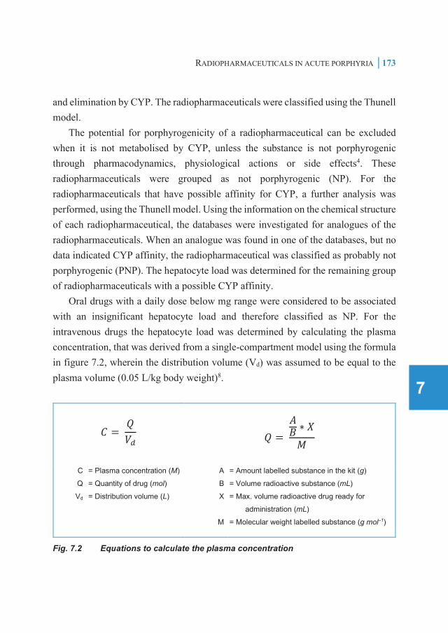
562641-L-bw-Schreuder562641-L-bw-Schreuder562641-L-bw-Schreuder562641-L-bw-SchreuderProcessed on: 25-8-2021Processed on: 25-8-2021Processed on: 25-8-2021Processed on: 25-8-2021 PDF page: 173PDF page: 173PDF page: 173PDF page: 173
RADIOPHARMACEUTICALS IN ACUTE PORPHYRIA | 173
7
and elimination by CYP. The radiopharmaceuticals were classified using the Thunell model. The potential for porphyrogenicity of a radiopharmaceutical can be excluded when it is not metabolised by CYP, unless the substance is not porphyrogenic through pharmacodynamics, physiological actions or side effects4. These radiopharmaceuticals were grouped as not porphyrogenic (NP). For the radiopharmaceuticals that have possible affinity for CYP, a further analysis was performed, using the Thunell model. Using the information on the chemical structure of each radiopharmaceutical, the databases were investigated for analogues of the radiopharmaceuticals. When an analogue was found in one of the databases, but no data indicated CYP affinity, the radiopharmaceutical was classified as probably not porphyrogenic (PNP). The hepatocyte load was determined for the remaining group of radiopharmaceuticals with a possible CYP affinity. Oral drugs with a daily dose below mg range were considered to be associated with an insignificant hepatocyte load and therefore classified as NP. For the intravenous drugs the hepatocyte load was determined by calculating the plasma concentration, that was derived from a single-compartment model using the formula in figure 7.2, wherein the distribution volume (Vd) was assumed to be equal to the plasma volume (0.05 L/kg body weight)8.
𝐶𝐶 = 𝑄𝑄𝑉𝑉𝑑𝑑
𝑄𝑄 = 𝐴𝐴𝐵𝐵 ∗ 𝑋𝑋𝑀𝑀
C
Q
Vd
= Plasma concentration (M)
= Quantity of drug (mol)
= Distribution volume (L)
A
B
X
M
= Amount labelled substance in the kit (g)
= Volume radioactive substance (mL)
= Max. volume radioactive drug ready for
administration (mL)
= Molecular weight labelled substance (g mol–1)
Fig. 7.2 Equations to calculate the plasma concentration

562641-L-bw-Schreuder562641-L-bw-Schreuder562641-L-bw-Schreuder562641-L-bw-SchreuderProcessed on: 25-8-2021Processed on: 25-8-2021Processed on: 25-8-2021Processed on: 25-8-2021 PDF page: 174PDF page: 174PDF page: 174PDF page: 174
174 | EXPLORING DRUG SAFETY OF RADIOPHARMACEUTICALS
When the plasma concentration was <1 µM and therefore the hepatocyte load was assumed to be insignificant, the drugs involved were consequently classified as PNP. However, when the plasma concentration was >1 µM, the hepatocyte load was considered as probably significant. In this case, the specific CYP3A4 or/and CYP2C9 affinity or the ability to induce ALAS1 transcription, such as activation via PXR and/or CAR of the substance, was determined by obtaining data from the SmPCs. The drug was classified as probably porphyrogenic (PSP) when there was affinity for CYP3A4/CYP2C9 or ability for ALAS1 induction. When information was lacking on the affinity for CYP, or the ability for ALAS1 induction, the drug was classified as possibly porphyrogenic. For drugs with a molecular weight of >50 kDa or blood cells labelled with a radioactive isotope, the passage into the capillary endothelium will be limited and drugs will be confined to the vascular compartment. As a result, the hepatic exposure of these drugs is insufficient to be potentially porphyrogenic. The SmPCs of the radiopharmaceuticals were used to obtain data on potential porphyrogenicity through inhibition of haem oxygenase, pharmacodynamics, physiological actions or side effects.
7.3 RESULTS
7.3.1 Databases and literature In the literature search a case report of a false-positive accumulation of [131I]iobenguane with acute intermittent porphyria was found9. Because this case was not related to porphyrogenicity of the radiopharmaceutical, it was excluded. No other articles were found using the described keywords. In the four porphyria databases used, the radiopharmaceuticals and the additives were neither identified in the safe nor in the unsafe lists.

562641-L-bw-Schreuder562641-L-bw-Schreuder562641-L-bw-Schreuder562641-L-bw-SchreuderProcessed on: 25-8-2021Processed on: 25-8-2021Processed on: 25-8-2021Processed on: 25-8-2021 PDF page: 175PDF page: 175PDF page: 175PDF page: 175
RADIOPHARMACEUTICALS IN ACUTE PORPHYRIA | 175
7
7.3.2 Analysing metabolism of the radiopharmaceuticals and classification using the Thunells model Because the radiopharmaceuticals were not classified in the databases, the metabolism of 41 radiopharmaceuticals were analysed, and the drugs were classified according to the Thunell model. The radiopharmaceuticals that do not have affinity for CYP are shown in table 7.1. They were considered as safe and classified as NP.
Table 7.1 The radiopharmaceuticals that do not have affinity for CYP and are classified as NOT porphyrogenic.
Product Indication* [51Cr]Cr-erythrocytes measurement of red blood cell volume [18F]fludeoxyglucose glucose metabolism imaging [67Ga]Ga-citrate inflammation and tumour scintigraphy [123I]sodium iodine thyroid scintigraphy [131I]sodium iodine thyroid therapy [111In]In-pentetic acid radionuclide cisternography [32P]sodium phosphate therapy of polycythemia vera [223Ra]Ra-dichloride therapy of prostate cancer [81Rb]Rb-krypton gas pulmonary ventilation scintigraphy [186Re]Re-etidronate palliative therapy of painful bone metastases [153Sm]Sm-lexidronam palliative therapy of painful bone metastases [89Sr]Sr-chloride palliative therapy of painful bone metastases [99mTc]Tc-bicisate brain scintigraphy [99mTc]Tc-medronic acid bone scintigraphy [99mTc]Tc-mertiatide dynamic renal scintigraphy [99mTc]Tc-oxidronic acid bone scintigraphy [99mTc]Tc-pentetic acid dynamic renal scintigraphy [99mTc]Tc-pertechnetate thyroid scintigraphy [99mTc]Tc-succimer static renal imaging
* Main indications noted
Table 7.2 presents the drugs that have a possible affinity for CYP. Radiopharmaceuticals [123I]iobenguane, [123I]iolopride and [131I]iobenguane were determined to be analogues of a drug known in the porphyria databases. [123I]iobenguane and [131I]iobenguane are norepinephrine analogues. [123I]iolopride is a dopamine analogue. For these analogues, no data indicating CYP affinity were found.

562641-L-bw-Schreuder562641-L-bw-Schreuder562641-L-bw-Schreuder562641-L-bw-SchreuderProcessed on: 25-8-2021Processed on: 25-8-2021Processed on: 25-8-2021Processed on: 25-8-2021 PDF page: 176PDF page: 176PDF page: 176PDF page: 176
176 | EXPLORING DRUG SAFETY OF RADIOPHARMACEUTICALS
Tabl
e 7.
2 Th
e ra
diop
harm
aceu
tical
s w
ith p
ossi
ble
affin
ity fo
r CYP
and
thei
r por
phyr
ogen
ic c
lass
ifica
tion
Prod
uct
Indi
catio
n*
Info
rmat
ion
on C
YP
affin
ity a
nd a
bilit
y to
in
duce
ALA
S1
tran
scrip
tion
Larg
e pa
rtic
le o
r pr
otei
n Pl
asm
a co
ncen
trat
ion
Pr
obab
le
insi
gnifi
cant
he
patic
load
Dru
g cl
assi
ficat
ion
[123 I]
iobe
ngua
ne
tum
our s
cint
igra
phy
CYP
affi
nity
unl
ikel
y -
- -
PNP
[123 I]
ioflu
pane
br
ain
scin
tigra
phy
Poss
ible
sub
stra
te o
f C
YP3A
4 -
0.6
nM
Yes
PNP
[123 I]
iolo
prid
e do
pam
ine
rece
ptor
sci
ntig
raph
y C
YP a
ffini
ty u
nlik
ely
-
PN
P [12
5 I]I-a
lbum
in
mea
sure
men
t of p
lasm
a vo
lum
e N
o da
ta
Mol
ecul
e >
50 k
Da
- Ye
s PN
P [13
1 I]io
beng
uane
th
erap
y of
neu
robl
asto
ma
CYP
affi
nity
unl
ikel
y -
PNP
[131 I]
iodo
met
hyl
norc
hole
ster
ol
adre
nal s
cint
igra
phy
poss
ible
sub
stra
te o
f C
YP3A
4 -
11.5
µM
N
o PS
P
[111 In
]In-o
xine
leuk
ocyt
es
infla
mm
atio
n sc
intig
raph
y N
o da
ta
Bloo
d ce
ll -
Yes
PNP
[111 In
]In-p
ente
treot
ide
som
atos
tatin
rece
ptor
sc
intig
raph
y C
YP a
ffini
ty u
nlik
ely
- -
- PN
P
[75Se
]taur
osel
chol
ic a
cid
inve
stig
atio
n of
bile
aci
d m
alab
sorp
tion
CYP
affi
nity
unl
ikel
y -
Ora
l use
; dai
ly
dose
< m
g ra
nge
Yes
PNP
[99mTc
]Tc-
albu
min
bl
ood
pool
imag
ing
No
data
M
olec
ule
> 50
kD
a
Yes
PNP
[99mTc
]Tc-
eryt
hroc
ytes
bl
ood
pool
imag
ing
No
data
Bl
ood
cell
- Ye
s PN
P [99
mTc
]Tc-
exam
etaz
ime
brai
n sc
intig
raph
y N
o da
ta
- 0.
5 µM
Ye
s PN
P [99
mTc
]Tc-
mac
rosa
lb
pulm
onar
y pe
rfusi
on s
cint
igra
phy
No
data
M
olec
ule
> 50
kD
a
Yes
PNP
[99mTc
]Tc-
meb
rofe
nin
hepa
tobi
lliary
imag
ing
No
data
-
103.
34 µ
M
No
PSP
[99mTc
]Tc-
nano
collo
id
sent
inel
nod
e sc
intig
raph
y N
o da
ta
Mol
ecul
e >
50 k
Da
- Ye
s PN
P [99
mTc
]Tc-
phyt
ate
liver
sci
ntig
raph
y N
o da
ta
- 4.
66 µ
M
No
PSP
[99mTc
]Tc-
sest
amib
i m
yoca
rdia
l per
fusi
on
scin
tigra
phy
No
data
-
1.57
µM
N
o PS
P
[99mTc
]Tc-
stan
nous
col
loid
liv
er a
nd s
plee
n sc
intig
raph
y N
o da
ta
- 0.
27 µ
M
Yes
PNP
[99mTc
]Tc-
sule
som
ab
infla
mm
atio
n sc
intig
raph
y N
o da
ta
Mol
ecul
e >
50 k
Da
- Ye
s PN
P [99
mTc
]Tc-
tetro
fosm
in
myo
card
ial p
erfu
sion
sc
intig
raph
y N
o da
ta
- 0.
07 µ
M
Yes
PNP
[201 T
l]Tl-c
hlor
ide
myo
card
ial p
erfu
sion
sc
intig
raph
y N
o da
ta
- 22
.77
µM
No
PSP
[90Y]
Y-ci
trate
ra
dios
ynov
iorth
esis
N
o da
ta
Mol
ecul
e >
50 k
Da
- Ye
s PN
P * M
ain
indi
catio
ns n
oted
. Abb
revi
atio
ns: A
LAS1
, 5-a
min
olev
ulin
ic a
cid
synt
hase
; PN
P, p
ossi
bly
not p
orph
yrog
enic
; PS
P, p
ossi
bly
porp
hyro
geni
c.

562641-L-bw-Schreuder562641-L-bw-Schreuder562641-L-bw-Schreuder562641-L-bw-SchreuderProcessed on: 25-8-2021Processed on: 25-8-2021Processed on: 25-8-2021Processed on: 25-8-2021 PDF page: 177PDF page: 177PDF page: 177PDF page: 177
RADIOPHARMACEUTICALS IN ACUTE PORPHYRIA | 177
7
CYP affinity for the corresponding radiopharmaceuticals seems unlikely and therefore they were classified as PNP10. [111In]In-pentetreotide is an octreotide analogue. Octreotide is a synthetic peptide and was considered to be safe in one reference10. As a consequence, CYP affinity for [111In]In-pentetreotide seems unlikely (i.e., [111In]In-pentetreotide was classified as PNP). In table 7.2, eight radiopharmaceuticals with a molecular size >50 kDa or blood cells labelled with a radioactive isotope were presented and classified as PNP. [75Se]tauroselcholic acid is an oral drug with a daily dose below the micromolar range. Hence, it is considered to be associated with an insignificant hepatocyte load and was classified as possibly not PNP. [123I]ioflupane was determined to be an analogue of cocaine and hence a possible substrate of CYP3A411, 12. Furthermore, [131I]iodomethyl norcholesterol was determined to be a cholesterol analogue and therefore a possible substrate of CYP3A413. For the remaining radiopharmaceuticals no data on CYP affinity were found. For the radiopharmaceuticals with a possible affinity for CYP or for which no data on CYP affinity was available, the plasma concentrations were calculated to determine whether these concentrations exceed the estimated porphyrogenic micromolar range (>1 µM). For [123I]ioflupane, [99mTc]Tc-exametazime, [99mTc]Tc-stannous colloid and [99mTc]Tc-tetrofosmin, plasma concentrations below 1 µM were calculated and these agents were subsequently classified as PNP. For [131I]iodomethyl norcholesterol, [99mTc]Tc-mebrofenin, [99mTc]Tc-phytate, [99mTc]Tc-sestamibi and [201Tl]Tl-chloride, plasma concentrations were estimated to be high enough (>1 µM) to trigger a porphyrogenic ALAS1 transcription. Because there was no information available for these drugs regarding the CYP3A4/2C9 affinity and the ability to induce ALAS1 transcription (e.g., such as activation via PXR and/or CAR) they were classified as possibly porphyrogenic.
7.3.3 Potential porphyrogenicity through inhibition of haem oxygenase, pharmacodynamics, physiological actions or side effects None of the radiopharmaceuticals assessed were found to be inhibitors of haem oxygenase. No data was found in the SmPCs indicating potential porphyrogenicity

562641-L-bw-Schreuder562641-L-bw-Schreuder562641-L-bw-Schreuder562641-L-bw-SchreuderProcessed on: 25-8-2021Processed on: 25-8-2021Processed on: 25-8-2021Processed on: 25-8-2021 PDF page: 178PDF page: 178PDF page: 178PDF page: 178
178 | EXPLORING DRUG SAFETY OF RADIOPHARMACEUTICALS
through pharmacodynamics, physiological actions or side effects. Because radiopharmaceuticals are administered with low mass doses (i.e., doses in or below the microgram range) pharmacological effects are not expected. Side effects as described in the SmPCs did not relate to responses via the sympatico-adrenal system, hypothalamic-pituary-adrenal axis, or disturbance of the energy homeostasis.
7.4 DISCUSSION
Our analysis on the porphyrogenicity of 41 radiopharmaceuticals revealed that five radiopharmaceuticals can be classified as possibly porphyrogenic. The assessment was based on information on inhibition of haem oxygenase, metabolism by cytochrome P450 (CYP), CYP affinity, ability to induce ALAS1 transcription, (particle) size and plasma concentration of the drug. It is important to understand that the outcome of the method used depends on the accuracy of the available pharmacokinetic and pharmacodynamic data. During our study we noticed that these data for radiopharmaceuticals are not always complete. Information on metabolism by CYP and the ability to induce ALAS1 transcription in particular (e.g., such as activation via PXR and/or CAR) is often incomplete or even absent. It is also worth noting that the plasma concentrations that were calculated are very strict. The human body was considered as a single compartment model and the plasma volume was set at 2500 mL, assuming a body weight of 50 kg. For assessing the level of drug exposure the maximum dose given to a patient was used. As a consequence, the plasma concentrations are probably overestimated in order to achieve a safety margin. The assessment in this study has not taken differences in response of individual patients to radiopharmaceuticals into account. Although individuals might have the same genetic predisposition, responses may be different from what is expected2. Other factors may contribute to the increase of the porphyric vulnerability of an individual patient such as the use of other drugs or alcohol, an altered hormone balance, experiencing stress, or suffering from an infection14. Therefore, an individual assessment that takes all factors into account is important for each patient.

562641-L-bw-Schreuder562641-L-bw-Schreuder562641-L-bw-Schreuder562641-L-bw-SchreuderProcessed on: 25-8-2021Processed on: 25-8-2021Processed on: 25-8-2021Processed on: 25-8-2021 PDF page: 179PDF page: 179PDF page: 179PDF page: 179
RADIOPHARMACEUTICALS IN ACUTE PORPHYRIA | 179
7
With regard to the five radiopharmaceuticals being classified as possibly porphyrogenic, further information is needed to assess whether they possess a clinically relevant CYP3A4 or CYP2C9 affinity or have the ability to induce ALAS1 transcription. If this is not the case, they can be classified as probably not porphyrogenic. Furthermore, we should be aware of the possible side effects of all radiopharmaceuticals inducing ALAS1 transcription that may provoke an attack. Most radiopharmaceuticals are used for diagnostic purposes and are commonly administered to patients in a fasted state. It has been suggested that glucose deprivation induces ALAS1 transcription and may well have porphyrogenic consequences14. It is therefore important to be aware of the risk of fasting associated with diagnostic procedures for carriers of the acute porphyria gene. A few radiopharmaceuticals are used for therapeutic purposes, sometimes resulting in lasting physiological changes. An example is treatment of the thyroid with [131I] sodium iodine, resulting in a partial of total thyroid ablation. Physiological changes could potentially alter the susceptibility to porphyrogenic substances of an individual. Therefore, monitoring patients for development of new symptoms after treatment with a therapeutic radiopharmaceutical is required. Based on the evidence available, classification of these radiopharmaceuticals as PNP or PSP as presented is appropriate. Although it has been shown that the application of the algorithm provides a correct classification in most cases, some examples were found wherein the clinical outcome did not match the algorithm classification2, 7. It is for this reason that, clinical confirmation is needed (e.g., listing the number of patients to which the drug has been administered safely by expert physicians). Until then, the radiopharmaceuticals being considered should be used with care as these substances are possibly harmful in acute porphyria patients. A potentially porphyrogenic drug is not to be prescribed for a carrier of the acute porphyria gene other than upon urgent indication and where safer alternatives are unavailable4. When a potentially porphyrogenic radiopharmaceutical must be prescribed, it is best to consult a porphyria expert16, as preventive measures need to be taken and doctors should be prepared for a potentially acute attack. Preventive measures are adapted to the individual porphyric vulnerability of the patient and the porphyrogenicity of the drug. In general, monitoring for porphyric symptoms can be

562641-L-bw-Schreuder562641-L-bw-Schreuder562641-L-bw-Schreuder562641-L-bw-SchreuderProcessed on: 25-8-2021Processed on: 25-8-2021Processed on: 25-8-2021Processed on: 25-8-2021 PDF page: 180PDF page: 180PDF page: 180PDF page: 180
180 | EXPLORING DRUG SAFETY OF RADIOPHARMACEUTICALS
recommended, such as red urine, as well as monitoring the concentration of porphobilinogen in the urine. When the patient is probably or highly susceptible, prolonged fasting must be avoided by ensuring the patient is treated early in the morning. They are also advised to seek medical advice in case of any symptoms suggestive of porphyria15.
7.5 CONCLUSION
From the 41 radiopharmaceuticals assessed, five of them, i.e. [131I]iodomethyl norcholesterol, [99mTc]Tc-mebrofenin, [99mTc]Tc-phytate, [99mTc]Tc-sestamibi and [201Tl]Tl-chloride, are classified as possible porphyrogenic according to the Thunell method and may be unsafe for a carrier of the acute porphyria gene. [131I]Iodomethyl norcholesterol, [99mTc]Tc-mebrofenin, [99mTc]Tc-phytate, [99mTc]Tc-sestamibi and [201Tl]Tl-chloride should not be prescribed for acute porphyria patients unless an urgent indication is present and a safer alternative not available. In such cases potential users should seek advice from a porphyria expert, and preventive measures may be required. The rest of the radiopharmaceuticals are classified as not or probably not porphyrogenic.

562641-L-bw-Schreuder562641-L-bw-Schreuder562641-L-bw-Schreuder562641-L-bw-SchreuderProcessed on: 25-8-2021Processed on: 25-8-2021Processed on: 25-8-2021Processed on: 25-8-2021 PDF page: 181PDF page: 181PDF page: 181PDF page: 181
RADIOPHARMACEUTICALS IN ACUTE PORPHYRIA | 181
7
7.6 REFERENCES
1. Puy H, Gouya L, Devbach JC. Porphyrias. Lancet 2010;375:924–37. 2. Hift RJ, Thunell S, Brun A. Drugs in porphyria: From observation to a modern
algorithm-based system for the prediction of porphyrogenicity. Pharmacol Ther 2011;132:158–69.
3. Thunell S. Genomic approach to acute porphyria. Physiol Res 2006;55(Suppl.2):S43–S66.
4. Thunell S, Pomp E, Brun A. Guide to drug porphyrogenicity prediction and drug prescription in the acute porphyrias. Br J Clin Pharmacol 2007;64:668–79.
5. Maltby P, Theobald T. Survey of current diagnostic radiopharmaceuticals. In: Theobald T, editor. Sampson’s textbook of radiopharmacy. 4th edition. London: Pharmaceutical Press; 2011. pp. 277–306.
6. Chan P, Croasdale J. Survey of current therapeutic radiopharmaceuticals. In: Theobald T, editor. Sampson’s textbook of radiopharmacy. 4th edition. London: Pharmaceutical Press; 2011. pp. 303–23.
7. Brun A, Gilleshammer L, Skeide, Kjome RL. The EPNET project to improve drug information for patients with an acute porphyria: The current role of clinical drug reporting. Br J Dermatol 2011;164:1137.
8. Rang H, Dale M, Ritter J, Flower R. Pharmacology. Edinburgh: Churchill Livingstone Elsevier; 2007. pp. 113–127.
9. Masuda T, Ota R, Ando T, Maeda N, Horie Y, Yoshimura T, Motomura M, Kawakami A. False-Positive accumulation of metaiodobenzylguanidine in a case with acute intermittent porphyria. Intern Med 2011;50:1029–1032.
10. The Norwegian Porphyria Centre (NAPOS). The Drug Database for Acute Porphyria. http://www.drugs-porphyria.org/. Accessed 24 Mar 2015.
11. LeDuc BW, Sinclair PR, Shuster L, Sinclair JF, Evans JE, Greenblatt DJ. Norcocaine and N-hydroxynorcocaine formation in human liver microsomes: role of cytochrome P-450 3A4. Pharmacology 1993;46(5):294–300.

562641-L-bw-Schreuder562641-L-bw-Schreuder562641-L-bw-Schreuder562641-L-bw-SchreuderProcessed on: 25-8-2021Processed on: 25-8-2021Processed on: 25-8-2021Processed on: 25-8-2021 PDF page: 182PDF page: 182PDF page: 182PDF page: 182
182 | EXPLORING DRUG SAFETY OF RADIOPHARMACEUTICALS
12. Ladona MG, Gonzalez ML, Rane A, Peter RM, de la Torre R. Cocaine metabolism in human fetal and adult liver microsomes is related to cytochrome P450 3A expression. Life Sci 2000;68(4):431–43.
13. Honda A, Miyazaki T, Ikegami T, Iwamoto J, Maeda T, Hirayama T, Saito Y, Teramoto T, Matsuzaki Y. Cholesterol 25-hydroxylation activity of CYP3A. J Lipid Res 2011;52(8):1509–16.
14. Matkovic LB, D'Andrea F, Fornes D, San Martín de Viale LC, Mazzetti MB. How porphyrinogenic drugs modelling acute porphyria impair the hormonal status that regulates glucose metabolism. Their relevance in the onset of this disease. Toxicology 2011;290:22–30.
15. Stein P, Badminton M, Barth J, Rees D, Stewart MF. Best practice guidelines on clinical management of acute attacks of porphyria and their complications. Ann Clin Biochem 2013;50:217–23.
16. List of porphyria centres with contact details. www.porphyria-europe.org. European Porphyria Network. Accessed 6 Mar 2015.

562641-L-bw-Schreuder562641-L-bw-Schreuder562641-L-bw-Schreuder562641-L-bw-SchreuderProcessed on: 25-8-2021Processed on: 25-8-2021Processed on: 25-8-2021Processed on: 25-8-2021 PDF page: 183PDF page: 183PDF page: 183PDF page: 183
Chapter 8
Lack of consistent dose recommendations for
radiopharmaceuticals in patients with renal insufficiency: results of a
systematic review
Nanno Schreuder 1,2, Iris de Romijn 3, Pieter L. Jager 4, Jos G.W. Kosterink 1,5,
Eugène P. van Puijenbroek 1,6 1 Groningen Research Institute of Pharmacy, PharmacoTherapy, -Epidemiology & -Economics,
University of Groningen, Groningen, the Netherlands 2 GE Healthcare Radiopharmacy Zwolle, Zwolle, the Netherlands 3 Division of Pharmacoepidemiology and Clinical Pharmacology, Utrecht Institute for Pharmaceutical Sciences, Utrecht University, Utrecht , The Netherlands 4 Department of Nuclear Medicine, Isala Hospital, Zwolle, the Netherlands 5 University of Groningen, University Medical Center Groningen, Department of Clinical Pharmacy
and Pharmacology, Groningen, The Netherlands 6 Netherlands Pharmacovigilance Centre Lareb, ‘s-Hertogenbosch, the Netherlands
Accepted for publication in EJNMMI Radiopharmacy and Chemistry

562641-L-bw-Schreuder562641-L-bw-Schreuder562641-L-bw-Schreuder562641-L-bw-SchreuderProcessed on: 25-8-2021Processed on: 25-8-2021Processed on: 25-8-2021Processed on: 25-8-2021 PDF page: 184PDF page: 184PDF page: 184PDF page: 184
184 | EXPLORING DRUG SAFETY OF RADIOPHARMACEUTICALS
ABSTRACT Introduction: Patients with renal insufficiency may need to have their radiopharmaceutical dosage adjusted to prevent adverse effects and poor outcomes, but there are few recommendations on radiopharmaceutical dosing for this group of patients. Objective: The aim of this study is to provide an overview of the available information on radiopharmaceutical dose recommendations for patients with renal insufficiency and, for those radiopharmaceuticals that are found in the literature to assess the dosing recommendations in this group of patients. Methods: We performed a systematic literature review according to the Preferred Reporting Items for Systematic Reviews and Meta-Analyses (PRISMA) statement. We conducted a literature search in the MEDLINE (PubMed) and Embase databases and screened potentially relevant studies using inclusion and exclusion criteria. We independently assessed the included observational studies’ methodologies and extracted relevant data. Results: Of the 5,795 studies first identified, 34 were included in this systematic review. These studies described three radiopharmaceuticals: [131I]sodium iodine, [18F]fludeoxyglucose, and [131I]iobenguane. Twenty-nine studies (85.3%) reported data on patients with stage 5 chronic kidney disease (CKD), while only three studies mentioned CKD patients in other stages (8.8%). Conclusion: We found no consistent recommendations for radiopharmaceutical dosing in patients with renal insufficiency. Although some studies do mention dosing difficulties in patients with insufficient renal function, information is available for only a few radiopharmaceuticals, and recommendations are sometimes contradictory. Further research on radiopharmaceutical dosing in patients with renal function insufficiency is needed to determine whether these patients require specific dosing.

562641-L-bw-Schreuder562641-L-bw-Schreuder562641-L-bw-Schreuder562641-L-bw-SchreuderProcessed on: 25-8-2021Processed on: 25-8-2021Processed on: 25-8-2021Processed on: 25-8-2021 PDF page: 185PDF page: 185PDF page: 185PDF page: 185
RADIOPHARMACEUTICALS IN RENAL INSUFFIENCY: A SYSTEMATIC REVIEW | 185
8
8.1 INTRODUCTION
Nuclear medicine plays an important role in the diagnosis and therapy of diseases, particularly in the field of oncology. The field of nuclear medicine relies on radioactive compounds, so-called radiopharmaceuticals1. Selecting the right dose of a radiopharmaceutical, expressed in becquerels (Bq) as the activity of the compound’s radionuclide, is of high importance. For diagnostic radiopharmaceuticals, the ideal dose will provide accurate, useful diagnostic information while keeping the radiation dose to the patient low2, 3. The efficacy will depend on the biodistribution of the diagnostic radiopharmaceutical. Important aspects include localisation in a target organ, localisation in non-target organs, and the mechanisms—such as biological excretion—for removing non-target radioactivity. Advantageous biodistribution will contribute to a good target-to-non-target activity ratio, ensuring optimal image quality which allows a clear diagnostic outcome4. For therapeutic radiopharmaceuticals, the ideal dose will deliver the right therapeutic activity without causing adverse effects and with a minimum radiation dose to non-target organs or tissues5–7. The biodistribution of the therapeutic radiopharmaceutical is important because localisation in the target organ will determine the therapeutic response, and non-target organs are at risk of toxicity4. Renal insufficiency is a growing health problem with an estimated prevalence of 11%–13% in the general population and can result from diseases, such as diabetes mellitus and hypertension, or from aging8–10. It may reduce the excretion rate of pharmaceuticals and their metabolites, elevating plasma concentrations and requiring the dose to be adjusted11, 12. Similarly, for radiopharmaceuticals that are cleared by the kidneys, the biodistribution of the radioactive drug is likely to be altered in patients with renal insufficiency. For diagnostic radiopharmaceuticals, decreased clearance may lead to prolonged blood pool activity and subsequently to a poor target-to-non-target ratio, which may decrease image quality and ultimately affect the diagnostic outcome. For therapeutic radiopharmaceuticals, decreased clearance may lead to increased activity at the target organs or non-target organs, which increases the risk of toxicity. Therefore, it is expected that the

562641-L-bw-Schreuder562641-L-bw-Schreuder562641-L-bw-Schreuder562641-L-bw-SchreuderProcessed on: 25-8-2021Processed on: 25-8-2021Processed on: 25-8-2021Processed on: 25-8-2021 PDF page: 186PDF page: 186PDF page: 186PDF page: 186
186 | EXPLORING DRUG SAFETY OF RADIOPHARMACEUTICALS
radiopharmaceutical dosage in patients with renal insufficiency will have to be adjusted13, 14. At present, the dose of a radiopharmaceutical is fixed in most cases, although it is sometimes adjusted for body weight, as in the case of children15. However, standards for radiopharmaceutical dosing in patients with renal insufficiency are lacking. One review describes treatment with radioiodine for hyperthyroidism and thyroid cancer in end-stage renal disease. The review mentions that the available literature is scarce and that standards, based only on analysis of single case reports, are not coherent16. Some available nuclear medicine guidelines contain only one paragraph on the use of radiopharmaceuticals in patients with renal insufficiency. Only one nuclear medicine guideline gives a specific dose recommendation for these patients, advising that the administered dose of bone-seeking therapeutic radiopharmaceuticals for palliation of bone pain should be lowered by 50% in patients with creatinine clearance of less than 50 mL/min17. Other guidelines provide only general, nonspecific comments, such as recommending that renal function should be assessed, that a nephrologist should be consulted, that the administration of the radiopharmaceutical should be carefully planned and managed, or even that patients with renal insufficiency should be excluded17–24. While these effects are particularly relevant in therapeutic applications because a change in biodistribution may affect therapy outcomes or increase the risk of toxicity in these patients, diagnostic radiopharmaceutical guidelines also indicate that scans obtained in renal insufficiency patients may be suboptimal due to a change in biodistribution. Some suggest increasing the time between administration of the radiopharmaceutical and imaging19, 25–28. However, several guidelines for both therapeutic and diagnostic radiopharmaceuticals mention that while dose adjustment may be needed in this group of patients, little is known about this topic18, 20, 22, 29–31. Therefore, our aim in this systematic review is to provide an overview of the available information on radiopharmaceutical dose recommendations for patients with renal insufficiency and, for those radiopharmaceuticals that are found in the literature, to assess the dose recommendations in this group of patients.

562641-L-bw-Schreuder562641-L-bw-Schreuder562641-L-bw-Schreuder562641-L-bw-SchreuderProcessed on: 25-8-2021Processed on: 25-8-2021Processed on: 25-8-2021Processed on: 25-8-2021 PDF page: 187PDF page: 187PDF page: 187PDF page: 187
RADIOPHARMACEUTICALS IN RENAL INSUFFIENCY: A SYSTEMATIC REVIEW | 187
8
8.2 METHODS
8.2.1 Study design We conducted this systematic literature review according to the Preferred Reporting Items for Systematic Reviews and Meta-Analyses (PRISMA) statement32, and the review was registered in the International Prospective Register of Systematic Reviews (PROSPERO) under number CRD4201913610733.
8.2.2 Search strategy We performed a computerised literature search using the databases MEDLINE (PubMed) and Embase. Two researchers (I.d.R. and N.S.) developed a search string for each database (Table 8.1) using keywords for both renal insufficiency and radiopharmaceuticals. No publication year limits were applied. Only studies in the English language were included, and a filter was applied to exclude animal-only studies. An additional filter was applied in Embase to exclude studies available in MEDLINE. We screened the selected studies and review articles to identify additional relevant studies and references. The initial search was completed on 10 May 2019 and updated with recent articles until 7 October 2020.
Table 8.1 Search strategies employed for PubMed and Embase
Database Search string PubMed (((((Radiopharmaceuticals(MeSH) OR radiopharmaceutical*(tiab) OR radioactive
drug*(tiab) OR radioiodine(tiab)))) AND ((Kidney Diseases(MeSH) OR kidney disease*(tiab) OR Renal Insufficiency(MeSH) OR renal insufficien*(tiab) OR renal impairment(tiab) OR Glomerular Filtration Rate(Mesh) OR glomerular filtration rate*(tiab) OR eGFR(tiab) OR Metabolic Clearance Rate(MeSH) OR renal clearance(tiab)))) NOT ("Animals"(Mesh) NOT "Humans"(Mesh))) AND English(Language)
Embase ('radiopharmaceutical agent'/exp OR 'radiopharmaceutical agent':ti,ab) AND ('kidney disease'/exp OR 'kidney disease':ti,ab OR 'kidney failure'/exp OR 'kidney failure':ti,ab OR 'renal impairment':ti,ab OR 'glomerulus filtration rate'/exp OR 'glomerular filtration rate':ti,ab OR 'estimated glomerular filtration rate':ti,ab OR 'metabolic clearance'/exp OR 'renal clearance':ti,ab) NOT ('animal'/exp NOT 'human'/exp) AND (english)/lim AND (embase)/lim NOT ((embase)/lim AND (medline)/lim)

562641-L-bw-Schreuder562641-L-bw-Schreuder562641-L-bw-Schreuder562641-L-bw-SchreuderProcessed on: 25-8-2021Processed on: 25-8-2021Processed on: 25-8-2021Processed on: 25-8-2021 PDF page: 188PDF page: 188PDF page: 188PDF page: 188
188 | EXPLORING DRUG SAFETY OF RADIOPHARMACEUTICALS
8.2.3 Study selection All titles and abstracts were screened, and we retrieved the full text of potentially relevant studies. Two researchers (I.d.R. and N.S.) independently assessed the full text of each study for relevance. We included studies that met the following inclusion criteria: They described patients diagnosed or treated with a radiopharmaceutical and who suffered from a renal insufficiency, they used a radiopharmaceutical that is (at least partly) cleared renally, and they made recommendations for an adequate dose in these patients or gave additional advice. Studies were excluded if they gave no relevant information on dosing or aspects related to dosing, addressed only the radiation safety of staff, described renal imaging, or were review articles.
8.2.4 Assessment of methodological quality Two researchers (I.d.R. and N.S.) independently assessed the methodological quality of the included observational studies using the Newcastle-Ottawa Scale (NOS): Quality Assessment Form for Cohort and Case-Control Studies34. For each study, we scored nine items in three domains: selection, comparability, and exposure or outcome. Scores were added to create an aggregate score. The NOS scores were converted to ratings of ‘good’, ‘fair’, or ‘poor’ according to Agency for Healthcare Research and Quality standards35. Studies of good quality were defined as those awarded 3–4 stars in the selection domain and 1–2 stars in the comparability domain and 2 stars in the exposure or outcome domain. Fair studies were defined as those awarded 2 stars in the selection domain and 1–2 stars in the comparability domain and 1–2 stars in the exposure or outcome domain. Poor-quality studies were defined as those awarded 0–1 stars in the selection domain or 0 in the comparability domain or 0 in the exposure or outcome domain. Where opinions on a score differed, we consulted a third reviewer (E.v.P.) to reach consensus.

562641-L-bw-Schreuder562641-L-bw-Schreuder562641-L-bw-Schreuder562641-L-bw-SchreuderProcessed on: 25-8-2021Processed on: 25-8-2021Processed on: 25-8-2021Processed on: 25-8-2021 PDF page: 189PDF page: 189PDF page: 189PDF page: 189
RADIOPHARMACEUTICALS IN RENAL INSUFFIENCY: A SYSTEMATIC REVIEW | 189
8
8.2.5 Data collection For studies meeting the selection criteria, we extracted data using a standardised approach. Two researchers (I.d.R. and N.S.) independently extracted the following data: (1) author and journal, (2) year of publication, (3) study design, (4) name(s) of radiopharmaceutical(s) and the administered dose(s), (5) indication, (6) number of patients with renal insufficiency, (7) stage of renal insufficiency, (8) where applicable, the type and timing of dialysis after administration of the radiopharmaceutical, (9) recommendation(s) for adjustment of dose, (10) other advice on aspects such as adjustment of dialysis or scintigraphy, (11) reasons for dose adjustment, and (12) study limitations. We standardised the radiopharmaceuticals’ names according to the Anatomical Therapeutic Chemical classification system36, and the International Consensus Radiochemistry Nomenclature Guidelines37. Where studies reported the administered dose using the unit curie (Ci), we converted this to the SI derived unit becquerel (Bq) for uniformity of outcome38. Patients with renal insufficiency were classified using the terminology of the chronic kidney disease (CKD) standard (Table 8.2), which includes five stages of kidney damage, from kidney damage with normal kidney function in stage 1 to kidney failure in stage 539, 40. We classified patients on dialysis as stage 5 if they had not been assigned to a specific stage in a study. When the extracted data were not in agreement and consensus could not be reached between the two researchers, a third researcher (E.v.P.) was consulted to resolve discrepancies.
Table 8.2 Classification of CKD by GFR39, 40
Stage Description GFR, mL/min/1.73 m2 1 Kidney damage with normal or increased GFR ≥ 90 2 Kidney damage with mild decreased GFR 60–89 3 Moderately decreased GFR 30–59 4 Severely decreased GFR 15–29 5 Kidney failure < 15 (or dialysis)
Abbreviations: CKD, chronic kidney diseases; GFR, glomerular filtration rate

562641-L-bw-Schreuder562641-L-bw-Schreuder562641-L-bw-Schreuder562641-L-bw-SchreuderProcessed on: 25-8-2021Processed on: 25-8-2021Processed on: 25-8-2021Processed on: 25-8-2021 PDF page: 190PDF page: 190PDF page: 190PDF page: 190
190 | EXPLORING DRUG SAFETY OF RADIOPHARMACEUTICALS
8.3 RESULTS
8.3.1 Search results The literature search identified a total of 5,795 studies in PubMed (n = 2,684) and Embase (n = 3,111); another 11 studies were identified from references. After removing duplicates (n = 81), we screened 5,725 studies by title (and abstract, where necessary), resulting in 65 potentially relevant studies. After a full-text screening we excluded another 31 studies for various reasons: they gave no relevant information on dosing or aspects related to dosing (n = 18), they addressed only the radiation safety of staff (n = 5), described renal imaging (n = 5), or were reviews (n = 3). A total of 34 studies remained for inclusion in this systematic review41–74. The selection process is illustrated in a PRISMA flow diagram (Figure 8.1).
Fig. 8.1 Selection of studies according to the PRISMA statement32

562641-L-bw-Schreuder562641-L-bw-Schreuder562641-L-bw-Schreuder562641-L-bw-SchreuderProcessed on: 25-8-2021Processed on: 25-8-2021Processed on: 25-8-2021Processed on: 25-8-2021 PDF page: 191PDF page: 191PDF page: 191PDF page: 191
RADIOPHARMACEUTICALS IN RENAL INSUFFIENCY: A SYSTEMATIC REVIEW | 191
8
The 34 selected studies included 12 case reports (35.3%), 11 case series (32.4%), five case-control studies (14.7%), two cohort studies (5.9%), three theoretical models (8.8%), and one case report with a theoretical model (2.9%). The radiopharmaceuticals reported in these studies are [18F]fludeoxyglucose (FDG) (n = 5; 14.7%), [131I]sodium iodine (n = 28; 82.4%), and [131I]iobenguane (n = 1; 2.9%). Twenty-nine studies reported data for patients with CKD stage 5 (85.3%), while three studies included patients in other stages (8.8%). In two studies, the CKD stage was not identified, or it was determined in a non-standard fashion (5.9%). Patients in the 29 studies reporting data for CKD stage 5 were on renal replacement therapy. Nineteen studies described patients on haemodialysis (HD) (65.5%), four studies described patients on continuous ambulatory peritoneal dialysis (CAPD) (13.8%), three studies described patients on HD or CAPD (10.3%), one study described patients on HD or intermittent peritoneal dialysis (IPD) (3.4%), one study described patients on continuous haemodialysis (3.4%), and one study did not specify the type of dialysis (3.4%). In these studies, the timing of dialysis varied. For HD, the start of dialysis varied from 15 to 72 hours after administration of the radiopharmaceutical, the number of dialyses varied from one to five times, and the timing intervals varied. For CAPD, the fluid changes varied from four to eight times a day. An overview of the included studies’ characteristics is presented in Table 8.3.
8.3.2 Quality of the observational studies We performed a methodological quality assessment of the included observational studies, namely five case-control studies (Table 8.4) and two cohort studies (Table 8.5). Our assessment yielded one study rated ‘good’ (14.3%), one study rated ‘fair’ (14.3%), and five studies rated ‘poor’ (71.4%).

562641-L-bw-Schreuder562641-L-bw-Schreuder562641-L-bw-Schreuder562641-L-bw-SchreuderProcessed on: 25-8-2021Processed on: 25-8-2021Processed on: 25-8-2021Processed on: 25-8-2021 PDF page: 192PDF page: 192PDF page: 192PDF page: 192
192 | EXPLORING DRUG SAFETY OF RADIOPHARMACEUTICALS
Tabl
e 8.
3 O
verv
iew
of i
nclu
ded
stud
ies
with
thei
r cha
ract
eris
tics
Ref
eren
ce
Year
St
udy
desi
gn
Num
ber
of
patie
nts
Rad
ioph
arm
aceu
tical
In
dica
tion
Dos
e (M
Bq)
St
age
of
rena
l fa
ilure
(C
KD
)
Type
of
dial
ysis
(n
umbe
r of
pa
tient
s)
Tim
ing
of d
ialy
sis
afte
r ad
min
istr
atio
n ra
diop
harm
aceu
tical
Aker
s41
2016
C
OS
58
[18F]
flude
oxyg
luco
se
PET/
CT
370–
555
1–5
NA
NA
Akta
ş42
2008
C
CS
10
[131 I]
sodi
um io
dine
TC
11
10–3
700
5 H
D (6
); C
APD
(4
)
24h
cont
inue
d ev
ery
day
for 5
d; C
APD
in
crea
sed
from
4 to
6–8
tim
es a
day
Alev
izak
i43
2006
C
S 5
[131 I]
sodi
um io
dine
PT
C
1110
–259
0 5
HD
(4);
IPD
(1)
48h,
and
2 p
atie
nts
also
96h
Bhat
44
2017
C
R
1 [13
1 I]so
dium
iodi
ne
PTC
18
50
5 H
D
15h,
27h
, 43h
C
ourb
on45
19
97
CR
1
[131 I]
sodi
um io
dine
TC
37
00
5 H
D
2d, 4
d C
ourb
on46
20
06
CS
2 [13
1 I]so
dium
iodi
ne
TC
3700
5
HD
72
h, 1
22h–
144h
C
ulpe
pper
47
1992
C
R
1 [13
1 I]so
dium
iodi
ne
FTC
47
73
5 H
D
24h
,43h
,66h
D
aum
erie
48
1996
C
S 3
[131 I]
sodi
um io
dine
PT
C
2 tre
atm
ents
of
925
5 H
D
2d, 3
d
Dem
ko49
19
98
CR
1
[131 I]
sodi
um io
dine
TM
NG
10
45.6
2 5
HD
24
h D
riedg
er50
20
06
CS
3 [13
1 I]so
dium
iodi
ne
PTC
37
00; 3
700;
25
00
5 H
D (2
); C
APD
(1
)
NA
El-Z
efta
wy51
20
17
CC
S 27
[13
1 I]so
dium
iodi
ne
DTC
M
ean
dose
55
50
3 an
d 4
NA
NA
Fofi52
20
13
CS
2 [13
1 I]so
dium
iodi
ne
PTC
18
50
5 C
HD
24
h, 4
8h
Hol
st53
20
05
CR
and
TM
1
[131 I]
sodi
um io
dine
PT
C*
3637
5
HD
2d
, 3d,
4d
How
ard54
19
81
CR
1
[131 I]
sodi
um io
dine
PT
C
740
5 H
D
NA
Jim
énez
55
2001
C
S 3
[131 I]
sodi
um io
dine
PT
C
2775
; 321
9;
4440
5
HD
24
h, 4
8h, 7
2h, 9
6h, 1
44h
Kapt
ein56
20
00
CS
2 [13
1 I]so
dium
iodi
ne
PTC
98
0; 1
110
5 C
APD
3–
5 tim
es a
day
* Al
thou
gh th
e st
udy
desc
ribes
hyp
erth
yroi
dism
as
wel
l, th
e re
porte
d ca
se w
as tr
eate
d w
ith [13
1 I]so
dium
iodi
ne fo
r thy
roid
can
cer.
† 12
pat
ient
s w
ere
on d
ialy
sis,
type
an
d tim
ing
wer
e no
t spe
cifie
d. ǂ
In th
is s
tudy
a p
harm
acok
inet
ic s
oftw
are
mod
el w
as d
evel
oped
and
val
idat
ed w
ith d
ata
of 13
1 I, 12
3 I an
d 12
4 I. ¶
patie
nts
with
a b
lood
se
rum
cre
atin
ine
leve
l > 1
.1 m
g/dl
. #
The
findi
ngs
in o
ne p
atie
nt w
ere
com
pare
d w
ith t
wo
patie
nts
with
mild
to
mod
erat
e re
nal f
unct
ion.
Abb
revi
atio
ns:
CAP
D,
cont
inuo
us a
mbu
lato
ry p
erito
neal
dia
lysi
s; C
CS,
cas
e-co
ntro
l stu
dy; C
HD
, con
tinuo
us h
aem
odia
lysi
s; C
KD, c
hron
ic k
idne
y di
seas
es; C
OS,
coh
ort s
tudy
; CR
, cas
e re
port;
CS,
cas
e se
ries;
DTC
, diff
eren
tiate
d th
yroi
d ca
ncer
; FTC
, fol
licul
ar th
yroi
d ca
ncer
; GD
, Gra
ves'
dis
ease
; HD
, hae
mod
ialy
sis;
IPD
, int
erm
itten
t per
itone
al
dial
ysis
; N
A, n
ot a
vaila
ble;
PC
, ph
eoch
rom
ocyt
oma;
PET
/CT,
pos
itron
em
issi
on t
omog
raph
y/co
mpu
ted
tom
ogra
phy;
PTC
, pa
pilla
ry t
hyro
id c
ance
r; TC
, th
yroi
d ca
ncer
; TD
, thy
roid
dis
ease
; TM
, The
oret
ical
mod
el; T
MN
G, t
oxic
mul
tinod
ular
goi
ter.

562641-L-bw-Schreuder562641-L-bw-Schreuder562641-L-bw-Schreuder562641-L-bw-SchreuderProcessed on: 25-8-2021Processed on: 25-8-2021Processed on: 25-8-2021Processed on: 25-8-2021 PDF page: 193PDF page: 193PDF page: 193PDF page: 193
RADIOPHARMACEUTICALS IN RENAL INSUFFIENCY: A SYSTEMATIC REVIEW | 193
8
Tabl
e 8.
3 (C
ontin
ued)
Ove
rvie
w o
f inc
lude
d st
udie
s w
ith th
eir c
hara
cter
istic
s
Ref
eren
ce
Year
St
udy
desi
gn
Num
ber
of
patie
nts
Rad
ioph
arm
aceu
tical
In
dica
tion
Dos
e (M
Bq)
St
age
of re
nal
failu
re
(CK
D)
Type
of
dial
ysis
(n
umbe
r of
pa
tient
s)
Tim
ing
of d
ialy
sis
afte
r ad
min
istr
atio
n ra
diop
harm
aceu
tical
Kode
57
2017
C
CS
30
[18F]
flude
oxyg
luco
se
PET/
CT
5.18
/kg
4 an
d 5
NA†
NA
Laffo
n58
2008
TM
N
A [18
F]flu
deox
yglu
cose
N
A N
A N
A N
A N
A M
agné
59
2002
C
R
1 [13
1 I]so
dium
iodi
ne
PTC
18
50
5 H
D
24h,
72h
, 144
h M
cKay
60
2019
TM
N
A [13
1 I]so
dium
iodi
neǂ
TD
NA
5 N
A se
vera
l tim
ing
inte
rval
s w
ere
sim
ulat
ed
McK
illop61
19
85
CR
1
[131 I]
sodi
um io
dine
G
D
462.
5 5
HD
3d
M
ello
62
1994
C
R
1 [13
1 I]so
dium
iodi
ne
PTC
2
treat
men
ts o
f 37
00
5 H
D
41h,
89h
Min
amim
oto63
20
07
CO
S 20
[18
F]flu
deox
yglu
cose
PE
T/C
T 21
0–36
0 N
A¶ N
A N
A M
iyas
aka64
19
97
CR
1
[131 I]
sodi
um io
dine
G
D
740
5 H
D
24h
Mor
rish65
19
90
CR
1
[131 I]
sodi
um io
dine
PT
C
1850
; 444
0;
5550
; 925
0 5
HD
48
h, 9
6h, 1
44h
Pahl
ka66
20
06
TM
NA
[131 I]
sodi
um io
dine
TC
N
A 5
HD
; CAP
D
seve
ral t
imin
g in
terv
als
wer
e si
mul
ated
Si
nsak
ul67
20
04
CS
2 [13
1 I]so
dium
iodi
ne
PTC
37
00; 5
809
5 H
D
20h–
24h
Tobe
s68, #
19
89
CC
S 1
[131 I]
iobe
ngua
ne
PC
18.5
5
HD
N
A To
riiha
ra69
20
15
CC
S 24
[18
F]flu
deox
yglu
cose
PE
T/C
T 3.
7 /k
g 5
HD
N
A To
uber
t70
2001
C
R
1 [13
1 I]so
dium
iodi
ne
FTC
81
4 5
CAP
D
4 tim
es a
day
Ve
rman
del71
20
20
CS
6 [13
1 I]so
dium
iodi
ne
TC
1842
–374
7 5
HD
42
h, 9
0h
Wan
g72
2003
C
S 2
[131 I]
sodi
um io
dine
PT
C
3700
; 555
0 5
CAP
D
4 tim
es a
day
W
illega
igno
n73
2010
C
R
1 [13
1 I]so
dium
iodi
ne
DTC
37
00
5 C
APD
N
A Ye
yin74
20
16
CS
3 [13
1 I]so
dium
iodi
ne
DTC
27
75 a
nd
1850
; 277
5;
1850
5 H
D
24h,
48h
* Al
thou
gh th
e st
udy
desc
ribes
hyp
erth
yroi
dism
as
wel
l, th
e re
porte
d ca
se w
as tr
eate
d w
ith [13
1 I]so
dium
iodi
ne fo
r thy
roid
can
cer.
† 12
pat
ient
s w
ere
on d
ialy
sis,
type
an
d tim
ing
wer
e no
t spe
cifie
d. ǂ
In th
is s
tudy
a p
harm
acok
inet
ic s
oftw
are
mod
el w
as d
evel
oped
and
val
idat
ed w
ith d
ata
of 13
1 I, 12
3 I an
d 12
4 I. ¶
patie
nts
with
a b
lood
se
rum
cre
atin
ine
leve
l > 1
.1 m
g/dl
. # Th
e fin
ding
s in
one
pat
ient
wer
e co
mpa
red
with
two
patie
nts
with
mild
to m
oder
ate
rena
l fun
ctio
n. A
bbre
viat
ions
: CAP
D, c
ontin
uous
am
bula
tory
per
itone
al d
ialy
sis;
CC
S, c
ase-
cont
rol s
tudy
; CH
D, c
ontin
uous
hae
mod
ialy
sis;
CKD
, chr
onic
kid
ney
dise
ases
; CO
S, c
ohor
t stu
dy; C
R, c
ase
repo
rt; C
S,
case
ser
ies;
DTC
, diff
eren
tiate
d th
yroi
d ca
ncer
; FTC
, fol
licul
ar th
yroi
d ca
ncer
; GD
, Gra
ves'
dis
ease
; HD
, hae
mod
ialy
sis;
IPD
, int
erm
itten
t per
itone
al d
ialy
sis;
NA,
not
av
aila
ble;
PC
, phe
ochr
omoc
ytom
a; P
ET/
CT,
pos
itron
em
issi
on to
mog
raph
y/co
mpu
ted
tom
ogra
phy;
PTC
, pap
illary
thyr
oid
canc
er; T
C, t
hyro
id c
ance
r; TD
, thy
roid
di
seas
e; T
M, T
heor
etic
al m
odel
; TM
NG
, tox
ic m
ultin
odul
ar g
oite
r.

562641-L-bw-Schreuder562641-L-bw-Schreuder562641-L-bw-Schreuder562641-L-bw-SchreuderProcessed on: 25-8-2021Processed on: 25-8-2021Processed on: 25-8-2021Processed on: 25-8-2021 PDF page: 194PDF page: 194PDF page: 194PDF page: 194
194 | EXPLORING DRUG SAFETY OF RADIOPHARMACEUTICALS
Tabl
e 8.
4 N
ewca
stle
-Otta
wa
Scal
e as
sess
men
t for
cas
e-co
ntro
l stu
dies
34, 3
5
Stud
y
Sele
ctio
n
Com
para
bilit
y
Expo
sure
Tota
l sco
re
Adequate definition of case
Representativeness of cases
Selection of controls
Definition of controls
Comparability of cases and controls on the basis of the
design or analysis
Study controls for any additional factor
Ascertainment of exposure
Same method of ascertainment for cases and
controls
Non-response rate
Akta
ş42
0
0 0
0
0 0
0
* 0
1
El-Z
efta
wy51
0 *
0 0
*
*
0 *
0
4
Kode
57
0
* 0
0
* *
0
* 0
4
Tobe
s68
0
0 0
0
0 0
0
* 0
1
Torii
hara
69
0
* 0
*
* *
0
* 0
5
'*' m
eans
one
aw
arde
d po
int.
Goo
d-qu
ality
: aw
arde
d 3–
4 st
ars
in th
e se
lect
ion
dom
ain
and
1–2
star
s in
the
com
para
bilit
y do
mai
n an
d 2
star
s in
the
expo
sure
do
mai
n. F
air s
tudi
es: a
war
ded
2 st
ars
for s
elec
tion
and
1–2
star
s fo
r com
para
bilit
y an
d 1–
2 st
ars
for e
xpos
ure.
Poo
r-qua
lity:
aw
arde
d 0–
1 st
ars
for s
elec
tion
or
0 fo
r com
para
bilit
y or
0 fo
r exp
osur
e35.

562641-L-bw-Schreuder562641-L-bw-Schreuder562641-L-bw-Schreuder562641-L-bw-SchreuderProcessed on: 25-8-2021Processed on: 25-8-2021Processed on: 25-8-2021Processed on: 25-8-2021 PDF page: 195PDF page: 195PDF page: 195PDF page: 195
RADIOPHARMACEUTICALS IN RENAL INSUFFIENCY: A SYSTEMATIC REVIEW | 195
8
Tabl
e 8.
5 N
ewca
stle
-Otta
wa
Scal
e as
sess
men
t for
coh
ort s
tudi
es34
, 35
Stud
y
Sele
ctio
n
Com
para
bilit
y
Out
com
e
Tota
l sc
ore
Representativeness of the exposed cohort
Selection of the non-exposed cohort
Ascertainment of exposure
Demonstration that outcome of interest was not present at
start of study
Comparability of cohorts on the basis of the design or
analysis
Study controls for any additional factor
Assessment of outcome
Follow-up long enough for outcomes to occur
Adequacy of follow-up of cohorts
Aker
s41
*
* 0
0
0 0
0
* *
4
Min
amim
oto63
* *
0 *
*
0
0 *
*
6
'*' m
eans
one
aw
arde
d po
int.
Goo
d-qu
ality
: aw
arde
d 3–
4 st
ars
in th
e se
lect
ion
dom
ain
and
1–2
star
s in
the
com
para
bilit
y do
mai
n an
d 2
star
s in
the
outc
ome
dom
ain.
Fai
r stu
dies
: aw
arde
d 2
star
s fo
r sel
ectio
n an
d 1–
2 st
ars
for c
ompa
rabi
lity
and
1–2
star
s fo
r out
com
e. P
oor-q
ualit
y: a
war
ded
0–1
star
s fo
r sel
ectio
n or
0
for c
ompa
rabi
lity
or 0
for o
utco
me35
.

562641-L-bw-Schreuder562641-L-bw-Schreuder562641-L-bw-Schreuder562641-L-bw-SchreuderProcessed on: 25-8-2021Processed on: 25-8-2021Processed on: 25-8-2021Processed on: 25-8-2021 PDF page: 196PDF page: 196PDF page: 196PDF page: 196
196 | EXPLORING DRUG SAFETY OF RADIOPHARMACEUTICALS
8.3.3 Diagnostic radiopharmaceuticals and renal insufficiency Regarding diagnostic radiopharmaceuticals in patients with renal insufficiency, we found dose recommendations and advice only for the radiopharmaceutical FDG (Table 8.6). The following section summarises our findings for this radiopharmaceutical.
[18F]fludeoxyglucose (FDG) A total of five studies reported the use of FDG in a total of 132 patients with renal insufficiency in several stages of renal failure. Two of these studies recommended that no adjustment in dose or protocol is needed for patients with renal insufficiency. These studies based their recommendation on evidence that standardised uptake values in patients with renal insufficiency were comparable to those in patients with normal kidney function41, 57. One study suggested that a slight decrease in uptake in the brain and a slight increase in normal blood pool activity were caused by a higher FDG concentration in the blood and decreased uptake by tissues, and it concluded that these changes would not have a large impact on the assessment of the scan63. However, another study indicated that FDG uptake in background organs or blood pool might influence interpretation of the scan. In this case-control study, the standard uptake values (SUV)—normalised by body weight, as the control subjects had a greater body weight—in the gluteal muscles, subcutaneous fat, spleen, and right atrium were higher in patients on HD than in control subjects. The increased background uptake may influence quantitative measurements when the SUV of the background is used as a reference to assess tumour treatment response69. A last study suggested, based on a theoretical assessment, to increase the time between radiopharmaceutical administration and imaging58.

562641-L-bw-Schreuder562641-L-bw-Schreuder562641-L-bw-Schreuder562641-L-bw-SchreuderProcessed on: 25-8-2021Processed on: 25-8-2021Processed on: 25-8-2021Processed on: 25-8-2021 PDF page: 197PDF page: 197PDF page: 197PDF page: 197
RADIOPHARMACEUTICALS IN RENAL INSUFFIENCY: A SYSTEMATIC REVIEW | 197
8
Tabl
e 8.
6 O
verv
iew
of d
ose
reco
mm
enda
tions
and
oth
er a
dvic
e fo
r dia
gnos
tic ra
diop
harm
aceu
tical
s
Rad
ioph
arm
aceu
tical
St
age
of re
nal
failu
re (C
KD)
Dos
e re
com
men
datio
n O
ther
adv
ice
[18F]
flude
oxyg
luco
se
1, 2
, 3, 4
or 5
N
o ad
just
men
t in
do
se
or
prot
ocol
is n
eede
d41, 5
7 . N
o ad
just
men
t in
imag
e tim
e41, 5
7 .
D
ose
adju
stm
ent
shou
ld
be
base
d on
th
e op
timiz
ed
radi
atio
n do
se58
.
The
mor
e se
vere
the
rena
l fai
lure
, the
late
r the
imag
ing
shou
ld
be, w
ithou
t nec
essa
rily
begi
nnin
g th
e ac
quis
ition
bey
ond
160
min
afte
r inj
ectio
n58.
ND
N
o la
rge
impa
ct o
n as
sess
men
t of s
can63
.
5
ND
Th
e ef
fect
of e
leva
ted
FDG
upt
ake
in th
e ba
ckgr
ound
org
ans
or
bloo
d po
ol m
ay in
fluen
ce in
terp
reta
tion
of th
e im
age
in p
atie
nts
with
rena
l im
pairm
ent o
n ha
emod
ialy
sis69
.
Abbr
evia
tions
: CKD
, chr
onic
kid
ney
dise
ases
; FD
G, [
18F]
flude
oxyg
luco
se; N
D, ‘
not d
eter
min
ed’.

562641-L-bw-Schreuder562641-L-bw-Schreuder562641-L-bw-Schreuder562641-L-bw-SchreuderProcessed on: 25-8-2021Processed on: 25-8-2021Processed on: 25-8-2021Processed on: 25-8-2021 PDF page: 198PDF page: 198PDF page: 198PDF page: 198
198 | EXPLORING DRUG SAFETY OF RADIOPHARMACEUTICALS
8.3.4 Therapeutic radiopharmaceuticals and renal insufficiency For the use of therapeutic radiopharmaceuticals in patients with renal insufficiency, we found dose recommendations and other advice for the treatment of hyperthyroidism and thyroid cancer with [131I]sodium iodine and for the treatment of pheochromocytoma with [131I]iobenguane (Table 8.7). The following sections summarise findings for these treatments.
[131I]sodium iodine for the treatment of hyperthyroidism Three case reports reported a total of three patients with renal insufficiency (CKD stage 5) on HD treated with [131I]sodium iodine for hyperthyroidism. Two of these studies calculated the dose of [131I]sodium iodine based on the 24-hour radioiodine uptake and the weight of the thyroid gland49, 64. In one of these two studies, HD was started after 24 hours. Approximately one-third of the calculated dose was administered, although the reason for reducing the dose in this way was not given64. The other study emphasised the importance of consistency in the timing of dialysis for both the iodine uptake assessment and the treatment, and it stated that the most reasonable time for dialysis is 24 hours after administration49. The third case-report study did not specify dose calculations, but it stated that standard management for hyperthyroidism is effective61. Of the three patients in these case reports, one developed hypothyroidism three months after treatment and later reached a euthyroid state61, and two patients remained in a euthyroid state64, 49. Exact follow-up times were not given for these three patients.
[131I]sodium iodine for the treatment of thyroid cancer A total of 25 studies and case reports reported a total of 80 patients with renal insufficiency treated with [131I]sodium iodine for thyroid cancer. Of these studies, 24 (96.0%) included patients with CKD stage 5 who were on dialysis. Only one study included patients with CKD stages 3 and 4. The latter study described the treatment of 27 patients with [131I]sodium iodine for ablation after thyroid cancer and reported a longer hospital stay and delayed renal clearance. It concluded that guidelines should consider adjusting the dose of [131I]sodium iodine in these patients to avoid increased radiation exposure51, but it did not specify the exact adjustment needed.

562641-L-bw-Schreuder562641-L-bw-Schreuder562641-L-bw-Schreuder562641-L-bw-SchreuderProcessed on: 25-8-2021Processed on: 25-8-2021Processed on: 25-8-2021Processed on: 25-8-2021 PDF page: 199PDF page: 199PDF page: 199PDF page: 199
RADIOPHARMACEUTICALS IN RENAL INSUFFIENCY: A SYSTEMATIC REVIEW | 199
8
The dose recommendations differed in the studies with patients with CKD stage 5. Eleven studies (45.8%) recommended a lower therapeutic dose, whereas two studies (8.3%) recommended a higher therapeutic dose. Eight studies (33.3%) indicated that the therapeutic dose should be calculated by individual patient dosimetry, and three studies (12.5%) did not give dose recommendations but offered other advice. One study recommended that when it is not possible to calculate the therapeutic dose by individual patient dosimetry, a lower dose of 25% of the normal dose should be given53. In the following paragraphs we describe the various dose recommendations for [131I]sodium iodine treatment of thyroid cancer in patients with CKD stage 5. Two studies recommended a lower therapeutic dose in patients with CKD stage 5 but did not quantify the optimal dose42, 66. One of these studies based this recommended dose on a theoretical pharmacokinetic model that included both CAPD and HD with several regimes starting after 24 hours or 48 hours66. The other study—performed in 10 patients, with four patients on CAPD and six patients on HD starting after 24 hours—based their advice on higher and more persistent salivary gland, nasal, oral, and gastrointestinal uptake of [131I]sodium iodine in this group of patients. In this study, six patients experienced persistent xerostomia and one patient a transient epistaxis42. One study reduced the dose to 75% of the standard dose in two patients, based on literature also included in our review, and started HD 24 hours after administration of the dose, followed by daily HD until a safe radiation dose rate was reached52. Another study recommended a 30% reduction in dose for ablative or adjuvant therapies and a dose based on pretherapeutic dosimetry studies for metastatic patients. This study based these recommendations on absorbed dose in the bone marrow estimated from normalised measured whole-body activity71. Two studies recommended a dose reduction of up to 50% of the dose given to individuals without renal insufficiency. In the first study, in five patients, the dose was reduced by 40% up to 50%, based on a previous case report and the researchers’ own experience. Dialysis in the patients in this study was started 48 hours after treatment. The patients did not experience discomfort during hospitalisation, and four were reported to be free of recurrence after a follow-up period of three years.

562641-L-bw-Schreuder562641-L-bw-Schreuder562641-L-bw-Schreuder562641-L-bw-SchreuderProcessed on: 25-8-2021Processed on: 25-8-2021Processed on: 25-8-2021Processed on: 25-8-2021 PDF page: 200PDF page: 200PDF page: 200PDF page: 200
200 | EXPLORING DRUG SAFETY OF RADIOPHARMACEUTICALS
For one patient, treatment was too recent for a valid follow-up43. The second study described the successful treatment of one patient with a 50% reduction in dose based on maintaining a comparable area under the curve of a plot of 131I-iodine activity as function of time44. Five studies recommended a dose reduction to 25% of a standard dose, but the type and timing of dialysis varied by study. One of these studies successfully treated three patients with this dose based on a blood activity concentration–time curve48. The second study successfully treated a patient with a lower dose based on the literature, although this patient did experience mild transient sialadenitis, which is a known adverse effect75. The authors provided a mathematical analysis showing that a patient on HD receiving 21%–28% of a normal dose in combination with dialysis on days 2, 3, and 4 receives the same dose as a patient with normal kidney function53. The third study used a dose of 25% of the normal dose based on the measurement of blood activity of a small tracing dose of [131I]sodium iodine in a patient with HD and reported successful ablation of tumour remnants54. The fourth study successfully used 22% of a normal dose in a patient on CAPD70. The last of these five studies reduced the doses given to two CAPD patients to 18%–20% of the dose given to a patient with normal kidney function. There was no recurrence of thyroid cancer in either patient after treatment after a follow-up period of seven to eight years56. Two studies recommended higher doses for dialysis patients than the doses used in patients with normal kidney function. Surprisingly, in one of these studies the authors suggested a dose of 125% of the normal dose, although they had treated a patient successfully with only 50% of a normal dose and started dialysis at 24 hours. They based their recommendation for the higher dose on the shorter half-life of 131I calculated from dialysate samples (2.7 +/- 0.8 hours) in comparison with the half-life of 131I in a patient with normal kidney function (11.4 hours)59. The second study treated a patient with a higher dose than normal after an unsuccessful first treatment with a lower than normal dose65. The two studies disagreed on the timing of dialysis. One study recommended initiating dialysis right after [131I]sodium iodine administration59, while the other study suggested delaying dialysis to 48 hours after treatment to achieve a higher dose65. Another study indicated that the two studies recommending higher doses erred in their assumptions by not including a true

562641-L-bw-Schreuder562641-L-bw-Schreuder562641-L-bw-Schreuder562641-L-bw-SchreuderProcessed on: 25-8-2021Processed on: 25-8-2021Processed on: 25-8-2021Processed on: 25-8-2021 PDF page: 201PDF page: 201PDF page: 201PDF page: 201
RADIOPHARMACEUTICALS IN RENAL INSUFFIENCY: A SYSTEMATIC REVIEW | 201
8
effective half-life and not accounting for the almost complete lack of iodine clearance between dialysis sessions53. Eight studies did not give a dose recommendation but mentioned that the administered dose must be determined using individual patient dosimetry. Dosimetry prior to therapy aims to calculate the required dose by estimating the absorbable doses of radiation by internal organs and organs of interest based on the individual patient’s iodine kinetics using a low dose of 131I46, 47, 53, 55, 62, 67, 73, 74. These studies indicated that it is difficult to make a standard recommendation because of the large range in effective half-life and other variables between patients, such as differences in the amount of thyroid remnant or residual kidney function53, and differences in dialysis protocols46. Based on a mathematical analysis, one study—also discussed in the section about studies recommending a dose reduction to 25% of a standard dose—stated that when individual calculations are not available, 25% of the normal dose should be administered53. In addition to the recommendation to use individual dosimetry calculations, some studies also offered safety advice, such as stating proper precautions against contamination of dialysis and staff exposure46, 67. One study emphasised that discussions between personnel from the dialysis department, radiation safety, and nuclear medicine are essential in planning and executing the treatment with [131I]sodium iodine62. Three studies did not make dose recommendations but offered other recommendations (Table 8.7). One study described a theoretical model of [131I]sodium iodine dosing in thyroid disease but did not specify which disease60.
[131I]iobenguane One case report reported the use of [131I]iobenguane in a patient with kidney failure and compared the findings with data from two patients with mild to moderate loss of kidney function. This study did not make a specific dose recommendation, but it did indicate that the administered dose of [131I]iobenguane in patients with renal insufficiency should be reduced, given their reduced renal clearance of [131I]iobenguane68.

562641-L-bw-Schreuder562641-L-bw-Schreuder562641-L-bw-Schreuder562641-L-bw-SchreuderProcessed on: 25-8-2021Processed on: 25-8-2021Processed on: 25-8-2021Processed on: 25-8-2021 PDF page: 202PDF page: 202PDF page: 202PDF page: 202
202 | EXPLORING DRUG SAFETY OF RADIOPHARMACEUTICALS
Tabl
e 8.
7 O
verv
iew
of d
ose
reco
mm
enda
tions
and
oth
er a
dvic
e fo
r the
rape
utic
al ra
diop
harm
aceu
tical
s
Rad
ioph
arm
aceu
tical
In
dica
tion
Stag
e of
re
nal
failu
re
(CK
D)
Dos
e re
com
men
datio
n O
ther
adv
ice
[131 I]
sodi
um io
dine
H
yper
thyr
oidi
sm
5 Th
is s
tudy
use
d a
dose
bas
ed o
n th
e 24
-h ra
dioi
odin
e up
take
and
the
wei
ght o
f the
gla
nd49
.
Tim
ing
of d
ialy
sis
shou
ld b
e co
nsis
tent
for b
oth
24-h
upt
ake
stud
y an
d tre
atm
ent,
and
the
mos
t rea
sona
ble
time
for d
ialy
sis
is 2
4 ho
urs
afte
r adm
inis
tratio
n49.
Th
is s
tudy
use
d a
dose
of
appr
oxim
atel
y on
e-th
ird o
f the
do
se b
ased
on
the
24-h
ra
dioi
odin
e up
take
and
the
wei
ght
of th
e gl
and64
.
ND
N
D
Stan
dard
met
hods
of m
anag
emen
t for
hyp
erth
yroi
dism
are
ef
fect
ive61
.
Thyr
oid
canc
er
3 an
d 4
Gui
delin
es s
houl
d co
nsid
er a
djus
ting
the
dose
of [
131 I]
sodi
um io
dine
to
avoi
d po
ssib
le h
arm
ful e
ffect
s of
ex
cess
[131 I]
sodi
um io
dine
on
vita
l or
gans
51.
ND
5 Lo
wer
ther
apeu
tic d
oses
are
re
com
men
ded42
, 66 .
Rec
onsi
der a
ltern
ate
treat
men
t42.
The
dial
ysis
freq
uenc
y an
d th
e tim
e in
terv
al b
etw
een
dose
ad
min
istra
tion
and
dial
ysis
can
bot
h be
use
d ef
fect
ivel
y66.
Lo
wer
ther
apeu
tic d
oses
are
re
com
men
ded.
The
pat
ient
has
to
be a
dmin
iste
red
arou
nd 7
5% o
f no
rmal
dos
e52, 7
1 .
Dai
ly H
D u
ntil
a sa
fe v
alue
of r
adio
activ
ity fo
r dis
char
ge w
as
reac
hed52
.
For m
etas
tatic
pat
ient
s pr
ethe
rape
utic
dos
imet
ry s
tudi
es a
re
reco
mm
ende
d71.
Lo
wer
ther
apeu
tic d
oses
are
re
com
men
ded.
The
pat
ient
has
to
be a
dmin
iste
red
arou
nd 5
0% o
f no
rmal
dos
e43, 4
4 .
Adm
inis
ter t
he d
ose
as s
oon
as p
ossi
ble
afte
r dia
lysi
s, w
hile
the
48-h
our d
ialy
sis
sche
dule
of t
he p
atie
nt c
ould
be
carri
ed o
n af
ter [
131 I]
sodi
um io
dine
trea
tmen
t43.
Fo
r met
asta
tic p
atie
nts
pret
hera
peut
ic d
osim
etry
stu
dies
are
re
com
men
ded71
. Ab
brev
iatio
ns: C
APD
, con
tinuo
us a
mbu
lato
ry p
erito
neal
dia
lysi
s; C
KD, c
hron
ic k
idne
y di
seas
es; H
D, h
aem
odia
lysi
s; N
D, ‘
not d
eter
min
ed’;
rhTS
H, r
ecom
bina
nt
hum
an th
yroi
d st
imul
atin
g ho
rmon
e.

562641-L-bw-Schreuder562641-L-bw-Schreuder562641-L-bw-Schreuder562641-L-bw-SchreuderProcessed on: 25-8-2021Processed on: 25-8-2021Processed on: 25-8-2021Processed on: 25-8-2021 PDF page: 203PDF page: 203PDF page: 203PDF page: 203
RADIOPHARMACEUTICALS IN RENAL INSUFFIENCY: A SYSTEMATIC REVIEW | 203
8
Tabl
e 8.
7 (C
ontin
ued)
Ove
rvie
w o
f dos
e re
com
men
datio
ns a
nd o
ther
adv
ice
for t
hera
peut
ical
radi
opha
rmac
eutic
als
Rad
ioph
arm
aceu
tical
In
dica
tion
Stag
e of
re
nal
failu
re
(CK
D)
Dos
e re
com
men
datio
n O
ther
adv
ice
[131 I]
sodi
um io
dine
Th
yroi
d ca
ncer
5
Low
er th
erap
eutic
dos
es a
re
reco
mm
ende
d. T
he p
atie
nt
has
to b
e ad
min
iste
red
arou
nd 2
5% o
f nor
mal
do
se48
, 53,
54,
56,
70 .
Firs
t dia
lysi
s 24
hou
rs a
fter r
adio
iodi
ne a
dmin
istra
tion48
.
Dia
lysi
s id
eally
sho
uld
be p
erfo
rmed
just
prio
r to
the
dose
of [
131 I]
sodi
um
iodi
ne. F
or ra
diat
ion
mon
itorin
g an
d pr
ecau
tions
sho
uld
be u
sed
for
the
first
3-4
dia
lysi
s se
ssio
ns a
fter t
reat
men
t for
thyr
oid
canc
er53
.
Trea
tmen
t req
uire
s m
ultid
isci
plin
ary
appr
oach
invo
lvin
g th
e en
docr
inol
ogis
t, nu
clea
r med
icin
e ph
ysic
ian,
nep
hrol
ogis
t, ra
diat
ion
safe
ty te
am, a
nd d
ialy
sis
team
53.
Arra
nge
dial
ysis
48
hour
s af
ter t
he d
ose54
.
Base
d on
the
calc
ulat
ions
for C
APD
56.
H
ighe
r the
rape
utic
dos
es a
re
reco
mm
ende
d59, 6
5 . In
itiat
e th
e fir
st d
ialy
sis
afte
r adm
inis
tratio
n59.
Tr
eatm
ent p
roce
dure
can
be
perfo
rmed
eas
ily w
ithou
t sig
nific
ant
radi
atio
n co
ntam
inat
ion
or d
ange
r to
pers
onne
l if p
rope
r pre
caut
ions
ar
e ob
serv
ed59
.
Del
ayin
g di
alys
is to
48
hour
s65.
In
divi
dual
pat
ient
do
sim
etry
/cal
cula
tions
are
ne
eded
to m
ake
a do
se
reco
mm
enda
tion46
, 47,
53,
55,
62, 6
7, 7
3, 7
4 .
Prop
er p
reca
utio
ns fo
r con
tam
inat
ion
of d
ialy
sis
equi
pmen
t and
sta
ff ex
posu
re s
houl
d be
take
n46.
If
indi
vidu
al d
osim
etry
is n
ot a
vaila
ble,
adm
inis
ter 2
5% o
f the
nor
mal
do
se53
.
Dai
ly H
D d
urin
g th
e fir
st 5
day
s of
trea
tmen
t55.
D
iscu
ssio
ns w
ith p
erso
nnel
from
the
dial
ysis
dep
artm
ent,
radi
atio
n sa
fety
and
nuc
lear
med
icin
e ar
e es
sent
ial i
n pl
anni
ng a
nd e
xecu
tion62
.
Car
eful
con
side
ratio
ns re
gard
ing
the
timin
g of
dia
lysi
s m
ust b
e m
ade67
.
Con
side
ratio
ns a
bout
saf
ety
mus
t be
mad
e67.
N
D
Dia
lysi
s co
uld
be d
one
earli
er to
dec
reas
e th
e ab
sorb
ed d
ose45
.
Stim
ulat
ion
with
rhTS
H s
impl
ifies
the
sele
ctio
n of
131 I-
dose
s in
eut
hyro
id
dial
ysis
pat
ient
s50.
U
se o
f CAP
D b
ecau
se o
f eas
e w
ith w
hich
con
tam
inat
ion
with
radi
atio
n co
uld
be p
reve
nted
72.
Th
yroi
d di
seas
e (n
ot s
peci
fied)
Fact
or o
f 3x
dose
redu
ctio
n60.
Opt
imum
thyr
oid/
rem
inde
r cum
ulat
ed a
ctiv
ity ra
tio fo
r dia
lysi
s st
artin
g be
twee
n 36
and
48
hour
s60.
[131 I]
iobe
ngua
ne
Pheo
chro
moc
ytom
a 5
It m
ay b
e pr
uden
t to
redu
ce
the
dose
of [
131 I]
iobe
ngua
ne
in re
nal i
nsuf
ficie
ncy68
.
The
alte
ratio
n in
bio
dist
ribut
ion
in th
e fa
ce o
f ren
al d
isea
se m
ust b
e co
nsid
ered
in th
e in
terp
reta
tion
of [13
1 I]io
beng
uane
sci
ntig
raph
y an
d in
th
e ra
diat
ion
dosi
met
ry68
. Ab
brev
iatio
ns: C
APD
, con
tinuo
us a
mbu
lato
ry p
erito
neal
dia
lysi
s; C
KD, c
hron
ic k
idne
y di
seas
es; H
D, h
aem
odia
lysi
s; N
D, ‘
not d
eter
min
ed’;
rhTS
H, r
ecom
bina
nt
hum
an th
yroi
d st
imul
atin
g ho
rmon
e.

562641-L-bw-Schreuder562641-L-bw-Schreuder562641-L-bw-Schreuder562641-L-bw-SchreuderProcessed on: 25-8-2021Processed on: 25-8-2021Processed on: 25-8-2021Processed on: 25-8-2021 PDF page: 204PDF page: 204PDF page: 204PDF page: 204
204 | EXPLORING DRUG SAFETY OF RADIOPHARMACEUTICALS
8.4 DISCUSSION
Based on a systematic review of the literature, which included 34 studies, consistent recommendations about radiopharmaceutical dosing in patients with renal insufficiency cannot be given. While studies do mention difficulties with the dosing of these medicines in patients with insufficient renal function, information is available for only a few radiopharmaceuticals, and recommendations are often contradictory. Results for the diagnostic radiopharmaceutical FDG suggest that adjustment of the dose is not required, but some effect on the uptake of FDG must be considered in interpreting the scan. We found no results for other diagnostic radiopharmaceuticals. This finding was unexpected and may suggest that, even though altered biodistribution due to renal insufficiency may lead to a poor target-to-non-target ratio with diagnostic radiopharmaceuticals, no significant influence on image quality is apparent in daily practice. We hypothesise that for most diagnostic radiopharmaceuticals insufficient renal function may be less important because inadequate tissue distribution and background clearance do not lead to clinically significant issues published, and technical and patient-related factors have a more important influence on scan quality. Further work is needed to investigate the effect of insufficient renal function on the biodistribution of diagnostical radiopharmaceuticals and the diagnostic outcome. For the therapeutic radiopharmaceutical [131I]sodium iodine, the dosing recommendations are not in agreement. For treatment of thyroid cancer, most studies recommend dosing based on individual patient dosimetry calculations. Other studies recommend changing the dose, with advice varying from lowering the dose by 75% to increasing the dose. The variations in dose recommendations for [131I]sodium iodine might be explained by variability in individual patients and in used methods, including (a) differences in the amount of remnant thyroid tissue and tumour stage in general; (b) variation in residual kidney function; (c) variability in effective half-life; and (d) differences in method, timing, and frequency of dialysis treatment43, 44,
66, 67, 76.

562641-L-bw-Schreuder562641-L-bw-Schreuder562641-L-bw-Schreuder562641-L-bw-SchreuderProcessed on: 25-8-2021Processed on: 25-8-2021Processed on: 25-8-2021Processed on: 25-8-2021 PDF page: 205PDF page: 205PDF page: 205PDF page: 205
RADIOPHARMACEUTICALS IN RENAL INSUFFIENCY: A SYSTEMATIC REVIEW | 205
8
Although the findings for therapeutic radiopharmaceuticals were limited, some studies indicated that dose adjustments in patients with insufficient renal function are important, and that altered biodistribution does affect therapeutic response and the risk of toxicity for non-target organs. A non-optimised dose in patients with renal insufficiency may lead to inadequate treatment42, increased radiation exposure51, or an increased risk of adverse effects (for example, bone marrow toxicity, xerostomia, epistaxis, sialadenitis, or xerostomia from treatment with [131I]sodium iodine)42, 43, 53,
71. We found studies for only two therapeutic radiopharmaceuticals, and none providing dose recommendations for radiopharmaceuticals, such as [177Lu]Lu-oxodotreotide or [90Y]Y-octreotide, even though renal insufficiency is described as a risk factor in 177Lu-somatostatin analogue treatment77, 78, and the radiopharmaceutical’s Summary of Product Characteristics warns that special consideration regarding the dose is required in these patients79. In this regard, the European Medicines Agency has indicated that safety and efficacy studies often exclude patients with renal insufficiency80. Therefore, we recommend including patients with insufficient renal function in studies, listing pharmacokinetic information regarding the influence of decreased kidney function, and providing specific dosing recommendations for therapeutic radiopharmaceuticals. With the introduction of new therapeutic radiopharmaceuticals, there is increased interest in patient-specific dosimetry. Although the practice of patient-specific dosimetry for therapeutic radiopharmaceuticals has been shown to vary across Europe81, efforts are underway to optimise and standardise this practice82, 83. Patient-specific dosimetry could play a role in predicting or verifying the dose of therapeutic radiopharmaceuticals in patients with renal insufficiency, and we suggest including renal insufficiency in future work on this topic. Another finding is that most studies describe patients with CKD stage 5; only a few studies (8.8%) include patients in other CKD stages. As the estimated prevalence of CKD is 11%–13% in the general population, with the majority in CKD stage 3 and only 0.1% in CKD stage 5, one would expect more information to be available on patients in CKD stages 1–48. It is difficult to explain these results. Possible explanations are that the effects of CKD stages 1–4 on the results of the nuclear

562641-L-bw-Schreuder562641-L-bw-Schreuder562641-L-bw-Schreuder562641-L-bw-SchreuderProcessed on: 25-8-2021Processed on: 25-8-2021Processed on: 25-8-2021Processed on: 25-8-2021 PDF page: 206PDF page: 206PDF page: 206PDF page: 206
206 | EXPLORING DRUG SAFETY OF RADIOPHARMACEUTICALS
medicine examination are less pronounced than those of CKD stage 5, that information on kidney function in patients in lower CKD stages is not available to the nuclear medicine staff, or that to date kidney function has not been considered when planning for a patient. To develop a full picture of the dosing of radiopharmaceuticals in all CKD stages future studies should include not only patients with CKD stage 5, but also those in other stages. Regarding patients in CKD stage 5 on renal treatment therapy, studies indicate that dialysis complicates the dosing of the radiopharmaceutical. Clearance of the radiopharmaceutical during dialysis may be altered or influenced by many aspects. First, characteristics of the substance such as molecular weight, solubility, binding to proteins, and volume of distribution are important to consider. In addition, dialysis-specific aspects such as the characteristics of the dialysis membrane, transmembrane pressure, dialysate flow rate, and timing and frequency of dialysis impact the clearance of the pharmaceutical84. These aspects complicate the treatment of patients with a therapeutic radiopharmaceutical and necessitate careful consideration of the timing and frequency of dialysis66, 67. Other challenges for patients on renal treatment therapy receiving a therapeutic radiopharmaceutical include contamination of dialysis equipment and radiation exposure to staff. However, studies have shown that with proper precautions in planning and execution, the procedure can be performed safely46, 59, 62, 67. A multidisciplinary approach, including the nuclear medicine physician, endocrinologist, nephrologist, radiation safety team, and dialysis team, is advised for treatment53. We believe our systematic review identifies an important gap in research for the dosing of radiopharmaceuticals in patients with renal insufficiency. The strength of our review is that we used a systematic approach and formulated a well-defined search string to identify available studies on radiopharmaceuticals and renal insufficiency. The researchers screened the included studies independently, which decreased the possibility of bias in this review. A limitation of our review is that we only included published information on the dosing of radiopharmaceuticals in patients with renal insufficiency. Other data, such as data on file available from marketing authorisation holders or studies published in another language, may provide more insight into this subject. Although outside

562641-L-bw-Schreuder562641-L-bw-Schreuder562641-L-bw-Schreuder562641-L-bw-SchreuderProcessed on: 25-8-2021Processed on: 25-8-2021Processed on: 25-8-2021Processed on: 25-8-2021 PDF page: 207PDF page: 207PDF page: 207PDF page: 207
RADIOPHARMACEUTICALS IN RENAL INSUFFIENCY: A SYSTEMATIC REVIEW | 207
8
the scope of this study, an evaluation by an expert panel with diverse areas of expertise may also aid in compiling recommendations. Forming such an expert opinion is described for the safety evaluation of other drugs in combination with disease conditions and may well be a next step in clarifying the dosing of radiopharmaceuticals in patients with renal insufficiency85. The quality of the included studies is not adequate for making reliable radiopharmaceutical dosing recommendations for patients with renal insufficiency. Most of the observational studies (71.4%) were determined to be of ‘poor’ quality. For many included studies (79.4%), a quality assessment using the NOS was not possible because they were case reports and theoretical models. The quality of evidence is low, as many studies report only one case (such as dialysis patients) or consider only one factor (such as one radiopharmaceutical). It is apparent that more well-designed research needs to be done to include all radiopharmaceuticals and to include patients with renal insufficiency in various CKD stages. Uniform dose recommendations for radiopharmaceuticals based on kidney function are difficult to provide. Therefore, we recommend that additional research should be conducted to address this absence of information about radiopharmaceuticals and renal function insufficiency and to provide appropriate dose recommendations for clinical practice. With the introduction of new therapeutic radiopharmaceuticals, such as those based on alpha-emitting radionuclides, including patients with renal insufficiency may become even more important86.
8.5 CONCLUSION
This study has determined that information on the dosing of radiopharmaceuticals in patients with renal insufficiency is limited. While some studies do mention difficulties with dosing these medicines in these patients, information is available for only a few radiopharmaceuticals and for only some CKD stages. Moreover, recommendations are sometimes contradictory. Further research on the dosing of radiopharmaceuticals in patients with insufficient renal function is needed to determine whether specific dosing is required.

562641-L-bw-Schreuder562641-L-bw-Schreuder562641-L-bw-Schreuder562641-L-bw-SchreuderProcessed on: 25-8-2021Processed on: 25-8-2021Processed on: 25-8-2021Processed on: 25-8-2021 PDF page: 208PDF page: 208PDF page: 208PDF page: 208
208 | EXPLORING DRUG SAFETY OF RADIOPHARMACEUTICALS
8.6 REFERENCES
1. Smith BT. Radioactive drugs in medicine: radiopharmaceuticals. In: Smith BT, Weatherman KD, editors. Diagnostic Imaging for pharmacists. 1st edition. Washington DC: American Pharmacists Association; 2012. pp. 2–8.
2. Pickett R. Radiopharmacokinetics. In: Theobald T, editor. Sampson’s textbook of radiopharmacy. 4th ed. London: Pharmaceutical Press; 2011. pp. 219–220.
3. Fahey F, Stabin M. Dose optimization in nuclear medicine. Semin Nucl Med 2014; 44:193–201.
4. Pickett RD. Radiopharmacokinetics. In: Theobald T, editor. Sampson’s textbook of radiopharmacy. 4th ed. London: Pharmaceutical Press; 2011. pp. 219–47.
5. Chan P, Croasdale J. Survey of current therapeutic radiopharmaceuticals. In: Theobald T, editor. Sampson’s textbook of radiopharmacy. 4th ed. London: Pharmaceutical Press; 2011. pp. 303–23.
6. Kassis AI, Adelstein SJ. Radiobiologic principles in radionuclide therapy. J Nucl Med 2005; 46 Suppl 1:4S–12S.
7. Meredith R, Wessels B, Knox S. Risks to normal tissues from radionuclide therapy. Semin Nucl Med 2008;38:347–357.
8. Hill NR, Fatoba ST, Oke JL, Hirst JA, O'Callaghan CA, Lasserson DS, et al. Global Prevalence of Chronic Kidney Disease - A Systematic Review and Meta-Analysis. PLoS One 2016; 11:e0158765.
9. Girndt M, Trocchi P, Scheidt-Nave C, Markau S, Stang A. The Prevalence of Renal Failure. Results from the German Health Interview and Examination Survey for Adults, 2008–2011 (DEGS1). Dtsch Arztebl Int 2016; 113:85–91.
10. Nissenson AR, Pereira BJ, Collins AJ, Steinberg EP. Prevalence and characteristics of individuals with chronic kidney disease in a large health maintenance organization. Am J Kidney Dis 2001; 37:1177–1183.
11. Matzke GR, Frye RF. Drug administration in patients with renal insufficiency. Minimising renal and extrarenal toxicity. Drug Saf 1997; 16:205–231.

562641-L-bw-Schreuder562641-L-bw-Schreuder562641-L-bw-Schreuder562641-L-bw-SchreuderProcessed on: 25-8-2021Processed on: 25-8-2021Processed on: 25-8-2021Processed on: 25-8-2021 PDF page: 209PDF page: 209PDF page: 209PDF page: 209
RADIOPHARMACEUTICALS IN RENAL INSUFFIENCY: A SYSTEMATIC REVIEW | 209
8
12. Dreisbach AW, Lertora JJ. The effect of chronic renal failure on drug metabolism and transport. Expert Opin Drug Metab Toxicol 2008; 4:1065–1074.
13. Krens SD, Lassche G, Jansman FGA, Desar IME, Lankheet NAG, Burger DM, et al. Dose recommendations for anticancer drugs in patients with renal or hepatic impairment. Lancet Oncol 2019; 20:e200–e207.
14. Munar MY, Singh H. Drug dosing adjustments in patients with chronic kidney disease. Am Fam Physician 2007;75:1487–1496.
15. Lassmann M, Treves ST, EANM/SNMMI Paediatric Dosage Harmonization Working Group. Paediatric radiopharmaceutical administration: harmonization of the 2007 EANM paediatric dosage card (version 1.5.2008) and the 2010 North American consensus guidelines. Eur J Nucl Med Mol Imaging 2014; 41:1036–1041.
16. Saracyn M, Bilski M, Kaminski G, Niemczyk S. Can radioiodine be administered effectively and safely to a patient with severe chronic kidney disease? Clin Endocrinol (Oxf) 2014; 81:169–174.
17. Bodei L, Lam M, Chiesa C, Flux G, Brans B, Chiti A, et al. EANM procedure guideline for treatment of refractory metastatic bone pain. Eur J Nucl Med Mol Imaging 2008; 35:1934–1940.
18. Poeppel TD, Handkiewicz-Junak D, Andreeff M, Becherer A, Bockisch A, Fricke E, et al. EANM guideline for radionuclide therapy with radium-223 of metastatic castration-resistant prostate cancer. Eur J Nucl Med Mol Imaging 2018; 45:824–845.
19. Van den Wyngaert T, Strobel K, Kampen WU, Kuwert T, van der Bruggen W, Mohan HK, et al. The EANM practice guidelines for bone scintigraphy. Eur J Nucl Med Mol Imaging 2016; 43:1723–1738.
20. Bombardieri E, Ambrosini V, Aktolun C, Baum RP, Bishof-Delaloye A, Del Vecchio S, et al. 111In-pentetreotide scintigraphy: procedure guidelines for tumour imaging. Eur J Nucl Med Mol Imaging 2010; 37:1441–1448.
21. Silberstein EB, Alavi A, Balon HR, Clarke SE, Divgi C, Gelfand MJ, et al. The SNMMI practice guideline for therapy of thyroid disease with 131I 3.0. J Nucl Med 2012; 53:1633–1651.

562641-L-bw-Schreuder562641-L-bw-Schreuder562641-L-bw-Schreuder562641-L-bw-SchreuderProcessed on: 25-8-2021Processed on: 25-8-2021Processed on: 25-8-2021Processed on: 25-8-2021 PDF page: 210PDF page: 210PDF page: 210PDF page: 210
210 | EXPLORING DRUG SAFETY OF RADIOPHARMACEUTICALS
22. Zaknun JJ, Bodei L, Mueller-Brand J, Baum RP, Pavel ME, Hörsch D, et al. The joint IAEA, EANM, and SNMMI practical guidance on peptide receptor radionuclide therapy (PRRNT) in neuroendocrine tumours. Eur J Nucl Med Mol Imaging 2013; 40:800–816.
23. Hope TA, Abbott A, Colucci K, Bushnell DL, Gardner L, Graham WS, et al. NANETS/SNMMI Procedure Standard for Somatostatin Receptor-Based Peptide Receptor Radionuclide Therapy with (177)Lu-DOTATATE. J Nucl Med 2019; 60:937–943.
24. Luster M, Clarke SE, Dietlein M, Lassmann M, Lind P, Oyen WJ, et al. Guidelines for radioiodine therapy of differentiated thyroid cancer. Eur J Nucl Med Mol Imaging 2008; 35:1941–1959.
25. Boellaard R, Delgado-Bolton R, Oyen WJ, Giammarile F, Tatsch K, Eschner W, et al. FDG PET/CT: EANM procedure guidelines for tumour imaging: version 2.0. Eur J Nucl Med Mol Imaging 2015; 42:328–354.
26. Bartel TB, Kuruva M, Gnanasegaran G, Beheshti M, Cohen EJ, Weissman AF, et al. SNMMI Procedure Standard for Bone Scintigraphy 4.0. J Nucl Med Technol 2018; 46:398–404.
27. Dam HQ, Brandon DC, Grantham VV, Hilson AJ, Howarth DM, Maurer AH, et al. The SNMMI procedure standard/EANM practice guideline for gastrointestinal bleeding scintigraphy 2.0. J Nucl Med Technol 2014; 42:308–317.
28. ACR-SNM-SPR Practice Guideline for the performance of Cardiac Scintigraphy. ACR Practice Guideline Oct. 2009. https://www.snmmi.org/ClinicalPractice/content.aspx?ItemNumber=6414. Accessed August 13, 2020.
29. Silberstein E.B. , Buscombe J.R., McEwans A., Taylor A. T. Society of nuclear medicine procedure guideline for palliative treatment of painful bone metastases. In: Society of nuclear medicine procedure guidelines manual. 2003. Pp. 145–153.
30. Balon HR, Brown TL, Goldsmith SJ, Silberstein EB, Krenning EP, Lang O, et al. The SNM practice guideline for somatostatin receptor scintigraphy 2.0. J Nucl Med Technol 2011; 39:317–324.

562641-L-bw-Schreuder562641-L-bw-Schreuder562641-L-bw-Schreuder562641-L-bw-SchreuderProcessed on: 25-8-2021Processed on: 25-8-2021Processed on: 25-8-2021Processed on: 25-8-2021 PDF page: 211PDF page: 211PDF page: 211PDF page: 211
RADIOPHARMACEUTICALS IN RENAL INSUFFIENCY: A SYSTEMATIC REVIEW | 211
8
31. Djang DS, Janssen MJ, Bohnen N, Booij J, Henderson TA, Herholz K, et al. SNM practice guideline for dopamine transporter imaging with 123I-ioflupane SPECT 1.0. J Nucl Med 2012; 53:154–163.
32. Moher D., Liberati A., Tetzlaff J., Altman DG., The PRISMA Group (2009). Preferred reporting items for systematic reviews and meta-analyses: the PRISMA statement. PLoS Med 6(7): e1000097. doi: 10.1371/journal.pmed1000097. Available at: http://www.prismastatement.org/. Accessed May 20, 2019.
33. Centre for Reviews and Dissemination, University of York. PROSPERO, International prospective register of systematic reviews. Available at: https://www.crd.york.ac.uk/prospero/. Accessed May 20, 2019.
34. Ottawa Hospital Research Institute. Newcastle-Ottawa Scale. The Newcastle-Ottawa Scale (NOS) for assessing the quality of nonrandomised studies in meta-analyses. Available at: www.ohri.ca/programs/clinical_epidemiology/oxford.asp. Accessed June 5, 2019.
35. Singh M., Kaur J., Singh S., Thumburu K., Jaiswal N. et al. Comparison of Newcastle Ottawa scale (NOS) and Agency for Health Research and Quality (AHRQ) as risk of bias assessment tools for cohort studies. Filtering the information overload for better decisions. Abstracts of the 23rd Cochrane Colloquium. 2015. John Wiley and Sons, Vienna, Austria.
36. WHO Collaborating Centre for Drug Statistics Methodology. ATC/DDD Index 2019. Available at: https://www.whocc.no/atc_ddd_index/. Accessed June 14, 2019.
37. Coenen HH, Gee AD, Adam M, Antoni G, Cutler CS, Fujibayashi Y, et al. Open letter to journal editors on: International Consensus Radiochemistry Nomenclature Guidelines. EJNMMI Radiopharm Chem 2019; 4:7-018-0047-y.
38. Allisy A. From the curie to the becquerel. Metrologia 1995; 31:467–479. 39. National Kidney Foundation, Inc. Kidney Disease. How to Classify CKD.
Available at: https://www.kidney.org/professionals/explore-your-knowledge/how-to-classify-ckd. Accessed June 5, 2019.

562641-L-bw-Schreuder562641-L-bw-Schreuder562641-L-bw-Schreuder562641-L-bw-SchreuderProcessed on: 25-8-2021Processed on: 25-8-2021Processed on: 25-8-2021Processed on: 25-8-2021 PDF page: 212PDF page: 212PDF page: 212PDF page: 212
212 | EXPLORING DRUG SAFETY OF RADIOPHARMACEUTICALS
40. International Society of Nephrology. Chapter 1: Definition and classification of CKD. Kidney international supplements 2013; 3:19–62. doi:10.1038/kisup.2012.64.
41. Akers SR, Werner TJ, Rubello D, Alavi A, Cheng G. 18F-FDG uptake and clearance in patients with compromised renal function. Nucl Med Commun 2016; 37:825–832.
42. Aktaş A, Kavak K, Kocabaş B, Aras M, Tütüncü NB, Gençoǧlu A. The biodistribution of radioiodine on posttherapy iodine-131 scans in thyroid cancer patients with chronic renal failure. Nucl Med Commun 2008; 29:943–948.
43. Alevizaki C, Molfetas M, Samartzis A, Vlassopoulou B, Vassilopoulos C, Rondogianni P, et al. Iodine 131 treatment for differentiated thyroid carcinoma in patients with end stage renal failure: dosimetric, radiation safety, and practical considerations. Hormones 2006; 5:276–287.
44. Bhat M, Mozzor M, Chugh S, Buddharaju V, Schwarcz M, Valiquette G. Dosing of radioactive iodine in end-stage renal disease patient with thyroid cancer. Endocrinol Diabetes Metab Case Rep 2017;2017:10.1530/EDM-17-0111. eCollection 2017.
45. Courbon F, Hoff M, Caselles O, Duthil P, Zedoud S, Berry I et al. Iodine-131 treatment of well differenciated thyroid carcinoma in patient on dialysis for chronic renal failure. Eur J Nucl med 1997; 24:985.
46. Courbon F, Caselles O, Zerdoud S, Duthil P, Regis H, Berry I, et al. Iodine-131 pharmacokinetics in patients on hemodialysis for end stage renal disease: clinical implications. Q J Nucl Med Mol Imaging 2006; 50:363–370.
47. Culpepper RM, Hirsch JI, Fratkin MJ. Clearance of 131I by hemodialysis. Clin Nephrol 1992; 38:110–114.
48. Daumerie C, Vynckier S, Caussin J, Jadoul M, Squifflet JP, de Patoul N, et al. Radioiodine treatment of thyroid carcinoma in patients on maintenance hemodialysis. Thyroid 1996; 6:301–304.
49. Demko TM, Tulchinsky M, Miller KL, Cheung JY, Groff JA. Diagnosis and radioablation treatment of toxic multinodular goiter in a hemodialysis patient. Am J Kidney Dis 1998; 31:698–700.

562641-L-bw-Schreuder562641-L-bw-Schreuder562641-L-bw-Schreuder562641-L-bw-SchreuderProcessed on: 25-8-2021Processed on: 25-8-2021Processed on: 25-8-2021Processed on: 25-8-2021 PDF page: 213PDF page: 213PDF page: 213PDF page: 213
RADIOPHARMACEUTICALS IN RENAL INSUFFIENCY: A SYSTEMATIC REVIEW | 213
8
50. Driedger AA, Quirk S, McDonald TJ, Ledger S, Gray D, Wall W, et al. A pragmatic protocol for I-131 rhTSH-stimulated ablation therapy in patients with renal failure. Clin Nucl Med 2006; 31:454–457.
51. El-Zeftawy HA, Zekri JM, Farag K, Al-Saadi R, Sadiq BMB, Miliebari S. Radioiodine I-131 ablation therapy and impaired renal function: Possible impact on guidelines. J Clin Oncol 2017; 2017/06;35(15).
52. Fofi C, Festuccia F, Barberi S, Apponi F, Chiacchiararelli L, Scopinaro F, et al. Hemodialysis in patients requiring 131I treatment for thyroid carcinoma. Int J Artif Organs 2013; 36:439–443.
53. Holst JP, Burman KD, Atkins F, Umans JG, Jonklaas J. Radioiodine therapy for thyroid cancer and hyperthyroidism in patients with end-stage renal disease on hemodialysis. Thyroid 2005; 15:1321–1331.
54. Howard N, Glasser M. Iodine 131 ablation therapy for a patient on maintenance haemodialysis. Br J Radiol 1981; 54:259-1285-54-639-259.
55. Jiménez RG, Moreno AS, Gonzalez EN, Simón FJL, Rodriguez JR, Jimenez JC, et al. Iodine-131 treatment of thyroid papillary carcinoma in patients undergoing dialysis for chronic renal failure: a dosimetric method. Thyroid 2001; 11:1031–1034.
56. Kaptein EM, Levenson H, Siegel ME, Gadallah M, Akmal M. Radioiodine dosimetry in patients with end-stage renal disease receiving continuous ambulatory peritoneal dialysis therapy. J Clin Endocrinol Metab 2000; 85:3058–3064.
57. Kode V, Karsch H, Osman MM, Muzaffar R. Impact of Renal Failure on F18-FDG PET/CT Scans. Front Oncol 2017;7:155.
58. Laffon E, Cazeau AL, Monet A, de Clermont H, Fernandez P, Marthan R, et al. The effect of renal failure on 18F-FDG uptake: a theoretic assessment. J Nucl Med Technol 2008; 36:200–202.
59. Magné N, Magné J, Bracco J, Bussière F. Disposition of radioiodine (131)I therapy for thyroid carcinoma in a patient with severely impaired renal function on chronic dialysis: a case report. Jpn J Clin Oncol 2002; 32:202–205.
60. McKay E, Malaroda A. Predicting the biodistribution of radioiodide in the setting of renal impairment and dialysis. Intern Med J 2019; 2019/04;49:22.

562641-L-bw-Schreuder562641-L-bw-Schreuder562641-L-bw-Schreuder562641-L-bw-SchreuderProcessed on: 25-8-2021Processed on: 25-8-2021Processed on: 25-8-2021Processed on: 25-8-2021 PDF page: 214PDF page: 214PDF page: 214PDF page: 214
214 | EXPLORING DRUG SAFETY OF RADIOPHARMACEUTICALS
61. McKillop JH, Leung AC, Wilson R. Successful management of Graves' disease in a patient undergoing regular dialysis therapy. Arch Intern Med 1985; 145:337–339.
62. Mello AM, Isaacs R, Petersen J, Kronenberger S, McDougall IR. Management of thyroid papillary carcinoma with radioiodine in a patient with end stage renal disease on hemodialysis. Clin Nucl Med 1994; 19:776–781.
63. Minamimoto R, Takahashi N, Inoue T. FDG-PET of patients with suspected renal failure: standardized uptake values in normal tissues. Ann Nucl Med 2007; 21:217–222.
64. Miyasaka Y, Yoshimura M, Tabata S, Shozu A, Nishikawa M, Iwasaka T, et al. Successful treatment of a patient with Graves' disease on hemodialysis complicated by antithyroid drug-induced granulocytopenia and angina pectoris. Thyroid 1997; 7:621–624.
65. Morrish DW, Filipow LJ, McEwan AJ, Schmidt R, Murland KR, von Westarp C, et al. 131I treatment of thyroid papillary carcinoma in a patient with renal failure. Cancer 1990; 66:2509–2513.
66. Pahlka RB, Sonnad JR. The effects of dialysis on 131I kinetics and dosimetry in thyroid cancer patients--a pharmacokinetic model. Health Phys 2006; 91:227–237.
67. Sinsakul M, Ali A. Radioactive 131I use in end-stage renal disease: nightmare or nuisance? Semin Dial 2004; 17:53–56.
68. Tobes MC, Fig LM, Carey J, Geatti O, Sisson JC, Shapiro B. Alterations of iodine-131 MIBG biodistribution in an anephric patient: Comparison to normal and impaired renal function. J Nucl Med 1989;30:1476–1482.
69. Toriihara A, Kitazume Y, Nishida H, Kubota K, Nakadate M, Tateishi U. Comparison of FDG-PET/CT images between chronic renal failure patients on hemodialysis and controls. Am J Nucl Med Mol Imaging 2015; 5:204–211.
70. Toubert ME, Michel C, Metivier F, Peker MC, Rain JD. Iodine-131 ablation therapy for a patient receiving peritoneal dialysis. Clin Nucl Med 2001; 26:302–305.

562641-L-bw-Schreuder562641-L-bw-Schreuder562641-L-bw-Schreuder562641-L-bw-SchreuderProcessed on: 25-8-2021Processed on: 25-8-2021Processed on: 25-8-2021Processed on: 25-8-2021 PDF page: 215PDF page: 215PDF page: 215PDF page: 215
RADIOPHARMACEUTICALS IN RENAL INSUFFIENCY: A SYSTEMATIC REVIEW | 215
8
71. Vermandel M, Debruyne P, Beron A, Devos L, Talbot A, Legrand JF, et al. Management of Patients with Renal Failure Undergoing Dialysis During (131)I Therapy for Thyroid Cancer. J Nucl Med 2020; 61:1161–1170.
72. Wang TH, Lee CH, Tseng LM, Liu RH. The management of well-differentiated thyroid cancer with end-stage renal disease. Endocrine 2003; 21:227–231.
73. Willegaignon J, Ribeiro VP, Sapienza M, Ono C, Watanabe T, Buchpiguel C. Is it necessary to reduce the radioiodine dose in patients with thyroid cancer and renal failure? Arq Bras Endocrinol Metabol 2010; 54:413–418.
74. Yeyin N, Cavdar I, Uslu L, Abuqbeitah M, Demir M. Effects of hemodialysis on iodine-131 biokinetics in thyroid carcinoma patients with end-stage chronic renal failure. Nucl Med Commun 2016; 37:283–287.
75. GE Healthcare B.V. Theracap 131 - Summary of Product Characteristics (SPC). https://www.geneesmiddeleninformatiebank.nl/smpc/h57765_smpc.pdf. Accessed August 14, 2020.
76. Meller J, Sahlmann CO, Becker W. Nuclear medicine studies in the dialysis patient. Semin Dial 2002; 15:269–276.
77. Bodei L, Cremonesi M, Ferrari M, Pacifici M, Grana CM, Bartolomei M, et al. Long-term evaluation of renal toxicity after peptide receptor radionuclide therapy with 90Y-DOTATOC and 177Lu-DOTATATE: the role of associated risk factors. Eur J Nucl Med Mol Imaging 2008; 35:1847–1856.
78. Svensson J, Hermann R, Larsson M, Forssell-Aronsson E, Wängberg B, Ahlman H, et al. Impairment in renal function predicts higher absorbed doses to the kidneys in peptide receptor radionuclide therapy. Eur J Nucl Med Mol Imaging 2012; 39:S307.
79. Advances Accelerator Applications. Lutathera - Summary of Product Characteristics (SPC). https://www.ema.europa.eu/en/documents/product-information/lutathera-epar-product-information_en.pdf. Accessed May 22, 2020.

562641-L-bw-Schreuder562641-L-bw-Schreuder562641-L-bw-Schreuder562641-L-bw-SchreuderProcessed on: 25-8-2021Processed on: 25-8-2021Processed on: 25-8-2021Processed on: 25-8-2021 PDF page: 216PDF page: 216PDF page: 216PDF page: 216
216 | EXPLORING DRUG SAFETY OF RADIOPHARMACEUTICALS
80. European Medicines Agency: Guideline on the evaluation of the pharmacokinetics of medicinal products in patients with decreased renal function. London, European Medicines Agency, 2015 https://www.ema.europa.eu/en/documents/scientific-guideline/guideline-evaluation-pharmacokinetics-medicinal-products-patients-decreased-renal-function_en.pdf. Accessed May 28, 2020.
81. Sjögreen Gleisner K, Spezi E, Solny P, Gabina PM, Cicone F, Stokke C, et al. Variations in the practice of molecular radiotherapy and implementation of dosimetry: results from a European survey. EJNMMI Phys 2017;4:28.
82. Stokke C, Gabiña PM, Solný P, Cicone F, Sandström M, Gleisner KS, et al. Dosimetry-based treatment planning for molecular radiotherapy: a summary of the 2017 report from the Internal Dosimetry Task Force. EJNMMI Phys 2017;4:27.
83. Konijnenberg M, Herrmann K, Kobe C, Verburg F, Hindorf C, Hustinx R, et al. EANM position paper on article 56 of the Council Directive 2013/59/Euratom (basic safety standards) for nuclear medicine therapy. Eur J Nucl Med Mol Imaging 2021;48:67–72.
84. Velenosi TJ, Urquhart BL. Pharmacokinetic considerations in chronic kidney disease and patients requiring dialysis. Expert Opin Drug Metab Toxicol 2014; 10:1131–1143.
85. Weersink RA, Bouma M, Burger DM, Drenth JPH, Harkes-Idzinga SF, Hunfeld NGM, et al. Evidence-Based Recommendations to Improve the Safe Use of Drugs in Patients with Liver Cirrhosis. Drug Saf 2018; 41:603–613.
86. Langbein T, Weber WA, Eiber M. Future of Theranostics: An Outlook on Precision Oncology in Nuclear Medicine. J Nucl Med 2019; 60(Suppl 2):13S–19S.

562641-L-bw-Schreuder562641-L-bw-Schreuder562641-L-bw-Schreuder562641-L-bw-SchreuderProcessed on: 25-8-2021Processed on: 25-8-2021Processed on: 25-8-2021Processed on: 25-8-2021 PDF page: 217PDF page: 217PDF page: 217PDF page: 217
Chapter 9
Summary, future perspectives, and conclusions
Nanno Schreuder 1,2 1 Groningen Research Institute of Pharmacy, PharmacoTherapy, -Epidemiology & -Economics,
University of Groningen, Groningen, the Netherlands 2 GE Healthcare Radiopharmacy Zwolle, Zwolle, the Netherlands
Unpublished

562641-L-bw-Schreuder562641-L-bw-Schreuder562641-L-bw-Schreuder562641-L-bw-SchreuderProcessed on: 25-8-2021Processed on: 25-8-2021Processed on: 25-8-2021Processed on: 25-8-2021 PDF page: 218PDF page: 218PDF page: 218PDF page: 218

562641-L-bw-Schreuder562641-L-bw-Schreuder562641-L-bw-Schreuder562641-L-bw-SchreuderProcessed on: 25-8-2021Processed on: 25-8-2021Processed on: 25-8-2021Processed on: 25-8-2021 PDF page: 219PDF page: 219PDF page: 219PDF page: 219
SUMMARY, FUTURE PERSPECTIVES, AND CONCLUSIONS | 219
9
9.1 INTRODUCTION AND AIM
Nuclear medicine plays an important role in the diagnosis and treatment of patients. The field relies on radioactive compounds called radiopharmaceuticals, consisting of a tracer part that is linked to a radioactive marker or nuclide. The tracer part is a molecule or particle and determines where the radiopharmaceutical will localise in the body. The radioactive marker is a radionuclide and emits radiation that can be detected with a scan or delivers a radiation dose to a target area. In that way, healthcare professionals can visualise diseases with an imaging technique or treat disease, depending on the characteristics of the radiopharmaceutical1. Most radiopharmaceuticals are used for diagnostic purposes and enable visualisation of most physiological processes in the human body using imaging techniques such as planar imaging, SPECT, or PET. Today, SPECT and PET are often combined with CT. These hybrid cameras are then referred to as SPECT/CT or PET/CT and offer better imaging than SPECT or PET alone, because they have the ability to image both processes in the body as well as anatomy, which allows for accurate localization of abnormalities1–3. Healthcare professionals use other radiopharmaceuticals for therapeutic purposes to deliver a radiation dose to a specific target in the body in order to damage or eradicate abnormal tissues4. Drug safety is an important aspect of medical practice. As with any drug, radiopharmaceuticals go through extensive safety evaluation which must show a favourable benefit-risk profile before manufacturers can market them. One aspect of drug safety relates to adverse reactions. An adverse reaction is defined by the WHO as ‘a response to a medicine which is noxious and unintended, and which occurs at doses normally used in man’. We need to distinguish this from an adverse event. An adverse event is not necessarily related to the drug and is defined by the WHO as ‘any untoward medical occurrence that may present during treatment with a medicine but which does not necessarily have a causal relationship with this treatment’5, 6. During the past decades, radiopharmaceuticals have proven to be very safe7. Undoubtedly, the reason for this is that radiopharmaceuticals generally do not have a pharmacologic effect due to use in very low quantities—in the range of

562641-L-bw-Schreuder562641-L-bw-Schreuder562641-L-bw-Schreuder562641-L-bw-SchreuderProcessed on: 25-8-2021Processed on: 25-8-2021Processed on: 25-8-2021Processed on: 25-8-2021 PDF page: 220PDF page: 220PDF page: 220PDF page: 220
220 | EXPLORING DRUG SAFETY OF RADIOPHARMACEUTICALS
micrograms1. Therefore, adverse reactions due to a pharmacologic action will not occur for most radiopharmaceuticals. However, adverse events with radiopharmaceuticals may occur. These adverse events are unusual, relate to an individual patient and cannot be predicted on the basis of the pharmacological action. Examples of these unusual adverse events are allergic reactions. Furthermore, radiopharmaceuticals are often used only once or a few times in an individual patient since the goal is generally diagnostic imaging. Accordingly, problems with drug safety, such as those associated with long-term use of other drugs, are unlikely to occur with radiopharmaceuticals. However, another reason for this low occurrence may be that these studies mainly rely on voluntary identification and reporting of adverse events by healthcare professionals, which is subject to underreporting. Furthermore, little research has been done on adverse events of radiopharmaceuticals from the patient’s perspective. Current studies do not report detailed information about such aspects as severity and impact to the patient8–15. Patients themselves can be a valuable source of information about adverse events16–18. Assessing adverse events from the perspective of a patient may yield important information about severity and the impact on their lives and may shed light on the true frequency of adverse reactions. Another aspect of drug safety of radiopharmaceuticals is the biodistribution (i.e., the distribution of the drug in the body). After all, the biodistribution will also determine the localisation of the radiopharmaceutical and is crucial to diagnostic or therapeutic outcomes. Interactions with other drugs may alter the biodistribution and might ultimately affect diagnostic or therapeutic outcomes19, 20. In the case of interactions of a diagnostic radiopharmaceutical with another drug, the interaction might influence the scan—e.g., reduce the uptake in areas of interest or cause abnormal uptake in non-relevant areas—and in that way lead to wrong or incomplete information that could ultimately affect the patient’s diagnosis21–23. Even though many drugs that interact with the biodistribution of radiopharmaceuticals are known, for some interactions the mechanism is still unknown and recommendations for clinical practice are lacking. Research in this area may help in providing clear guidance.

562641-L-bw-Schreuder562641-L-bw-Schreuder562641-L-bw-Schreuder562641-L-bw-SchreuderProcessed on: 25-8-2021Processed on: 25-8-2021Processed on: 25-8-2021Processed on: 25-8-2021 PDF page: 221PDF page: 221PDF page: 221PDF page: 221
SUMMARY, FUTURE PERSPECTIVES, AND CONCLUSIONS | 221
9
In addition to the aspect of biodistribution, certain groups of patients need additional attention in order to ensure optimal outcome and avoid safety issues. These patients need extra precautions, such as special preparations or a change in dose, to ensure the safe use of radiopharmaceuticals. For some groups of patients, such as children and pregnant or breastfeeding women, guidelines are available24, 25. For other groups of patients, such as patients with renal insufficiency or patients carrying the acute porphyria gene, clear recommendations are not yet available. Finally, patients’ radiation exposure may be a concern with the use of radiopharmaceuticals. Nevertheless, patients’ exposure to radiation from nuclear medicine procedures is not excessive and the incremental risks are small and outweighed by the benefits26. Despite the fact that radiopharmaceuticals have an excellent safety profile, relatively little is known about adverse events, interactions and use in some specific patient groups. Research on these aspects of drug safety in radiopharmaceuticals can help to gain more knowledge and improve the drug safety of radiopharmaceuticals. For this reason, we studied drug safety issues of radiopharmaceuticals in this thesis, with the main topics being adverse reactions, interactions with drugs, and considerations in specific patient populations. We aim to contribute to a better understanding of drug safety of radiopharmaceuticals and with this to improve the safe use of radiopharmaceuticals and increase patients’ and health care professionals’ awareness. In this final chapter we describe the main findings of the studies that are presented in this thesis. Subsequently we describe the implications of the findings for future research.
9.2 MAIN FINDINGS
9.2.1 Adverse reactions of radiopharmaceuticals The first drug safety topic we cover in this thesis concerns adverse reactions with the use of radiopharmaceuticals. Although healthcare professionals know that radiopharmaceuticals can cause adverse events, information is only scarcely

562641-L-bw-Schreuder562641-L-bw-Schreuder562641-L-bw-Schreuder562641-L-bw-SchreuderProcessed on: 25-8-2021Processed on: 25-8-2021Processed on: 25-8-2021Processed on: 25-8-2021 PDF page: 222PDF page: 222PDF page: 222PDF page: 222
222 | EXPLORING DRUG SAFETY OF RADIOPHARMACEUTICALS
available in case reports and databases and may not be readily accessible to healthcare professionals. We performed a systematic review in Chapter 2 and provided an overview of adverse events of radiopharmaceuticals for diagnostic purposes along with their characteristics as presented in literature. We found a median frequency for adverse events in diagnostic radiopharmaceuticals reported in literature of 0.0016%. Most reported adverse events are skin disorders (such as rash and itching) and general disorders (such as fever). Interestingly, very little attention has been paid to the patient’s experience with adverse events from radiopharmaceuticals. To our knowledge, only one small study from a patient perspective, using one radiopharmaceutical was performed (in contrast, in our review we found 100 studies written from the perspective of the healthcare professionals). However, the researchers did not specify detailed information about the method of recording and analysing the adverse events27. In order to study adverse events of radiopharmaceuticals from a patient perspective, the development of a validated questionnaire is needed to collect reliable information from patients about adverse events, time course, severity, and outcome. In Chapter 3, we describe the development and validation of such a questionnaire. The questionnaire was developed with input from patients undergoing a nuclear medicine examination. Some questions and answer options were revised, partly due to the input from patients, in the initial questionnaire to improve clarity. One hundred patients undergoing nuclear medicine examination tested this version and demonstrated the acceptability of the questionnaire. The developed questionnaire proved to be a suitable and valid instrument. In Chapter 4, we used the developed questionnaire in a larger study of 1,002 patients in a regional hospital in the Netherlands (Isala Hospital in Zwolle). In this study we aimed to determine the characteristics and frequency of patient-reported adverse events of radiopharmaceuticals and to assess the outcome of these adverse events from the patient’s perspective. Using the questionnaire, we collected patient-reported information on adverse events that patients attributed to the radiopharmaceutical; we included events that occurred immediately after administration of the radiopharmaceutical as well as those that occurred later. Two independent researchers analysed, coded, and assessed the adverse events for

562641-L-bw-Schreuder562641-L-bw-Schreuder562641-L-bw-Schreuder562641-L-bw-SchreuderProcessed on: 25-8-2021Processed on: 25-8-2021Processed on: 25-8-2021Processed on: 25-8-2021 PDF page: 223PDF page: 223PDF page: 223PDF page: 223
SUMMARY, FUTURE PERSPECTIVES, AND CONCLUSIONS | 223
9
causality. Most patient-reported adverse events of radiopharmaceuticals were a hot feeling, a sense of oppressed breathing, chest discomfort, headache, and fatigue. Of the patient-reported adverse events, 43.0% were possibly or probably causally related to radiopharmaceuticals. The other adverse events were not or were unlikely to be causally related to radiopharmaceuticals. We found the frequency of patient-reported adverse drug reactions to diagnostic radiopharmaceuticals to be 2.8%. This is higher than the frequency of 0.0016% as we found in the literature. No important medical events were related to the administration of diagnostical radiopharmaceuticals. Most adverse events (80%) occurred shortly after administration of the radiopharmaceutical and resolved within a few hours. Some events (20%) emerged after patients had left the nuclear medicine department, took longer to resolve, and sometimes (27.5%) prompted the patient to consult a healthcare professional. Our study findings imply that adverse events of diagnostic radiopharmaceuticals, as experienced by patients, are more common than previously assumed. Our study shows that patients can provide useful information about radiopharmaceuticals adverse events, complementing the information available in the literature. In addition to this systematic research, the analysis of individual cases can also contribute to more knowledge about adverse reactions. In Chapter 5, we describe an example of a case report of a 49-year-old woman developing an anaphylactic reaction to [99mTc]Tc-macrosalb used for pulmonary scintigraphy (a nuclear medicine examination to evaluate the blood supply to the lungs).
9.2.2 Interactions of radiopharmaceuticals with drugs The second drug safety topic we cover in this thesis concerns interactions with other drugs. One well-known interaction is the high uptake of [18F]fludeoxyglucose (FDG) in the colon seen on PET scans in patients using the oral antidiabetic drug metformin. Although healthcare professionals do not generally use FDG PET to study primary colorectal cancer, the increased uptake of FDG in the colon could still obscure lesions and cause findings to be missed. While several studies recommended that metformin should be discontinued before the FDG PET scan, there is no consensus on the optimal discontinuation period28–30. In Chapter 6, we therefore describe our

562641-L-bw-Schreuder562641-L-bw-Schreuder562641-L-bw-Schreuder562641-L-bw-SchreuderProcessed on: 25-8-2021Processed on: 25-8-2021Processed on: 25-8-2021Processed on: 25-8-2021 PDF page: 224PDF page: 224PDF page: 224PDF page: 224
224 | EXPLORING DRUG SAFETY OF RADIOPHARMACEUTICALS
study examining whether discontinuing metformin for at least 48 hours prevents metformin-induced FDG uptake in all segments of the colon. For this, we included patients with type 2 diabetes who were using metformin before undergoing an FDG PET/CT scan and created two groups: patients who discontinued metformin for less than 48 hours and patients who discontinued metformin for between 48 and 72 hours. We included a control group comprised of non-diabetic patients who were not using metformin before undergoing an FDG PET/CT. We visually scored the uptake of FDG in four segments of the colon. We found that discontinuing metformin for 48 hours before undergoing an FDG PET/CT still gives a high uptake in the distal parts of the colon when compared with non-diabetic patients who are not using metformin. This raises the question of whether patients should discontinue metformin even longer than 48 hours. However, longer discontinuation periods may not be feasible for patients and could influence their diabetic control and health. Discontinuing metformin for 48 hours did normalise colonic uptake in the more proximal segments of the colon and thus 48 hours seems to be useful.
9.2.3 Specific patient populations and radiopharmaceuticals A third and final drug safety topic we cover in this thesis is the safe use of radiopharmaceuticals in specific patient populations. For patients with renal insufficiency or patients carrying the acute porphyria gene, clear recommendations are not yet available. For this reason, we have further investigated the use of radiopharmaceuticals in these two specific patient populations. The acute porphyrias are a group of rare metabolic disorders of the haem biosynthetic pathway. Carriers of the acute porphyria gene are prone to acute attacks, which can be precipitated by drug exposure. The symptoms of an attack of acute porphyria may include severe abdominal pain, constipation, nausea, confusion, and seizures. These attacks can be life-threatening. Hence, knowing whether a drug is safe for carriers of acute porphyria genes is important. In Chapter 7, we discuss our assessment of radiopharmaceuticals on their porphyrogenicity, i.e., the potential of a drug to induce an attack. For this assessment, we used an algorithm for predicting the risk that a certain drug may activate the disease in a gene carrier of acute

562641-L-bw-Schreuder562641-L-bw-Schreuder562641-L-bw-Schreuder562641-L-bw-SchreuderProcessed on: 25-8-2021Processed on: 25-8-2021Processed on: 25-8-2021Processed on: 25-8-2021 PDF page: 225PDF page: 225PDF page: 225PDF page: 225
SUMMARY, FUTURE PERSPECTIVES, AND CONCLUSIONS | 225
9
porphyria31. We based our assessment on information about metabolism, (particle) size, and plasma concentration of the drug. Of the 41 radiopharmaceuticals assessed, most radiopharmaceuticals are probably safe to use. We classified five radiopharmaceuticals ([131I]Iodomethyl norcholesterol, [99mTc]Tc-mebrofenin, [99mTc]Tc-phytate, [99mTc]Tc-sestamibi and [201Tl]Tl-chloride) as possible porphyrogenic and concluded they should not be prescribed for patients suffering from acute porphyria unless an urgent indication is present and no safer alternative is available. In such cases, potential users should seek advice from a porphyria expert; preventive measures may be required. Another specific group of patients where extra precautions may be needed are those suffering from renal insufficiency. As renal insufficiency will reduce excretion and elevate plasma concentrations of some pharmaceuticals and their metabolites, dose adjustment of the radiopharmaceutical may be necessary, especially when cleared by the kidney32–35. However, standards for dosing of radiopharmaceuticals in renal insufficient patients are still lacking. In Chapter 8, we therefore discuss the systematic review we performed aiming to provide dose recommendations of radiopharmaceuticals in renal insufficient patients. Surprisingly, we could find no consistent recommendations about radiopharmaceutical dosing in patients with renal insufficiency. Although some studies mention difficulties with dosing in patients with insufficiency in renal function, information about only a few radiopharmaceuticals is available and recommendations are often contradictory.
9.3 FUTURE PERSPECTIVES
Even though radiopharmaceuticals, both for diagnostics and therapy, are in use for many decades and generally have an excellent safety track record, we do find some gaps in our knowledge and in our opinion more research could contribute to further understand and improve drug safety of radiopharmaceuticals. Currently, promising new radiopharmaceuticals are under development, such as PET radiopharmaceuticals for myocardial perfusion imaging, dementia, and prostate cancer imaging36. Furthermore, researchers are putting much effort into the development of ‘theragnostics’. The term theragnostic (sometimes theranostic) is a

562641-L-bw-Schreuder562641-L-bw-Schreuder562641-L-bw-Schreuder562641-L-bw-SchreuderProcessed on: 25-8-2021Processed on: 25-8-2021Processed on: 25-8-2021Processed on: 25-8-2021 PDF page: 226PDF page: 226PDF page: 226PDF page: 226
226 | EXPLORING DRUG SAFETY OF RADIOPHARMACEUTICALS
combination of the words ‘therapeutics’ and ‘diagnostics’ and refers to a methodology that delivers diagnostic and therapeutic properties with one compound37. Theragnostic radiopharmaceuticals bring diagnosis and therapy even closer, as they use an (almost) identical tracer part that can be bound either to a diagnostic radionuclide or to a therapeutic radionuclide. In that way using a scan we can predict where the therapeutic will localise and to what extent. This approach will improve the method for selecting patients for effective therapy and may predict adverse events38. Yet another development is the introduction of new therapeutic radiopharmaceuticals based on alpha-radiation-emitting radionuclides, which have the potential to locally eradicate tumour cells with a high radiation dose while saving healthy tissue adjacent to the tumour as much as possible39. All these new radiopharmaceuticals will undergo extensive safety studies and must show a favourable benefit-risk profile. Contemporary knowledge of drug safety will also lead to more focus on drug safety aspects with the development of new radiopharmaceuticals nowadays compared to the development in the past40. Nevertheless, some safety aspects will probably only become apparent during post-marketing surveillance or follow-up studies. We show in this thesis that the patient can play an important role during these studies. We believe that researchers should include patients more with future studies than they have in the past, as patients themselves can indicate adverse events. This information can help healthcare professionals with patient care and in the management of adverse events. We show that the frequency of patient-reported adverse drug reactions to diagnostic radiopharmaceuticals is much higher than reported in literature, i.e., 2.8% versus 0.0016%. This suggests underreporting of adverse events of radiopharmaceuticals in literature, as is well known for other drugs41. We recommend research to determine the reasons for this underreporting and identify possible areas for improvement in reporting. In general, we believe that researchers should pay more attention to the area of interactions and radiopharmaceuticals. Research in this area could focus on describing new or unknown interactions, unravelling the exact mechanisms of these interactions, and ultimately aiding healthcare professionals with substantiated and clear advice for management in patients. Regarding the high uptake of FDG on PET

562641-L-bw-Schreuder562641-L-bw-Schreuder562641-L-bw-Schreuder562641-L-bw-SchreuderProcessed on: 25-8-2021Processed on: 25-8-2021Processed on: 25-8-2021Processed on: 25-8-2021 PDF page: 227PDF page: 227PDF page: 227PDF page: 227
SUMMARY, FUTURE PERSPECTIVES, AND CONCLUSIONS | 227
9
in the colon with the use of metformin, we showed that discontinuing metformin for 48 hours seems to be useful for the more proximal segments of the colon; however, FDG uptake remained high in the more distal segments of the colon. This raises the question of whether patients should discontinue metformin even longer than 48 hours, with risk of influence on their diabetic control. For now, the mechanism behind the relationship between metformin and FDG uptake in the colon remains unclear. Understanding the exact mechanism of the interaction may aid in answering the question of whether patients using metformin and undergoing a FDG PET/CT scan should discontinue metformin even longer than 48 hours. Finally, future research should place more emphasis on the use of radiopharmaceuticals in specific groups. In our work, we assessed porphyrogenicity of radiopharmaceuticals and classified them according to a theoretical model; however, clinical conformation is still needed. Further, we reviewed the literature on dosing of radiopharmaceuticals in patients with renal insufficiency and found that little is known. Ideally, trials with new radiopharmaceuticals should also include specific groups of patients, such as acute porphyria patients and renal insufficient patients. Another option would be to evaluate radiopharmaceuticals use in these groups with an expert panel with diverse areas of expertise. Forming such an expert opinion is reported for the safety evaluation of other drugs in combination with disease conditions42 and may shed light on the use of radiopharmaceuticals in specific groups of patients and aid in translating findings into clear recommendations for patients and healthcare professionals.
9.4 CONCLUSIONS
Nuclear medicine provides essential information for diagnosis and treatment of patients. The radiopharmaceuticals used are safe but not entirely free of adverse events and interactions in all patients. In this thesis we studied several topics related to radiopharmaceuticals drug safety. We show that patients themselves can make an important contribution to knowledge in this area. Interestingly, patients reported adverse reactions that they attributed to the use of diagnostic radiopharmaceuticals

562641-L-bw-Schreuder562641-L-bw-Schreuder562641-L-bw-Schreuder562641-L-bw-SchreuderProcessed on: 25-8-2021Processed on: 25-8-2021Processed on: 25-8-2021Processed on: 25-8-2021 PDF page: 228PDF page: 228PDF page: 228PDF page: 228
228 | EXPLORING DRUG SAFETY OF RADIOPHARMACEUTICALS
with a frequency of 2.8%, which is much higher than assumed in the literature. However, it remains difficult to establish the causal relationship with certainty. We hope that this thesis contributes to a better understanding of the safety of radiopharmaceuticals, especially of some topics, such as adverse reactions, metformin induced FDG uptake in the colon, the use of radiopharmaceuticals in renal insufficiency and porphyria. This may increase awareness among patients and healthcare providers.

562641-L-bw-Schreuder562641-L-bw-Schreuder562641-L-bw-Schreuder562641-L-bw-SchreuderProcessed on: 25-8-2021Processed on: 25-8-2021Processed on: 25-8-2021Processed on: 25-8-2021 PDF page: 229PDF page: 229PDF page: 229PDF page: 229
SUMMARY, FUTURE PERSPECTIVES, AND CONCLUSIONS | 229
9
9.5 REFERENCES
1. Weatherman K, Crips W, Weber H. The physiological basis of radiopharmaceuticals. In: Smith BT, editor. Nuclear Pharmacy: Concepts and Applications in Nuclear Pharmacy. 1st edition. London: Pharmaceutical Press; 2010. pp. 55–66.
2. Tomographic Imaging Devices. In: Saha GB. Physics and radiobiology of nuclear medicine. 2nd edition. New York: Springer-Verlag; 2001. pp. 141–166.
3. Maltby P, Theobald T. Survey of current diagnostic radiopharmaceuticals. In: Theobald T, editor. Sampson’s textbook of radiopharmacy. 4th edition. London: Pharmaceutical Press; 2011. pp. 277–306.
4. Chan P, Croasdale J. Survey of current therapeutic radiopharmaceuticals. In: Theobald T, editor. Sampson’s textbook of radiopharmacy. 4th edition. London: Pharmaceutical Press; 2011. pp. 303–23.
5. World Health Organization: International Drug Monitoring, The Role of National Centres (Technical Report Series No. 498). Geneva, World Health Organisation, 1972.
6. World Health Organization: Safety of Medicines: a guide to detecting and reporting adverse drug reactions. Geneva, World Health Organisation, 2002, whqlibdoc.who.int/hq/2002/WHO_EDM_QSM_2002.2.pdf, Accessed 05 Jun 2020.
7. Hesslewood SR, Keeling DH. Frequency of adverse reactions to radiopharmaceuticals in Europe. Eur J Nucl Med 1997;24:1179–1182.
8. Silberstein EB, Ryan J: Prevalence of adverse reactions in nuclear medicine: Pharmacopeia Committee of the Society of Nuclear Medicine. J Nucl Med 1996; 37:185–192.
9. Silberstein EB: Prevalence of adverse events to radiopharmaceuticals from 2007 to 2011. J Nucl Med 2014; 55:1308–1310.
10. Pinto SR, Santos LFC, Dos Reis SRR, Bastos MK, Gomes VDS, Vieira TO, et al. Adverse Reactions to Radiopharmaceuticals: A Survey Based on Clinical

562641-L-bw-Schreuder562641-L-bw-Schreuder562641-L-bw-Schreuder562641-L-bw-SchreuderProcessed on: 25-8-2021Processed on: 25-8-2021Processed on: 25-8-2021Processed on: 25-8-2021 PDF page: 230PDF page: 230PDF page: 230PDF page: 230
230 | EXPLORING DRUG SAFETY OF RADIOPHARMACEUTICALS
Cases Using Criteria of Systematic Review. Ther Innov Regul Sci. 2018;52:109–13.
11. Santos-Oliveira R, Machado M. Pitfalls with radiopharmaceuticals. Am J Med Sci 2011;342:50–3.
12. Silberstein EB: Prevalence of adverse reactions to positron emitting radiopharmaceuticals in nuclear medicine. Pharmacopeia Committee of the Society of Nuclear Medicine. J Nucl Med 1998; 39:2190–2192.
13. Santos-Oliveira R. Undesirable events with radiopharmaceuticals. Tohoku J Exp Med 2009;217:251–257.
14. Salvatori M, Treglia G, Mores N. Further considerations on adverse reactions to radiopharmaceuticals. Eur J Nucl Med Mol Imaging 2012;39:1360–1362.
15. Hesse B, Vinberg N, Berthelsen AK, Ballinger JR. Adverse events in nuclear medicine - cause for concern? Eur J Nucl Med Mol Imaging 2012;39:782–785.
16. Rolfes L, van Hunsel F, Wilkes S, van Grootheest K, van Puijenbroek E. Adverse drug reaction reports of patients and healthcare professionals-differences in reported information. Pharmacoepidemiol Drug Saf 2015;24:152–8.
17. Foster JM, Van Der Molen T, Caeser M, Hannaford P. The use of questionnaires for measuring patient-reported side effects of drugs: its importance and methodological challenges. Pharmacoepidemiol Drug Saf 2008;17:278–96.
18. Jarernsiripornkul N, Kakaew W, Loalukkana W, Krska J. Adverse drug reaction monitoring: comparing doctor and patient reporting for new drugs. Pharmacoepidemiol Drug Saf 2009;18:240–5.
19. Hladik WB,3rd, Norenberg JP. Problems associated with the clinical use of radiopharmaceuticals: a proposed classification system and troubleshooting guide. Eur J Nucl Med 1996;23:997–1002.
20. Vallabhajosula S, Killeen RP, Osborne JR. Altered biodistribution of radiopharmaceuticals: role of radiochemical/pharmaceutical purity, physiological, and pharmacologic factors. Semin Nucl Med 2010;40:220–241.
21. Hladik WB,3rd, Nigg KK, Rhodes BA. Drug-induced changes in the biologic distribution of radiopharmaceuticals. Semin Nucl Med 1982;12:184–218.

562641-L-bw-Schreuder562641-L-bw-Schreuder562641-L-bw-Schreuder562641-L-bw-SchreuderProcessed on: 25-8-2021Processed on: 25-8-2021Processed on: 25-8-2021Processed on: 25-8-2021 PDF page: 231PDF page: 231PDF page: 231PDF page: 231
SUMMARY, FUTURE PERSPECTIVES, AND CONCLUSIONS | 231
9
22. Sampson CB. Adverse reactions and drug interactions with radiopharmaceuticals. Drug Saf 1993;8:280–294.
23. Hesslewood S, Leung E. Drug interactions with radiopharmaceuticals. Eur J Nucl Med 1994;21:348–356.
24. Leide-Svegborn S, Ahlgren L, Johansson L, Mattsson S. Excretion of radionuclides in human breast milk after nuclear medicine examinations. Biokinetic and dosimetric data and recommendations on breastfeeding interruption. Eur J Nucl Med Mol Imaging 2016;43:808–821.
25. Lassmann M, Treves S.T. For the EANM/SNMMI Paediatric Dosage Harmonization Working Group. Paediatric radiopharmaceutical administration: harmonization of the 2007 EANM paediatric dosage card (version 1.5.2008) and the 2010 North American consensus guidelines. Eur J Nucl Med Mol Imaging 2014;41:1036–1041.
26. Adelstein SJ. Radiation risk in nuclear medicine. Semin Nucl Med 2014 May;44(3):187–192.
27. Dos Santos Almeida R, Mamede M, Santos-Oliveira R: Pharmacovigilance of radiopharmaceuticals used for prostate and breast cancer in Brazil. Adverse Drug React Bull 2013;283:1091–94.
28. Oh JR, Song HC, Chong A, et al. Impact of medication discontinuation on increased intestinal FDG accumulation in diabetic patients treated with metformin. AJR Am J Roentgenol 2010; 195:1404–1410.
29. Hamidizadeh R, Eftekhari A, Wiley EA, Wilson D, Alden T, Bénard F. Metformin discontinuation prior to FDG PET/CT: A randomized controlled study to compare 24- and 48-hour bowel activity. Radiology 2018; 289:418–425.
30. Lee SH, Jin S, Lee HS, Ryu JS, Lee JJ. Metformin discontinuation less than 72 h is suboptimal for F-18 FDG PET/CT interpretation of the bowel. Ann Nucl Med 2016; 30:629–636.
31. Thunell S, Pomp E, Brun A. Guide to drug porphyrogenicity prediction and drug prescription in the acute porphyrias. Br J Clin Pharmacol 2007;64:668-79.
32. Matzke GR, Frye RF. Drug administration in patients with renal insufficiency. Minimising renal and extrarenal toxicity. Drug Saf 1997 Mar;16(3):205–231.

562641-L-bw-Schreuder562641-L-bw-Schreuder562641-L-bw-Schreuder562641-L-bw-SchreuderProcessed on: 25-8-2021Processed on: 25-8-2021Processed on: 25-8-2021Processed on: 25-8-2021 PDF page: 232PDF page: 232PDF page: 232PDF page: 232
232 | EXPLORING DRUG SAFETY OF RADIOPHARMACEUTICALS
33. Dreisbach AW, Lertora JJ. The effect of chronic renal failure on drug metabolism and transport. Expert Opin Drug Metab Toxicol 2008 Aug;4(8):1065–1074.
34. Krens SD, Lassche G, Jansman FGA, Desar IME, Lankheet NAG, Burger DM, et al. Dose recommendations for anticancer drugs in patients with renal or hepatic impairment. Lancet Oncol 2019 Apr;20(4):e200–e207.
35. Munar MY, Singh H. Drug dosing adjustments in patients with chronic kidney disease. Am Fam Physician 2007 May 15;75(10):1487–1496.
36. Clarke BN. PET Radiopharmaceuticals: What's new, what's reimbursed, and what's next? J Nucl Med Technol 2018 Feb 2.
37. Funkhouser J. Reintroducing pharma: theranostic revolution. Curr Drug Discov 2002;2:17–19.
38. Langbein T, Weber WA, Eiber M. Future of Theranostics: An Outlook on Precision Oncology in Nuclear Medicine. J Nucl Med 2019 Sep;60(Suppl 2):13S–19S.
39. Badrising SK, Louhanepessy RD, van der Noort V, Coenen JLLM, Hamberg P, Beeker A, et al. A prospective observational registry evaluating clinical outcomes of Radium-223 treatment in a nonstudy population. Int J Cancer 2019 Dec 25. doi: 10.1002/ijc.32851. Online ahead of print.
40. Jones JK, Kingery E. History of pharmacovigilance, In Andrews EB, Moore N, editors. Mann’s pharmacovigilance. New York: Wiley Blackwell; 2014. P. 11–23.
41. Hazell L, Shakir SA. Under-reporting of adverse drug reactions: a systematic review. Drug Saf 2006;29:385–96.
42. Weersink RA, Bouma M, Burger DM, Drenth JPH, Harkes-Idzinga SF, Hunfeld NGM, et al. Evidence-Based Recommendations to Improve the Safe Use of Drugs in Patients with Liver Cirrhosis. Drug Saf 2018 Jun;41(6):603–613.

562641-L-bw-Schreuder562641-L-bw-Schreuder562641-L-bw-Schreuder562641-L-bw-SchreuderProcessed on: 25-8-2021Processed on: 25-8-2021Processed on: 25-8-2021Processed on: 25-8-2021 PDF page: 233PDF page: 233PDF page: 233PDF page: 233
Chapter 10
Nederlandse samenvatting
Nanno Schreuder 1,2 1 Groningen Research Institute of Pharmacy, PharmacoTherapy, -Epidemiology & -Economics,
University of Groningen, Groningen, the Netherlands 2 GE Healthcare Radiopharmacy Zwolle, Zwolle, the Netherlands
Unpublished

562641-L-bw-Schreuder562641-L-bw-Schreuder562641-L-bw-Schreuder562641-L-bw-SchreuderProcessed on: 25-8-2021Processed on: 25-8-2021Processed on: 25-8-2021Processed on: 25-8-2021 PDF page: 234PDF page: 234PDF page: 234PDF page: 234

562641-L-bw-Schreuder562641-L-bw-Schreuder562641-L-bw-Schreuder562641-L-bw-SchreuderProcessed on: 25-8-2021Processed on: 25-8-2021Processed on: 25-8-2021Processed on: 25-8-2021 PDF page: 235PDF page: 235PDF page: 235PDF page: 235
NEDERLANDSE SAMENVATTING | 235
10
10.1 INTRODUCTIE EN DOEL
Nucleaire geneeskunde speelt een belangrijke rol bij de diagnostiek en behandeling van patiënten. Het vakgebied gebruikt radioactieve verbindingen, die radiofarmaca—in enkelvoudsvorm radiofarmacon—genoemd worden en bestaan uit een tracerdeel dat gebonden is aan een radioactief deel. Het tracerdeel is een molecuul of deeltje, en zorgt ervoor dat het radiofarmacon zich richt op een specifieke plek in het lichaam en daar lokaliseert. Het radioactieve deel is een radionuclide (een radioactief atoom) en zendt straling uit die kan worden gedetecteerd met een scanner of welke een stralingsdosis afgeeft op een specifieke plek. Op deze manier kunnen aandoeningen in beeld gebracht worden of worden behandeld. De meeste radiofarmaca worden gebruikt voor diagnostische doeleinden en maken het mogelijk om afwijkende processen in het menselijk lichaam zichtbaar te maken. Hierbij worden technieken gebruikt, zoals planaire scintigrafie, ‘Single Photon Emission Computed Tomography’ (SPECT) of ‘Positron Emission Tomography’ (PET). Voor een betere beeldvorming worden tegenwoordig SPECT en PET ook vaak gecombineerd met computertomografie (CT), waarbij gebruikt wordt gemaakt van röntgenstraling. Deze hybride camera’s worden dan SPECT/CT of PET/CT genoemd, en kunnen zowel processen in het lichaam als de anatomie afbeelden, waardoor er een nauwkeurige lokalisatie van afwijkingen kan worden bepaald. Naast het gebruik van de diagnostische radiofarmaca zijn er ook therapeutische radiofarmaca die gebruikt worden voor het behandelen van aandoeningen. Deze therapeutische radiofarmaca geven een stralingsdosis af welke dan bijvoorbeeld kankercellen kan beschadigen of vernietigen. Medicatieveiligheid is een belangrijk aspect in de medische praktijk. Zoals bij elk geneesmiddel, ondergaat ook een radiofarmacon een uitgebreide veiligheidsevaluatie. Voordat een radiofarmacon wordt toegelaten tot de markt moet er te zijn aangetoond dat er een gunstig werkzaamheid-risico profiel is. Een van de aspecten van medicatieveiligheid heeft betrekking op bijwerkingen. Een ‘bijwerking’ kan worden gedefinieerd als een onbedoelde schadelijke gebeurtenis bij gebruik van een geneesmiddel in een normale dosering. We dienen hierbij onderscheid te maken tussen een ‘bijwerking’ en een ‘vermoede bijwerking’. Een

562641-L-bw-Schreuder562641-L-bw-Schreuder562641-L-bw-Schreuder562641-L-bw-SchreuderProcessed on: 25-8-2021Processed on: 25-8-2021Processed on: 25-8-2021Processed on: 25-8-2021 PDF page: 236PDF page: 236PDF page: 236PDF page: 236
236 | EXPLORING DRUG SAFETY OF RADIOPHARMACEUTICALS
vermoede bijwerking hoeft namelijk niet gerelateerd te zijn aan het geneesmiddel en wordt dan ook gedefinieerd als een gebeurtenis die optreedt bij het gebruik van het geneesmiddel, maar waarbij het niet duidelijk is of er daadwerkelijk een verband tussen de bijwerking en het geneesmiddel is. Gedurende het gebruik van radiofarmaca in de afgelopen decennia is gebleken dat zij over het algemeen erg veilig in het gebruik zijn. De reden hiervan is dat radiofarmaca in het algemeen geen farmacologisch effect hebben door het gebruik van zeer lage hoeveelheden—vaak in de ordegrootte van microgrammen of nog minder. Hierdoor zullen bijwerkingen ten gevolge van een farmacologisch effect bij de meeste radiofarmaca niet optreden. Toch kunnen bijwerkingen bij radiofarmaca voorkomen. Deze bijwerkingen zijn ongebruikelijk, hebben betrekking op een individuele patiënt en zijn op grond van het farmacologisch effect vaak niet te voorspellen. Voorbeelden van deze ongebruikelijke bijwerkingen zijn allergische reacties. Verder worden radiofarmaca vaak maar één of enkele keren gebruikt bij een individuele patiënt, aangezien het doel in het algemeen diagnostische beeldvorming is. Daarom is het uitermate onwaarschijnlijk dat er problemen met de geneesmiddelenveiligheid op zullen treden bij radiofarmaca, zoals die wel bekend zijn bij langdurig gebruik en in veel hogere doses van andere geneesmiddelen. Echter een andere reden waarom verondersteld wordt dat bijwerkingen zeldzaam zijn, zou kunnen zijn dat de beschreven studies voornamelijk gebruik maken van vrijwillige herkenning en rapportage van bijwerkingen door zorgverleners, waarbij er sprake kan zijn van een onderrapportage. Daarnaast is er tot op heden nog maar weinig onderzoek uitgevoerd naar bijwerkingen van radiofarmaca vanuit het perspectief van de patiënt. Huidige studies geven geen gedetailleerde informatie over aspecten zoals de ernst en impact voor de patiënt. De patiënt zelf kan een waardevolle bijdrage leveren aan de informatie over bijwerkingen. Beoordeling van bijwerkingen vanuit dit perspectief zou mogelijk belangrijke gegevens kunnen opleveren over de ernst en impact op hun welzijn en betere informatie geven over de frequentie daarvan. Een ander aspect van medicatieveiligheid van radiofarmaca heeft betrekking op de biodistributie (de verspreiding van een geneesmiddel in het lichaam). De biodistributie bepaalt immers mede het lokaliseren van het radiofarmacon en daarmee een succesvolle beeldvorming of therapie uitkomst. Interacties met

562641-L-bw-Schreuder562641-L-bw-Schreuder562641-L-bw-Schreuder562641-L-bw-SchreuderProcessed on: 25-8-2021Processed on: 25-8-2021Processed on: 25-8-2021Processed on: 25-8-2021 PDF page: 237PDF page: 237PDF page: 237PDF page: 237
NEDERLANDSE SAMENVATTING | 237
10
geneesmiddelen kunnen de biodistributie van radiofarmaca mogelijk veranderen en daarmee de beeldvorming of behandeling beïnvloeden. De scan wordt dan verstoord of de behandeling beïnvloed. In het geval van interacties van een diagnostisch radiofarmacon met een ander geneesmiddel kan de opname op de bedoelde plekken afnemen of juist een abnormale opname geven op niet gewenste plekken. Dit kan leiden tot foutieve of onvolledige informatie die uiteindelijk de diagnose zou kunnen beïnvloeden. Ondanks dat voor veel geneesmiddelen de interacties met radiofarmaca bekend zijn, zijn voor een aantal interacties de mechanismen onbekend en ontbreken adviezen voor de klinische praktijk. Onderzoek in dit gebied kan helpen om tot duidelijke adviezen te komen. Naast het aspect van biodistributie is er in het kader van medicatieveiligheid ook aandacht nodig voor een aantal specifieke patiëntgroepen om een optimaal resultaat te garanderen en veiligheidsproblemen te voorkomen. Extra voorzorgen, zoals bijzondere bereidingen of dosisaanpassingen, zijn nodig om het veilig gebruik van radiofarmaca bij deze patiënten mogelijk te maken. Voor een aantal patiëntgroepen, zoals moeders die borstvoeding geven of kinderen, zijn duidelijke richtlijnen beschikbaar. Voor andere groepen zijn deze richtlijnen nog niet beschikbaar, zoals bijvoorbeeld voor patiënten met nierinsufficiëntie of patiënten die drager zijn van het gen dat verantwoordelijk is voor acute porfyrie. Tenslotte kan er bij het gebruik van radiofarmaca bezorgdheid zijn over de stralingsbelasting voor de patiënt. In het algemeen echter is de stralingsbelasting voor patiënten van nucleair geneeskundige onderzoeken laag en is de toename van het risico op significante schade als gevolg van deze straling zeer laag. Dit kleine risico weegt niet op tegen de voordelen die een diagnostisch onderzoek voor de patiënt oplevert. Ondanks het feit dat radiofarmaca een excellent veiligheidsprofiel hebben, is er relatief weinig bekend over bijwerkingen, interacties en het gebruik in een aantal specifieke patiëntengroepen. Onderzoek naar deze aspecten van medicatieveiligheid bij radiofarmaca kan ertoe bijdragen dat er meer bekend wordt en medicatieveiligheid van radiofarmaca verbeterd. Vandaar dat wij in dit proefschrift nader onderzoek hebben uitgevoerd naar medicatieveiligheidsaspecten van radiofarmaca, met als belangrijkste onderwerpen: bijwerkingen zelf, interacties met

562641-L-bw-Schreuder562641-L-bw-Schreuder562641-L-bw-Schreuder562641-L-bw-SchreuderProcessed on: 25-8-2021Processed on: 25-8-2021Processed on: 25-8-2021Processed on: 25-8-2021 PDF page: 238PDF page: 238PDF page: 238PDF page: 238
238 | EXPLORING DRUG SAFETY OF RADIOPHARMACEUTICALS
geneesmiddelen, en afwegingen in speciale patiëntgroepen. Wij beogen hiermee een bijdrage te leveren aan een beter begrip van de medicatieveiligheid van radiofarmaca en hiermee het veilig gebruik van radiofarmaca en het bewustzijn onder patiënten en zorgverleners te verbeteren. In dit laatste hoofdstuk beschrijven wij de belangrijkste bevindingen van de studies die opgenomen zijn in dit proefschrift. Vervolgens beschrijven wij wat de bevindingen kunnen betekenen voor toekomstig onderzoek.
10.2 BELANGRIJKSTE BEVINDINGEN
10.2.1 Bijwerkingen van radiofarmaca Het eerste onderwerp over medicatieveiligheid dat wij in dit proefschrift bespreken, zijn bijwerkingen. Hoewel het bekend is dat bijwerkingen kunnen optreden bij het gebruik van radiofarmaca, is er slechts weinig informatie in de literatuur beschikbaar, is deze slechts beperkt beschikbaar en vaak afkomstig uit casusbesprekingen of niet eenvoudig toegankelijke databanken en niet altijd makkelijk beschikbaar voor zorgverleners. In hoofdstuk 2 hebben wij een systematische review van de literatuur uitgevoerd en geven wij een overzicht van bijwerkingen van diagnostische radiofarmaca en hun karakteristieken. Wij vonden een mediane frequentie van bijwerkingen van diagnostische radiofarmaca, zoals gerapporteerd in de literatuur, van 0,0016%, en vonden als meest gerapporteerde bijwerkingen huidaandoeningen (zoals huiduitslag en jeuk) en algemene aandoeningen (zoals koorts). Opmerkelijk genoeg is er weinig aandacht voor de ervaringen van de patiënt rond de bijwerkingen van radiofarmaca. Voor zover wij weten is er slechts één kleine studie vanuit het perspectief van de patiënt verricht waarbij ook slechts één radiofarmacon werd gebruikt (in tegenstelling tot de 100 studies welke wij in onze review vonden en die geschreven zijn vanuit het gezichtspunt van de zorgverlener). De onderzoekers van deze studie gaven echter geen gedetailleerde beschrijving van de methode van het noteren en analyseren van de bijwerkingen. Om de bijwerkingen van radiofarmaca vanuit het perspectief van de patiënt goed te kunnen onderzoeken moest eerst een gevalideerde vragenlijst worden ontwikkeld,

562641-L-bw-Schreuder562641-L-bw-Schreuder562641-L-bw-Schreuder562641-L-bw-SchreuderProcessed on: 25-8-2021Processed on: 25-8-2021Processed on: 25-8-2021Processed on: 25-8-2021 PDF page: 239PDF page: 239PDF page: 239PDF page: 239
NEDERLANDSE SAMENVATTING | 239
10
die betrouwbare informatie van patiënten over de bijwerkingen, het verloop in de tijd, de ernst en uitkomst oplevert. In hoofdstuk 3 beschrijven wij de ontwikkeling van een dergelijke vragenlijst. De vragenlijst werd ontwikkeld met inbreng van zowel experts als patiënten. Een aantal vragen en antwoordopties in de eerste versie van de vragenlijst werden, mede door de input van patiënten, verbeterd. Vervolgens testten honderd patiënten die een nucleair geneeskundig onderzoek ondergingen deze vragenlijst. In deze testfase lieten we zien dat de vragenlijst goed werd geaccepteerd door de patiënten, en toonden we aan dat de ontwikkelde vragenlijst bruikbaar en valide was. In hoofdstuk 4 gebruikten we de ontwikkelde vragenlijst in een grotere studie onder 1002 patiënten in een groot regionaal ziekenhuis in Nederland (Isala in Zwolle). Hierbij hadden wij als doel om de karakteristieken en frequentie van bijwerkingen van radiofarmaca vanuit het perspectief van de patiënt te bepalen en de uitkomst van deze bijwerkingen te beoordelen. Met de vragenlijst verzamelden wij informatie over bijwerkingen die patiënten toeschreven aan het gebruikte radiofarmacon. Hierbij beoordeelden wij zowel de bijwerkingen direct na toediening als de bijwerkingen die plaatsvonden na het verlaten van de afdeling Nucleaire Geneeskunde. Vermoede bijwerkingen werden geanalyseerd, gecodeerd en beoordeeld op de sterkte van het oorzakelijk verband. De meeste door de patiënt gerapporteerde vermoede bijwerkingen waren: een heet gevoel, benauwdheid, druk op de borst, hoofdpijn en vermoeidheid. Van de door de patiënten gemelde vermoede bijwerkingen bleken na beoordeling van het oorzakelijke verband 43% mogelijk of waarschijnlijk gerelateerd aan het radiofarmacon. De overige vermoede bijwerkingen waren niet gerelateerd aan het radiofarmacon, of was het onwaarschijnlijk dat dit het geval was. Zo vonden wij een frequentie van patiënt-gerapporteerde bijwerkingen van diagnostische radiofarmaca van 2,8%. Dit is hoger dan de frequentie van 0,0016% zoals wij die vonden in de literatuur. In onze studie vonden wij geen reacties gerelateerd aan diagnostische radiofarmaca die volgens formele criteria als ernstig geclassificeerd worden. De meeste vermoede bijwerkingen (80%) traden kort na de toediening van het radiofarmacon op en patiënten herstelden over het algemeen binnen een aantal uren weer. Sommige vermoede bijwerkingen (20%) traden op nadat de patiënten de afdeling Nucleaire

562641-L-bw-Schreuder562641-L-bw-Schreuder562641-L-bw-Schreuder562641-L-bw-SchreuderProcessed on: 25-8-2021Processed on: 25-8-2021Processed on: 25-8-2021Processed on: 25-8-2021 PDF page: 240PDF page: 240PDF page: 240PDF page: 240
240 | EXPLORING DRUG SAFETY OF RADIOPHARMACEUTICALS
Geneeskunde hadden verlaten. Het duurde langer voordat deze hersteld waren en in een aantal gevallen (27,5%) consulteerde de patiënt een zorgverlener. De bevindingen van onze studie impliceren dat bijwerkingen van diagnostische radiofarmaca zoals de patiënt ze ervaart, vaker voorkomen dan voorheen aangenomen. Onze studie laat zien dat patiënten waardevolle informatie over bijwerkingen bij radiofarmaca kunnen verstrekken, welke een nuttige aanvulling is op wat we vanuit de literatuur weten. Naast dit systematisch onderzoek kan ook de analyse van individuele casussen bijdragen aan meer kennis over bijwerkingen. Een voorbeeld van een casusbespreking presenteren wij in hoofdstuk 5, waar wij beschrijven hoe een 49 jarige vrouw een anafylactische reactie ontwikkelde na toediening van [99mTc]Tc-macrosalb, dat gebruikt werd voor longscintigrafie (een nucleair geneeskundig onderzoek waarbij de bloedvoorziening in de longen wordt onderzocht).
10.2.2 Interacties van radiofarmaca met geneesmiddelen Het tweede onderwerp over medicatieveiligheid dat wij in dit proefschrift bespreken gaat over interacties met andere geneesmiddelen. Een bekende interactie is de verhoogde opname van [18F]fludeoxyglucose (FDG) in de dikke darm op PET-scans bij patiënten die het orale antidiabetes geneesmiddel metformine gebruiken. Hoewel FDG PET in zijn algemeenheid niet wordt gebruikt om kanker van de dikke darm te onderzoeken, zou de verhoogde opname van FDG in de dikke darm poliepen en voorstadia van darmkanker kunnen overstralen waardoor ze worden gemist. Hoewel verschillende studies aangeven dat metformine gestopt dient te worden voor de FDG PET-scan, is er op dit moment geen consensus over de optimale duur van stoppen. Daartoe onderzochten we in hoofdstuk 6 of het stoppen van metformine gedurende in elk geval 48 uur de opname van FDG in alle delen van de dikke darm voorkomt. Hierbij includeerden wij patiënten met diabetes type 2 die metformine gebruikten en een FDG PET/CT-scan ondergingen. Wij creëerden twee groepen: patiënten die metformine korter dan 48 uur gestopt hadden en patiënten die metformine tussen 48 uur en 72 uur gestopt hadden. Een controlegroep werd samengesteld uit niet-diabetes patiënten die geen metformine gebruikten en een FDG PET/CT-scan ondergingen.

562641-L-bw-Schreuder562641-L-bw-Schreuder562641-L-bw-Schreuder562641-L-bw-SchreuderProcessed on: 25-8-2021Processed on: 25-8-2021Processed on: 25-8-2021Processed on: 25-8-2021 PDF page: 241PDF page: 241PDF page: 241PDF page: 241
NEDERLANDSE SAMENVATTING | 241
10
De opname van de FDG in vier verschillende delen van de dikke darm werd geanalyseerd. We vonden dat zelfs het stoppen van metformine gedurende 48 uur of langer nog steeds een verhoogde FDG opname gaf in de distale delen van de dikke darm, vergeleken met niet-diabeten die geen metformine gebruiken. Hierbij kan men zich afvragen of het zinvol is om patiënten nog langer te laten stoppen met de metformine. Dit langer stoppen is onwenselijk voor patiënten, omdat dit de controle van hun diabetes en daarmee gezondheid kan beïnvloeden. Het stoppen van metformine gedurende 48 uur gaf wel een normalisering van de storende FDG opname voor de proximale delen van de dikke darm en daarom lijkt 48 uur stoppen wel een nuttig advies.
10.2.3 Speciale patiëntgroepen en radiofarmaca Een derde en laatste onderwerp over medicatieveiligheid dat wij belichten in dit proefschrift gaat over het gebruik van radiofarmaca bij specifieke patiëntgroepen. Voor patiënten met nierinsufficiëntie of patiënten die drager zijn van het gen dat verantwoordelijk is voor acute porfyrie zijn duidelijk aanbevelingen nog niet beschikbaar. Vandaar dat wij nader onderzoek hebben gedaan naar het gebruik van radiofarmaca bij patiënten met nierinsufficiëntie en bij patiënten met acute porfyrie. Acute porfyrie omvat een groep van zeldzame metabole stofwisselingsziekten die te maken hebben met de biosynthese van heem (welke onder andere nodig is voor hemoglobine in rode bloedcellen). Dragers van het acute porfyrie gen zijn vatbaar voor acute aanvallen, die kunnen worden aangezet door blootstelling aan geneesmiddelen. De symptomen van een aanval van acute porfyrie kunnen zijn: ernstige buikpijn, obstipatie, misselijkheid, verwardheid en toevallen. Deze aanvallen kunnen levensbedreigend zijn. Vandaar dat het van belang is te weten of een geneesmiddel veilig is voor dragers van het acute porfyrie gen. Daarom beoordeelden we in hoofdstuk 7 radiofarmaca op hun potentieel om een aanval te veroorzaken. Bij deze beoordeling maakten we gebruik van een algoritme voor het voorspellen van het risico dat een bepaald geneesmiddel de ziekte activeert in een drager van het acute porfyrie gen. Informatie over geneesmiddel, zoals het metabolisme, (deeltjes)grootte en plasmaconcentratie, wordt meegenomen in dit

562641-L-bw-Schreuder562641-L-bw-Schreuder562641-L-bw-Schreuder562641-L-bw-SchreuderProcessed on: 25-8-2021Processed on: 25-8-2021Processed on: 25-8-2021Processed on: 25-8-2021 PDF page: 242PDF page: 242PDF page: 242PDF page: 242
242 | EXPLORING DRUG SAFETY OF RADIOPHARMACEUTICALS
algoritme. Van de 41 beoordeelde radiofarmaca bleken de meeste radiofarmaca waarschijnlijk veilig gebruikt te kunnen worden. Van vijf radiofarmaca ([131I]iodomethyl norcholesterol, [99mTc]Tc-mebrofenin, [99mTc]Tc-phytate, [99mTc]Tc-sestamibi and [201Tl]Tl-chloride) werd echter gevonden dat zij mogelijk de ziekte kunnen activeren. Deze radiofarmaca kunnen beter niet worden gebruikt bij dragers van het acute porfyrie gen, tenzij er een urgente indicatie aanwezig is en er geen alternatief beschikbaar is. In deze gevallen dient er advies gezocht te worden van een porfyrie specialist; mogelijk dat voorzorgsmaatregelen getroffen dienen te worden. Een andere speciale patiëntengroep waar mogelijk extra voorzorgen genomen moeten worden, zijn patiënten met nierinsufficiëntie. Aangezien nierinsufficiëntie de uitscheiding van een aantal geneesmiddelen en hun metabolieten vermindert en de concentratie in het plasma verhoogt, kan het nodig zijn om de dosering van het radiofarmacon aan te passen, vooral als deze door de nier worden geklaard. Echter richtlijnen voor dosering van radiofarmaca in patiënten met nierinsufficiëntie ontbreken op dit moment. Vandaar dat wij in hoofdstuk 8 een systematische review uitvoerden met als doel om te komen tot doseringsadviezen voor radiofarmaca bij patiënten met nierinsufficiëntie. Verassend genoeg konden wij echter geen consistente algemene aanbevelingen over de dosering van radiofarmaca in deze groep patiënten geven. Hoewel er studies zijn die de moeilijkheden van het doseren bij nierinsufficiëntie beschrijven, is er slechts van enkele radiofarmaca daadwerkelijk informatie beschikbaar waarbij deze informatie ook niet eens eenduidig is.
10.3 TOEKOMSTPERSPECTIEF
Hoewel radiofarmaca zowel voor diagnostiek als therapie al vele decennia in gebruik zijn en in het algemeen uiterst veilig zijn gebleken, vinden we toch lacunes op dit gebied en kan meer onderzoek naar onze mening bijdragen om de medicatieveiligheid van radiofarmaca te begrijpen en te verbeteren. Momenteel zijn er veelbelovende nieuwe radiofarmaca in ontwikkeling, zoals PET-radiofarmaca voor hartonderzoek, dementie en prostaatkanker. Verder wordt er

562641-L-bw-Schreuder562641-L-bw-Schreuder562641-L-bw-Schreuder562641-L-bw-SchreuderProcessed on: 25-8-2021Processed on: 25-8-2021Processed on: 25-8-2021Processed on: 25-8-2021 PDF page: 243PDF page: 243PDF page: 243PDF page: 243
NEDERLANDSE SAMENVATTING | 243
10
op dit moment veel onderzoek gedaan naar de ontwikkeling van zogenaamde ‘theragnostica’. De term theragnostica (soms theranostica) is een combinatie van de woorden ‘therapie’ en ‘diagnostiek’, en beschrijft een methode waarbij met één verbinding zowel diagnostiek als therapie kan worden uitgevoerd. Bij theragnostische radiofarmaca worden hierbij diagnostiek en therapie nog dichter bij elkaar gebracht en wordt er gebruik gemaakt van een (nagenoeg) gelijk tracer deel, dat kan worden gebonden aan hetzij een diagnostisch radionuclide hetzij een therapeutisch radionuclide. Zo kan doormiddel van de scan voorspeld worden waar het therapeuticum terecht komt en in welke mate. Dit kan ervoor zorgen dat patiënten makkelijker geselecteerd kunnen worden voor een effectieve therapie en bijwerkingen mogelijk kunnen worden voorspeld. Een andere ontwikkeling zijn de nieuwste therapeutische radiofarmaca, die gebaseerd zijn op alfastraling-uitzendende radionucliden en de mogelijkheid geven om kankercellen heel lokaal te vernietigen met hoge stralingsdoses waardoor aangrenzend gezond weefsel zoveel mogelijk wordt gespaard. Al deze nieuwe radiofarmaca zullen eerst een veiligheidsevaluatie ondergaan, waarbij aangetoond moet worden dat er een gunstig werkzaamheid-risico profiel is. Hedendaagse inzichten op het gebied van veiligheid van geneesmiddelen zorgen ervoor dat er tegenwoordig ook meer aandacht is voor medicatieveiligheidsaspecten bij de ontwikkeling van nieuwe radiofarmaca dan dat er in het verleden was. Toch zullen sommige veiligheidsaspecten mogelijk pas aan het licht komen als het radiofarmacon op de markt is. In dit proefschrift lieten we zien dat de patiënt een belangrijke rol kan spelen bij studies naar ervaringen bij het gebruik van het radiofarmacon. Wij vinden dat de patiënt meer betrokken moet worden bij toekomstige studies, juist de patiënt zelf kan vermoede bijwerkingen aangeven. Deze informatie kan de zorgverlener in staat te stellen bijwerkingen beter te herkennen, te begrijpen, hierop te anticiperen en op een juiste manier te handelen. In ons proefschrift toonden we aan dat de frequentie van door patiënten gerapporteerde bijwerkingen op diagnostische radiofarmaca aanzienlijk hoger is dan gerapporteerd in de literatuur, namelijk 2,8% versus 0,0016%. Dit wijst op een onderrapportage in de literatuur, zoals ook bekend bij andere geneesmiddelen. Er

562641-L-bw-Schreuder562641-L-bw-Schreuder562641-L-bw-Schreuder562641-L-bw-SchreuderProcessed on: 25-8-2021Processed on: 25-8-2021Processed on: 25-8-2021Processed on: 25-8-2021 PDF page: 244PDF page: 244PDF page: 244PDF page: 244
244 | EXPLORING DRUG SAFETY OF RADIOPHARMACEUTICALS
zou onderzoek gedaan moeten worden om de redenen voor deze onderrapportage vast te stellen en te komen tot mogelijke verbeteringen. In zijn algemeenheid denken wij dat er meer aandacht moet worden gevestigd op het gebied van interacties van radiofarmaca met geneesmiddelen. Onderzoek op dit gebied zou zich kunnen concentreren op het beschrijven van nieuwe of onbekende interacties en het ontrafelen van de exacte mechanismen van deze interacties. Verkregen resultaten kunnen de zorgprofessional helpen met onderbouwde en duidelijke adviezen voor de behandeling van patiënten. Met betrekking tot een hogere FDG opname bij PET in de dikke darm bij het gebruik van metformine toonden we aan dat ondanks het stoppen gedurende 48 uur, de FDG-opname hoog blijft in de distale delen van de dikke darm. Hierbij kan men zich afvragen of het zinvol is om patiënten nog langer te laten stoppen met de metformine, met het risico op beïnvloeding van de controle van hun diabetes. Aangezien het mechanisme achter de relatie tussen metformine en FDG opname in de dikke darm tot op heden onopgehelderd is, zou inzicht hierin kunnen helpen bij het beantwoorden van de vraag wat het beste beleid is voor patiënten die metformine gebruiken en een FDG PET/CT-scan moeten ondergaan. Tenslotte moet er meer nadruk worden gelegd op het gebruik van radiofarmaca bij specifieke groepen patiënten in onderzoek. In ons werk hebben we de porfyrogeniciteit van radiofarmaca beoordeeld en deze geclassificeerd volgens een theoretisch model, maar het is nog steeds nodig om dit ook in patiënten te onderzoeken. Daarnaast hebben we de literatuur over de dosering van radiofarmaca bij patiënten met nierinsufficiëntie bestudeerd en vastgesteld dat er weinig bekend is. Idealiter zouden bij trials met nieuwe radiofarmaca ook specifieke groepen patiënten meegenomen moeten worden, zoals patiënten met nierinsufficiëntie en patiënten met acute porfyrie. Een andere mogelijkheid is om het gebruik in deze groepen te evalueren met een expertpanel vanuit diverse expertisegebieden. Het vormen van een dergelijk deskundig oordeel kan helpen om te komen tot duidelijke aanbevelingen voor de patiënten en zorgverleners.

562641-L-bw-Schreuder562641-L-bw-Schreuder562641-L-bw-Schreuder562641-L-bw-SchreuderProcessed on: 25-8-2021Processed on: 25-8-2021Processed on: 25-8-2021Processed on: 25-8-2021 PDF page: 245PDF page: 245PDF page: 245PDF page: 245
NEDERLANDSE SAMENVATTING | 245
10
10.4 CONCLUSIES
Nucleaire geneeskunde levert essentiële informatie voor de diagnostiek en behandeling van vele soorten patiënten. De gebruikte radiofarmaca zijn veilig maar toch niet helemaal vrij van bijwerkingen en interacties in alle groepen patiënten. In dit proefschrift hebben wij verschillende aspecten van medicatieveiligheid van radiofarmaca onderzocht. Wij hebben laten zien dat de patiënten zelf een belangrijke bijdrage kunnen leveren aan de kennis op dit gebied. Hierbij viel op dat patiënten zelf in 2,8% bijwerkingen meldden welke zij toeschrijven aan het gebruik van het diagnostische radiofarmacon, veel meer dan wat er in de literatuur bekend was. Het blijft echter moeilijk het oorzakelijk verband met zekerheid vast te stellen. Wij hopen met dit proefschrift te hebben bijgedragen aan een beter begrip van medicatieveiligheid, en speciaal ook een beter begrip van enkele onderwerpen, zoals bijwerkingen, de FDG opname in de dikke darm bij het gebruik van metformine, het gebruik van radiofarmaca bij nierinsufficiëntie en porfyrie. Wellicht leidt dit tot meer aandacht voor medicatieveiligheid onder patiënten en zorgverleners.

562641-L-bw-Schreuder562641-L-bw-Schreuder562641-L-bw-Schreuder562641-L-bw-SchreuderProcessed on: 25-8-2021Processed on: 25-8-2021Processed on: 25-8-2021Processed on: 25-8-2021 PDF page: 246PDF page: 246PDF page: 246PDF page: 246

562641-L-bw-Schreuder562641-L-bw-Schreuder562641-L-bw-Schreuder562641-L-bw-SchreuderProcessed on: 25-8-2021Processed on: 25-8-2021Processed on: 25-8-2021Processed on: 25-8-2021 PDF page: 247PDF page: 247PDF page: 247PDF page: 247
Appendices
List of publications Curriculum Vitae
Dankwoord Supplementary material
Index of radiopharmaceuticals

562641-L-bw-Schreuder562641-L-bw-Schreuder562641-L-bw-Schreuder562641-L-bw-SchreuderProcessed on: 25-8-2021Processed on: 25-8-2021Processed on: 25-8-2021Processed on: 25-8-2021 PDF page: 248PDF page: 248PDF page: 248PDF page: 248

562641-L-bw-Schreuder562641-L-bw-Schreuder562641-L-bw-Schreuder562641-L-bw-SchreuderProcessed on: 25-8-2021Processed on: 25-8-2021Processed on: 25-8-2021Processed on: 25-8-2021 PDF page: 249PDF page: 249PDF page: 249PDF page: 249
LIST OF PUBLICATIONS | 249
LIST OF PUBLICATIONS
Publications in this thesis 1. Schreuder N, Jacobs NA, Jager PL, Kosterink JGW, van Puijenbroek EP.
Patient-Reported Adverse Events of Radiopharmaceuticals: A Prospective Study of 1002 Patients. Drug Saf 2021;44(2):211–222.
2. Schreuder N, de Hoog Q, de Vries ST, Jager PL, Kosterink JGW, van Puijenbroek EP. Patient-Reported Adverse Events of Radiopharmaceuticals: Development and Validation of a Questionnaire. Drug Saf 2020;43:319–328.
3. Schreuder N, Klarenbeek H, Vendel BN, Jager PL, Kosterink JGW, van Puijenbroek EP. Discontinuation of metformin to prevent metformin-induced high colonic FDG uptake: is 48 h sufficient? Ann Nucl Med 2020;34:833–839.
4. Schreuder N, Koopman D, Jager PL, Kosterink JGW, van Puijenbroek E. Adverse Events of Diagnostic Radiopharmaceuticals: A Systematic Review. Semin Nucl Med 2019;49:382–410.
5. Schreuder N, de Hoog Q, van der Bruggen W, van Puijenbroek EP. Anaphylactic Reaction to Tc-99m Macrosalb. Drug Saf Case Rep 2019;6:4-019-0097-4.
6. Schreuder N, Mamedova I, Jansman FG. Radiopharmaceuticals in Acute Porphyria. Clin Ther 2016;38:2239–2247.
Other publications 1. van den Heuvel J, Lub-deHooge M, Boersman H, Prins A, Lange R, ter Heine
R, Janssen PJJM, Schreuder N. Radiopharmaceuticals. In: Esser J, editor. Procedure Guidelines Nuclear Medicine. 1st ed.: Kloosterhof; 2016. p. 445–591.
2. Mouden M, Rijkee KS, Schreuder N, Timmer JR, Jager PL. Influence of proton-pump inhibitors on stomach wall uptake of 99mTc-tetrofosmin in cadmium-zinc-telluride SPECT myocardial perfusion imaging. Nucl Med Commun 2015;36:143–147.

562641-L-bw-Schreuder562641-L-bw-Schreuder562641-L-bw-Schreuder562641-L-bw-SchreuderProcessed on: 25-8-2021Processed on: 25-8-2021Processed on: 25-8-2021Processed on: 25-8-2021 PDF page: 250PDF page: 250PDF page: 250PDF page: 250
250 | EXPLORING DRUG SAFETY OF RADIOPHARMACEUTICALS
3. Schreuder N. Radiofarmaca medicatiebewaking. Eerste complete overzicht van interacties en contra-indicaties bij radiofarmaca. 1st ed.: Probook; 2013.
4. Schreuder N. Radiofarmaca apotheken. Gamma 2012;62:8–12. 5. Ensing K, de Boer T, Schreuder N, de Zeeuw RA. Separation and
identification of neuropeptide Y, two of its fragments and their degradation products using capillary electrophoresis-mass spectrometry. J Chromatogr B Biomed Sci Appl 1999;727:53–61.
6. Schreuder N, Van Der Meer YG. Are we injecting where we think we are? Pharm Weekbl 1997;132:709.
Patents 1. Schreuder N, Swiers G, Herbrink M. Shielding device (Capsule Filling Shield).
WO2017072279A1, 2017. 2. Schreuder N, van den Heuvel L, Swiers G. Device (Speed-dipper).
WO2013045421A1, 2013.

562641-L-bw-Schreuder562641-L-bw-Schreuder562641-L-bw-Schreuder562641-L-bw-SchreuderProcessed on: 25-8-2021Processed on: 25-8-2021Processed on: 25-8-2021Processed on: 25-8-2021 PDF page: 251PDF page: 251PDF page: 251PDF page: 251
CURRICULUM VITAE | 251
CURRICULUM VITAE
Nanno Schreuder was born on 28 April 1972 in Bathmen, The Netherlands. After attending high school (VWO) at ‘het Wagenings Lyceum’ in Wageningen he studied Pharmacy at the University of Groningen. In 1995 he received his Master’s degree with specialisation Bioanalysis and Toxicology. After obtaining his Pharmacist degree in 1997, he started working as a pharmacy manager in a community pharmacy in Rosmalen, with a special interest in the compounding of pharmaceuticals. In 2002 he made a switch in his career and started working for Amersham (now GE Healthcare). In 2003 he obtained his radiation safety level 3 examination and was certified as Nuclear Pharmacist at Purdue University in West Lafayette (IN), the United States. In the period of 2002 to 2004 he was responsible for setting up the GE Healthcare Radiopharmacy in Zwolle. Since the opening of this pharmacy in 2004 he is the manager of this facility and responsible for all pharmaceutical, radiation safety, clinical trial and operational aspects. In addition to these managerial tasks, he has a special focus on learning and development, is responsible for the training program of the GE Healthcare radiopharmacies and provides guest lectures about radiopharmacy at several institutes including the University of Groningen. During the last few years, he developed a special interest in drug safety of radiopharmaceuticals. In 2016 he started his PhD research at the Groningen Research Institute of Pharmacy, PharmacoTherapy, -Epidemiology & -Economics of the University of Groningen under supervision of Prof. E.P. van Puijenbroek, Prof. J.G.W. Kosterink and Dr. P.L. Jager. The outcomes of his research are presented in this thesis.

562641-L-bw-Schreuder562641-L-bw-Schreuder562641-L-bw-Schreuder562641-L-bw-SchreuderProcessed on: 25-8-2021Processed on: 25-8-2021Processed on: 25-8-2021Processed on: 25-8-2021 PDF page: 252PDF page: 252PDF page: 252PDF page: 252

562641-L-bw-Schreuder562641-L-bw-Schreuder562641-L-bw-Schreuder562641-L-bw-SchreuderProcessed on: 25-8-2021Processed on: 25-8-2021Processed on: 25-8-2021Processed on: 25-8-2021 PDF page: 253PDF page: 253PDF page: 253PDF page: 253
DANKWOORD | 253
DANKWOORD
Het schrijven van dit proefschrift zou niet mogelijk geweest zijn zonder de positieve steun van heel veel mensen in mijn leven. Ik ben iedereen erg dankbaar voor deze steun op verschillende momenten tijdens mijn promotietraject de afgelopen vijf jaar. Allereerst gaat mijn dank uit naar de patiënten die mee hebben gedaan aan de onderzoeken. Dankzij hun deelname hebben wij waardevolle gegevens verkregen, wat heeft geleid tot nieuwe inzichten. Vervolgens wil ik graag mijn promotoren en co-promotor bedanken. Eugène van Puijenbroek, Jos Kosterink en Piet Jager: bedankt voor de fijne samenwerking de afgelopen jaren. Ik denk met veel plezier terug aan onze overleggen, inspirerende discussies en jullie positief opbouwende feedback. Beste Eugène, ik herinner mij nog goed het moment dat jij opperde of ik niet zelf onderzoek kon gaan doen op het gebied van radiofarmaca en medicatieveiligheid. Dit was naar aanleiding van onze begeleiding van een aantal farmaciestudenten met hun stage. Het was het startmoment van een tijd die ik als erg leuk en leerzaam heb ervaren. Jij nam altijd de tijd om mij advies te geven over het promotietraject. Het feit dat je zelf ook ooit als buitenpromovendus onderzoek hebt gedaan, zorgde ervoor dat je uit eigen ervaring tips kon geven over hoe ik het traject het beste aan kon pakken. Tijdens de hele periode bleef je altijd optimistisch en dat heeft mij gedurende het hele traject erg gemotiveerd. Bedankt voor al je steun! Beste Jos, ondanks jouw drukke baan vond je tijd om mijn promotietraject te begeleiden. Behalve kennis vanuit de ziekenhuisfarmacie bracht je mij ook praktische tips om de voortgang van het promotietraject goed te kunnen borgen. Ik ben ook dankbaar voor jouw contacten binnen het UMCG, waardoor ik gebruik kon maken van expertise in voor mij minder bekende disciplines. Bedankt voor al je begeleiding!

562641-L-bw-Schreuder562641-L-bw-Schreuder562641-L-bw-Schreuder562641-L-bw-SchreuderProcessed on: 25-8-2021Processed on: 25-8-2021Processed on: 25-8-2021Processed on: 25-8-2021 PDF page: 254PDF page: 254PDF page: 254PDF page: 254
254 | EXPLORING DRUG SAFETY OF RADIOPHARMACEUTICALS
Beste Piet, wij werken al een flink aantal jaren plezierig samen voor een optimale patiëntenzorg op het gebied van nucleaire geneeskunde in de regio. Het lag dan ook voor de hand om voor mijn promotietraject jou met jouw expertise als nucleair geneeskundige te vragen om mee te werken. Tijdens het opzetten van de onderzoeken was jouw inbreng van essentieel belang. Hierbij hield je het belang van de patiënten en de belasting van de medewerkers op de afdeling Nucleaire Geneeskunde altijd goed in het oog. Tijdens het schrijven van de artikelen was je altijd erg kritisch en hiervan heb ik veel geleerd. Jouw complimenten na de acceptatie van een artikel waren voor mij een belangrijke motivator om weer door te gaan. Bedankt voor al je tijd en advies! Graag wil ik de leden van de beoordelingscommissie, prof. dr. R.A.J.O. Dierckx, prof. dr. A.C.G. Egberts en prof. dr. M.N. Lub-de Hooge, hartelijk bedanken voor het lezen en beoordelen van mijn proefschrift. Verder wil ik graag de co-auteurs bedanken voor hun bijdragen: Daniëlle Koopman, Quincy de Hoog, Sieta de Vries, Niels Jacobs, Wouter van der Bruggen, Hedwig Klarenbeek, Brian Vendel, Frank Jansman, Ilahä Mamedova en Iris de Romijn. Ik vind het ook erg fijn dat mijn twee beste vrienden Adriaan en Mechiel mij vandaag vergezellen als paranimfen. Wij kennen elkaar sinds het begin van de studietijd in Groningen en hebben de afgelopen jaren veel samen meegemaakt. Vandaag is er in feite nog een derde paranimf bij. Ik weet namelijk zeker dat Chu met ons meekijkt vanuit de hemel. Jullie steun en belangstelling is belangrijk voor mij geweest tijdens mijn promotietraject. Maar ook de ontspanning tijdens de verschillende leuke uitjes die we samen hebben gemaakt. En wat is er leuker dan na een zeiltocht in een pittige storm een warm en veilig havencafé op te zoeken! My enthusiasm for the field of nuclear pharmacy started with my certification program at the Purdue University in West-Lafayette. Thank you, Kara Duncan Weatherman for all your teachings and practical training. And I would like to thank Stan Shaw, who, although no longer with us, continues to inspire by his example.

562641-L-bw-Schreuder562641-L-bw-Schreuder562641-L-bw-Schreuder562641-L-bw-SchreuderProcessed on: 25-8-2021Processed on: 25-8-2021Processed on: 25-8-2021Processed on: 25-8-2021 PDF page: 255PDF page: 255PDF page: 255PDF page: 255
DANKWOORD | 255
I would also like to thank Greg Ryan for providing my internship at the Phoenix nuclear pharmacy. My curiosity for drug safety and radiopharmaceuticals was encouraged by one of the pioneers in this field, Stuart Hesslewood. Thank you, Stuart, for your introduction into the aspects of drug safety and radiopharmaceuticals and for all the years we worked together. Dear Dharmesh, thank you for the wonderful times we had together working at GE Healthcare. As you finished your PhD some time ago, I could reflect together with you on my own PhD project. Somehow this was often combined with a night out on the town! Het onderzoek wat wij mochten uitvoeren was alleen mogelijk door een continue steun vanuit de organisaties GE Healthcare, Isala en het bijwerkingencentrum LAREB. Iedereen die op de een of andere manier heeft bijgedragen aan dit proefschrift, wil ik dan ook hartelijk bedanken. In het bijzonder wil ik mijn collega’s van GE Healthcare en het team in Zwolle bedanken voor hun steun en belangstelling gedurende de afgelopen jaren. Dankbaar ben ik ook voor de hulp en inzet van de vele ziekenhuismedewerkers van het Isala ziekenhuis in Zwolle. Dit onderzoek was niet mogelijk geweest zonder de hulp van de nucleair geneeskundigen en medisch nucleair werkers van dit ziekenhuis. In het bijzonder wil ik Stoffer Reiffers bedanken voor alle steun tijdens het opzetten van de radiofarmaca apotheek in Zwolle en de continue belangstelling, zelfs na je pensionering. Ook wil ik Siert Knollema bedanken voor de fijne samenwerking en de steun voor mijn promotieonderzoek binnen Isala. Verder wil ik Amarins Blaauwbroek en Joris van Dijk bedanken voor hun steun vanuit het Isala researchteam. Ook ben ik dankbaar voor de hulp van de medewerkers van het bijwerkingencentrum LAREB. Jullie instructiemomenten hebben mij goed op weg geholpen bij mijn onderzoek. Bij het uitvoeren van het onderzoek zijn ook diverse andere organisaties erg behulpzaam geweest. Ik wil graag deze organisaties bedanken voor hun bijdrage. In het bijzonder wil ik noemen de Universiteitsbibliotheek van de RuG voor hun hulp bij de literatuurreviews, EuroQol voor het gebruik van het EQ-5D instrument, Researchmanager voor het gebruik van hun online tool, en MedDRA voor het gebruik van de database. Ik wil graag Truus van Ittersum bedanken voor haar hulp

562641-L-bw-Schreuder562641-L-bw-Schreuder562641-L-bw-Schreuder562641-L-bw-SchreuderProcessed on: 25-8-2021Processed on: 25-8-2021Processed on: 25-8-2021Processed on: 25-8-2021 PDF page: 256PDF page: 256PDF page: 256PDF page: 256
256 | EXPLORING DRUG SAFETY OF RADIOPHARMACEUTICALS
bij het opzetten van de literatuur zoekstrategie. Saskia Abbes wil ik graag bedanken voor haar hulp met het opzetten van de vragenlijst in researchmanager. I would like to thank Jane Knight for her excellent instruction on the use of MedDRA. Dank ook aan de vele stagiaires die ik de afgelopen jaren heb mogen begeleiden. Dankzij jullie bleef ik up-to-date met de ontwikkelingen in Groningen. Speciaal wil ik hier noemen Willemijn en Sharon. Jullie verkennende literatuurstudie naar radiofarmaca en bijwerkingen is een eerste aanzet geweest voor mij om dit onderwerp nader te bekijken. De vele bezoeken aan Groningen afgelopen jaar waren ook een mooie gelegenheid om vrienden vaker te zien. Na mijn afspraken op de Universiteit vond er steevast een ‘Culinair recept’ plaats en ik wil dan ook Monique en Wianda, naast Adriaan en Mechiel, bedanken voor de gezelligheid en ook de belangstelling naar mijn promotiewerk. Het dankwoord is niet compleet zonder de mentorgroep te noemen. Het is toch heel bijzonder dat wij nog steeds na al die jaren contact houden. Esther, Magda, Marleen, Martine, Melanie en Renate bedankt daarvoor! Lieve familie en vrienden. Ik wil jullie bedanken voor jullie continue interesse voor mijn onderzoek en ook de vele momenten samen. Rudolf, papa, bedankt voor de afgelopen jaren! Jouw steun bij mijn ontwikkeling was en is erg belangrijk voor mij. Tijdens mijn promotietraject had je veel interesse voor het werk en de gepubliceerde artikelen. Maar daarnaast is het ook altijd gezellig als je op bezoek komt en helpt om de tuin weer op orde te krijgen! Een speciaal plekje in dit proefschrift is gereserveerd voor Hanna en Ria. Het boekje is af! De avonden en weekenden op zolder coderen, lezen en schrijven zijn afgelopen. Ook de steeds maar toenemende hoeveelheid artikelen en boeken kunnen nu worden opgeruimd. Bedankt voor jullie steun en ik hoop nog vele jaren te mogen genieten van jullie gezelschap!

562641-L-bw-Schreuder562641-L-bw-Schreuder562641-L-bw-Schreuder562641-L-bw-SchreuderProcessed on: 25-8-2021Processed on: 25-8-2021Processed on: 25-8-2021Processed on: 25-8-2021 PDF page: 257PDF page: 257PDF page: 257PDF page: 257
SUPPLEMENTARY MATERIAL | 257
Supplementary material 1: Final version of the questionnaire
Questionnaire in Dutch Vragenlijst ‘ervaringen met nucleair geneeskundig onderzoek’†
Informatie over vragenlijst Bedankt dat u mee doet aan dit wetenschappelijke onderzoek. Dit onderzoek bestaat uit een aantal vragen met betrekking op uw recente nucleaire onderzoek. Voor het invullen van deze vragenlijst heeft u ongeveer 10 tot 20 minuten nodig. U gaat naar de volgende pagina door op de knop 'verder' te klikken. Als u alle vragen heeft beantwoord kan de vragenlijst worden verstuurd. Dit kan op de laatste pagina door op de knop 'verstuur' te klikken. Bij het geven van bepaalde antwoorden kunnen extra vragen verschijnen. Wij willen u verzoeken deze ook in te vullen. Als u tijdens het invullen van de vragen uw antwoord wilt verwijderen, klik dan op het rode knopje voor de vraag. Lees voor het beantwoorden van de vragen eerst de uitleg bovenaan de pagina, dit kan verwarring voorkomen. Begin aan de vragenlijst door op de knop 'verder' rechts onderin uw scherm te klikken.
† All rights reserved © 2019 Nanno Schreuder, Quincy de Hoog, Sieta T. de Vries, Pieter L. Jager, Jos Kosterink, Eugène van Puijenbroek

562641-L-bw-Schreuder562641-L-bw-Schreuder562641-L-bw-Schreuder562641-L-bw-SchreuderProcessed on: 25-8-2021Processed on: 25-8-2021Processed on: 25-8-2021Processed on: 25-8-2021 PDF page: 258PDF page: 258PDF page: 258PDF page: 258
258 | EXPLORING DRUG SAFETY OF RADIOPHARMACEUTICALS
Algemeen Onderstaande vragen worden gesteld om meer over u te weten te komen. Met behulp van deze informatie kunnen wij u indelen in een bepaalde groep.
1. Wat is uw geslacht? a. Man b. Vrouw c. Anders/wil niet zeggen
2. Wat is uw geboortedatum?
a. ../../….
3. Wat is uw gewicht? a. …… kg
4. Wat is uw lengte?
a. …… cm
5. Wat is uw hoogst behaalde opleiding? a. Geen onderwijs afgerond b. Basisonderwijs c. Lager beroepsonderwijs (LBO, MAVO, VMBO) d. HAVO of VWO e. Middelbaar beroepsonderwijs (MBO) f. Hoger beroepsonderwijs (HBO) g. Universiteit (WO)
6. Gebruikt u middelen van de drogist of zelfzorgmiddelen (bijvoorbeeld;
paracetamol)?‡ a. Ja
i. Welke middelen van de drogist of zelfzorg gebruikt u? b. Nee
‡ Question was added during content validation

562641-L-bw-Schreuder562641-L-bw-Schreuder562641-L-bw-Schreuder562641-L-bw-SchreuderProcessed on: 25-8-2021Processed on: 25-8-2021Processed on: 25-8-2021Processed on: 25-8-2021 PDF page: 259PDF page: 259PDF page: 259PDF page: 259
SUPPLEMENTARY MATERIAL | 259
Gezondheid§ Onderstaande vragen worden gesteld om een beeld te krijgen van uw huidige gezondheidstoestand.
7. Klik bij iedere groep in de lijst hieronder een hokje voor de zin die het best past bij uw eigen gezondheidstoestand vandaag.
Mobiliteit
a. Ik heb geen problemen met lopen b. Ik heb enige problemen met lopen c. Ik ben bedlegering
Zelfzorg a. Ik heb geen problemen om mijzelf te wassen of aan te kleden b. Ik heb enige problemen om mijzelf te wassen of aan te kleden c. Ik ben niet in staat mijzelf te wassen of aan te kleden
Dagelijkse activiteiten (bijv. werk, studie, huishouden, gezins- en vrijetijdsactiviteiten)
a. Ik heb geen problemen met mijn dagelijkse activiteiten b. Ik heb enige problemen met mijn dagelijkse activiteiten c. Ik ben niet in staat mijn dagelijkse activiteiten uit te voeren
Pijn/klachten a. Ik heb geen pijn of andere klachten b. Ik heb matige pijn of andere klachten c. Ik heb zeer ernstige pijn of andere klachten
Stemming a. Ik ben niet angstig of somber b. Ik ben matig angstig of somber c. Ik ben erg angstig of somber
§ © EuroQol Research Foundation. EQ-5D™ is a trade mark of the EuroQol Research Foundation

562641-L-bw-Schreuder562641-L-bw-Schreuder562641-L-bw-Schreuder562641-L-bw-SchreuderProcessed on: 25-8-2021Processed on: 25-8-2021Processed on: 25-8-2021Processed on: 25-8-2021 PDF page: 260PDF page: 260PDF page: 260PDF page: 260
260 | EXPLORING DRUG SAFETY OF RADIOPHARMACEUTICALS
We willen weten hoe goed of slecht uw gezondheid VANDAAG is. Deze meetschaal loopt van 0 tot 100. 100 staat voor de beste gezondheid die u zich kunt voorstellen. 0 staat voor de slechtste gezondheid die u zich kunt voorstellen. Verschuif het balkje van de meetschaal om aan te geven hoe uw gezondheid VANDAAG is.
a. Meetschaal (0–100) Onderzoek/ Verleden Onderstaande vraag gaat over nucleaire onderzoeken die u eventueel in het verleden heeft gehad. Deze vraag gaat NIET over uw meest recente nucleaire onderzoek.
8. Heeft u eerder een nucleair geneeskundig onderzoek ondergaan? a. Weet ik niet b. Nee c. Ja
i. Welk type nucleair onderzoek is er eerder bij u uitgevoerd? (meerdere antwoorden mogelijk)
1. Bot 2. Hart 3. Lymfe 4. Schildklier 5. Long 6. Nier 7. PET/CT 8. Anders
a. Welk type nucleair onderzoek is bij u uitgevoerd?
ii. Heeft u direct of binnen 7 dagen na dit onderzoek één of meerdere bijwerkingen of lichamelijke veranderingen ervaren die u in verband brengt met het nucleaire onderzoek?
1. Weet ik niet 2. Nee 3. Ja

562641-L-bw-Schreuder562641-L-bw-Schreuder562641-L-bw-Schreuder562641-L-bw-SchreuderProcessed on: 25-8-2021Processed on: 25-8-2021Processed on: 25-8-2021Processed on: 25-8-2021 PDF page: 261PDF page: 261PDF page: 261PDF page: 261
SUPPLEMENTARY MATERIAL | 261
a. Bij welk type nucleair onderzoek trad de mogelijke bijwerking of verandering op?
i. Bot ii. Hart
iii. Lymfe iv. Schildklier v. Long
vi. Nier vii. PET/CT
viii. Anders 1. Welk type nucleair
onderzoek is bij u uitgevoerd?
b. Op welke datum vond het onderzoek plaats? (bij voorkeur de exacte datum, anders een schatting)
i. ……. c. Kunt u kort de mogelijke bijwerking of
verandering beschrijven? i. …….
d. Binnen hoeveel tijd na het onderzoek trad de mogelijke bijwerking of verandering op? (minuten, uren, dagen)
i. ….. e. Heeft u uw behandelend arts/huisarts of
andere zorgverlener geïnformeerd? i. Geen zorgverlener geïnformeerd
ii. Behandelend arts ziekenhuis iii. Huisarts iv. Apotheker v. Verpleegkundige
vi. Anders

562641-L-bw-Schreuder562641-L-bw-Schreuder562641-L-bw-Schreuder562641-L-bw-SchreuderProcessed on: 25-8-2021Processed on: 25-8-2021Processed on: 25-8-2021Processed on: 25-8-2021 PDF page: 262PDF page: 262PDF page: 262PDF page: 262
262 | EXPLORING DRUG SAFETY OF RADIOPHARMACEUTICALS
1. Welke zorgverlener heeft u nog meer geïnformeerd?
f. Is de mogelijke bijwerking of verandering behandeld?
i. Ja 1. Op wat voor manier is de
mogelijke bijwerking of verandering behandeld?
ii. Nee g. Heeft u zelf iets gedaan om de mogelijke
bijwerking of verandering te behandelen? i. Ja
1. Hoe heeft u de mogelijke bijwerking of verandering zelf behandeld?
ii. Nee h. Hoe gaat het op dit moment met de
mogelijke bijwerking? i. Hersteld
1. Binnen hoeveel tijd is de mogelijke bijwerking hersteld (minuten, uren, dagen)?
ii. Een deel van de bijwerking is hersteld, maar nog niet volledig
1. Wat zijn de resterende klachten?
iii. De bijwerking is nog niet hersteld i. Heeft de mogelijke bijwerking of
verandering geleid tot een van de volgende criteria?

562641-L-bw-Schreuder562641-L-bw-Schreuder562641-L-bw-Schreuder562641-L-bw-SchreuderProcessed on: 25-8-2021Processed on: 25-8-2021Processed on: 25-8-2021Processed on: 25-8-2021 PDF page: 263PDF page: 263PDF page: 263PDF page: 263
SUPPLEMENTARY MATERIAL | 263
i. Ziekenhuisopname ii. Levensbedreigende situatie
iii. Blijvende arbeidsongeschiktheid iv. Aangeboren afwijking
j. Zijn er naar uw mening mogelijk andere omstandigheden of oorzaken die deze mogelijke bijwerking of lichamelijke verandering kunnen hebben veroorzaakt of verergerd?
i. Ja 1. Wat heeft volgens u de
mogelijk bijwerking veroorzaakt?
ii. Nee Onderzoek/ Voorbereiding Onderstaande vragen gaan over uw voorbereiding op uw meest RECENTE nucleaire onderzoek.
9. Wat vond u van de patiënteninformatie die u voorafgaand aan het onderzoek heeft gekregen?
a. Heel duidelijk b. Duidelijk c. Neutraal d. Onduidelijk e. Heel onduidelijk f. Ik heb de verstrekte patiënteninformatie niet gelezen
i. Wat was de reden dat u de verstrekte patiënteninformatie niet heeft gelezen? (meerdere antwoorden mogelijk)
1. Patiënteninformatie was niet beschikbaar 2. Tijdgebrek 3. Geen interesse 4. Moeite met het lezen van de tekst

562641-L-bw-Schreuder562641-L-bw-Schreuder562641-L-bw-Schreuder562641-L-bw-SchreuderProcessed on: 25-8-2021Processed on: 25-8-2021Processed on: 25-8-2021Processed on: 25-8-2021 PDF page: 264PDF page: 264PDF page: 264PDF page: 264
264 | EXPLORING DRUG SAFETY OF RADIOPHARMACEUTICALS
5. Anders a. Wat was nog meer de reden dat u de
verstrekte patiënteninformatie niet heeft gelezen?
10. Heeft u voor het onderzoek één of meer van de onderstaande voorbereidende
handelingen moeten verrichten? (meerdere antwoorden mogelijk) a. Nuchter b. Extra vocht inname c. Extra zoutinname d. Gestopt met innemen geneesmiddel(en) e. Geen koffie, thee, cola of energiedrankjes gedronken f. Geen chocolade of bananen gegeten g. Telebrix gastro gedronken h. Voor het onderzoek waren geen voorbereidende handelingen nodig i. Anders
i. Welke andere voorbereidende handelingen heeft u voor het onderzoek moeten verrichten?
Onderzoek/Toediening Onderstaande vragen gaan over uw meest RECENTE nucleaire onderzoek.
11. Welk type nucleair onderzoek is er als laatste bij u uitgevoerd?** a. Bot b. Hart c. Lymfe d. Schildklier e. Long f. Nier g. PET/CT h. Anders
** Question was added during content validation

562641-L-bw-Schreuder562641-L-bw-Schreuder562641-L-bw-Schreuder562641-L-bw-SchreuderProcessed on: 25-8-2021Processed on: 25-8-2021Processed on: 25-8-2021Processed on: 25-8-2021 PDF page: 265PDF page: 265PDF page: 265PDF page: 265
SUPPLEMENTARY MATERIAL | 265
i. Welk type nucleair onderzoek is bij u uitgevoerd? i. Weet ik niet
12. Is er tijdens het onderzoek een radioactieve stof bij u toegediend?
a. Nee b. Weet ik niet c. Ja
i. Heeft u kort na toediening van de radioactieve stof een mogelijke bijwerking of lichamelijke verandering gemerkt?
1. Nee 2. Ja
a. Kunt u kort beschrijven wat er gebeurde? b. Hoeveel tijd zat er tussen de injectie met de
radioactieve stof en deze mogelijke bijwerking of verandering?
i. …… min c. Heeft u deze mogelijke bijwerking of
verandering gemeld aan het ziekenhuispersoneel?
i. Ja ii. Nee
d. Is de mogelijke bijwerking of verandering behandeld?
i. Ja 1. Op wat voor manier is de
mogelijke bijwerking of verandering behandeld?
ii. Nee e. Hoe gaat het op dit moment met de mogelijke
bijwerking? i. Hersteld

562641-L-bw-Schreuder562641-L-bw-Schreuder562641-L-bw-Schreuder562641-L-bw-SchreuderProcessed on: 25-8-2021Processed on: 25-8-2021Processed on: 25-8-2021Processed on: 25-8-2021 PDF page: 266PDF page: 266PDF page: 266PDF page: 266
266 | EXPLORING DRUG SAFETY OF RADIOPHARMACEUTICALS
1. Binnen hoeveel tijd is de mogelijk bijwerking hersteld? (minuten, uren, dagen)
ii. Een deel van de bijwerking is hersteld, maar nog niet volledig
1. Wat zijn de resterende klachten?
iii. De bijwerking is nog niet hersteld f. Zijn er naar uw mening mogelijk andere
omstandigheden of oorzaken die deze mogelijke bijwerking kunnen hebben veroorzaakt of verergerd?
i. Ja 1. Wat heeft volgens u de
mogelijke bijwerking veroorzaakt?
ii. Nee
13. Heeft u tijdens het onderzoek röntgencontrastmiddel toegediend gekregen? a. Nee b. Weet ik niet c. Ja
i. Heeft u kort na toediening van het röntgencontrastmiddel een mogelijke bijwerking of lichamelijke verandering gemerkt?
1. Nee 2. Ja
a. Kunt u kort beschrijven wat er gebeurde? b. Hoeveel tijd zat er tussen de injectie van
het röntgencontrastmiddel en deze mogelijke bijwerking of verandering?

562641-L-bw-Schreuder562641-L-bw-Schreuder562641-L-bw-Schreuder562641-L-bw-SchreuderProcessed on: 25-8-2021Processed on: 25-8-2021Processed on: 25-8-2021Processed on: 25-8-2021 PDF page: 267PDF page: 267PDF page: 267PDF page: 267
SUPPLEMENTARY MATERIAL | 267
i. …… min c. Heeft u deze mogelijke bijwerking of
verandering gemeld aan het ziekenhuispersoneel?
i. Ja ii. Nee
d. Is de mogelijke bijwerking of verandering behandeld?
i. Ja 1. Op wat voor manier is de
mogelijke bijwerking of verandering behandeld?
ii. Nee e. Hoe gaat het op dit moment met de mogelijke
bijwerking? i. Hersteld
1. Binnen hoeveel tijd is de mogelijke bijwerking hersteld? (minuten, uren, dagen)
ii. Een deel van de bijwerking is hersteld, maar nog niet volledig
1. Wat zijn de resterende klachten?
iii. De bijwerking is nog niet hersteld f. Heeft u eerder een bijwerking ervaren van
röntgencontrastmiddel? i. Ja
1. Was de bijwerking vergelijkbaar?
a. Ja b. Nee

562641-L-bw-Schreuder562641-L-bw-Schreuder562641-L-bw-Schreuder562641-L-bw-SchreuderProcessed on: 25-8-2021Processed on: 25-8-2021Processed on: 25-8-2021Processed on: 25-8-2021 PDF page: 268PDF page: 268PDF page: 268PDF page: 268
268 | EXPLORING DRUG SAFETY OF RADIOPHARMACEUTICALS
i. Wat waren de verschillen met de vorige keer?
ii. Nee g. Zijn er naar uw mening mogelijk andere
omstandigheden of oorzaken die deze mogelijke bijwerking kunnen hebben veroorzaakt of verergerd?
i. Ja 1. Wat heeft volgens u de
mogelijke bijwerking veroorzaakt?
ii. Nee
14. Zijn er naar uw mening ongewone dingen gebeurd tijdens het onderzoek? a. Ja
i. Kunt u uitleggen wat volgens u afwijkend was? b. Nee

562641-L-bw-Schreuder562641-L-bw-Schreuder562641-L-bw-Schreuder562641-L-bw-SchreuderProcessed on: 25-8-2021Processed on: 25-8-2021Processed on: 25-8-2021Processed on: 25-8-2021 PDF page: 269PDF page: 269PDF page: 269PDF page: 269
SUPPLEMENTARY MATERIAL | 269
Thuis na het onderzoek Onderstaande vragen gaan over de periode thuis na uw meest RECENTE nucleaire onderzoek.
15. Heeft u thuis na het onderzoek bepaalde handelingen verricht die aangegeven waren vanuit het ziekenhuis?
a. Ja i. Wat waren deze handelingen?
b. Nee
16. Heeft u binnen 7 dagen na het onderzoek een of meerdere bijwerkingen of lichamelijke veranderingen ervaren die u in verband brengt met het nucleaire onderzoek?
a. Nee b. Ja
i. Kunt u kort de mogelijke bijwerking of verandering beschrijven?
ii. Binnen hoeveel tijd na het onderzoek trad de mogelijke bijwerking of verandering op (minuten, uren, dagen)?
1. ….. iii. Heeft u uw behandelend arts/huisarts of andere
zorgverlener geïnformeerd? 1. Geen zorgverlener geïnformeerd 2. Behandelend arts ziekenhuis 3. Huisarts 4. Apotheker 5. Verpleegkundige 6. Anders
a. Welke zorgverlener heeft u nog meer geïnformeerd?
iv. Is de mogelijke bijwerking of verandering behandeld? 1. Ja

562641-L-bw-Schreuder562641-L-bw-Schreuder562641-L-bw-Schreuder562641-L-bw-SchreuderProcessed on: 25-8-2021Processed on: 25-8-2021Processed on: 25-8-2021Processed on: 25-8-2021 PDF page: 270PDF page: 270PDF page: 270PDF page: 270
270 | EXPLORING DRUG SAFETY OF RADIOPHARMACEUTICALS
a. Op wat voor manier is de mogelijke bijwerking of verandering behandeld?
2. Nee v. Heeft u zelf iets gedaan om mogelijke bijwerking of
verandering te behandelen? 1. Ja
a. Hoe heeft u de mogelijk bijwerking of verandering zelf behandeld?
2. Nee vi. Hoe gaat het op dit moment met de mogelijke bijwerking of
verandering? 1. Hersteld
a. Binnen hoeveel tijd is de mogelijke bijwerking hersteld? (minuten, uren, dagen)
2. Een deel van de bijwerking is hersteld, maar nog niet volledig
a. Wat zijn de resterende klachten? 3. De bijwerking is nog niet hersteld
vii. Zijn er naar uw mening mogelijk andere omstandigheden of oorzaken die deze mogelijke bijwerking kunnen hebben veroorzaakt of verergerd?
1. Ja a. Wat heeft volgens u de mogelijke bijwerking
veroorzaakt? 2. Nee

562641-L-bw-Schreuder562641-L-bw-Schreuder562641-L-bw-Schreuder562641-L-bw-SchreuderProcessed on: 25-8-2021Processed on: 25-8-2021Processed on: 25-8-2021Processed on: 25-8-2021 PDF page: 271PDF page: 271PDF page: 271PDF page: 271
SUPPLEMENTARY MATERIAL | 271
Opmerkingen Tot slot kunt u hieronder nog dingen kwijt die u niet in de rest van de vragenlijst kwijt kon.
17. Heeft u nog op/of aanmerkingen over het complete nucleaire onderzoek? a. Ja
i. Wat voor op/of aanmerkingen heeft u over het complete nucleaire onderzoek?
b. Nee 18. Heeft u verder nog opmerkingen?
a. Ja i. Wat voor opmerkingen heft u verder nog?
b. Nee Bedankt voor het invullen van deze vragenlijst. U kunt de vragenlijst versturen door eerste op 'volgende' te klikken en vervolgens op de knop 'verstuur' te klikken. Aanvullende informatie††
a. Radiofarmacon b. Activiteit c. Type onderzoek d. Nierfunctie e. Geneesmiddelen f. Indicaties
†† Section to be completed by researchers

562641-L-bw-Schreuder562641-L-bw-Schreuder562641-L-bw-Schreuder562641-L-bw-SchreuderProcessed on: 25-8-2021Processed on: 25-8-2021Processed on: 25-8-2021Processed on: 25-8-2021 PDF page: 272PDF page: 272PDF page: 272PDF page: 272
272 | EXPLORING DRUG SAFETY OF RADIOPHARMACEUTICALS
English Translation of Questionnaire Questionnaire: ‘Experiences with Nuclear Medicine Examinations’‡‡
Information about the questionnaire Thank you for participating in this scientific research. This research comprises questions related to your recent nuclear examination. You will need approximately 10 to 20 minutes to complete the questionnaire. Clicking the ‘continue’ button will take you to the next page. When you have answered all of the questions, the questionnaire can be sent by clicking the ‘send’ button on the final page. Additional questions may appear when certain answers are given. Please complete these as well. If you wish to delete your answer while filling in the questions, click the red button in front of the question. Before answering each question, please first read the explanation at the top of the page. Start the questionnaire by clicking the ‘continue’ button at the bottom right of your screen.
‡‡ All rights reserved © 2019 Nanno Schreuder, Quincy de Hoog, Sieta T. de Vries, Pieter L. Jager, Jos Kosterink, Eugène van Puijenbroek

562641-L-bw-Schreuder562641-L-bw-Schreuder562641-L-bw-Schreuder562641-L-bw-SchreuderProcessed on: 25-8-2021Processed on: 25-8-2021Processed on: 25-8-2021Processed on: 25-8-2021 PDF page: 273PDF page: 273PDF page: 273PDF page: 273
SUPPLEMENTARY MATERIAL | 273
General The questions below are asked to learn more about you. With the help of this information, we can classify you into a specific group.
1. What is your gender? a. Male b. Female c. Other/I don’t want to say
2. What is your date of birth?
a. …/…/…
3. What is your weight? a. …… kg
4. What is your height?
a. …… cm
5. What is your highest level of education? a. Have not completed any education b. Primary education c. Lower secondary education d. Upper secondary education e. Post-secondary non-tertiary education f. Bachelor’s or equivalent level g. Master’s or equivalent level
6. Do you use drugs from the drugstore or self-care products (e.g.,
paracetamol)?§§ a. Yes
i. What drugs from the drugstore or self-care products do you use? b. No
§§ Question was added during content validation

562641-L-bw-Schreuder562641-L-bw-Schreuder562641-L-bw-Schreuder562641-L-bw-SchreuderProcessed on: 25-8-2021Processed on: 25-8-2021Processed on: 25-8-2021Processed on: 25-8-2021 PDF page: 274PDF page: 274PDF page: 274PDF page: 274
274 | EXPLORING DRUG SAFETY OF RADIOPHARMACEUTICALS
Health status*** The questions below are asked to get an idea of your current health status.
7. By ticking a box in each of the below groups, please indicate which statements best describe your state of health today.
Mobility
a. I have no problems with walking about b. I have some problems with walking about c. I am confined to bed
Self-Care a. I have no problems with washing or dressing myself b. I have some problems with washing or dressing myself c. I am unable to wash or dress myself
Usual Activities (e.g., work, study, housework, family or leisure activities) a. I have no problems with performing my usual activities b. I have some problems with performing my usual activities c. I am unable to perform my usual activities
Pain/Discomfort a. I have no pain or discomfort b. I have moderate pain or discomfort c. I have extreme pain or discomfort
Anxiety/Depression a. I am not anxious or depressed b. I am moderately anxious or depressed c. I am extremely anxious or depressed
*** © EuroQol Research Foundation. EQ-5D™ is a trade mark of the EuroQol Research Foundation

562641-L-bw-Schreuder562641-L-bw-Schreuder562641-L-bw-Schreuder562641-L-bw-SchreuderProcessed on: 25-8-2021Processed on: 25-8-2021Processed on: 25-8-2021Processed on: 25-8-2021 PDF page: 275PDF page: 275PDF page: 275PDF page: 275
SUPPLEMENTARY MATERIAL | 275
Please indicate on a scale of 0 to 100 how good or bad your health is today, in your opinion (0 being the worst state you can imagine and 100 being the best state you can imagine). Slide the bar on the scale to indicate your health today.
a. Scale (0–100) Past nuclear examinations The question below is about nuclear examinations you may have had in the past. This question is NOT about your most recent nuclear medicine examination.
8. Have you previously undergone a nuclear medicine examination? a. I don’t know b. No c. Yes
i. What type of nuclear medicine examination has previously been performed on you? (multiple answers possible)
1. Bone 2. Heart 3. Lymph 4. Thyroid 5. Lung 6. Kidney 7. PET/CT 8. Other
a. What type of nuclear medicine examination has been performed on you?
ii. Have you experienced one or more side effects or physical changes that you relate to the nuclear medicine examination immediately after or within seven days of this examination?
1. I don’t know 2. No 3. Yes

562641-L-bw-Schreuder562641-L-bw-Schreuder562641-L-bw-Schreuder562641-L-bw-SchreuderProcessed on: 25-8-2021Processed on: 25-8-2021Processed on: 25-8-2021Processed on: 25-8-2021 PDF page: 276PDF page: 276PDF page: 276PDF page: 276
276 | EXPLORING DRUG SAFETY OF RADIOPHARMACEUTICALS
a. With what type of nuclear medicine examination did you notice possible side effects or physical changes?
i. Bone ii. Heart
iii. Lymph iv. Thyroid v. Lung
vi. Kidney vii. PET/CT
viii. Other 1. What type of nuclear
medicine examination was performed on you?
b. On what date did the examination take place? (preferably the exact date, otherwise an estimate)
i. ……. c. Can you briefly describe what happened?
i. ……. d. How much time passed between the
administration of the drug and the adverse events or changes? (minutes, hours, days)
i. ….. e. Have you informed your doctor or other
health care provider of these side effects or changes?
i. I did not inform a health care professional
ii. I informed my referring physician’s hospital
iii. I informed my general practitioner
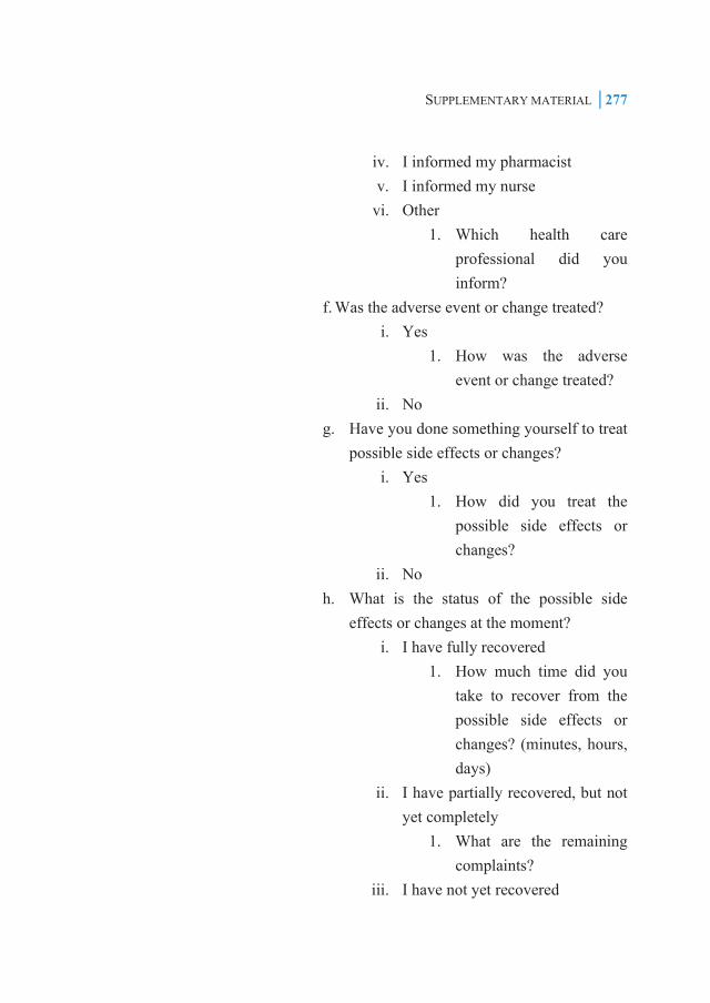
562641-L-bw-Schreuder562641-L-bw-Schreuder562641-L-bw-Schreuder562641-L-bw-SchreuderProcessed on: 25-8-2021Processed on: 25-8-2021Processed on: 25-8-2021Processed on: 25-8-2021 PDF page: 277PDF page: 277PDF page: 277PDF page: 277
SUPPLEMENTARY MATERIAL | 277
iv. I informed my pharmacist v. I informed my nurse
vi. Other 1. Which health care
professional did you inform?
f. Was the adverse event or change treated? i. Yes
1. How was the adverse event or change treated?
ii. No g. Have you done something yourself to treat
possible side effects or changes? i. Yes
1. How did you treat the possible side effects or changes?
ii. No h. What is the status of the possible side
effects or changes at the moment? i. I have fully recovered
1. How much time did you take to recover from the possible side effects or changes? (minutes, hours, days)
ii. I have partially recovered, but not yet completely
1. What are the remaining complaints?
iii. I have not yet recovered

562641-L-bw-Schreuder562641-L-bw-Schreuder562641-L-bw-Schreuder562641-L-bw-SchreuderProcessed on: 25-8-2021Processed on: 25-8-2021Processed on: 25-8-2021Processed on: 25-8-2021 PDF page: 278PDF page: 278PDF page: 278PDF page: 278
278 | EXPLORING DRUG SAFETY OF RADIOPHARMACEUTICALS
i. Did the adverse event or change lead to one of the following situations?
i. Hospitalisation ii. A life-threatening situation
iii. Persistent work disability iv. Congenital defect
j. In your opinion, are there other circumstances that could have caused or worsened these potential side effects or changes?
i. Yes 1. What do you think caused
the possible side effects? ii. No
Preparation for nuclear medicine examination The questions below are about the preparation for your most recent nuclear medicine examination.
9. What did you think of the patient information you received before the examination?
a. Very clear b. Clear c. Neutral d. Unclear e. Very unclear f. I did not read the patient information that was provided
i. Why did you not read the patient information provided? (multiple answers possible)
1. Patient information was not available 2. Lack of time 3. Not interested 4. Difficulty reading the text 5. Other

562641-L-bw-Schreuder562641-L-bw-Schreuder562641-L-bw-Schreuder562641-L-bw-SchreuderProcessed on: 25-8-2021Processed on: 25-8-2021Processed on: 25-8-2021Processed on: 25-8-2021 PDF page: 279PDF page: 279PDF page: 279PDF page: 279
SUPPLEMENTARY MATERIAL | 279
a. Why did you not read the patient information provided?
10. Before the examination, did you have to adhere to one or more of the
following preparatory instructions? (multiple answers possible) a. Attend the examination on an empty stomach b. Extra fluid intake c. Extra salt intake d. Stop taking medicine(s) e. Drink no caffeinated drinks (e.g., coffee, tea, cola or energy drinks) f. Do not eat chocolate or bananas g. Drink Telebrix gastro h. No preparatory measures were required for the examination i. Other
i. What other preparatory instructions did you have to follow for the examination?
Administration of the radiopharmaceutical The below questions are about your most recent nuclear medicine examination.
11. What type of nuclear medicine examination was performed on you?††† a. Bone b. Heart c. Lymph d. Thyroid e. Lung f. Kidney g. PET/CT h. Other
i. What type of nuclear medicine examination was performed on you?
††† Question was added during content validation

562641-L-bw-Schreuder562641-L-bw-Schreuder562641-L-bw-Schreuder562641-L-bw-SchreuderProcessed on: 25-8-2021Processed on: 25-8-2021Processed on: 25-8-2021Processed on: 25-8-2021 PDF page: 280PDF page: 280PDF page: 280PDF page: 280
280 | EXPLORING DRUG SAFETY OF RADIOPHARMACEUTICALS
i. I don’t know
12. Was a radioactive substance administered to you during the examination? a. No b. I don’t know c. Yes
i. Did you notice any possible side effects or physical changes shortly after the administration of the radioactive substance?
1. No 2. Yes
a. Can you briefly describe what happened? b. How much time passed between the
administration of the drug and the adverse events or changes?
i. …… min c. Did you report these adverse events or
changes to hospital staff? i. Yes
ii. No d. Were the adverse events or changes
treated? i. Yes
1. How were the adverse events or changes treated?
ii. No e. What is the status of the possible side effects
or changes at the moment? i. I have fully recovered
1. How much time did you take to recover from the possible side effects or

562641-L-bw-Schreuder562641-L-bw-Schreuder562641-L-bw-Schreuder562641-L-bw-SchreuderProcessed on: 25-8-2021Processed on: 25-8-2021Processed on: 25-8-2021Processed on: 25-8-2021 PDF page: 281PDF page: 281PDF page: 281PDF page: 281
SUPPLEMENTARY MATERIAL | 281
changes? (minutes, hours, days)?
ii. I have partially recovered, but not yet completely
1. What are the remaining complaints?
iii. I have not yet recovered f. In your opinion, are there other circumstances
that could have caused or worsened these possible side effects?
i. Yes 1. What do you think caused
the possible side effects? ii. No
13. Did you receive an X-ray contrast agent during the study?
a. No b. I don’t know c. Yes
i. Did you notice any possible side effects or physical changes shortly after the administration of the X-ray contrast agent?
1. No 2. Yes
a. Can you briefly describe what happened? i. …….
b. How much time passed between the administration of the X-ray contrast agent and the adverse events or changes? (minutes, hours, days)
i. …… c. Did you report these adverse events or
changes to hospital staff?

562641-L-bw-Schreuder562641-L-bw-Schreuder562641-L-bw-Schreuder562641-L-bw-SchreuderProcessed on: 25-8-2021Processed on: 25-8-2021Processed on: 25-8-2021Processed on: 25-8-2021 PDF page: 282PDF page: 282PDF page: 282PDF page: 282
282 | EXPLORING DRUG SAFETY OF RADIOPHARMACEUTICALS
i. Yes ii. No
d. Were the adverse events or changes treated?
i. Yes 1. How were the adverse
events or changes treated? 2.
ii. No e. What is the status of the possible side effects
or changes at the moment? i. I have fully recovered
1. How much time did you take to recover? (minutes, hours, days)?
ii. I have partially recovered, but not yet completely
1. What are the remaining complaints?
iii. I have not yet recovered f. Have you previously experienced any side
effects from X-ray contrast agents? i. Yes
1. Were the previous side effects comparable with the latest ones?
a. Yes b. No
i. How were the previous side

562641-L-bw-Schreuder562641-L-bw-Schreuder562641-L-bw-Schreuder562641-L-bw-SchreuderProcessed on: 25-8-2021Processed on: 25-8-2021Processed on: 25-8-2021Processed on: 25-8-2021 PDF page: 283PDF page: 283PDF page: 283PDF page: 283
SUPPLEMENTARY MATERIAL | 283
effects different from the latest ones?
ii. No g. In your opinion, are there other
circumstances that could have caused or worsened these potential side effects?
i. Yes 1. What do you think caused
the possible side effects? ii. No
14. In your opinion, did unusual things happen during the examination?
a. Yes i. What do you think was different?
b. No Time after nuclear examination The questions below are about the period at home after your most recent nuclear medicine examination.
15. Did you perform certain actions at home after the examination that were indicated by the hospital?
a. Yes i. What were these actions?
b. No
16. Did you experience one or more side effects or physical changes within 7 days of the examination that you relate to the nuclear examination?
a. No b. Yes

562641-L-bw-Schreuder562641-L-bw-Schreuder562641-L-bw-Schreuder562641-L-bw-SchreuderProcessed on: 25-8-2021Processed on: 25-8-2021Processed on: 25-8-2021Processed on: 25-8-2021 PDF page: 284PDF page: 284PDF page: 284PDF page: 284
284 | EXPLORING DRUG SAFETY OF RADIOPHARMACEUTICALS
i. Can you briefly describe what happened? 1. …….
ii. How much time passed between the administration of the drug and adverse events or changes? (minutes, hours, days)
1. ….. iii. Have you informed your referring physician/doctor or other
health care provider? 1. I did not inform a health care professional 2. I informed my referring physician’s hospital 3. I informed my general practitioner 4. I informed my pharmacist 5. I informed my nurse 6. Other
a. Which other health care professional did you inform?
iv. Were the adverse events or changes treated? 1. Yes
a. How were the adverse events or changes treated?
2. No v. Have you done something yourself to treat possible side
effects or changes? 1. Yes
a. How did you treat the possible side effects or changes?
2. No vi. What is the status of the possible side effects or changes at
the moment? 1. I have fully recovered

562641-L-bw-Schreuder562641-L-bw-Schreuder562641-L-bw-Schreuder562641-L-bw-SchreuderProcessed on: 25-8-2021Processed on: 25-8-2021Processed on: 25-8-2021Processed on: 25-8-2021 PDF page: 285PDF page: 285PDF page: 285PDF page: 285
SUPPLEMENTARY MATERIAL | 285
a. How much time did you take to recover from the possible side effects or changes? (minutes, hours, days)
2. I have partially recovered, but not yet completely a. What are the remaining complaints?
3. I have not yet recovered vii. In your opinion, are there other circumstances that could
have caused or worsened these potential side effects? 1. Yes
a. What do you think caused the possible side effects?
2. No Other remarks Finally, you can add comments below that you could not include in the other parts of the questionnaire.
17. Do you have any comments on the complete nuclear medicine examination? a. Yes
i. What are your comments/remarks about the complete nuclear medicine examination?
b. No 18. Do you have any further remarks?
a. Yes i. What other remarks do you have?
b. No Thank you for completing this questionnaire. You can send the questionnaire by first clicking ‘next’ and then clicking the ‘send’ button.

562641-L-bw-Schreuder562641-L-bw-Schreuder562641-L-bw-Schreuder562641-L-bw-SchreuderProcessed on: 25-8-2021Processed on: 25-8-2021Processed on: 25-8-2021Processed on: 25-8-2021 PDF page: 286PDF page: 286PDF page: 286PDF page: 286
286 | EXPLORING DRUG SAFETY OF RADIOPHARMACEUTICALS
Additional information‡‡‡ a. Radiopharmaceutical b. Activity c. Type of examination d. Renal function e. Drugs f. Indications
‡‡‡ Section to be completed by researchers

562641-L-bw-Schreuder562641-L-bw-Schreuder562641-L-bw-Schreuder562641-L-bw-SchreuderProcessed on: 25-8-2021Processed on: 25-8-2021Processed on: 25-8-2021Processed on: 25-8-2021 PDF page: 287PDF page: 287PDF page: 287PDF page: 287
SUPPLEMENTARY MATERIAL | 287
Supplementary material 2: Questions that were removed during the validation process
1. Heeft u allergieën die bekend zijn? a. Ja
i. Allergie voor Hooikoorts ii. Allergie voor Huisstofmijt
iii. Allergie voor Huisdieren iv. Voedselallergie v. Contactallergie
vi. Allergie voor contrastmiddel vii. Anders, namelijk; …
b. Nee
2. Wat was uw mening voor het onderzoek dat u zou worden ingespoten met een radioactieve stof?
3. Heeft u zich op de dag van het onderzoek ruim op tijd kunnen melden op de
afdeling nucleaire geneeskunde? a. Ja b. Nee c. Anders, namelijk;……
4. Hoe voelde u zich tijdens het wachten voor het onderzoek?
a. 0 (niet zenuwachtig) – 7 (heel erg zenuwachtig)
5. Wat vond u van de injectie met de radioactieve stof?
6. Wat was de plaats van de injectie?
7. Na toediening van de radioactieve stof heeft u ongeveer 1 uur gewacht in de voorbereidingsruimte, wat vond u hiervan?

562641-L-bw-Schreuder562641-L-bw-Schreuder562641-L-bw-Schreuder562641-L-bw-SchreuderProcessed on: 25-8-2021Processed on: 25-8-2021Processed on: 25-8-2021Processed on: 25-8-2021 PDF page: 288PDF page: 288PDF page: 288PDF page: 288
288 | EXPLORING DRUG SAFETY OF RADIOPHARMACEUTICALS
8. Was voor u de reden voor het verblijf in de voorbereidingsruimte duidelijk?
a. Ja b. Nee c. Namelijk;
9. Hoe voelde u zich tijdens het wachten in de voorbereidingsruimte?
a. 0 (niet zenuwachtig) – 10 (heel erg zenuwachtig)
10. Heeft u tijdens het wachten in de voorbereidingsruimte een lichamelijke verandering gemerkt?
a. Nee b. Ja
i. Kunt u kort beschrijven wat er gebeurde? ii. Hoeveel tijd zat er tussen de injectie en deze verandering?
iii. Heeft u deze verandering gemeld aan het ziekenhuispersoneel?
1. Ja 2. Nee
iv. Is de reactie behandeld? 1. Ja 2. Nee
v. Hoe is de reactie afgelopen? 1. Hersteld 2. Herstellende 3. Hersteld met rest verschijnselen 4. Niet hersteld 5. Onbekend
11. Hoe voelde u zich tijdens het scannen?
a. 0 (niet zenuwachtig) – 7 (heel erg zenuwachtig)

562641-L-bw-Schreuder562641-L-bw-Schreuder562641-L-bw-Schreuder562641-L-bw-SchreuderProcessed on: 25-8-2021Processed on: 25-8-2021Processed on: 25-8-2021Processed on: 25-8-2021 PDF page: 289PDF page: 289PDF page: 289PDF page: 289
SUPPLEMENTARY MATERIAL | 289
12. Heeft u tijdens het scannen in de een lichamelijke verandering gemerkt? a. Nee b. Ja
i. Kunt u kort beschrijven wat er gebeurde? ii. Hoeveel tijd zat er tussen de injectie en deze verandering?
1. …… iii. Heeft u deze verandering gemeld aan het
ziekenhuispersoneel? 1. Ja 2. Nee
iv. Is de reactie behandeld? 1. Ja 2. Nee
v. Hoe is de reactie afgelopen? 1. Hersteld 2. Herstellende 3. Hersteld met rest verschijnselen 4. Niet hersteld 5. Onbekend
13. Heeft u na de scan een korte verbleven in een ruimte voordat u naar huis
mocht? a. Nee b. Ja
i. Was voor u de reden voor het verblijf in deze ruimte duidelijk?
1. Ja 2. Nee
a. Want; …... c. Hoe voelde u zich tijdens het wachten?
i. 0 (niet zenuwachtig) – 10 (heel erg zenuwachtig) d. Heeft u tijdens het wachten een lichamelijke verandering gemerkt?

562641-L-bw-Schreuder562641-L-bw-Schreuder562641-L-bw-Schreuder562641-L-bw-SchreuderProcessed on: 25-8-2021Processed on: 25-8-2021Processed on: 25-8-2021Processed on: 25-8-2021 PDF page: 290PDF page: 290PDF page: 290PDF page: 290
290 | EXPLORING DRUG SAFETY OF RADIOPHARMACEUTICALS
i. Nee ii. Ja
e. Heeft u kort na toediening van het contrastmiddel een lichamelijke verandering gemerkt?
i. Kunt u kort beschrijven wat er gebeurde? ii. Hoeveel tijd zat er tussen de injectie en deze verandering?
1. …… iii. Heeft u deze verandering gemeld aan het
ziekenhuispersoneel? 1. Ja 2. Nee
iv. Is de reactie behandeld? 1. Ja 2. Nee
v. Hoe is de reactie afgelopen? 1. Hersteld 2. Herstellende 3. Hersteld met rest verschijnselen 4. Niet hersteld 5. Onbekend
14. Heeft u dezelfde mening over radioactieve stoffen als voorheen?
a. Ja i. Namelijk; ….
b. Nee i. Namelijk; ….

562641-L-bw-Schreuder562641-L-bw-Schreuder562641-L-bw-Schreuder562641-L-bw-SchreuderProcessed on: 25-8-2021Processed on: 25-8-2021Processed on: 25-8-2021Processed on: 25-8-2021 PDF page: 291PDF page: 291PDF page: 291PDF page: 291
SUPPLEMENTARY MATERIAL | 291
English translation of questions that were removed during the validation process
1. Do you have any known allergies? a. Yes
i. Hay fever ii. Dust mite allergy
iii. Pet allergy iv. Food allergy v. Contact dermatitis
vi. Allergic reaction to contrast agent vii. Otherwise, namely: …
b. No
2. What was your opinion before the examination that you would be injected with a radioactive substance?
3. Were you able to report to the nuclear medicine department in time on the
day of the examination? a. Yes b. No c. Otherwise, namely: …
4. How did you feel while waiting for the examination?
a. 0 (not nervous) – 7 (very nervous)
5. What did you think of the injection with the radioactive substance?
6. What was the site of injection?
7. After the administration of the radioactive substance, you have waited about 1 hour in the preparation room, what did you think about this?

562641-L-bw-Schreuder562641-L-bw-Schreuder562641-L-bw-Schreuder562641-L-bw-SchreuderProcessed on: 25-8-2021Processed on: 25-8-2021Processed on: 25-8-2021Processed on: 25-8-2021 PDF page: 292PDF page: 292PDF page: 292PDF page: 292
292 | EXPLORING DRUG SAFETY OF RADIOPHARMACEUTICALS
8. Was the reason for your stay in the preparation room clear to you? a. Yes b. No c. Namely: …
9. How did you feel while waiting in the preparation room?
a. 0 (not nervous) – 10 (very nervous)
10. Did you notice a physical change while waiting in the preparation room? a. No b. Yes
i. Can you describe briefly what happened? ii. How much time was there between the injection and this
change? iii. Have you reported this change to hospital staff?
1. Yes 2. No
iv. Has the reaction been treated? 1. Yes 2. No
v. What was the outcome of the reaction? 1. Recovered 2. Recovering 3. Recovered with residual symptoms 4. Not recovered 5. Unknown
11. How did you feel during the scan?
a. 0 (not nervous) – 7 (very nervous)
12. Did you notice a physical change during the scan? a. No

562641-L-bw-Schreuder562641-L-bw-Schreuder562641-L-bw-Schreuder562641-L-bw-SchreuderProcessed on: 25-8-2021Processed on: 25-8-2021Processed on: 25-8-2021Processed on: 25-8-2021 PDF page: 293PDF page: 293PDF page: 293PDF page: 293
SUPPLEMENTARY MATERIAL | 293
b. Yes i. Can you describe briefly what happened?
ii. How much time was there between the injection and this change?
1. …… iii. Have you reported this change to hospital staff?
1. Yes 2. No
iv. Has the reaction been treated? 1. Yes 2. No
v. What was the outcome of the reaction? 1. Recovered 2. Recovering 3. Recovered with residual symptoms 4. Not recovered 5. Unknown
13. Did you stay in a room after the scan before you were allowed to go home?
a. No b. Yes
i. Was the reason for your stay in this room clear to you? 1. Yes 2. No
a. Because: … c. How did you feel while waiting?
i. 0 (not nervous) – 10 (very nervous) d. Did you notice a physical change while waiting?
i. No ii. Yes
e. Have you noticed a physical change shortly after the administration of the contrast agent?

562641-L-bw-Schreuder562641-L-bw-Schreuder562641-L-bw-Schreuder562641-L-bw-SchreuderProcessed on: 25-8-2021Processed on: 25-8-2021Processed on: 25-8-2021Processed on: 25-8-2021 PDF page: 294PDF page: 294PDF page: 294PDF page: 294
294 | EXPLORING DRUG SAFETY OF RADIOPHARMACEUTICALS
i. Can you describe briefly what happened? ii. How much time was there between the injection and this
change? 1. ……
iii. Have you reported this change to hospital staff? 1. Yes 2. No
iv. Has the reaction been treated? 1. Yes 2. No
v. What was the outcome of the reaction? 1. Recovered 2. Recovering 3. Recovered with residual symptoms 4. Not recovered 5. Unknown
14. Do you have the same opinion about radioactive substances as before?
a. Yes i. Namely: ...
b. No i. Namely: ...

562641-L-bw-Schreuder562641-L-bw-Schreuder562641-L-bw-Schreuder562641-L-bw-SchreuderProcessed on: 25-8-2021Processed on: 25-8-2021Processed on: 25-8-2021Processed on: 25-8-2021 PDF page: 295PDF page: 295PDF page: 295PDF page: 295
SUPPLEMENTARY MATERIAL | 295
Supplementary material 3: Reported adverse events of the radiopharmaceuticals and causality assessment using the Naranjo and Silberstein algorithms
Radiopharmaceutical AEs Naranjo category Silberstein [18F]fludeoxyglucose Paraesthesia Possible Unlikely [18F]fludeoxyglucose Fatigue Possible Unlikely [223Ra]Ra-dichloride Nausea Possible Unlikely [223Ra]Ra-dichloride Backpain Probable Possible [99mTc]Tc-oxidronic acid Fatigue Possible Unlikely [99mTc]Tc-tetrofosmin Dyspnoea Possible Unlikely [99mTc]Tc-tetrofosmin Chest discomfort Possible Unlikely [99mTc]Tc-tetrofosmin Nausea Possible Unlikely [99mTc]Tc-tetrofosmin Fatigue Possible Unlikely [99mTc]Tc-tetrofosmin Feeling hot Possible Unlikely [99mTc]Tc-tetrofosmin Abdominal discomfort Possible Unlikely [99mTc]Tc-tetrofosmin Fatigue Possible Unlikely [99mTc]Tc-tetrofosmin Pain in extremity Possible Unlikely [99mTc]Tc-tetrofosmin Nausea Possible Unlikely [99mTc]Tc-tetrofosmin Feeling hot Possible Unlikely [99mTc]Tc-tetrofosmin Abdominal discomfort Possible Unlikely [99mTc]Tc-tetrofosmin Fatigue Possible Unlikely [99mTc]Tc-tetrofosmin Limb discomfort, Possible Unlikely [99mTc]Tc-tetrofosmin Sense of oppression, Possible Unlikely [99mTc]Tc-tetrofosmin Chest discomfort Possible Unlikely [99mTc]Tc-tetrofosmin Fatigue Possible Unlikely [99mTc]Tc-exametazime-labelled cells Fatigue Possible Unlikely

562641-L-bw-Schreuder562641-L-bw-Schreuder562641-L-bw-Schreuder562641-L-bw-SchreuderProcessed on: 25-8-2021Processed on: 25-8-2021Processed on: 25-8-2021Processed on: 25-8-2021 PDF page: 296PDF page: 296PDF page: 296PDF page: 296

562641-L-bw-Schreuder562641-L-bw-Schreuder562641-L-bw-Schreuder562641-L-bw-SchreuderProcessed on: 25-8-2021Processed on: 25-8-2021Processed on: 25-8-2021Processed on: 25-8-2021 PDF page: 297PDF page: 297PDF page: 297PDF page: 297
INDEX OF RADIOPHARMACEUTICALS | 297
Index of radiopharmaceuticals
[198Au]Au-colloid, 70 [51Cr]chromate labelled cells and [125I]I-
human albumin, 64 [51Cr]Cr-edetate, 58 [51Cr]Cr-erythrocytes, 175 [18F]fluciclovine, 35, 37, 51, 68, 75, 127 [18F]fludeoxyglucose, 11, 17, 19, 35, 36,
44, 52, 68, 73, 75, 103, 105, 106, 123, 127, 128, 134, 149–162, 175, 184, 191, 192, 193, 196, 197, 204, 223, 224, 226, 227, 228, 240, 241, 244, 245, 295
[18F]fluorocholine, 16, 105, 127 [18F]fluorodihydroxyphenylalanine
(DOPA), 18, 36, 52, 68, 75 [67Ga]Ga-citrate, 35, 50, 51, 67, 72, 175 [68Ga]Ga-DOTA-NOC, 52, 68 [68Ga]Ga-DOTA-TATE, 35, 52, 68 [68Ga]Ga-edotreotide (DOTA-TOC), 36,
52, 68, 127 [308Hg]chlormerodrin, 70 [111In]In-colloid, 71 [111In]In-oxinate-labelled cells, 51, 67,
176 [111In]In-pentetic acid, 35, 36, 44, 45, 55,
175 [111In]In-pentetreotide, 52, 127, 176, 177 [111In]In-platelets, 71 [111In]In-satumomab pendetide, 35, 53,
69 [113mIn]In-colloid, 60 [113mIn]In-pentetic acid, 71 [125I]I-albumin, 176 [131I]I-human albumin, 35, 36, 70, 73 [131I]I-macrosalb, 35, 36, 44, 62, 73 [123I]iobenguane, 35, 50, 53, 68, 127, 175,
176 [131I]iobenguane, 35, 69, 174, 175, 176,
184, 191, 193, 198, 201, 203
[123I]iodofiltic acid (BMIPP), 64 [123I]iodohippurate, 58 [125I]iodohippurate, 17 [131I]iodohippurate, 17, 37, 58 [131I]iodomethyl norcholesterol, 36, 37,
54, 70, 168, 176, 177, 180, 225, 242 [123I]iofetamine (IMP), 55 [123I]ioflupane, 11, 16, 44, 55, 127, 176,
177 [123I]iolopride, 175, 176 [131I]I-rose bengal, 60 [123I]sodium iodine, 49, 63, 72, 105, 127,
128, 131, 134, 175 [124I]sodium iodine, 127, 128 [131I]sodium iodine, 17, 36, 63, 72, 125,
127, 128, 175, 184, 191, 192, 193, 198–205
[177Lu]Lu-oxodotreotide, 205 [32P]sodium phosphate, 175 Pyrophosphate (non-radioactive), 33, 49,
64 [223Ra]Ra-dichloride, 11, 106, 125, 127,
128, 131, 175, 295 [81Rb]Rb-krypton gas, 175 [82Rb]Rb-chloride, 123, 127 [186Re]Re-etidronate, 175 [75Se]tauroselcholic acid, 46, 60, 176, 177 [153Sm]Sm-lexidronam, 175 [89Sr]Sr-chloride, 175 Stannous agent (non-radioactive), 33, 49,
64 [99mTc]Tc-albumin, 176 [99mTc]Tc-albumin colloid, 60 [99mTc]Tc-antimony sulfide colloid, 60 [99mTc]Tc-arcitumomab, 69 [99mTc]Tc-bicisate, 175 [99mTc]Tc-butedronic acid, 56

562641-L-bw-Schreuder562641-L-bw-Schreuder562641-L-bw-Schreuder562641-L-bw-SchreuderProcessed on: 25-8-2021Processed on: 25-8-2021Processed on: 25-8-2021Processed on: 25-8-2021 PDF page: 298PDF page: 298PDF page: 298PDF page: 298
298 | EXPLORING DRUG SAFETY OF RADIOPHARMACEUTICALS
[99mTc]Tc-diethylenetriaminepentaacetic acid-galactosyl human serum albumin (GSA), 60
99mTc-diphosphonates, 16, 56 [99mTc]Tc-erythrocytes, 176 [99mTc]Tc-ethylenedicysteine, 58 [99mTc]Tc-exametazime, 45, 55, 176, 177 [99mTc]Tc-exametazime-labelled cells,
51, 67, 106, 127, 295 [99mTc]Tc-fanolesomab, 35, 36, 44, 67, 73 [99mTc]Tc-gluceptate, 58 [99mTc]Tc-human albumin, 64 [99mTc]Tc-human albumin - DTPA, 64 [99mTc]Tc-human immunoglobulin
(HIG), 67 [99mTc]Tc-iron hydroxide, 71 [99mTc]Tc- or [113mIn]In-iron precipitate,
71 [99mTc]Tc-macrosalb, 17, 19, 35, 37, 44,
47, 62, 73, 127, 141–146, 176, 223, 240
[99mTc]Tc-mebrofenin, 168, 176, 177, 180, 225, 242
[99mTc]Tc-medronic acid, 15, 35, 36, 37, 45, 56, 72, 94, 118, 132, 175
[99mTc]Tc-mertiatide, 36, 46, 58, 127, 175 [99mTc]Tc-microspheres, 36, 62 [99mTc]Tc-nanocolloid, 35, 47, 60, 105,
127, 176 [99mTc]Tc-oxidronic acid, 35, 45, 57, 103,
105, 106, 123, 127, 128, 129, 134, 175, 295
[99mTc]Tc-pentetic acid, 35, 46, 59, 175 [99mTc]Tc-pertechnetate, 16, 17, 33, 49,
63, 75, 105, 127, 175 [99mTc]Tc-phytate, 60, 168, 176, 177,
180, 225, 242 [99mTc]Tc-plasmin, 71 [99mTc]Tc-pyrophosphate, 57 [99mTc]Tc-rheniumsulfide colloid, 60 [99mTc]Tc-sestamibi, 35, 36, 37, 49, 50,
65, 127, 128, 168, 176, 177, 180, 225, 242
[99mTc]Tc-stannous agent labelled cells, 64
[99mTc]Tc-stannous colloid, 176, 177 [99mTc]Tc-succimer, 46, 59, 127, 175 [99mTc]Tc-sulesomab, 51, 67, 176 [99mTc]Tc-sulfur colloid, 35, 36, 37, 47,
61 [99mTc]technegas, 48, 62 [99mTc]Tc-tetrofosmin, 11, 50, 65, 103,
105, 106, 123, 125, 127, 128, 129, 131, 176, 177, 295
[99mTc]Tc-tilmanocept, 36, 54, 69 [99mTc]Tc-tin colloid, 61 [201Tl]Tl-chloride, 35, 50, 66, 168, 176,
177, 180, 225, 242 [90Y]Y-citrate, 176 [90Y]Y-octreotide, 205 [169Yb]Yb-pentetic acid, 55