university of groningen staphylococcus aureus fibronectin
TRANSCRIPT
University of Groningen
Staphylococcus aureus fibronectin binding protein-A induces motile attachment sites andcomplex actin remodeling in living endothelial cellsSchroeder, Andreas; Schroeder, Barbara; Roppenser, Bernhard; Linder, Stefan; Sinha,Bhanu; Faessler, Reinhard; Aepfelbacher, MartinPublished in:Molecular Biology of the Cell
DOI:10.1091/mbc.E06-05-0463
IMPORTANT NOTE: You are advised to consult the publisher's version (publisher's PDF) if you wish to cite fromit. Please check the document version below.
Document VersionPublisher's PDF, also known as Version of record
Publication date:2006
Link to publication in University of Groningen/UMCG research database
Citation for published version (APA):Schroeder, A., Schroeder, B., Roppenser, B., Linder, S., Sinha, B., Faessler, R., & Aepfelbacher, M.(2006). Staphylococcus aureus fibronectin binding protein-A induces motile attachment sites and complexactin remodeling in living endothelial cells. Molecular Biology of the Cell, 17(12), 5198-5210.https://doi.org/10.1091/mbc.E06-05-0463
CopyrightOther than for strictly personal use, it is not permitted to download or to forward/distribute the text or part of it without the consent of theauthor(s) and/or copyright holder(s), unless the work is under an open content license (like Creative Commons).
The publication may also be distributed here under the terms of Article 25fa of the Dutch Copyright Act, indicated by the “Taverne” license.More information can be found on the University of Groningen website: https://www.rug.nl/library/open-access/self-archiving-pure/taverne-amendment.
Take-down policyIf you believe that this document breaches copyright please contact us providing details, and we will remove access to the work immediatelyand investigate your claim.
Downloaded from the University of Groningen/UMCG research database (Pure): http://www.rug.nl/research/portal. For technical reasons thenumber of authors shown on this cover page is limited to 10 maximum.
Molecular Biology of the CellVol. 17, 5198–5210, December 2006
Staphylococcus aureus Fibronectin Binding Protein-AInduces Motile Attachment Sites and Complex ActinRemodeling in Living Endothelial Cells□D □V
Andreas Schroder,*† Barbara Schroder,‡§ Bernhard Roppenser,* Stefan Linder,‡Bhanu Sinha,� Reinhard Fassler,¶ and Martin Aepfelbacher*
*Institut fur Medizinische Mikrobiologie, Virologie und Hygiene, Universitatsklinikum Hamburg-Eppendorf,20246 Hamburg, Germany; †Max von Pettenkofer-Institut, Ludwig-Maximilians-Universitat Munchen, 80336Munich, Germany; ‡Institut fur Prophylaxe der Kreislaufkrankheiten, Ludwig-Maximilians-UniversitatMunchen, 80336 Munich, Germany; �Institut fur Medizinische Mikrobiologie, Universitatsklinikum Munster,48149 Munster, Germany; and ¶Max-Planck-Institut fur Biochemie, Abteilung fur Molekulare Medizin, 82152Martinsried, Germany
Submitted May 31, 2006; Revised September 14, 2006; Accepted September 26, 2006Monitoring Editor: Ralph Isberg
Staphylococcus aureus fibronectin binding protein-A (FnBPA) stimulates �5�1-integrin signaling and actin rearrange-ments in host cells. This eventually leads to invasion of the staphylococci and their targeting to lysosomes. Using live cellimaging, we found that FnBPA-expressing staphylococci induce formation of fibrillar adhesion-like attachment sites andtranslocate together with them on the surface of human endothelial cells (velocity �50 �m/h). The translocating bacteriarecruited cellular actin and Rab5 in a cyclic and alternating manner, suggesting unsuccessful attempts of phagocytosis bythe endothelial cells. Translocation, actin recruitment, and eventual invasion of the staphylococci was regulated by thefibrillar adhesion protein tensin. The staphylococci also regularly produced Neural Wiskott-Aldrich syndrome protein-controlled actin comet tails that further propelled them on the cell surface (velocity up to 1000 �m/h). Thus, S. aureusFnBPA produces attachment sites that promote bacterial movements but subvert actin- and Rab5 reorganization duringinvasion. This may constitute a novel strategy of S. aureus to postpone invasion until its toxins become effective.
INTRODUCTION
For the purpose of tissue penetration, invasion of the vas-culature, and dissemination via the bloodstream, the bacte-rial pathogen S. aureus produces a plethora of virulencefactors, including teichoic acids, exotoxins, and adhesins(Lowy, 1998). Collagen-binding Cna, fibrinogen-bindingclumping factor-A and -B, and fibronectin binding protein-Aand -B (FnBPA and -B) are among the most important ad-hesins of S. aureus (Jonsson et al., 1991; Patti et al., 1992;McDevitt et al., 1994; Menzies, 2003). Although S. aureus isconsidered an extracellular pathogen, it has been well doc-umented that its fibronectin (Fn) binding adhesins FnBPAand -B also mediate bacterial invasion of cells (Sinha et al.,1999, 2000; Fowler et al., 2000; Massey et al., 2001). However,after invasion of human endothelial cells and keratinocytes,the majority of the internalized staphylococci are effectively
eliminated by lysosomal effector mechanisms (Krut et al.,2003; Schroder et al., 2006).
In a recent animal study, staphylococci were shown topreferably adhere to capillaries and postcapillary venulesafter intraarterial injection (Laschke et al., 2005). Most staph-ylococci (�99%) disappeared from the blood vessels within20 min, suggesting internalization by the endothelium. Ananimal endocarditis model demonstrated that expression ofFnBPA conferred on staphylococci and lactococci the capa-bility to infect heart valves, resulting in invasion of thevalvular endothelium (Que et al., 2005). Hence, the adhesinsFnBPA and -B crucially determine interaction of S. aureuswith endothelial cells in vivo and in vitro.
FnBPs contain multiple modules, each of which can bindto a stretch of type I (F1) repeats at the N terminus of Fn(Massey et al., 2001; Schwarz-Linek et al., 2004). Presumably,FnBPs are thereby capable of associating with several Fnmolecules at once (Schwarz-Linek et al., 2004). Furthermore,the FnBP binding site is located at some distance from thecentral integrin binding RGD motif in Fn. Therefore, Fn canform a bridge between FnBPs and integrins (Sinha et al.,1999; Fowler et al., 2000). Apparently through these features,FnBPs are capable of inducing integrin clustering and sig-naling in cells. This leads to invasion of staphylococci via srctyrosine kinase activation and reorganization of the actincytoskeleton (Agerer et al., 2003; Fowler et al., 2003).
Physiological clustering of integrins by extracellular cuescauses formation of adhesion structures that differ by mor-phology, molecular composition, and spatiotemporal dy-
This article was published online ahead of print in MBC in Press(http://www.molbiolcell.org/cgi/doi/10.1091/mbc.E06–05–0463)on October 4, 2006.□D □V The online version of this article contains supplemental mate-rial at MBC Online (http://www.molbiolcell.org).§ Present address: Center for Basic Research in Digestive Diseasesand Department of Biochemistry and Molecular Biology, MayoClinic, Rochester, MN 55905.
Address correspondence to: Martin Aepfelbacher ([email protected]).
5198 © 2006 by The American Society for Cell Biology
namics. Focal adhesions are tight clusters of cytoskeletal andsignaling proteins, including focal adhesion kinase (FAK),vinculin, paxillin, talin, and tensin. These proteins coopera-tively organize the association of actin stress fibers with thecytoplasmic domains of integrins (Zamir and Geiger, 2001;Brakebusch and Fassler, 2003; DeMali et al., 2003). It has beenproposed that fibrillar adhesions develop by translocation ofFn-ligated �5�1 integrins out of focal adhesions. Fibrillaradhesions have an elongated or beaded morphology andcontain tensin, but they are devoid of most other focaladhesion components. The current notion is that fibrillaradhesions stretch extracellular Fn molecules and therebyperpetuate Fn fibrillogenesis (Pankov et al., 2000; Zamir et al.,2000; Zamir and Geiger, 2001; Danen et al., 2002).
So far, the molecular mechanisms controlling adhesionand internalization of staphylococci were visualized mainlyin fixed cells. Naturally, this could only provide snapshots ofhighly dynamic processes. Based mainly on live cell imagingdata we report here that the staphylococcal adhesin FnBPAtriggers formation of fibrillar adhesion-like attachment sitesthat encompass the bacteria and mediate their centripetaltranslocation on the cell surface. The translocating staphy-lococci induced multiple consecutive actin cups as well asactin comet tails propelling the bacteria on the cell surface.Movements, actin reorganization and final internalization ofthe bacteria were regulated by the fibrillar adhesion proteintensin.
We propose that FnBPA-stimulated integrin signalingpromotes bacterial motility on the endothelial cell surfacebut retards bacterial invasion. Thus, disturbance of the cel-lular phagocytic machinery may determine the outcome ofendothelium infection by S. aureus in disease states such assepsis or endocarditis.
MATERIALS AND METHODS
Bacterial Strains and Culture ConditionsEscherichia coli HB101 strain expressing Invasin of Yersinia enterocolitica namedInvasin-E. coli was cultured in LB medium as described previously (Schulte etal., 1998). S. aureus DU 5883(pFnBA4) named FnBPA-S. aureus expressingfull-length FnBPA (Greene et al., 1995) was cultured in LB medium containing20 �g/ml tetracycline, 5 �g/ml erythromycin, and 20 �g/ml chlorampheni-col. S. carnosus TM300(pFNBA4) named FnBPA-S. carnosus heterologouslyexpressing full-length FnBPA from S. aureus (Sinha et al., 2000) was culturedin LB medium containing 20 �g/ml chloramphenicol. For infection assays,overnight cultures were diluted 1:10 in LB medium and grown to an OD600 of0.8. Subsequently, strains were centrifuged and bacterial pellets were care-fully resuspended in sterile phosphate-buffered saline (PBS), pH 7.4. Forinfection assays, the volume of PBS containing bacteria for an infection ratio(multiplicity of infection) of 100 bacteria per cell was added to the cell culturemedium.
Cell Culture and Infection AssaysIsolation of human umbilical vein endothelial cells (HUVECs) was performedas described previously (Essler et al., 2003). Cells were cultured in endothelialcell growth medium (Promocell, Heidelberg, Germany) containing 2% fetalcalf serum at 37°C, 5% CO2, and 90% humidity. For infection assays, HUVECs(2 � 104 cells) were seeded onto gelatin-coated glass coverslips (12 mm;Hartenstein, Wurzburg, Germany) kept in eight-well plates (Nalge NuncInternational, Rochester, NY) 1 d before the experiment. Thirty minutesbefore adding the bacteria, the medium was replaced by antibiotic-free me-dium. In some experiments cells were centrifuged at 44g for 5 min afteradding bacteria to allow simultaneous attachment of the bacteria. For live cellimaging, HUVECs (2 � 104 cells) were seeded onto glass-bottomed dishes(MatTek, Ashland, MA) 1 d before the experiment. The medium was replacedby antibiotic- and phenol red-free medium 30 min before start of imaging.Bacteria were pipetted in appropriate amounts to the medium and sedi-mented onto the cells.
Floxed Fn cells were isolated from mice carrying a conditional Fn allele,immortalized, and cloned. Subsequently, the Fn alleles were deleted byadenoviral Cre transduction (Fn�/� cells). The cells were grown in DMEMsupplemented with 10% fetal calf serum (FCS) or alternatively, in serumreplacement medium (Sigma Aldrich, Taufkirchen, Germany).
Transfection and PlasmidsHUVECs were transfected with the Amaxa nucleofection system (Amaxa,Cologne, Germany) applying the standard protocol as specified by the man-ufacturer. The green fluorescent protein (GFP) fusion vector GFP-C1 andGFP-actin construct were obtained commercially (BD Clontech, Heidelberg,Germany). Constructs encoding for chicken GFP-tensin (Chuang et al., 1995)and chicken GFP-tensin AH2 (corresponding to residues 659-762) were giftsof Dr. Shin Lin (University of California, Irvine, Irvine, CA), GFP-Arp3 waskindly provided by Dr. Dorothy Schafer (University of Virginia, Charlottes-ville, VA), GFP-neural-Wiskott-Aldrich syndrome (N-WASp) was donated byDr. Silvia Lommel (Helmholtz Centre for Infection Research, Braunschweig,Germany), and GFP-Rab5 was a generous gift of Dr. Craig Roy (Yale Univer-sity School of Medicine, New Haven, CT). The construct encoding for mono-meric red fluorescent protein (mRFP)-actin has been described previously(Osiak et al., 2005).
Antibodies and ReagentsAntibodies used for immunofluorescence staining: anti-fibronectin (1:500; BDTransduction Laboratories, Heidelberg, Germany) anti-paxillin (1:200 BDTransduction Laboratories), anti-vinculin (1:50; Sigma Aldrich), anti-pFAK(1:50; BioSource International, Camarillo, CA) and anti-glutathione S-trans-ferase (GST) (diluted 1:100; Invitrogen, Leiden, The Netherlands). Inside/outside staining for S. aureus was performed with a commercially availableantibody against whole bacterial lysate (1:2000; Biodesign International, Saco,ME); for invasin detection, a rabbit polyclonal antiserum was used (1:500; akind gift of Dr. G. Grassl, Max von Pettenkofer-Institut, University of Munich,Germany). Wiskostatin (Merck Biosciences, Bad Soden, Germany) was usedat a final concentration of 2.5 �M.
Expression of GST, GST-Inv397, and GST-FnBPA-Du-D4Expression and purification of GST and GST-Inv397 was performed as describedpreviously (Wiedemann et al., 2001). GST-FnBPA-Du-D4 was expressed accord-ing to the same protocol, except that E. coli BL21 harboring pGEX-4T-FnBPA-Du-D4 was grown at 37°C, and induction of expression was induced withisopropyl-�-d-thiogalactopyranoside at a final concentration of 1 mM. Purity andidentity of GST-Inv397 and GST-FnBPA-Du-D4 fusion proteins were analyzed bySDS-PAGE and Western blot by using goat anti-GST (diluted 1:1000; MolecularProbes, Eugene, OR) and anti-invasin antiserum (diluted 1:5000; see above).Protein concentrations were determined using the BCA protein assay (PierceChemical, Rockford, IL). C3-transferase, N17Rac, and N17CDC42 were expressedand purified as GST-fusions as described previously (Wiedemann et al., 2001).For microinjection, proteins were dialyzed against buffer (50 mM Tris-HCl, pH7.5, 150 mM NaCl, and 5 mM MgCl2), concentrated in Centricon or Microcon(Millipore, Billerica, MA), shock frozen, and stored at �80°C. Purity was testedusing SDS-PAGE and Coomassie staining.
Coating of Proteins to Latex BeadsAbout 1 � 109 latex beads (1 �m in diameter, sulfate microspheres; Invitro-gen) were washed and resuspended in 1 ml containing 1 mg each of GST,GST-Inv397, or GST-FnBPA-Du-D4. The protein was allowed to adsorb to thebeads for 3 h at room temperature. After adding 500 �l of 20 mg/ml bovineserum albumin (BSA), the solution was incubated at room temperature foranother 1 h. Beads were washed in PBS containing 1 mg/ml BSA and storedat 4°C in 500 �l of PBS containing 0.2 mg ml/BSA. To determine the couplingefficiency, the protein concentration of the starting solution and of the super-natant after coupling was determined. Integrity of coated fusion protein waschecked using Western blot analysis. Beads coated with GST, GST-Inv397, orGST-FnBPA-Du-D4 were referred to as GST-beads, Invasin-beads, or FnBPA-beads, respectively. For infection assays, the volume giving rise to a beads-to-cell ratio of 100:1 was added to the cells.
Microinjection of ProteinsFor microinjection experiments cells were seeded onto glass coverslips 1 dbefore the experiment. Microinjection was performed by using a transjector5246 (Eppendorf, Hamburg, Germany) and a Compic inject micromanipulator(Cell Biology Trading, Hamburg, Germany). Proteins were injected into thecytoplasm at 0.7–1 mg/ml. Injected cells were identified by coinjected rat IgG(0.5 mg/ml; Dianova, Hamburg, Germany) after staining with fluoresceinisothiocyanate (FITC)-labeled goat anti-rat IgG (Dianova).
Ligand Overlay AssayThe ligand overlay assay was performed as described previously (Hussain etal., 2001).
ImmunofluorescenceInside/outside staining for the discrimination between intracellular and ex-tracellularly attached beads or bacteria was performed as described previ-ously (Wiedemann et al., 2001). Briefly, cells were fixed with PBS containing
S. aureus FnBPA-induced Actin Dynamics in Endothelial Cells
Vol. 17, December 2006 5199
4% paraformaldehyde for 5 min and then incubated with antibodies directedeither against S. aureus, invasin or GST in the appropriate dilutions (seeabove). Subsequently, cells were washed three times followed by incubationwith Alexa Fluor 568-labeled goat anti-rabbit IgG (diluted 1:200) (Invitrogen).After cell permeabilization with ice-cold acetone for 5 min, coverslips wereincubated with antibodies directed either against S. aureus, invasin, or GSTfollowed by incubation with Alexa Fluor 488-labeled goat anti-rabbit IgG (1:200)(Invitrogen). Thereby, intracellular bacteria are in green and extracellular bacteriaare in yellow/orange due to overlay of two fluorescence dyes. For evaluating theamount of invaded beads or bacteria, the number of invaded and total cell-associated beads/bacteria was counted microscopically.
Images from fixed samples were acquired with a Leica DMRBE microscopeequipped with a Spot camera (Visitron Systems, Puchheim, Germany) con-trolled by MetaMorph (Molecular Devices, Sunnyvale, CA), except Figure 5B,which was imaged with an UltraView LCI spinning disk confocal system (seebelow). Live cell experiments and z-staples for the three-dimensional (3D)-model in Figure 5B were performed with an UltraView LCI spinning diskconfocal system (PerkinElmer Life and Analytical Sciences, Rodgau-Jug-esheim, Germany) fitted on a Nikon TE300 microscope equipped with atemperature- and CO2-controllable environment chamber. Images were takenwith the black/white ORCA ER camera (Hamamatsu, Herrsching, Germany).
Software and CalculationsProcessing of the z-staples for 3D-reconstruction was performed with Voloc-ity (Improvision, Tubingen, Germany). For tracking of beads and evaluationof velocity and distance to origin values, MetaMorph software (MolecularDevices) was used. Briefly, an overlay of the threshold image depicting thebeads (orange), and the phase contrast was generated. The distance of beadsbetween single frames was determined using the software, and then meanspeed was calculated. Distance to origin is the mean distance of all beads atthe given time points. For evaluating the fluorescence intensity traces, Ultra-view LCI software (PerkinElmer Life and Analytical Sciences) was used. Aregion of interest was defined around the bacteria, and gray level values werelogged for each wavelength. Quicktime movies were generated with ImageJ(Rasband, 2006).
RESULTS
Kinetics of FnBPA-mediated Internalization ofStaphylococci by Primary Human Endothelial CellsInvasion of cells via S. aureus FnBPA requires an Fn bridgebetween FnBPA and the �5�1 integrin (Sinha et al., 1999). Incomparison, the surface protein invasin of enteropathogenicyersiniae directly binds to �1-integrins to trigger cell inva-sion (Dersch and Isberg, 1999; Isberg et al., 2000). To get afirst idea about the dynamics of these two bacterial invasionprocesses, we used S. aureus DU 5883(pFnBA4), which ex-presses FnBPA (FnBPA-S. aureus; Greene et al., 1995), and E.coli HB101, which expresses invasin (Invasin-E. coli; Schulteet al., 1998), and we performed time courses of bacterialinvasion into primary HUVECs.
As documented in Figure 1, A and B, the invasion rates ofFnBPA-S. aureus and Invasin-E. coli into HUVECs differedmarkedly. After 10 min, �80% of cell attached Invasin-E. coliwere internalized, whereas in the same time period only�20% of FnBPA-S. aureus were taken up. Further internal-ization of FnBPA-S. aureus proceeded almost linearly, reach-ing �80% at 120 min (Figure 1B). The internalization kineticsof S. carnosus TM300(pFNBA4), which heterologously ex-presses FnBPA (Sinha et al., 2000), was indistinguishablefrom that of FnBPA-S. aureus (our unpublished data).
To verify these results in a bacteria-free system, we testedinvasion of 1-�m-diameter latex beads coated with the 31-kDa Du-D4 segment of S. aureus FnBPA (FnBPA-beads). TheDu-D4 segment harbors most of the Fn-binding repeats ofFnBPA (Massey et al., 2001), and we confirmed its Fn-bind-ing activity by ligand overlay assay (our unpublished data;Hussain et al., 2001). Likewise, invasion of beads coated withthe 42-kDa �1-integrin binding fragment of Yersinia entero-colitica invasin was tested (Invasin-beads; Wiedemann et al.,2001). As shown in Figure 1B, internalization kinetics ofFnBPA-beads and Invasin-beads closely resembled that ofFnBPA-S. aureus and Invasin-E. coli, respectively. These data
show that staphylococci expressing FnBPA spend in themean �45 min on the endothelial cell surface before beingingested. Because most Invasin-E. coli were internalizedwithin 5–10 min, we conclude that the molecular mecha-nisms regulating invasin- and FnBPA/Fn-mediated inva-sion are different.
In our assay FnBPA on the surface of staphylococci orbeads binds to serum Fn thus producing Fn-coated particles(Figure 1C; our unpublished data). We were interested tofind out whether the Fn coat is necessary or sufficient for cellinvasion and whether Fn aggregates and fibrils that areusually exposed on the surface of cultured endothelial cellsalso promote invasion. Because endogenous Fn productioncannot be completely eliminated in HUVECs, we deter-mined invasion of FnBPA-S. aureus pretreated with or with-out fetal calf serum (producing Fn-coated- and uncoatedbacteria, respectively) into Fn�/� fibroblasts (Nyberg et al.,2004) As shown in Figure 1C, Fn�/� cells cultured in serum-free medium are devoid of cellular Fn. Fn-coated FnBPA-S.aureus was readily internalized by Fn�/� fibroblasts, but nointernalization of uncoated bacteria was detected (Figure1D). We next tested whether uncoated FnBPA-S. aureus wasinternalized by HUVECs that expose Fn matrix on the sur-face (Figure 1C). The HUVECs internalized uncoatedFnBPA-S. aureus with comparable efficiency as the Fn-coatedbacteria (Figure 1D). These experiments demonstrate thatboth serum Fn and cellular Fn are competent to promoteFnBPA-mediated cellular invasion.
S. aureus FnBPA Induces Formation of FibrillarAdhesion-like Structures on the Surfaceof Endothelial CellsClustering of integrins by extracellular matrix proteins leads tothe formation of distinct adhesion structures (Zamir andGeiger, 2001), and we therefore asked what type of adhesionsare induced by FnBPA/Fn. First, we devised an assay that candistinguish between focal adhesions/focal complexes andfibrillar adhesions (also termed ECM contacts) in HUVECs. Forthis, a GFP-fusion of the focal- and fibrillar adhesion proteintensin (GFP-tensin) was expressed in HUVECs, and the latterwere concomitantly stained for the focal adhesion proteinsvinculin, paxillin, or phosphorylated focal adhesion kinase(pFAK). Streak-like enrichments of vinculin in the cell periph-ery that colocalized with GFP-tensin were identified as focaladhesions (Figure 2A). These structures also contained paxillinand pFAK (our unpublished data). In comparison, streaky andbeaded accumulations of GFP-tensin that mostly localized to-ward the cell center represent fibrillar adhesions (Figure 2A).As in other cell types, the fibrillar adhesions of HUVECs con-tained no paxillin or pFAK but colocalized with �5 integrinsubunits (our unpublished data). Consistent with its proposedtranslocation out of focal adhesions, GFP-tensin often seemedto form a centripetal extension of vinculin streaks (Figure 2A,overlay 2).
Next, we evaluated which adhesion proteins were re-cruited by FnBPA- and Invasin-beads. As demonstrated inFigure 2B, GFP-tensin accumulated in cup-like structuresaround both types of beads, whereas vinculin was onlyrecruited by the Invasin-beads. Quantitative analyses veri-fied that vinculin, paxillin, and pFAK only rarely were core-cruited with GFP-tensin by the FnBPA-beads. In contrast, inthe majority of cases these adhesion proteins were core-cruited with GFP-tensin by the Invasin-beads (Figure 2C).For example, 17% of the GFP-tensin cups induced byFnBPA-beads compared with 97% induced by the Invasin-beads recruited vinculin (Figure 2C).
A. Schroder et al.
Molecular Biology of the Cell5200
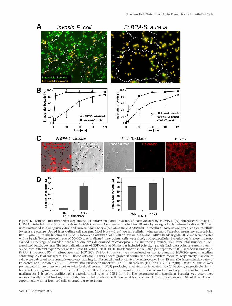
Figure 1. Kinetics and fibronectin dependence of FnBPA-mediated invasion of staphylococci by HUVECs. (A) Fluorescence images ofHUVECs infected with Invasin-E. coli or FnBPA-S. aureus. Cells were infected for 10 min by using a bacteria-to-cell ratio of 30:1 andimmunostained to distinguish extra- and intracellular bacteria (see Materials and Methods). Intracellular bacteria are green, and extracellularbacteria are orange. Dotted lines outline cell margins. Most Invasin-E. coli are intracellular, whereas most FnBPA-S. aureus are extracellular.Bar, 10 �m. (B) Uptake kinetics of FnBPA-S. aureus and Invasin-E. coli (left) or Invasin-beads and FnBPA-beads (right). HUVECs were infectedwith a beads/bacteria-to-cell ratio of 50–100:1. At indicated time points, cells were fixed, and extracellular bacteria/beads were immuno-stained. Percentage of invaded beads/bacteria was determined microscopically by subtracting extracellular from total number of cell-associated beads/bacteria. The internalization rate of GST-beads at 60 min was included (x in right panel). Each data point represents mean �SD of three different experiments with at least 100 cells (�5000–10,000 beads/bacteria) evaluated per experiment. (C) Fibronectin staining ofFnBPA-S. carnosus, FN�/� fibroblasts and HUVECs. FnBPA-S. carnosus was transferred or not to standard HUVECs growth mediumcontaining 2% fetal calf serum. Fn�/� fibroblasts and HUVECs were grown in serum-free- and standard medium, respectively. Bacteria orcells were subjected to immunofluorescence staining for fibronectin and evaluated by microscopy. Bars, 10 �m. (D) Internalization rates ofFn-coated and uncoated FnBPA-S. aureus into fibronectin-knockout (Fn�/�) fibroblasts (left) or HUVECs (right). FnBPA-S. aureus werepreincubated in medium without or with fetal calf serum (�FCS) producing uncoated- or Fn-coated (see C) bacteria, respectively. Fn�/�
fibroblasts were grown in serum-free medium, and HUVECs pregrown in standard medium were washed and kept in serum-free standardmedium for 1 h before addition of a bacteria-to-cell ratio of 100:1 for 1 h. The percentage of intracellular bacteria was determinedmicroscopically by subtracting extracellular from total number of cell-associated bacteria. Each bar represents mean � SD of three differentexperiments with at least 100 cells counted per experiment.
S. aureus FnBPA-induced Actin Dynamics in Endothelial Cells
Vol. 17, December 2006 5201
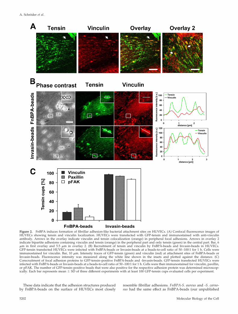
These data indicate that the adhesion structures producedby FnBPA-beads on the surface of HUVECs most closely
resemble fibrillar adhesions. FnBPA-S. aureus and -S. carno-sus had the same effect as FnBPA-beads (our unpublished
Figure 2. FnBPA induces formation of fibrillar adhesion-like bacterial attachment sites on HUVECs. (A) Confocal fluorescence images ofHUVECs showing tensin and vinculin localization. HUVECs were transfected with GFP-tensin and immunostained with anti-vinculinantibody. Arrows in the overlay indicate vinculin and tensin colocalization (orange) in peripheral focal adhesions. Arrows in overlay 2indicate bipartite adhesions containing vinculin and tensin (orange) in the peripheral part and only tensin (green) in the central part. Bar, 6�m in first overlay and 5.5 �m in overlay 2. (B) Recruitment of tensin and vinculin by FnBPA-beads and Invasin-beads in HUVECs.GFP-tensin transfected HUVECs were infected with FnBPA-beads or Invasin-beads at a beads-to-cell ratio of 50–100:1 for 1 h. Cells wereimmunostained for vinculin. Bar, 10 �m. Intensity traces of GFP-tensin (green) and vinculin (red) at attachment sites of FnBPA-beads orInvasin-beads. Fluorescence intensity was measured along the white line shown in the insets and plotted against the distance. (C)Corecruitment of focal adhesion proteins to GFP-tensin–positive FnBPA-beads and -Invasin-beads. GFP-tensin transfected HUVECs wereinfected with FnBPA-beads or Invasin-beads at a beads-to-cell ratio of 50–100:1 for 1 h. Cells were then immunostained for vinculin, paxillin,or pFAK. The number of GFP-tensin–positive beads that were also positive for the respective adhesion protein was determined microscop-ically. Each bar represents mean � SD of three different experiments with at least 100 GFP-tensin cups evaluated cells per experiment.
A. Schroder et al.
Molecular Biology of the Cell5202
data). In contrast, the attachment sites generated by Invasin-beads and Invasin-E. coli resemble classical focal adhesions(Figure 2, B and C).
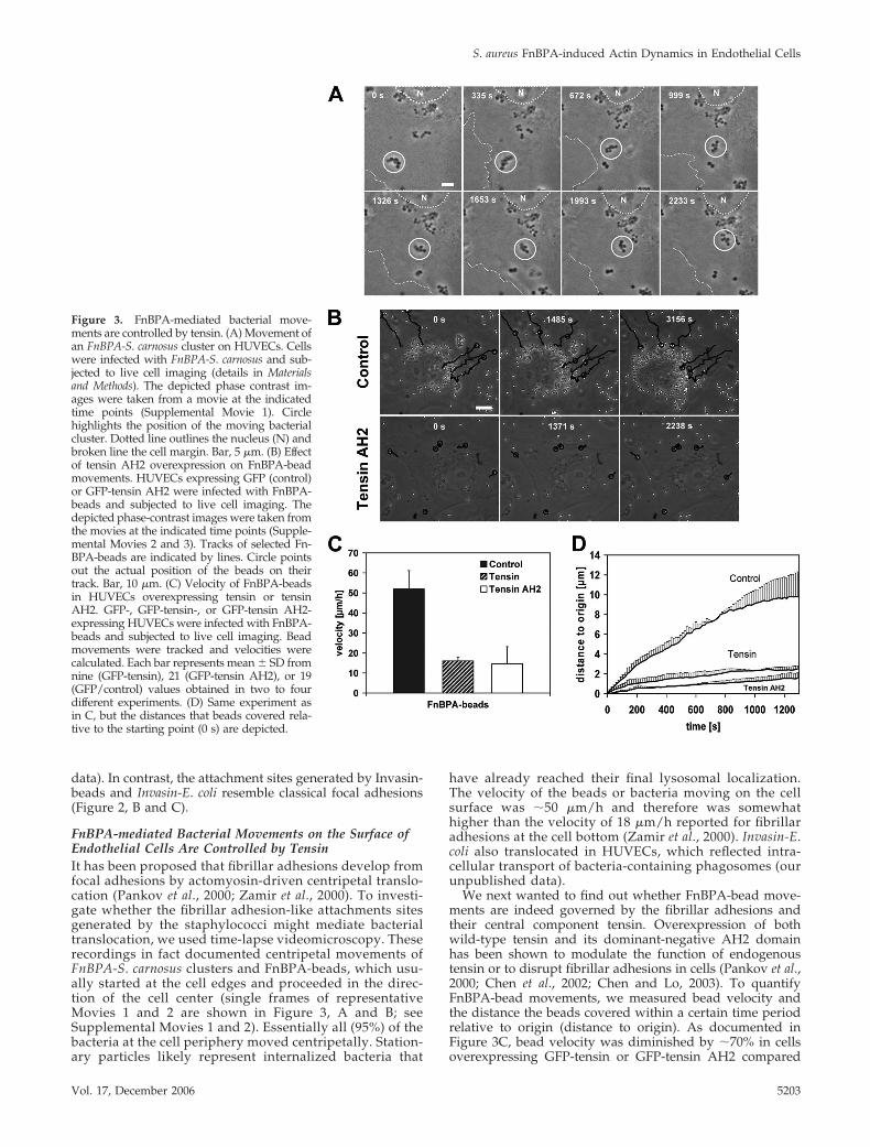
FnBPA-mediated Bacterial Movements on the Surface ofEndothelial Cells Are Controlled by TensinIt has been proposed that fibrillar adhesions develop fromfocal adhesions by actomyosin-driven centripetal translo-cation (Pankov et al., 2000; Zamir et al., 2000). To investi-gate whether the fibrillar adhesion-like attachments sitesgenerated by the staphylococci might mediate bacterialtranslocation, we used time-lapse videomicroscopy. Theserecordings in fact documented centripetal movements ofFnBPA-S. carnosus clusters and FnBPA-beads, which usu-ally started at the cell edges and proceeded in the direc-tion of the cell center (single frames of representativeMovies 1 and 2 are shown in Figure 3, A and B; seeSupplemental Movies 1 and 2). Essentially all (95%) of thebacteria at the cell periphery moved centripetally. Station-ary particles likely represent internalized bacteria that
have already reached their final lysosomal localization.The velocity of the beads or bacteria moving on the cellsurface was �50 �m/h and therefore was somewhathigher than the velocity of 18 �m/h reported for fibrillaradhesions at the cell bottom (Zamir et al., 2000). Invasin-E.coli also translocated in HUVECs, which reflected intra-cellular transport of bacteria-containing phagosomes (ourunpublished data).
We next wanted to find out whether FnBPA-bead move-ments are indeed governed by the fibrillar adhesions andtheir central component tensin. Overexpression of bothwild-type tensin and its dominant-negative AH2 domainhas been shown to modulate the function of endogenoustensin or to disrupt fibrillar adhesions in cells (Pankov et al.,2000; Chen et al., 2002; Chen and Lo, 2003). To quantifyFnBPA-bead movements, we measured bead velocity andthe distance the beads covered within a certain time periodrelative to origin (distance to origin). As documented inFigure 3C, bead velocity was diminished by �70% in cellsoverexpressing GFP-tensin or GFP-tensin AH2 compared
Figure 3. FnBPA-mediated bacterial move-ments are controlled by tensin. (A) Movement ofan FnBPA-S. carnosus cluster on HUVECs. Cellswere infected with FnBPA-S. carnosus and sub-jected to live cell imaging (details in Materialsand Methods). The depicted phase contrast im-ages were taken from a movie at the indicatedtime points (Supplemental Movie 1). Circlehighlights the position of the moving bacterialcluster. Dotted line outlines the nucleus (N) andbroken line the cell margin. Bar, 5 �m. (B) Effectof tensin AH2 overexpression on FnBPA-beadmovements. HUVECs expressing GFP (control)or GFP-tensin AH2 were infected with FnBPA-beads and subjected to live cell imaging. Thedepicted phase-contrast images were taken fromthe movies at the indicated time points (Supple-mental Movies 2 and 3). Tracks of selected Fn-BPA-beads are indicated by lines. Circle pointsout the actual position of the beads on theirtrack. Bar, 10 �m. (C) Velocity of FnBPA-beadsin HUVECs overexpressing tensin or tensinAH2. GFP-, GFP-tensin-, or GFP-tensin AH2-expressing HUVECs were infected with FnBPA-beads and subjected to live cell imaging. Beadmovements were tracked and velocities werecalculated. Each bar represents mean � SD fromnine (GFP-tensin), 21 (GFP-tensin AH2), or 19(GFP/control) values obtained in two to fourdifferent experiments. (D) Same experiment asin C, but the distances that beads covered rela-tive to the starting point (0 s) are depicted.
S. aureus FnBPA-induced Actin Dynamics in Endothelial Cells
Vol. 17, December 2006 5203
with control cells expressing GFP. Moreover, in the GFP-tensin– or GFP-tensin AH2-overexpressing cells, the dis-tance to origin covered by the FnBPA-beads was reduceddrastically by 80–90% (Figure 3D). In these cells, bead move-ments were mainly confined to the cell margins (Figure 3Band Supplemental Movie 3).
Together, these experiments indicate that the fibrillar ad-hesion protein tensin controls centripetal movements ofFnBPA-exposing staphylococci and -beads on the endothe-lial cell surface.
FnBPA-stimulated Integrin Signaling Induces Formationof Multiple Actin CupsIt has been reported that FnBPA-mediated cellular invasionof staphylococci is associated with formation of actin-richphagocytic cups (Agerer et al., 2005). We confirmed this byrhodamine phalloidin staining of FnBPA-S. carnosus-infectedHUVECs and also verified by selective immunofluorescencestaining that all bacteria encompassed by actin cups arelocalized extracellularly (our unpublished data).
For monitoring how actin cup formation is coordinatedwith bacterial movement, live cell imaging of GFP-actin–expressing cells infected with FnBPA-S. carnosus was used. Itwas revealed that GFP-actin repeatedly accumulated aroundclusters of staphylococci, whereas these were translocatingon the cell surface (single frames of a representative movieare depicted in Figure 4A; see Supplemental Movie 4). Ahigher spatiotemporal resolution showed that during oneaccumulation event GFP-actin in fact advances in a wave-like manner from one end of the staphylococcal chain to theother. By this, cup formation around individual bacteria inthe chain was accomplished (Figure 4B, top; see Supplemen-tal Movie 5). In some cases, it even seemed as if vigorousactin bursts were capable of separating bacterial clusters(Figure 4B, bottom). In experiments using single FnBPA-beads, it was found that these beads triggered up to 10separate GFP-actin cups, whereas Invasin-beads never pro-duced more than two actin cups (Figure 4C). A fluorescenceintensity trace of GFP-actin repeatedly accumulating arounda FnBPA-bead doublet is depicted in Figure 4D. From thisand similar experiments, it was determined that the dura-tion of the actin accumulation at single cups is very constant(80 � 33 s; mean � SD; n � 44), whereas the intermittentperiods vary widely (range 21–1200 s).
To show the relevance of the described actin reorganiza-tions for FnBPA-mediated invasion, we tested the effect ofRho GTPase- and N-WASp inhibitors. Rho GTP-bindingproteins are universal regulators of actin and for integrin-triggered phagocytic cup formation they have been shownto engage N-WASp and Arp2/3 complex (May andMachesky, 2001; Wiedemann et al., 2001). Microinjection ofthe Rho-inhibitor C3-transferase, dominant inhibitoryN17Rac, or N17Cdc42, or treatment with the N-WASp in-hibitor wiskostatin all reduced internalization of FnBPA-S.aureus by 50–60% (Figure 4E). This indicates that the actincups assembled by cooperation of different RhoGTPases andN-WASp crucially control FnBPA-mediated invasion. Con-sistent with this, GFP-fusions of Rac, Cdc42, and N-WASptranslocated together with mRFP-actin to phagocytic cups(our unpublished data).
FnBPA-stimulated Actin Comet Tails Propel Bacteria onthe Cell SurfaceWhile analyzing phase-contrast movies of infected cells, weregularly observed motions of FnBPA-expressing staphylo-cocci that seemed undirected and were much faster than thecentripetal movements described above. Such motions were
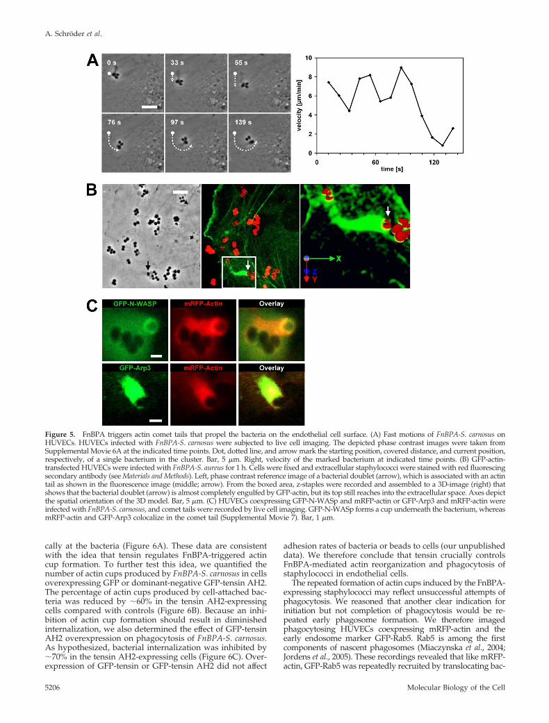
also seen with FnBPA-beads; they occurred about once ev-ery 10 min per recorded cell (mean of 13 single cell record-ings) and displayed a velocity of 400-1000 �m/h (singleframes of a representative movie are depicted in Figure 5A;see corresponding Supplemental Movie 6A). An explanationfor these fast bacterial movements was obtained in record-ings of GFP-actin–expressing HUVECs. These movies visu-alized GFP-actin comet tails that propelled bacteria forwardwith a velocity of up to 1000 �m/h (14 events analyzed; seeSupplemental Movie 6B, which is the parallel recording ofSupplemental Movie 6A). Rhodamine-phalloidin staining ofinfected HUVECs confirmed the existence of bacterial comettails consisting of endogenous actin (our unpublished data).Quantification revealed that 27 � 15% of all cell-associatedbacteria showed actin accumulations (mean � SD of 28recordings), and of these roughly 13% were comet tails(mean of 7 recordings). Comet tails could constitute one ofseveral cycles of actin accumulation at FnBPA-expressingbacteria or beads, in a way that an actin cup was followed bya comet tail and again by an actin cup. Comet tails couldform spontaneously, but they could also directly form froman actin cup. Thus, actin cups and comet tails are likelydifferent expressions of the same phenomenon, with thecomet tails being polarized, thereby causing propulsion ofbacteria or beads. Because actin comet tails have so far beendescribed mainly on intracellular bacteria, we testedwhether the tails we observed were induced by extra- orintracellular staphylococci (Stevens et al., 2006). Selectiveextracellular immunofluorescence staining of staphylococciand 3D reconstruction of a comet tail documents that al-though the bacterial cell is largely engulfed by GFP-actin, itstop still reaches into the extracellular space (Figure 5B).These experiments strongly support the idea that actincomet tails are produced by extracellular staphylococci.
Many pathogen-triggered actin comet tails are generatedby activation of N-WASp that stimulates the actin nucleationactivity of Arp2/3 complex (Stevens et al., 2006). We there-fore visualized staphylococcal comet tails in cells coexpress-ing mRFP-actin and GFP-N-WASp or mRFP-actin and GFP-Arp3, the latter being a subunit of Arp2/3 complex (Welchet al., 1997). As depicted exemplary in Figure 5C, GFP-N-WASp was enriched in cup-like structures underneath bac-teria producing an mRFP-actin comet tail. In comparison,GFP-Arp3 colocalized with mRFP-actin in the comet tail(Supplemental Movie 7). This suggests that N-WASp is re-cruited to the bacterial attachment site and assembles actincups and comet tails via Arp2/3 complex. Consistent withthis notion, no comet tails could be observed in cells treatedwith the N-WASp inhibitor wiskostatin.
Dynamic Interplay of Tensin, Actin, and Rab5 inFnBPA-stimulated Endothelial Cell Activationand PhagocytosisOur data suggest that FnBPA induces an interplay of tensinand actin in endothelial cell phagocytic cups that eventuallyleads to bacterial invasion. To visualize the spatiotemporalregulation of this interplay, live cell imaging of FnBPA-S.aureus–infected HUVECs coexpressing mRFP-actin andGFP-tensin was performed. As documented in Figure 6A(single frames of representative Movie 8; see SupplementalMaterials), mRFP-actin and GFP-tensin dynamically redis-tributed around bacterial clusters translocating on the cellsurface. When mRFP-actin accumulated at one or more cellsof the bacterial tetrad, there was a discrete to extensivecolocalization with GFP-tensin. Intensity traces demonstratethat during the phases of mRFP-actin accumulation atphagocytic cups the level of GFP-tensin also increased lo-
A. Schroder et al.
Molecular Biology of the Cell5204
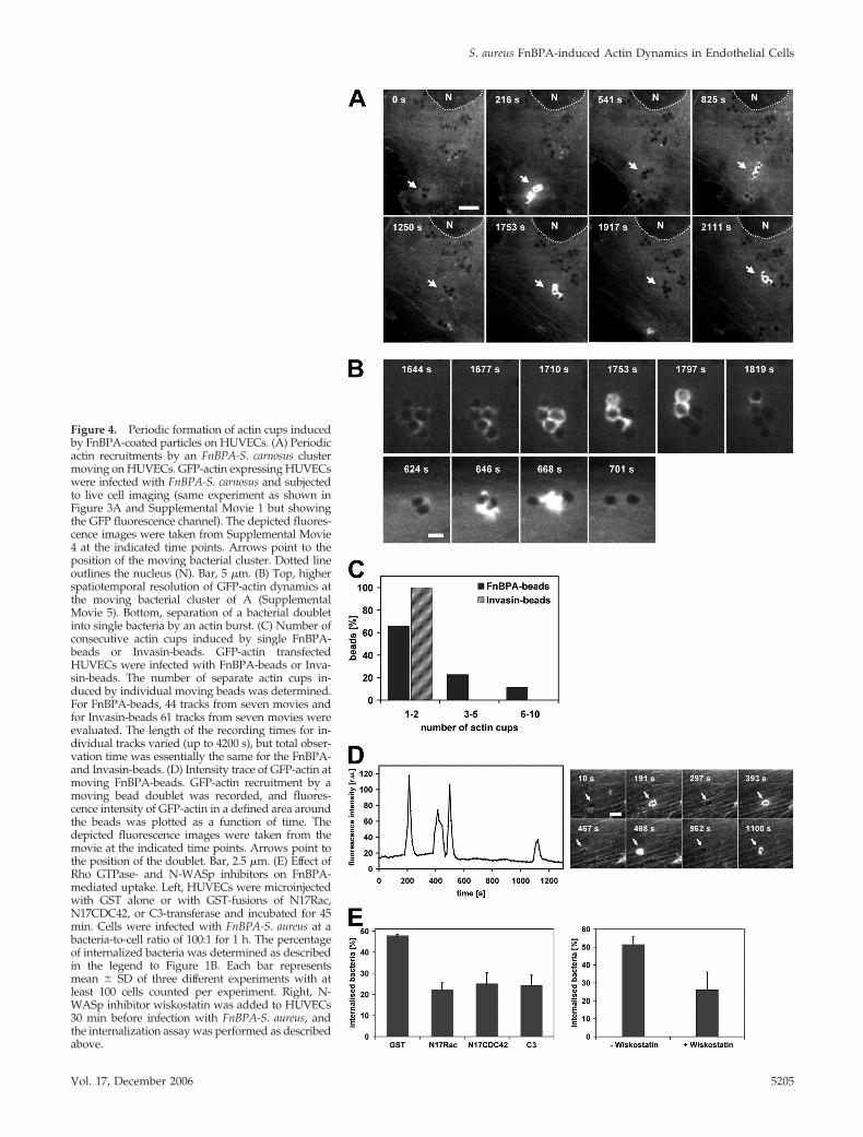
Figure 4. Periodic formation of actin cups inducedby FnBPA-coated particles on HUVECs. (A) Periodicactin recruitments by an FnBPA-S. carnosus clustermoving on HUVECs. GFP-actin expressing HUVECswere infected with FnBPA-S. carnosus and subjectedto live cell imaging (same experiment as shown inFigure 3A and Supplemental Movie 1 but showingthe GFP fluorescence channel). The depicted fluores-cence images were taken from Supplemental Movie4 at the indicated time points. Arrows point to theposition of the moving bacterial cluster. Dotted lineoutlines the nucleus (N). Bar, 5 �m. (B) Top, higherspatiotemporal resolution of GFP-actin dynamics atthe moving bacterial cluster of A (SupplementalMovie 5). Bottom, separation of a bacterial doubletinto single bacteria by an actin burst. (C) Number ofconsecutive actin cups induced by single FnBPA-beads or Invasin-beads. GFP-actin transfectedHUVECs were infected with FnBPA-beads or Inva-sin-beads. The number of separate actin cups in-duced by individual moving beads was determined.For FnBPA-beads, 44 tracks from seven movies andfor Invasin-beads 61 tracks from seven movies wereevaluated. The length of the recording times for in-dividual tracks varied (up to 4200 s), but total obser-vation time was essentially the same for the FnBPA-and Invasin-beads. (D) Intensity trace of GFP-actin atmoving FnBPA-beads. GFP-actin recruitment by amoving bead doublet was recorded, and fluores-cence intensity of GFP-actin in a defined area aroundthe beads was plotted as a function of time. Thedepicted fluorescence images were taken from themovie at the indicated time points. Arrows point tothe position of the doublet. Bar, 2.5 �m. (E) Effect ofRho GTPase- and N-WASp inhibitors on FnBPA-mediated uptake. Left, HUVECs were microinjectedwith GST alone or with GST-fusions of N17Rac,N17CDC42, or C3-transferase and incubated for 45min. Cells were infected with FnBPA-S. aureus at abacteria-to-cell ratio of 100:1 for 1 h. The percentageof internalized bacteria was determined as describedin the legend to Figure 1B. Each bar representsmean � SD of three different experiments with atleast 100 cells counted per experiment. Right, N-WASp inhibitor wiskostatin was added to HUVECs30 min before infection with FnBPA-S. aureus, andthe internalization assay was performed as describedabove.
S. aureus FnBPA-induced Actin Dynamics in Endothelial Cells
Vol. 17, December 2006 5205
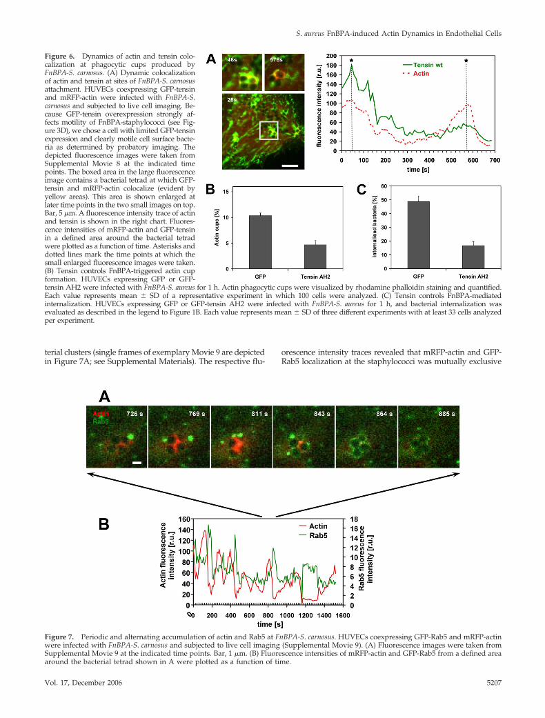
cally at the bacteria (Figure 6A). These data are consistentwith the idea that tensin regulates FnBPA-triggered actincup formation. To further test this idea, we quantified thenumber of actin cups produced by FnBPA-S. carnosus in cellsoverexpressing GFP or dominant-negative GFP-tensin AH2.The percentage of actin cups produced by cell-attached bac-teria was reduced by �60% in the tensin AH2-expressingcells compared with controls (Figure 6B). Because an inhi-bition of actin cup formation should result in diminishedinternalization, we also determined the effect of GFP-tensinAH2 overexpression on phagocytosis of FnBPA-S. carnosus.As hypothesized, bacterial internalization was inhibited by�70% in the tensin AH2-expressing cells (Figure 6C). Over-expression of GFP-tensin or GFP-tensin AH2 did not affect
adhesion rates of bacteria or beads to cells (our unpublisheddata). We therefore conclude that tensin crucially controlsFnBPA-mediated actin reorganization and phagocytosis ofstaphylococci in endothelial cells.
The repeated formation of actin cups induced by the FnBPA-expressing staphylococci may reflect unsuccessful attempts ofphagocytosis. We reasoned that another clear indication forinitiation but not completion of phagocytosis would be re-peated early phagosome formation. We therefore imagedphagocytosing HUVECs coexpressing mRFP-actin and theearly endosome marker GFP-Rab5. Rab5 is among the firstcomponents of nascent phagosomes (Miaczynska et al., 2004;Jordens et al., 2005). These recordings revealed that like mRFP-actin, GFP-Rab5 was repeatedly recruited by translocating bac-
Figure 5. FnBPA triggers actin comet tails that propel the bacteria on the endothelial cell surface. (A) Fast motions of FnBPA-S. carnosus onHUVECs. HUVECs infected with FnBPA-S. carnosus were subjected to live cell imaging. The depicted phase contrast images were taken fromSupplemental Movie 6A at the indicated time points. Dot, dotted line, and arrow mark the starting position, covered distance, and current position,respectively, of a single bacterium in the cluster. Bar, 5 �m. Right, velocity of the marked bacterium at indicated time points. (B) GFP-actin-transfected HUVECs were infected with FnBPA-S. aureus for 1 h. Cells were fixed and extracellular staphylococci were stained with red fluorescingsecondary antibody (see Materials and Methods). Left, phase contrast reference image of a bacterial doublet (arrow), which is associated with an actintail as shown in the fluorescence image (middle; arrow). From the boxed area, z-staples were recorded and assembled to a 3D-image (right) thatshows that the bacterial doublet (arrow) is almost completely engulfed by GFP-actin, but its top still reaches into the extracellular space. Axes depictthe spatial orientation of the 3D model. Bar, 5 �m. (C) HUVECs coexpressing GFP-N-WASp and mRFP-actin or GFP-Arp3 and mRFP-actin wereinfected with FnBPA-S. carnosus, and comet tails were recorded by live cell imaging. GFP-N-WASp forms a cup underneath the bacterium, whereasmRFP-actin and GFP-Arp3 colocalize in the comet tail (Supplemental Movie 7). Bar, 1 �m.
A. Schroder et al.
Molecular Biology of the Cell5206
terial clusters (single frames of exemplary Movie 9 are depictedin Figure 7A; see Supplemental Materials). The respective flu-
orescence intensity traces revealed that mRFP-actin and GFP-Rab5 localization at the staphylococci was mutually exclusive
Figure 6. Dynamics of actin and tensin colo-calization at phagocytic cups produced byFnBPA-S. carnosus. (A) Dynamic colocalizationof actin and tensin at sites of FnBPA-S. carnosusattachment. HUVECs coexpressing GFP-tensinand mRFP-actin were infected with FnBPA-S.carnosus and subjected to live cell imaging. Be-cause GFP-tensin overexpression strongly af-fects motility of FnBPA-staphylococci (see Fig-ure 3D), we chose a cell with limited GFP-tensinexpression and clearly motile cell surface bacte-ria as determined by probatory imaging. Thedepicted fluorescence images were taken fromSupplemental Movie 8 at the indicated timepoints. The boxed area in the large fluorescenceimage contains a bacterial tetrad at which GFP-tensin and mRFP-actin colocalize (evident byyellow areas). This area is shown enlarged atlater time points in the two small images on top.Bar, 5 �m. A fluorescence intensity trace of actinand tensin is shown in the right chart. Fluores-cence intensities of mRFP-actin and GFP-tensinin a defined area around the bacterial tetradwere plotted as a function of time. Asterisks anddotted lines mark the time points at which thesmall enlarged fluorescence images were taken.(B) Tensin controls FnBPA-triggered actin cupformation. HUVECs expressing GFP or GFP-tensin AH2 were infected with FnBPA-S. aureus for 1 h. Actin phagocytic cups were visualized by rhodamine phalloidin staining and quantified.Each value represents mean � SD of a representative experiment in which 100 cells were analyzed. (C) Tensin controls FnBPA-mediatedinternalization. HUVECs expressing GFP or GFP-tensin AH2 were infected with FnBPA-S. aureus for 1 h, and bacterial internalization wasevaluated as described in the legend to Figure 1B. Each value represents mean � SD of three different experiments with at least 33 cells analyzedper experiment.
Figure 7. Periodic and alternating accumulation of actin and Rab5 at FnBPA-S. carnosus. HUVECs coexpressing GFP-Rab5 and mRFP-actinwere infected with FnBPA-S. carnosus and subjected to live cell imaging (Supplemental Movie 9). (A) Fluorescence images were taken fromSupplemental Movie 9 at the indicated time points. Bar, 1 �m. (B) Fluorescence intensities of mRFP-actin and GFP-Rab5 from a defined areaaround the bacterial tetrad shown in A were plotted as a function of time.
S. aureus FnBPA-induced Actin Dynamics in Endothelial Cells
Vol. 17, December 2006 5207
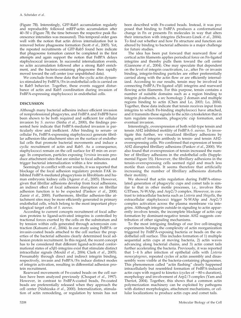
(Figure 7B). Interestingly, GFP-Rab5 accumulation regularlyand reproducibly followed mRFP-actin accumulation after40–50 s (Figure 7B; the time between the respective peak flu-orescence intensities was measured). This temporal order goeswell with the notion that actin drives internalization but isremoved before phagosome formation (Scott et al., 2005). Yet,the repeated recruitments of GFP-Rab5 found here indicatethat phagosome formation cannot be completed in the firstattempts and again support the notion that FnBPA delaysstaphylococcal invasion. In successful internalization events,no actin accumulation followed after a strong Rab5 enrich-ment, and the bacteria-containing phagosome then rapidlymoved toward the cell center (our unpublished data).
We conclude from these data that the cyclic actin dynam-ics stimulated by FnBPA/Fn in endothelial cells is carried onto Rab5 behavior. Together, these events suggest distur-bance of actin and Rab5 coordination during invasion ofFnBPA-expressing staphylococci in endothelial cells.
DISCUSSION
Although many bacterial adhesins induce efficient invasionof nonprofessional phagocytes, and FnBPA and FnBPB havebeen shown to be both required and sufficient for cellularinvasion by S. aureus (Sinha et al., 2000), the findings pre-sented here indicate that FnBPA-mediated invasion is par-ticularly slow and inefficient. After binding to serum- orcellular Fn, FnBPA-expressing staphylococci generate fibril-lar adhesion-like attachment sites on the surface of endothe-lial cells that promote bacterial movements and induce acyclic recruitment of actin and Rab5. As a consequence,staphylococci remain on the cell surface for 45 min on aver-age. In comparison, particles exposing Yersinia invasin pro-duce attachment sites that are similar to focal adhesions andtrigger bacterial internalization within a few minutes.
Seemingly in conflict with our results, it was reported thatblockage of the focal adhesion regulatory protein FAK in-hibited FnBPA-mediated phagocytosis in fibroblasts and hu-man embryonic kidney cells (Agerer et al., 2005). However,given that fibrillar adhesions develop from focal adhesions,an indirect effect of focal adhesion disruption on fibrillaradhesion function is to be expected (Pankov et al., 2000;Zamir et al., 2000). Furthermore, fibrillar adhesion-like at-tachment sites may be more efficiently generated in primaryendothelial cells, which belong to the most important phys-iological target cells of S. aureus.
According to current concepts recruitment of focal adhe-sion proteins to ligand-activated integrins is controlled bytractional forces exerted by the cells on the substratum andby tension within cells generated through actomyosin con-traction (Katsumi et al., 2004). In our study using FnBPA- orinvasin-coated beads attached to the cell surface the prop-erties of the bacterial adhesins clearly determined focal ad-hesion protein recruitment. In this regard, the recent concepthas to be considered that different ligand-activated confor-mational states of �5�1-integrins exist that stimulate distinctintracellular signals (Mould et al., 2004; Clark et al., 2005).Presumably through direct and indirect integrin binding,respectively, invasin and FnBPA/Fn induce distinct modesof integrin activation, resulting in differential adhesion pro-tein recruitment.
Rearward movements of Fn-coated beads on the cell sur-face have been analyzed previously (Choquet et al., 1997).However, in contrast to FnBPA-coated beads, Fn-coatedbeads are preferentially released when they approach thecell center (Nishizaka et al., 2000). Internalization, stimula-tion of actin remodeling, or regulation by tensin has not
been described with Fn-coated beads. Instead, it was pro-posed that binding to FnBPA produces a conformationalchange in Fn or presents Fn molecules in way that alterstheir interaction with integrins (Schwarz-Linek et al., 2004).To find out whether and how Fn structure and function arealtered by binding to bacterial adhesins is a major challengefor future studies.
The idea has been put forward that rearward flow ofcortical actin filaments applies periodical force on Fn-ligatedintegrins and thereby pulls them toward the cell center(Giannone et al., 2004). One may speculate that dependenton the level of integrin coactivation, i.e., after Fn- or invasinbinding, integrin-binding particles are either preferentiallycarried along with the actin flow or are efficiently internal-ized. According to our results, tensin may be involved inconnecting FnBPA/Fn-ligated �5�1 integrins and rearwardflowing actin filaments. For this purpose, tensin contains anumber of suitable domains such as a region binding tointegrin �-subunits, a src homology 2 domain and multipleregions binding to actin (Chen and Lo, 2003; Lo, 2004).Together, these data indicate that tensin receives input fromintegrins to which Fn-binding staphylococci have attached,and it transmits these signals to the actin cytoskeleton that inturn regulate movements, phagocytic cup formation, andeventual invasion.
Interestingly, overexpression of both wild-type tensin andtensin AH2 inhibited motility of FnBPA-S. aureus. To inves-tigate this further, we visualized fibrillary adhesions byusing anti-�5 integrin antibody in tensin- and tensin AH2-overexpressing cells. We confirmed that expression of tensinAH2 disrupted fibrillary adhesions (Pankov et al., 2000). Wealso found that overexpression of tensin increased the num-ber of fibrillary adhesions in the endothelial cells (Supple-mental Figure 1S). However, the fibrillary adhesions in thetensin-overexpressing cells seemed rigid and much lessmotile than controls. It seems therefore that artificiallyincreasing the number of fibrillary adhesions disturbstheir motility.
We showed that actin regulation during FnBPA-stimu-lated generation of phagocytic cups and comet tails is sim-ilar to that in other motile processes, i.e., involves RhoGTPases, N-WASp, and Arp2/3 complex. However, in con-trast to intracellular bacteria such as Listeria and Shigella, theextracellular staphylococci trigger N-WASp and Arp2/3complex activation across the plasma membrane via inte-grins. Although integrin outside-in signaling to actin appar-ently involves tensin, the incomplete blockage of actin cupformation by dominant-negative tensin AH2 suggests con-tribution of other signaling mechanisms.
To the most intriguing findings of our live cell-imagingexperiments belongs the complexity of actin reorganizationtriggered by FnBPA-exposing bacteria or beads on the en-dothelial cell surface. This includes formation of 1) multiplesequential actin cups at moving bacteria, 2) actin wavesadvancing along bacterial chains, and 3) actin comet tailsfurther accelerating the bacteria. Previously, it was reportedthat 1–4 h after infection of epithelial cells with Listeriamonocytogenes, repeated cycles of actin assembly and disas-sembly were visible at the bacteria-containing phagosomes.This phenomenon called “actin flashing” clearly happenedintracellularly but resembled formation of FnBPA-inducedactin cups with regard to kinetics (cycles of �80-s duration),morphology and involvement of Arp2/3 complex (Yam andTheriot, 2004). Together, this shows that a common actinpolymerization machinery can be exploited by pathogenswith distinct morphologies, attachment mechanisms, or cel-lular localizations to produce actin cups and comet tails.
A. Schroder et al.
Molecular Biology of the Cell5208
Finally, the repeated assembly of actin phagocytic cupsand early phagosomes may be purposely induced by thestaphylococci to delay phagocytosis. This may give thestaphylococci enough time to produce critical amounts ofcell damaging toxins. Consistent with this idea, it was re-ported that unless they exerted a high cellular toxicity beforeuptake, most S. aureus strains got efficiently eliminated inlysosomes of keratinocytes or endothelial cells (Krut et al.,2003; Schroder et al., 2006). Yet, for a host cell, the cyclic actincup and phagosome formation may also somehow preparethe bacteria for eventual invasion. For example, proteasesreleased from early phagosomes could digest and repro-gram the FnBPA-bound Fn, enabling its uptake togetherwith the bacteria (Sottile and Chandler, 2005).
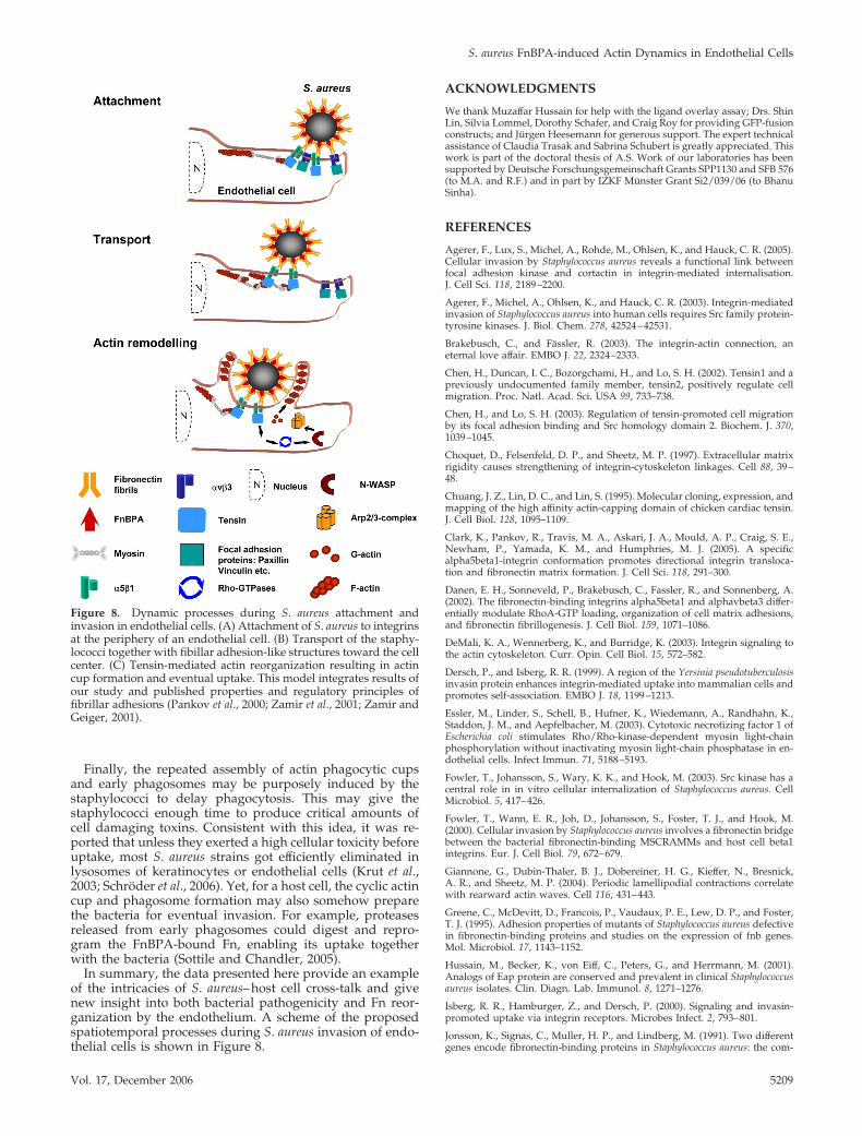
In summary, the data presented here provide an exampleof the intricacies of S. aureus–host cell cross-talk and givenew insight into both bacterial pathogenicity and Fn reor-ganization by the endothelium. A scheme of the proposedspatiotemporal processes during S. aureus invasion of endo-thelial cells is shown in Figure 8.
ACKNOWLEDGMENTS
We thank Muzaffar Hussain for help with the ligand overlay assay; Drs. ShinLin, Silvia Lommel, Dorothy Schafer, and Craig Roy for providing GFP-fusionconstructs; and Jurgen Heesemann for generous support. The expert technicalassistance of Claudia Trasak and Sabrina Schubert is greatly appreciated. Thiswork is part of the doctoral thesis of A.S. Work of our laboratories has beensupported by Deutsche Forschungsgemeinschaft Grants SPP1130 and SFB 576(to M.A. and R.F.) and in part by IZKF Munster Grant Si2/039/06 (to BhanuSinha).
REFERENCES
Agerer, F., Lux, S., Michel, A., Rohde, M., Ohlsen, K., and Hauck, C. R. (2005).Cellular invasion by Staphylococcus aureus reveals a functional link betweenfocal adhesion kinase and cortactin in integrin-mediated internalisation.J. Cell Sci. 118, 2189–2200.
Agerer, F., Michel, A., Ohlsen, K., and Hauck, C. R. (2003). Integrin-mediatedinvasion of Staphylococcus aureus into human cells requires Src family protein-tyrosine kinases. J. Biol. Chem. 278, 42524–42531.
Brakebusch, C., and Fassler, R. (2003). The integrin-actin connection, aneternal love affair. EMBO J. 22, 2324–2333.
Chen, H., Duncan, I. C., Bozorgchami, H., and Lo, S. H. (2002). Tensin1 and apreviously undocumented family member, tensin2, positively regulate cellmigration. Proc. Natl. Acad. Sci. USA 99, 733–738.
Chen, H., and Lo, S. H. (2003). Regulation of tensin-promoted cell migrationby its focal adhesion binding and Src homology domain 2. Biochem. J. 370,1039–1045.
Choquet, D., Felsenfeld, D. P., and Sheetz, M. P. (1997). Extracellular matrixrigidity causes strengthening of integrin-cytoskeleton linkages. Cell 88, 39–48.
Chuang, J. Z., Lin, D. C., and Lin, S. (1995). Molecular cloning, expression, andmapping of the high affinity actin-capping domain of chicken cardiac tensin.J. Cell Biol. 128, 1095–1109.
Clark, K., Pankov, R., Travis, M. A., Askari, J. A., Mould, A. P., Craig, S. E.,Newham, P., Yamada, K. M., and Humphries, M. J. (2005). A specificalpha5beta1-integrin conformation promotes directional integrin transloca-tion and fibronectin matrix formation. J. Cell Sci. 118, 291–300.
Danen, E. H., Sonneveld, P., Brakebusch, C., Fassler, R., and Sonnenberg, A.(2002). The fibronectin-binding integrins alpha5beta1 and alphavbeta3 differ-entially modulate RhoA-GTP loading, organization of cell matrix adhesions,and fibronectin fibrillogenesis. J. Cell Biol. 159, 1071–1086.
DeMali, K. A., Wennerberg, K., and Burridge, K. (2003). Integrin signaling tothe actin cytoskeleton. Curr. Opin. Cell Biol. 15, 572–582.
Dersch, P., and Isberg, R. R. (1999). A region of the Yersinia pseudotuberculosisinvasin protein enhances integrin-mediated uptake into mammalian cells andpromotes self-association. EMBO J. 18, 1199–1213.
Essler, M., Linder, S., Schell, B., Hufner, K., Wiedemann, A., Randhahn, K.,Staddon, J. M., and Aepfelbacher, M. (2003). Cytotoxic necrotizing factor 1 ofEscherichia coli stimulates Rho/Rho-kinase-dependent myosin light-chainphosphorylation without inactivating myosin light-chain phosphatase in en-dothelial cells. Infect Immun. 71, 5188–5193.
Fowler, T., Johansson, S., Wary, K. K., and Hook, M. (2003). Src kinase has acentral role in in vitro cellular internalization of Staphylococcus aureus. CellMicrobiol. 5, 417–426.
Fowler, T., Wann, E. R., Joh, D., Johansson, S., Foster, T. J., and Hook, M.(2000). Cellular invasion by Staphylococcus aureus involves a fibronectin bridgebetween the bacterial fibronectin-binding MSCRAMMs and host cell beta1integrins. Eur. J. Cell Biol. 79, 672–679.
Giannone, G., Dubin-Thaler, B. J., Dobereiner, H. G., Kieffer, N., Bresnick,A. R., and Sheetz, M. P. (2004). Periodic lamellipodial contractions correlatewith rearward actin waves. Cell 116, 431–443.
Greene, C., McDevitt, D., Francois, P., Vaudaux, P. E., Lew, D. P., and Foster,T. J. (1995). Adhesion properties of mutants of Staphylococcus aureus defectivein fibronectin-binding proteins and studies on the expression of fnb genes.Mol. Microbiol. 17, 1143–1152.
Hussain, M., Becker, K., von Eiff, C., Peters, G., and Herrmann, M. (2001).Analogs of Eap protein are conserved and prevalent in clinical Staphylococcusaureus isolates. Clin. Diagn. Lab. Immunol. 8, 1271–1276.
Isberg, R. R., Hamburger, Z., and Dersch, P. (2000). Signaling and invasin-promoted uptake via integrin receptors. Microbes Infect. 2, 793–801.
Jonsson, K., Signas, C., Muller, H. P., and Lindberg, M. (1991). Two differentgenes encode fibronectin-binding proteins in Staphylococcus aureus: the com-
Figure 8. Dynamic processes during S. aureus attachment andinvasion in endothelial cells. (A) Attachment of S. aureus to integrinsat the periphery of an endothelial cell. (B) Transport of the staphy-lococci together with fibillar adhesion-like structures toward the cellcenter. (C) Tensin-mediated actin reorganization resulting in actincup formation and eventual uptake. This model integrates results ofour study and published properties and regulatory principles offibrillar adhesions (Pankov et al., 2000; Zamir et al., 2001; Zamir andGeiger, 2001).
S. aureus FnBPA-induced Actin Dynamics in Endothelial Cells
Vol. 17, December 2006 5209
plete nucleotide sequence and characterization of the second gene. Eur.J. Biochem. 202, 1041–1048.
Jordens, I., Marsman, M., Kuijl, C., and Neefjes, J. (2005). Rab proteins,connecting transport and vesicle fusion. Traffic 6, 1070–1077.
Katsumi, A., Orr, A. W., Tzima, E., and Schwartz, M. A. (2004). Integrins inmechanotransduction. J. Biol. Chem. 279, 12001–12004.
Krut, O., Utermohlen, O., Schlossherr, X., and Kronke, M. (2003). Strain-specific association of cytotoxic activity and virulence of clinical Staphylococ-cus aureus isolates. Infect. Immun. 71, 2716–2723.
Laschke, M. W., Kerdudou, S., Herrmann, M., and Menger, M. D. (2005).Intravital fluorescence microscopy: a novel tool for the study of the interactionof Staphylococcus aureus with the microvascular endothelium in vivo. J. Infect.Dis. 191, 435–443.
Lo, S. H. (2004). Tensin. Int. J. Biochem. Cell Biol. 36, 31–34.
Lowy, F. D. (1998). Staphylococcus aureus infections. N. Engl. J. Med. 339,520–532.
Massey, R. C., Kantzanou, M. N., Fowler, T., Day, N. P., Schofield, K., Wann,E. R., Berendt, A. R., Hook, M., and Peacock, S. J. (2001). Fibronectin-bindingprotein A of Staphylococcus aureus has multiple, substituting, binding regionsthat mediate adherence to fibronectin and invasion of endothelial cells. CellMicrobiol. 3, 839–851.
May, R. C., and Machesky, L. M. (2001). Phagocytosis and the actin cytoskel-eton. J. Cell Sci. 114, 1061–1077.
McDevitt, D., Francois, P., Vaudaux, P., and Foster, T. J. (1994). Molecularcharacterization of the clumping factor (fibrinogen receptor) of Staphylococcusaureus. Mol. Microbiol. 11, 237–248.
Menzies, B. E. (2003). The role of fibronectin binding proteins in the patho-genesis of Staphylococcus aureus infections. Curr. Opin. Infect. Dis. 16, 225–229.
Miaczynska, M., Pelkmans, L., and Zerial, M. (2004). Not just a sink: endo-somes in control of signal transduction. Curr. Opin. Cell Biol. 16, 400–406.
Mould, A. P., and Humphries, M. J. (2004). Regulation of integrin functionthrough conformational complexity: not simply a knee-jerk reaction? Curr.Opin. Cell Biol. 16, 544–551.
Nishizaka, T., Shi, Q., and Sheetz, M. P. (2000). Position-dependent linkages offibronectin-integrin-cytoskeleton. Proc. Natl. Acad. Sci. USA 97, 692–697.
Nyberg, P., Sakai, T., Cho, K. H., Caparon, M. G., Fassler, R., and Bjorck, L.(2004). Interactions with fibronectin attenuate the virulence of Streptococcuspyogenes. EMBO J. 23, 2166–2174.
Osiak, A. E., Zenner, G., and Linder, S. (2005). Subconfluent endothelial cellsform podosomes downstream of cytokine and RhoGTPase signaling. Exp.Cell Res. 307, 342–353.
Pankov, R., Cukierman, E., Katz, B. Z., Matsumoto, K., Lin, D. C., Lin, S.,Hahn, C., and Yamada, K. M. (2000). Integrin dynamics and matrix assembly:tensin-dependent translocation of alpha(5)beta(1) integrins promotes earlyfibronectin fibrillogenesis. J. Cell Biol. 148, 1075–1090.
Patti, J. M., et al. (1992). Molecular characterization and expression of a geneencoding a Staphylococcus aureus collagen adhesin. J. Biol. Chem. 267, 4766–4772.
Que, Y. A., et al. (2005). Fibrinogen and fibronectin binding cooperate forvalve infection and invasion in Staphylococcus aureus experimental endocar-ditis. J Exp. Med. 201, 1627–1635.
Rasband, W. S. (2006). ImageJ. National Institutes of Health, Bethesda, MD.http://rsb.info.nih.gov/ij (1997–2006).
Schroder, A., Kland, R., Peschel, A., von Eiff, C., and Aepfelbacher, M. (2006).Live cell imaging of phagosome maturation in Staphylococcus aureus infectedhuman endothelial cells: small colony variants are able to survive in lyso-somes. Med. Microbiol. Immunol. 195, 185–194. Epub 2006 April 5.
Schulte, R., Zumbihl, R., Kampik, D., Fauconnier, A., and Autenrieth, I. B.(1998). Wortmannin blocks Yersinia invasin-triggered internalization, but notinterleukin-8 production by epithelial cells. Med. Microbiol. Immunol. 187,53–60.
Schwarz-Linek, U., Hook, M., and Potts, J. R. (2004). The molecular basis offibronectin-mediated bacterial adherence to host cells. Mol. Microbiol. 52,631–641.
Scott, C. C., Dobson, W., Botelho, R. J., Coady-Osberg, N., Chavrier, P.,Knecht, D. A., Heath, C., Stahl, P., and Grinstein, S. (2005). Phosphatidylino-sitol-4,5-bisphosphate hydrolysis directs actin remodeling during phagocyto-sis. J. Cell Biol. 169, 139–149.
Sinha, B., Francois, P. P., Nusse, O., Foti, M., Hartford, O. M., Vaudaux, P.,Foster, T. J., Lew, D. P., Herrmann, M., and Krause, K. H. (1999). Fibronectin-binding protein acts as Staphylococcus aureus invasin via fibronectin bridgingto integrin alpha5beta1. Cell Microbiol. 1, 101–117.
Sinha, B., Francois, P., Que, Y. A., Hussain, M., Heilmann, C., Moreillon, P.,Lew, D., Krause, K. H., Peters, G., and Herrmann, M. (2000). Heterologouslyexpressed Staphylococcus aureus fibronectin-binding proteins are sufficient forinvasion of host cells. Infect. Immun. 68, 6871–6878.
Sottile, J., and Chandler, J. (2005). Fibronectin matrix turnover occurs througha caveolin-1-dependent process. Mol. Biol. Cell 16, 757–768.
Stevens, J. M., Galyov, E. E., and Stevens, M. P. (2006). Actin-dependentmovement of bacterial pathogens. Nat. Rev. Microbiol. 4, 91–101.
Welch, M. D., DePace, A. H., Verma, S., Iwamatsu, A., and Mitchison, T. J.(1997). The human Arp2/3 complex is composed of evolutionarily conservedsubunits and is localized to cellular regions of dynamic actin filament assem-bly. J. Cell Biol. 138, 375–384.
Wiedemann, A., Linder, S., Grassl, G., Albert, M., Autenrieth, I., and Aepfel-bacher, M. (2001). Yersinia enterocolitica invasin triggers phagocytosis via beta1integrins, CDC42Hs and WASp in macrophages. Cell Microbiol. 3, 693–702.
Yam, P. T., and Theriot, J. A. (2004). Repeated cycles of rapid actin assemblyand disassembly on epithelial cell phagosomes. Mol. Biol. Cell 15, 5647–5658.
Zamir, E., and Geiger, B. (2001). Molecular complexity and dynamics ofcell-matrix adhesions. J. Cell Sci. 114, 3583–3590.
Zamir, E., et al. (2000). Dynamics and segregation of cell-matrix adhesions incultured fibroblasts. Nat. Cell Biol. 2, 191–196.
A. Schroder et al.
Molecular Biology of the Cell5210