university of warwick institutional repository: · investigations of siderophore and tetronic acid...
TRANSCRIPT

University of Warwick institutional repository: http://go.warwick.ac.uk/wrap
A Thesis Submitted for the Degree of PhD at the University of Warwick
http://go.warwick.ac.uk/wrap/55795
This thesis is made available online and is protected by original copyright.
Please scroll down to view the document itself.
Please refer to the repository record for this item for information to help you to cite it. Our policy information is available from the repository home page.

Investigations of Siderophore and Tetronic acid
Biosynthesis in Streptomyces scabies 87.22
Joanna Bicz
Thesis submitted in partial fulfilment of the requirements for
the degree of Doctor of Philosophy in Chemistry.
University of Warwick
Department of Chemistry
March 2013

PhD Thesis: Joanna Bicz Contents
i
Contents
Contents .......................................................................................................................i
List of Figures..............................................................................................................v
List of Tables ............................................................................................................xii
Acknowledgements..................................................................................................xiv
Declaration................................................................................................................xv
Abstract....................................................................................................................xvi
Abbreviations .........................................................................................................xvii
1. Introduction .......................................................................................................... 1
1.1 Streptomyces .................................................................................................. 2
1.2 Streptomyces scabies .......................................................................................... 3 1.3 Secondary metabolites of S. scabies 87.22 ........................................................ 5
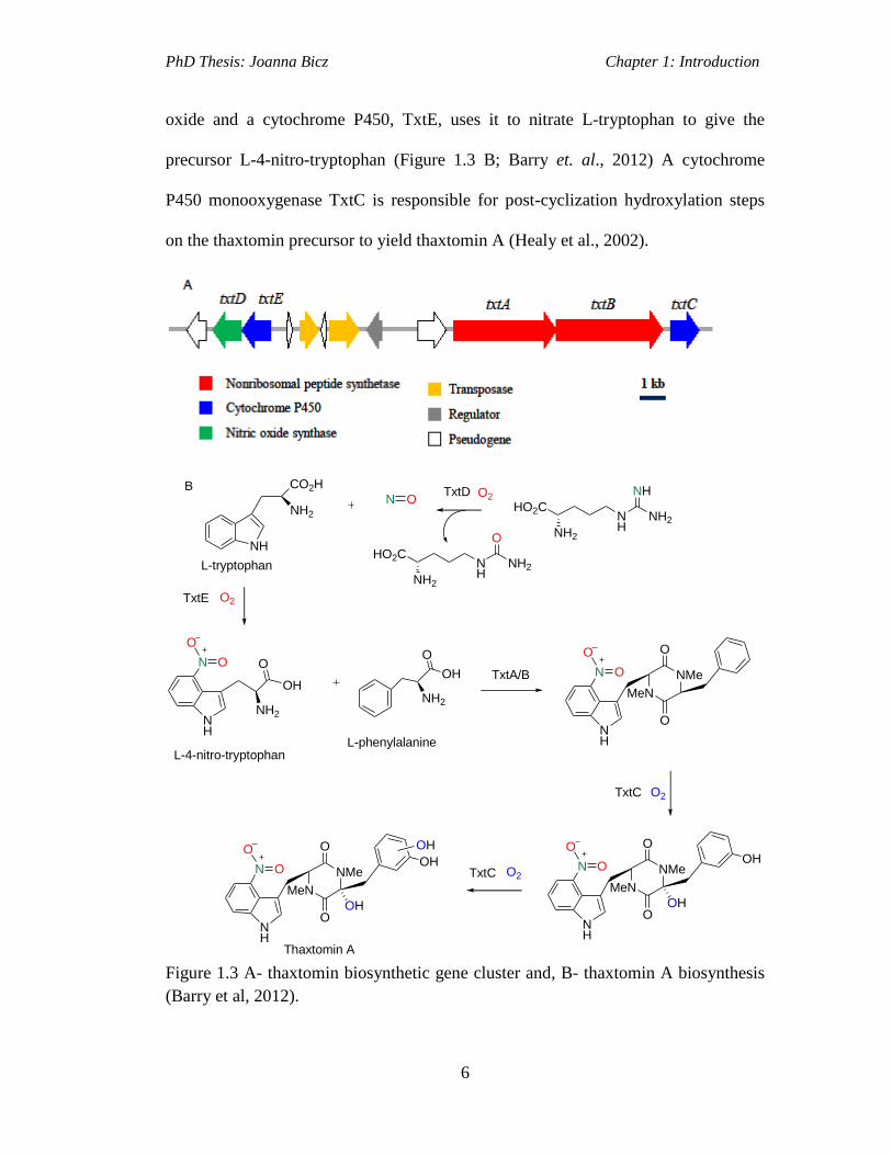
1.3.1 Thaxtomins .................................................................................................. 5
1.3.2 Concanamycins ........................................................................................... 7 1.4 Secondary Metabolites of S. scabies 87.22 Discovered by Genome Mining. . 13
1.4.1 Coronofacic acid ....................................................................................... 16 1.4.2 Bottromycin .............................................................................................. 20
1.5 Siderophores ..................................................................................................... 26
1.5.1 Siderophores Biosynthesised via NRPS-Dependent Pathways................. 28 1.5.1.1 NRPS siderophore pathway-Enterobactin Biosynthesis...................32
1.5.2 Siderophores Assembled via NRPS-Independent Siderophore (NIS)
Pathway .............................................................................................................. 36
1.5.2.1 Aerobactin Biosynthesis- an archetypal NIS pathway.....................37 1.6 Investigations of Cryptic Siderophore Biosynthetic Pathways encoded for in
the S. scabies 87.22 genome. ................................................................................. 39 1.6.1 Pyochelin ................................................................................................... 39
1.6.1.1 Pyochelin Biosynthesis in P. aeruginosa.........................................40
1.6.1.2 Sequence Analysis of the S. scabies Pyochelin Biosynthetic Gene
Cluster...........................................................................................................43 1.6.1.3 Biosynthesis of Enantio-Pyochelin in Pseudomonas fluorescens....45
1.6.2 Desferrioxamines. ..................................................................................... 47

PhD Thesis: Joanna Bicz Contents
ii
1.6.3 Novel Putative Hydroxamate Siderophore................................................ 53 1.7 Products of Other Cryptic Pathways ................................................................ 56
1.7.1 RK-682 ...................................................................................................... 61 1.7.2 Agglomerins .............................................................................................. 63 1.8.3 Cryptic Tetronate Biosynthetic Gene Cluster in S. scabies 87.22. ........... 64
1.8 Aims of the Project........................................................................................... 68 Results and Discussion I: Investigation of siderophore biosynthetic gene clusters in
S. scabies. ................................................................................................................... 70 2. Pyochelin as a Product of Pyochelin Biosynthetic Gene Cluster in S. scabies. ..... 71
2.1 Analysis of the Pyochelin Biosynthetic Gene Cluster in S. scabies 87.22. ..... 71
2.2 Creation of Pyochelin Mutants in S. scabies 87.22. ......................................... 75
2.3 Analysis of Pyochelin Production in S. scabies ............................................... 76
2.4 Discussion ........................................................................................................ 80 3. Investigation of a Putative Hydroxamate Siderophore Biosynthetic Gene Cluster in
S. scabies 87.22 .......................................................................................................... 83 3.1 PCR Targeting Strategy ................................................................................... 83
3.2 Investigation of Putative Hydroxamate Siderophore Production in S. scabies 87 3.2.1 Isolation and Structure Elucidation of the Hydroxamate Siderophore ..... 88 3.2.2 Scabichelin Biosynthetic Gene Cluster ..................................................... 89
3.2.3 Creation of a scab85471 Mutant ............................................................... 92 3.2.4 LC-MS Analysis of Scabichelin Mutant ................................................... 99
3.2.5 Discussions .............................................................................................. 100
4. Desferrioxamine production in S. scabies 87.22 .................................................. 112
4.1 The Aims of This Work ................................................................................. 112 4.2 Putative Desferrioxamine Biosynthetic Gene Cluster in S. scabies ............... 113
4.3 Construction of desC Mutant in S. scabies 87.22 .......................................... 116 4.3.1 LC-MS Analysis of desC Mutant ............................................................ 119
4.4 Chemical Complementation of desC Mutant ................................................. 121
4.5 Genetic Complementation of desC Mutant .................................................... 123 4.5.1 LC-MS Analysis of Complemented Δ desC Mutant ............................... 126
4.6 Discussion ...................................................................................................... 127 Results and Discussion II: Investigation of Tetronate-like Biosynthetic Gene Cluster
in S. scabies 87.22. ................................................................................................... 130
5. Investigation of Tetronate-like Biosynthetic Gene Cluster in S. scabies 87.22. .. 131 5.1 Cryptic Polyketide Tetronate Biosynthetic Gene Cluster in S. scabies 87.22 131 5.2 Transcriptional Analysis of Cryptic Tetronate Biosynthetic Gene Cluster in S.
scabies .................................................................................................................. 132
5.3 Mutagenesis of scab63021 to Determine the Effect on Expression of Putative
Tetronate-like Gene Cluster in S. scabies. ........................................................... 135 5.3.1 SARP Family .......................................................................................... 135 5.3.2 Mutagenesis of scab63021 Gene ............................................................ 135
5.4 RT-PCR on Wild Type S. scabies and the Δscab63021 Mutant RNA .......... 141
5.5 Discussion ...................................................................................................... 142 6. Summary, Conclusions and Future Work ............................................................ 144

PhD Thesis: Joanna Bicz Contents
iii
6.1 Results and Discussion I: Investigation of Siderophore Biosynthetic Gene
Clusters in S. scabies. ........................................................................................... 145 6.1.1 Investigation of Pyochelin Biosynthetic Gene Cluster in S. scabies 87.22.
.......................................................................................................................... 145 6.1.2 Investigation of Scabichelin Biosynthetic Gene Cluster in S. scabies
87.22. ................................................................................................................ 147 6.1.3 Desferrioxamine Production in S. scabies .............................................. 149
6.2 Results and Discussion II: Investigation of Tetronate-like Biosynthetic Gene
Cluster in S. scabies 87.22. .................................................................................. 150 6.2.1 Investigation of Tetronate-like Biosynthetic Gene Cluster in S. scabies
87.22. ................................................................................................................ 150
6.3 Significance .................................................................................................... 152
7. Materials and Methods ......................................................................................... 155 7.1 Materials ......................................................................................................... 156
7.1.1 Enzymes, Chemicals and Equipment ...................................................... 156 7.1.2 General Solutions, Buffers and Antibiotics ............................................ 157
7.1.3 Bacterial Strains ...................................................................................... 158 7.1.4 Plasmids and Cosmids ............................................................................ 160 7.1.5 Primers .................................................................................................... 161
7.1.6 Growth Media ......................................................................................... 163 7.1.6.1 Recipes for Liquid Media...............................................................163
7.1.6.2 Recipes for Solid Media.................................................................165
7.2 Growth, Manipulation and Storage of E. coli ................................................ 167
7.2.1 Growth Conditions of E. coli .................................................................. 168 7.2.2 Preparation of Electrocompetent E. coli Cells ........................................ 168
7.2.3 Transformation of Electrocompetent E. coli Cells .................................. 168 7.2.4 Storage of E. coli Cells ........................................................................... 169
7.3 Growth, Manipulation and Storage of Streptomyces ..................................... 169
7.3.1 Spore Stock Collection from Agar Plates ............................................... 169 7.3.2 Liquid Cultures for Genomic DNA Extraction ....................................... 169
7.3.3 DNA Transfer from E. coli to S. scabies by Conjugation ....................... 169 7.4 Extraction and Manipulation of DNA ............................................................ 171
7.4.1 Isolation of Genomic DNA from S. scabies ........................................... 171
7.4.2 Plasmid/Cosmid Isolation from E. coli ................................................... 172 7.4.3 Digestion of DNA with Restriction Enzymes ......................................... 172 7.4.4 Ligation of DNA into Plasmids .............................................................. 173 7.4.5 Agarose Gel Electrophoresis ................................................................... 173
7.5 PCR Methods ................................................................................................. 173 7.5.1 General PCR Method .............................................................................. 174 7.5.2 PCR Amplification of the Gene Replacement Cassette. ......................... 175
7.6 PCR Targeting in S. scabies 87.22 ................................................................. 175 7.6.1 Design of PCR Primers ........................................................................... 175
7.6.2 Purification of the Resistance Cassette ................................................... 175 7.6.3 PCR Amplification of the Extended Resistance Cassette ....................... 176
PhD Thesis: Joanna Bicz Contents
iv
7.6.4 Introduction of Cosmid Clones into E. coli BW25113/pIJ790 by
Electroporation ................................................................................................. 176 7.6.5 PCR Targeting of Cosmids ..................................................................... 177 7.6.6 Transfer of the Mutant Cosmids into Streptomyces scabies ................... 178
7.7 Southern Blot Hybridisation .......................................................................... 179
7.7.1 Probe Labeling ........................................................................................ 179 7.7.2 Genomic DNA Digestion ........................................................................ 179 7.7.3 Capillary Transfer and DNA Fixation..................................................... 180 7.7.4 Hybridisation ........................................................................................... 181 7.7.5 Detection ................................................................................................. 181
7.8 RNA Methods ................................................................................................ 182
7.8.1 Total RNA Isolation from S. scabies ...................................................... 182
7.8.2 Reverse Transcriptase PCR Method ....................................................... 183 7.9 Growth of Streptomyces scabies; Extraction of Metabolites and Analysis of
Metabolite Production .......................................................................................... 185 7.9.1 Pyochelin ................................................................................................. 185
7.9.1.1 Culturing of S. scabies in Iron Deficient Medium..........................185 7.9.1.2 LC-MS Analysis.............................................................................185 7.9.1.3 Extraction of Pyochelin and High Resolution Mass Spectrometry186
7.9.2 Scabichelin .............................................................................................. 188 7.9.2.1 LC-MS Analysis.............................................................................188
7.9.3 Desferrioxamines .................................................................................... 189
7.9.3.1 Growing S. scabies in Supplemented Minimal Medium (SMM) and
LC-MS Analysis.........................................................................................189 References ................................................................................................................ 190
PhD Thesis: Joanna Bicz List of Figures
v
List of Figures
Figure 1.1 The Streptomycete life cycle (Anger, 2005)...............................................3
Figure 1.2 Potato and radish with scab disease (http://www.cals.ncsu.edu,
http://www.agroatlas.ru)...............................................................................................4
Figure 1.3 A- thaxtomin biosynthetic gene cluster and, B- thaxtomin A biosynthesis
(Barry et al, 2012).........................................................................................................6
Figure 1.4 Structure of concanamycin A (Haydock et al., 2005).................................7
Figure 1.5 Organization of the concanamycin biosynthetic gene cluster in: A-
Streptomyces neyagawaensis ATCC 27449 (Kinashi et al., 1984). The gene functions
are described in the text. B- S. scabies 87.22 (Yaxley,
2009).............................................................................................................................9
Figure 1.6 Proposed biosynthetic pathway for synthesis and addition of the sugar
moiety 4′-O-carbamoyl-2′-deoxyrhamnose................................................................11
Figure 1.7 Domain organisation of concanamycin A PKS genes (Haydock et al.,
2005)...........................................................................................................................13
Figure 1.8 A-Structure of coronafacic acid (CFA) from Pseudomonas syringae and,
B- A predicted structure for the CFA-like compound produced by S. scabies 87.22
(Bignell et al., 2010)………………………………………………………………...17
Figure 1.9 A-The cfa-like biosynthetic gene cluster of Streptomyces scabies 87.22;
B- The cfa biosynthetic cluster of Pseudomonas syringe pathovar of tomato DC3000;
and C-The putative cfa biosynthetic cluster of Pectobacterium atrosepticum
SCRI1043 (Bignell et al., 2010)…………………………………………………….18
Figure 1.10 Structure of the Pseudomonas syringae coronatine (COR) phytotoxin
consisting of coronafacic acid (CFA) linked to coronamic acid (CMA) (Bignell et al.,
2010)...........................................................................................................................19
Figure 1.11 Structure of bottromycin (Gomez-Escribano et al., 2012)......................21
Figure 1.12 Putative bottromycin biosynthetic gene clusters in S. bottropensis DSM
40262 (Gomez-Escribano et al., 2012) and S. scabies 87.22 (Crone et al.,
2012)...........................................................................................................................22
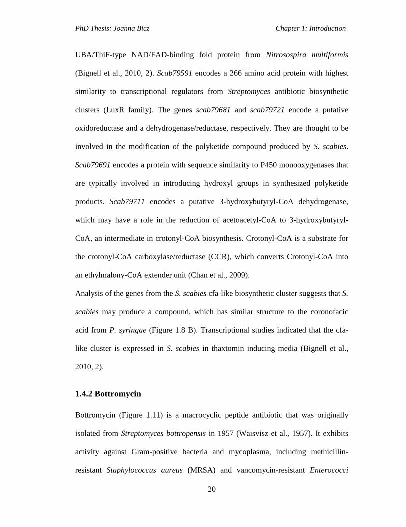
Figure 1.13 EIC for m/z = 405.2200 and 412.2300 (corresponding to the [M+2H]2+
ions for bottromycins A2 and C2, respectively) from LC-MS analyses of culture
supernatants of S. bottropensis DSM40262 (top two traces) and EIC for m/z =
405.2228 and 412.2314 (corresponding to the [M+2H]2+
ions for bottromycins A2
and C2, respectively) from LC-MS analyses of culture supernatants of S. scabies
(bottom two traces; Gomez-Escribano et al., 2012).………………….....……….…24

PhD Thesis: Joanna Bicz List of Figures
vi
Figure 1.14 Proposed pathway for bottromycin biosynthesis in S. bottropensis and S.
scabies.........................................................................................................................26
Figure 1.15 Structures of several siderophores (chelating atoms are highlighted in
blue; Miethke and Marahiel, 2007).............................................................................27
Figure 1.16 Steps of nonribosomal peptide biosynthesis, domains: A – adenylation, T
(PCP) – thiolation, C – condensation, TE – thioesterase............................................29
Figure 1.17 The A domain reaction catalysis: the activation of the amino acid
substrate through the formation of an aminoacyl adenylate followed by the covalent
binding via a thioester bond to the 4’Ppant cofactor of the
PCPdomain.................................................................................................................30
Figure 1.18 Reaction catalysed by PPTases. The apo is the inactive form, lacking the
phosphopantetheinyl arm and the holo is the active form..........................................31
Figure 1.19 The catalytic mechanism of condensation (C) domains..........................31
Figure 1.20 A – The release of peptide from NRPS catalysed by TE domain….......32
Figure 1.21 Biosynthetic pathway of enterobactin. Reactions catalysed by EntE and
EntF enzymes in the assembly of an L-N-(2, 3-dihydroxybenzoyl) serinyl thioester
intermediate (Oves-Costales et al., 2009). ICL (EntA)-Isochorismate lyase…...…..34
Figure 1.22 Biosynthetic pathway of enterobactin. Oligomerization and
macrocyclization reactions catalyzed by the TE domain of EntF from the L-N-(2, 3-
dihydroxybenzoyl) serinyl thioester intermediate…………………………………..35
Figure 1.23 Proposed pathway for aerobactin biosynthesis. A-Biosynthesis of N6-
acetyl-N6-hydroxylysine (AHL) from L-lysine catalysed by IucB and IucD. B-
biosynthesis of aerobactin catalysed by IucA and IucC (Challis, 2005)....................37
Figure 1.24 Aerobactin biosynthetic gene cluster…………………………………..38
Figure 1.25 Structure of pyochelin (Cox, 1982). .......................................................40
Figure 1.26 Organization of the PBGC in P. aeruginosa PAO1................................40
Figure 1.27 Biosynthesis of pyochelin. NRPS domains: T-thiolation, Cy-
heterocyclization, A-adenylation, MT-methyltransferase, TE-thioesterase…….......42
Figure 1.28 Organisation of pyochelin biosynthetic gene clusters in S. scabies 87.22,
P. aeruginosa PAO-1and in P. fluorescensPf-5 (Seipke et al., 2011, Youard et al.,
2007)…………………………………………...........................................................43
Figure 1.29 Stereoisomers of pyochelin (P. aeruginosa) and ent-pyochelin (P.
fluorescens)……………………………………………………………………….....46
Figure 1.30 Structure of desferrioxamines E, B and G1............................................48
Figure 1.31 Figure 1.31 Organistation of the gene cluster directing desferrioxamine
biosynthesis and ferrioxamine uptake in S. coelicolor M145 and in S. scabies 87.22..
....................................................................................................................................49
Figure 1.32 Proposed pathways for desferrioxamine E and B biosynthesis. Proposed
pathway for the biosynthesis of AHC and HSC. B-Biosynthesis of desferrioxamine E
by DesD. C- Biosynthesis of desferrioxamine B by DesD (Kadi et al., 2007)...........52
PhD Thesis: Joanna Bicz List of Figures
vii
Figure 1.33 Organization of the scabichelin biosynthetic gene cluster in S. scabies
87.22...........................................................................................................................53
Figure 1.34 Organisation of modules and domains of the NRPS encoded by
scab85471...................................................................................................................56
Figure 1.35 Structures of tetronate polyketides..........................................................57
Figure 1.36 Reaction catalysed by AT domains. A-loading. B-chain extension........59
Figure 1.37 Reaction catalysed by KS domain...........................................................59
Figure 1.38 Reaction catalysed by KR domain..........................................................60
Figure 1.39 Reaction catalysed by DH domain..........................................................60
Figure 1.40 Reaction catalysed by ER domain...........................................................60
Figure 1.41 RK-682 biosynthetic gene cluster in Streptomyces sp. 88-682…...........62
Figure 1.42 Proposed biosynthetic pathway of RK-682……………………..….......63
Figure 1.43 Structure of agglomerin A.......................................................................64
Figure 1.44 Cryptic polyketide tetronate biosynthetic gene cluster in S. scabies and
RK-682 biosynthetic gene cluster in Streptomyces sp. 88-682 (Challis,
unpublished)................................................................................................................65
Figure 1.45 Proposed biosynthetic pathway for cryptic tetronate product in S. scabies
87.22...........................................................................................................................67
Figure 2.1 Organization of the PBGCs in S. scabies 87.22 and P. aeruginosa
PAO1………………………………………………………………………………..72
Figure 2.2 A proposed pathway for pyochelin biosynthesis in S. scabies 87.22 based
on analysis of biosynthesis by ortologous proteins from P. aeruginosa....................74
Figure 2.3 RT-PCR analysis of ΔScab1401 mutant strain showing that Scab1401
represses transcription of S. scabies 87.22 pyochelin biosynthetic genes. Lane 1- S.
scabies wild-type cDNA, 2- ΔScab1401, 3- ΔScab1401 with pAU3-45 or 4-
pRFSRL34 (plasmids containing the copy of scab1401 gene) grown in liquid
minimal medium+100 mM FeCl3 was used as template for PCRs. The murX gene -a
loading control (Seipke et al., 2011)…………...........................................................75
Figure 2.4 A- Measured mass spectrum of pyochelin from Δscab1401 supernatant
(top panel), pyochelin standard (middle panel), and ent-pyochelin (bottom
panel)..........................................................................................................................78
Figure 2.4 B- Extracted Ion Chromatograms (EICs) at m/z=325 from analyses of ent-
pyochelin standard (top trace), pyochelin standard (middle trace) and S. scabies
pyochelin (bottom trace).............................................................................................78
Figure 2.4C- Pyochein and ent-pyochelin stereoisomers. Metal induced shift at C-2’’
results in isomerisation of pyochelin (Ino, and Murabayashi, 2001; Schlegel et al.,
2004)...........................................................................................................................79
Figure 2.5 EICs at m/z= 325 from analyses of supernatants from the Δscab1401….81
Figure 3.1 Map of pIJ773 (Gust et al., 2003)……………….....................................79
Figure 3.1 Map of pIJ773 (Gust et al., 2003).............................................................84
PhD Thesis: Joanna Bicz List of Figures
viii
Figure 3.2 Design of PCR primers for making a gene replacement (Gust et al., 2002).
....................................................................................................................................85
Figure 3.3 Creation of a gene deletion in the cosmid via homologous recombination
of the disruption cassette with cosmid DNA and then conjugation from E. coli into S.
scabies and double homologous recombination resulting in a replacement of the gene
of interest with the disruption cassette creating an S. scabies mutant. Yellow gene –
apramycin resistance, orange region – origin of transfer (oriT), green regions = FRT
sites.............................................................................................................................86
Figure 3.4 Structure of a novel siderophore scabichelin………................................88
Figure 3.5 Organization of the scabichelin biosynthetic gene cluster in S. scabies
87.22...........................................................................................................................89
Figure 3.6 A-Proposed role of the enzymes encoded by scab85521 and scab85511 in
the biosynthesis of the non-proteinogenic amino acids L-N5-hydroxyornithine (L-
hOrn) and L-N5-formyl-N5-hydroxyornithine (L-fhOrn). B-Organisation of modules
and domains of the NRPS encoded by scab85471. The residues attached to the
thiolation domains (black circles) of module 1, 2, 3 are the aminoacids predicted to
be incorporated into scabichelin by each A domain. The substrates loaded by A
domains of the modules 4 and 5 could not be unambiguously predicted. ……….....91
Figure 3.7 The enzymatic domains of scab85471. The fragment deleted is module
2..................................................................................................................................93
Figure 3.8 Restriction analysis of the wild type S57 cosmid. Lanes from left to
right:M- 1kb DNA molecular size marker, BamHI, PstI, NotI, NcoI
digests.........................................................................................................................93
Figure 3.9 Restriction digest pattern of scabichelin cosmid (S57) with BamH, PstI,
NotI and NcoI restriction enzymes (the band sizes in bp that are displayed red in the
diagram indicate bands present on the gel picture in figure
3.7)…..........................................................................................................................94
Figure 3.10 Restriction analysis of wild type vs mutagenised S57 cosmid
(S57::scab85471::apr-OriT. Lanes: M- 1kb DNA molecular size marker, WT- wild
type S57 cosmid BamHI cut, Δ- cosmid + oriT-apramycin cassette BamHI
cut................................................................................................................................95
Figure 3.11 PCR analyses of the wild type and the mutagenised S57 cosmid with the
test primers. Lanes: M- 1kb DNA molecular size marker, WT gDNA- PCR band for
the wild type S. scabies DNA, WT cos- PCR band for the wild type S57cosmid, Δ
cos-PCR band for the S57 cosmid + oriT-apramycin cassette……….......................96
Figure 3.12 PCR analysis of the genomic DNA from the putative scabichelin mutant.
Lanes: M- 1kb DNA molecular size marker, WT cos- PCR band for the wild type
scabichelin cosmid, Δ cos-PCR band for the scabichelin cosmid with OriT-
apramycin cassette, Δ gDNA- PCR for the for the scabichelin mutant genomic DNA.
....................................................................................................................................97
PhD Thesis: Joanna Bicz List of Figures
ix
Figure 3.13 Southern blot hybridization using labelled S57::oriT-apr cosmid as a
probe confirming the nature of S.scabiesW100 mutant with 3 kb fragment deleted
within scab85471 gene. Bands in white colour are all present in the S.scabies wild
type and the mutant. Band 7827 bp in green is only present in the wild type strain.
Bands 1412 bp and 7575 bp coloured in red are characteristic for the mutant.
....................................................................................................................................98
Figure 3.14 EICs at m/z=351.14 (corresponding to the [M+2H]2+
ion for the ferric-
scabichelin) from analyses of the culture supernatant of S. scabies 87.22 (top panel),
Fe-scabichelin purified from S. antibioticus (middle panel) and the culture
supernatant of S. scabies W1000 (bottom panel)………...........................................99
Figure 3.15 MS/MS spectra of ferric-scabichelin purified from S. antibioticus (top
panel) and from ferrated supernatants from iron-deficient cultures of S. scabies 87.22
(bottompanel)............................................................................................................100
Figure 3.16 Structures of coelichelin and a novel siderophore scabichelin..............101
Figure 3.17 A-putative scabichelin biosynthetic gene cluster. B-Organisation of the
coelichelin biosynthetic gene cluster and the module and domain organisation of the
NRPS encoded by cchH. The residues attached to the thiolation domains (black
circles) of module 1, 2, 3 are the aminoacids predicted to be incorporated into
coelichelin by each A domain...................................................................................102
Figure 3.18 Proposed coelichelin biosynthetic pathway (Lautru et al., 2005). The
stages 1-5 are described in the text. The domains active in each step are shaded gray.
The black circles are the thiolation domains in each module of CcH......................104
Figure 3.19 A-Proposed role of the enzymes encoded by scab85521 and scab85511
in the biosynthesis of the non-proteinogenic amino acids L-N5-hydroxyornithine (L-
hOrn) and L-N5-formyl-N5-hydroxyornithine (L-fhOrn). B-Organisation of modules
and domains of the NRPS encoded by scab85471. The residues attached to the
thiolation domains (black circles) of the five modules are the aminoacids proposed to
be incorporated into scabichelin………………………………………………..….107
Figure 3.20 Amychelin biosynthetic gene cluster in Amycolatopsis sp. AA4
(Seyedsayamdost at al., 2011)……………..............................................................108
Figure 3.21 Proposed biosynthesis of amychelin (Seyedsayamdost at al.,
2011).........................................................................................................................111
Figure 4.1 Putative desferrioxamine biosynthetic gene cluster in S. scabies
87.22……………………………………………………………………………….113
Figure 4.2 Proposed desferrioxamine biosynthetic pathway in S. coelicolor (Kadi et
al., 2007)…………………………………………………………………………...115
Figure 4.3 PCR analysis of genomic DNA from the putative desC mutant colony.
Lanes M1- i: (M1) Middle Range DNA molecular size marker (M2) 1kb DNA
molecular size marker, (a, b, c) desC mutant genomic DNA Tm= 57, 61, 63, (d, e, f)
desC cosmid with apramycin cassette, ( g, h, i) wild type S. scabies 87.22 genomic
DNA………………………………………………………………………………..117

PhD Thesis: Joanna Bicz List of Figures
x
Figure 4.4 Southern blot hybridization using labeled des::oriT-apr cosmid as a probe
confirming the nature of S.scabies desC mutant. Band 14716 bp in green is only
present in wild type strain. Bands 1412 bp and 7575 bp coloured in red are
characteristic for the mutant.....................................................................................118
Figure 4.5 A – EIC (m/z = 614) for ferrioxamine B from LC-MS analyses of
acidified organic extracts from the S. scabies wild type (green) and S. scabies desC
mutant (blue). B – EIC (m/z = 614) for ferrioxamine E from LC-MS analyses of
acidified organic extracts from the S. scabies wild type (red) and S. scabies desC
mutant (green)...........................................................................................................120
Figure 4.6 Pathway for the biosynthesis of desferrioxamines B, G1 and E (Kadi et
al., 2007).A-reactions catalysed by DesA, DesB enzymes and reaction proposed to
be catalysed by DesC enzyme. B-Reaction catalysed by DesD enzyme. yielding
desferrioxamine B, G1 and E.............................…………………………………...122
Figure 4.7 EIC (m/z = 614) for ferrioxamine B from LC-MS analyses of acidified
organic extracts from the S. scabies wild type (top trace), S. scabies desC mutant
(middle trace) and Δ desC mutant + HSC (bottom trace).........................................123
Figure 4.8 Map of pOSV556 plasmid……………………………………………...124
Figure 4.9 Left- a- an XhoI-HindIII restriction digest and b- PCR analysis with desC
gene cloning primers of pOSV556::desC plasmid. Right-PCR analysis with desC
gene cloning primers of: c- S. scabies wild type chromosomal DNA, d- S. scabies
ΔdesC mutant DNA and e- S. scabies ΔdesC::pOSV556::desC complemented
mutant. M- 1 kb molecular DNA ladder.....................................................………..125
Figure 4.10 EICs (m/z = 614) for ferrioxamine B from LC-MS analyses of acidified
organic extracts from the S. scabies wild type (top trace), S. scabies desC mutant
(middle trace) and Δ desC ::pOSV556/63021::apr (bottom trace)...........................126
Figure 5.1 Cryptic tetronate polyketide biosynthetic gene cluster in S.
scabies……………………………………………………………………………...131
Figure 5.2 The 1% agarose gel of total RNA extracted from wild type S. scabies.
Lanes 1-4: 1- RNA extracted from 3 days old mycelium; 2- RNA extracted from 5
days old mycelium; 3- RNA extracted from 7 days old mycelium; 4- RNA extracted
from 14 days old mycelium.23S and 16S are two bands of total RNA...................133
Figure 5.3 RT-PCR analysis of S. scabies wild type RNA. A- the putative tetronate
gene cluster in S. scabies 87.22; B- RT-PCR: 1- with hrdB primers (positive control);
2- with scab62881 primers; 3- with scab62951 primers. 3, 5, 7, 14- the S. scabies
cells were grown for 3, 5, 7 and 14 days before the RNA
extraction……………………………………………………..........................…....134
Figure 5.4 Map of pUC57/scab63021. Legend: oriT-origin of replication, ampR-
ampicillin resistance gene.........................................................................................137
Figure 5.5 PCR analysis of genetically-engineered pUC57/scab63021 plasmid with
test primers. Lanes: M-b. M- 1 kb molecular weight marker, a- PCR band for
unmutagenised pUC57/scab63021, b- PCR for pUC57/scab63021::oriT-

PhD Thesis: Joanna Bicz List of Figures
xi
apra...........................................................................................................................138
Figure 5.6 Agarose gel electrophoresis analysis of restriction enzymes digest of
genetically-engineered pUC57/scab63021 plasmid used to disrupt the scab63021 in
the S. scabies genome within putative tetronate –like cluster. Lanes M-d: M- 1kb
molecular weight marker, a- undigested pUC57/scab63021::oriT-apra, b- digestion
with EcoRI, c- digestion with BamHI, d- digestion with SacI…………………….139
Figure 5.7 A- PCR analysis of genetically-engineered pUC57/scab63021 plasmid
and genomic DNA of scab63021 mutant with oriT- apramycin casette internal
primers; M-1 kb molecular weight marker, a –PCR of genomic DNA of Δscab63021
mutant; b- pUC57/scab63021::apra plasmid with apramycin internal primers. B-PCR
analysis of putative scab63021 mutant genomic DNA with test primers………….140
Figure 5.8 RT-PCR analysis of S. scabies wild type and Δscab63021 RNA, A: RT-
PCR control, 1317bp product size (hrdB primers); B: RT-PCR with scab62951
primers, 615 bp product size; C: RT-PCR with scab62881 primers, 617 bp product
size.………………………………...................................................................……142
Figure 6.1 Structures of investigated siderophores………………………...……....145

PhD Thesis: Joanna Bicz List of Tables
xii
List of Tables
Table 1.1 Overview of S. scabies known and putative natural product biosynthetic
gene clusters (Yaxley, 2009). Yellow- biosynthetic gene clusters conserved both in S.
coelicolor and S. avermitilis; white-biosynthetic gene clusters conserved either in S.
coelicolor or in S. avermitilis; blue- clusters conserved in neither of the two
species………………………………………………………….................................14
Table 1.2 Genes and proteins involved in bottromycin biosynthesis and their putative
functions (adapted from Gomez-Escribano et al., 2012)…………………………....23
Table 1.3 Putative proteins function encoded by the desferrioxamine biosynthetic
gene cluster in S. scabies 87.22…………………………………………………......50
Table 1.4 Putative proteins function encoded by the putative novel hydroxamate
siderophore biosynthetic gene cluster in S. scabies
87.22…………………...................................................................................………54
Table 1.5 Predicted substrate specificity of adenylation domains in the Scab85471
protein……………………………………………………….………………………56
Table 1.6 Putative proteins function encoded by the putative tetronate biosynthetic
gene cluster in S. scabies 87.22…………………………………………..…………66
Table 4.1 Putative proteins function encoded by the desferrioxamine biosynthetic
gene cluster in S. scabies 87.22…………………………………………..……......114
Table 7.1General stock solutions………………………………...………………..157
Table 7.2 Antibiotics stock solutions………………………….…….....……….....158
Table 7.3 The E. coli strains used……………………………..……………..........158
Table 7.4 The S. scabies strains used………………………………………….......159
Table 7.5 Plasmids and cosmids used……………………………………..............160
Table 7.6 Oligonucleotide primers used...................................................................161
Table 7.7 RT-PCR primers used...............................................................................162
Table 7.8 PCR reaction conditions……………………………....………..…….…174
Table 7.9 RT-PCR reaction conditions…………………………...…………....…..184
Table 7.10 Gradient elution profile used in LC-MS analyses of pyochelin
production................................................................................................................186

PhD Thesis: Joanna Bicz List of Tables
xiii
Table 7.11 Gradient elution profile used in LC-MS analyses of pyochelin
production.................................................................................................................187
PhD Thesis: Joanna Bicz Acknowledgements
xiv
Acknowledgements
Firstly I would like to thank Professor Greg Challis, who gave me the opportunity to
work on this interesting project and for all his help and advice throughout the whole
period of my PhD. I would also like to thank all the members of the Challis’ group
for their support and creation of great working atmosphere. I would also like to thank
Dr. Paulina Sydor for her guidance throughout duration of my studies, especially
during the first year. To Dr. Christoph Corre, Dr. Nadia Kadi and Dr. Orestis Lazos
for their tips on molecular biology work and to Dr Lijiang Song and Nicolas for all
their help with chemistry. To Dr. Sarah Barry, Lauren, Lona and Dr. Orestis Lazos
for proof-reading of my thesis; to Mansoor and David and to all other group
members past and present. Big thanks also for our technician Anne for dealing with
many practical matters in the lab and for keeping it running on a daily basis.
A very special thank you goes to all the friends I have met while at Warwick, which
helped me greatly during the times of my studies, and for their encouragement. A
special thank you also goes to my friend Ifti for all the support at the time I needed it.
Finally, I would like to thank the Warwick University and US Department of
Agriculture for providing funding for this work.

PhD Thesis: Joanna Bicz Declaration
xv
Declaration
Experimental work contained in this thesis is original research carried out by the
author, unless otherwise stated, in the Department of Chemistry at the University of
Warwick, between October 2008 and November 2011. No material contained herein
has been submitted for any other degree, or at any other institution.
Results from other authors are referenced in the usual manner throughout the text.
_________________________________ Date: ___________________
Joanna Bicz

PhD Thesis: Joanna Bicz Abstract
xvi
Abstract Streptomyces are Gram-positive bacteria, usually found living within the soil and
they are saphrophytes. Among this class of bacteria are some plant pathogenic
species, which cause infection of the roots or the tubers of some plants. The model
Streptomycete plant pathogen is Streptomyces scabies; this infects root crops, such as
potato or radish and is a known cause of scab disease. Most Streptomyces species are
producers of secondary metabolites, many of which possess important biological
activities, such as antibacterial, iron-chelating, anticancer or immunosuppressant.
One group of these secondary metabolites are called siderophores. These are small
organic molecules, which can chelate ferric iron. The iron in the environment is
mainly present as iron (III) hydroxide, which is not very water soluble and cannot,
therefore, be taken up directly by microorganisms. Some bacteria solve this problem
through production of siderophores. The siderophores are released into the
environment by the microorganisms to chelate iron (III) from the environment and
transport it into the cell across the cell membrane. Iron is required for many life
processes.
Analysis of the Streptomyces scabies genome sequence resulted in the identification
of gene clusters predicted to direct the biosynthesis of known siderophores, e.g.
desferrioxamines and pyochelin, as well as, potentially novel siderophores. A gene
inactivation and comparative metabolic profiling approach has been employed to
identify the metabolic products of these gene clusters.
A PCR-targeting method was used to replace part of or whole genes in the S. scabies
87.22 putative secondary metabolite gene clusters. An internal fragment of the
scabichelin biosynthetic gene scab85471 and the putative S. scabies desC gene were
deleted using this method. The scabichelin and desC gene mutants were subsequently
analysed by LC-MS allowing confirmation of the function of the genes investigated.
Production of scabichelin by S. scabies 87.22 wild type was analysed by comparing
it with the authentic standard. The chemical and genetic complementation of the Δ
desC mutant was carried out to establish the involvement of the desC gene in the
biosynthesis of desferrioxamines.
The S. scabies 87.22 cryptic tetronate biosynthetic gene cluster predicted to encode a
novel agglomerin-like product, which could potentially be involved in plant
pathogenicity was also investigated. The expression of the gene cluster was first
analysed using reverse transcriptase PCR (RT-PCR) which was carried out on the
total RNA isolated from the wild type S. scabies. Following this, an attempt was
made to disrupt the scab63021 gene, a putative transcriptional activator of the cryptic
tetronate-like cluster in the S. scabies genome. Transcriptional analysis of the wild
type S. scabies and the putative Δscab63021 mutant genomes did not show any
difference in the expression of the tetronate genes between the wild type strain and
the Δ scab63021 mutant.

PhD Thesis: Joanna Bicz Abbreviations
xvii
Abbreviations
A Adenylation (domain)
aa amino acid
ACP Acyl Carrier Protein
ADP Adenosine diphosphate
amp Ampicillin
apra Apramycin
AT Acyl Transferase (domain)
ATP Adenosine triphosphate
BLAST Basic Local Alignment Search Tool
bp Base pairs
C Condensation (domain)
CoA Coenzyme A
DH Dehydratase (domain)
DNA Deoxyribonucleic acid
E Epimerisation (domain)
EDTA Ethylenediaminetetraacetic acid
EIC Extracted ion chromatogram
ER Enoyl Reductase (domain)
ESI-MS Electrospray ionization – Mass spectrometry
EtOAc ethyl acetate
EtOH Ethanol
FAD Flavin Adenine Dinucleotide
FRT FLP recognition targets

PhD Thesis: Joanna Bicz Abbreviations
xviii
GC Guanine-Cytosine
L-fhOrn L-N5-formyl-N5-hydroxyornithine
L-hOrn L-N5-hydroxyornithine
HPLC High Pressure Liquid Chromatography
HRMS High Resolution Mass Spectrometry
hyg Hygromycin
kan Kanamycin
kb kilo base pairs
KR Ketoreductase (domain)
KS Ketosynthase (domain)
LB Luria-Bertani (Medium)
LC (High Pressure) Liquid Chromatography
LC-MS Liquid Chromatography – Mass Spectrometry
Lys lysine
M Methylation (domain)
Me Methyl
min. minute
MS Mass Spectroscopy
NADH Nicotinamide adenine dinucleotide
NADPH Nicotinamide adenine dinucleotide phosphate
NMR Nuclear Magnetic Resonance
NRPS Nonribosomal peptide synthetase
nt Nucleotide
OBB Oat Bran Broth (medium)
PhD Thesis: Joanna Bicz Abbreviations
xix
OD Optical Density
ORF Open Reading Frame
oriT Origin of Transfer
PBGC pyochelin biosynthetic gene cluster
PCR Polymerase Chain Reaction
PCP Peptidyl Carrier Protein
PKS Polyketide synthase
PLP Pyridoxal phosphate
PPTase Phosphopantethienyl transferaze
R resistance
RBS Ribosome binding site
rpm revolutions per minute
RT-PCR Reverse Transcriptase Polymerase Chain Reaction
S sensitive
SAM S-adenosylmethionine
Ser serine
SFM Soya Flour Mannitol medium
SMM Supplemented Minimal Medium
T Thiolation (domain)
Taq Thermus aquaticus (polymerase)
TBE Tris-boric acid EDTA buffer
TE Thioesterase (domain)
tet tetracycline
TO Time-of-flight

PhD Thesis: Joanna Bicz Abbreviations
xx
Tris Tris(hydroxymethyl)aminomethane
UV ultra-violet

PhD Thesis: Joanna Bicz Chapter 1: Introduction
1
1. Introduction
PhD Thesis: Joanna Bicz Chapter 1: Introduction
2
1.1 Streptomyces
Streptomyces belong to the phylum of Actinobacteria and to the family of
Streptomycetaceae (Kampfer, 2006). Streptomyces are Gram-positive bacteria, their
genomes contain a high content of guanine and cytosine and they have a linear
chromosome. They live mainly in soil, where they decompose organic matter to
produce their characteristic earthy odour of which the main compound is volatile
substance called geosmin (Meyer et al., 1996). Most Streptomyces species produce
biologically active secondary metabolites and more than two thirds of clinically used
antibiotics of microbial origin are produced by Streptomyces spp. The biological
activities of secondary metabolites also include antimalarial, antifungal and
antitumour, as well as some immunosuppressants (Kieser et al., 2000). Some
Streptomyces spp. are pathogenic, for example, S. somaliensis and S. sudanensis
cause mycetoma in humans (Lichon et al., 2006) ; and S. acidiscabies, S.
turgidiscabies and S. scabies cause scab disease in plants (Bignell et al., 2010, 2).
Streptomyces have a complex life cycle (Figure1.1). It begins with the single spore,
which germinates to form several multi-nucleoid filaments. These filaments
elongate, producing branches, which form hyphae known as vegetative mycelia.
After several days the vegetative mycelia grow into air forming aerial hyphae. When
the growth of the aerial hyphae is completed it undergoes septation and produces
prespore compartments. The spores are assigned into chains and in this step, which is
known as a stationary phase, secondary metabolites are produced. The mature spores
are then released to start a new life cycle (Kieser et al., 2000).

PhD Thesis: Joanna Bicz Chapter 1: Introduction
3
Figure 1.1 The life cycle of Streptomyces (Angert, 2005).
1.2 Streptomyces scabies
There are over 900 Streptomyces species described to date (Labeda et al., 2012), and
these include some plant pathogenic species. Three species are Streptomyces scabies
and S. turgidiscabies and S. acidiscabies, which cause infection of root vegetables,
such as potato or radish, known as scab disease (Loria and Kempter, 1986; Figure
1.2). Streptomyces scabies was one of the first plant pathogens characterised and has
a broad world distribution, whereas S. turgidiscabies and S. acidiscabies species

PhD Thesis: Joanna Bicz Chapter 1: Introduction
4
have been described more recently and have been found only in several isolated
world areas (Bignell et al., 2010, 1).
Figure 1.2 Potato and radish with scab disease (http://www.cals.ncsu.edu,
http://www.agroatlas.ru).
Some of the secondary metabolites produced by S. scabies were known prior to the
availability of its genome sequence, including thaxtomins and concanamycins (Kers
et al., 2004, Natsume et al., 2005).
One of the first reports of secondary metabolites produced by scab causing
streptomycetes was thaxtomins, a family of phytotoxins isolated from S. scabies in
1989 (King et al., 1989). It was also shown that S. scabies produces other
phytotoxins, such as concanamycin A and B (Natsume et al. 1996, Natsume et al.
1998), a macrolide antibiotic first isolated from Streptomyces diastatochromogenes
S-45 (Kinashi et al., 1984).

PhD Thesis: Joanna Bicz Chapter 1: Introduction
5
1.3 Secondary metabolites of S. scabies 87.22
1.3.1 Thaxtomins
The plant pathogenic Streptomyces species produce the family of phytotoxins called
thaxtomins (Figure 1.3; Loria et al., 2008, Fry and Loria, 2002; Johnson et al., 2009).
These toxins, present in infected host tissue (Lawrence et al., 1990), are nitrated
dipeptides biosynthesised from L-phenylalanine and L-tryptophan. The 4-nitroindole
moiety is required for phytotoxicity (King and Calhoun, 2009). To date, eleven
members of the thaxtomin family have been identified, which differ only in the
presence or absence of an N-methyl group and the pattern of hydroxylation.
Thaxtomin A is the most abundant member of the thaxtomin family (Figure 1.3 B)
and is produced by all three plant pathogenic Streptomyces species, S. scabies, S.
acidiscabies and S. turgidiscabies (Loria et al., 2008), whereas the main product of S.
ipomeae is thaxtomin C (King et al., 1994). Thaxtomin A is essential for
development of scab disease in plant tissue (King and Calhoun, 2009). The main
mode of action of thaxtomins is inhibition of cellulose biosynthesis in growing plant
tissue, causing hyperthropy of plant cells, tissue necrosis and cell death (Loria et al.,
2008).
Thaxtomin is biosynthesised by non-ribosomal peptide synthetases (NRPSs) encoded
by the txtA and txtB genes (Figure 1.3 A). TxtA and TxtB are responsible for
producing the cyclic dipeptide from L-4-nitro-tryptophan and L-phenylalanine. The
region close to the txtA and txtB genes indicated the presence of a gene highly
homologous to the oxygenase domain of mammalian nitric oxide synthases (NOSs,
Kers et al., 2004). This gene encodes the enzyme TxtD, which provides the nitric
PhD Thesis: Joanna Bicz Chapter 1: Introduction
6
oxide and a cytochrome P450, TxtE, uses it to nitrate L-tryptophan to give the
precursor L-4-nitro-tryptophan (Figure 1.3 B; Barry et. al., 2012) A cytochrome
P450 monooxygenase TxtC is responsible for post-cyclization hydroxylation steps
on the thaxtomin precursor to yield thaxtomin A (Healy et al., 2002).
L-4-nitro-tryptophan
NH
CO2H
NH2
N OTxtD
NH
NH2
HO2C
NH2
NH
NH
NH2
HO2C
NH2
O
O2
TxtE O2
NH
NH2
N
O
O
OH
OOH
NH2
O
TxtA/B
NH
N
O
O
MeN
NMe
O
O
TxtC O2
NH
N
O
O
MeN
NMe
O
O
OH
OH
TxtC O2
NH
N
O
O
MeN
NMe
O
O
OH
OH
OH
L-phenylalanine
L-tryptophan
Thaxtomin A
B
Figure 1.3 A- thaxtomin biosynthetic gene cluster and, B- thaxtomin A biosynthesis
(Barry et al, 2012).

PhD Thesis: Joanna Bicz Chapter 1: Introduction
7
1.3.2 Concanamycins
Concanamycins are macrolide antibiotics primarily isolated from S.
diastatochromogenes (Kinashi et al., 1984), consisting of an 18-membered macrolide
ring and a β-hydroxyhemiacetal side chain (Figure 1.4; Haydock et al., 2005). They
have a broad range of biological activities, such as antiviral, antiprotozoal,
antifungal, and antineoplastic (Kinashi et al., 1984; Omura et al., 1982; Seki-Asano,
1994). Their activity is thought to be due to their inhibition of vacuolar ATP-ases (V-
ATP-ases; Drose et al., 1993), the role of which is to actively transport protons into
the lumen of various cellular compartments such as endosomes, lysosomes and the
vacuoles of plants and fungi (Mellmann et al., 1986; Nelson, 1995). Concanamycins
inhibit the acidification of the lysosomes by binding to the V0 membrane component
of the ATP-ase (Drose et al., 2001). Research into these compounds has increased
since it was discovered that they may have some application in the treatment of
osteoporosis, as V-type ATP-ases play a key role in osteoclast-mediated bone
resorption (Laitala-Leinonen et al., 1999).
O
OO
HOCO
O
H2N
OOH
H3COOH
OCH3
O OH
HO
Figure 1.4 Structure of concanamycin A (Haydock et al., 2005).
PhD Thesis: Joanna Bicz Chapter 1: Introduction
8
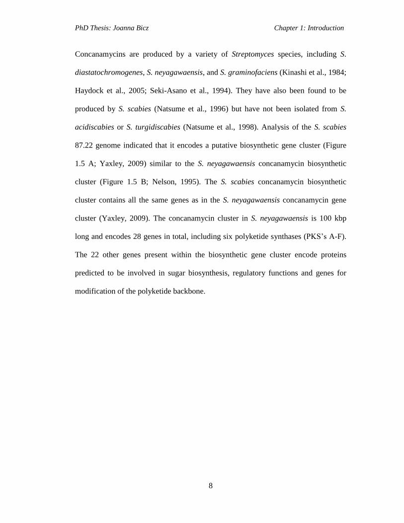
Concanamycins are produced by a variety of Streptomyces species, including S.
diastatochromogenes, S. neyagawaensis, and S. graminofaciens (Kinashi et al., 1984;
Haydock et al., 2005; Seki-Asano et al., 1994). They have also been found to be
produced by S. scabies (Natsume et al., 1996) but have not been isolated from S.
acidiscabies or S. turgidiscabies (Natsume et al., 1998). Analysis of the S. scabies
87.22 genome indicated that it encodes a putative biosynthetic gene cluster (Figure
1.5 A; Yaxley, 2009) similar to the S. neyagawaensis concanamycin biosynthetic
cluster (Figure 1.5 B; Nelson, 1995). The S. scabies concanamycin biosynthetic
cluster contains all the same genes as in the S. neyagawaensis concanamycin gene
cluster (Yaxley, 2009). The concanamycin cluster in S. neyagawaensis is 100 kbp
long and encodes 28 genes in total, including six polyketide synthases (PKS’s A-F).
The 22 other genes present within the biosynthetic gene cluster encode proteins
predicted to be involved in sugar biosynthesis, regulatory functions and genes for
modification of the polyketide backbone.

PhD Thesis: Joanna Bicz Chapter 1: Introduction
9
Figure 1.5 Organization of the concanamycin biosynthetic gene cluster in: A-
Streptomyces neyagawaensis ATCC 27449 (Kinashi et al., 1984). The genes
functions are described in the text. B- S. scabies 87.22 (Yaxley, 2009).
Concanamycin A contains deoxysugar moiety 4’-O-carbamoyl-2’-deoxyrhamnose,
the biosynthesis and attachement of which is encoded for by seven genes ORF5*-
ORF11*. ORF5* and ORF6* encode proteins with high homology to a TDP-glucose
4, 6-dehydratase and TDP glucose synthase, respectively (Figure 1.5). ORFs 8*, 9*
and 10* encode proteins with high homology to a dTDP-hexose 4-ketoreductase, d-

PhD Thesis: Joanna Bicz Chapter 1: Introduction
10
TDP-sugar-3-ketoreductase and dTDP-sugar-2, 3-dehydratase respectively. ORF7*
is highly homologous to carbamoyltransferases and ORF11* is homologous to
glycosyltransferases. Assignment of the genes allows predictions to be made with
regards to the biosynthetic route towards the deoxysugar moiety. ORF6* is proposed
to activate glucose and ORF5* to catalyze 4, 6-dehydration of TDP glucose to form
TDP-4-keto-6-deoxyglucose (Figure 1.6). ORF10* is then proposed to catalyse the
formation of the 2-deoxy intermediate, which is in equilibrium with the 2, 4-diketo
derivative. The ORFs 8*, 9* are then proposed to reduce each keto group at C3 and
C4 to give the 2-deoxy derivative with the appropriate stereochemistry at the C3 and
C4 positions. ORF7* is proposed to catalyse the addition of a carbamoyl unit to form
4’-O-carbaomyl-2’-deoxyrhamnose. ORF11* is then predicted to catalyse the
transfer of the sugar moiety to concanamycin A aglycone (Figure 1.6).

PhD Thesis: Joanna Bicz Chapter 1: Introduction
11
O
OH
OHOPO3
2-
D-glucose1-phosphate
ORF6* O
OHOH
OHONDP
OH OH
ORF5* O
OH
OHONDP
O
ORF10*
NDP-4-keto-6-deoxy-D-glucose
OONDP
O
OH
Spontaneoustautomerisation
ORF8*
OONDP
O
O
O ONDP
O
OONDP
O
ORF9*
OHHO
ORF8* ORF9*
OONDP
OHORF11*
OONDP
OH
HOH2NOOC
ORF7* Sugar moietytransferred toconcanamycin A aglycone
4'-O-carbamoyl-2'-deoxyrhamnose
OH
Figure 1.6 Proposed biosynthetic pathway for synthesis and addition of the sugar
moiety 4′-O-carbamoyl-2′-deoxyrhamnose.
The regulatory functions are proposed to be encoded for by ORF’s 2, 3 and 17*
(Haydock et al., 2005). ORF3 has high sequence homology to members of the LuxR
regulatory family proteins, which are found in the secondary metabolite gene clusters
of many Gram-positive bacteria, and ORFs 2 and 17* are similar to the regulatory
genes in other PKS gene clusters. ORF17* has 33% identity to AfsR family regulator
from Streptomyces griseus (Haydock et al., 2005).
ORF1 is highly homologous to malonyl transferase enzymes and its function in the
concanamycin A gene cluster is unclear. A putative ORF4B*, located between
ORF4* and ORF5*, has no match to any other genes in the databases and its function
is unknown. ORFs 18* and 19* encode proteins proposed to be involved in

PhD Thesis: Joanna Bicz Chapter 1: Introduction
12
biosynthesis of extender units. The enzymes are predicted to be a crotonyl-CoA
reductase and a β-hydroxybutyryl-CoA reductase, respectively, which could both
play a role in the biosynthesis of a butyrate extender unit for module 10. The
extender unit selected for by modules 6 and 13 of the PKS could be a result of direct
incorporation of methoxymalonyl-ACP. ORF12*, is homologous to FkbG and may
be involved in the biosynthesis of methoxymalonyl-ACP. ORF1* is homologous to
FkbB, ORF2* to FkbJ, ORF3* to FkbI and ORF4* to FkbH. These proteins are
predicted to catalyse the conversion of primary metabolites into methoxymalonyl-
ACP.
The biosynthesis of the concanamycin A polyketide backbone in S. neyagawaensis is
directed by the operon of six genes conA to conF (Figure 1.7), which encode the
PKS enzymes CON1-6, respectively. CON1 (2841 aa, approx. 300 kDa) contains the
loading module and module 1 for chain extension. CON2 (4825 aa, approx. 500 kDa)
contains extension modules 2-4. CON3 (4970 aa, approx. 520 kDa) contains
extension modules 5–7, CON4 (4490 aa, approx. 430 kDa) contains modules 8–9
and CON5 (5599 aa, approx.530 kDa) contains modules 10–12. CON6 (2056 aa,
approx. 215 kDa) contains the final condensation module 13 and a C-terminal
thioesterase domain for cyclization and release of the polyketide chain (Figure 1.7;
Haydock et al., 2005). More about PKSs family proteins is discussed in detail in
section 1.8.

PhD Thesis: Joanna Bicz Chapter 1: Introduction
13
KS AT ACP KSDH KR
ACPKS AT KS
KRACP AT
KRACP KS AT ACP
KS AT KSKR
ACP ATKR
ACP KS ATDH KR
ACP KS ATDH KR
ACP ACPKS ATDH ER KR
KS AT KSKR
ACP ATKR
ACP KS ATDH KR
ACPKS AT
DH KRACP TE
CON1 CON2
CON3 CON4
CON5 CON6
loadmodule 1 module 2 module 3 module 4
module 5module 6
module 7 module 8module 9
module 10
module 11
module 12 module 13 end
Figure 1.7 Domain organisation of concanamycin A PKS genes (Haydock et al.,
2005).
1.4 Secondary Metabolites of S. scabies 87.22 Discovered by Genome
Mining.
S. scabies 87.22 is a model organism for studying plant pathogenicity in Gram-
positive bacteria (Lambert and Loria, 1989). The sequence of the genome of S.
scabies became available through the S. scabies genome-sequencing project
(www.sanger.ac.uk/Projects/S.scabies). The genome is 10.1 Mb long with a GC
content of 72%. The availability of the genome sequence for S. scabies allowed
comparative genomic analyses with other bacterial genomes, to identify several
biosynthetic gene clusters proposed to encode either known (such as
PhD Thesis: Joanna Bicz Chapter 1: Introduction
14
desferrioxamines, pyochelin) or novel (agglomerin-like product, coronofacic acid-
like product) secondary metabolites
(http://www.ncbi.nlm.nih.gov/nuccore/260644157,
http://strepdb.streptomyces.org.uk).
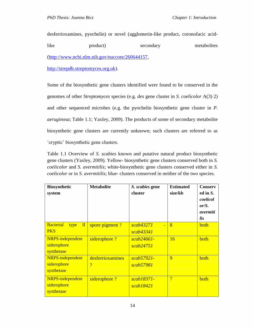
Some of the biosynthetic gene clusters identified were found to be conserved in the
genomes of other Streptomyces species (e.g. des gene cluster in S. coelicolor A(3) 2)
and other sequenced microbes (e.g. the pyochelin biosynthetic gene cluster in P.
aeruginosa; Table 1.1; Yaxley, 2009). The products of some of secondary metabolite
biosynthetic gene clusters are currently unknown; such clusters are referred to as
‘cryptic’ biosynthetic gene clusters.
Table 1.1 Overview of S. scabies known and putative natural product biosynthetic
gene clusters (Yaxley, 2009). Yellow- biosynthetic gene clusters conserved both in S.
coelicolor and S. avermitilis; white-biosynthetic gene clusters conserved either in S.
coelicolor or in S. avermitilis; blue- clusters conserved in neither of the two species.
Biosynthetic
system
Metabolite S. scabies gene
cluster
Estimated
size/kb
Conserv
ed in S.
coelicol
or/S.
avermiti
lis
Bacterial type II
PKS
spore pigment ?
scab43271 -
scab43341
8
both
NRPS-independent
siderophore
synthetase
siderophore ?
scab24661-
scab24751
16
both
NRPS-independent
siderophore
synthetase
desferrioxamines
?
scab57921-
scab57981
9
both
NRPS-independent
siderophore
synthetase
siderophore ? scab18371-
scab18421
7
both

PhD Thesis: Joanna Bicz Chapter 1: Introduction
15
Ectoine synthase
ectoine
compatible solute
scab70711-
scab70751
4
both
Hopene/squalene
synthase
pentacyclic
hopanoids
scab12951-
scab13061
14 both
Phytoene/polyprenyl
synthase
carotenoid
pigment
scab5431-
scab5511
12 both
Sesquiterpene
synthase
geosmin?
scab20121
2 both
Tyrosinase melanin pigment scab59231-
scab59241
2 both
Tyrosinase melanin pigment scab85681-
scab85691
2 both
Bacterial type III
polyketide synthase
germicidins? scab80171
4 in S.
coelicol
or
NRPS-independent
siderophore
synthetase
siderophore ? scab84501-
scab84521
8 in S.
avermiti
lis
NRPS unknown scab72991
4 in S.
avermiti
lis
Polysaccharide
exopolysaccharide scab23341-
scab23551
29 in S.
coelicol
or
NRPS pyochelin
siderophore ?
scab1411-
scab1571
35 no
NRPS NRPS
siderophore?
scab85431-
scab85521
42 no
NRPS lipopeptide?
scab19681-
scab19731
34 no
NRPS unknown
scab19681-
scab19731
8 no
NRPS thaxtomin
scab31761-
scab31841
19 no
Hybrid NRPS/PKS
system
unknown
scab62901-
scab63011
14 no
Hybrid NRPS/PKS
system
unknown
scab78961-
scab78981
8 no

PhD Thesis: Joanna Bicz Chapter 1: Introduction
16
Hybrid NRPS/PKS
system
unknown
scab43961
13 no
Bacterial type I PKS
macrolide
phytotoxin ?
scab83871-
scab84091
95 no
Bacterial type I PKS
coronofacic acid
derivative ?
scab79581-
scab79721
31 no
Class II DAHP
synthase/AfsA
homologue
butyrolactone ?
scab12021-
scab12111
12 no
Lantibiotic
lantibiotic ?
scab31961-
scab32051
14 no
Mixed unknown
scab63251-
scab63401
8 no
Mixed unknown
scab69771-
scab69871
13 no
Other bothromycin scab56711-
scab56591
18 no
1.4.1 Coronofacic acid
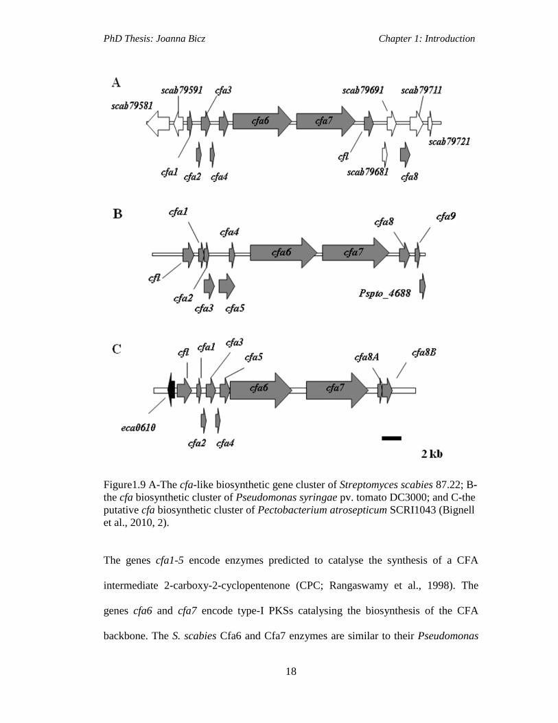
Analysis of the S. scabies 87.22 genome sequence revealed the presence of a
secondary metabolite gene cluster that is predicted to encode a compound
homologous to coronofacic acid (CFA). CFA forms part of coronatine (COR), a
phytotoxin produced by some plant pathogenic bacteria such as Pseudomonas
syringae (Figure 1.8 A; Bignell et al., 2010, 2). In this strain COR is an important
virulence factor and the cfa biosynthetic cluster is conserved in that strain as well as
in the plant pathogen, Pectobacterium atrosepticum. Analysis of genomes of
nonpathogenic Streptomyces (S. coelicolor, S. avermitilis, S. griseus) showed that
they do not encode the cfa biosynthetic cluster. As cfa biosynthetic cluster is
conserved amongst some plant pathogenic bacteria, it is possible that the S. scabies
cfa cluster may also be important for virulence in this strain.

PhD Thesis: Joanna Bicz Chapter 1: Introduction
17
H3C
CO
OH
O
H3C
CO
OH
O
A B
H H H H
Figure 1.8 A-Structure of coronafacic acid (CFA) from Pseudomonas syringae and,
B- A predicted structure for the CFA-like compound produced by S. scabies 87.22
(Bignell et al., 2010, 2).
The cfa biosynthetic cluster in S. scabies is 31 kb long and includes 15 genes (Figure
1.9; Bignell et al., 2010, 2), nine of which are homologous to those in the
Pseudomonas syringae and the Pectobacterium atrosepticum cfa clusters.
PhD Thesis: Joanna Bicz Chapter 1: Introduction
18
Figure1.9 A-The cfa-like biosynthetic gene cluster of Streptomyces scabies 87.22; B-
the cfa biosynthetic cluster of Pseudomonas syringae pv. tomato DC3000; and C-the
putative cfa biosynthetic cluster of Pectobacterium atrosepticum SCRI1043 (Bignell
et al., 2010, 2).
The genes cfa1-5 encode enzymes predicted to catalyse the synthesis of a CFA
intermediate 2-carboxy-2-cyclopentenone (CPC; Rangaswamy et al., 1998). The
genes cfa6 and cfa7 encode type-I PKSs catalysing the biosynthesis of the CFA
backbone. The S. scabies Cfa6 and Cfa7 enzymes are similar to their Pseudomonas

PhD Thesis: Joanna Bicz Chapter 1: Introduction
19
syringae and Pectobacterium atrosepticum analogs, however, the enoyl reductase
(ER2) domain in the S. scabies Cfa7 PKS is not present in the Pseudomonas
syringae and Pectobacterium atrosepticum equivalent PKS (Bignell et al., 2010, 2).
One more gene in the S. scabies cfa cluster that is homologous to the genes in the
Pseudomonas syringae and Pectobacterium atrosepticum cluster is coronofactate
ligase (cfl). Its function in Pseudomonas syringae is predicted to be ligation of CFA
to coronamic acid (CMA) to produce coronatine (Figure 1.10). Although cfl is
present in S. scabies and Pectobacterium atrosepticum these strains are not thought
to produce coronatine as they do not encode genes for CMA biosynthesis (Bell et al.,
2004).
H3C
O
H H
O
NHCH3
OH O
CFA CMA
Figure 1.10 Structure of the Pseudomonas syringae coronatine (COR) phytotoxin
consisting of coronafacic acid (CFA) linked to coronamic acid (CMA) (Bignell et al.,
2010, 2).
The six other genes that are encoded in the S. scabies biosynthetic cluster and which
have no homologues in Pseudomonas syringae or Pectobacterium atrosepticum
clusters are: scab79581, scab79591, scab79681, scab79691, scab79711, scab79721.
Scab79581 encodes a ~640 amino acid protein the function of which is unknown. It
has low homology to other proteins from the BlastP database. It is 25% identical to a
PhD Thesis: Joanna Bicz Chapter 1: Introduction
20
UBA/ThiF-type NAD/FAD-binding fold protein from Nitrosospira multiformis
(Bignell et al., 2010, 2). Scab79591 encodes a 266 amino acid protein with highest
similarity to transcriptional regulators from Streptomyces antibiotic biosynthetic
clusters (LuxR family). The genes scab79681 and scab79721 encode a putative
oxidoreductase and a dehydrogenase/reductase, respectively. They are thought to be
involved in the modification of the polyketide compound produced by S. scabies.
Scab79691 encodes a protein with sequence similarity to P450 monooxygenases that
are typically involved in introducing hydroxyl groups in synthesized polyketide
products. Scab79711 encodes a putative 3-hydroxybutyryl-CoA dehydrogenase,
which may have a role in the reduction of acetoacetyl-CoA to 3-hydroxybutyryl-
CoA, an intermediate in crotonyl-CoA biosynthesis. Crotonyl-CoA is a substrate for
the crotonyl-CoA carboxylase/reductase (CCR), which converts Crotonyl-CoA into
an ethylmalony-CoA extender unit (Chan et al., 2009).
Analysis of the genes from the S. scabies cfa-like biosynthetic cluster suggests that S.
scabies may produce a compound, which has similar structure to the coronofacic
acid from P. syringae (Figure 1.8 B). Transcriptional studies indicated that the cfa-
like cluster is expressed in S. scabies in thaxtomin inducing media (Bignell et al.,
2010, 2).
1.4.2 Bottromycin
Bottromycin (Figure 1.11) is a macrocyclic peptide antibiotic that was originally
isolated from Streptomyces bottropensis in 1957 (Waisvisz et al., 1957). It exhibits
activity against Gram-positive bacteria and mycoplasma, including methicillin-
resistant Staphylococcus aureus (MRSA) and vancomycin-resistant Enterococci

PhD Thesis: Joanna Bicz Chapter 1: Introduction
21
(VRE; Kobayashi et al., 2010). Its antibacterial properties result from inhibiting
bacterial protein synthesis by blocking an aminoacyl-tRNA binding to the A site of
bacterial ribosomes (Otaka and Kaji., 1976; Otaka and Kaji., 1981; Otaka and Kaji.,
1983). It is comprised of a macrolactamidine, non-proteinogenic amino acids and a
thiazole ring and its total synthesis was elucidated in 2009 (Shimamura et al., 2009).
NH
NH
N
HN
O
N
O
O
R1
HHN
O
NH
O
S
NH
Ph
OMe
OR2
bottromycin A2 (R1 = Me, R2 = H)bottromycin B2 (R1 = R2 = H)bottromycin C2 (R1 = R2 = Me)
Figure 1.11 Structure of bottromycin (Gomez-Escribano et al., 2012).
Using a combination of high-throughput DNA sequencing, genetic manipulation and
comparative metabolite profiling the bottromycin biosynthetic gene cluster was
identified in S. bottropensis (Figure 1.12 A; Gomez-Escribano et al., 2012).
Bioinformatic analysis of bottromycin biosynthetic gene cluster unveiled an almost
identical gene cluster within the genome of S. scabies 87.22 (Figure 1.12 B; Crone et
al., 2012). S. scabies had not been reported to produce bottromycin, consequently it
was analysed for bottromycin production alongside S. bottropensis using LC-MS
(Figure 1.13). This demonstrated that bottromycins are also produced by S. scabies
(Gomez-Escribano et al., 2012; Crone et al., 2012).

PhD Thesis: Joanna Bicz Chapter 1: Introduction
22
Figure 1.12 Putative bottromycin biosynthetic gene clusters in S. bottropensis DSM
40262 (Gomez-Escribano et al., 2012) and S. scabies 87.22 (Crone et al., 2012).
Analysis of the S. bottropensis genome identified eleven putative biosynthetic genes
(bmbA-K, Figure 1.12A), as well as, gene encoding a putative pathway specific
regulatory protein (bmbR) and a gene encoding a putative exporter of the assembled
product (bmbT). The gene cluster appears to be organised into two operons: bmbA-T
and bmbB-K. The putative bottromycin biosynthetic gene cluster in S. scabies
comprises of the genes scab56711 to scab56591, which were denoted btmA-M
(Figure 1.12 B). The functions of bottromycin biosynthetic genes from S.
bottropensis, as well as, their S. scabies homologs are listed in Table 1.2.

PhD Thesis: Joanna Bicz Chapter 1: Introduction
23
Table 1.2 Genes and proteins involved in bottromycin biosynthesis and their putative
functions (adapted from Gomez-Escribano et al., 2012).
Gene
name
S. scabies
homolog
Motifs Putative function
bmbT Scab56711
(btmA)
putative integral membrane protein Bottromycin export
and immunity
bmbA Scab56701
(btmB)
Leucine carboxyl methyltransferase O-methylation of Asp
residue
bmbB Scab56691
(btmC)
B12 binding/Radical SAM β-methylation of
Phe/Val/Pro residues
bmbC Scab56681
(btmD)
Contains core peptide sequence
GPVVVFDC
Precursor peptide
bmbD Scab56671
(btmE)
YcaO-like family Thiazoline
formation?
bmbE Scab56661
(btmF)
YcaO-like family (shows similarity to
microcin B17 thiazoline/oxazoline
synthetase McbD)
Thiazoline formation
bmbF Scab56651
(btmG)
B12 binding/Radical SAM O-methylation of
Phe/Val/Pro residues
bmbG Scab56641
(btmH)
Abhydrolase 1 family, alpha/beta
hydrolase fold domain
Peptidase involved in
macrolactamidine
formation
bmbH Scab56631
(btmI)
Metal-dependent hydrolase Peptidase involved in
macrolactamidine
formation
bmbI Scab56621
(btmJ)
Cytochrome P450 Oxidative
decarboxylation of
thiazoline to form
thiazole
bmbJ Scab56611
(btmK)
B12 binding/Radical SAM β-methylation of
Phe/Val/Pro residues
bmbR Scab56601
(btmL)
DNA-binding domain of ArsR;
DUF2087
Putative
transcriptional
regulator
bmbK Scab56591
(btmM)
Peptidase M17 cytosolic
aminopeptidase family
Removal of N-
terminal Met residue
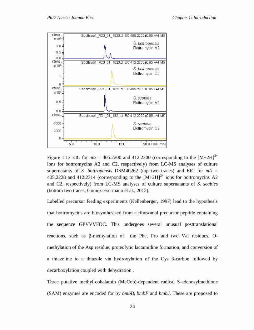
PhD Thesis: Joanna Bicz Chapter 1: Introduction
24
Figure 1.13 EIC for m/z = 405.2200 and 412.2300 (corresponding to the [M+2H]2+
ions for bottromycins A2 and C2, respectively) from LC-MS analyses of culture
supernatants of S. bottropensis DSM40262 (top two traces) and EIC for m/z =
405.2228 and 412.2314 (corresponding to the [M+2H]2+
ions for bottromycins A2
and C2, respectively) from LC-MS analyses of culture supernatants of S. scabies
(bottom two traces; Gomez-Escribano et al., 2012).
Labelled precursor feeding experiments (Kellenberger, 1997) lead to the hypothesis
that bottromycins are biosynthesised from a ribosomal precursor peptide containing
the sequence GPVVVFDC. This undergoes several unusual posttranslational
reactions, such as β-methylation of the Phe, Pro and two Val residues, O-
methylation of the Asp residue, proteolytic lactamidine formation, and conversion of
a thiazoline to a thiazole via hydroxylation of the Cys β-carbon followed by
decarboxylation coupled with dehydration .
Three putative methyl-cobalamin (MeCob)-dependent radical S-adenosylmethione
(SAM) enzymes are encoded for by bmbB, bmbF and bmbJ. These are proposed to

PhD Thesis: Joanna Bicz Chapter 1: Introduction
25
catalyse β-methylation of the Phe, Pro and two of the three Val residues in BmbC
(Figure 1.14). The putative SAM-dependent O-methyltransferase, encoded for by
bmbB, is proposed to convert the carboxylic acid group of the Asp residue to the
corresponding methyl ester (Figure 1.14). The protein similar to oxazoline/thiazoline
synthetases (encoded for bmbC gene) is involved in the biosynthesis of many other
heterocycle-containing ribosomal peptides (Dunbar et al., 2012). It is hypothesised
that BmbE forms a complex with BmbD, similar to oxazoline/thiazoline synthetases,
which then catalyses ATP-dependent thiazoline formation. Proteases thought to be
encoded for by bmbG, bmbH and bmbK are proposed to catalyse the removal of the
N-terminal N-formyl-Met residue and the 35 amino acids located downstream of the
Cys residue. Removal of the N-terminal N-formyl-Met residue enables nucleophlic
attack of the Gly amino group on the carbonyl group of the appropriate β-methyl-Val
residue with subsequent collapse of the resulting intermediate with loss of water,
which results in macrolactamidine formation. This reaction is proposed to be
catalysed by the third protease. The putative cytochrome P450 is encoded for by
bmbI and it is thought to catalyse the conversion of the thiazoline to a thiazole via
hydroxylation of the β-carbon of the Cys residue, followed by elimination of water
and subsequent decarboxylation. The final reaction in the biosynthetic pathway is
proposed to be the epimerisation of the Asp β-carbon, yielding bottromycin A2. It is
not known whether this reaction is catalysed by a specific enzyme or is spontaneous.
Bottromycins B2 and C2 arise from the same pathway, except that C-methylation of
the Pro residue is skipped in the biosynthesis of the former and occurs twice in the
assembly of the latter. bmbT encodes a putative integral membrane protein that may
be involved in the export of bottromycin. bmbR is proposed to encode a negative
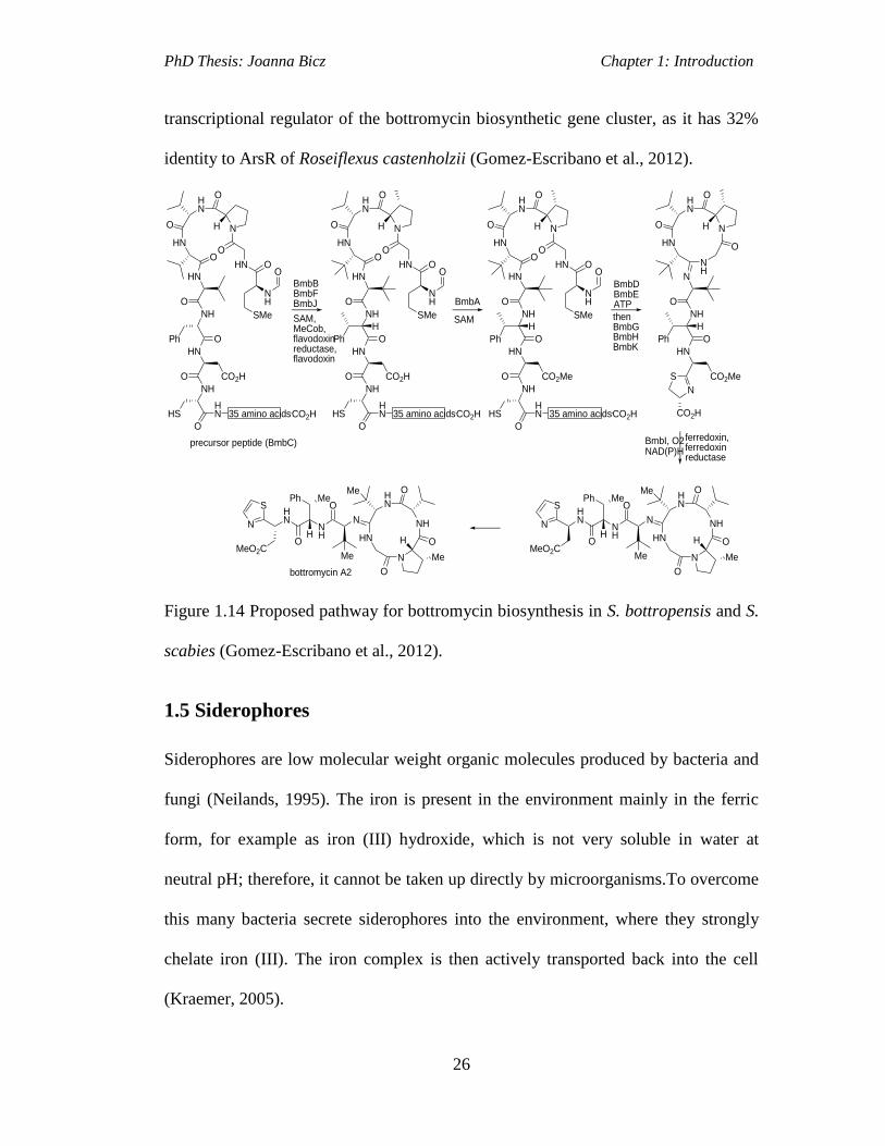
PhD Thesis: Joanna Bicz Chapter 1: Introduction
26
transcriptional regulator of the bottromycin biosynthetic gene cluster, as it has 32%
identity to ArsR of Roseiflexus castenholzii (Gomez-Escribano et al., 2012).
HN
N
HN
HN
O
O
H
NH
O
HN
OPh
CO2HO
NH
HN
O
HS
O
35 amino acidsCO2H
O
HN
SAM,MeCob,flavodoxin reductase,flavodoxin
precursor peptide (BmbC)
O
NH
SMe
BmbBBmbFBmbJ BmbA
HN
NH
N
HN
O
N
O
O
H
NH
O
HN
O
H
Ph
CO2MeS
BmbDBmbEATP
N
CO2H
HN
HN
N
NH
O
N
O
O
Me
H
Me
NH
OHN
OH
Ph Me
MeMeO2C
S
ferredoxin,ferredoxin reductase
N
HN
HN
N
NH
O
N
O
O
Me
H
Me
NH
OHN
OH
Ph Me
MeMeO2C
S
N
bottromycin A2
BmbI, O2NAD(P)H
thenBmbGBmbHBmbK
O
HN
N
HN
HN
O
O
H
NH
O
HN
OPh
CO2HO
NH
HN
O
HS
O
35 amino acidsCO2H
O
HN O
NH
SMe
O
H
HN
N
HN
HN
O
O
H
NH
O
HN
OPh
CO2MeO
NH
HN
O
HS
O
35 amino acidsCO2H
O
HN O
NH
SMe
O
HSAM
Figure 1.14 Proposed pathway for bottromycin biosynthesis in S. bottropensis and S.
scabies (Gomez-Escribano et al., 2012).
1.5 Siderophores
Siderophores are low molecular weight organic molecules produced by bacteria and
fungi (Neilands, 1995). The iron is present in the environment mainly in the ferric
form, for example as iron (III) hydroxide, which is not very soluble in water at
neutral pH; therefore, it cannot be taken up directly by microorganisms.To overcome
this many bacteria secrete siderophores into the environment, where they strongly
chelate iron (III). The iron complex is then actively transported back into the cell
(Kraemer, 2005).

PhD Thesis: Joanna Bicz Chapter 1: Introduction
27
The ability of the siderophore to chelate iron results from formation of stable
multidentate (e.g. hexadentate) complexes with iron (III). This is possible due to the
presence of iron-chelating ligands in the siderophores structure. The most commonly
observed are one to several bidentate oxygen ligands, such as hydroxamates (e.g. in
desferrioxamine E), catecholates (e.g. in enterobactin), or α-hydroxycarboxylates
(e.g. in achromobactin; Figure 1.15; Miethke and Marahiel, 2007). Some
siderophores contain oxygen and nitrogen heterocyclic structures to chelate ferric
iron (e.g. pyochelin; Figure 1.15; Miethke and Marahiel, 2007).
N NH
O
OH
O
O
NOH
HN
O NH
O
N
OHO
desferrioxamine E
O
O
N
O
NH
N
achromobactin
HO2C
O
O
O O
O
O
O
O
HN NH
HN
O
OH
OH
O
HO
HO
O
OH
OH
enterobactin
SN
SN
O
HO
Me
OH
pyochelin
H
H
OH
O
OH
HO
HO
O
HO
O
OH
Figure 1.15 Structures of several siderophores (chelating atoms are highlighted in
blue; Miethke and Marahiel, 2007).
PhD Thesis: Joanna Bicz Chapter 1: Introduction
28
Iron is required for many mammalian cell processes, such as gene regulation, DNA
synthesis, oxygen transport, respiration and lipid metabolism to name a few
(Recalcati et al., 2006). To perform these functions iron is incorporated into
haemoglobin, myoglobin and cytochromes. It is also a part of non-haeme structures,
e.g.iron-sulfur motifs found in the prosthetic groups of some proteins (Meyer, 2008).
In plants, iron is required for chlorophyll formation and for metabolism (Marsh et al.,
1963). Iron is also essential for bacterial growth. Iron acquisition, through the
production and release of siderophores, is necessary for the succesful infection of
mammalian hosts by bacterial pathogens (Miethke and Marahiel, 2007). For
example, mycobactin, a siderophore produced by M. tuberculosis, is able to extract
iron from macrophages (Luo et al., 2005). Siderophore-mediated iron acquisition can
be inhibited by the mammalian host protein siderocalin, which binds to siderophores
released by the pathogen. However, some bacteria have developed a way to
circumvent this defense mechanism through the production of siderophores, to
which, siderocalin cannot bind. An example of this resistance mechanism is found in
Vibrio cholera, which produces the siderophore vibriobactin (Li et al., 2012).
1.5.1 Siderophores Biosynthesised via NRPS-Dependent Pathways
Many siderophores are biosynthesised by enzymes belonging to the family of
nonribosomal peptide synthetases (NRPSs; Challis, 2005). They catalyse the
condensation of amino acids to form biologically-active peptides. These molecules
commonly possess structural features, such as non-proteinogenic amino acids,
hydroxy acids, heterocyclic rings and other unusual modifications in their backbone.
Products of NRPSs include not only siderophores but other pharmacologically
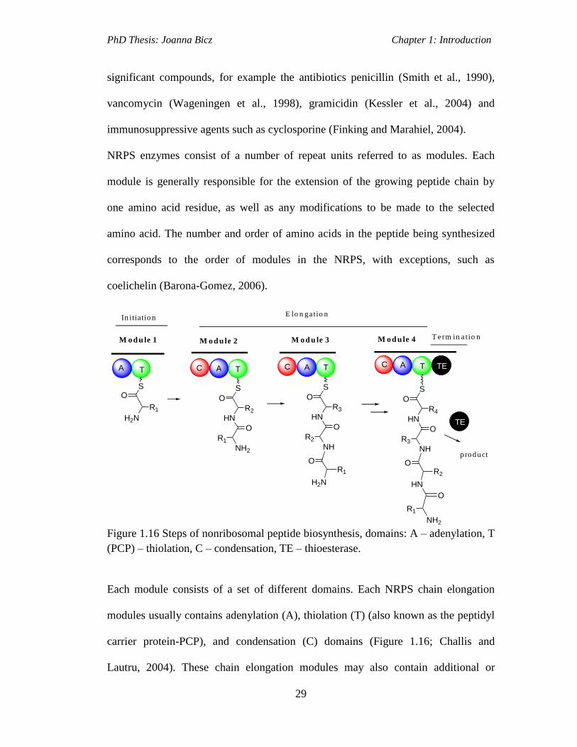
PhD Thesis: Joanna Bicz Chapter 1: Introduction
29
significant compounds, for example the antibiotics penicillin (Smith et al., 1990),
vancomycin (Wageningen et al., 1998), gramicidin (Kessler et al., 2004) and
immunosuppressive agents such as cyclosporine (Finking and Marahiel, 2004).
NRPS enzymes consist of a number of repeat units referred to as modules. Each
module is generally responsible for the extension of the growing peptide chain by
one amino acid residue, as well as any modifications to be made to the selected
amino acid. The number and order of amino acids in the peptide being synthesized
corresponds to the order of modules in the NRPS, with exceptions, such as
coelichelin (Barona-Gomez, 2006).
T
A
C TC T
S S
In itiatio nE lo n gatio n
T erm in a t io n
product
M o d u le 1 M o d u le 2 M o d u le 3 M o d u le 4
O
H2N
R1
HN
NH2
R2
R1
O
O
S
HN
NH
R3
R2
O
O
H2N
OR1
S
HN
NH
R4
R3
O
O
HN
OR2
NH2
O
R1
C T TE
TE
A A A A
Figure 1.16 Steps of nonribosomal peptide biosynthesis, domains: A – adenylation, T
(PCP) – thiolation, C – condensation, TE – thioesterase.
Each module consists of a set of different domains. Each NRPS chain elongation
modules usually contains adenylation (A), thiolation (T) (also known as the peptidyl
carrier protein-PCP), and condensation (C) domains (Figure 1.16; Challis and
Lautru, 2004). These chain elongation modules may also contain additional or

PhD Thesis: Joanna Bicz Chapter 1: Introduction
30
alternative domains such as epimerisation (E), methylation (M), and
heterocyclization (Cy), which introduce modifications to the amino acid incorporated
by the module into the biosynthesized polypeptide chain. Another two modules
usually present in the NRPS are a loading module, consisting only of A and T
domains; and a chain terminating module containing the thioesterase (TE) domain
(Figure 1.16).
The adenylation (A) domain is considered to have a primary role in substrate
selectivity of the module. During nonribosomal peptide biosynthesis (Figure 1.16),
the A domain firstly recognises the amino acid being incorporated, activates it as an
amino acyl adenylate and then transfers it to the adjoining T domain (Figure 1.17).
The activated amino acid is linked to the T domain via a covalent bond as a thioester
to the 4’-phosphopantetheinyl (4’Ppant) arm of the T domain. The 4’Ppant is
posttranslationally attached to a conserved serine of the T domain. This reaction is
carried out by a broad specificity phosphopantetheinyl transferase, which catalyses
the attack of a conserved serine residue in the T domain on the diphosphate group of
coenzyme A. 3', 5'-adenosine diphosphate (ADP) is released and the T domain with a
phosphopantetheine thiol group is generated (Figure 1.18; Walsh et al., 1997).
O
R
O
H3NO
R
PATP
PPi
PPi
Adenosine
HS
AMP
H3NS
R
O
H3N
O
O
O-
O
H3NO
R
P Adenosine
O
O
O-
O
PCP PCP
Figure 1.17 The A domain reaction catalysis: the activation of the amino acid
substrate through the formation of an aminoacyl adenylate followed by the covalent
binding via a thioester bond to the 4’Ppant cofactor of the PCPdomain.

PhD Thesis: Joanna Bicz Chapter 1: Introduction
31
HS
HN
HN
O O
OH
Adenosine
3'5'-ADP
HS
HN
HN
O O
OH
PCP
SH
PCP
OHO P
O
O-
O P
O-
O
apo-Peptidyl Carrier Protein Coenzyme A
holo-Peptidyl Carrier Protein
O P
O
O-
O
PCP
=
Figure 1.18 Reaction catalysed by PPTases. The apo is the inactive form, lacking the
phosphopantetheinyl arm and the holo is the active form.
Elongation of the growing peptide chain is then carried out by the condensation (C)
domain, which joins the amino acid attached to the T domain of its module with an
amino acid attached to the T domain of the downstream module. This is achieved by
the nucleophilic attack of α-amino group of the peptidyl chain, tethered to the
downstream T domain, on the thioester carbonyl group of the the amino acid bound
to the upstream T domain (Figure 1.19; Challis and Lautru, 2004).
SO
H2N
R1
SO
H2N
R2
SH S
HN
NH2
R2
R1
O
O
C T TA ATA C TA
Figure 1.19 The catalytic mechanism of condensation (C) domains.

PhD Thesis: Joanna Bicz Chapter 1: Introduction
32
The final step of polypeptide biosynthesis is release of the peptide from the NRPS,
which is usually catalysed by a TE domain located at the C terminal end of the last
module of the NRPS. The first structure of a TE domain was solved in 2002
(Brunner et al., 2002). It showed that TE domains contain the catalytic residues
Ser80, His207 and Asp107, which catalyse the cleavage of the peptidyl thioester
bond resulting in the formation of a peptidyl-O-TE intermediate. This intermediate
can be attacked by an external nucleophile, usually a water molecule (hydrolysis) or
by an intramolecular nucleophile (cyclization) resulting in the release of the peptide
(Figure 1.20; Donadio et al., 2007; Challis and Lautru, 2004).
O
R2
HN
NH2
R1
O
O
His
OH
O
R2
HN
NH2
R1
O
O
R2
HN
NH2
R1
O
HN
NH
O
R1
R2
SHS
last module End last module
hydrolysis
cyclisation
TE
TE
End
H
OH
C TAC TA
Figure 1.20 The release of peptide from NRPS catalysed by TE domain.
1.5.1.1 NRPS siderophore pathway-Enterobactin Biosynthesis
One of the earliest and most comprehensive studies of an NRPS pathway is the
biosynthesis of catecholate siderophore enterobactin (Oves-Costales et al., 2009).
The discovery of enterobactin (also named enterochelin) was first reported in 1970
and this compound was found to be a metabolite produced by Escherichia coli. The
biosynthesis of enterobactin and its ferric complex uptake mechanism have since
been elucidated. The total chemical synthesis of enterobactin was reported in 2003

PhD Thesis: Joanna Bicz Chapter 1: Introduction
33
(Raymond et al., 2003). The three catecholate moieties in the enterobactin structure
allow it to form a very stable ferric complex.
The entABCDEF gene cluster of E. coli encodes the enzymes responsible for
enterobactin biosynthesis. Chorismate, a precursor in aromatic amino acid
biosynthesis, is converted into 2, 3-dihydroxybenzoic acid (2, 3-DHB) by EntA,
which is the N-terminal domain of EntB and EntC (Rusnak et al., 1990). The three
molecules of 2,3-DHB are joined with three molecules of L-serine via three amide
and three ester linkages to form enterobactin (Figure 1.21; Liu et al., 1990; Rusnak et
al., 1989). EntE is a stand-alone adenylation (A) domain catalysing the acylation of
the C-terminal thiolation (T) domain of EntB with 2, 3-DHB (Rusnak et al., 1989;
Reichert et al., 1992). The EntF protein contains C, A and T domains and a TE
domain is also present at its C-end. The adenylation (A) domain of EntF catalyses
the adenylation of the carboxyl group of L-serine and subsequent acylation of the T
domain of EntF. The EntF condensation (C) domain catalyses the formation of an
amide bond between the amino group in the L-serinyl thioester with the DHB–EntB
thioester to form a 2, 3-DHB-L-serinyl-EntF thioesters (Figure 1.21). This is then
transacylated with the active Ser residue of the EntF thioesterase (TE) domain to
form a 2, 3-DHB-L-serinyl-TE ester (Reichert et al., 1992).
The formation of second 2, 3-DHB-L-serinyl thioester is catalysed by EntF
condensation domain and then the TE domain catalyses its condensation with the 2,
3-DHB-L-serinyl-TE ester (Figure 1.22; Shaw-Reid et al., 1999). In this way the
dimeric ester is formed. The same process is repeated to form a trimeric ester, which
then undergoes macrocyclisation catalysed by the EntF TE domain to release
enterobactin (Figure 1.22).
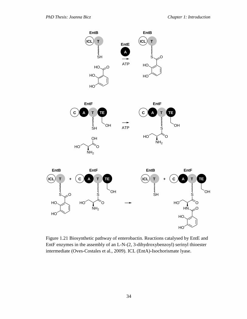
PhD Thesis: Joanna Bicz Chapter 1: Introduction
34
TE
OH
O
HN O
HO
HO
HO
S
TC
SH
TICL +TE
OH
O
NH2
HO
S
TCTICL + A
S O
HO
HO
TICL
S O
HO
HO
TICL
SH
O
HO
HO
HO
TE
OH
O
NH2
HO
S
TC A
A
EntE
ATP
EntB EntB
EntF
ATP
TE
OHSH
TC A
EntF
O
NH2
HO
OH
EntF EntFEntBEntB
A
Figure 1.21 Biosynthetic pathway of enterobactin. Reactions catalysed by EntE and
EntF enzymes in the assembly of an L-N-(2, 3-dihydroxybenzoyl) serinyl thioester
intermediate (Oves-Costales et al., 2009). ICL (EntA)-Isochorismate lyase.
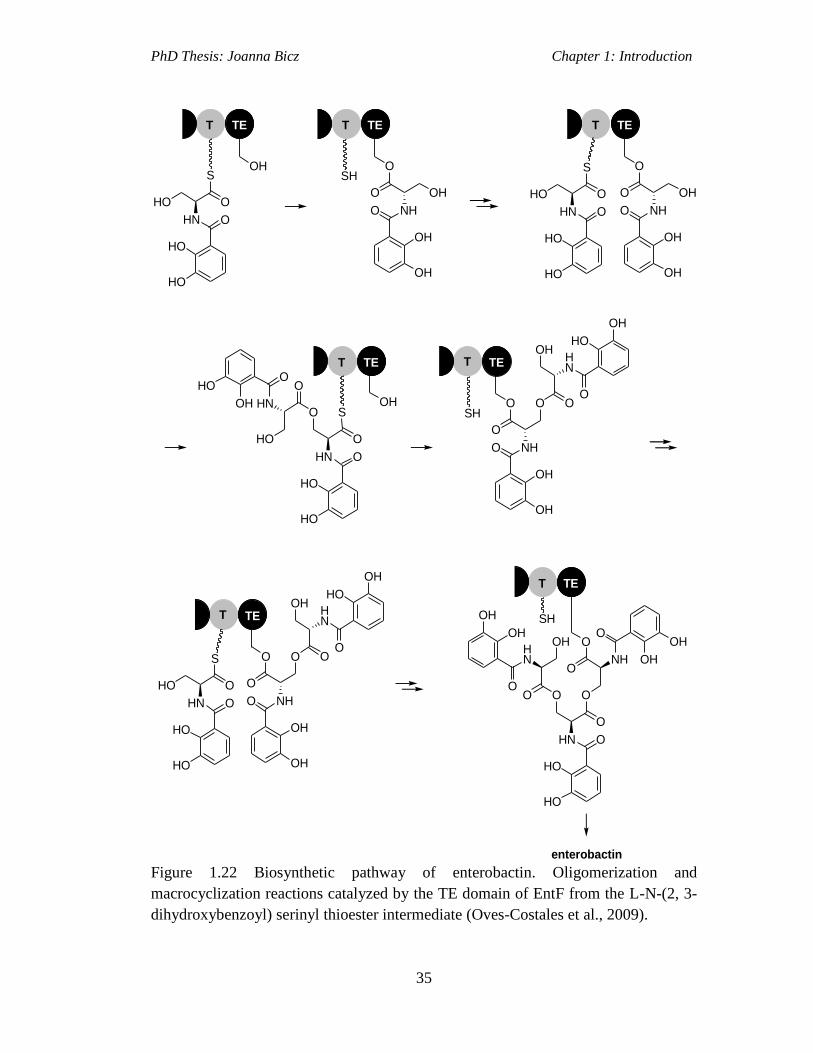
PhD Thesis: Joanna Bicz Chapter 1: Introduction
35
TE
OH
O O
O
O
O
O
HN NH
HN
O
OH
OH
O
HO
HO
O
OH
OH
O
HN O
HO
HO
HO
S
TE
SH
O
NHO
OH
OH
OH
O
TE
O
NHO
OH
OH
OH
O
O
HN O
HO
HO
HO
S
TE
OH
TE
O O O
O
NH
HN
O
OH
OH
O
HO
OH
SH
OH
O
O
HN O
HO
HO
S
TE
O O O
O
NH
HN
O
OH
OH
O
HO
OH
OH
O
HN O
HO
HO
HO
S
OH
TE
SH
enterobactin
T T
T T
T
T
T
O
HN
O
OH
HO
HO
Figure 1.22 Biosynthetic pathway of enterobactin. Oligomerization and
macrocyclization reactions catalyzed by the TE domain of EntF from the L-N-(2, 3-
dihydroxybenzoyl) serinyl thioester intermediate (Oves-Costales et al., 2009).
PhD Thesis: Joanna Bicz Chapter 1: Introduction
36
1.5.2 Siderophores Assembled via NRPS-Independent Siderophore (NIS)
Pathway
Siderophores containing dicarboxylic acid and diamine building blocks are
biosynthesised by a structurally distinct family of synthetases, the NRPS-
independent siderophore synthetases (NISs). In the last two decades over 40 species
of bacteria were found to contain genes encoding for NIS synthetases, which are
involved in biosynthesis no less than ten different siderophores (Challis, 2005). The
first characterised NIS pathway was the aerobactin biosynthetic pathway (Neilands
1986). Following on from this discovery, several more NIS-derived siderophores
were identified including rhizobactin, alcaligin, desferrioxamines, vibrioferrin,
staphylobactin, anthrachelin and achromobactin. Genetic studies demonstrated that
all these siderophore biosynthetic gene clusters encode at least one enzyme with high
sequence similarity to the aerobactin siderophore synthetases. Biosynthesis of
siderophores via an NIS pathway involves the activation of carboxylic acid
substrates to their corresponding acid adenylates. The NISs can be divided into three
subtypes depending on which type of carboxylic acid substrate is recognised and
activated (Berti and Thomas, 2009). Type A NISs are specific for citrate, type B for
α-ketoglutarate and type C for ester or amid derivatives of carboxylic acid (Oves-
Costales et al., 2009). The type A NISs are homologs of the IucA synthetase, type B
are homologs of IucD and the type C enzymes are similar to IucC. All of which are
present in the aerobactin biosynthetic pathway.
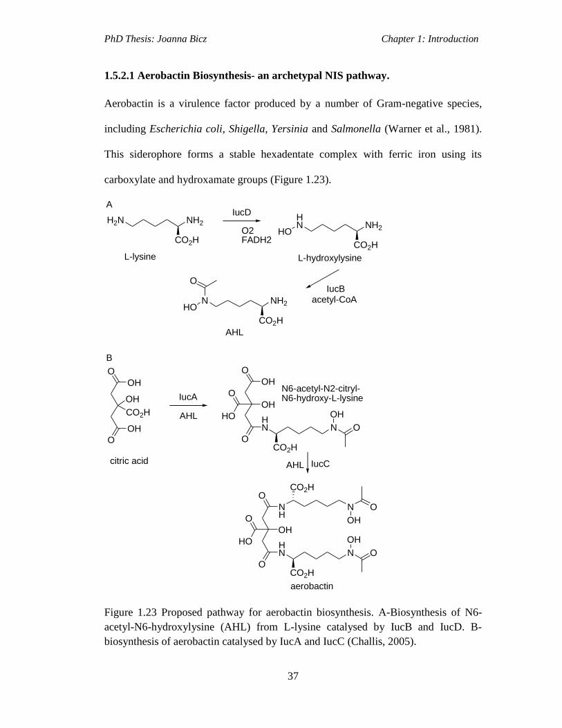
PhD Thesis: Joanna Bicz Chapter 1: Introduction
37
1.5.2.1 Aerobactin Biosynthesis- an archetypal NIS pathway.
Aerobactin is a virulence factor produced by a number of Gram-negative species,
including Escherichia coli, Shigella, Yersinia and Salmonella (Warner et al., 1981).
This siderophore forms a stable hexadentate complex with ferric iron using its
carboxylate and hydroxamate groups (Figure 1.23).
NH2H2N
CO2HO2FADH2
IucD
NH2
HN
CO2H
HO
IucB
NH2N
CO2H
HO
O
acetyl-CoA
AHL
A
L-lysine L-hydroxylysine
O
O
OH
IucA
OH
O
OH
OH
CO2H
O
IucC
AHL
OH
HN N
OH
O
CO2H
HO
O
AHL
O
O
OH
HN N
OH
O
CO2H
HO
O
NH
N
OH
O
CO2H
aerobactin
B
citric acid
N6-acetyl-N2-citryl-N6-hydroxy-L-lysine
Figure 1.23 Proposed pathway for aerobactin biosynthesis. A-Biosynthesis of N6-
acetyl-N6-hydroxylysine (AHL) from L-lysine catalysed by IucB and IucD. B-
biosynthesis of aerobactin catalysed by IucA and IucC (Challis, 2005).

PhD Thesis: Joanna Bicz Chapter 1: Introduction
38
The aerobactin biosynthetic gene cluster was first identified on the pColV-K30
plasmid in E. coli. It consists of five genes, of which four (iucABCD) direct
aerobactin biosynthesis (Figure 1.24), while the fifth (iutA) encodes an outer-
membrane receptor protein involved in the uptake of the ferric-aerobactin complex
(Challis, 2005). The iucD and iucB genes encode a FADH2-dependent
monooxygenase and an acetyl-CoA-dependent acyltransferase, respectively. They
catalyse the first two steps of aerobactin biosynthesis (Figure 1.23 A). IucD catalyses
the conversion of L-lysine to L-N6-hydroxylysine and IucB catalyses the conversion
of L-N6-hydroxylysine to L-N6-acetyl-N6-hydroxylysine (AHL; Carbonetti and
Williams, 1984; Cox, 1982). The iucA is proposed to encode a synthetase catalysing
the condensation of the α-amino group in L-N6-acetyl-N6-hydroxylysine with one of
the prochiral carboxyl groups of citric acid to form N6-acetyl-N2-citryl-N6-hydroxy-
L-lysine (Figure 1.24). The iucC is proposed to encode a homologue of IucA
synthetase, which catalyses the condensation of the product of the IucA-catalysed
reaction with a second molecule of L-N6-acetyl-N6-hydroxylysine to form
aerobactin (Challis, 2005; Figure 1.23).
Figure 1.24 Aerobactin biosynthetic gene cluster.

PhD Thesis: Joanna Bicz Chapter 1: Introduction
39
1.6 Investigations of Cryptic Siderophore Biosynthetic Pathways
encoded for in the S. scabies 87.22 genome.
Not much is known about the significance of siderophores in bacterial plant
pathogenicity. Investigations into siderophore mediated iron acquisition and its role
in plant pathogenicity has only been explored in Gram-negative bacteria known to be
plant pathogens. For example, the siderophore pyoverdine plays a key role in the
infection of tobacco by Pseudomonas syringae pv. tabaci 6605 (Sriyosachati and
Cox, 1986). The presence of iron aids the transcription of virulence genes during
speck disease in tomato by Pseudomonas syringae pv. tomato DC3000 (Kim et al.,
2009). However, to date, siderophore-mediated iron uptake and its role in plant
pathogenicity has not been explored as extensively in Gram-positive bacteria. One
Gram-positive strain, where studies into this have been undertaken, is the plant
pathogen S. scabies 87.22.
1.6.1 Pyochelin
Analysis of the S. scabies 87.22 genome (http://strepdb.streptomyces.org.uk/)
uncovered a putative gene cluster for the biosynthesis of pyochelin, a previously
characterised siderophore. Pyochelin (Figure 1.25) produced by the mammalian
pathogen Pseudomonas aeruginosa (Cox et al., 1981) is known to be a virulence
factor in these bacteria, as it plays an important role in the infection in mice (Cox,
1982). This is because pyochelin is able to take up iron from transferrin and
lactoferrin present in the blood serum of mammals and use it for bacterial growth
(Akenbauer et al., 1995).

PhD Thesis: Joanna Bicz Chapter 1: Introduction
40
SN
SN
O
HO
Me
OH
H
H
Figure 1.25 Structure of pyochelin (Cox, 1982).
1.6.1.1 Pyochelin Biosynthesis in P. aeruginosa
Pyochelin is a siderophore that contains a thiazoline/thiazolidine moiety and its
biosynthesis in Pseudomonas aeruginosa, has been fully characterised (Cox, 1982;
Takase et al., 2000). The pyochelin biosynthetic gene cluster in P. aeruginosa
(Figure 1.26) encodes seven proteins PchABCDEFG, which are required for
pyochelin biosynthesis (Figure 1.27; Cox et al., 1981; Reinmann et al., 2001).
Figure 1.26 Organization of the pyochelin biosynthetic gene cluster in P. aeruginosa
PAO1.
PchA and PchB catalyse the biosynthesis of salicylate from chorismate (Serino et al.,
1995). A stand-alone A domain, PchD, then adenylates salicylate and subsequently
loads it onto the thiolation domain of PchE NRPS multienzyme (Figure 1.27). The A
domain of PchE catalyses the acylation of cysteine onto the C-terminal T domain,
and the Cy (heterocyclization) domain catalyses condensation of the cysteinyl
thioester with the salicylic thioester to form a peptide bond. An MT-like domain

PhD Thesis: Joanna Bicz Chapter 1: Introduction
41
embedded within the A domain of PchE is then proposed to catalyse the
epimerization of the α-carbon of the resulting thioester (Patel et al., 2003). The Cy
domain, then, catalyses the cyclodehydration to yield a 2-hydroxyphenyl-thiazolinyl
thioester intermediate.
The A domain of PchF catalyses the cis loading of cysteine onto its T domain, and
the Cy domain of PchF catalyses amide bond formation between the cysteinyl
thioester and the 2-hydroxyphenyl-thiazolinyl thioester on PchE. Subsequent
cyclodehydration results in a formation of a bis-thiazolinyl thioester intermediate.
PchG catalyses NADPH-dependent reduction of one of the thiazoline rings to the
corresponding thiazolidine and an MT domain in PchF catalyzes the S-
adenosylmethionine (SAM)-dependent N-methylation of the thiazolidine. The TE
domain of PchF catalyses hydrolysis of the thioester bond in the resulting
intermediate yielding pyochelin (Reinmann et al., 2001; Serino et al., 1995; Serino et
al., 2003).

PhD Thesis: Joanna Bicz Chapter 1: Introduction
42
O
HO
HO
A
PchD
ATP
SH
TCy A
PchE
SH
T MT
O
HO ATP
SH
TCy A
PchE
S
T MT
O
NH2
HS
OH
O
HO
TCy A
PchE
S
T MT
O
NH2
HS
S
TCy A
PchE
SH
T MT
O
S
SN
OH
H
ATPSH
TCy A
PchF
MT
O
NH2
HS
OH
TE
OH
TCy A
PchF
MT
O
NH2
HS
S
TE
OH
T
O
S
SN
OH
H
PchE
+
TCy A
PchF
MT
O
S
TE
OH
SN
S N
OH
H
T
PchF
O
S
TE
OH
SN
S N
OH
H
H H
Me
T
PchF
SH
TE
OSN
S N
OH
H
H
Me
O
pyochelin
PchG
NADPH SAM
Figure 1.27 Biosynthesis of pyochelin. NRPS domains: T-thiolation, Cy-
heterocyclization, A-adenylation, MT-methyltransferase, TE-thioesterase (Reinmann
et al., 2001; Serino et al., 1995; Serino et al., 2003).

PhD Thesis: Joanna Bicz Chapter 1: Introduction
43
1.6.1.2 Sequence Analysis of the S. scabies Pyochelin Biosynthetic Gene Cluster.
The pyochelin biosynthetic gene cluster (PBGC) in S. scabies contains 12 genes
(Figure 1.28; Cox, 1982). Six of these genes encode proteins (Scab1381, Scab1411,
Scab1421, Scab1461, Scab1471 and Scab1481) that are proposed to be analogous in
function to the pyochelin biosynthetic proteins PchABCDEFG from P. aeruginosa
(Patel and Walsh, 2001; Quadri et al., 1999; Serino et al., 1997).
Figure 1.28 Organisation of pyochelin biosynthetic gene clusters in S. scabies 87.22,
P. aeruginosa PAO-1and P. fluorescens Pf-5 (Seipke et al., 2011; Youard et al.,
2007).
Scab1381 encodes for a putative salicylate synthase and it contains an anthranilate
synthase domain. Scab1411 is predicted to be AMP-ligase, which contains putative
active site residues that are highly similar to the active site residues of MbtA, an
AMP-ligase from Mycobacterium tuberculosis. MbtA catalyses the adenylation of
salicylate in mycobactin biosynthesis (Challis et al., 2000; Quadri et al., 1998;
Rausch et al., 2005). Two genes scab1481 and scab1471, encode proteins with
putative functions analogous to PchF and PchE, respectively, both of which are
PhD Thesis: Joanna Bicz Chapter 1: Introduction
44
NRPS enzymes in P. aeruginosa (Figure 1.28). Scab1481, which is analogous to
PchE, is proposed to catalyse the formation of a 2-hydroxyphenylthiazolinyl
thioester from the salicyl adenylate and Scab1471 adds a second molecule of the
cysteine to the thioester to form 2-hydroxyphenylbis-thiazolinyl thioester attached to
the T domain of Scab1471 in a manner analogous to PchF (Figure 1.27). Scab1461 is
predicted to encode a reductase enzyme homologous to PchG. It is proposed to
catalyse the reduction of one of the thiazoline rings in the thioester. N-methylation of
the reduced thiazoline ring and the release of the pyochelin by the N-terminal
thioesterase domain is catalysed by Scab1471 (Patel and Walsh, 2001). Scab1421
protein is predicted to be a homolog of PchC, a proofreading thioesterase, which
catalyses removal of wrongly charged molecules from the PchE and PchF thiolation
domains (Reinmann et al., 2004). The analysis of the S. scabies genes indicates that
they encode proteins, which have identical properties to the pyochelin biosynthetic
proteins PchABCDEFG from P. aeruginosa and indicates that S. scabies may be a
producer of pyochelin.
Other genes from the putative pyochelin gene cluster, such as scab1401 encodes a
pathway specific regulator homologous to PchR, a pyochelin-responsive repressor
protein in P. aeruginosa and Scab1401 is a TetR family transcriptional regulator.
Analysis of the transcription of the PBGC revealed that scab1371 is a positive
transcriptional regulator of the cluster and it is co-transcribed with the scab1381,
predicted to encode salicylate synthase. Putative transport proteins are encoded for
by scab1431, scab1441 and scab1451.
PhD Thesis: Joanna Bicz Chapter 1: Introduction
45
There are also some differences between the biosynthetic proteins in P. aeruginosa
and S. scabies. Formation of salicylate from chorismate is catalysed in P. aeruginosa
by two proteins PchA and PchB (Serino et al., 1995) while in S. scabies 87.22 a
single protein, Scab1381, is predicted to catalyse the same reaction. Another
difference is that the S. scabies pyochelin gene cluster does not contain an ortholog
of the P. aeruginosa fptX gene encoding the inner membrane pyochelin permease.
The gene is not encoded anywhere else in the S. scabies genome and therefore, it is
speculated that one of the putative transport proteins Scab1431, Scab1441 or
Scab1451 within the cluster may be used for the import of ferric-pyochelin in S.
scabies (Seipke et al., 2011).
1.6.1.3 Biosynthesis of Enantio-Pyochelin in Pseudomonas fluorescens
Pyochelin is biosynthesised from salicylate and two molecules of cysteine. During
thiazoline ring formation in P. aeruginosa, the α-carbon of the first cysteine residue
undergoes epimerisation, which results in the conversion of cysteine to its D-
isoform, whereas the second cysteine remains as L-isoform (Figure 1.27). This
results in the formation of two pyochelin diastereoisomers with the stereochemistry
4’R, 2’’R, 4’’R for pyochelin I and 4’R, 2’’S, 4’’R for pyochelin II (Youard et al.,
2007). Other closely related strains P. fluorescens Pf-5 and CHA0 were found to
produce an enantiomer of pyochelin named enantiopyochelin (Figure 1.29).
Enantiopyochelin was found to facilitate growth under iron-deficient conditions in P.
fluorescens and to induce the expression of its biosynthetic genes, however, it did not
promote growth in P. aeruginosa. On the other hand, pyochelin from P. aeruginosa
did not promote growth, nor induce the expression in P. fluorescens. Comparison of

PhD Thesis: Joanna Bicz Chapter 1: Introduction
46
the pyochelin biosynthetic gene cluster between P. aeruginosa and P. fluorescens
(Figure 1.28) highlighted three main differences. Firstly, in P. aeruginosa the
biosynthetic genes are organised in two operons, while in P. fluorescens the genes
are in one operon. Secondly, PchE in P. fluorescens does not contain the
epimerisation domain and finally the predicted function of PchK in P. fluorescens is
not homologous to PchG in P. aeruginosa. PchG is a thiazoline reductase and PchK
is predicted to have reductase and/or epimerase function. The amino acid sequences
of other of P. aeruginosa and P. fluorescens pch genes are between 35 and 60%
identical (Youard et al., 2007).
C4' C2''
OH
N
SN
S
COOHH3C
OH
N
SN
S
H3C
C4''
OH
N
SN
S
COOH
H3C
H H
H
COOH
H
H
H
Pyochelin I (4'R, 2''R, 4''R) Pyochelin II (4'R, 2''S, 4''R)
OH
N
SN
S
CH3
H
COOH
HH
ent-Pyochelin I (4'S, 2''S, 4''S)
OH
N
SN
S
CH3
H
COOH
H
H
ent-Pyochelin II (4'S, 2''R, 4''S)
Figure 1.29 Stereoisomers of pyochelin (P. aeruginosa) and ent-pyochelin (P.
fluorescens).
PhD Thesis: Joanna Bicz Chapter 1: Introduction
47
1.6.2 Desferrioxamines.
Desferrioxamines are a group of siderophores containing three hydroxamic acid
ligands, which form a hexadentate complex with ferric iron (Miller, 1989).
Desferrioxamines include the linear trimer desferrioxamine G1, the hetero-oligomer
desferrioxamine B and the 33-membered macrocyclic desferrioxamine E (Figure
1.30). They are biosynthesised by several Streptomyces species (Bickel et al., 1960).
S. coelicolor has been shown to produce desferrioxamine G1 and E (Imbert et al.,
1995) and S. viridosporus has been shown to produce desferrioxamine B and E
(Imbert et al., 1995). S. pilosus has been found to take up ferrioxamines B and E, S.
lividans B and G1 and S. viridosporus B, E and G1 (Muller and Raymond, 1984,
Imbert et al., 1995). Desferrioxamine B is used clinically for the treatment of iron
poisoning in human body.
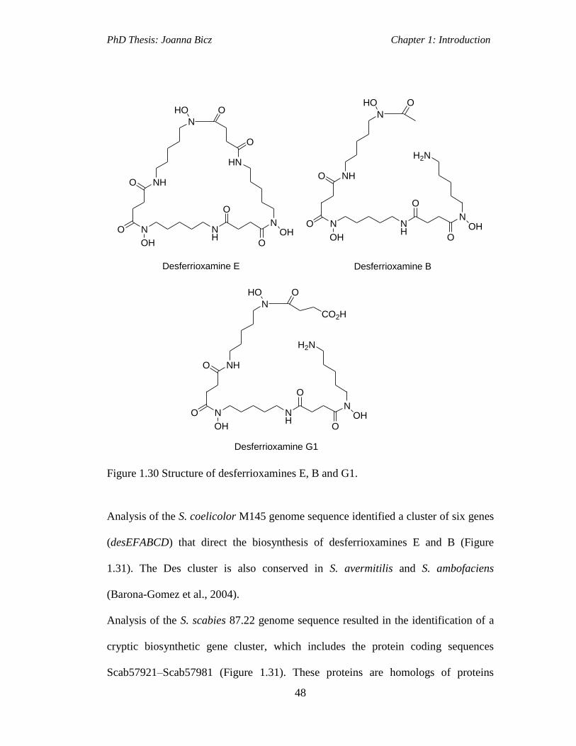
PhD Thesis: Joanna Bicz Chapter 1: Introduction
48
N NH
N
NH
N
HN
HO O
O
OHO
O
OH
O
O
Desferrioxamine E
N NH
N
NH
N
H2N
HO O
OHO
O
OH
O
O
Desferrioxamine B
Desferrioxamine G1
N NH
N
NH
N
H2N
HO O
OHO
O
OH
O
O
CO2H
Figure 1.30 Structure of desferrioxamines E, B and G1.
Analysis of the S. coelicolor M145 genome sequence identified a cluster of six genes
(desEFABCD) that direct the biosynthesis of desferrioxamines E and B (Figure
1.31). The Des cluster is also conserved in S. avermitilis and S. ambofaciens
(Barona-Gomez et al., 2004).
Analysis of the S. scabies 87.22 genome sequence resulted in the identification of a
cryptic biosynthetic gene cluster, which includes the protein coding sequences
Scab57921–Scab57981 (Figure 1.31). These proteins are homologs of proteins
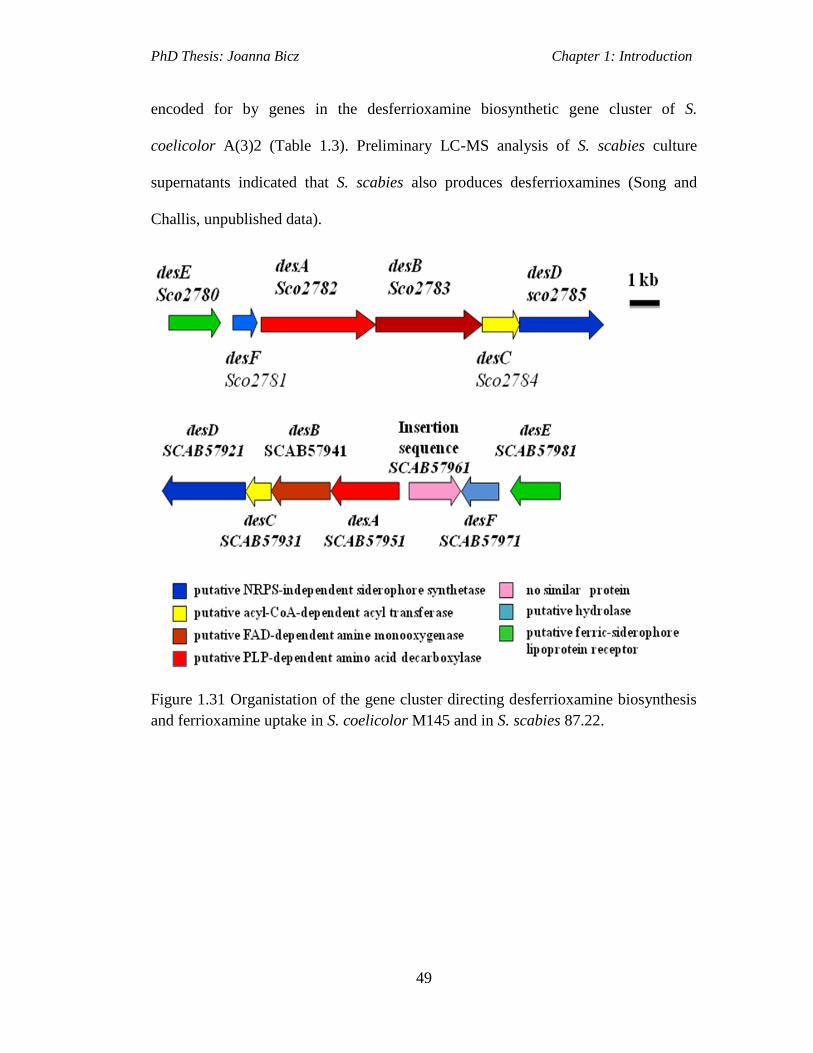
PhD Thesis: Joanna Bicz Chapter 1: Introduction
49
encoded for by genes in the desferrioxamine biosynthetic gene cluster of S.
coelicolor A(3)2 (Table 1.3). Preliminary LC-MS analysis of S. scabies culture
supernatants indicated that S. scabies also produces desferrioxamines (Song and
Challis, unpublished data).
Figure 1.31 Organistation of the gene cluster directing desferrioxamine biosynthesis
and ferrioxamine uptake in S. coelicolor M145 and in S. scabies 87.22.

PhD Thesis: Joanna Bicz Chapter 1: Introduction
50
Table 1.3 Putative proteins function encoded by the desferrioxamine biosynthetic
gene cluster in S. scabies 87.22
S. scabies
gene
desferrioxamine
biosynthesis protein (Des)
Sequence similarity with S.
coelicolor A3(2) des cluster (%)
scab57921 DesD hypothetical protein SCO2785 (81%)
scab57931 DesC aceytltranferase SCO2784 (79%)
scab57941 DesB monooxygenase SCO2783 (80%)
scab57951 DesA pyridoxal-dependent
decarboxylase SCO2782 (86%)
scab57961 IS630 family insertion
sequence
no similar protein
scab57971 DesE hypothetical protein SCO2781 (75%)
The first steps of desferrioxamine biosynthesis were investigated in Streptomyces
pilosus, and involved elucidation of reactions catalysed by the enzymes DesA and
DesB (Schupp at al., 1987, 1988), which have high sequence identity to PLP-
dependent amino acid decarboxylases and FAD-dependent amine monooxygenases,
respectively. DesA and DesB are involved in the first two steps of desferrioxamine
biosynthesis to form N-hydroxycadaverine from L-lysine (Figure 1.32 A).
Investigations into the roles of DesC and DesD were carried out in the
desferroxamine E biosynthetic pathway of S. coelicolor. The desC gene encodes an
enzyme similar to acyl-CoA-dependent acyl transferases (Kadi et al., 2007) and is
proposed to catalyse the N-acylation of N-hydroxycadaverine (N. Kadi and G. L.
Challis, unpublished data). DesC is predicted to have relaxed substrate specificity in

PhD Thesis: Joanna Bicz Chapter 1: Introduction
51
those bacteria, which also produce desferrioxamine B, catalysing acylation of N-
hydroxycadaverine with acetyl-CoA as well as succinyl-CoA, giving N-hydroxy-N-
acetylcadaverine (HAC) and N-hydroxy-N-succinylcadaverine (HSC), respectively
(Figure 1.32 A; Barona-Gomez et al., 2004).
The gene desD encodes a protein that has a high percentage sequence similarity to
IucA and IucC from the aerobactin biosynthetic pathway, the first NIS enzymes to be
characterised. Purified recombinant DesD was shown to catalyse the ATP-dependent
conversion of chemically-synthesized HSC to desferrioxamines G1 and E. DesD
catalyses the ATP-dependent trimerisation and macrocyclisation of HSC to yield
desferrioxamine E (Figure 1.32 B) and the ATP-dependent condensation of two units
of HSC with one unit of HAC to yield desferrioxamine B (Figure 1.32C; Barona-
Gomez et al., 2006).
DesE is similar to ferric-siderophore lipoprotein receptors and has been shown to
bind ferrioxamines with high affinity and selectivity (Patel et al., 2010). DesF is
similar to ViuB, which is proposed to be a hydrolase involved in the release of iron
from ferric-vibriobactin. However, more recent sequence analyses suggest it is a
ferric-siderophore reductase (Barona-Gomez et al., 2006).
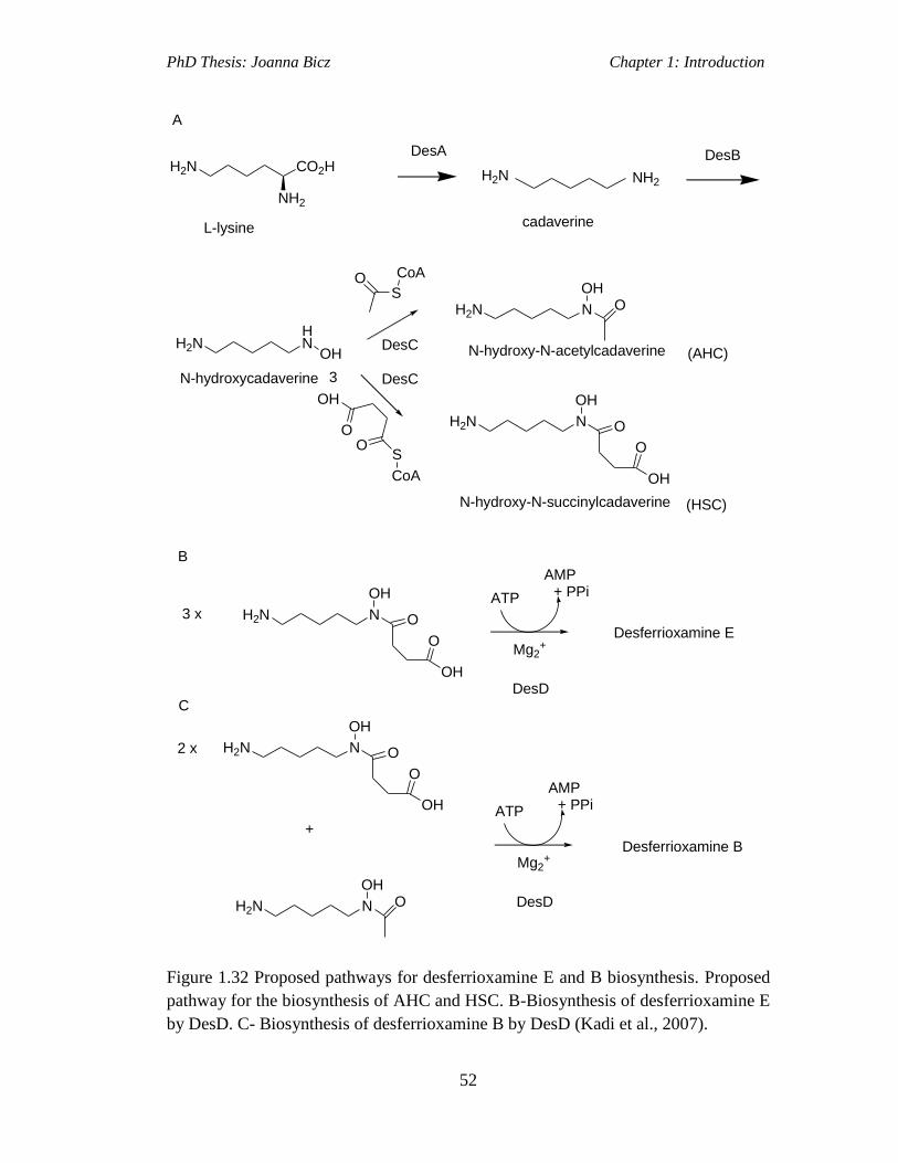
PhD Thesis: Joanna Bicz Chapter 1: Introduction
52
H2NHN
OH
S
CoAO
OO
OH
S
CoA
H2N N
OHO
H2N N
OH
O
O
OH
H2N N
OH
O
O
OH
Mg2+
ATP
AMP
H2N N
OH
O
O
OH
H2N N
OHO
Mg2+
ATP
AMP
H2N NH2
H2N CO2H
NH2
N-hydroxy-N-acetylcadaverine
N-hydroxy-N-succinylcadaverine
3 x
+ PPi
DesD
Desferrioxamine E
2 x
+
+ PPi
DesD
Desferrioxamine B
cadaverine
DesB
L-lysine
DesA
DesC
DesCN-hydroxycadaverine
(AHC)
(HSC)
A
B
C
3
Figure 1.32 Proposed pathways for desferrioxamine E and B biosynthesis. Proposed
pathway for the biosynthesis of AHC and HSC. B-Biosynthesis of desferrioxamine E
by DesD. C- Biosynthesis of desferrioxamine B by DesD (Kadi et al., 2007).

PhD Thesis: Joanna Bicz Chapter 1: Introduction
53
1.6.3 Novel Putative Hydroxamate Siderophore
A novel tris-hydroxamate iron-chelating metabolite was originally discovered in
Streptomyces antibioticus NBRC 13838 by Dr Shinya Kodani and coworkers
(Shizuoka University, Japan), collaborators of the Challis group. Analysis of the S.
scabies 87.22 genome sequence unveiled a cryptic NRPS biosynthetic gene cluster
(Figure 1.33), which includes the protein coding sequences from Scab85431 to
Scab85521, suggesting that it may encode an NRPS that directs the production of a
hydroxamate-containing metabolite. All of these proteins, present within the NRPS
biosynthetic gene cluster, are homologous to proteins encoded for by genes within
the coelichelin biosynthetic gene cluster from S. coelicolor M145 (Table 1.4)
(http://www.ncbi.nlm.nih.gov/nuccore/260644157).
Figure 1.33 Organization of the scabichelin biosynthetic gene cluster in S. scabies
87.22.

PhD Thesis: Joanna Bicz Chapter 1: Introduction
54
Table 1.4 Putative proteins function encoded by the putative novel hydroxamate
siderophore biosynthetic gene cluster in S. scabies 87.22.
Protein Putative function Homolog; origin Similarity/
identity
(%)
Scab85431 ABC transporter
permease/ATPase component
CchI; S. coelicolor 53/38
Scab85441 ABC transporter
permease/ATPase component
CchG; S. coelicolor 58/46
Scab85451 Ferric-siderophore
lipoprotein receptor
CchF; S. coelicolor 53/38
Scab85461
MbtH-like protein CchK; S. coelicolor 72/57
Scab85471 Nonribosomal peptide
synthetase
CchH; S. coelicolor 60/48
Scab85481 ABC transporter permease
component
CchC; S. coelicolor 65/47
Scab85491 ABC transporter permease
component
CchD; S.coelicolor 60/45
Scab85501 ABC transporter ATPase
component
CchE; S. coelicolor 74/60
Scab85511 Formyl-tetrahydrofolate-
dependent formyl transferase
CchA; S. coelicolor 85/76
Scab85521 Flavin-dependent
monooxygenase
CchB; S. coelicolor 73/60
The genes Scab85511 and scab85521 encode proteins homologous to CchA and
CchB from S. coelicolor, respectively. CchA and CchB are involved in the
biosynthesis of the non-proteogenic amino acid residues hydroxyornithine (hOrn)
and formylhydroxyornithine (fhOrn), which are take part in the biosynthesis of
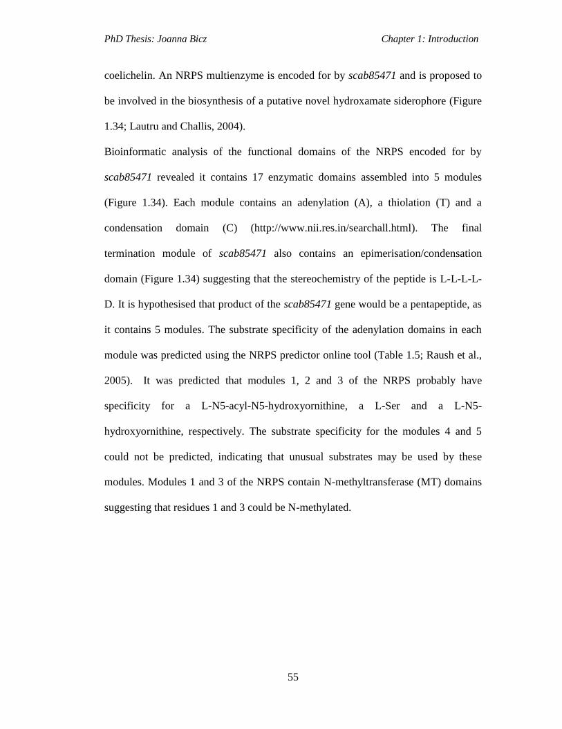
PhD Thesis: Joanna Bicz Chapter 1: Introduction
55
coelichelin. An NRPS multienzyme is encoded for by scab85471 and is proposed to
be involved in the biosynthesis of a putative novel hydroxamate siderophore (Figure
1.34; Lautru and Challis, 2004).
Bioinformatic analysis of the functional domains of the NRPS encoded for by
scab85471 revealed it contains 17 enzymatic domains assembled into 5 modules
(Figure 1.34). Each module contains an adenylation (A), a thiolation (T) and a
condensation domain (C) (http://www.nii.res.in/searchall.html). The final
termination module of scab85471 also contains an epimerisation/condensation
domain (Figure 1.34) suggesting that the stereochemistry of the peptide is L-L-L-L-
D. It is hypothesised that product of the scab85471 gene would be a pentapeptide, as
it contains 5 modules. The substrate specificity of the adenylation domains in each
module was predicted using the NRPS predictor online tool (Table 1.5; Raush et al.,
2005). It was predicted that modules 1, 2 and 3 of the NRPS probably have
specificity for a L-N5-acyl-N5-hydroxyornithine, a L-Ser and a L-N5-
hydroxyornithine, respectively. The substrate specificity for the modules 4 and 5
could not be predicted, indicating that unusual substrates may be used by these
modules. Modules 1 and 3 of the NRPS contain N-methyltransferase (MT) domains
suggesting that residues 1 and 3 could be N-methylated.

PhD Thesis: Joanna Bicz Chapter 1: Introduction
56
A MT C A C A MT
S S
O
HN
H2N
Module 1 Module 2 Module 3
85471
S
O
N
H2N
OHO
H
O
H2N
OH
HO
C A
Module 4
C A
Module 5
E
?
L-fhOrn L-hOrnL-Ser
Figure 1.34 Organisation of modules and domains of the NRPS encoded by
scab85471.
Table 1.5 Predicted substrate specificity of adenylation domains in the Scab85471
protein.
Module number Substrate specificity Residue position
1 hforn DVWILGAT
2 ser DVWHLSLV
3 horn DMENLGLI
4 ? DGEDLGLS
5 ? DAQXXXLV
1.7 Products of Other Cryptic Pathways
A family of polyketide natural products, which all contain a five-membered tetronate
ring are known to have apoptotic and antibacterial properties (Hamaguchi et al.,
1995). All these
natural products were found to be biosynthesised by both Gram-
negative and Gram-positive bacteria, including various Streptomyces species.

PhD Thesis: Joanna Bicz Chapter 1: Introduction
57
Examples of tetronate containing natural products are RK-682 (Figure 1.35) from
Streptomyces sp. 88-68 and agglomerin antibiotics (Figure 1.35) from the Gram-
negative Pantoea agglomerans (Sun et al., 2010).
O
OO
HOOH
RK-682
O
OO
HO
agglomerin A
Figure 1.35 Structures of tetronate polyketides.
The common structural feature of tetronate natural products is the tetronic acid ring
(Coloured blue in Figure 1.35). The mechanism of tetronic acid formation was
elucidated during investigations into the biosynthesis of RK-682. This is proposed to
involve an FabH-like protein, which is an enzyme belonging to a family of fatty acid
synthases (Sun et al., 2010).
Another common feature of these natural products is a polyketide backbone, which is
assembled by enzymes belonging to the family of polyketide synthases (PKSs).
Polyketides comprise a major group of structurally and functionally diverse natural
products that include a broad range of clinically important compounds, such as
antibiotics, immunosuppressants, antitumour, antifungal and antiparasitic agents
(McMurry and Begley, 2005). Polyketides are biosynthesized by the condensation of
extender units malonyl-, methylmalonyl- or ethylmalonyl-CoA with an acyl-CoA
starter unit and are classified according to their structure into three types. Type I
PKSs contain multiple catalytic domains for each reaction in the polyketide chain

PhD Thesis: Joanna Bicz Chapter 1: Introduction
58
assembly and are classified into modular and iterative systems. In type I modular
PKSs each module is used only once to catalyse the elongation of the polyketide
chain by a malonate extender unit, whereas in iterative systems the PKS modules are
used for more than one elongation step. Type II PKSs are a group of monofunctional
proteins where each protein catalyses a different reaction in the chain assembly.
Type III PKSs are homodimers of identical keto synthase (KS) domains that do not
use ACP domains in the polyketide chain assembly. They posses a single active site
to catalyse different types of reactions, such as decarboxylations, condensations and
cyclisations (Austin and Noel, 2002).
Type I PKSs are involved in tetronate polyketide chain assembly. In a type I PKS the
loading module contains an acyl transferase (AT) and an acyl carrier protein (ACP)
A chain extension module consists of a minimum of three domains: an acyl
transferase (AT), an acyl carrier protein (ACP) and a β-ketoacyl synthase (KS). In
the first stage, the AT domain of the loading module loads the acyl-CoA starter unit
and then transfers it to the adjacent ACP domain (Figure 1.36 A; McMurry and
Begley, 2005). During chain extension the malonate extender unit is loaded onto the
AT domain of the module 1 and then transferred onto the ACP domain within the
same module (Figure 1.36 B; McMurry and Begley, 2005). The KS domain of
module 1 then catalyses the decarboxylative Claisen condensation between the ACP
bound acyl thioester of the module 1 with the acyl-ACP of the upstream loading
module to extend the chain forming a β-ketothioester (Figure 1.37; McMurry and
Begley, 2005). Additional domains, such as a ketoreductase (KR), a dehydratase
(DH) and an enoyl reductase (ER) may be present in some extension modules. The
KR domain catalyses the NADPH-dependent reduction of the β-carbon to form a β-

PhD Thesis: Joanna Bicz Chapter 1: Introduction
59
hydroxy thioester (Figure 1.38; McMurry and Begley, 2005). The DH domain
catalyses the conversion of the β-hydroxy thioester to an α, β-unsaturated thioester
by removal of water (Figure 1.39; McMurry and Begley, 2005). The ER domain
catalyses the NADPH-dependent reduction of the α, β-unsaturated double bond to
form a saturated bond (Figure 1.40; McMurry and Begley, 2005).
Load
O
propionyl CoA
(starter unit)
O
H
HisSCoA
AT ACP
SH
Load
AT ACP
O S
His
HO
Load
AT ACP
OH
Loading module
S
O
A
Chain extension module
Module 1
OH
His
AT
O SCoA
O
methylmalonyl CoA(extender unit)
O
Module 1
O
AT ACP
S
Module 1
ACP
S
O
Module 1
O
O
O
H
H
HisO
O
B
Figure 1.36 Reaction catalysed by AT domains. A-loading, B-chain extension
Load
ACP
S
O
H S
H
Module 1
S
O
O
O
SH
Load
S
Module 1
S
O
O
O
H
O
CO2 HS
Module 1
S
O
O
KS AT ACP KS AT ACP KS AT ACPACP
Figure 1.37 Reaction catalysed by KS domain.
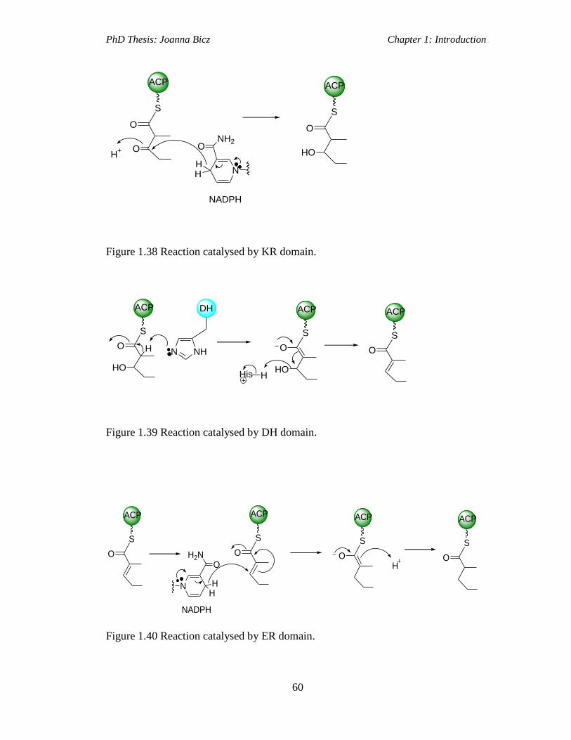
PhD Thesis: Joanna Bicz Chapter 1: Introduction
60
ACP
S
O
OH
N
NH2
HH
O
ACP
S
O
HO
NADPH
Figure 1.38 Reaction catalysed by KR domain.
ACP
S
O
HO
H
DH
NHN
ACP
S
O
HOHis H
ACP
S
O
Figure 1.39 Reaction catalysed by DH domain.
ACP
S
O
NADPH
ACP
S
O
NHH
H2NO
ACP
S
OH
ACP
S
O
Figure 1.40 Reaction catalysed by ER domain.

PhD Thesis: Joanna Bicz Chapter 1: Introduction
61
1.7.1 RK-682
RK-682 (3-hexadecanoyl-5-hydroxymethyl-tetronic acid; Figure 1.35) is a model
tetronate containing polyketide, originally isolated from Streptomyces sp. 88-682
(Hamaguchi et al., 1995). It is an inhibitor of protein phosphatases and of HIV-1
proteinase. The RK-682 biosynthetic gene cluster was heterologously expressed in
Saccharopolyspora erythraea JCB2, which does not produce erythromycin, thus
allowing the production of RK-682, to be easily detected (Sun et al., 2010).
Nine genes, RkA-RkI, were identified in the RK-682 biosynthetic gene cluster in
Streptomyces sp. 88–682 (Figure 1.41). The genes from this cluster code for the
following proteins: rkI-TetR-family transcriptional regulator; rkH- antibiotic efflux
protein; rkA- palmitoyl-CoA synthetase; rkB and rkF- acyl carrier proteins; rkC,
modular PKS containing KS, AT and ACP domains; rkD-FabH-like protein; rkE-
FkbH-like glyceryl-S-ACP synthase; rkG -type II thioesterase (TEII). It was
determined that expression of all nine genes from the RK-682 cluster from the
original host is required for similar production levels in Saccharopolyspora
erythraea. The putative Rk proteins were later expressed and purified from E. coli
allowing the steps of the RK-682 biosynthetic pathway to be reconstituted in vitro
(Sun et al., 2010).

PhD Thesis: Joanna Bicz Chapter 1: Introduction
62
Figure 1.41 RK-682 biosynthetic gene cluster in Streptomyces sp. 88-682 (Sun et al.,
2010).
RkA, which is a putative acyl-CoA synthetase, would activate palmitic acid and load
it onto the stand-alone ACP, RkB, forming a palmitoyl-S-RkB complex (Figure
1.42). The chain would then be elongated by the PKS, RkC, which catalyses the
incorporation of one malonyl-CoA unit, resulting in the formation of 3-oxo-stearoyl-
S-RkC. In parallel, RkE would catalyse both the transfer of D-1,3-
bisphosphoglycerate to RkF, a second stand-alone ACP, and its subsequent
dephosphorylation, to form glyceryl-S-RkF. RkD, which is a putative 3-oxoacyl-
ACP synthase (FabH-like), would then catalyse the condensation of glyceryl-S-RkF
and 3-oxo-stearoyl-S-RkC to yield RK-682. RkD is proposed to to catalyse the C-C
and C-O tetronate bond formation in RK-682 (Sun et al., 2010).
PhD Thesis: Joanna Bicz Chapter 1: Introduction
63
OO
O OH
OH
RK-682
S
O
RkC
O
S
O
OH
OH
+
O
OPO3=
OH
OPO3=
RkE(FkbH)
RkF
S
O
+
OH
O
RkA
RkB
RkD(FabH)
ATP
KS AT ACP ACPACP
KSAT
Figure 1.42 Proposed biosynthetic pathway of RK-682 (Sun et al., 2010).
1.7.2 Agglomerins
Agglomerins A (Figure 1.43), B, C and D were originally extracted from the culture
broth of a bacterial strain, identified as Pantoea agglomerans PB-6042 (formerly
Enterobacter agglomerans), in the late 80’s by Shoji and co-workers (Shoji et al
1989). P. agglomerans belong to a family of Gram-negative aerobic bacteria, known
as Enterobacteriaceae. This strain can either be commensal or pathogenic to plants
and is commonly found in diverse environments, including water, soil or sediments
(Cruz et al., 2007; Shoji et al., 1989). Several strains of P. agglomerans are used
comercially in the United States as biological control agents, against the fire blight
pathogen (Erwinia amylovora) on apple trees (Wodzinski et al., 1994; Johnson and
Stockwell et al., 1998). This species is currently also considered as an opportunistic
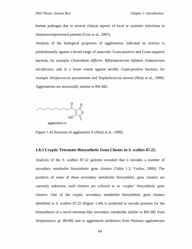
PhD Thesis: Joanna Bicz Chapter 1: Introduction
64
human pathogen due to several clinical reports of local or systemic infections in
immunocompromised patients (Cruz et al., 2007).
Analysis of the biological properties of agglomerins indicated its activity is
predominantly against a broad range of anaerobic Gram-positive and Gram-negative
bacteria, for example Clostridium difficile, Bifidobacterium bifidum, Eubacterium
aerofaciens, and to a lesser extent against aerobic Gram-positive bacteria, for
example Streptococcus pneumoniae and Staphylococcus aureus (Shoji et al., 1989).
Agglomerins are structurally similar to RK-682.
O
OO
HO
agglomerin A
Figure 1.43 Structure of agglomerin A (Shoji et al., 1989).
1.8.3 Cryptic Tetronate Biosynthetic Gene Cluster in S. scabies 87.22.
Analysis of the S. scabies 87.22 genome revealed that it encodes a number of
secondary metabolite biosynthetic gene clusters (Table 1.2; Yaxley, 2009). The
products of some of these secondary metabolite biosynthetic gene clusters are
currently unknown; such clusters are referred to as ‘cryptic’ biosynthetic gene
clusters. One of the cryptic secondary metabolite biosynthetic gene clusters
identified in S. scabies 87.22 (Figure 1.44) is predicted to encode proteins for the
biosynthesis of a novel tetronate-like secondary metabolite similar to RK-682 from
Streptomyces sp. 88-682 and to agglomerin antibiotics from Pantoea agglomerans
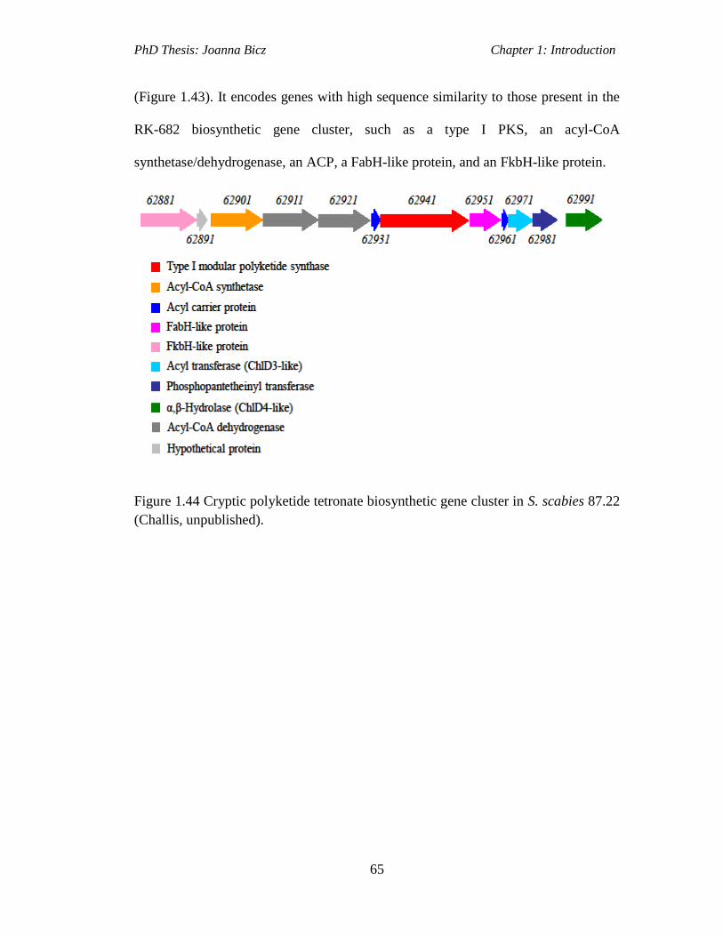
PhD Thesis: Joanna Bicz Chapter 1: Introduction
65
(Figure 1.43). It encodes genes with high sequence similarity to those present in the
RK-682 biosynthetic gene cluster, such as a type I PKS, an acyl-CoA
synthetase/dehydrogenase, an ACP, a FabH-like protein, and an FkbH-like protein.
Figure 1.44 Cryptic polyketide tetronate biosynthetic gene cluster in S. scabies 87.22
(Challis, unpublished).

PhD Thesis: Joanna Bicz Chapter 1: Introduction
66
Table 1.6 Putative proteins function encoded by the putative tetronate biosynthetic
gene cluster in S. scabies 87.22.
Protein Putative function Homolog; origin Identity (%)
scab62881 FkbH-like protein
RkE; Streptomyces sp.88-682 66.1
scab62891 Unknown
- -
scab62901 Acyl-CoA synthetase
RkA; Streptomyces sp.88-682 66.4
scab62911
Acyl-CoA dehydrogenase Acyl-CoA dehydrogenase,
S. clavuligerus ATCC 27064
83
scab62921 Acyl-CoA dehydrogenase
Acyl-CoA dehydrogenase,
S. clavuligerus ATCC 27064
85
scab62931 Acyl carrier protein
RkB; Streptomyces sp.88-682 63.9
scab62941 Type I modular PKS
RkC; Streptomyces sp.88-682 67.9
scab62951 FabH-like protein
RkD; Streptomyces sp.88-682 71.7
scab62961 Acyl carrier protein
RkF; Streptomyces sp.88-682 64.8
scab62971 Acyl transferase (ChlD3-
like protein)
Acyl transferase,
S. clavuligerus ATCC 27064
82
scab62981 PPtase
PPtase, S. clavuligerus ATCC
27064
83
scab62991 α,β-hydrolase (ChlD4-
like protein)
α,β-hydrolase, S. clavuligerus
ATCC 27064
86
Based on the sequence analysis of the RK-682 biosynthetic gene cluster and the S.
scabies 87.22 putative tetronate biosynthetic gene cluster it is likely that the S.
scabies tetronate metabolite is assembled in a way analogous to RK-682.

PhD Thesis: Joanna Bicz Chapter 1: Introduction
67
The starting point of the S. scabies tetronate assembly would be the activation of an
acyl group by the putative acyl-CoA synthetase, Scab62901, followed by loading
onto the ACP, Scab62961, to form an acyl-S-ACP intermediate (Figure 1.44). The
chain would then be elongated by the putative PKS, Scab62941, which would result
in the formation of a β-ketothioester. In parallel, an FkbH-like protein, Scab62881,
would catalyse the transfer of 3-bisphosphoglycerate to the ACP, Scab62931 and
dephosphorylation by the same enzyme yields the glyceryl-S-ACP intermediate.
Scab62951, which is a FabH-like protein, would then catalyse the condensation of
the glyceryl-S-ACP intermediate with the polyketide chain attached to Scab62941, to
form the tetronate ring. A further two steps are likely to be involved in S. scabies
tetronate product formation, which are not present in RK-682 biosynthesis. These are
reactions catalysed by a putative acyl-transferase, Scab62971, (ChlD3-like), and by a
putative hydrolase, Scab62991, (ChlD4-like).
ATO
R
O
O OH
OHS
O
62941
R
O
S
O
OH
OH
+
O
OPO3=
OH
OPO3=
62881(FkbH)
62931
ACP
S
O
R
+
OH
O
R
62901
62961
62951(FabH)
ATP
product ?
ACP KS AT ACP
KS
ACP
Figure 1.45 Proposed biosynthetic pathway for cryptic tetronate product in S. scabies
87.22 (Challis, unpublished).

PhD Thesis: Joanna Bicz Chapter 1: Introduction
68
1.8 Aims of the Project
The aim of this project is to investigate cryptic secondary metabolite biosynthetic
gene clusters in S. scabies 87.22.
S. scabies 87.22 has become a model organism for studying plant-microbe
interactions among Gram-positive plant pathogenic bacteria and the genome
sequence for this strain is now published (GenBank Accession no. FN554889). Three
out of four of the biosynthetic gene clusters investigated in this work are responsible
for the production of the siderophores pyochelin, scabichelin and desferrioxamines,
which are iron-chelating molecules. Iron acquisition through the use of siderophores
is crucial for the development of infection in the mammalian host by pathogenic
bacteria (Miethke and Marahiel, 2007). Studies of iron uptake were mostly
conducted in Gram negative bacteria, whereas investigations into iron acquisition by
Gram positive plant pathogens remained limited. Analysis of the S. scabies
secondary metabolite biosynthetic gene clusters and its metabolic products is
important to increase the understanding of its potential role in plant pathogenicity.
For example, several genes encoding for the biosynthesis of plant toxins in these S.
scabies secondary metabolite biosynthetic gene clusters, were found to be significant
virulence factors. Awareness of these virulence factors can be used in the future in
the agrochemical development to prevent infection by that pathogen potentially
saving important agricultural crops from damage.
The first objective of this project was to confirm that pyochelin is the product of the
putative pyochelin biosynthetic gene cluster in S. scabies. Analysis of the pyochelin

PhD Thesis: Joanna Bicz Chapter 1: Introduction
69
gene cluster mutants was planned to determine if the gene knockouts affected the
production of pyochelin either by abolishing (deletion of NRPS scab1471) or
upregulating production (deletion of scab1401). Comparisons of the compound from
the pyochelin overproducing mutant, ∆scab1401, with pyochelin and
enantiopyochelin authentic standards from P. aeruginosa were planned.
The second objective was to investigate whether the cryptic NRPS cluster in S.
scabies directs the production of the putative hydroxamate siderophore, by creation
of a partial cryptic NRPS gene deletion (scab85471) using a PCR targeting strategy.
The mutant strain would also be examined for the production of scabichelin by
comparisons of the S. scabies NRPS product with an authentic standard from S.
antibioticus.
The third objective was to confirm that the putative desferrioxamine biosynthetic
gene cluster in S. scabies directs desferrioxamine production and to investigate the
role of the desC gene in desferrioxamine biosynthesis. The aim was to create a desC
gene deletion in S. scabies using PCR targeting technology and to examine
desferrioxamine production in the desC mutant. The function of the desC gene in the
mutant would then be restored via genetic complementation by introducing another
copy of desC gene into the mutant genome. The complemented mutant could then be
analysed for restoration of desferrioxamine production.
The final objective was to investigate the expression of the cryptic tetronate-
containing polyketide biosynthetic gene cluster in S. scabies. The role of the SARP
family of proteins encoded for within the gene cluster in the regulation of expression
of the cluster was also investigated by performing reverse transcriptase PCR
reactions on the total RNA isolated from wild type S. scabies.

PhD Thesis: Joanna Bicz Chapter 2: Results and Discussion
70
Results and Discussion I: Investigation of
siderophore biosynthetic gene clusters in S. scabies.
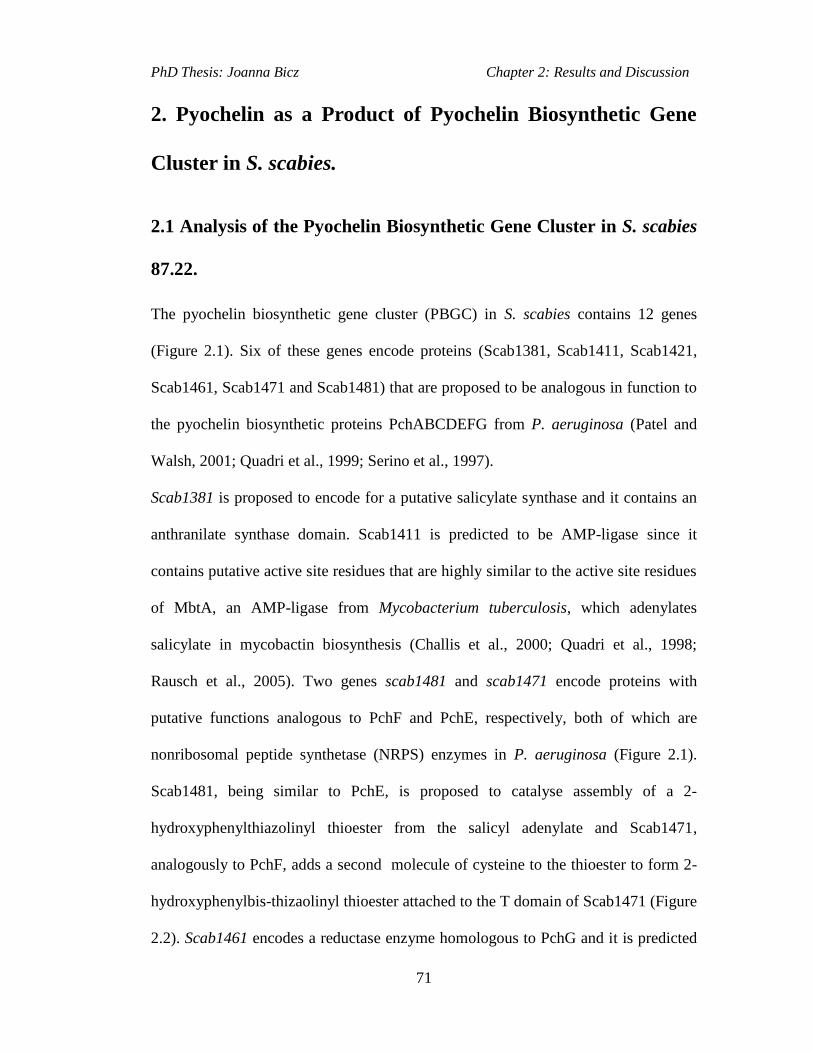
PhD Thesis: Joanna Bicz Chapter 2: Results and Discussion
71
2. Pyochelin as a Product of Pyochelin Biosynthetic Gene
Cluster in S. scabies.
2.1 Analysis of the Pyochelin Biosynthetic Gene Cluster in S. scabies
87.22.
The pyochelin biosynthetic gene cluster (PBGC) in S. scabies contains 12 genes
(Figure 2.1). Six of these genes encode proteins (Scab1381, Scab1411, Scab1421,
Scab1461, Scab1471 and Scab1481) that are proposed to be analogous in function to
the pyochelin biosynthetic proteins PchABCDEFG from P. aeruginosa (Patel and
Walsh, 2001; Quadri et al., 1999; Serino et al., 1997).
Scab1381 is proposed to encode for a putative salicylate synthase and it contains an
anthranilate synthase domain. Scab1411 is predicted to be AMP-ligase since it
contains putative active site residues that are highly similar to the active site residues
of MbtA, an AMP-ligase from Mycobacterium tuberculosis, which adenylates
salicylate in mycobactin biosynthesis (Challis et al., 2000; Quadri et al., 1998;
Rausch et al., 2005). Two genes scab1481 and scab1471 encode proteins with
putative functions analogous to PchF and PchE, respectively, both of which are
nonribosomal peptide synthetase (NRPS) enzymes in P. aeruginosa (Figure 2.1).
Scab1481, being similar to PchE, is proposed to catalyse assembly of a 2-
hydroxyphenylthiazolinyl thioester from the salicyl adenylate and Scab1471,
analogously to PchF, adds a second molecule of cysteine to the thioester to form 2-
hydroxyphenylbis-thizaolinyl thioester attached to the T domain of Scab1471 (Figure
2.2). Scab1461 encodes a reductase enzyme homologous to PchG and it is predicted

PhD Thesis: Joanna Bicz Chapter 2: Results and Discussion
72
to catalyse the reduction of one of the thiazoline rings in the thioester. Scab1471
catalyses N-methylation of the reduced thiazoline ring, followed by the release of
pyochelin by its N-terminal thioesterase domain (Patel and Walsh, 2001). Scab1421
gene is proposed to encode for a putative thioesterase analogous to PchC. PchC can
catalyse removal of wrongly charged molecules from the thiolation domains of PchE
and PchF (Reinmann et al., 2004).
Figure 2.1 Organization of the PBGCs in S. scabies 87.22 and P. aeruginosa PAO1.
Other genes from the putative pyochelin gene cluster include scab1401, which
encodes a pathway specific regulator homologous to PchR, a pyochelin-responsive
repressor protein in P. aeruginosa. Scab1401 belongs to the TetR family of
transcriptional regulators. Analysis of the transcription of PBGC revealed that
Scab1371 is a positive transcriptional regulator of the cluster and it is co-transcribed
with the predicted salicylate synthase gene scab1381. Scab1431, scab1441 and
scab1451 encode putative transport proteins. Proposed functions of the proteins
encoded by the S. scabies 87.22 pyochelin biosynthetic gene cluster are displayed in
the Table 1.7 (Seipke et al., 2011).

PhD Thesis: Joanna Bicz Chapter 2: Results and Discussion
73
Table 1.7 Putative proteins function encoded by the putative pyochelin biosynthetic
gene cluster in S. scabies 87.22 (adapted from Seipke et al., 2011).
Protein Putative function Homolog; origin Identity
(%)
Scab1371 AfsR-family regulator
AfsR; S. coelicolor 40
Scab1381 Salicylate synthase
TrpE; Saccharopolyspora
erythraea
51
Scab1391 Unknown
Sav760; S. avermitilis 72
Scab1401
TetR-family regulator OrfH2; S. griseoruber 55
Scab1411 Salicyl-AMP-ligase
DhbE; B. subtilis 50
Scab1421 Thioesterase
PchC; P. fluorescens 44
Scab1431 ABC transporter permease/
ATPase
Nfa28020; Nocardia
farcinica
66
Scab1441 ABC transporter permease/
ATPase
Nfa28010; N. farcinica 65
Scab1451 Major facilitator
superfamily protein
Fraal6304; Frankia alni 46
Scab1461 Thiazoline reductase
Strop2818; Salinispora
tropica
49
Scab1471
Nonribosomal peptide
synthetase
PchF; P. fluorescens 45
Scab1481
Nonribosomal peptide
synthetase
Pst2602; P. syringae 40

PhD Thesis: Joanna Bicz Chapter 2: Results and Discussion
74
O
HO
HO
A
Scab1411
ATP
SH
TCy A
Scab1481
SH
T MT
O
HO ATP
SH
TCy A
Scab1481
S
T MT
O
NH2
HS
OH
O
HO
TCy A
Scab1481
S
T MT
O
NH2
HS
S
TCy A
Scab1481
SH
T MT
O
S
SN
OH
H
ATP
SH
TCy A
Scab1471
MT
O
NH2
HS
OH
TE
OH
TCy A
Scab1471
MT
O
NH2
HS
S
TE
OH
T
O
S
SN
OH
H
Scab1481
+
TCy A
Scab1471
MT
O
S
TE
OH
SN
S N
OH
H
T
Scab1471
O
S
TE
OH
SN
S N
OH
H
H H
Me
T
Scab1471
SH
TE
OSN
S N
OH
H
H
Me
O
pyochelin
Scab1461
NADPH SAM
Figure 2.2 A proposed pathway for pyochelin biosynthesis in S. scabies 87.22 based
on analysis of biosynthesis by orthologous proteins from P. aeruginosa (Seipke et
al., 2011).

PhD Thesis: Joanna Bicz Chapter 2: Results and Discussion
75
2.2 Creation of Pyochelin Mutants in S. scabies 87.22.
Mutants of S. scabies 87.22 were created using PCR-targeting REDIRECT
technology (Gust et al., 2003) in the lab of Rose Loria (Cornell University). As
discussed in section 2.1 scab1401 is a gene encoded for in the putative pyochelin
biosynthetic gene cluster (PBGC) in S. scabies 87.22 wild type strain. Analysis of
pyochelin production in the wild type strain and in the scab1401 mutant revealed
that Scab1401 is a transcriptional repressor of the S. scabies PBGC (Seipke et al.,
2011). The scab1401 mutant contains a cassette consisting of oriT and an
apramycin resistance gene from pIJ773 in place of the scab1401 gene (Gust et al.,
2003) Deletion of scab1401 induces expression of the putative pyochelin
biosynthetic gene cluster (confirmed by RT-PCR analysis in Loria lab; Figure 2.3;
Seipke et al., 2011).
Figure 2.3 RT-PCR analysis of ΔScab1401 mutant strain showing that Scab1401
represses transcription of S. scabies 87.22 pyochelin biosynthetic genes. Lane 1- S.
scabies wild-type cDNA, 2- ΔScab1401, 3- ΔScab1401 with pAU3-45 or 4-
pRFSRL34 (plasmids containing the copy of scab1401 gene) grown in liquid
minimal medium+100 mM FeCl3 was used as template for PCRs. The murX gene -a
loading control (Seipke et al., 2011).

PhD Thesis: Joanna Bicz Chapter 2: Results and Discussion
76
scab1471 encodes a putative NRPS multienzyme complex homologous to PchF in
the P. aeruginosa PBGC, involved in catalysing the assembly of pyochelin. The
scab1471 mutant contains a cassette with oriT and a hygromycin B resistance gene
from pIJ10700 plasmid, in place of scab1471 (Gust et al., 2003). Deletion of
scab1471 was created to investigate the effect on pyochelin production in S. scabies.
scab1471 was also replaced with a gene conferring resistance to hygromycin B in the
Δscab1401 mutant, creating the double mutant Δscab1401-Δscab1471 by the group
of Rose Loria. The creation of the double mutant was required for investigations into
the function of Scab1471 NRPS under conditions giving constitutive expression of
the S. scabies PBGC.
2.3 Analysis of Pyochelin Production in S. scabies
The production of pyochelin in S. scabies wild type and mutant strains scab1401,
scab1471 and Δscab1401-Δscab1471 was examined by growing the strains in a
liquid iron deficient medium (IDM) and then analysing the culture supernatants for
the presence of pyochelin by LC-MS. Iron deficient media is a liquid minimal media
in which all traces of iron are removed by Chelex resin (section 7.1.6.1; Barona-
Gomez et al., 2004).
LC-MS analyses of the scab1401 mutant supernatant unveiled the presence of a
compound with m/z of 325.0669 and a retention time of 23.5 min (Figure 2.4 A).
This m/z correlates with the m/z for the [M+H]+ ion of pyochelin. This compound
from the Δscab1401 mutant culture supernatant was also compared with the

PhD Thesis: Joanna Bicz Chapter 2: Results and Discussion
77
simulated mass spectrum of pyochelin, with the molecular formula of
C14H17N2O3S2+. It indicated that the m/z for each isotopic isomer of S. scabies
compound correlates and agrees with the predicted m/z values for pyochelin. The
relative abundances of these isotopic isomers in both the measured and the simulated
spectra also agree with each other.
LC-MS analysis of the scab1471 mutant and of scab1401-1471 double mutant
culture supernatants revealed that pyochelin was not produced (Figure 2.5).
The stereochemistry of the compound produced by S. scabies was also investigated.
Research recently unveiled that the plant pathogen P. fluorescens, which is a Gram-
negative bacterium, produces diastereoisomers of pyochelin, referred to as
enantiopyochelins (ent-pyochelin I and II; Figure 2.4 C; Youard et al., 2007).
Comparison of the scab1401 mutant culture supernatant by LC-MS using chiral
stationary phase with the synthetic standards of pyochelin and ent-pyochelin
(standards kindly provided by Cornelia Reinmann) proved that S. scabies produce
pyochelin, not enantiopyochelin (Figure 2.4 B).

PhD Thesis: Joanna Bicz Chapter 2: Results and Discussion
78
Figure 2.4 A- Measured mass spectrum of pyochelin from Δscab1401 supernatant
(top panel), pyochelin standard (middle panel), and ent-pyochelin (bottom panel).
Figure 2.4 B- Extracted Ion Chromatograms (EICs) at m/z=325 from analyses of ent-
pyochelin standard (top trace), pyochelin standard (middle trace) and S. scabies
pyochelin (bottom trace).
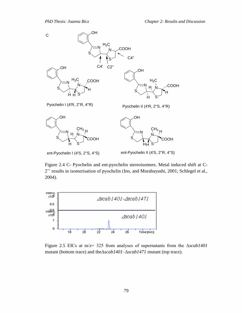
PhD Thesis: Joanna Bicz Chapter 2: Results and Discussion
79
C4' C2''
OH
N
SN
S
COOHH3C
OH
N
SN
S
H3C
C4''
OH
N
SN
S
COOH
H3C
H H
H
COOH
H
H
H
Pyochelin I (4'R, 2''R, 4''R) Pyochelin II (4'R, 2''S, 4''R)
OH
N
SN
S
CH3
H
COOH
HH
ent-Pyochelin I (4'S, 2''S, 4''S)
OH
N
SN
S
CH3
H
COOH
H
H
ent-Pyochelin II (4'S, 2''R, 4''S)
C
Figure 2.4 C- Pyochelin and ent-pyochelin stereoisomers. Metal induced shift at C-
2’’ results in isomerisation of pyochelin (Ino, and Murabayashi, 2001; Schlegel et al.,
2004).
Figure 2.5 EICs at m/z= 325 from analyses of supernatants from the Δscab1401
mutant (bottom trace) and theΔscab1401–Δscab1471 mutant (top trace).
PhD Thesis: Joanna Bicz Chapter 2: Results and Discussion
80
2.4 Discussion
Pyochelin production by Gram-negative bacterial pathogens has been reported
previously (Cox et al., 1981; Thomas, 2007; Castignetti, 1997). S. scabies is the first
Gram-postive plant pathogen, which has been found to produce pyochelin. Mining of
the S. scabies genome for putative secondary metabolite clusters identified a putative
PBGC. Five genes from that cluster scab1411, scab1421, scab1461, scab1471 and
scab1481 are predicted to encode proteins homologous to well characterised
pyochelin biosynthetic proteins PchCDEFG from P. aeruginosa. The main
differences between the S. scabies PBGC and the P. aeruginosa pyochelin
biosynthetic gene cluster are in the organisation of the genes and in the regulation of
transcription of the gene clusters.
Expression studies of the S. scabies PBGC, conducted by the research group of our
collaborator professor Rose Loria revealed that it is transcribed as two operons. One
of these is ~19 kb in size and includes the pyochelin biosynthetic genes, and the
other smaller operon, which is ~3kb, consists of scab1371 and the salicylate synthase
gene scab1381. The two genes scab1391 and scab1401 are not transcribed in either
operon and RT-PCR did not give any PCR products for scab1391-scab1401 region.
Scab1401 is a member of the TetR family of proteins and represses the transcription
of the large biosynthetic operon during growth in high iron conditions. The other
regulator Scab1371 protein is an AfsR-family regulator, which activates transcription
of the large operon if the availability of iron is low (Seipke et al., 2011). In contrast,
the pyochelin cluster in P. aeruginosa is regulated by the AraC-family protein PchR

PhD Thesis: Joanna Bicz Chapter 2: Results and Discussion
81
and the ferric uptake protein, Fur (Heinrichs and Poole, 1993, 1996; Michel et al.,
2005).
Pyochelin biosynthesis differs slightly between S. scabies and P. aeruginaosa. The
synthesis of salicylate, a substrate for pyochelin biosynthesis in the S. scabies PBGC
is thought to be catalysed by one protein Scab1381, while in P. aeruginosa the
biosynthesis of salicylate is catalysed by two proteins PchA and PchB (Serino et al.,
1995).
LC-MS analysis of the ∆scab1401 mutant indicated the presence of a compound with
an m/z value corresponding to that of pyochelin. The compound with the same m/z
was not consistently detected in the supernatants of wild type S. scabies when grown
in iron deficient conditions. Pyochelin should have been produced consistently in the
wild type strain in iron deficient media as the pyochelin biosynthesis genes have
been found to be expressed under these growth conditions. LC-MS analyses of the
∆scab1401-1471 mutant culture supernatant, in which the pyochelin biosynthetic
gene is deleted, revealed that pyochelin is not produced, further confirming that
pyochelin is the product of the putative pyochelin biosynthetic gene cluster identified
in S. scabies. Further LC-MS analysis of the ∆scab1401 mutant culture supernatant
alongside authentic standards of pyochelin and enantiopyochelin confirmed that S.
scabies produces pyochelin and not its enantiomer enantiopyochelin.
Plant assays were used (by Prof R. Loria’s group at Cornell University; Seipke et al.,
2011) to analyse the importance of pyochelin in plant pathogenicity. Incubation of
the scab1471 mutant (scab1471 codes for one of the NRPS enzymes from the
pyochelin biosynthetic pathway) and of the S. scabies 87.22 wild type strain, both
on potato tissue and radish seedlings, showed tissue necrosis by both wild type and
PhD Thesis: Joanna Bicz Chapter 2: Results and Discussion
82
scab1471 mutant strains indicating that pyochelin is not required for pathogenicity
of potato tissue and radish. One conclusion that can be drawn from these results is
that iron may not have been essential for S. scabies growth in these assays. The
genome of S. scabies possesses several more biosynthetic gene clusters coding for
other siderophores, such as putative genes for the biosynthesis of desferroxamines
(scab57921–scab57951). To determine which iron-chelating metabolites produced
by S. scabies are required for plant pathogenicity, analysis of a desferrioxamine
mutant and other siderophore mutants in S. scabies is needed, as well as, in planta
transcriptional analysis of the pyochelin, desferrioxamine and other putative
siderophore biosynthetic gene clusters.
PhD Thesis: Joanna Bicz Chapter 3: Results and Discussion
83
3. Investigation of a Putative Hydroxamate Siderophore
Biosynthetic Gene Cluster in S. scabies 87.22
3.1 PCR Targeting Strategy
Mutagenesis of S. scabies, to create gene knockouts, was achieved using a PCR
targeting protocol (Gust et al., 2002; Gust et al., 2003). This strategy allows the
replacement of a genomic DNA sequence, located in the cosmid genomic library of
S. scabies, with a cassette containing an apramycin resistance gene. This cassette is
amplified by PCR using primers, which have 39 nt 5’ regions homologous to the
regions adjacent to the targeted gene. The cassette also contains the origin of transfer
(oriT) allowing for the transfer of the PCR targeted cosmid from an E. coli strain to
S. scabies via conjugation.
Plasmid pIJ773 (Figure 3.1) was used for obtaining the resistance cassette. The
plasmid containing an apramycin gene and oriT was digested using restriction
enzymes HindIII and EcoRI resulting in two fragments. The fragment of 1.4 kb,
containing the apramycin resistance gene and oriT, was purified from an agarose gel
and subsesquently used as a PCR template.

PhD Thesis: Joanna Bicz Chapter 3: Results and Discussion
84
pIJ773
4334 bps
1000
2000
3000
4000
SacI 70
SacI 821
HindIII 1374
SacI 4272EcoRI 4326
FRT
aac(3)IV
oriTFRT
amp
pIJ773
4334 bps
1000
2000
3000
4000
SacI 70
SacI 821
HindIII 1374
SacI 4272EcoRI 4326
FRT
aac(3)IV
oriTFRT
amp
Figure 3.1 Map of pIJ773 (Gust et al., 2003).
The PCR primers for amplification of the disruption cassette were 59 nt (upstream
primer) and 58 nt (downstream primer). The sequence of each set of primers had at
the 5’ end, 39 nt homologous to the S. scabies DNA region adjacent to the gene to be
inactivated and at the 3’ end, 19 or 20 nt same as the right or left end of the
resistance cassette (Figure 3.2).
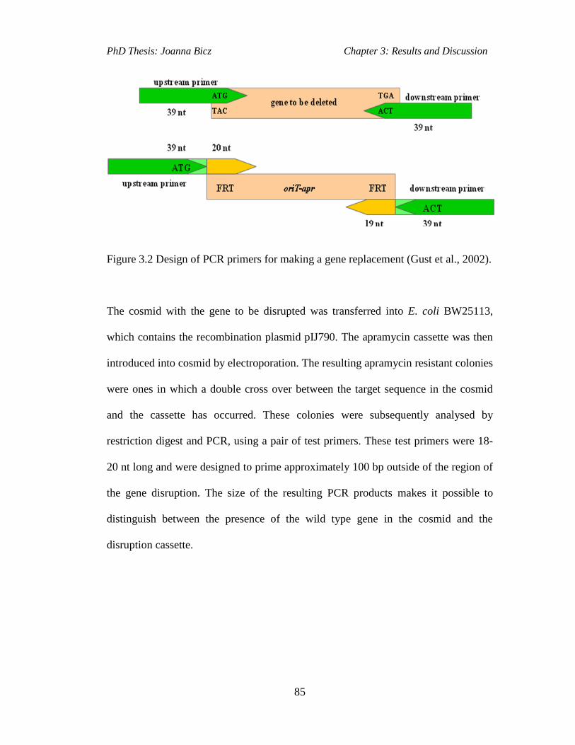
PhD Thesis: Joanna Bicz Chapter 3: Results and Discussion
85
Figure 3.2 Design of PCR primers for making a gene replacement (Gust et al., 2002).
The cosmid with the gene to be disrupted was transferred into E. coli BW25113,
which contains the recombination plasmid pIJ790. The apramycin cassette was then
introduced into cosmid by electroporation. The resulting apramycin resistant colonies
were ones in which a double cross over between the target sequence in the cosmid
and the cassette has occurred. These colonies were subsequently analysed by
restriction digest and PCR, using a pair of test primers. These test primers were 18-
20 nt long and were designed to prime approximately 100 bp outside of the region of
the gene disruption. The size of the resulting PCR products makes it possible to
distinguish between the presence of the wild type gene in the cosmid and the
disruption cassette.

PhD Thesis: Joanna Bicz Chapter 3: Results and Discussion
86
Figure 3.3 Creation of a gene deletion in the cosmid via homologous recombination
of the disruption cassette with cosmid DNA and then conjugation from E. coli into S.
scabies and double homologous recombination resulting in a replacement of the gene
of interest with the disruption cassette creating an S. scabies mutant. Yellow gene –
apramycin resistance, orange region – origin of transfer (oriT), green regions = FRT
sites.
The cosmid with the replaced gene was then introduced via transformation into E.
coli ET12567/pUZ8002, which is a DNA non-methylating strain (Figure 3.3). It is
necessary to use this strain as S. scabies carries a methylated DNA-sensing
restriction system, which degrades methylated DNA from ordinary E. coli strains.
The mutagenised cosmid was then transferred from E. coli ET12567/pUZ8002 to S.
scabies via conjugation. This was completed by mixing the E. coli
ET12567/pUZ8002 cells containing the mutagenised cosmid with S. scabies spores,
and then plating a dilution series on SFM plates. Following an overnight incubation

PhD Thesis: Joanna Bicz Chapter 3: Results and Discussion
87
the plates were overlaid with nalidixic acid, killing only the E. coli, and with
apramycin, which selectively kills the Streptomyces that did not contain introduced
cosmid. As a result of the conjugal transfer of cosmid, the apramycin resistant S.
scabies colonies on the SFM plates should have the target gene replaced with the
resistance cassette by double crossover. The double cross over was recognized by
apramycin resistance (cassette contains apramycin) and kanamycin sensitivity (the
cosmid backbone containing kanamycin resistance gene has been lost during cross
over). The replacement of the gene was confirmed by PCR and Southern blot
hybridization.
3.2 Investigation of Putative Hydroxamate Siderophore Production
in S. scabies
The aim of this work was to examine whether the putative hydroxamate siderophore
(Figure 3.4) is present in culture supernatants of S. scabies 87.22 grown under iron
deficient conditions. It has been reported by our collaborators Kodani and co-
workers that the siderophore was isolated from the culture supernatants of S.
antibioticus NBRC 13838 (the strains that they used in their work). That siderophore
is also proposed to be the product of a cryptic NRPS enzyme encoded by scab85471,
discovered by genome mining of S. scabies 87.22.

PhD Thesis: Joanna Bicz Chapter 3: Results and Discussion
88
3.2.1 Isolation and Structure Elucidation of the Hydroxamate
Siderophore
Analysis of the culture supernatants of S. antibioticus NBRC 13838, via HPLC by
our collaborators, Dr Shinya Kodani and his co-workers, indicated that this strain
produces a putative siderophore that was named scabichelin. Our collaborators also
carried out the structural characterisation of scabichelin. The metabolite was
converted into a complex with Galium before the NMR analysis, which helped signal
dispersion and aided stereochemical analysis. The Ga-scabichelin complex structure
was elucidated by H NMR AND 2D NMR. The NMR spectra analysis indicated that
the compound consists of the five amino acids: N-methylated hydroxy-formyl-
ornithine, serine, N-methylated hydroxy-ornithine, ornithine and cyclic hydroxyl-
ornithine. The mass spectrum had a parent ion present for scabichelin at the m/z
648.3671, which agreed well with the calculated value of 648.3675 corresponding to
[C26H50N9O10]+. The stereochemistry of scabichelin was determined by chemical
analysis using Marfey method (Marfey, 1984) and 3D modelling (Deeth, 2001) as
LLLLD.
HN
N
H O
OH
NH
O
O
OH
NOH
NH
HNO
NH
O
N
O
OH
NH2
Figure 3.4 Structure of a novel siderophore scabichelin.

PhD Thesis: Joanna Bicz Chapter 3: Results and Discussion
89
3.2.2 Scabichelin Biosynthetic Gene Cluster
Analysis of the S. scabies 87.22 genome sequence unveiled a cryptic NRPS
biosynthetic gene cluster, which includes the protein coding sequences from
Scab85431 to Scab85521 (Figure 3.5). All of these proteins, present within the
NRPS biosynthetic gene cluster, are homologous to proteins encoded for by genes
within the coelichelin biosynthetic gene cluster from S. coelicolor M145 (section
1.7.3; Table 3.1; http://www.ncbi.nlm.nih.gov/nuccore/260644157).
Figure 3.5 Organization of the scabichelin biosynthetic gene cluster in S. scabies
87.22.
Bioinformatic analysis of the functional domains of the NRPS encoded for by
scab85471 revealed it contains 17 enzymatic domains organized into 5 modules
(Figure 3.6 B). Each module contains an adenylation (A), a thiolation (T) and a
condensation domain (C) (http://www.nii.res.in/searchall.html). The final
termination module of scab85471 also contains an epimerization/condensation
domain (Figure 3.6 B) suggesting that the stereochemistry of the peptide is L-L-L-L-
D. It is hypothesised that product of the scab85471 gene would be a pentapeptide, as
it contains 5 modules.

PhD Thesis: Joanna Bicz Chapter 3: Results and Discussion
90
Two genes from the putative scabichelin biosynthetic cluster, scab85511 and
scab85521, encode proteins homologous to CchA and CchB from S. coelicolor
M145, respectively. These enzymes have sequence similarity to formyl transferase
and NADH/FAD-dependent monoxygenases, respectively. CchB catalyses the
hydroxylation of L-ornithine (L-Orn) to yield L-N5-hydroxyornithine (L-hOrn) and
CchA subsequently formylates it yielding L-N5-formyl-N5-hydroxyornithine (L-
fhOrn; Pohlman and Marahiel, 2008). These two non-proteinogenic amino acids are
later incorporated into coelichelin. Analogous reactions are predicted to be catalysed
by Scab85511 and Scab85521 in the biosynthesis of the novel hydroxamate
siderophore in S. scabies (Figure 3.6 A).
The amino acids, which are the building blocks for the putative S. scabies product
are loaded onto the thiolation (T) domains by the adenylation (A) domains of the
NRPS encoded by scab85471 (Figure 3.6 B).

PhD Thesis: Joanna Bicz Chapter 3: Results and Discussion
91
A MT C A C A MT
S S
O
HN
H2N
Module 1 Module 2 Module 3
85471
S
O
N
H2N
OHO
H
O
H2N
OH
HO
C A
Module 4
C A
Module 5
E
HO
O
N
H2N
OHO
H
HO
O
HN
H2N
OH
HO
O
H2N
H2N85521 85511
FADNADHO2
fTHF
L-Orn L-hOrn L-fhOrn
A
B
?
L-fhOrn L-hOrnL-Ser
Figure 3.6 A-Proposed role of the enzymes encoded by scab85521 and scab85511 in
the biosynthesis of the amino acids L-N5-hydroxyornithine (L-hOrn) and L-N5-
formyl-N5-hydroxyornithine (L-fhOrn). B-Organisation of modules and domains of
the NRPS encoded by scab85471. The residues attached to the thiolation domains
(black circles) of module 1, 2, 3 are the aminoacids predicted to be incorporated into
scabichelin by each A domain. The substrates loaded by A domains of the modules 4
and 5 could not be unambiguously predicted.
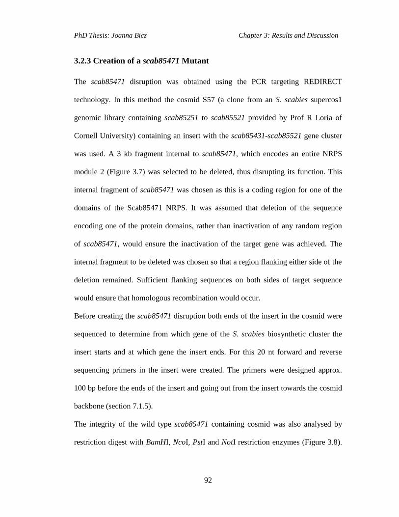
PhD Thesis: Joanna Bicz Chapter 3: Results and Discussion
92
3.2.3 Creation of a scab85471 Mutant
The scab85471 disruption was obtained using the PCR targeting REDIRECT
technology. In this method the cosmid S57 (a clone from an S. scabies supercos1
genomic library containing scab85251 to scab85521 provided by Prof R Loria of
Cornell University) containing an insert with the scab85431-scab85521 gene cluster
was used. A 3 kb fragment internal to scab85471, which encodes an entire NRPS
module 2 (Figure 3.7) was selected to be deleted, thus disrupting its function. This
internal fragment of scab85471 was chosen as this is a coding region for one of the
domains of the Scab85471 NRPS. It was assumed that deletion of the sequence
encoding one of the protein domains, rather than inactivation of any random region
of scab85471, would ensure the inactivation of the target gene was achieved. The
internal fragment to be deleted was chosen so that a region flanking either side of the
deletion remained. Sufficient flanking sequences on both sides of target sequence
would ensure that homologous recombination would occur.
Before creating the scab85471 disruption both ends of the insert in the cosmid were
sequenced to determine from which gene of the S. scabies biosynthetic cluster the
insert starts and at which gene the insert ends. For this 20 nt forward and reverse
sequencing primers in the insert were created. The primers were designed approx.
100 bp before the ends of the insert and going out from the insert towards the cosmid
backbone (section 7.1.5).
The integrity of the wild type scab85471 containing cosmid was also analysed by
restriction digest with BamHI, NcoI, PstI and NotI restriction enzymes (Figure 3.8).
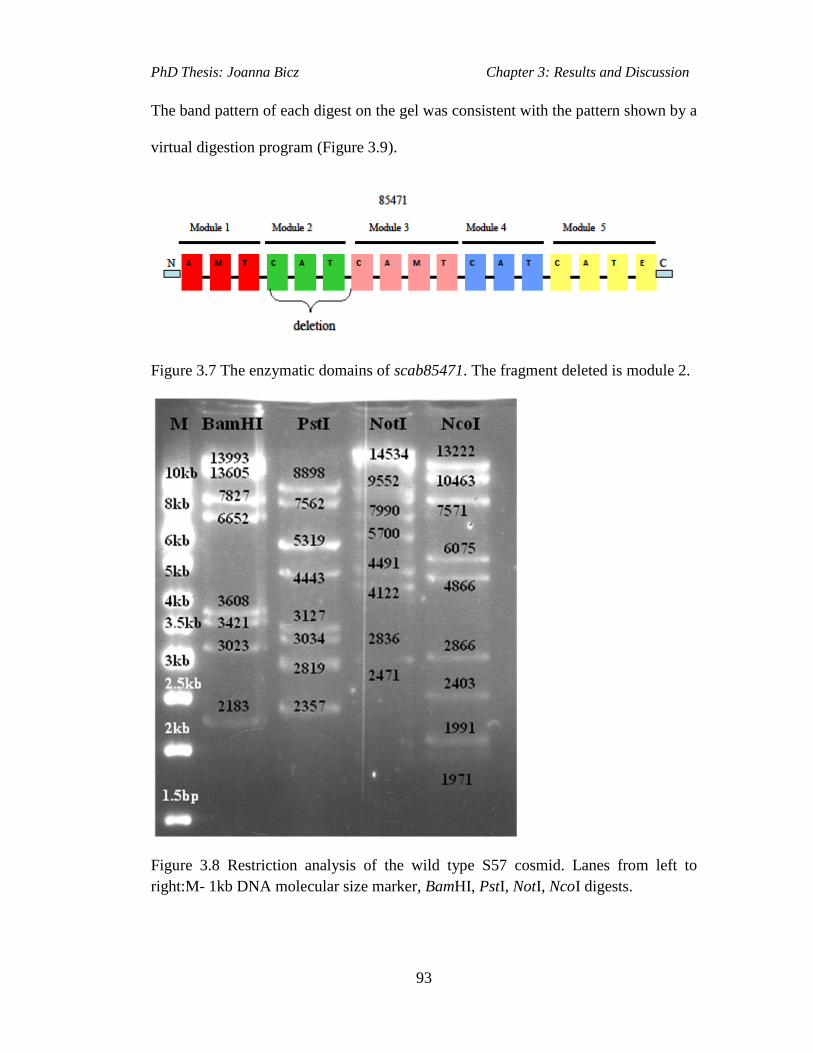
PhD Thesis: Joanna Bicz Chapter 3: Results and Discussion
93
The band pattern of each digest on the gel was consistent with the pattern shown by a
virtual digestion program (Figure 3.9).
Figure 3.7 The enzymatic domains of scab85471. The fragment deleted is module 2.
Figure 3.8 Restriction analysis of the wild type S57 cosmid. Lanes from left to
right:M- 1kb DNA molecular size marker, BamHI, PstI, NotI, NcoI digests.

PhD Thesis: Joanna Bicz Chapter 3: Results and Discussion
94
Figure 3.9 Restriction digest pattern of scabichelin cosmid (S57) with BamH, PstI,
NotI and NcoI restriction enzymes (the band sizes in bp that are displayed red in the
diagram indicate bands present on the gel picture in figure 3.8).
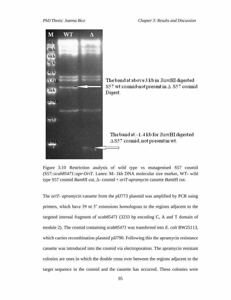
PhD Thesis: Joanna Bicz Chapter 3: Results and Discussion
95
Figure 3.10 Restriction analysis of wild type vs mutagenised S57 cosmid
(S57::scab85471::apr-OriT. Lanes: M- 1kb DNA molecular size marker, WT- wild
type S57 cosmid BamHI cut, Δ- cosmid + oriT-apramycin cassette BamHI cut.
The oriT- apramycin cassette from the pIJ773 plasmid was amplified by PCR using
primers, which have 39 nt 5’ extensions homologous to the regions adjacent to the
targeted internal fragment of scab85471 (3233 bp encoding C, A and T domain of
module 2). The cosmid containing scab85471 was transferred into E. coli BW25113,
which carries recombination plasmid pIJ790. Following this the apramycin resistance
cassette was introduced into the cosmid via electroporation. The apramycin resistant
colonies are ones in which the double cross over between the regions adjacent to the
target sequence in the cosmid and the cassette has occurred. These colonies were

PhD Thesis: Joanna Bicz Chapter 3: Results and Discussion
96
analysed by restriction digest (Figure 3.10) and PCR with a pair of test primers
(Figure 3.11). These primers were 18-20 nt long (Section 7.1.5) and were designed to
prime approximately 100 bp outside of the region of the gene disruption. The size of
the PCR allowed for comparison between the wild type gene in the cosmid and the
gene in the disruption cassette. The test primers generated the expected ~1.6 kb
fragment after gene disruption (1.4kb-the size of apramycin cassette plus ~ 200bp
distance of primers from the cassette and approximately 3kb for the wild type -
Figure 3.11).
Figure 3.11 PCR analyses of the wild type and the mutagenised S57 cosmid with the
test primers. Lanes: M- 1kb DNA molecular size marker, WT gDNA- PCR band for
the wild type S. scabies DNA, WT cos- PCR band for the wild type S57cosmid, Δ
cos-PCR band for the S57 cosmid + oriT-apramycin cassette.
The cosmid with replaced gene was introduced by transformation into E. coli
ET12567/pUZ8002, which is a DNA non-methylating strain. The mutagenised

PhD Thesis: Joanna Bicz Chapter 3: Results and Discussion
97
cosmid was then transferred from E. coli ET12567/pUZ8002 to S. scabies by
conjugation where a double cross over between the homologous regions in S. scabies
genomic DNA and scab85471 on cosmid would occur, yielding a mutant with
inactivated scab85471. The replacement of the gene was confirmed by PCR and
Southern blot hybridization. The test primers (Test_fw and Test_rv; Table 7.6) were
used to analyse replacement of the target sequence in the genome by PCR (Figure
3.12). The mutant was named S. scabies W1000.
Figure 3.12 PCR analysis of the genomic DNA from the putative scabichelin mutant.
Lanes: M- 1kb DNA molecular size marker, WT cos- PCR band for the wild type
scabichelin cosmid, Δ cos-PCR band for the scabichelin cosmid with OriT-
apramycin cassette, Δ gDNA- PCR for the for the scabichelin mutant genomic DNA.
Southern blot hybridization analysis was carried out to analyse the position of the
resistance cassette within the S. scabies W1000 genome. Genomic DNA and the

PhD Thesis: Joanna Bicz Chapter 3: Results and Discussion
98
cosmid containing mutagenised scabichelin gene were digested by BamHI. A DIG-
labelled mutagenised scabichelin cosmid was used as a probe, which was hybridised
to the digested DNA. Comparisons could then be made between the wild type and
mutant on an X-ray film. Bands that could be seen on the x-ray film were highlited in
white, red or green (Figure 3.13).
Figure 3.13 Southern blot hybridization using labelled S57::oriT-apr cosmid as a
probe confirming the nature of S.scabiesW100 mutant with 3 kb fragment deleted
within scab85471 gene. Bands in white colour are all present in the S.scabies wild
type and the mutant. Band 7827 bp in green is only present in the wild type strain.
Bands 1412 bp and 7575 bp coloured in red are characteristic for the mutant.

PhD Thesis: Joanna Bicz Chapter 3: Results and Discussion
99
The mutant was also analysed by sequencing using Test_fw and Test_rv primers
(Section 7.1.5), which confirmed the presence of the apramycin resistance gene in
the S. scabies W1000 genome.
3.2.4 LC-MS Analysis of Scabichelin Mutant
LC-MS analysis of the W1000 scabichelin mutant culture supernatant compared with
that of wild type culture supernatant, with the ferric iron added before analysis,
indicated the presence of a ferric complex in S. scabies 87.22 but not in the W1000
mutant (Figure 3.14). This complex had identical retention time (Figure 3.14) and
MS/MS fragmentation pattern (Figure 3.15) to scabichelin isolated and purified from
S. antibioticus (provided by our collaborators), which was used as an authentic
standard. These data conclusively proves that scabichelin is the metabolic product of
the Scab85471 NRPS in S. scabies 87.22.
Figure 3.14 EICs at m/z=351.14 (corresponding to the [M+2H]
2+ ion for the ferric-
scabichelin) from analyses of the culture supernatant of S. scabies 87.22 (top panel),
Fe-scabichelin purified from S. antibioticus (middle panel) and the culture
supernatant of S. scabies W1000 (bottom panel).
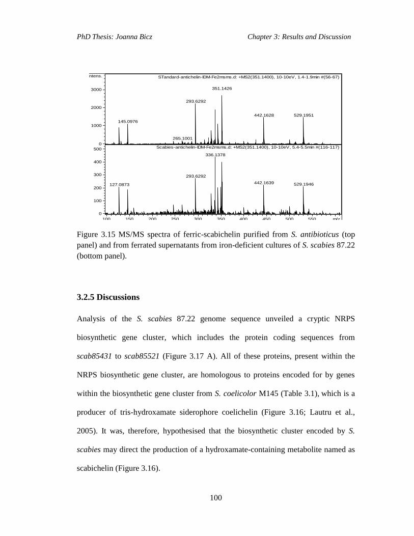
PhD Thesis: Joanna Bicz Chapter 3: Results and Discussion
100
145.0976
265.1001
293.6292
351.1426
442.1628 529.1951
STandard-antichelin-IDM-Fe2msms.d: +MS2(351.1400), 10-10eV, 1.4-1.9min #(56-67)
127.0873
293.6292
336.1378
442.1639 529.1946
Scabies-antichelin-IDM-Fe2msms.d: +MS2(351.1400), 10-10eV, 5.4-5.5min #(116-117)0
1000
2000
3000
Intens.
0
100
200
300
400
500
100 150 200 250 300 350 400 450 500 550 m/z
Figure 3.15 MS/MS spectra of ferric-scabichelin purified from S. antibioticus (top
panel) and from ferrated supernatants from iron-deficient cultures of S. scabies 87.22
(bottom panel).
3.2.5 Discussions
Analysis of the S. scabies 87.22 genome sequence unveiled a cryptic NRPS
biosynthetic gene cluster, which includes the protein coding sequences from
scab85431 to scab85521 (Figure 3.17 A). All of these proteins, present within the
NRPS biosynthetic gene cluster, are homologous to proteins encoded for by genes
within the biosynthetic gene cluster from S. coelicolor M145 (Table 3.1), which is a
producer of tris-hydroxamate siderophore coelichelin (Figure 3.16; Lautru et al.,
2005). It was, therefore, hypothesised that the biosynthetic cluster encoded by S.
scabies may direct the production of a hydroxamate-containing metabolite named as
scabichelin (Figure 3.16).

PhD Thesis: Joanna Bicz Chapter 3: Results and Discussion
101
N
H O
OH
NH
O
O
NOH
H2N
OH
H
N
HO
OHO
ONH2
HO
HN
N
H O
OH
NH
O
O
OH
NOH
NH
HNO
NH
O
N
O
OH
NH2
scabichelin
coelichelin
NH
Figure 3.16 Structures of coelichelin and a novel siderophore scabichelin.
PCR targeting method (Gust et al., 2003) was used to create a mutant of S. scabies
87.22 in order to investigate the role of the cryptic nonribosomal peptide biosynthetic
gene cluster in scabichelin biosynthesis. The mutant, named as S. scabies W1000,
was obtained via replacement of an internal fragment of the scab85471 gene with an
oriT-apr cassette. LC-MS/MS analyses of culture supernatants of the 87.22 and
W1000 strains grown in an iron-deficient medium, to which ferric iron was added
before analysis, indicated the presence of a ferric complex that is produced by S.
scabies 87.22, but not the W1000 mutant. LC-MS retention time and MS/MS
fragmentation pattern of ferri-scabichelin are the same for S. scabies 87.22
compound and the authentic standard. The experimental data confirm that
scab85431-scab85521 gene cluster is involved in the production of scabichelin.
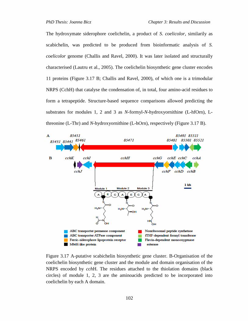
PhD Thesis: Joanna Bicz Chapter 3: Results and Discussion
102
The hydroxymate siderophore coelichelin, a product of S. coelicolor, similarily as
scabichelin, was predicted to be produced from bioinformatic analysis of S.
coelicolor genome (Challis and Ravel, 2000). It was later isolated and structurally
characterised (Lautru et al., 2005). The coelichelin biosynthetic gene cluster encodes
11 proteins (Figure 3.17 B; Challis and Ravel, 2000), of which one is a trimodular
NRPS (CchH) that catalyse the condensation of, in total, four amino-acid residues to
form a tetrapeptide. Structure-based sequence comparisons allowed predicting the
substrates for modules 1, 2 and 3 as N-formyl-N-hydroxyornithine (L-hfOrn), L-
threonine (L-Thr) and N-hydroxyornithine (L-hOrn), respectively (Figure 3.17 B).
Figure 3.17 A-putative scabichelin biosynthetic gene cluster. B-Organisation of the
coelichelin biosynthetic gene cluster and the module and domain organisation of the
NRPS encoded by cchH. The residues attached to the thiolation domains (black
circles) of module 1, 2, 3 are the aminoacids predicted to be incorporated into
coelichelin by each A domain.
PhD Thesis: Joanna Bicz Chapter 3: Results and Discussion
103
Unlike the proposed biosynthesis of scabichelin, where C domains of five modules
of the NRPS are predicted to catalyse condensation of five amino acyl thioesters,
coelichelin biosynthetic model (Figure 3.18) involves iterative usage of module 1 to
incorporate two molecules of N-formyl-N-hydroxyornithine into the peptide (Lautru
et al., 2005). In the first step, the A domains of modules 1, 2, 3 activate the three
substrates, L-hfOrn, L-Thr and L-hOrn as aminoacyl adenylates and subsequently
transfer them onto the adjacent T domains to form the corresponding aminoacyl
thioesters (Figure 3.18; 1). The α-carbons of the thioesters undergo epimerization
catalysed by E domains of module 1 and 2. In the second step, the C domains of the
module 2 and 3 catalyse sequential condensation of the thioesters of the three
substrates, L-hfOrn, L-Thr and L-hOrn to form a tripeptide tethered to the T domain
of module 3 (Figure 3.18; 2). In the third step, a second molecule of L-hfOrn is
loaded onto T domain of module 1 and the α-carbon of the resulting thioester is
epimerized by the module 1 E domain (Figure 3.18; 3). In the fourth step the C
domain of either module 2 or module 3 catalyses the condensation of the module 3
attached tripeptide thioester with the module 1 attached hOrn thioester yielding
tetrapeptide thioester attached to the T domain of module 3 (Figure 3.18; 4). In the
final stage, the tetrapeptide thioester is released from the NRPS by hydrolysis
catalysed by CchJ (Figure 3.18, 5), a protein encoded by one of the genes in the cch
biosynthetic cluster. Usually the C-terminal TE domain is used by NRPS for the
cleavage of the assembled peptide and the CchH was not found to contain one.
However, the examination of other genes in the cch cluster identified an orthologue
of an esterase, which catalyses hydrolytic cleavage of an enterobactin siderophore
into smaller subunits in order to release iron from the ferric-enterobactin complex
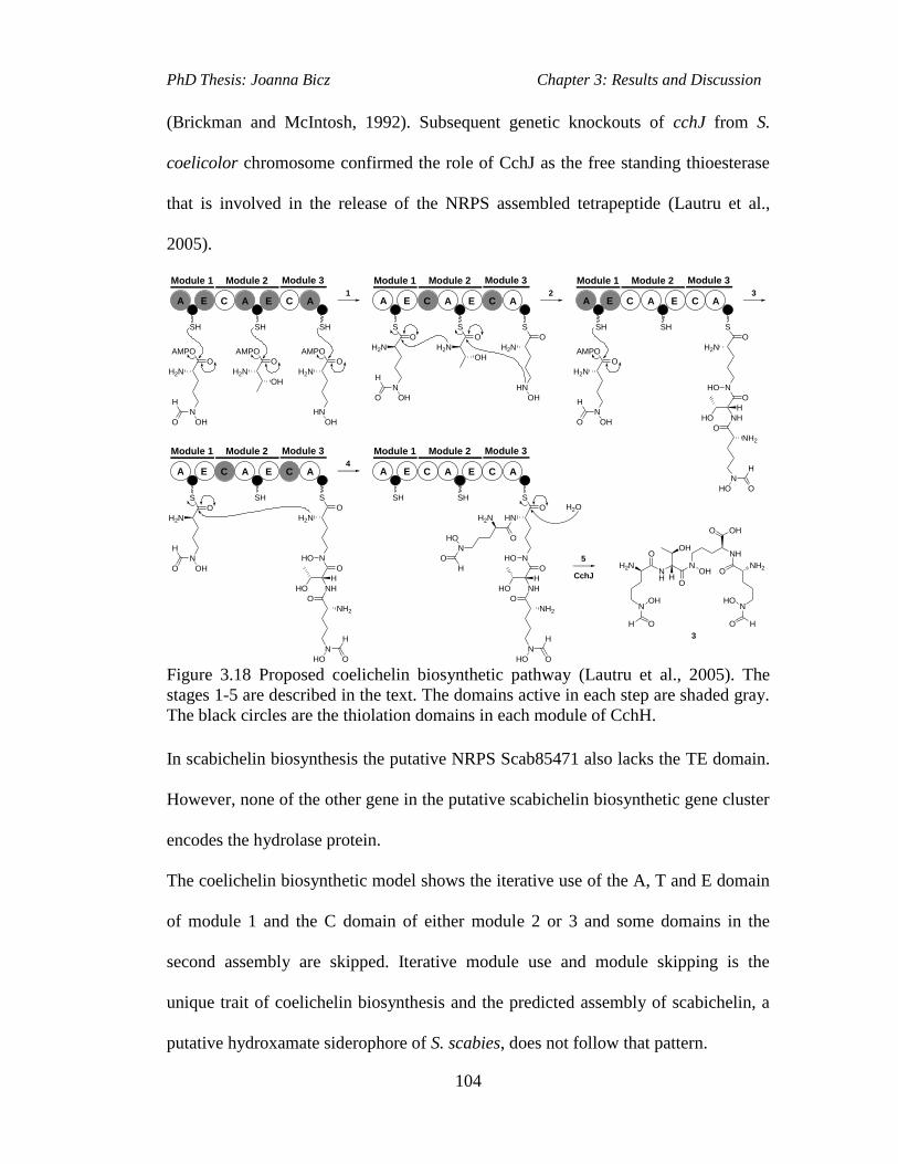
PhD Thesis: Joanna Bicz Chapter 3: Results and Discussion
104
(Brickman and McIntosh, 1992). Subsequent genetic knockouts of cchJ from S.
coelicolor chromosome confirmed the role of CchJ as the free standing thioesterase
that is involved in the release of the NRPS assembled tetrapeptide (Lautru et al.,
2005).
A E C A E C A
SH SH SH
A E C A E C A
S
O
N
H2N
OHO
S
O
H2N
OH
S
O
HN
H2N
OH
A E C A E C A
SH SH S
O
N
H2N
HO
O
NHHO
O
N
NH2
HO O
H
H
H
A E C A E C A
SH S
O
N
H2N
HO
O
NHHO
O
N
NH2
HO O
H
H
S
O
N
H2N
OHO
H
AMPO
O
N
H2N
OHO
H
AMPO
O
H2N
OH
AMPO
O
HN
H2N
OH
1 2 3
4A E C A E C A
SH S
O
N
HN
HO
O
NHHO
O
N
NH2
HO O
H
H
5
O
N
H2N
HO
H
O
SH
CchJ
H2NNH
OOH
N
O
OH
NH
ONH2
NOH
O
NHO
O
O OH
H
H H
H2O
AMPO
O
N
H2N
OHO
H
Module 1 Module 2 Module 3 Module 1 Module 2 Module 3 Module 1 Module 2 Module 3
Module 1 Module 2 Module 3 Module 1 Module 2 Module 3
3
Figure 3.18 Proposed coelichelin biosynthetic pathway (Lautru et al., 2005). The
stages 1-5 are described in the text. The domains active in each step are shaded gray.
The black circles are the thiolation domains in each module of CchH.
In scabichelin biosynthesis the putative NRPS Scab85471 also lacks the TE domain.
However, none of the other gene in the putative scabichelin biosynthetic gene cluster
encodes the hydrolase protein.
The coelichelin biosynthetic model shows the iterative use of the A, T and E domain
of module 1 and the C domain of either module 2 or 3 and some domains in the
second assembly are skipped. Iterative module use and module skipping is the
unique trait of coelichelin biosynthesis and the predicted assembly of scabichelin, a
putative hydroxamate siderophore of S. scabies, does not follow that pattern.
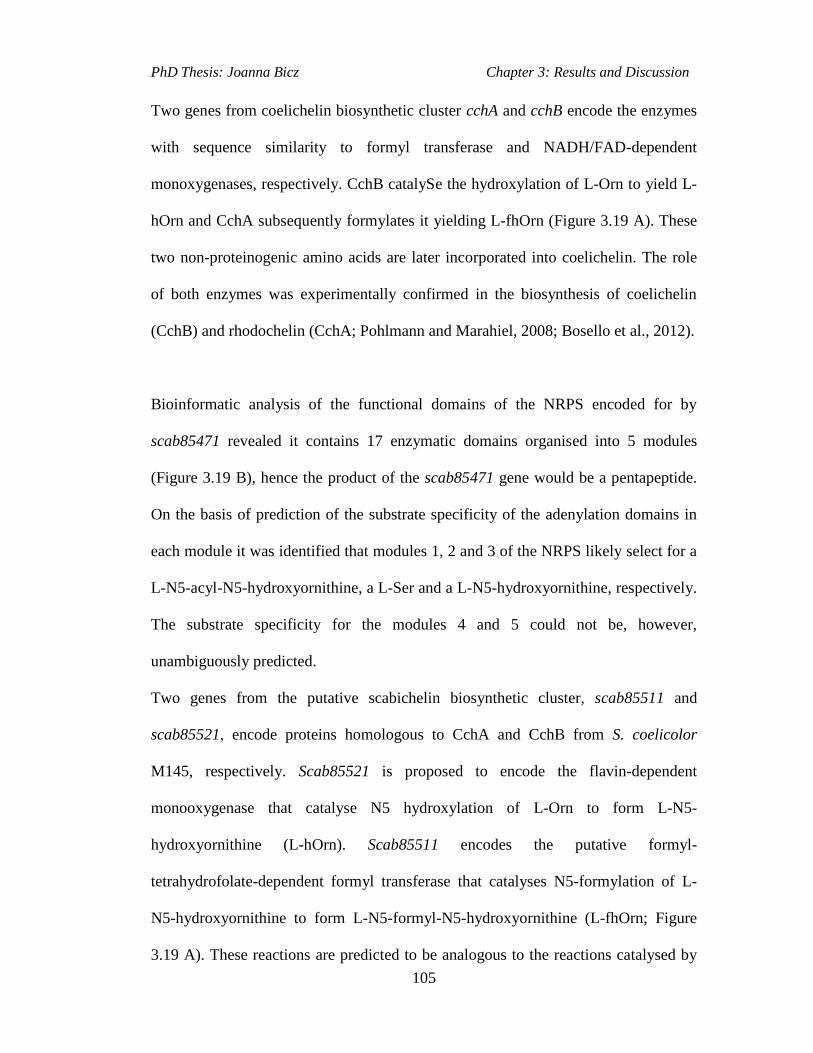
PhD Thesis: Joanna Bicz Chapter 3: Results and Discussion
105
Two genes from coelichelin biosynthetic cluster cchA and cchB encode the enzymes
with sequence similarity to formyl transferase and NADH/FAD-dependent
monoxygenases, respectively. CchB catalySe the hydroxylation of L-Orn to yield L-
hOrn and CchA subsequently formylates it yielding L-fhOrn (Figure 3.19 A). These
two non-proteinogenic amino acids are later incorporated into coelichelin. The role
of both enzymes was experimentally confirmed in the biosynthesis of coelichelin
(CchB) and rhodochelin (CchA; Pohlmann and Marahiel, 2008; Bosello et al., 2012).
Bioinformatic analysis of the functional domains of the NRPS encoded for by
scab85471 revealed it contains 17 enzymatic domains organised into 5 modules
(Figure 3.19 B), hence the product of the scab85471 gene would be a pentapeptide.
On the basis of prediction of the substrate specificity of the adenylation domains in
each module it was identified that modules 1, 2 and 3 of the NRPS likely select for a
L-N5-acyl-N5-hydroxyornithine, a L-Ser and a L-N5-hydroxyornithine, respectively.
The substrate specificity for the modules 4 and 5 could not be, however,
unambiguously predicted.
Two genes from the putative scabichelin biosynthetic cluster, scab85511 and
scab85521, encode proteins homologous to CchA and CchB from S. coelicolor
M145, respectively. Scab85521 is proposed to encode the flavin-dependent
monooxygenase that catalyse N5 hydroxylation of L-Orn to form L-N5-
hydroxyornithine (L-hOrn). Scab85511 encodes the putative formyl-
tetrahydrofolate-dependent formyl transferase that catalyses N5-formylation of L-
N5-hydroxyornithine to form L-N5-formyl-N5-hydroxyornithine (L-fhOrn; Figure
3.19 A). These reactions are predicted to be analogous to the reactions catalysed by

PhD Thesis: Joanna Bicz Chapter 3: Results and Discussion
106
CchA and CchB, enzymes involved in the biosynthesis of non-proteogenic amino
acid residues hOrn and fhOrn that are utilised in the biosynthesis of coelichelin
(Pohlmann and Marahiel, 2008). The amino acids, which are the building blocks for
the putative S. scabies product are loaded onto the thiolation (T) domains by the
adenylation (A) domains of the NRPS encoded by scab85471 (Figure 3.19 B). The
methylation (MT) domains of the NRPS are proposed to catalyse the N-methylation
of the aminoacyl thioesters attached to the T domains within module 1 and 3. The
epimerization (E) domain within module 5 is proposed to catalyze α-carbon
epimerization of the L-hOrn residue attached to the T domain of module 5 and the
condensation (C) domains of modules 2 to 5 are proposed to catalyse peptide bond
formation between the T domain-bound aminoacyl thioesters to yield pentapeptidyl
thioester attached to the T domain of module 5 before it is finally released from the
NRPS (Figure 3.19 B).

PhD Thesis: Joanna Bicz Chapter 3: Results and Discussion
107
A MT C A C A MT
S S
O
HN
MeHN
Module 1 Module 2 Module 3
85471
S
O
N
MeHN
OHO
H
O
H2N
OH
HO
C A
S
Module 4
O
H2N
H2N
C A
S
Module 5
H2N
HN
OH
E
NHMe
HN
O
N
O
HO
NHMe
N
OHO
H
OH
O
HNNH
N
OHO
O
NH2
O
HO
O
N
H2N
OHO
H
HO
O
HN
H2N
OH
HO
O
H2N
H2N85521 85511
FADNADHO2
fTHF
L-Orn L-hOrn L-fhOrn
A
B
-5H+
Figure 3.19 A-Proposed role of the enzymes encoded by scab85521 and scab85511
in the biosynthesis of the amino acids L-N5-hydroxyornithine (L-hOrn) and L-N5-
formyl-N5-hydroxyornithine (L-fhOrn). B-Organisation of modules and domains of
the NRPS encoded by scab85471. The residues attached to the thiolation (T)
domains (black circles) of the five modules are the aminoacids proposed to be
incorporated into scabichelin.
There are two hypothetical ways of scabichelin release from the NRPS. One of them
is via intramolecular addition of the δ-hydroxylamino group of the D-hOrn residue to
the carbonyl group of the thioester, and subsequent deprotonation and release of the
resulting product (Figure 3.19 B). This mechanism for peptide chain release is
analogous to that proposed for another putative siderophore called amychelin (Figure
3.21) from Amycolatopsis sp. AA4 (Seyedsayamdost et al., 2011). Both amychelin
and scabichelin, contains D-cyclo-hOrn residue and both of the putative siderophore
NRPSs lack the C-terminal thioesterase (TE) domain, which is responsible for chain
release in nonribosomal peptide biosynthesis. In amychelin that reaction is proposed

PhD Thesis: Joanna Bicz Chapter 3: Results and Discussion
108
to be catalysed by AcmB, a putative standalone α, β-hydrolase (Figure 3.20).
Scabichelin biosynthetic gene cluster, however, does not encode a protein with
sequence similarity to an α, β-hydrolase. Therefore, the reaction may be catalysed by
α, β-hydrolase encoded elsewhere in the S. scabies genome. Some epimerization
domains may also be involved in the peptide release. The epimerization (E) domain
of module 5 of the scabichelin NRPSs may not only catalyse the epimerization of the
module 5 bound L-hOrn residue, but it may also catalyse the subsesquent cleavage of
the assembled peptide. In vitro experiments are required to elucidate the peptide
release mechanisms.
Further work, in collaboration with Dr. Bignell (former member of professor R.
Loria group at Cornell University) and co-workers, would be to examine the effect of
scabichelin biosynthetic gene inactivation on plant pathogenicity and would involve
the use of a plant assay to compare the necrotic effect of S. scabies 87.22 and W1000
strains grown in parallel on tuber and root vegetables such as potato and radish,
respectively.
Figure 3.20 Amychelin biosynthetic gene cluster in Amycolatopsis sp. AA4
(Seyedsayamdost at al., 2011).

PhD Thesis: Joanna Bicz Chapter 3: Results and Discussion
109
The amychelin biosynthetic gene cluster is 35.2 kb in size and consists of 16 genes
(SSMG_02531 to SSMG_02546). It includes four NRPS genes (amcDEFG), amino
acid tailoring genes (amcKP), chain initiation and termination genes (amcBCHL) and
transport genes (amcAIJMON). AmcK is 61% identical to CchB in S. coelicolor a
flavin-dependent monooxygenase and amcP is encoded outside of the amc gene
cluster and it encodes a protein that is 74% identical to CchA the formyltransferase
in coelichelin biosynthetic pathway. Thus, AmcK and AmcP are proposed to catalyse
formation of the two nonproteinogenic precursor amino acids N-hydroxyornithine
(hOrn) and N-hydroxy-N-formylornithine (hfOrn), respectively.
AmcL is proposed to encode salicylate synthase that catalyses the salicylate from
chorismate in proposed amychelin biosynthesis (Figure 3.21; Seyedsayamdost et al.,
2011). AmcH is proposed to encode hydroxybenzoyl-AMP ligase, which likely
catalyses adenylation of salicylate and its subsequent loading onto the aryl carrier
protein (ArCP) domain of AmcG. The substrate specificity of A domain in AmcG is
predicted to be cysteine, though the serine is incorporated. The cyclisation domain
(Cy) in AmcG is then proposed to catalyse oxazoline ring formation and the chain is
then transferred to AmcF. The two A domains of AmcF have substrate specificity for
serine and after the incorporation of second serine the E domain in AmcF catalyzes
epimerization of second serine α-carbon. The chain is then passed on to AmcD,
where it is condensed with another serine and with N-hydroxy-N-formylornithine
(hfOrn) residue, a substrates predicted to be selected by the A domains of AmcD.
The E domain in AmcD generates D form of hfOrn. The A domain of the final NRPS
AmcE is predicted to select for glutaminic acid. After condensation of hfOrn and

PhD Thesis: Joanna Bicz Chapter 3: Results and Discussion
110
epimerization of its α-carbon the peptide is proposed to be released from the AmcE
by AmcB a putative α, β hydrolase encoded in amc cluster. AmcE does not contain
the C-terminal TE domain and the amychelin release mechanisms resembles that of
coelichelin, where free standing esterase performs that function (Seyedsayamdost et
al., 2011).
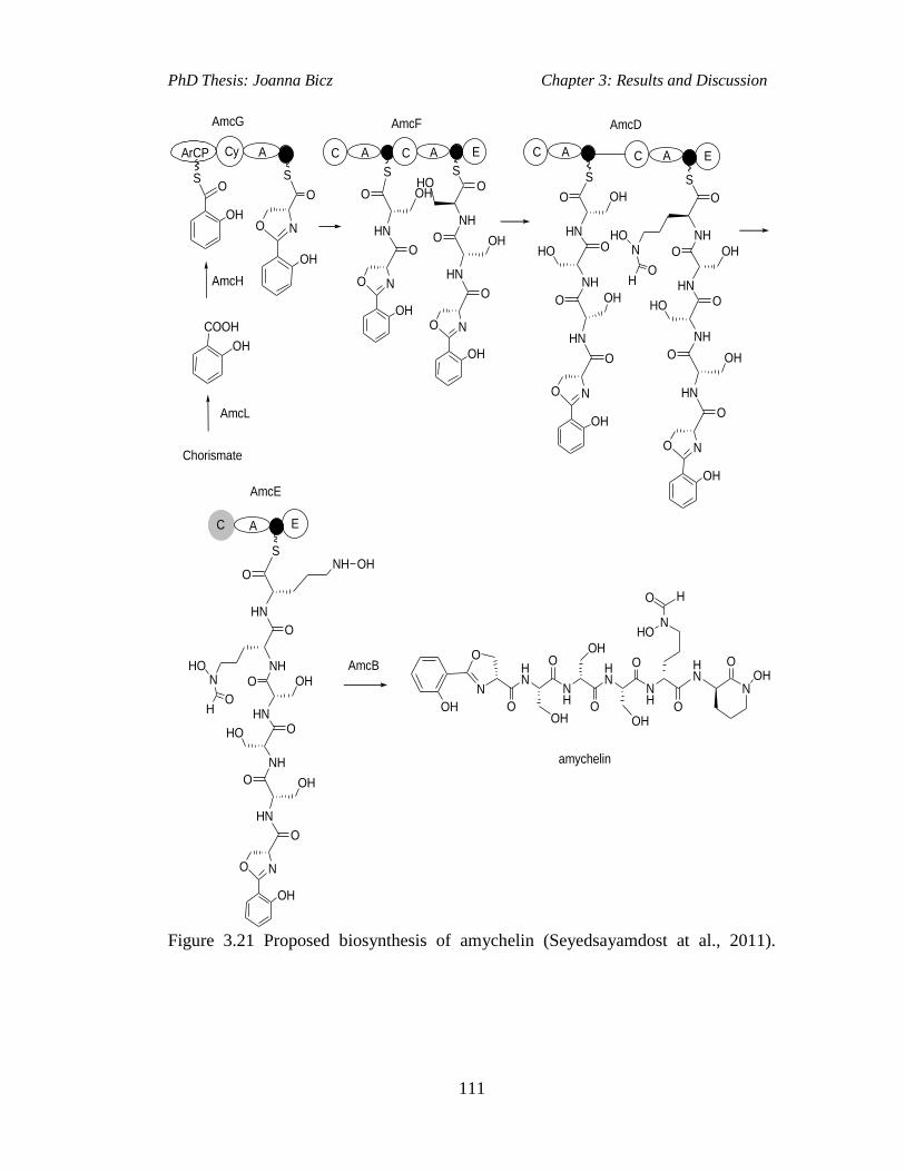
PhD Thesis: Joanna Bicz Chapter 3: Results and Discussion
111
COOH
CyArCP
OH
Chorismate
AmcL
AmcH
O
OH
O
NO
OH
A
SS
AmcG
E
S
HN
O
NO
OH
O
S
NH
O
HN
O
NO
OH
O OH
A A
OHHO
AmcF
A
S
NH
O
HN
O
NO
OH
O
O
HO
HN
OH
NH
O
HN
O
NO
OH
O OH
O
HO
HN
OH
S
NH
A A E
OH
O
NHO
OH
AmcD
C C C C
A E
A
NH
O
HN
O
NO
OH
O OH
O
HO
HN
OHNH
O
NHO
OH
S
HN
NH OHO
AmcB
C
AmcE
OH
N
OHN
NH
HN
NH
HN
OO
O O
OOH
OH
OH
N
HO
HO
N
OOH
amychelin
Figure 3.21 Proposed biosynthesis of amychelin (Seyedsayamdost at al., 2011).

PhD Thesis: Joanna Bicz Chapter 4: Results and Discussion
112
4. Desferrioxamine production in S. scabies 87.22
4.1 The Aims of This Work
Desferrioxamines are a group of ferric iron binding molecules consisting of
hydroxamic acid ligands (Challis and Kadi, 2009). Unlike pyochelin and scabichelin,
which are synthesised by multidomain NRPS enzymes, desferrioxamines are
synthesised via an NRPS-independent pathway (Challis and Kadi, 2009).
Desferrioxamines are produced by a broad range of bacteria including many
Streptomyces species (Bickel et al., 1960) and analysis of the S. scabies genome
unveiled a putative desferrioxamine biosynthetic gene cluster.
The aim of this work was to establish if the putative desferrioxamine biosynthetic
gene cluster in S. scabies directs the biosynthesis of desferrioxamine and to
investigate the role of the desC gene in desferrioxamine biosynthesis. This was
achieved by creating a desC gene deletion in S. scabies using the PCR targeting
technology and examining the production of desferrioxamine in the desC mutant
strain. Subsequent chemical complementation of the desC mutant by the introduction
of a precursor from the desferrioxamine biosynthetic pathway into desC mutant
liquid culture was analysed to establish if desferrioxamine production could be
restored. The desC mutant in S. scabies was also genetically complemented by in
trans expression of the desC gene from the integrative plasmid pOSV556.

PhD Thesis: Joanna Bicz Chapter 4: Results and Discussion
113
4.2 Putative Desferrioxamine Biosynthetic Gene Cluster in S. scabies
Analysis of the S. scabies 87.22 genome sequence identified a cryptic biosynthetic
gene cluster, which includes the protein coding sequences Scab57921-Scab57981
(Figure 4.1). These proteins are homologs of proteins encoded by genes in
desferrioxamne biosynthetic gene cluster of S. coelicolor A (3)2 (Table 4.1).
Figure 4.1 Putative desferrioxamine biosynthetic gene cluster in S. scabies 87.22.

PhD Thesis: Joanna Bicz Chapter 4: Results and Discussion
114
Table 4.1 Putative proteins functions encoded by the desferrioxamine biosynthetic
gene cluster in S. scabies 87.22.
S. scabies
gene
desferrioxamine
biosynthesis protein (Des)
Sequence similarity with S. coelicolor
A3(2) des cluster (%)
scab57921 DesD hypothetical protein SCO2785 (81%)
scab57931 DesC aceytltranferase SCO2784 (79%)
scab57941 DesB monooxygenase SCO2783 (80%)
scab57951 DesA pyridoxal-dependent decarboxylase
SCO2782 (86%)
scab57961 IS630 family insertion
sequence
no similar protein
scab57971 DesE hypothetical protein SCO2781 (75%)
scab57981 DesF secreted protein SCO2780 (73%)
The desC gene, which is of interest, encodes for an enzyme responsible for
catalysing the acylation of N-hydroxycadaverine to form N-hydroxy-N-
succinylcadaverine, a precursor in desferrioxamines biosynthesis in Streptomyces
coelicolor (Kadi amd Challis, unpublished data; Figure 4.2).

PhD Thesis: Joanna Bicz Chapter 4: Results and Discussion
115
H2N CO2H
NH2
L-lysine
H2NHN
OH
O OOH S
CoA
DesC ?
N-hydroxycadaverine
H2N N
OH
O
O
OH
N-hydroxy-N-succinylcadaverine (HSC)
DesA
H2N NH2
cadaverine
DesB
N NH
N
NH
N
HN
O
O
O
O
O
O
OHOH
HO
Mg2+
ATP
AMP
+ PPi
DesD
Desferrioxamine E
Figure 4.2 Proposed desferrioxamine biosynthetic pathway in S. coelicolor (Kadi et
al., 2007).

PhD Thesis: Joanna Bicz Chapter 4: Results and Discussion
116
4.3 Construction of desC Mutant in S. scabies 87.22
The desC mutant was previously made with S. scabies ATCC 49173 (Loria group,
unpublished data). However, before continuing with the desC complementation
experiment, it has been decided that the desC mutant should be made again in S.
scabies 87.22, which is the wild type strain used by our collaborators at Cornell
University. The reason for making desC mutant in S. scabies 87.22 was to investigate
function of that gene and desferrioxamine production in this strain. Specifically after
the desC mutant of S. scabies 87.22 has been made, the complementation of this
mutant was attempted.
The desC mutant in S. scabies 87.22 was created using the same strategy as that used
to create the scabichelin mutant i.e. PCR targeting and subsequent conjugation to the
wild type S. scabies 87.22 strain. The cosmid containing the whole des cluster with
the oriT-apramycin resistance cassette in place of the desC gene, which is only 555
bases in length, was used for the construction of the mutant (the sequence of des
cluster and the cosmid was obtained from our collaborators). This cosmid was
introduced into E. coli ET12567 by electroporation and then transferred by
conjugation from E. coli ET12567 to S. scabies 87.22. The kanamycin sensitive,
apramycin resistant Streptomyces colonies were then selected and their genomic
DNA was isolated and analysed by PCR. The PCR (Figure 4.3) was performed using
18 nt long test primers (Table 7.6) designed within 100 bp region upstream and
downstream of desC gene in S. scabies 87.22 genome.
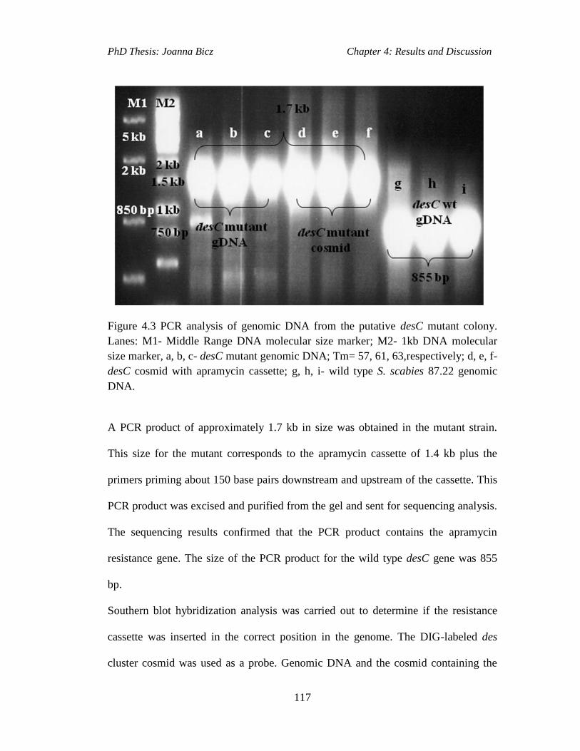
PhD Thesis: Joanna Bicz Chapter 4: Results and Discussion
117
Figure 4.3 PCR analysis of genomic DNA from the putative desC mutant colony.
Lanes: M1- Middle Range DNA molecular size marker; M2- 1kb DNA molecular
size marker, a, b, c- desC mutant genomic DNA; Tm= 57, 61, 63,respectively; d, e, f-
desC cosmid with apramycin cassette; g, h, i- wild type S. scabies 87.22 genomic
DNA.
A PCR product of approximately 1.7 kb in size was obtained in the mutant strain.
This size for the mutant corresponds to the apramycin cassette of 1.4 kb plus the
primers priming about 150 base pairs downstream and upstream of the cassette. This
PCR product was excised and purified from the gel and sent for sequencing analysis.
The sequencing results confirmed that the PCR product contains the apramycin
resistance gene. The size of the PCR product for the wild type desC gene was 855
bp.
Southern blot hybridization analysis was carried out to determine if the resistance
cassette was inserted in the correct position in the genome. The DIG-labeled des
cluster cosmid was used as a probe. Genomic DNA and the cosmid containing the
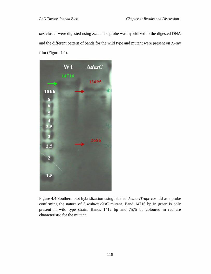
PhD Thesis: Joanna Bicz Chapter 4: Results and Discussion
118
des cluster were digested using SacI. The probe was hybridized to the digested DNA
and the different pattern of bands for the wild type and mutant were present on X-ray
film (Figure 4.4).
Figure 4.4 Southern blot hybridization using labeled des::oriT-apr cosmid as a probe
confirming the nature of S.scabies desC mutant. Band 14716 bp in green is only
present in wild type strain. Bands 1412 bp and 7575 bp coloured in red are
characteristic for the mutant.

PhD Thesis: Joanna Bicz Chapter 4: Results and Discussion
119
4.3.1 LC-MS Analysis of desC Mutant
The metabolic profile of the desC mutant was analysed by LC-MS. The desC mutant
strain was grown in 50 ml Minimal Liquid Medium (SMM) in parallel with 50 ml
SMM culture of S. scabies 87.22 wild type at 30° C, 180 rpm for 5 days. The culture
extracts were then analysed via LC-MS for the presence of ferrioxamine B and E.
Analysis of the extracted ion chromatograms for the m/z of 614 (ferrioxamine B;
Figure 4.5 A) and 654 (ferrioxamine E; Figure 4.5 B) indicated that these compounds
are present in cultures of the wild type strain. However, compounds with the same
m/z and retention time could not be seen in the desC mutant (Figure 4.5 A, B).
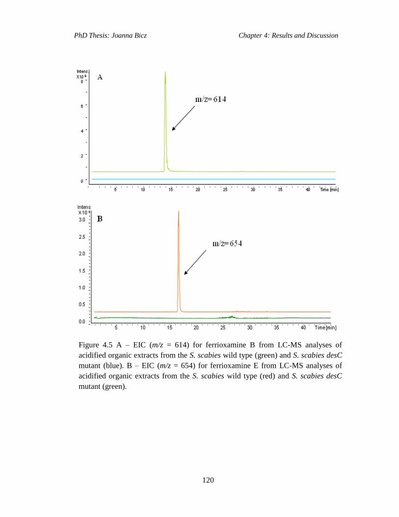
PhD Thesis: Joanna Bicz Chapter 4: Results and Discussion
120
Figure 4.5 A – EIC (m/z = 614) for ferrioxamine B from LC-MS analyses of
acidified organic extracts from the S. scabies wild type (green) and S. scabies desC
mutant (blue). B – EIC (m/z = 654) for ferrioxamine E from LC-MS analyses of
acidified organic extracts from the S. scabies wild type (red) and S. scabies desC
mutant (green).
PhD Thesis: Joanna Bicz Chapter 4: Results and Discussion
121
4.4 Chemical Complementation of desC Mutant
The aim of this work was chemical complementation of the S. scabies desC mutant
to illustrate that reintroduction of the N-hydroxy-N-succinylcadaverine (HSC)
precursor (Figure 4.6) into the desferrioxamine biosynthetic pathway would restore
the production of desferrioxamines in this mutant. This was carried out by setting up
three tubes with 5 ml of SMM medium each. Two were inoculated with the desC
mutant spores, and S. scabies 87.22 wild type strain was used to inoculate the third.
The HSC precursor was added to one of the cultures in which the desC mutant was
growing. The other culture with desC mutant and the one with wild type S. scabies
remained as the controls with no HSC added to either one. Following 72 hours
incubation the culture supernatants were analysed by LC-MS. The analysis indicated
restoration of desferrioxamine B production in the culture of desC mutant chemically
complemented with HSC (Figure 4.7). However, desferrioxamine E (m/z=654) was
not detected in the culture of desC mutant chemically complemented with HSC.
Desferrioxamine was not detected in the culture supernatant of desC mutant with no
HSC added (Figure 4.7).
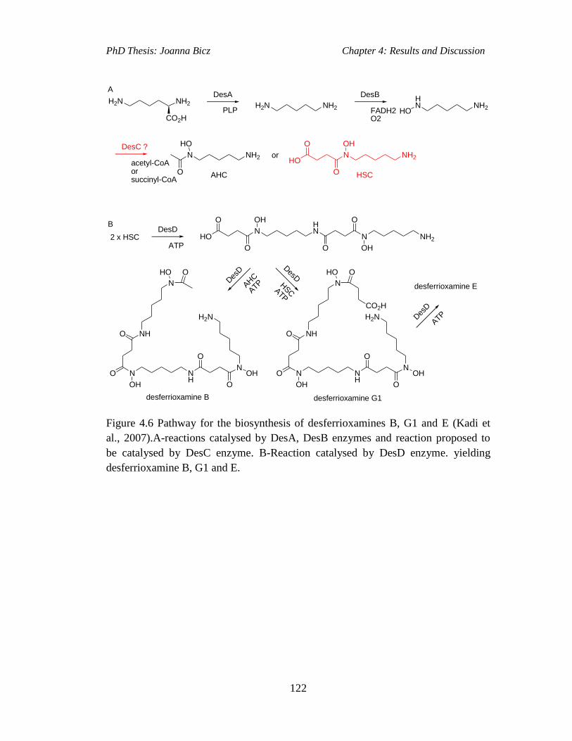
PhD Thesis: Joanna Bicz Chapter 4: Results and Discussion
122
NH2H2N
CO2HPLP
DesA
NH2H2N
DesB
FADH2O2
NH2
HN
HO
DesC ?
acetyl-CoAorsuccinyl-CoA
NH2N
HO
O
NH2N
OH
O
HO
O
or
AHC
2 x HSCDesD
ATP
N
OH
O
HO
O
NH2N
OH
OHN
O
N
OH
OHN
O
NH
O
O
NHO
H2N
N
HO O
N
OH
OHN
O
NH
O
O
NHO
H2N
N
HO O
CO2H
Des
D
AHC
ATP desferrioxamine E
DesD
HSCATP
Des
D
ATP
HSC
A
B
desferrioxamine B desferrioxamine G1
Figure 4.6 Pathway for the biosynthesis of desferrioxamines B, G1 and E (Kadi et
al., 2007).A-reactions catalysed by DesA, DesB enzymes and reaction proposed to
be catalysed by DesC enzyme. B-Reaction catalysed by DesD enzyme. yielding
desferrioxamine B, G1 and E.
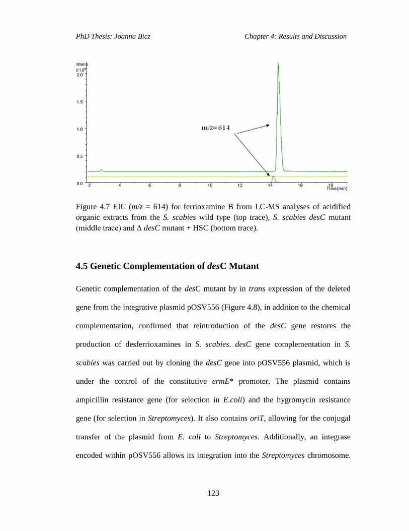
PhD Thesis: Joanna Bicz Chapter 4: Results and Discussion
123
Figure 4.7 EIC (m/z = 614) for ferrioxamine B from LC-MS analyses of acidified
organic extracts from the S. scabies wild type (top trace), S. scabies desC mutant
(middle trace) and Δ desC mutant + HSC (bottom trace).
4.5 Genetic Complementation of desC Mutant
Genetic complementation of the desC mutant by in trans expression of the deleted
gene from the integrative plasmid pOSV556 (Figure 4.8), in addition to the chemical
complementation, confirmed that reintroduction of the desC gene restores the
production of desferrioxamines in S. scabies. desC gene complementation in S.
scabies was carried out by cloning the desC gene into pOSV556 plasmid, which is
under the control of the constitutive ermE* promoter. The plasmid contains
ampicillin resistance gene (for selection in E.coli) and the hygromycin resistance
gene (for selection in Streptomyces). It also contains oriT, allowing for the conjugal
transfer of the plasmid from E. coli to Streptomyces. Additionally, an integrase
encoded within pOSV556 allows its integration into the Streptomyces chromosome.
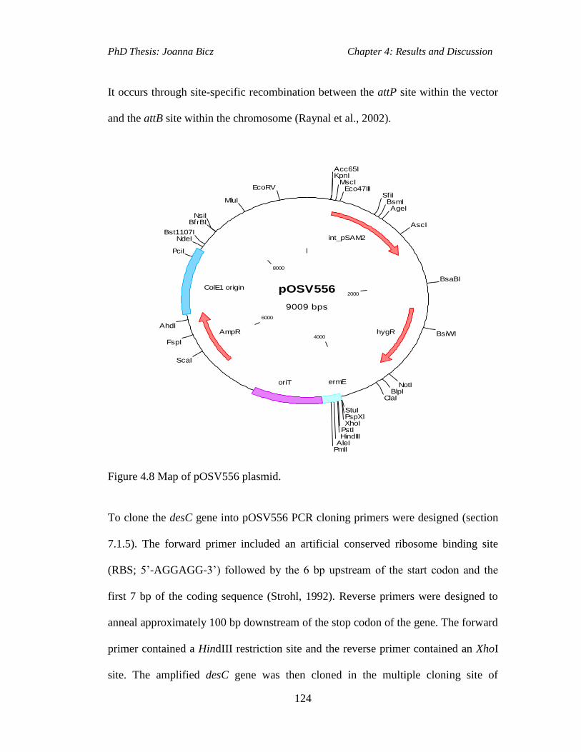
PhD Thesis: Joanna Bicz Chapter 4: Results and Discussion
124
It occurs through site-specific recombination between the attP site within the vector
and the attB site within the chromosome (Raynal et al., 2002).
pOSV556
9009 bps
2000
4000
6000
8000
Acc65IKpnI
MscIEco47III
SfiIBsmIAgeI
AscI
BsaBI
BsiWI
NotIBlpI
ClaI
StuIPspXIXhoI
PstIHindIII
AleIPmlI
ScaI
FspI
AhdI
PciI
NdeIBst1107I
BfrBINsiI
MluI
EcoRV
int_pSAM2
hygR
ermEoriT
AmpR
ColE1 origin
Figure 4.8 Map of pOSV556 plasmid.
To clone the desC gene into pOSV556 PCR cloning primers were designed (section
7.1.5). The forward primer included an artificial conserved ribosome binding site
(RBS; 5’-AGGAGG-3’) followed by the 6 bp upstream of the start codon and the
first 7 bp of the coding sequence (Strohl, 1992). Reverse primers were designed to
anneal approximately 100 bp downstream of the stop codon of the gene. The forward
primer contained a HindIII restriction site and the reverse primer contained an XhoI
site. The amplified desC gene was then cloned in the multiple cloning site of
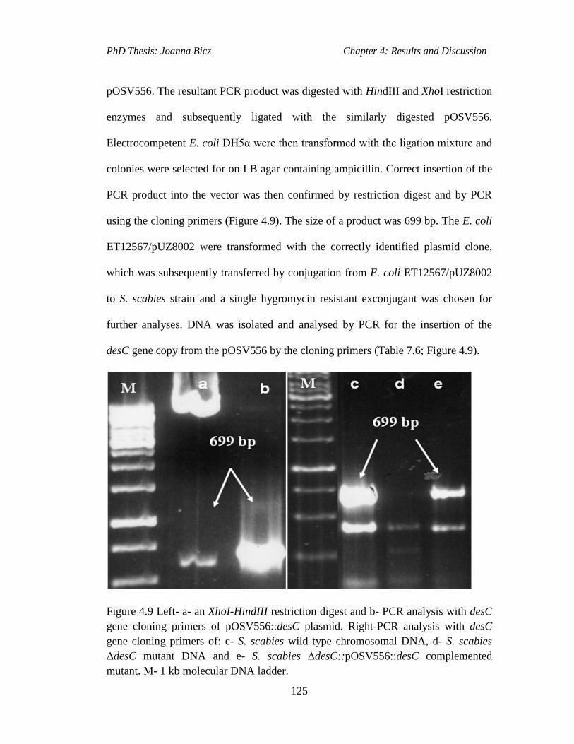
PhD Thesis: Joanna Bicz Chapter 4: Results and Discussion
125
pOSV556. The resultant PCR product was digested with HindIII and XhoI restriction
enzymes and subsequently ligated with the similarly digested pOSV556.
Electrocompetent E. coli DH5α were then transformed with the ligation mixture and
colonies were selected for on LB agar containing ampicillin. Correct insertion of the
PCR product into the vector was then confirmed by restriction digest and by PCR
using the cloning primers (Figure 4.9). The size of a product was 699 bp. The E. coli
ET12567/pUZ8002 were transformed with the correctly identified plasmid clone,
which was subsequently transferred by conjugation from E. coli ET12567/pUZ8002
to S. scabies strain and a single hygromycin resistant exconjugant was chosen for
further analyses. DNA was isolated and analysed by PCR for the insertion of the
desC gene copy from the pOSV556 by the cloning primers (Table 7.6; Figure 4.9).
Figure 4.9 Left- a- an XhoI-HindIII restriction digest and b- PCR analysis with desC
gene cloning primers of pOSV556::desC plasmid. Right-PCR analysis with desC
gene cloning primers of: c- S. scabies wild type chromosomal DNA, d- S. scabies
ΔdesC mutant DNA and e- S. scabies ΔdesC::pOSV556::desC complemented
mutant. M- 1 kb molecular DNA ladder.

PhD Thesis: Joanna Bicz Chapter 4: Results and Discussion
126
4.5.1 LC-MS Analysis of Complemented Δ desC Mutant
The LC-MS analysis (Figure 4.10) indicated restoration of desferrioxamine B
production in the culture of the desC mutant complemented with pOSV556::desC,
however, this is at a lower level when in comparison to the S. scabies 87.22 wild
type strain. Desferrioxamine B (m/z=654) was not detected in the culture of the desC
mutant complemented with pOSV556::desC. Desferrioxamine was not detected in
the culture supernatant of desC mutant (Figure 4.10).
Figure 4.10 EICs (m/z = 614) for ferrioxamine B from LC-MS analyses of acidified
organic extracts from the S. scabies wild type (top trace), S. scabies desC mutant
(middle trace) and Δ desC ::pOSV556/63021::apr (bottom trace).
PhD Thesis: Joanna Bicz Chapter 4: Results and Discussion
127
4.6 Discussion
The PCR-targeting method was used to create a deletion of the desC gene within the
des cluster of S. scabies (Gust et al., 2002). The desC gene in S. scabies genome was
replaced with an apramycin resistance cassette containing oriT. The constructed
mutant was analysed via PCR and Southern blot hybridisation methods.
LC-MS analysis of the S. scabies wild type strain and the S. scabies ΔdesC mutant
strain liquid cultures was undertaken. Before analysis, the supernatants of the wild
type and the mutant strain were incubated with ferric iron to obtain the ferric
complexes with desferrioxamines, which were then examined via LC-MS. The
results indicated that wild type S. scabies produced desferrioxamines, whereas the
ΔdesC mutant S. scabies strain did not, indicating that disruption of the desC gene in
S. scabies abolished desferrioxamine production.
Chemical complementation was carried out to further analyse the function of desC
gene within the des cluster in S. scabies. The DesC enzyme is thought to catalyse the
acylation of N-hydroxycadaverine to form precursors in desferrioxamine E and B
biosynthesis in S. coelicolor (N. Kadi and G. L. Challis unpublished data; Figure
4.6).
Chemical complementation using HSC restored desferrioxamine B production in the
S. scabies desC mutant, although to a lower level than in a wild type S. scabies
strain. The lower level of desferrioxamine B production in S. scabies desC
chemically complemented strain could have been due to the amount of HSC
precursor added to desC mutant liquid culture. The concentration of HSC in desC

PhD Thesis: Joanna Bicz Chapter 4: Results and Discussion
128
mutant culture was only 1µmol/ml. The reason for the small concentration of HSC
was that it was not readily available during the experiment. Had more HSC been
available the chemical complementation of S. scabies desC mutant could have been
repeated several times to validate the LC-MS analysis results and optimise the
concentration needed.
The introduction of desC copy from pOSV556 vector into the ΔdesC mutant S.
scabies genome was confirmed by PCR and LC-MS analyses. The LC-MS of
complemented ΔdesC mutant strain indicated restoration of desferrioxamines
biosynthesis.
Investigation into the role of DesC was carried out in S. coelicolor (N. Kadi and G.
L. Challis, unpublished data). The desC gene encodes an enzyme similar to acyl-
CoA-dependent acyl transferases (Kadi et al., 2007), and catalyses the N-acylation of
N-hydroxycadaverine (Figure 4.6; N. Kadi and G. L. Challis, unpublished data). In
those organisms that also produce desferrioxamine B, DesC has relaxed substrate
specificity, allowing acylation of N-hydroxycadaverine with acetyl-CoA as well as
succinyl-CoA, giving N-hydroxy-N-acetylcadaverine (AHC) and N-hydroxy-N-
succinylcadaverine (HSC), respectively (Figure 4.6; Kadi et al., 2007). To prove
directly that DesC catalyses these reactions the DesC enzyme purified from S.
coelicolor was incubated with succinyl coenzyme A and with N-hydroxycadaverine
substrate in one reaction and with acetyl coenzyme A and with N-hydroxycadaverine
in another reaction (Kadi and Challis, unpublished data). LC-MS analysis of the
reaction mixture with succinyl coenzyme A and another with acetyl coenzyme A
showed respectively the presence of the peaks, which had the same m/z and retention
PhD Thesis: Joanna Bicz Chapter 4: Results and Discussion
129
times as the authentic standards of the products HSC and AHC (Kadi and Challis,
data not shown).
In conclusion, desC gene in S. scabies is involved in desferrioxamine biosynthesis as
its deletion resulted in the abolition of desferrioxamine production. Chemical
complementation of the desC mutant with HSC restored to some level
desferrioxamine biosynthesis (Figure 4.10) suggesting that the DesC protein in S.
scabies catalyses the acylation of N-hydroxycadaverine to yield HSC, which is
utilised in subsequent reaction catalysed by DesD to yield desferrioxamine B.

PhD Thesis: Joanna Bicz Chapter 5: Results and Discussion
130
Results and Discussion II: Investigation of Tetronate-
like Biosynthetic Gene Cluster in S. scabies 87.22.
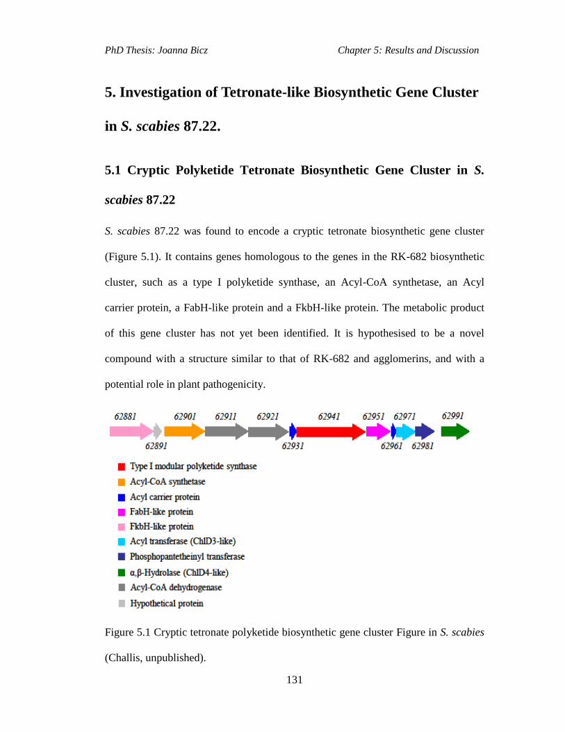
PhD Thesis: Joanna Bicz Chapter 5: Results and Discussion
131
5. Investigation of Tetronate-like Biosynthetic Gene Cluster
in S. scabies 87.22.
5.1 Cryptic Polyketide Tetronate Biosynthetic Gene Cluster in S.
scabies 87.22
S. scabies 87.22 was found to encode a cryptic tetronate biosynthetic gene cluster
(Figure 5.1). It contains genes homologous to the genes in the RK-682 biosynthetic
cluster, such as a type I polyketide synthase, an Acyl-CoA synthetase, an Acyl
carrier protein, a FabH-like protein and a FkbH-like protein. The metabolic product
of this gene cluster has not yet been identified. It is hypothesised to be a novel
compound with a structure similar to that of RK-682 and agglomerins, and with a
potential role in plant pathogenicity.
Figure 5.1 Cryptic tetronate polyketide biosynthetic gene cluster Figure in S. scabies
(Challis, unpublished).

PhD Thesis: Joanna Bicz Chapter 5: Results and Discussion
132
5.2 Transcriptional Analysis of Cryptic Tetronate Biosynthetic Gene
Cluster in S. scabies
The aim of this work was to determine if the genes from the putative tetronate-like
cluster in S. scabies are expressed under laboratory conditions. Currently the
metabolic products of this cluster have not been identified. To check the expression
of the putative tetronate-like cluster in S. scabies reverse transcriptase PCR (RT-
PCR) was carried out on the total RNA isolated from wild type S. scabies.
PCR primers approximately 20 bp in length were designed to amplify a 500-600 bp
regions of several genes found within the putative tetronate biosynthetic gene cluster,
such as scab62881 (encoding a FkbH-like protein), scab62951 (encoding a FabH-
like protein), scab62971 (encoding a ChlD3-like protein), scab62991 (encoding a
ChlD4-like protein) and also from the hrdB gene (an essential gene encoding sigma-
like transcription factor) as a positive control. The total RNA was isolated from S.
scabies mycelium grown for 3, 5, 7 and 14 days. The RNA was then analysed using
agarose gel electrophoresis (Figure 5.2) and treated with DNAse to remove all traces
of DNA from the RNA samples. RT-PCR was carried out on a purified RNA with
the hrdB, scab62881, scab62951, scab62971 and scab62991 primers (Section 7.1.5)
to examine the transcription of the genes (Figure 5.3).
The first step in RT-PCR involves the use of reverse transcriptase polymerase and a
primer to anneal and extend the targeted mRNA sequence. The mRNA is transcribed
to a complementary strand of DNA (cDNA), which is subsequently amplified with
primers and DNA polymerase in a standard PCR. The PCR products are analysed
using agarose gel electrophoresis. If a band of the desired molecular weight is
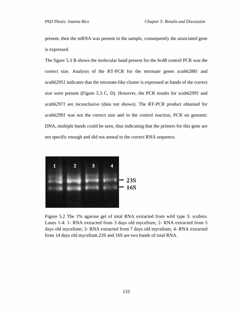
PhD Thesis: Joanna Bicz Chapter 5: Results and Discussion
133
present, then the mRNA was present in the sample, consequently the associated gene
is expressed.
The figure 5.3 B shows the molecular band present for the hrdB control PCR was the
correct size. Analysis of the RT-PCR for the tetronate genes scab62881 and
scab62951 indicates that the tetronate-like cluster is expressed as bands of the correct
size were present (Figure 5.3 C, D). However, the PCR results for scab62991 and
scab62971 are inconclusive (data not shown). The RT-PCR product obtained for
scab62991 was not the correct size and in the control reaction, PCR on genomic
DNA, multiple bands could be seen, thus indicating that the primers for this gene are
not specific enough and did not anneal to the correct RNA sequence.
Figure 5.2 The 1% agarose gel of total RNA extracted from wild type S. scabies.
Lanes 1-4: 1- RNA extracted from 3 days old mycelium; 2- RNA extracted from 5
days old mycelium; 3- RNA extracted from 7 days old mycelium; 4- RNA extracted
from 14 days old mycelium.23S and 16S are two bands of total RNA.
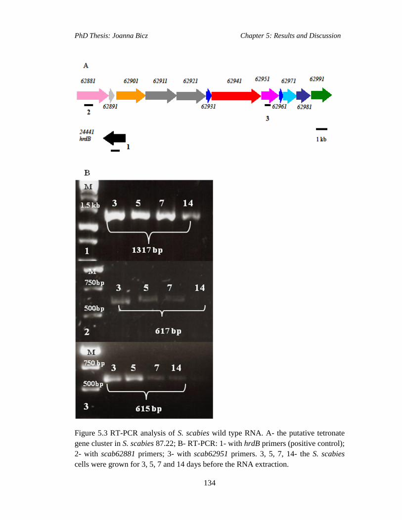
PhD Thesis: Joanna Bicz Chapter 5: Results and Discussion
134
Figure 5.3 RT-PCR analysis of S. scabies wild type RNA. A- the putative tetronate
gene cluster in S. scabies 87.22; B- RT-PCR: 1- with hrdB primers (positive control);
2- with scab62881 primers; 3- with scab62951 primers. 3, 5, 7, 14- the S. scabies
cells were grown for 3, 5, 7 and 14 days before the RNA extraction.
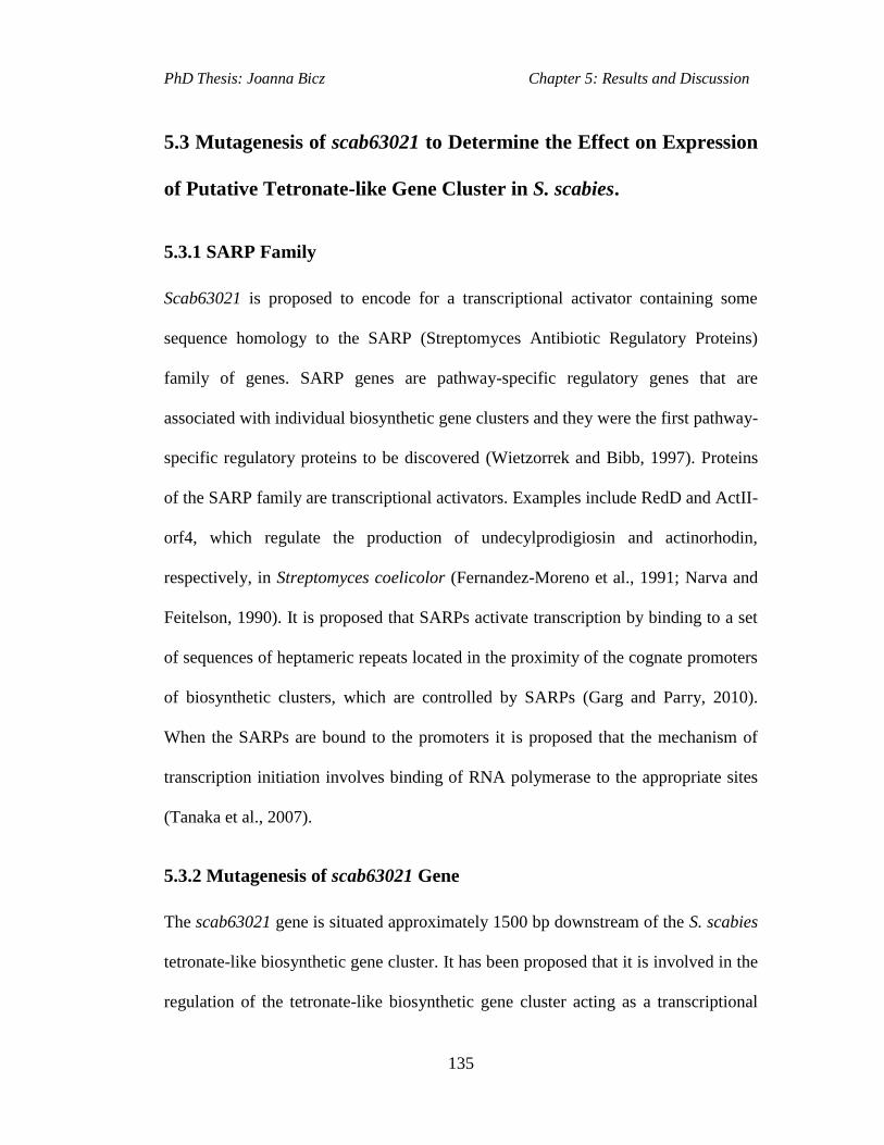
PhD Thesis: Joanna Bicz Chapter 5: Results and Discussion
135
5.3 Mutagenesis of scab63021 to Determine the Effect on Expression
of Putative Tetronate-like Gene Cluster in S. scabies.
5.3.1 SARP Family
Scab63021 is proposed to encode for a transcriptional activator containing some
sequence homology to the SARP (Streptomyces Antibiotic Regulatory Proteins)
family of genes. SARP genes are pathway-specific regulatory genes that are
associated with individual biosynthetic gene clusters and they were the first pathway-
specific regulatory proteins to be discovered (Wietzorrek and Bibb, 1997). Proteins
of the SARP family are transcriptional activators. Examples include RedD and ActII-
orf4, which regulate the production of undecylprodigiosin and actinorhodin,
respectively, in Streptomyces coelicolor (Fernandez-Moreno et al., 1991; Narva and
Feitelson, 1990). It is proposed that SARPs activate transcription by binding to a set
of sequences of heptameric repeats located in the proximity of the cognate promoters
of biosynthetic clusters, which are controlled by SARPs (Garg and Parry, 2010).
When the SARPs are bound to the promoters it is proposed that the mechanism of
transcription initiation involves binding of RNA polymerase to the appropriate sites
(Tanaka et al., 2007).
5.3.2 Mutagenesis of scab63021 Gene
The scab63021 gene is situated approximately 1500 bp downstream of the S. scabies
tetronate-like biosynthetic gene cluster. It has been proposed that it is involved in the
regulation of the tetronate-like biosynthetic gene cluster acting as a transcriptional

PhD Thesis: Joanna Bicz Chapter 5: Results and Discussion
136
activator. To prove this hypothesis the gene was disrupted in S. scabies using PCR
targeting protocol (section 7.6.5).
In this method the cosmid containing the target gene is usually used. However, the
cosmid containing scab63021 was not available. Instead, primers were designed to
encompass the start and stop codons of the target gene and to amplify it by PCR from
S. scabies 87.22 genomic DNA. The amplified scab63021 gene would be then
cloned into plasmid vector and that construct could subsequently be used to
transform E.coli BW25113/pIJ790 strain. From then on the further steps in the
construction of the mutant would be carried as in a standard PCR Redirect protocol.
According to the above the cloning primers were designed to amplify scab63021
gene via PCR. These were designed in the same way as the primers used to clone the
S. scabies desC gene into pOSV556 plasmid (section 4.6), however, the PCR with
primers designed to amplify the scab63021 gene did not yield a product with the
correct molecular weight. For that reason the scab63021 gene was not cloned into
plasmid vector. As the alternative, the scab63021 gene was synthesised by a gene
synthesis company (Genewiz). The whole scab63021 gene was synthesised and
cloned into pUC57 plasmid by Genewiz (Figure 5.4).
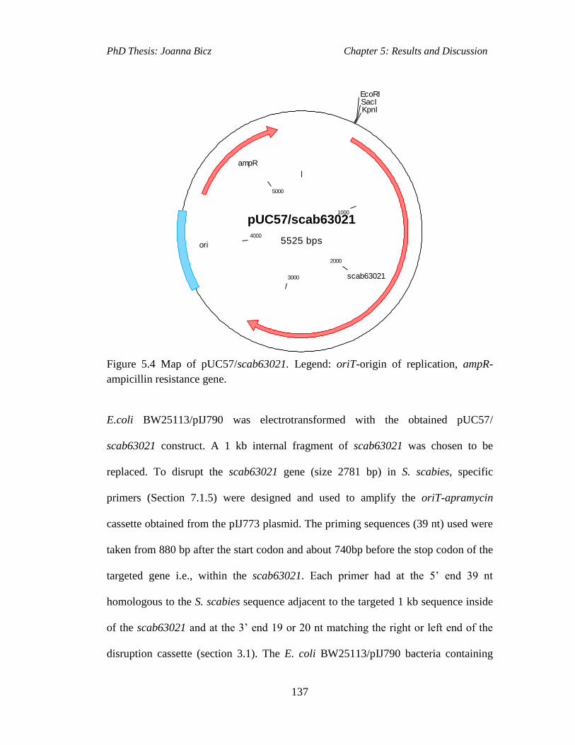
PhD Thesis: Joanna Bicz Chapter 5: Results and Discussion
137
pUC57/scab63021
5525 bps
1000
2000
3000
4000
5000
EcoRISacIKpnI
scab63021
ori
ampR
Figure 5.4 Map of pUC57/scab63021. Legend: oriT-origin of replication, ampR-
ampicillin resistance gene.
E.coli BW25113/pIJ790 was electrotransformed with the obtained pUC57/
scab63021 construct. A 1 kb internal fragment of scab63021 was chosen to be
replaced. To disrupt the scab63021 gene (size 2781 bp) in S. scabies, specific
primers (Section 7.1.5) were designed and used to amplify the oriT-apramycin
cassette obtained from the pIJ773 plasmid. The priming sequences (39 nt) used were
taken from 880 bp after the start codon and about 740bp before the stop codon of the
targeted gene i.e., within the scab63021. Each primer had at the 5’ end 39 nt
homologous to the S. scabies sequence adjacent to the targeted 1 kb sequence inside
of the scab63021 and at the 3’ end 19 or 20 nt matching the right or left end of the
disruption cassette (section 3.1). The E. coli BW25113/pIJ790 bacteria containing
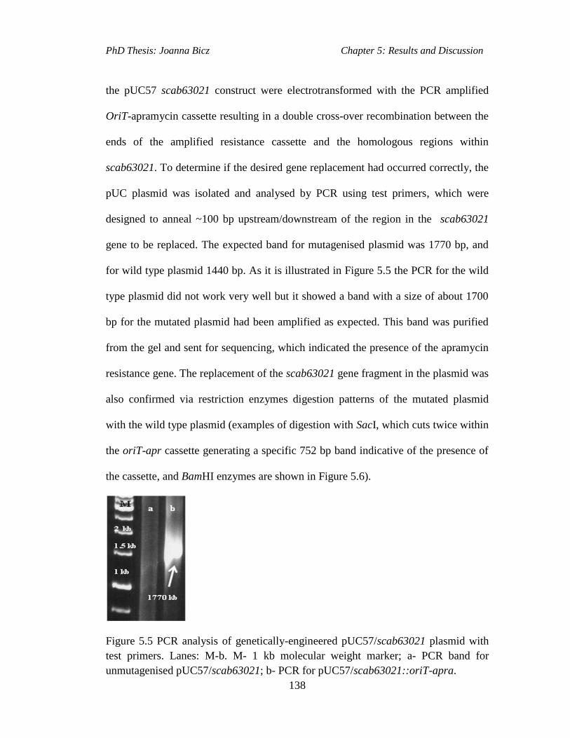
PhD Thesis: Joanna Bicz Chapter 5: Results and Discussion
138
the pUC57 scab63021 construct were electrotransformed with the PCR amplified
OriT-apramycin cassette resulting in a double cross-over recombination between the
ends of the amplified resistance cassette and the homologous regions within
scab63021. To determine if the desired gene replacement had occurred correctly, the
pUC plasmid was isolated and analysed by PCR using test primers, which were
designed to anneal ~100 bp upstream/downstream of the region in the scab63021
gene to be replaced. The expected band for mutagenised plasmid was 1770 bp, and
for wild type plasmid 1440 bp. As it is illustrated in Figure 5.5 the PCR for the wild
type plasmid did not work very well but it showed a band with a size of about 1700
bp for the mutated plasmid had been amplified as expected. This band was purified
from the gel and sent for sequencing, which indicated the presence of the apramycin
resistance gene. The replacement of the scab63021 gene fragment in the plasmid was
also confirmed via restriction enzymes digestion patterns of the mutated plasmid
with the wild type plasmid (examples of digestion with SacI, which cuts twice within
the oriT-apr cassette generating a specific 752 bp band indicative of the presence of
the cassette, and BamHI enzymes are shown in Figure 5.6).
Figure 5.5 PCR analysis of genetically-engineered pUC57/scab63021 plasmid with
test primers. Lanes: M-b. M- 1 kb molecular weight marker; a- PCR band for
unmutagenised pUC57/scab63021; b- PCR for pUC57/scab63021::oriT-apra.

PhD Thesis: Joanna Bicz Chapter 5: Results and Discussion
139
Figure 5.6 Agarose gel electrophoresis analysis of restriction enzymes digest of
genetically-engineered pUC57/scab63021 plasmid used to disrupt the scab63021 in
the S. scabies genome within putative tetronate-like cluster. Lanes M-d: M- 1kb
molecular weight marker; a- undigested pUC57/scab63021::oriT-apra; b- digestion
with EcoRI; c- digestion with BamHI d- digestion with SacI.
The correctly modified plasmids were introduced into S. scabies by intergenic
conjugation from E. coli ET12567/pUZ8002. Apramycin resistant, Streptomyces
exconjugants were analysed by PCR to confirm that a double cross-over gene
replacement had taken place (Figure 5.7). The first PCR using genomic DNA from
the scab63021 mutant colonies and with the primers annealing inside of the oriT-
apramycin cassette, confirmed the presence of the oriT-apramycin cassette in place
of the scab63021 internal fragment in the genome of the mutant. The expected size
of the PCR product was ~400 bp, which was then compared to the PCR of the

PhD Thesis: Joanna Bicz Chapter 5: Results and Discussion
140
mutagenised plasmid pUC/scab63021::apr, which also gave band of ~400 bp
(Figure 5.7 A). This indicate that introduction of oriT-apramycin cassette into the
genome of putative scab63021 mutant was successful. They do not prove, however,
that a double cross-over between scab63021 and cassette had occured. The putative
scab63021 mutant genomic DNA was further analysed with the test primers (Section
7.1.5) designed to anneal ~100 bp upstream and downstream of the replaced internal
fragment within scab63021. The expected band size was ~1.5 kb for the wild type S.
scabies genomic DNA, and 1.8 kb for scab63021 mutant. The PCR results showed
no band for wild type, however, the expected band was present for the mutant strain
(only mutant band is displayed-Figure 5.7 B).
Figure 5.7 A- PCR analysis of genetically-engineered pUC57/scab63021 plasmid
and genomic DNA of scab63021 mutant with oriT- apramycin casette internal
primers; M-1 kb molecular weight marker, a –PCR of genomic DNA of Δscab63021
mutant; b- pUC57/scab63021::apra plasmid with apramycin internal primers. B-PCR
analysis of putative scab63021 mutant genomic DNA with test primers.
PhD Thesis: Joanna Bicz Chapter 5: Results and Discussion
141
5.4 RT-PCR on Wild Type S. scabies and the Δscab63021 Mutant
RNA
Although the mutation of the scab63021 gene had not been proved conclusively,
analysis of the wild type S. scabies and the putative scab63021 mutant genomes has
been carried out to allow for comparisons in the patterns of expression of the genes
in the putative tetronate-like gene cluster.
The RT-PCR was carried on wild type and mutant S. scabies purified RNA using the
same method previously described (section 5.3). The RNA was extracted from
mycelia 3, 5, 7 and 14 days old. In this experiment the expression was analysed only
for two genes from S. scabies tetronate-like cluster; scab62881 (encoding a FkbH-
like protein), and scab62951 (encoding a FabH-like protein) as the RT-PCR in wild
type S. scabies yielded specific bands only for these two genes within the cluster.
The RT-PCR with the scab62881 primers yielded specific as well as unspecific
bands. The right size for the specific band of RT-PCR product on wild type RNA
was 617 bp for the scab62881 primers. The ~20 bp PCR primers designed for the
previous analysis to amplify regions of scab62881 and scab62951 approximately 600
bp in length were used in this experiment. The hrdB gene RT-PCR was again used as
a positive control.
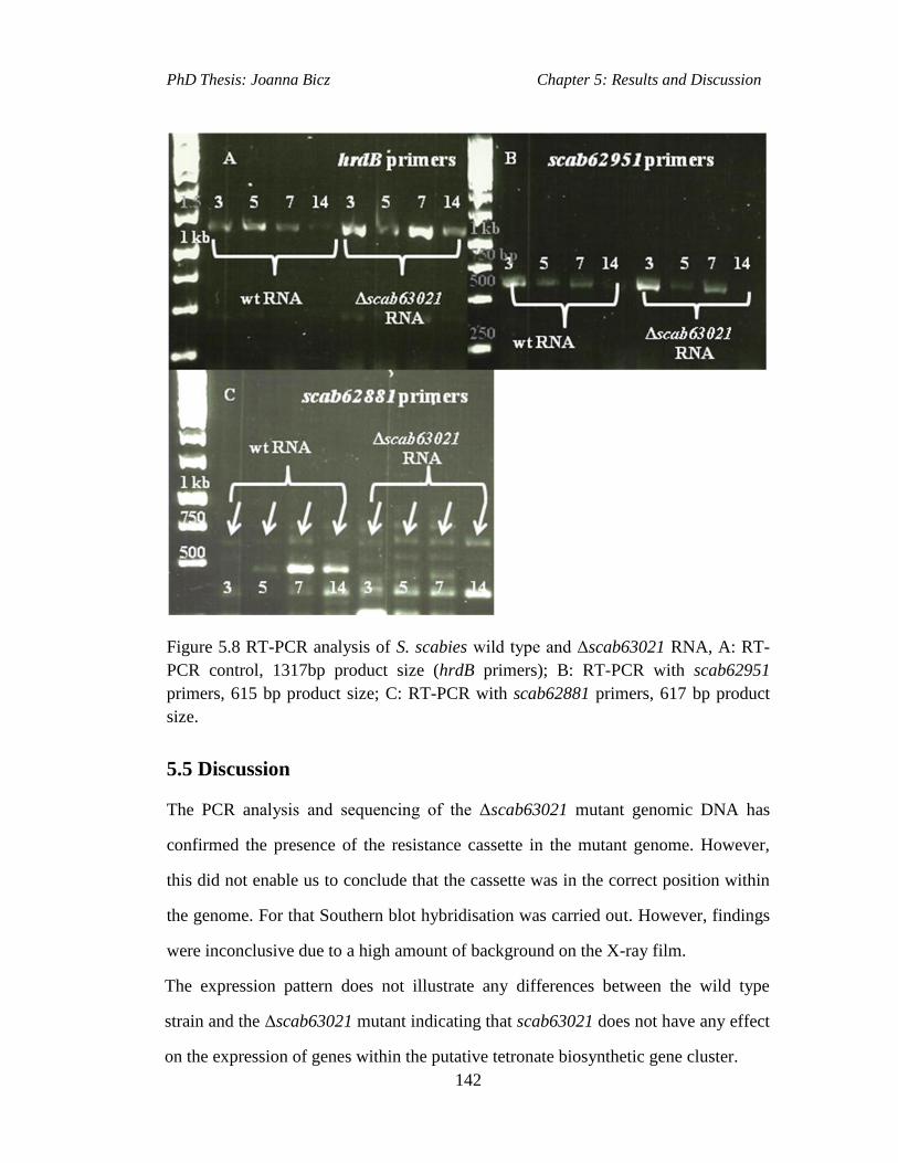
PhD Thesis: Joanna Bicz Chapter 5: Results and Discussion
142
Figure 5.8 RT-PCR analysis of S. scabies wild type and Δscab63021 RNA, A: RT-
PCR control, 1317bp product size (hrdB primers); B: RT-PCR with scab62951
primers, 615 bp product size; C: RT-PCR with scab62881 primers, 617 bp product
size.
5.5 Discussion
The PCR analysis and sequencing of the Δscab63021 mutant genomic DNA has
confirmed the presence of the resistance cassette in the mutant genome. However,
this did not enable us to conclude that the cassette was in the correct position within
the genome. For that Southern blot hybridisation was carried out. However, findings
were inconclusive due to a high amount of background on the X-ray film.
The expression pattern does not illustrate any differences between the wild type
strain and the Δscab63021 mutant indicating that scab63021 does not have any effect
on the expression of genes within the putative tetronate biosynthetic gene cluster.
PhD Thesis: Joanna Bicz Chapter 5: Results and Discussion
143
An alternative method could have been employed to construct the Δscab63021
mutant. The scab63021 gene could have been obtained within a cosmid, which could
subsequently be used as a template for PCR. The cosmid would be more useful as the
insert is bigger and the PCR primers could be designed to anneal outside of the
scab63021 gene and the sufficient flanking sequences on both sides of the target
sequence would ensure that the homologous recombination between the investigated
gene and the resistance cassette PCR product ends would occur.
Further work would be to carry out the comparative analysis of the S. scabies wild
type strain and Δscab63021 mutant culture supernatants via LC-MS to find the
putative tetronate product. If there would be no differences between the metabolic
profiles of the two strains, the Δscab63021 mutant could be constructed again by
another method. Alternatively, the deletion of the scab62941, encoding for a putative
PKS within the S. scabies tetronate-like gene cluster could be obtained and the
metabolic profile of the wild type strain and the mutant could be compared again via
LC-MS in search of the cryptic tetronate metabolite.

PhD Thesis: Joanna Bicz Chapter 6. Summary, Conclusions and Future Work
144
6. Summary, Conclusions and Future Work

PhD Thesis: Joanna Bicz Chapter 6. Summary, Conclusions and Future Work
145
6.1 Results and Discussion I: Investigation of Siderophore
Biosynthetic Gene Clusters in S. scabies.
N NH
N
NH
N
HN
HO O
O
OHO
O
OH
O
O
Desferrioxamine E
SN
SN
O
HO
Me
OH
H
H
HN
N
H O
OH
NH
O
O
OH
NOH
NH
HNO
NH
O
N
O
OH
NH2
scabichelin
pyochelin
Figure 6.1 Structures of investigated siderophores.
6.1.1 Investigation of Pyochelin Biosynthetic Gene Cluster in S. scabies
87.22.
Putative functions for most of the twelve genes within the putative pyochelin
biosynthetic gene cluster (PBGC) in S. scabies 87.22 have been proposed (Seipke et
al., 2011) and a biosynthetic pathway for pyochelin has been suggested on the basis
of bioinformatics and experimental studies. However, the regulation of pyochelin
PhD Thesis: Joanna Bicz Chapter 6. Summary, Conclusions and Future Work
146
production was not fully understood. Scab1401 was found to encode a pathway
specific regulator, which was predicted to repress transcription of PBGC in iron rich
conditions. To determine the function of scab1401, the gene was replaced with the
oriT-apramycin cassette creating Δscab1401 mutant by our collaborators (Loria’s
group; Seipke et al., 2011). The mutant was then analysed via LC-MS for pyochelin
production in iron deficient conditions. To prove that pyochelin is a product of S.
scabies PBGC the putative NRPS encoding gene scab1471 in Δscab1401 mutant was
replaced with oriT-hygromycin resistance cassette creating a scab1401- scab1471
double mutant. The metabolic profile of the mutant was analysed via LC-MS
alongside of Δscab1401 mutant and the S. scabies wild type strain.
LC-MS analyses of the scab1401 mutant supernatant unveiled the presence of a
compound with the same molecular mass as pyochelin. The stereochemistry of the
compound produced by S. scabies was also investigated. Comparison of the
scab1401 mutant culture supernatant by LC-MS using chiral stationary phase with
the authentic standards of pyochelin and ent-pyochelin proved that S. scabies
produce pyochelin, not enantio-pyochelin. The LC-MS analysis of culture
supernatants of a scab1401- scab1471 double mutant indicated no pyochelin
production. These data indicate that pyochelin is produced by PBGC in S. scabies
and Scab1401 represses transcription of pyochelin in iron-deficient conditions. The
compound with the same mass as pyochelin was not consistently detected in the
supernatants of wild type S. scabies when grown in iron-deficient conditions. It was
postulated to be that it is due to that the pyochelin gene cluster in the wild type strain
was not induced enough under the used growth conditions.

PhD Thesis: Joanna Bicz Chapter 6. Summary, Conclusions and Future Work
147
Plant assays were used (by Prof R. Loria’s group at Cornell University; Seipke et al.,
2011) to analyse the importance of pyochelin in plant pathogenicity. Incubation of
scab1471 mutant (scab1471 encodes one of the NRPS enzymes from the pyochelin
biosynthetic pathway) and of S. scabies wild type, both on potato tissue and radish
seedlings, indicated that pyochelin is not required for pathogenicity of the potato
tissue and radish. One conclusion that can be drawn from these results is that iron
may have not been essential for S. scabies growth in the assays. The genome of S.
scabies possesses several more biosynthetic gene clusters encoding other
siderophores, such as putative genes for the biosynthesis of desferroxamines
(scab57921–scab57951). To determine which iron-chelating metabolites produced
by S. scabies are required for plant pathogenicity, analysis of desferrioxamine mutant
and other siderophores mutants in S. scabies is needed as well as, in planta
transcriptional analysis of the pyochelin, desferrioxamine and other putative
siderophore biosynthetic gene clusters.
6.1.2 Investigation of Scabichelin Biosynthetic Gene Cluster in S. scabies
87.22.
Mining of the S. scabies 87.22 genome has identified a gene cluster, which was
predicted to encode the nonribosomal peptide synthetase multienzymes responsible
for the biosynthesis of a novel iron-chelating metabolite named scabichelin.
Bioinformatic analysis of a cryptic NRPS biosynthetic gene cluster, which include
the protein coding sequences from scab85431 to scab85521 are homologous to
proteins encoded for by genes within the coelichelin biosynthetic gene cluster from

PhD Thesis: Joanna Bicz Chapter 6. Summary, Conclusions and Future Work
148
S. coelicolor M145. Analysis of the culture supernatants of S. antibioticus, used by
our collaborators, via HPLC and subsequent analysis by NMR indicated that these
strains produce a tris-hydroxamate siderophore, which was named scabichelin. The
aim was to establish whether the product of S. antibioticus is also the product of a
cryptic NRPS encoded by the cryptic biosynthetic gene cluster in S. scabies 87.22.
Bioinformatic analysis of the functional domains of the NRPS encoded for by
scab85471 revealed it contains 17 enzymatic domains organized into 5 modules.
Therefore, it was hypothesised that product of the scab85471 gene would be a
pentapeptide,
PCR analysis and sequencing confirmed the presence of apramycin cassette in
scab85471::oriT-apr (W1000 mutant). The correct construction of the mutant was
also confirmed by Southern blot hybridisation analysis.
Comparative LC-MS/MS analyses of the W1000 mutant, S. scabies 87.22 wild type
and scabichelin purified from S. antibioticus indicated that scabichelin is the
metabolic product of the S. scabies 87.22 cryptic gene cluster. LC-MS analyses of
culture supernatants of the 87.22 and W1000 strains grown in an iron-deficient
medium, to which ferric iron was added before analysis, indicated the presence of a
ferric complex that is produced by S. scabies 87.22, but not the W1000 mutant. LC-
MS retention time and MS/MS fragmentation pattern of ferri-scabichelin are the
same for the S. scabies 87.22 compound and the authentic standard.
Bioinformatic analysis of the scab85431-scab85521 gene cluster and LC-MS
analysis of S. scabies 87.22 scabichelin indicated its potential function as an iron
chelator. However, the essentiality of that metabolite for growth in iron-deficient

PhD Thesis: Joanna Bicz Chapter 6. Summary, Conclusions and Future Work
149
conditions has not yet been investigated. These findings may be helpful to establish
the function of that siderophore in S. scabies and its potential role in plant
pathogenicity. The further work with scabichelin would be conducted in
collaboration with Dr.Bignell’s research group (former member of Prof. Loria’s
group) and would involve the use of a plant assay to compare the necrotic effect of S.
scabies 87.22 and the W1000 strains grown in parallel on tuber and root vegetables
such as potato and radish, respectively.
6.1.3 Desferrioxamine Production in S. scabies
The PCR-targeting method was used to create a deletion of the desC gene within the
des cluster of S. scabies (Gust et al., 2002). The desC gene in the S. scabies genome
was replaced with an apramycin resistance cassette containing oriT. The constructed
mutant was analysed via PCR and Southern blot hybridisation methods.
LC-MS analysis of the S. scabies wild type strain and the S. scabies ΔdesC mutant
strain liquid cultures was undertaken. Before analysis, the supernatants of the wild
type and the mutant strain were incubated with ferric iron to obtain the ferric
complexes with desferrioxamines, which were then examined via LC-MS.
Chemical complementation was carried out to further analyse the function of the
desC gene within the des cluster in S. scabies.
The introduction of the desC copy from the pOSV556 vector into the ΔdesC mutant
S. scabies genome was confirmed by PCR and LC-MS analyses. The results
indicated that the wild type S. scabies produced desferrioxamines, whereas the
ΔdesC mutant S. scabies strain did not, indicating that disruption of the desC gene in

PhD Thesis: Joanna Bicz Chapter 6. Summary, Conclusions and Future Work
150
S. scabies abolished desferrioxamine production. Chemical complementation using
desferrioxamine pathway precursor N-hydroxy-N-succinylcadaverine (HSC) restored
desferrioxamine B production in the S. scabies desC mutant, although to a lower
level than in a wild type S. scabies strain. The lower level of desferrioxamine B
production in S. scabies desC chemically complemented strain could have been due
to the amount of HSC precursor added to ΔdesC mutant liquid culture. The
concentration of HSC in ΔdesC mutant culture was only 1 µmol/ml. The reason for
the small concentration of HSC was that it was not readily available during the
experiment.
The LC-MS of genetically complemented ΔdesC mutant strain indicated restoration
of desferrioxamine biosynthesis.
The further work with desC mutant would be conducted in collaboration with Prof.
Rose Loria’s research group and would involve use of a plant assay to compare the
necrotic effect of the S. scabies 87.22 wild type and ΔdesC mutant grown in parallel
on root vegetables.
6.2 Results and Discussion II: Investigation of Tetronate-like
Biosynthetic Gene Cluster in S. scabies 87.22.
6.2.1 Investigation of Tetronate-like Biosynthetic Gene Cluster in S.
scabies 87.22.
The S. scabies 87.22 genome was found to encode cryptic tetronate biosynthetic gene
cluster, which is predicted to encode genes homologous to genes in the RK-682
biosynthetic gene cluster, such as a type I polyketide synthase, an Acyl-CoA
PhD Thesis: Joanna Bicz Chapter 6. Summary, Conclusions and Future Work
151
synthetase/dehydrogenase, an Acyl carrier protein, a FabH-like protein, and a FkbH-
like protein. The aim of this work was to determine if the genes from the putative
tetronate-like cluster in S. scabies are expressed under laboratory conditions.
Currently, the metabolic product of this cluster has not been identified. To analyse
the expression of the putative tetronate-like cluster in S. scabies, reverse transcriptase
PCR (RT-PCR) was carried out on the total RNA isolated from the wild type S.
scabies.
The scab63021 gene has been proposed to be involved in the regulation of the
tetronate-like biosynthetic gene cluster acting as a transcriptional activator. To prove
this hypothesis, the attempt was made to disrupt the gene in S. scabies using PCR
targeting protocol. Analysis of the wild type S. scabies and the putative scab63021
mutant genomes has been carried out to allow for comparisons in the patterns of
expression of the genes in the putative tetronate-like gene cluster.
The transcriptional analysis of the S. scabies wild type RNA revealed that the cluster
is expressed. Transcripts of some genes (listed in section 5.2) were obtained and
confirmed by sequencing. The PCR analysis and sequencing of the Δscab63021
mutant genomic DNA has confirmed the presence of the resistance cassette in the
mutant genome. However, this did not enable us to conclude whether the cassette
was in the correct position within the genome. The expression pattern does not
illustrate any differences between the wild type strain and the Δscab63021 mutant
indicating that scab63021 does not have any effect on the expression of genes within
the putative tetronate biosynthetic gene cluster.

PhD Thesis: Joanna Bicz Chapter 6. Summary, Conclusions and Future Work
152
Further work would be to carry out the comparative analysis of the S. scabies wild
type strain and the Δscab63021 mutant culture supernatants via LC-MS to find the
putative tetronate product. If there would be no differences between the metabolic
profiles of the two strains, the Δscab63021 mutant could be constructed again by
another method. Alternatively, the deletion of the scab62941, encoding for a putative
PKS within the S. scabies tetronate-like gene cluster could be obtained and the
metabolic profile of the wild type strain and the mutant could be compared again via
LC-MS in search of the cryptic tetronate metabolite.
6.3 Significance
Among actinomycetes, members of the genus Streptomyces are considered to be the
major producers of secondary metabolites (Challis and Hopwood, 2003). They have
evolved the ability to biosynthesize multiple biologically active compounds. These
compounds are not made during the vegetative growth phase and their production is
usually switched on at a point when nutrients needed for a primary metabolism are
depleted and during the aerial mycelium growth phase and during spore growth
initiation. Their biological functions include, among the others, killing of
competitive microorganisms, regulation of bacterial metabolism and metal ion
uptake. It was argued that possible reasons for the biosynthesis of secondary
metabolites may be the selectional advantages these compounds confer to the
producing organisms such as the defence mechanism against the environmental
stresses (Stone and Williams, 1992).
Siderophores are one class of secondary metabolites. They are produced by many
bacteria to increase the solubility of iron from the soil. The analysis of several

PhD Thesis: Joanna Bicz Chapter 6. Summary, Conclusions and Future Work
153
Streptomyces species genomes were found to encode for the biosynthesis of multiple
siderophores (Challis and Hopwood, 2003; Barona-Gomez et al., 2006), for example,
S. coelicolor produces two hydroxamate siderophores: coelichelin and
desferrioxamine. S. avermitilis produces desferrioxamine and is also predicted to
produce the siderophore myxochelin (Ikeda et al., 2003). The reason for the
production of more than one kind of iron-chelating metabolite is not exactly clear.
However, it is known that many bacteria that do not produce desferrioxamine
siderophore posses the uptake systems for it (Barona-Gomez et al., 2006). It was,
therefore, hypothesized that streptomycetes, which produce desferrioxamine, also
need to possess a biosynthetic gene cluster responsible for the production of another
siderophore, which is not taken up into the cell by the ferroxamine uptake system
(Challis and Hopwood, 2003).
Multiple pathways for the assembly of various siderophores were also found in
several plant pathogenic microorganisms (Franza and Expert, 2012). It is known that
iron is essential for the development of bacterial infections in mammalian hosts (Cox
et al 1981). However, the role of iron uptake in the invasion of plants by pathogenic
bacteria has only been investigated in recent years (Franza and Expert, 2012). These
investigations have provided some evidence for the role of iron acquisition in the
virulence of Gram-negative plant pathogenic microorganisms. For example, Dickeya
dadantii a soft-rotting Gram-negative soil bacterium is a plant pathogen of many
plant species, including some vegetables. The two siderophores that it produces-
chrysobactin and achromobactin- are required for the development of infection
symptoms in the plant tissues (Dellagi et al., 2005; Enard et al., 1988). Iron uptake is
also required for infection by Erwinia amylovora, which causes fire blight in apple
PhD Thesis: Joanna Bicz Chapter 6. Summary, Conclusions and Future Work
154
flowers (Dellagi et al., 1998). Pseudomonas syringae pv. tabaci 6605 produces the
siderophore pyoverdin, which is needed for development of wildfire disease on
tobacco plants (Taguchi et al., 2010). As iron is often critical for the virulence of
Gram-negative plant pathogens, it is suspected that siderophores are also important
for iron uptake in pathogenic Streptomyces species. Streptomyces scabies causes
disease in potatoes and root crops (Loria et al., 1998) and its genome encodes
biosynthetic pathways directing biosynthesis of several siderophores, including
pyochelin and desferrioxamines. The novel iron-chelating metabolite scabichelin was
also isolated from this strain. Siderophore-mediated iron acquisition has not been yet
confirmed to be a significant factor in pathogenicity of S. scabies. However, the
investigations of the siderophore biosynthetic pathways in this strain will enable
further elucidation of the role of iron acquisition in the virulence of plant pathogenic
streptomycetes. Analysis of the S. scabies genome also unveiled the presence of a
gene cluster encoding biosynthetic pathways for the production of secondary
metabolites that are not associated with iron uptake but, which were reported to be
present in other plant pathogens (Section 5). The investigation of these clusters and
their metabolites will contribute to further elucidation of the molecular mechanism of
S. scabies that are important for plant pathogenicity.

PhD Thesis: Joanna Bicz Chapter 7: Materials and Methods
155
7. Materials and Methods

PhD Thesis: Joanna Bicz Chapter 7: Materials and Methods
156
7.1 Materials
7.1.1 Enzymes, Chemicals and Equipment
Kits for DNA gel extraction and plasmid isolation from E. coli were purchased from
Qiagen (USA). The kit for DNA ligation was obtained from Roche (Germany).
Restriction enzymes were purchased from MBI Fermentas (Lithuania) and New
England Biolabs (USA). Oligonucleotide primers were ordered from Sigma Genosys
(USA). The gene synthesis was ordered from Genewiz. Taq polymerase was
purchased from Fermentas and High Fidelity Polymerase from Roche. The
ingredients for media preparation were purchased from Fisher BioReagents, Difco
and Becton, Dickinson and co. The rest of chemicals were bought from Difco (USA),
Sigma-Aldrich (USA), Fermentas (Lithuania), Fisher Scientific (UK), Biolab (New
Zealand), Lonza (UK).
The concentration of DNA and RNA was measured on a NanoDrop ND-1000
Spectrophotometer. An Eppendorf Mastercycler Personal was used for PCR
reactions. Agarose gel electrophoresis was performed on Bio-Rad Power Pac 300. A
Bio-Rad Gene Pulser II with the Bio-Rad Pulse Controller Plus was used for
electroporations. Thermo BioMate3 Spectrophotometer was used to check optical
density of bacterial cell cultures. The aseptic work with Streptomyces was performed
under Bassaire laminar flow hood. The microbial cell culture supernatants and
extracts were analysed for secondary metabolite production by LC-MS (LC –
Agilent 900, MS – Bruker HCT Plus) and Bruker MicroTOF mass spectrometer. For
Southern blot hybridisation a UV Stratalinker 2400 from Stratagene was used for
PhD Thesis: Joanna Bicz Chapter 7: Materials and Methods
157
DNA fixation to nylon membrane (Amersham). For hybridisation of probe to the
DNA a TechHybridiser HB-1D was used.
7.1.2 General Solutions, Buffers and Antibiotics
All stock solutions (Table 7.1) were prepared according to Sambrook and Russel
(2001). Distilled water was used for preparing all stock solutions. Autoclaving or
filtration through a 0.2 µm filter (Stericon) was used to sterilise the solutions.
Antibiotic stock solutions (Table 7.2) were dissolved in distilled water or other
appropriate solution and sterilised by filtration through a 0.2 µm filter.
Table 7.1General stock solutions.
Chemical Stock solution Solvent
TBE ( 1000ML 5x) Trizma base- 53g
Boric acid- 27.5g
EDTA- 10mM, pH 8
water
TE buffer Tris –HCL pH 8 100mM
EDTA – 10mM, pH 8
water
Tris-HCl (pH 8) 0.5M Water, pH adjusted with
HCL
Agarose gel loading dye Glycerol 50%
Bromophenol blue 0.1%
Xylene cyanol 0.1%
water
RNAse solution RNAse stock 50mg/ml water
PhD Thesis: Joanna Bicz Chapter 7: Materials and Methods
158
Table 7.2 Antibiotics stock solutions.
Antibiotic
Stock sol.
(mg/mL)
Solvent
E. coli
(μg/mL)
S. scabies
(μg/mL)
overlay
S. scabies
(mg/plate)
overlay
ampicillin 100 water 100 - -
apramycin 50 water 50 25 1.25
chloramphenicol 25 ethanol 25 - -
hygromycin 50 water 50 25 1.25
kanamycin 50 water 50 100 5.0
nalidixic acid 25 0.3M
NaOH
25 20 0.5
7.1.3 Bacterial Strains
Table 7.3 and table 7.4 list the bacterial strains used in this work.
Table 7.3 The E. coli strains used.
Escherichia coli strains
Name Genotype Source
DH5α
F- endA1 glnV44 thi-1 relA1
gyrA96 deoR nupG lacZdeltaM15
hsdR17
general cloning host
(Woodcock et al.,
1989), lab stock
MC1061
araD139 Δ(ara-leu)7696 galE15
galK16 Δ(lac)X74 rpsL(Strr) hsdR2
(rK–mK+) mcrA mcrB1
general cloning host,
lab stock
ET12567/pUZ8002
Non-methylating host for transfer
of DNA into Streptomyces spp.
plasmid pUZ8002: tra, KanR, RP4
23; Genome: dam, dcm, hsdS,
CmR, TetR, JIC, Norwich
JIC, Norwich
(Gust et al., 2004)
BW25113/pIJ790
Temperature sensitive strain of E.
coli, should be grown on rich
Plant Bioscience
PhD Thesis: Joanna Bicz Chapter 7: Materials and Methods
159
medium containing
chloramphenicol (25 μg/ml) at
30°C. Plasmid: pIJ790 [oriR101],
[repA101(ts)], araBp-gam-be-exo.
Chromosome: (Δ(araD-araB)567,
ΔlacZ4787(::rrnB-4), lacIp-
4000(lacIQ), λ-, rpoS369(Am),
rph-1, Δ(rhaD-rhaB)568, hsdR514;
Datsenko and Wanner, 2000).
Limited
(Datsenko and Wanner,
2000)
Table 7.4 The S. scabies strains used
Streptomyces scabies strains Source
87.22 Wild type Rose Loria
Cornell University
Δscab1401 87.22 scab1401 null mutant Rose Loria
Cornell University
Δscab1471 87.22 scab1471 null mutant Rose Loria
Cornell University
Δscab1401-
Δscab1471
87.22 scab1401-scab1471 double mutant Rose Loria
Cornell University
Δscab desC S. scabies 87.22 scab57931 null mutant
This study
Δscab desC
comp
S. scabies 87.22, scab57931 null mutant
complemented
This study
S. scabies
W1000
S. scabies scab85471::apr mutant This study
Δscab63021 S. scabies 87.22 scab63021 ::apr mutant Lab stock
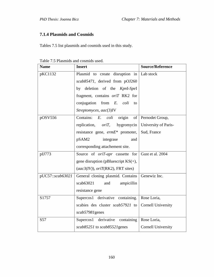
PhD Thesis: Joanna Bicz Chapter 7: Materials and Methods
160
7.1.4 Plasmids and Cosmids
Tables 7.5 list plasmids and cosmids used in this study.
Table 7.5 Plasmids and cosmids used.
Name Insert Source/Reference
pKC1132 Plasmid to create disruption in
scab85471, derived from pOJ260
by deletion of the KpnI-SpeI
fragment, contains oriT RK2 for
conjugation from E. coli to
Streptomyces, aac(3)IV
Lab stock
pOSV556 Contains: E. coli origin of
replication, oriT, hygromycin
resistance gene, ermE* promoter,
pSAM2 integrase and
corresponding attachement site.
Pernodet Group,
University of Paris-
Sud, France
pIJ773 Source of oriT-apr cassette for
gene disruption (pBluescript KS(+),
(aac3(IV)), oriT(RK2), FRT sites)
Gust et al. 2004
pUC57::scab63021 General cloning plasmid. Contains
scab63021 and ampicillin
resistance gene
Genewiz Inc.
S1757 Supercos1 derivative containing.
scabies des cluster scab57921 to
scab57981genes
Rose Loria,
Cornell University
S57 Supercos1 derivative containing
scab85251 to scab85521genes
Rose Loria,
Cornell University
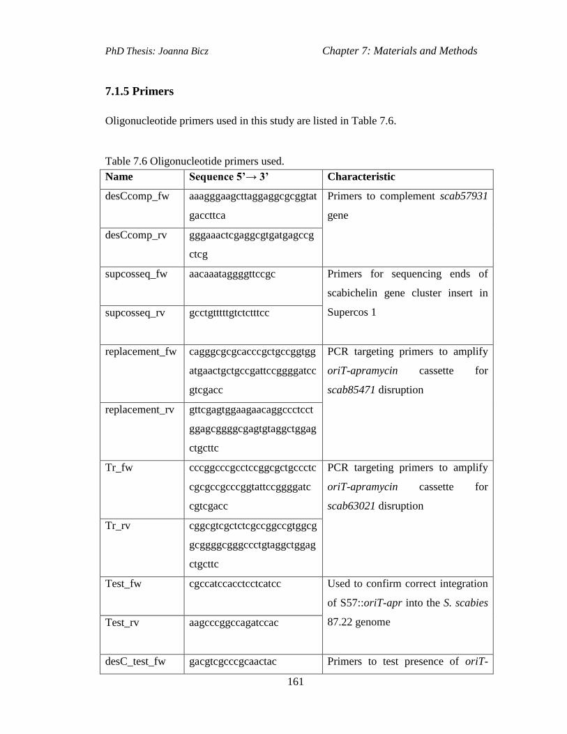
PhD Thesis: Joanna Bicz Chapter 7: Materials and Methods
161
7.1.5 Primers
Oligonucleotide primers used in this study are listed in Table 7.6.
Table 7.6 Oligonucleotide primers used.
Name Sequence 5’→ 3’ Characteristic
desCcomp_fw aaagggaagcttaggaggcgcggtat
gaccttca
Primers to complement scab57931
gene
desCcomp_rv gggaaactcgaggcgtgatgagccg
ctcg
supcosseq_fw
aacaaataggggttccgc Primers for sequencing ends of
scabichelin gene cluster insert in
Supercos 1 supcosseq_rv
gcctgtttttgtctctttcc
replacement_fw cagggcgcgcacccgctgccggtgg
atgaactgctgccgattccggggatcc
gtcgacc
PCR targeting primers to amplify
oriT-apramycin cassette for
scab85471 disruption
replacement_rv gttcgagtggaagaacaggccctcct
ggagcggggcgagtgtaggctggag
ctgcttc
Tr_fw
cccggcccgcctccggcgctgccctc
cgcgccgcccggtattccggggatc
cgtcgacc
PCR targeting primers to amplify
oriT-apramycin cassette for
scab63021 disruption
Tr_rv
cggcgtcgctctcgccggccgtggcg
gcggggcgggccctgtaggctggag
ctgcttc
Test_fw
cgccatccacctcctcatcc Used to confirm correct integration
of S57::oriT-apr into the S. scabies
87.22 genome Test_rv
aagcccggccagatccac
desC_test_fw gacgtcgcccgcaactac Primers to test presence of oriT-

PhD Thesis: Joanna Bicz Chapter 7: Materials and Methods
162
apramycin cassette in the place of
scab57931 desC_test_rv
gtgatgagccgctcgtgg
Tr_test_fw
cgggcgtggaagtaaccg Used to confirm correct integration
of pUC57::oriT-apr into the S.
scabies 87-22 genome Tr_test_rv
ctgcacgagcgtgtcctg
apr_test_fw
tacaccggaccttggagt
Used to confirm the presence of
oriT-apr cassette in the S. scabies
87.22 genome apr_test_rv
gaccgactggaccttccttc
Table 7.7 RT-PCR primers used.
Name Sequence 5’→ 3’ Characteristic
Scab62941_fw gctgcgccggctggagga primers to amplify internal 749 bp of
scab62941 gene Scab62941_rv tcttggtgcggttgctgctc
Scab62951_fw caacgaagagtggatcgacgt primers to amplify internal 615 bp of
scab62951 gene Scab62951_fw tcgatggccttgtagtcctc
Scab62971_fw ctcggtcgtcacctatctg primers to amplify internal 553 bp of
scab62971 gene Scab62971_rv gatgatccggtggtcgaag
Scab62981_fw gccgggaagctcaacctg primers to amplify internal 617 bp of
scab62981 gene Scab62981_rv cagttggcggtgacctgc
Scab62991_fw ggcatcgtcagcgtcaag primers to amplify internal 615 bp of
scab62991 gene Scab62991_rv cggagcagtgaggtgatcag
Scab63021_int
fw
gagtacccgttgcgggagagcc primers to amplify internal 615 bp of
scab63021 gene
Scab63021_int
rv
gatcaccgtgccctccgcgaag
hrdB_fw gcgtgtcgagcacggagtgcagc primers to amplify internal 1317 bp
of hrdB gene hrdB_rv ccgagtccgtctctgtcatggc

PhD Thesis: Joanna Bicz Chapter 7: Materials and Methods
163
7.1.6 Growth Media
Most growth media were prepared according to the manual by Kieser et al. (2000).
All media were sterilised by autoclaving at 121°C.
7.1.6.1 Recipes for Liquid Media
LB Broth
Luria-Bertani powder 25g/L
H2O to 1L
2X YT
Difco Bacto Tryptone 16g
Difco Bacto Yeast extract 10g
NaCl 5g
H2O to 1L
SMM (Suplement Minimal Medium)
MgSO4 .7H 2O (0.2M) 25 mL
TES Buffer (0.25M, pH 7.2) 100 mL
NaH 2 PO 4+ K 2HPO 4(50 mM each) 10 mL
Trace Element Solution 1 mL
Casaminoacids (20%) 10 mL
H2O 804 mL
Glucose (20%) 50 mL

PhD Thesis: Joanna Bicz Chapter 7: Materials and Methods
164
Each solution was sterilised by autoclaving or filtering and then added to sterile
water.
Trace Element Solution
ZnSO 4 .7 H2O 0.1 g/L
FeSO 4 .7 H2O 0.1 g/L
MnCl 2 .4 H2O 0.1 g/L
CaCl 2 .6 H2O 0.1 g/L
NaCl 0.1 g/L
H2O to 1 L
This solution is sterilised by filtration.
Iron Deficient Medium (IDM; Barona-Gomez et. al., 2004)
Salt concentration
K2SO4 2 g
K2HPO4 3 g
NaCl 1 g
NH4Cl 5 g
H2O to 1 L
Trace element concentration for IDM
Thiamine 2 mg/L
ZnSO4.7H2O 2 mg/L
CuSO4 (0.5 mg/mL) 10 µL
MnSO4.H2O (3.5mg/mL) 10 µL
MgSO4 .H2O 80 mg/L
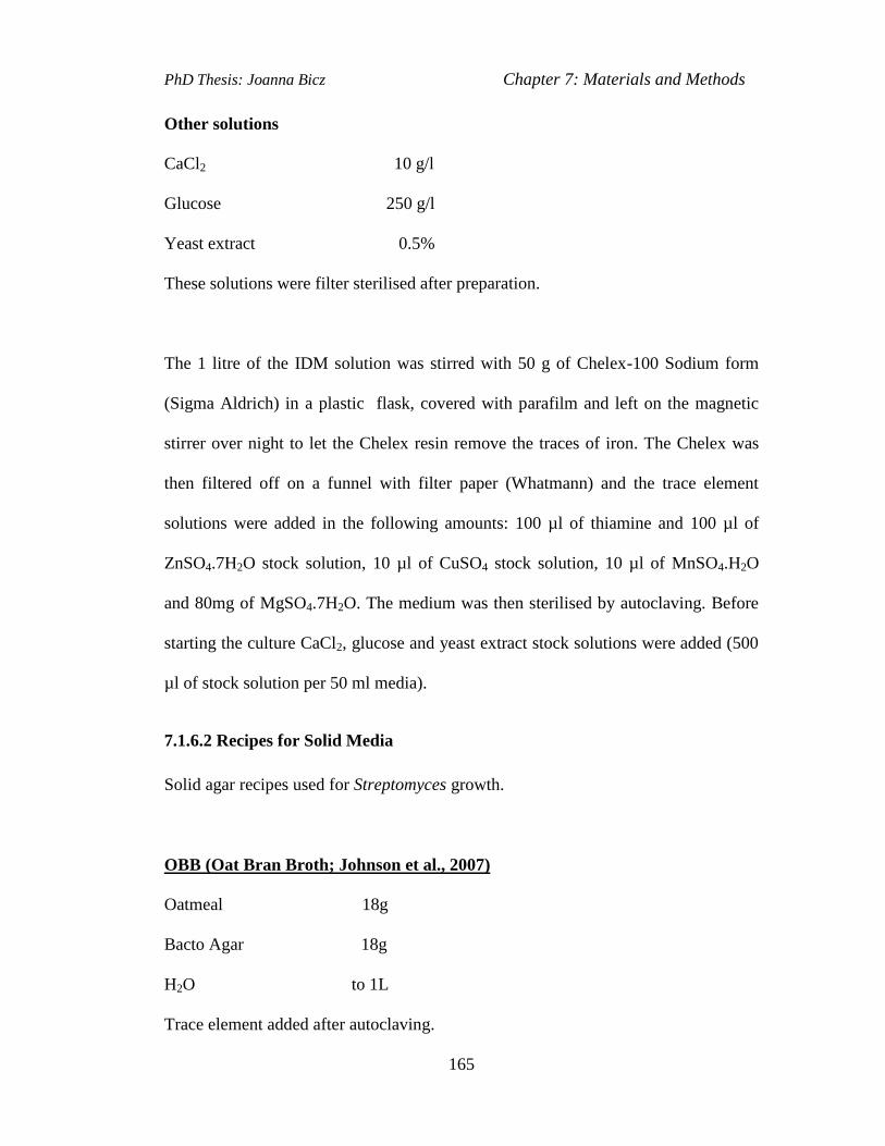
PhD Thesis: Joanna Bicz Chapter 7: Materials and Methods
165
Other solutions
CaCl2 10 g/l
Glucose 250 g/l
Yeast extract 0.5%
These solutions were filter sterilised after preparation.
The 1 litre of the IDM solution was stirred with 50 g of Chelex-100 Sodium form
(Sigma Aldrich) in a plastic flask, covered with parafilm and left on the magnetic
stirrer over night to let the Chelex resin remove the traces of iron. The Chelex was
then filtered off on a funnel with filter paper (Whatmann) and the trace element
solutions were added in the following amounts: 100 µl of thiamine and 100 µl of
ZnSO4.7H2O stock solution, 10 µl of CuSO4 stock solution, 10 µl of MnSO4.H2O
and 80mg of MgSO4.7H2O. The medium was then sterilised by autoclaving. Before
starting the culture CaCl2, glucose and yeast extract stock solutions were added (500
µl of stock solution per 50 ml media).
7.1.6.2 Recipes for Solid Media
Solid agar recipes used for Streptomyces growth.
OBB (Oat Bran Broth; Johnson et al., 2007)
Oatmeal 18g
Bacto Agar 18g
H2O to 1L
Trace element added after autoclaving.
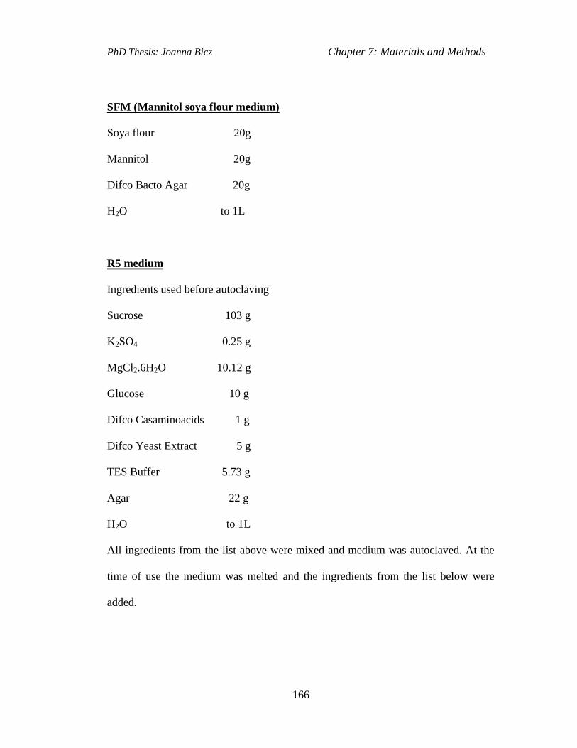
PhD Thesis: Joanna Bicz Chapter 7: Materials and Methods
166
SFM (Mannitol soya flour medium)
Soya flour 20g
Mannitol 20g
Difco Bacto Agar 20g
H2O to 1L
R5 medium
Ingredients used before autoclaving
Sucrose 103 g
K2SO4 0.25 g
MgCl2.6H2O 10.12 g
Glucose 10 g
Difco Casaminoacids 1 g
Difco Yeast Extract 5 g
TES Buffer 5.73 g
Agar 22 g
H2O to 1L
All ingredients from the list above were mixed and medium was autoclaved. At the
time of use the medium was melted and the ingredients from the list below were
added.
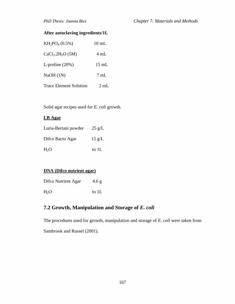
PhD Thesis: Joanna Bicz Chapter 7: Materials and Methods
167
After autoclaving ingredients/1L
KH2PO4 (0.5%) 10 mL
CaCl2.2H2O (5M) 4 mL
L-proline (20%) 15 mL
NaOH (1N) 7 mL
Trace Element Solution 2 mL
Solid agar recipes used for E. coli growth.
LB Agar
Luria-Bertani powder 25 g/L
Difco Bacto Agar 15 g/L
H2O to 1L
DNA (Difco nutrient agar)
Difco Nutrient Agar 4.6 g
H2O to 1L
7.2 Growth, Manipulation and Storage of E. coli
The procedures used for growth, manipulation and storage of E. coli were taken from
Sambrook and Russel (2001).

PhD Thesis: Joanna Bicz Chapter 7: Materials and Methods
168
7.2.1 Growth Conditions of E. coli
E. coli was cultured over night in LB broth (180 rpm, 37°C) or on LB agar plates at
37°C. Antibiotic selection was used for plasmid-containing cells at the
concentrations listed in Table 7.2.
7.2.2 Preparation of Electrocompetent E. coli Cells
E. coli was grown over night at 37°C in 10 ml LB shaking at 180 rpm. 100 µl of E.
coli overnight culture was then inoculated into 10 ml LB and grown for 3-4 hours
shaking at 200 rpm to an OD600 of ~ 0.4. The cells were then recovered by
centrifugation at 3000 rpm for 10 min at 4°C. After decanting the medium the cell
pellet was resuspended by mixing in 10 ml of ice-cold 10% glycerol. The cells were
then centrifuged again and resuspended in 5 ml of ice-cold 10% glycerol,
centrifuged, decanted and resuspended in the remaining ~100 µl 10% glycerol.
7.2.3 Transformation of Electrocompetent E. coli Cells
About 100 ng of DNA were added to 100 µl of E. coli cell suspension in a single
transformation. Electroporation was done in a 0.2 cm ice-cold electroporation cuvette
using a BioRad GenePulser II set to: 200 Ω, 25 μF and 2,5 kV and the expected time
constant of 4.5 – 4.9 ms. Immediately after electroporation, 1 ml of ice cold LB was
added to shocked cells. They were then incubated shaking for 1h at 37°C and spread
onto LB agar plates containing appropriate antibiotic for the selection of
transformants.
PhD Thesis: Joanna Bicz Chapter 7: Materials and Methods
169
7.2.4 Storage of E. coli Cells
E. coli overnight LB cultures were mixed with 70% glycerol to a final concentration
of 20% and stored at -78 °C for a long term.
7.3 Growth, Manipulation and Storage of Streptomyces
The procedures used for growth, manipulation and storage of Streptomyces were
described by Kieser et al. (2000). The protocol for genomic DNA extraction was
taken from Pospiech and Neumann (1995).
7.3.1 Spore Stock Collection from Agar Plates
S. scabies spores were grown on oat bran broth (OBB) agar. Spores were collected
from each plate by adding 3 ml of sterile water and scrapping the surface of the
mycelia with a sterile loop. The spore suspension was then filtered through sterile
cotton wool and centrifuged for 10 min at 3000 rpm. The supernatant was poured off
and the spore pellet was resuspended in approximately 500 µl of the remaining
water. The stock was then made by adding similar volume of 50% sterile glycerol to
the spore suspension. The spores were stored at -20ºC.
7.3.2 Liquid Cultures for Genomic DNA Extraction
S. scabies spores for genomic DNA extraction were grown in 50 ml of TSB medium
at 30ºC, 180 rpm for 72 hours.
7.3.3 DNA Transfer from E. coli to S. scabies by Conjugation
10μl of E. coli ET12567/pUZ8002 glycerol stock was added to 10 ml of a liquid LB
medium containing kanamycin (25 μg/ml) and chloramphenicol (25 μg/ml) and the

PhD Thesis: Joanna Bicz Chapter 7: Materials and Methods
170
culture was grown at 37ºC over night.Then, 100 μl of over night culture was added to
10 ml LB and incubated for 6 hours with the same antibiotics and at the same
conditions as above. After 6 hours the electrocompetent cells were prepared
according to the procedure used earlier (section7.2.2), transformed with the oriT-
resistance cassette containing cosmid or plasmid clone (section 7.2.3) and selected
for the incoming cosmid or plasmid by plating on LB agar with kanamycin (25
μg/ml) and chloramphenicol (25 μg/ml) as well as antibiotic appropriate to the
resistance of the cassette. One of the transformant colonies was inoculated into 10 ml
LB liquid medium containing the same antibiotics as those used on LB plates and
grown overnight at 37ºC. Then, 500 μl of overnight culture was inoculated into 50
ml of fresh LB plus antibiotics as above and grown for 6 h at 37°C, shaking at 180
rpm to an OD600 of 0.4. After 6 hours the cells were washed twice with 50 ml of LB
to remove antibiotics (might inhibit Streptomyces), and resuspended in ~2 ml of LB.
While washing the E. coli cells, 500μl (approximately 109) of freshly collected
Streptomyces scabies 87.22 spores were added to 0.5ml 2 × YT broth, heat shocked
at 50°C for 20 min, then cooled for 1 min on ice. Next, ~2 ml of E. coli cell
suspension were mixed with 500 μl heat-shocked spores in 500 2xYT and then
centrifuged briefly. The supernatant was then partially removed and the pellet was
resuspended in the c.a. 500 μl residual liquid. This liquid was then plated out without
diluting on several SFM agar + 10mM MgCl2 plates (without antibiotics) and
incubated at 30°C for 16-20 h. The next day each plate was overlayed with 1 ml of
water containing 0.5 mg nalidixic acid (20 μl of 25 mg/ml stock) to selectively kill
E. coli. The antibiotics in the appropriate concentrations were also added for the

PhD Thesis: Joanna Bicz Chapter 7: Materials and Methods
171
selection of the exconjugant Streptomyces containing the plasmid or cosmid of
interest.
7.4 Extraction and Manipulation of DNA
7.4.1 Isolation of Genomic DNA from S. scabies
The S. scabies DNA was isolated according to the method by Pospiech and Neumann
(1995).
The cells of S. scabies 87.22 strain were grown in 50ml of TSB medium at 30°C for
three days. The cells were harvested by centrifugation (10 min, 3000 rpm) and
resuspended in 5 ml of SET (75 mM NaCl, 25 mM EDTA, 20 mM Tris, pH 7.5).
Lysozyme was added to the cells to a concentration of 1 mg/ml and incubated at
37°C for 0.5 h. Next, l/10 volume of 10% SDS and 0.5 mg/ml of proteinase K were
added and the cells were incubated at 55°C with an occasional inversion for 2 h.
After that l/3 volumes of 5 M sodium chloride and one volume of chloroform were
added and the cells were incubated at room temperature for 0.5 h with frequent
inversion. The cells were then centrifuged at 4500 g for 15 min; the aqueous phase
was transferred to a new tube using a blunt-ended pipette tip. The DNA was
precipitated by adding 1 volume of isopropanol and gently inverting the tube. The
precipitated DNA was then transferred into a microfuge tube and rinsed with 70%
ethanol, dried on air and dissolved in 1ml of TE buffer. The amount of DNA
obtained was between 1-3 mg.

PhD Thesis: Joanna Bicz Chapter 7: Materials and Methods
172
7.4.2 Plasmid/Cosmid Isolation from E. coli
The E.coli cosmid or plasmid was isolated according to the method by Sambrook and
Russell (2001).
5 ml of liquid LB medium with added antibiotics was inoculated with E. coli cells
and they were grown overnight at 37°C, 180 rpm. The cells were then centrifuged
and the cell pellet was resuspended in 100 μl of solution I (50 mM Tris/HCl, pH 8;
10 mM EDTA). Then, 200 μl solution II (200 mM NaOH; 1% SDS) was added to it
and cells were mixed by inverting the tube several times. Then, 150 μl of solution III
(3 M potassium acetate, pH 5.5) were added to the tube and it was mixed by
inverting several times. The tube was centrifuged at a maximum speed in a
microcentrifuge for 5 min at room temperature. Then, the supernatant was
transferred to a new tube and 400 μl of phenol-chloroform-isoamyl alcohol (25:24:1)
was added, vortexed 2 min and centrifuged at a full speed in a microcentrifuge for 5
min, at room temperature. The upper phase was transferred to a new tube and 600 μl
of ice cold isopropanol was added to it. The tube was left on ice for 10 min to
precipitate the DNA, and the sample was centrifuged for 10 min at a full speed in a
minicentrifuge at 4°C. The supernatant was removed and the pellet was washed with
200 μl 70% ethanol, spinned in a microcentrifuge for 5 min at room temperature and
left for 30 min at room temperature to air-dry. The pellet was then resuspended in a
30 μl of sterile water with RNAse.
7.4.3 Digestion of DNA with Restriction Enzymes
Digestions by restriction enzymes were prepared following the manufacturer
instructions. Restriction enzymes always occupied 10% of the total volume and

PhD Thesis: Joanna Bicz Chapter 7: Materials and Methods
173
appropriate buffer also occupied 10% (all buffers were 10 x concentrated). If BSA
had to be added, it also occupied 10% of the total volume. The rest of the total
volume consisted of a DNA solution to be digested and, if necessary, water.
Digestions of plasmid/cosmid were carried out in a total volume of 10 µl.
7.4.4 Ligation of DNA into Plasmids
DNA to be ligated into plasmid vectors was purified using the Fermentas gel
extraction kit and eluted with 10 µl of distilled water. The concentration of vector
and insert was then estimated by running 1µl of each DNA sample on an agarose gel
along with a molecular size marker of a known DNA concentration. A molar ratio of
vector: insert of 1:4 was generally used for ligation reactions. The ligation reactions
were carried out in a 20 µl of a total volume for 30 min using Rapid Ligation kit
from Roche.
7.4.5 Agarose Gel Electrophoresis
1x TBE buffer was used to prepare and run agarose gels. Different percentage
agarose gels were prepared (0.8%, 1%, 1.5%) depending on the size of DNA
fragments. 2 µl of Gel Red dye (Biotium) was added to the gel to visualize DNA
under UV light. Electrophoresis was carried out for 40 min at 110 V. A. 1kb DNA
molecular size marker (Fermentas) was used as a size standard.
7.5 PCR Methods
The High Fidelity Polymerase (Roche) was used to amplify a gene for PCR targeting
or for cloning. The Taq polymerase (Fermentas) was used for analysis of constructs
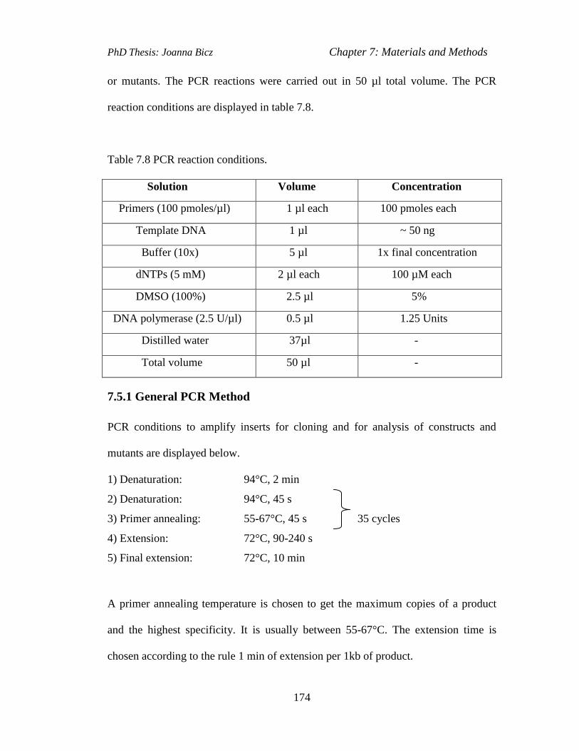
PhD Thesis: Joanna Bicz Chapter 7: Materials and Methods
174
or mutants. The PCR reactions were carried out in 50 µl total volume. The PCR
reaction conditions are displayed in table 7.8.
Table 7.8 PCR reaction conditions.
Solution Volume Concentration
Primers (100 pmoles/µl) 1 µl each 100 pmoles each
Template DNA 1 µl ~ 50 ng
Buffer (10x) 5 µl 1x final concentration
dNTPs (5 mM) 2 µl each 100 µM each
DMSO (100%) 2.5 µl 5%
DNA polymerase (2.5 U/µl) 0.5 µl 1.25 Units
Distilled water 37µl -
Total volume 50 µl -
7.5.1 General PCR Method
PCR conditions to amplify inserts for cloning and for analysis of constructs and
mutants are displayed below.
1) Denaturation: 94°C, 2 min
2) Denaturation: 94°C, 45 s
3) Primer annealing: 55-67°C, 45 s 35 cycles
4) Extension: 72°C, 90-240 s
5) Final extension: 72°C, 10 min
A primer annealing temperature is chosen to get the maximum copies of a product
and the highest specificity. It is usually between 55-67°C. The extension time is
chosen according to the rule 1 min of extension per 1kb of product.

PhD Thesis: Joanna Bicz Chapter 7: Materials and Methods
175
7.5.2 PCR Amplification of the Gene Replacement Cassette.
A PCR reaction (Gust et al., 2002) with designed primers was carried out to amplify
the cassette from the digested pIJ773 vector. The conditions of the reaction are as
follows:
1) Denaturation: 94°C, 2 min
2) Denaturation: 94°C, 45 s
3) Primer annealing: 50°C, 45 sec 10 cycles
4) Extension: 72°C, 90 sec
5) Denaturation: 94°C, 45 sec
6) Primer annealing: 55°C, 45 sec 25 cycles
7) Extension: 72°C, 90 sec
8) Final extension: 72°C, 5 min
7.6 PCR Targeting in S. scabies 87.22
PCR targeting technique was used to create in-frame deletions in S. scabies genome
(Gust et al., 2002).
7.6.1 Design of PCR Primers
Two targeting primers were designed for the deletion in each gene according to the
procedure described in section 3.1. The targeting primers are displayed in Table 7.6
(Section 7.1.5).
7.6.2 Purification of the Resistance Cassette
Approximately 3 μg of plasmid DNA pIJ773 was digested with 50 U EcoRI (Roche)
and 50 U HindIII (Roche), in 1 x buffer (Roche), in a 50 μl reaction for 1.5 hour at
37° C. A 2938 bp vector fragment and a 1400 bp for the cassette were generated. The

PhD Thesis: Joanna Bicz Chapter 7: Materials and Methods
176
digest was then run on a gel and then the band for the resistance cassette was cut out
and purified by Qiagen gel extraction kit. The purified fragment was stored in a 30 μl
of a sterile water, at –20°C.
7.6.3 PCR Amplification of the Extended Resistance Cassette
The purified resistance cassette was amplified by PCR with long targeting primers
(Table 7.6) using the reaction mixture shown in Table 7.9. The Expand High Fidelity
polymerase (Roche) was used in PCR reactions under the conditions described in
section 7.5.2. 5 µl of the PCR reaction was analysed by electrophoresis on a 1%
agarose gel for the presence of the cassette with the size of ~1476 bp. The rest of the
PCR product was purified from the reaction mixture using the Qiagen PCR
purification kit following the manufacturer’s instructions and eluted from the column
with 30 µl of water.
7.6.4 Introduction of Cosmid Clones into E. coli BW25113/pIJ790 by
Electroporation
pIJ790 vector contains the resistance marker cat (chloramphenicol resistance) and a
temperature sensitive origin of replication (requires 30°C for replication). E. coli
BW25113/pIJ790 cells were grown overnight at 30°C in a 10 ml LB containing
chloramphenicol (25 μg/ml). 100 μl of E. coli BW25113/pIJ790 from the overnight
culture were inoculated in a 10 ml LB containing 20 mM MgSO4 (200 μl of 1M
stock) and chloramphenicol (25 μg/ml) and grown for 4 hours at 30°C, shaking at
180 rpm to an OD600 of ~ 0.4. The competent cells were then prepared according to
the section 7.2.2 and the 100 μl of the cell suspension was mixed with ~ 100 ng (1-2

PhD Thesis: Joanna Bicz Chapter 7: Materials and Methods
177
μl) of a cosmid DNA. Electroporation was carried out as described in section 7.2.3
and the cells were incubated on an orbital shaker at 180 rpm for 1h at 30°C. The
culture was spread onto two plates of LB agar containing ampicillin (100 μg/ml),
kanamycin (50 μg/ml) and chloramphenicol (25 μg/ml) and incubated overnight at
30°C. The next day, the transformant colonies containing cosmid were observed.
One isolated colony was transferred into 5 ml of liquid LB containing antibiotics as
above and incubated overnight at 30°C. This culture was then used as a pre-culture
for generating competent cells to be transformed with the extended resistance
cassette.
7.6.5 PCR Targeting of Cosmids
10 ml of LB containing ampicillin (100 μg/ml), kanamycin (50 μg/ml) and
chloramphenicol (25 μg/ml) was inoculated with 1% of the overnight culture of E.
coli BW25113/pIJ790 and the cosmid. 100 μl of 1M L-arabinose stock solution was
added to the culture to induce the red genes. Then, the culture was grown for 4 hours
at 30°C shaking, at 180 rpm to an OD600 of ~ 0.4. Competent cells were prepared as
described in section 7.2.2 and the 50 μl of the cell suspension was mixed with ~ 100
ng (2 μl) of the extended resistance cassette PCR product. Electroporation was
carried out (section 7.2.3) and the cells were incubated shaking at 180rpm for 1h at
37°C (the temperature above 30°C causes degeneration and loss of pIJ790 plasmid).
The culture was spread onto two plates of LB agar containing ampicillin (100
μg/ml), kanamycin (50 μg/ml) and apramycin (50 μg/ml) and incubated overnight at
37°C. After 16 hours growth at 37°C different colony sizes were observed. The small
colonies were false positives and the larger the sizes of a colony, the more copies of

PhD Thesis: Joanna Bicz Chapter 7: Materials and Methods
178
the mutagenised cosmids were present. Several large colonies were inoculated in 5
ml LB liquid cultures containing ampicillin (100 μg/ml), kanamycin (50 μg/ml) and
apramycin (50 μg/ml) and grown at 37°C, shaking at 180 rpm to a cell density OD600
~ 0.1 – 0.3, for 6 h. After 6 hours, the cosmid DNA was isolated and tested by
restriction analysis and PCR.
About 2 µg (10 µl) of the wild type cosmid clone DNA and 2 µg (10 µl) of the
mutagenised cosmid (with introduced extended apramycin cassette and OriT) were
digested, each with BamHI or SacI restriction enzymes in a total volume of 30 µl.
Both digestions were left for a 3 hours at 37°C and then 5 µl of each digestion was
analysed on an agarose gel (1% agarose). The digested wild type and mutagenised
cosmid gave distinct patterns of bands. The expected digestion pattern for the
mutagenised cosmid was confirmed by an in silico prediction using program, such as
Clone Manager or NEB cutter. If the digestion pattern agreed with the program
prediction then the integration of the disruption cassette into cosmid was also
confirmed using PCR with suitable test primers (Table 7.6) priming within ~ 100 bp
upstream and downstream of the region, which recombined with the PCR product of
the extended OriT-apramycin resistance cassette. The PCR reaction was performed
under the conditions listed in the section 7.5.2. The mutagenised cosmid was
introduced into a non-methylating E. coli host ET12567/pUZ8002 by electroporation
(section 7.3.3).
7.6.6 Transfer of the Mutant Cosmids into Streptomyces scabies
The procedure for conjugal transfer of the engineered cosmid from E. coli
ET12567/pUZ8002 into S. scabies is described in section 7.3.3. After several days of

PhD Thesis: Joanna Bicz Chapter 7: Materials and Methods
179
incubation the grey, hydrophobic Streptomyces colonies were present on each plate.
Several colonies were picked and transferred onto DNA plates (Difco Nutrient Agar)
containing nalidixic acid (25µg/ml) and apramycin (50µg/ml) with and without
kanamycin (50µg/ml). Double cross-over exconjugants were kanamycin sensitive
(the cosmid backbone containing kanamycin resistance gene has been lost during
cross over) and apramycin resistant (cassette contains apramycin). The kanamycin
sensitive clones were picked from DNA plates and plated out on oat bran broth (the
solid medium used for growing of S. scabies). The spores were then collected;
genomic DNA isolated and analysed for the desired mutation by PCR (section 7.5.1)
with the test primers (section 7.1.5) and also by Southern blot hybridisation (section
7.7).
7.7 Southern Blot Hybridisation
DIG High Prime DNA Labelling and Detection Starter Kit (Roche) was used to carry
out Southern blot hybridisation.
7.7.1 Probe Labeling
1µg of cosmid DNA (5 µl) was added to a final volume of 16 µl of autoclaved miliQ
distilled water and denatured by boiling at 100°C for 10 min. The sample was then
rapidly cooled on ice, mixed with 4 µl of DIG High Prime solution and incubated
over night in 37°C water bath. Then, the labeling reaction was stopped by adding 2
µl of 0.2 M EDTA (pH 8.0) and then by heating to 65°C for 10 min.
7.7.2 Genomic DNA Digestion
20 µg of genomic DNA for each strain was digested with BamHI or SacI over night
at 37°C. The samples were then loaded on 0.7% agarose gel and run over night at 25

PhD Thesis: Joanna Bicz Chapter 7: Materials and Methods
180
V. The gel was then exposed to UV light for 5 minutes to help denature the DNA.
Then, the gel was rinsed in distilled water and washed twice 15 minutes in
denaturation buffer (2 x 15 min shaking at room temperature). After denaturation the
gel was rinsed again in distilled water and twice with neutralisation buffer (2x 15
shaking).
7.7.3 Capillary Transfer and DNA Fixation
A glass plate was placed across a plastic tray containing 20xSSC buffer. One sheet of
3mm Whatmann filter paper soaked in 20xSSC buffer was placed on the glass plate.
The ends of the filter paper touched the buffer in the container. On the top of this
filter paper another soaked piece of paper was put and on that the treated gel with the
wells side down. The piece of a nylon membrane with a positive charge cut to the
size of the gel was placed on the top of it. The gel was wrapped around on the edges
with a tape to prevent ‘short cirquiting.’ Then, three pieces of Whatmann 3 mm
paper the size of gel were placed on the top of a nylon membrane; after that, the 5 cm
high layer of paper towels and finally on that a glass plate with 2 litres flask half-
filled with water as a weight. The movement of the DNA to the membrane was
carried out by capillary transfer over night. The next day the membrane was removed
from the gel, placed on the piece of Whatmann 3mm paper soaked in 10xSSC buffer
and UVcross-linked to fix the DNA (auto-crosslinking on UV Stratalinker 2400 from
Stratagene).

PhD Thesis: Joanna Bicz Chapter 7: Materials and Methods
181
7.7.4 Hybridisation
The membrane with the DNA transferred to it was prehybridised in a hybridisation
tube in a 20 ml of DIG Easy Hyb solution ( DIG Easy Hyb granules mixed with 64
ml of sterile water for 10 min at 37°C) rotating for 2 hours at 42°C. The membrane
was then incubated in the hybridisation solution over night rotating at 42°C. The
hybridisation solution was prepared by adding the 20 µl of denatured DIG labeled
DNA probe (5µg/µl) into 7 ml of preheated DIG Easy Hyb solution. The probe was
denatured by boiling for 5 min at 98°C and then rapidly cooled on ice. The next day
the hybridization solution was removed and the membrane was washed 2x 5 minutes
in 50 ml preheated 2x SSC, 0.1% SDS rotating at 25°C; then 2x 15 min in 50 ml
preheated 0.5x SSC, 0.1% SDS at 58°C. The membrane was then washed in 30 ml of
washing buffer for 5 min at room temperature. Then, it was incubated at 50 ml of
blocking solution at room temperature; under constant rotation in the hybridisation
oven. After 30 minutes the blocking solution was replaced with a 30 ml of the fresh
blocking solution, mixed with a 3 µl of the anti-DIG antibody conjugate and the
membrane was incubated with it for another 30 min. Then, the antibody solution was
removed and the membrane was washed twice in 50 ml of washing buffer (2x15 min
rotating) and then equilibrated for 5 min in 20 ml of a detection buffer.
7.7.5 Detection
The wet membrane was placed with the DNA side up in a hybridisation bag and 2 ml
of chemiluminescence substrate CSPD (from DIG High Prime kit, Roche) was
applied onto it. The membrane was then immediately covered with the second sheet
of the development bag so that the CSPD substrate was spread evenly without an air
PhD Thesis: Joanna Bicz Chapter 7: Materials and Methods
182
bubbles over the membrane. The edges of the hybridisation bag were then sealed and
the membrane was incubated in the development folder in the dark. After 5 min the
membrane was placed inside a new hybridisation bag, sealed and incubated at 37°C
for 15 min to enhance the luminescence reaction. The membrane was then exposed
to film over night. The film was then developed according to standard procedures.
7.8 RNA Methods
7.8.1 Total RNA Isolation from S. scabies
The wild type S. scabies were grown on an oat bran broth plates at 30°C for 3, 5, 7
and 14 days. After the incubation, the mycelia from each plate were collected by a
plastic loop and ground in a mortar with the addition of a liquid nitrogen. The frozen
ground mycelium was transferred into a 1.5 ml Eppendorf tube containing 350 µl of
resuspension buffer RA1 from NucleoSpin RNA II kit (Macherey-Nagel) and 3.5 µl
of mercapthoethanol. The sample was then vortexed vigorously for 3 minutes to help
the cell wall breakage. The sample was processed further using NucleoSpin RNA II
kit. The RNA was finally eluted on the column with 60 µl of RNAse free distilled
sterile water. The RNA was always handled in gloves. All pipette tips and Eppendorf
tubes were autoclaved twice. The bench area and pipettes were wiped with the
RNAse inhibitor agent (RNaseZAP, Sigma Aldrich). The RNA was analysed using
agarose gel elecrophoresis on a 1% agarose gel. 1µl of RNA was mixed with the 4 µl
of the loading dye (Fermentas) and run on a gel with 50 V current for 1hour. The
RNA was then visualised under the UV light. The presence of the two sharp bands
and the third lower and more faded was indicating that the RNA is not degraded and

PhD Thesis: Joanna Bicz Chapter 7: Materials and Methods
183
it is suitable for downstream applications, such as PCR. The RNA samples were then
treated with DNAse (TurboDnase, Ambion) to remove any traces of genomic DNA.
They were then analysed using standard PCR method (section 7.5.1) with Taq
polymerase to ensure that no genomic DNA was present in the samples. The purified
RNA was then used as a template in reverse transcriptase PCR reactions (section
7.8.2) to analyse expression of the genes from the putative tetronate biosynthetic
gene cluster.
7.8.2 Reverse Transcriptase PCR Method
The Qiagen One Step PCR Kit was used for performing of PCR reactions.
Polymerase from that kit is a mix of reverse transcriptase (RT) polymerases
(Omniscript Reverse Transcriptase, Sensiscript ReverseTranscriptase) and the Taq
DNA polymerase (Hotstar Taq polymerase). The RT polymerase converts messenger
RNA (mRNA) into a complementary DNA (cDNA) in the initial part of the PCR
reaction carried out at 50°C in 30 minutes. In the next step, the temperature of the
reaction was raised to 95°C to activate the Taq DNA polymerase, deactivate the
reverse transcriptases and denature the cDNA. The cDNA template was then
amplified by the Taq polymerase in a standard PCR reaction.
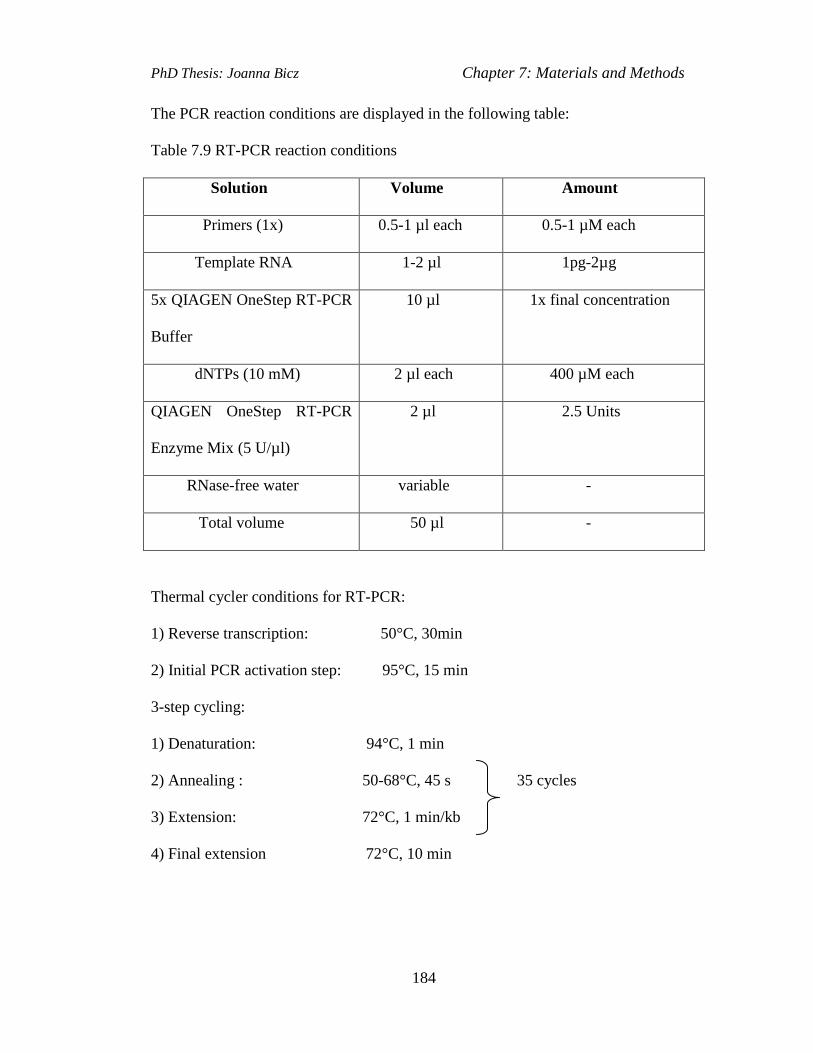
PhD Thesis: Joanna Bicz Chapter 7: Materials and Methods
184
The PCR reaction conditions are displayed in the following table:
Table 7.9 RT-PCR reaction conditions
Solution Volume Amount
Primers (1x) 0.5-1 µl each 0.5-1 µM each
Template RNA 1-2 µl 1pg-2µg
5x QIAGEN OneStep RT-PCR
Buffer
10 µl 1x final concentration
dNTPs (10 mM) 2 µl each 400 µM each
QIAGEN OneStep RT-PCR
Enzyme Mix (5 U/µl)
2 µl 2.5 Units
RNase-free water variable -
Total volume 50 µl -
Thermal cycler conditions for RT-PCR:
1) Reverse transcription: 50°C, 30min
2) Initial PCR activation step: 95°C, 15 min
3-step cycling:
1) Denaturation: 94°C, 1 min
2) Annealing : 50-68°C, 45 s 35 cycles
3) Extension: 72°C, 1 min/kb
4) Final extension 72°C, 10 min
PhD Thesis: Joanna Bicz Chapter 7: Materials and Methods
185
7.9 Growth of Streptomyces scabies; Extraction of Metabolites and
Analysis of Metabolite Production
7.9.1 Pyochelin
7.9.1.1 Culturing of S. scabies in Iron Deficient Medium
S. scabies 87.22 and and the ΔScab1401 and ΔScab1401–ΔScab1471 mutants were
cultured in Iron Deficient Medium for a detection of pyochelin. 50 ml of sterilised
IDM in each 250 ml sterile plastic Erlenmeyer flask were inoculated with 10 µl of
fresh spore stock of each S. scabies pyochelin mutant and wild type S. scabies strain.
The liquid cultures were incubated at 30°C shaking at 180 rpm for 4-5 days. 1 ml of
each cell culture was then taken to 1.5 ml Eppendorf tubes and centrifuged in a table
microfuge at 13000 rpm for 5 minutes at room temperature. Supernatant was placed
in 1 ml HPLC tubes. The samples were analysed using LC-MS.
7.9.1.2 LC-MS Analysis
Liquid chromatography –Mass Spectrometry (LC-MS) was used to analyse the
supernatants of the S. scabies 87.22 and the ΔScab1401 and ΔScab1401–ΔScab1471
mutants. LC-MS analysis of the reaction mixture was carried out using Eclipse
XDB-C18 column (150 x 4.6mm, 5μm, Agilent) connected to an Agilent 1100
HPLC instrument. The outflow was connected via a splitter (10% flow to MS, 90 %
flow to waste) to a Bruker HCT+ mass spectrometer fitted with an electrospray
source in positive ion mode with parameters: nebulizer flow 40 psi, dry gas flow 10.0
L/min, dry temperature 300 ºC, capillary – 4 kV, skimmer 40V, capillary exit 106 V,
ion charge control target (ICC) 100,000, spectral averages 3. Absorbance was

PhD Thesis: Joanna Bicz Chapter 7: Materials and Methods
186
monitored at 210 nm and 280 nm. The elution profile is displayed in the table below,
with a 1 ml/min flow rate. The retention time for pyochelin was 23 minutes.
Table 7.10 Gradient elution profile used in LC-MS analyses of pyochelin production.
Time (min) Water + 0.1% formic acid Methanol + 0.1% formic acid
0 100 0
5 100 0
25 0 100
30 0 100
35 100 0
7.9.1.3 Extraction of Pyochelin and High Resolution Mass Spectrometry
S. scabies 87.22 and the ΔScab1401 and ΔScab1401–ΔScab1471 mutants were
grown in an iron-deficient medium (Barona-Gomez et al., 2004) for 4 days. The
cultures were centrifuged for 10 min at 4000 r.p.m. at 4°C. The supernatants were
lyophilised,
resuspended in 5 ml of methanol and analysed using a Dionex Ultimate 3000RS
instrument fitted with Supelco Acentis Express C18 column (15062.1 mm, 2.7 mm);
coupled to a Bruker MaXis mass spectrometer with parameters: ESI in positive
mode; full scan 50–1500 m/z; End plate offset, 2500 V; Capillary, 24500 V;
Nebulizer gas (N2), 1.6 bar; Dry gas (N2), 8 l min21; Dry Temperature, 180 uC. The
elution profile is displayed in the table. The flow rate was 0.2 ml per minute.
Absorbance was monitored at 210 and 280 nm. The High Resolution Mass
spectrometry analyses were performed by our collaborator Dr. Lijiang Song

PhD Thesis: Joanna Bicz Chapter 7: Materials and Methods
187
Table 7.11 Gradient elution profile used in High Resolution MS analyses of
pyochelin production.
Time (min) Water + 0.1% formic acid Methanol + 0.1% formic acid
0 100 0
5 100 0
30 0 100
35 0 100
40 100 0
55 100 0
Pyochelin and enantio-pyochelin (ent-pyochelin) standard solutions (both 10 µl, 10
µg/ml in methanol) and concentrated culture supernatant from the ΔScab1401 mutant
were analysed using a homochiral stationary phase using an Agilent 1100 HPLC
coupled to a Bruker HCT plus mass spectrometer with parameters: ESI in positive
mode; full scan 50–1500 m/z; End plate offset, 2500 V; Capillary, 24500 V;
Nebulizer gas (N2), 40 p.s.i.; Dry gas (N2), 10 l min21; Dry Temperature, 300 uC.
The eluents and elution conditions are described in Table 7.11. The flow rate was 1
ml/min. All High Resolution Mass Spectrometry analyses for pyochelin and
scabichelin, as well as, chiral HPLC analyses, were carried out with the assistance of
our collaborator on the project Dr. Lijiang Song.
PhD Thesis: Joanna Bicz Chapter 7: Materials and Methods
188
7.9.2 Scabichelin
7.9.2.1 LC-MS Analysis
S. scabies 87.22 and W1000 were grown in iron deficient medium (Barona-Gomez et
al., 2004) according to the same protocol as in section 7.9.1.1. 1 ml of supernatants
from each of 50 ml cultures of S. scabies 87.22 and W1000 were analysed by high
resolution LC-MS on a Sigma Ascentis Express column (C18, 150 x 2.1mm, 2.7μm)
attached to a Dionex 3000RS UHPLC coupled to a Bruker MaXis ESI-Q-TOF mass
spectrometer. The mobile phases were water containing 0.1% formic acid (eluent A)
and methanol containing 0.1% formic acid (eluent B). A gradient of 5% B to 100% B
in 15 minutes was used with a flow rate of 0.2ml/min. Absorbance at 210nm was
monitored. The mass spectrometer was operated in a positive ion mode with a scan
range 50-2,000 m/z. Source conditions were: end plate offset at -500V; capillary at -
4500V; nebulizer gas (N2) at 1.6bar; dry gas (N2) at 8L/min; dry Temperature at 180
°C. Ion tranfer conditions were: ion funnel RF at 200Vpp; multiple RF at 200Vpp;
quadruple low mass at 55 m/z; collision energy at 5.0 eV; collision RF at 600 Vpp;
ion cooler RF at 50-350 Vpp; transfer time at 121μs; pre-Pulse storage time at 1 μs.
PhD Thesis: Joanna Bicz Chapter 7: Materials and Methods
189
7.9.3 Desferrioxamines
7.9.3.1 Growing S. scabies in Supplemented Minimal Medium (SMM) and LC-
MS Analysis
50 ml of sterilised SMM in each 250 ml sterile plastic Erlenmeyer flask was
inoculated with 10 µl of fresh spore stock of S. scabies ΔdesC mutant and wild type
S. scabies strain. The liquid cultures were then incubated at 30°C incubator, shaking
at 180 rpm for 3-4 days. 1 ml of each cell culture was then taken to 1.5 ml Eppendorf
tubes and 2µl of FeCl3 was added to them. The samples were incubated for 30 min at
room temperature and then centrifuged in a table microfuge at 13000 rpm for 5
minutes at room temperature. Supernatant was taken to 1 ml HPLC tubes. The
samples were then analysed using LC-MS. The LC-MS equipment and analysis
conditions were same as described for pyochelin LC-MS analysis (Section 7.9.1.2).
The absorbance was monitored at 435 nm.
PhD Thesis: Joanna Bicz References
190
References
Angert, E. R.; Nature Rev Microbiol 2005, 3, 214-224.
Ankenbauer, R.; Sriyosachati, S. and Cox, C. D. Infect Immun 1985, 49, 132–140.
Austin, M.B. and. Noel, J. P. Nat Prod Rep 2003, 20, 79-110.
Barona-Gomez F.; Lautru S.; Francou F. X.; Pernodet J. L.; Leblond P.; Challis G.
L. Microbiol 2006, 152, 3355-3366.
Barona-Gomez, F.; Wong, U.; Giannakopulos, A. E.; Derrick, P. J. and Challis, G. L.
J Am Chem Soc, 2004, 126, 16282–16283.
Barry S. M.; Kers J. A.; Johnson E. G.; Song L.; Aston P. R.; Patel B.; Krasnoff S.
B.; Crane B. R.; Gibson D. M.; Loria R.; Challis G. L. Nat Chem Biol 2012, 8, 814-
816.
Bell, K. S., Sebaihia, M., Pritchard, L., Holden, M. T., Hyman, L. J.,
Holeva, M. C., Thomson, N. R., Bentley, S. D., Churcher, L. J., Mungall,
K., Atkin, R., Bason, N., Brooks, K., Chillingworth, T., Clark, K.,
Doggett, J., Fraser, A., Hance, Z., Hauser, H., Jagels, K., Moule, S.,
Norbertczak, H., Ormond, D., Price, C., Quail, M. A., Sanders, M.,
Walker, D., Whitehead, S., Salmond, G. P., Birch, P. R., Parkhill, J., and
Toth, I. K. Proc. Natl. Acad. Sci. U.S.A., 2004, 101, 11105-11110.
Berti A. D. and Thomas M. G. J. Bacteriol 2009, 191, 4594-4604.
Bickel H.; Bosshardt R.; Gaumann E.; Reusser P.,Vischer E., Voser W., Wettstein
A., Zahner A. Helv Chim Acta 1960, 43, 2118–2128.
PhD Thesis: Joanna Bicz References
191
Bignell, D. R. D.; Huguet-Tapia J. C.; Joshi M. V.; Pettis, G. S.; Loria, R. Antonie
van Leeuwenhoek 2010, 98, 179–194. (1)
Bignell D. R. D., Seipke R. F., Huguet-Tapia J. C.,Chambers A. H., Parry R. J.,
Loria R The Am. Phytopathol Soc, 2010, 23, 161 – 175. (2)
Brickman, T. J. and McIntosh, M. A. J Biol Chem 1992, 267, 12350-12355.
Bruner S. D.; Weber T.; Kohli R. M., Schwarzer D., Marahiel M. A.,
Walsh C. T. and M. Stubbs T., Structure 2002, 10, 301-310.
Bukhalid, R. A.; Gibson D. M.; Crane, B R.; Loria R. Nature 2004, 429, 79–82.
Cairo, G., Bernuzz,i F. and Recalcati, S. Genes and Nutriton 2006, 1, 25-40.
Castignetti, D. Curr Microbiol 1997, 34, 250–257.
Carbonetti, N. H. and Williams, P. H. Infect. Immun 1984, 46, 7–12.
Cartinhour, S. W. and Shuler, M. L. Appl Environ Microbiol 2009, 75, 2720–2726.
Challis G. L., ChemBioChem 2005, 6, 601-611.
Challis, G. L. J Am Chem Soc 2004, 126, 16282–16283.
Challis, G. L. and Hopwood, D. A. Proc Natl Acad Sci U S A 2003, 100, 14555-61.
Challis, G. L. and Lautru, S. Microbiol 2004, 150, 1629-1636.
Challis, G. L. and Ravel, J. FEMS Microbiol Lett 2000, 187, 111-114.
Chan, Y. A., Podevels, A. M., Kevany, B. M. and Thomas, M. G.
Nat Prod Rep 2009, 26, 90-114.
Cox, C. D. Infect Immun 1982, 36, 17-23.
Cox, C. D; Rinehart, K. L.; Moore, M. L.; Cook, J. C. Proc Natl Acad Sci U S A
1981, 78, 4256-4260.
Crone, W. J. K.; Leeper, F. J.; Truman, A. W. Chem Sci 2012, 11, 3121-3127.

PhD Thesis: Joanna Bicz References
192
Cruz, A.T.; Cazacu, A. C.; Allen, C. H. J Clin Microbiol 2007, 45, 1989-1992.
Deeth, R.J. Coordin Chem Rev 2001, 212, 11-34.
Dellagi, A.; Brisset, M., N.; Paulin, J., P. and Expert, D. Mol Plant Microbe Interact
1998, 11, 734–742.
Dellagi, A.; Rigault, M.; Segond, D.; Roux, C.; Kraepiel, Y.; Cellier, F.; Briat, J.F.;
Gaymard, F. and Expert, D. Plant J. 2005, 43, 262–272.
Demydchuk, Y.; Sun, Y.; Hong, H.; Staunton, J. B.; Leadley, P. F. Chem biochem
2008, 9, 1136-1145.
Donadio, S.; Monciardini, P.; Sosio, M. Nat Prod Rep 2007, 24, 1073-109.
Donadio, S.; Sosio, M.; Lancini, G.; Appl Microbiol Biotechnol 2002, 60, 377–380.
Drose, S.; Bindseil, K. U.; Bowman, E. J.; Siebers, A.; Zeeck, A.
and Altendorf, K. Biochem, N Y 1993, 32, 3902–3906.
Drose, S.; Boddien, C.; Gassel, M.; Ingenhorst, G.; Zeeck, A.; Altendorf, K.
Biochemistry 2001, 40, 2816–2825.
Dunbar, K.L.; Melby, J. O.; Mitchell, D. A. Nat Chem Biol 2012, 8, 569.
Enard, C., Diolez, A. and Expert, D. J Bacteriol, 1988, 170, 2419–2426.
Fernandez-Moreno, M. A.; Caballero, J. L.; Hopwood, D. A.; Malpartida, F. Cell
1991, 66, 769-780.
Finking, R. and Marahiel, M. A. Ann Rev Microbiol 2004, 58, 453-488.
Franza, T. and Expert D. Mol Plant Pathol 2012, 1-10.
Fry, B. A.; Loria, R. Physiol Mol Plant Path 2002, 60, 1-8.
Garg, R. P. and Parry, R. J. Microbiol 2010, 156, 472-483.
Gehring, A. M.; Bradley, K. A. and Walsh, C. T. Biochem 1997, 36, 8495–8503
Gehring, A. M.; Mori I.; and Walsh, C. T. Biochem 1998, 37, 2648–2659.
PhD Thesis: Joanna Bicz References
193
Gibson, D. M. Mol Microbiol 2009, 73, 409-18.
Gomez-Escribano, J. P.; Song, L.; Bibb, M. J.; Challis, G. L. Chem Sci 2012
Gust, B.; Challis, G. L.; Fowler, K.; Kieser, T.; Chater, K. F. Proc Natl Acad Sci U S
A 2003, 100, 1541-6.
Gust, B.; Kieser, T.; Chater, K. F. REDIRECT(c) Technology: PCR-targeting system
in Streptomyces coelicolor. 2002.
Gust, B., O’Rourke, S., Bird, N., Kieser, T. and Chater, K. F. (2003b).
Recombineering in Streptomyces coelicolor. Norwich, UK: The John Innes
Foundation.
Hamaguchi, T.; Sudo, T.; Osada, H. FEBS Lett 1995, 18, 54-8.
Haydock, S.F.; Appleyard, A. N.; Mironenko, T.; Lester, J.; Scott, N.; Leadlay, P. F.
Microbiol 2005, 151, 3161–3169.
Healy, F. G.; Krasnoff, S. B.; Wach, M.; Gibson, D. M.; Loria, R. J Bacteriol 2002,
184, 2019–2029.
Heinrichs, D. E. and Poole, K. J Bacteriol 1993, 175, 5882–5889.
Heinrichs, D. E. and Poole, K. J Bacteriol 1996, 178, 2586–2592.
Hoegy, F.; Lee, X.; Noe, S.l; Rognan, D.; Mislin, G. L. A.; Reimmann, C. and
Schalk, I. J. J Biol Chem 2009, 284, 14949-14957.
Ikeda, H., Ishikawa, J., Hanamoto, A., Shinose, M., Kikuchi, H., Shiba, T., Sakaki,
Y., Hattori, M. and Omura, S. Nat. Biotechnol. 2003, 21, 526–531.
Imbert, M.; Bechet, M. and Blondeau, R. Curr Microbiol 1995, 31, 129–133.
Ino, A. and Murabayashi, A. Tetrahedron 2001, 57, 1897–1902
PhD Thesis: Joanna Bicz References
194
Johnson, E. G.; Krasnoff, S. B.; Bignell, D. R.; Chung, W. C.; Tao, T.; Parry, R. J.;
Loria, R.; Johnson, K. B. and Stockwell, V. O. Annu Rev Phytopathol 1998, 36, 227-
248.
Johnson, E. G.; Joshi, M. V.; Gibson, D. M. and Loria, R. Physiol Mol Plant Pathol
2007, 71, 18-25.
Kadi, N.; Oves-Costales, D.; Barona-Gomez, F.; Challis, G.L. Nat Chem Biol 2007,
3, 652-656.
Katz, E., and Demain, A.L. Bacteriological Rev 1977, 41, 449-474.
Kämpfer, P. The Prokaryotes: a Handbook on the Biology of Bacteria, Springer:
Berlin, 2006.
Kellenberger, J. L.; Ph.D Thesis. ETH, 1997.
Kers, J.A.; Wach, M.J.; Krasnoff, S.B.; Widom, J.; Cameron, K.D.; Bukhalid, R.A.;
Gibson, D.M.; Crane, B.R.; Loria, R. Nature 2004 429:79–82
Kers, J. A.; Wach, M. J.; Krasnoff, S.B.; Widom, J.; Cameron, K. D.; Kershaw, J. K.;
Lennard, N. J.; Jones, M.; Jones, S. J. M. and Solenberg, P. J. Chem Biol 1998, 5,
155-162.
Kessler, N.; Schuhmann, H.; Morneweg, S.; Linne, U. and Marahiel, M. A. J Biol
Chem 2004, 279, 7413-7419.
Kieser, T.; Bibb, M. J.; Buttner, M. J.; Chater, K. F.; Hopwood, D. A. Practical
Strepto-
myces Genetics. 2 ed.; The John Innes Centre Fundation: Norwich, 2000.
Kim, B. J.; Park, J. H.; Park, T. H.; Bronstein, P. A.; Schneider, D. J., Cartinhour, S.
W.; Shuler, M. L. Appl Environ Microbiol 2009, 75, 2720-6.
Kinashi, H; Someno, K.; Sakaguchi, K. J Antibiot, (Tokyo) 1984, 37, 1333–1343
PhD Thesis: Joanna Bicz References
195
King, R. R.; Calhoun, L. A. Phytochemistry 2009,70, 833-41.
King, R.R.; Lawrence, C. H.;Calhoun, L. A.; Ristaino, J. B. J Agric Food Chem
1994, 42, 1791–1794.
King, R.R.; Lawrence, C. H.; Clark, M.C.; Calhoun, L.A. J Chem Soc Chem
Commun 1989, 13, 849–850.
Kobayashi, Y. M.;. Icchioka, T.; Hirose, K.; Nagai, A.; Matsumoto, H.; Matsui, H.;
Hanaki, R.; Masuma, Y.; Takahasi Y. and Omura S. Bioorg Med Chem Lett 2010,
20, 6116
Kraemer, S. M. Aqua Sci 2005, 66, 3–18.
Labeda D.P., Goodfellow M., Brown R., Ward A. C., Lanoot B., Vanncanneyt M.,
Swings J., Kim S.B., Liu Z., Chun J., Tamura T., Oguchi A., Kikuchi T., Kikuchi H.,
Nishii T., Tsuji K., Yamaguchi Y., Tase A., Takahashi M., Sakane T., Suzuki K.I.,
Hatano K. Antonie Leeuwenhoek 2012, 101, 73-104.
Laitala-Leinonen, T.; Lowik, C.; Papapoulos, S. and Vaananen, H. K. J Cell Sci
1999, 112, 3657–3666.
Lambert, D. H. and Loria, R. Int J Syst Bacteriol, 1989, 39, 387-392.
Lautru, S. and Challis, G.L. Microbiol 2004, 150, 1629-1636.
Lautru, S.; Deeth, R. J.; Bailey, L. M. and Challis, G. L. Nat Chem Biol 2005, 1, 265-
269.
Lawrence, C. H.; Clark, M. C.; King, R. R. Phytopathology, 1990, 80, 606–608.
Li, N.; Zhang C.; Li, B.; Liu, X.; Huang, Y.; Xu, S.; Gu, L. J Biol Chem 2012, 287,
8912-8919.
Lichon, V.; Khachemoune, A. Am J Clin Dermatol 2006, 7, 315-21.
Liu, J.; Quinn, N.; Berchtold, G. A. and Walsh, C. T. Biochem 1990, 29, 1417–1425.
PhD Thesis: Joanna Bicz References
196
Loria, R.; Bignell, D. R.; Moll,S.; Huguet-Tapia, J. C.; Joshi, M. V.; Johnson, E. G.;
Seipke, R. F.; Gibson, D. M. Antonie van Leeuvenhoek, 2008, 94, 3-10.
Loria, R. and Kempter, B. A. Plant Dis 1986, 70, 1146–1148.
Loria, R.; Kers, J.; Joshi, M. Annu Rev of Phytopathol 2006, 44, 16.1-16.19.
Luo, M.; Fadeev, E. A.; Groves, J. T. Nat Chem Biol 2005, 1, 149-153.
Marfey, P.Carisberg Res Commun 1984, 49, 591-596.
Marsh, H. V.; Evan, H. J.; Matrone, G. Plant Physiol 1963, 38, 638–642.
McMurry, J.; Begley, T. The Organic Chemistry of Biological Pathways. Roberts
and Company Publisher: Englewood, CO, 2005.
Mellmann, I.; Fuchs, R. and Helenius, A. A Rev Biochem 1986, 55,663–700.
Meyer, J. M.; Neely, A.; Stintzi, A.; Georges, C.; Holder, I. A. Infect Immun 1996,
64, 518–523.
Michel, L.; Gonzalez, N.; Jagdeep, S.; Nguyen-Ngoc, T. and Reimmann, C. Mol
Microbiol 2005, 58, 495–509.
Miethke, M., and Marahiel, M.A. Microbiol Mol Biol Rev 2007, 71, 413-451.
Miller M. J. Chem Rev, 1989, 7, 1563-1579.
Muller, G. and Raymond, K. N. J Bacteriol 1984, 160, 304–312.
Narva, K. E. and Feitelson, J. S. J Bacteriol 1990, 172, 326-333.
Natsume, M.; Komiya, M.; Koyanagi, F.; Tashiro, N.; Kawaide, H.; Ab,e H.; J Gen
Plant Pathol 2005, 71, 364–369.
Natsume, M.; Ryu, R.; Abe, H. Ann Phytopathol Soc Jpn 1996, 62, 411–413.
Natsume, M.; Yamada, A.; Tashiro, N.; Abe, H. Ann Phytopathol Soc Jpn 1998, 64,
202–204.
PhD Thesis: Joanna Bicz References
197
Natsume, M.; Komiya, M.; Koyanagi, F.; Tashiro, N.; Kawaide H.;Abe, H. J Gen
Plant Pathol 2005, 71, 364–369.
Neilands, J. B. J Biol Chem 1995, 270, 26723–26726.
Nelson, N. (1995). Molecular and cellular biology of F- and VATPase.
In Organellar Poton-ATPases (ed. N. Nelson), pp. 1–27. New York: Springer.
Omura, S.; Imamura, N.; Hinotozawa, K.; Otaguro, K.; Hashimoto, K.; Nakagawa,
A. J Antibiot 1982, 35, 1632–1637.
Otaka, T. and Kaji, A. J. Biol Chem 1976, 251, 2299.
Otaka, T. and Kaji, A. FEBS Lett 1981, 123, 173.
Otaka, T. and Kaji, A. FEBS Lett 1983, 153, 53.
Oves-Costales D.; Kadi N.; Challis G. L.; Chem Comm 2009, 43, 6530-6541.
Patel, P.; Song, L. and Challis, G. L. Biochemistry 2010, 49, 8033-8042.
Patel, H. M. and Walsh, C. T. Biochem 2001, 40, 9023–9031.
Pohlmann, V. and Marahiel, M.A. Org Biomol Chem 2008, 6, 1843-1848.
Pospiech, A., and Neumann, B. Trends Genet 1995, 11, 217-218.
Rangaswamy, V.; Jiralerspong, S.; Parry, R.; and Bender, C. L. Proc Natl Acad Sci
U S A 1998, 95, 15469-15474.
Raymond, K. N.; Dertz, E. A.; Kim, S. S. Proc Natl Acad Sci U S A 2003, 100, 3584-
3588.
Reichert, J.; Sakitani, M.; and Walsh, C. T. Prot Sci 1992, 1, 549–556.
Reimann, C.; Patel, H. M.; Serino, L.; Barone, M.; Walsh, C. T.; Haas, D. J
Bacteriol 2001, 183, 813-820.
Reimmann, C.; Patel, H. M.; Walsh, C. T. and Haas, D. J Bacteriol 2004, 186,
6367–6373.

PhD Thesis: Joanna Bicz References
198
Rausch, C.; Weber, T.; Kohlbache,r O.; Wohlleben, W. and Huson, D. H. Nucleic
Acids Res 2005, 33, 5799-5808.
Rusnak, F.; Faraci, W. S. and Walsh, C. T. Biochemistry 1989, 28, 6827–6833.
Rusnak, F.; Liu, J.; Quinn, N.; Berchtold, G. A. and Walsh, C. T. Biochemistry 1990,
29, 1425–1435.
Quadri, L. E. N.; Keating, T. A.; Patel, H. M. and Walsh, C. T. Biochemistry 1999,
38,14941–14954.
Sakaitani, M.; Rusnak, F.; Quinn, B.; Frigo, T. B.; Tu, C.; Berchtold G. A. and
Walsh, C. T. Biochemistry 1990, 29, 6789–6798.
Sambrook, J.; Russell, D. W. Molecular Cloning - A Laboratory Manual. 3 ed.; Cold
Spring Harbor Laboratory Press: New York, 2001.
Schlegel, K.; Taraz, K. and Budzikiewicz, H. BioMetals 2004, 17, 409–414.
Schupp, T.; Waldmeier, U. and Divers, M. FEMS Microbiol Lett 1987, 42, 135–139.
Schupp, T.; Toupet, C. and Divers, M. Gene 1988, 64, 179–188.
Seipke, R.F.; Song, L.; Bicz, J.; Laskaris, P.; Yaxley, A.M.; Challis, G.L.; Loria, R.
Microbiol 2011, 157, 2681-93.
Seki-Asano, M.; Okazaki, T.; Yamagishi, M.; Sakai, N.; Hanada, K.; Mizoue, K. J
Antibiot 1994, 47, 1226–1233.
Serino, L.; Reimmann, C.; Baur, H.; Beyeler, H.; Visca, P. and Haas, D. Mol Gen
Genet 1995, 249, 217-228.
Serino, L.; Reimmann, C.; Visca, P.; Beyeler, M.; Chiesa, V. D. and Haas, D. J
Bacteriol 1997, 179, 248–257.
Serino, L.; Reimann, C.; Visca, P.; Beyeler, M.; Chiesa, D. V. and Haas D. J
Bacteriol 2003, 179, 248-257.
PhD Thesis: Joanna Bicz References
199
Seyedsayamdost, M. R.; Traxler, M. F.; Zheng, S.-L.; Kolter, R. and Clardy, J.
JACS 2011, 133, 11434-11437.
Shaw-Reid, C. A.; Kelleher, N. L.; Losey, H. C.; Gehring, A. M.; Berg, C.; Walsh,
C. T. Chem Biol 1999, 6, 355-400.
Shimamura, H.; Gouda, H.; Nagai, K.; Hirose, T.,; Ichioka, M.; Furuya, Y.;
Kobayashi, Y.; Hirono, S.; Sunazuka, T. and Ōmura, S. Angew Chem Int Ed 2009,
48, 914.
Shoji, J.; Sakazaki, R.; Hattori, T.; Matsumoto, K.; Uotani, N.; Yoshida, T. J
Antibiot 1989, 42, 1729-33.
Smith, D. J.; Earl, A. J. and Turner, G. EMBO J 1990, 9, 2743-2750.
Sriyosachati, S. and Cox, C. D. Infect Immun 1986, 52, 885–891.
Stone, M. J. and Williams, D. H. Mol Microbiol U S A 1992, 6, 29-34.
Sun, Y.; Hahn, F.; Demydchuk, Y.; Chettle, J.; Tosin, M.; Osada, H.; Leadlay, P. F.
Nat Chem Biol 2010, 6, 99-101.
Sun, Y.; He, X.; Liang, J.; Zhou, X.; Deng, Z. Appl Microbiol Biotechnol 2009, 82,
303.
Strohl, W. R. Nucleic Acids Res 1992, 20, 961-974.
Taguchi, F.; Suzuki, T.; Inagaki, Y.; Toyoda, K.; Shiraishi, T. and Ichinose, Y. J
Bacteriol 2010, 192, 117–126.
Takase, H.; Nitanai, H.; Hoshino, K. and Otani, T. Infect Immun 2000, 68, 1834-
1839.
Tanaka, A.; Takano, Y.; Ohnishi, Y.; Horinouchi, S. J Mol Biol 2007, 369, 322-333.
Thariath, A.; Socha, D.; Valvano, M. A. and Viswanatha, T. J Bacteriol 1993, 175,
589–596.
PhD Thesis: Joanna Bicz References
200
Thomas, M. S. BioMetals 2007, 20, 431–452.
Wageningen, A. A.; Kirkpatrick, P. N.; Williams, D. H., Harris B. R; Kershaw, J.K.;
Lennard, N. J.; Jones, M.; Jones, S. J. M. and Solenberg, P. J. Chem Biol 1998, 5,
155-162.
Waisvisz, J. M.; van der Hoeven, M. G.; van Peppen, J.; Zwennis, W. C. M. J Am
Chem Soc 1957,79, 4520.
Walsh, C. T.; Gehring, A. M.; Weinreb, P. H.; Quadri, L. E.; Flugel, R. S. Curr Opin
Chem Biol 1997, 1, 309-15.
Warner, P. J.; Williams, P. H.; Bindereif, A. and Neilands, J. B. Infect Immun 1981,
33, 540–545.
Wietzorrek, A.; Bibb, M.; Mol Microbiol 1997, 25, 1181–1184.Wodzinski, R. S.;
Umholtz, T. E.; Beer, S. V. J Appl Bacteriol 1994, 76, 22-29.
Yaxley, A. M.; Ph.D Thesis. University of Warwick, 2009.
Youard, Z. A.; Mislin, G. L. A.; Majcherczyk, P. A.; Schalk, I. J. and Reimmann, C.
J Biol Chem 2007, 282, 35546-35553.