university of zurich - zora.uzh.ch filenuclear targeting of adenovirus type 2 requires crm1-mediated...
TRANSCRIPT

University of ZurichZurich Open Repository and Archive
Winterthurerstr. 190
CH-8057 Zurich
http://www.zora.unizh.ch
Year: 2005
Nuclear targeting of adenovirus type 2 requires CRM1-mediated
nuclear export
Strunze, Sten; Trotman, Lloyd C; Boucke, Karin; Greber, Urs F
Strunze, Sten; Trotman, Lloyd C; Boucke, Karin; Greber, Urs F. Nuclear targeting of adenovirus type 2 requiresCRM1-mediated nuclear export. Mol. Biol. Cell 2005, 16(6):2999-3009.Postprint available at:http://www.zora.unizh.ch
Posted at the Zurich Open Repository and Archive, University of Zurich.http://www.zora.unizh.ch
Originally published at:Mol. Biol. Cell 2005, 16(6):2999-3009

Nuclear targeting of adenovirus type 2 requires CRM1-mediated
nuclear export
Abstract
Incoming adenovirus type 2 (Ad2) and Ad5 shuttle bidirectionally along microtubules, biased to themicrotubule-organizing center by the dynein/dynactin motor complex. It is unknown how the particlesreach the nuclear pore complex, where capsids disassemble and viral DNA enters the nucleus. Here, weidentified a novel link between nuclear export and microtubule-mediated transport. Two distinctinhibitors of the nuclear export factor CRM1, leptomycin B (LMB) and ratjadone A (RJA) orCRM1-siRNAs blocked adenovirus infection, arrested cytoplasmic transport of viral particles at themicrotubule-organizing center or in the cytoplasm and prevented capsid disassembly and nuclear importof the viral genome. In mitotic cells where CRM1 is in the cytoplasm, adenovirus particles were notassociated with microtubules but upon LMB treatment, they enriched at the spindle poles implying thatCRM1 inhibited microtubule association of adenovirus. We propose that CRM1, a nuclear factorexported by CRM1 or a protein complex containing CRM1 is part of a sensor mechanism triggering theunloading of the incoming adenovirus particles from microtubules proximal to the nucleus of interphasecells.

Molecular Biology of the CellVol. 16, 2999–3009, June 2005
Nuclear Targeting of Adenovirus Type 2 RequiresCRM1-mediated Nuclear Export!V
Sten Strunze,* Lloyd C. Trotman,*† Karin Boucke, and Urs F. Greber
University of Zurich, Institute of Zoology, CH-8057 Zurich, Switzerland
Submitted February 11, 2005; Revised March 24, 2005; Accepted March 29, 2005Monitoring Editor: Karsten Weis
Incoming adenovirus type 2 (Ad2) and Ad5 shuttle bidirectionally along microtubules, biased to the microtubule-organizing center by the dynein/dynactin motor complex. It is unknown how the particles reach the nuclear pore complex,where capsids disassemble and viral DNA enters the nucleus. Here, we identified a novel link between nuclear export andmicrotubule-mediated transport. Two distinct inhibitors of the nuclear export factor CRM1, leptomycin B (LMB) andratjadone A (RJA) or CRM1-siRNAs blocked adenovirus infection, arrested cytoplasmic transport of viral particles at themicrotubule-organizing center or in the cytoplasm and prevented capsid disassembly and nuclear import of the viralgenome. In mitotic cells where CRM1 is in the cytoplasm, adenovirus particles were not associated with microtubules butupon LMB treatment, they enriched at the spindle poles implying that CRM1 inhibited microtubule association ofadenovirus. We propose that CRM1, a nuclear factor exported by CRM1 or a protein complex containing CRM1 is part ofa sensor mechanism triggering the unloading of the incoming adenovirus particles from microtubules proximal to thenucleus of interphase cells.
INTRODUCTION
Molecular crowding restricts the diffusion of macromole-cules in the cytoplasm. Many cargo complexes use motorsfor effective cytoplasmic trafficking (reviewed in Luby-Phelps, 2000; Ploubidou and Way, 2001; Smith and Enquist,2002). Long-range cargo transport is typically mediated bymicrotubules (MTs) and MT-associated motors of the kine-sin and dynein families. These motors transport mRNAs forcell fate and polarity determinations (reviewed in Saxton,2001), transcriptional regulators, such as the NF kappa Binhibitor I!B (Crepieux et al., 1997), the tumor suppressorprotein p53 (Giannakakou et al., 2000; Giannakakou et al.,2002), the developmental factors "-catenin or adenomatouspoliposis coli protein (APC, reviewed in Bienz, 2002), theapoptosis effector bim (Puthalakath et al., 1999), and stress-related signaling kinases of the ERK and JNK families (re-viewed in Verhey and Rapoport, 2001). Many of these fac-tors act in the nucleus but it is not known how they arereleased from the MTs to gain access to the nucleoplasm.
Microorganisms and many viruses prominently abuse theMT shuttling system (Sodeik, 2000; Ploubidou and Way,
2001; Smith and Enquist, 2002; Grieshaber et al., 2003). Dur-ing entry, viruses take advantage of the dynein/dynactinmotor complex for directional transport to the MT minusends organized at the centrosome, the MT-organizing center(Bornens, 2002). This has been demonstrated for adenovi-ruses (Suomalainen et al., 1999; Leopold et al., 2000; Suoma-lainen et al., 2001; Mabit et al., 2002; Kelkar et al., 2004),herpes viruses (Dohner et al., 2002; Douglas et al., 2004), thelentivirus human immunodeficiency virus 1 (McDonald etal., 2002), the retroviruses human foamy virus 13 and Ma-son-Pfizer monkey virus (Petit et al., 2003; Sfakianos et al.,2003), canine parvovirus (Suikkanen et al., 2003), endosomalinfluenza virus (Lakadamyali et al., 2003), African swinefever virus (Alonso et al., 2001; Jouvenet et al., 2004), and theP-protein polymerase complex of rabies virus (Jacob et al.,2000; Raux et al., 2000; Finke et al., 2004). Like for cellularcargoes, it is unknown how the motor interactions with MTsare regulated and how the cargo is released from the motorsat the final destination.
Adenovirus enters the nucleus by binding to the cytoplas-mic fibril protein CAN/Nup214 of the nuclear pore com-plex, dismantles the capsid and releases the DNA into thenucleoplasm (Greber et al., 1996; Trotman et al., 2001; Martin-Fernandez et al., 2004). In this study, we asked if nuclearprotein export provides a link between the nucleus and theperinuclear cytoplasm to inform the virus about the nuclearposition in the cytoplasm. The nuclear export factor CRM1(chromosome region maintenance 1) predominantly local-izes to the nucleus and exports leucine-rich nuclear exportsequence (NES) containing proteins (Fornerod et al., 1997a).The loading of CRM1 to proteins containing physiologicalNESs in the nucleus is assisted by Ran:GTP, whereas cargodischarge occurs in the cytoplasm or the cytoplasmic face ofthe NPC upon stimulation of Ran:GTP hydrolysis by Ran-GAP (Mahajan et al., 1997; Gorlich and Kutay, 1999). Ourresults indicate that the targeting of incoming adenovirusfrom MTs to the NPC requires CRM1 activity. Treating avariety of different cells with CRM1-specific siRNAs or in-
This article was published online ahead of print in MBC in Press(http://www.molbiolcell.org/cgi/doi/10.1091/mbc.E05–02–0121)on April 6, 2005.!V The online version of this article contains supplemental materialat MBC Online (http://www.molbiolcell.org).
* These authors contributed equally to this work.† Present address: Cancer Biology and Genetics Program, MemorialSloan-Kettering Cancer Center, New York, NY 10021.
Address correspondence to: Urs F. Greber ([email protected]).
Abbreviations used: Ad, adenovirus; DAPI, 4!,6-diamidino-2-phenylindole dihydrochloride; DIC, differential interference contrast;LMB, leptomycin B; MT, microtubule; MTOC, microtubule organizingcenter; NPC, nuclear pore complex; RJA, ratjadone A; TR, Texas Red.
© 2005 by The American Society for Cell Biology 2999

hibitors of CRM1, such as leptomycin B (LMB) or ratjadoneA (RJA) strongly reduced adenovirus infection and blockedthe nuclear targeting of viral particles, the disassembly ofcapsids and the import of viral DNA. In most cell typestested, LMB or RJA blocked the cytoplasmic transport ofadenovirus at the MTOC and in one cell line the blockoccurred in the cytoplasm, precluding viral attachment tothe NPC. We suggest that in normal cells CRM1 or a nuclearfactor exported by CRM1 dissociates adenovirus particlesfrom MTs in the perinuclear region and thus enables viralbinding to NPCs and infection.
MATERIALS AND METHODS
Cells, Transfection, and VirusesTC7 (African green monkey kidney) cells were obtained from J. Bulinski(Columbia University, New York) and used as described (Suomalainen et al.,1999). HeLa cells (obtained from American Type Culture Collection, Rock-ville, MD), human lung carcinoma A549 cells, Ad5-E1 transfected humanembryonic retinoblast 911 cells (Fallaux et al., 1998), human epithelial KB cells(obtained from American Type Culture Collection), human epithelial carci-noma cells A431 (obtained from J. Pavlovic, Institute of Medical Virology,University of Zurich, Switzerland), African green monkey kidney cells CV1
(American Type Culture Collection) and COSN cells, a subline of COS7 cellscontaining origin-defective SV40 DNA (Gluzman, 1981) obtained from S.Hemmi (Institute of Molecular Biology, University of Zurich, Switzerland;Hemmi et al., 1994) were grown in 10% fetal bovine serum or 7% clone IIIserum (Hyclone, PerBio Science, Lausanne, Switzerland) on alcian blue-coated glass coverslips (Suomalainen et al., 1999). Freshly isolated, primaryhuman umbilical vein endothelial cells (HUVEC) were a gift of L. Jornot(Respiratory Division, University Hospital Geneva, Switzerland) and weregrown in RPMI 1640 plus 15% fetal calf serum and 15 #g/ml endothelialgrowth factor and 90 #g/ml heparin (Invitrogen, Basel, Switzerland). Primaryhuman fibroblasts isolated from human foreskin were provided by S. Hemmi(Institute of Molecular Biology, University of Zurich, Switzerland). PlasmidDNAs were transfected into 50% confluent HeLa or TC7 cells grown on12-mm coverslips using FuGENE 6 (Roche, Indianapolis, IN) according to themanufacture’s protocol. Wild-type Ad2 and the mutant temperature sensitive(ts) Ad2 ts1 were grown, isolated, and labeled with TR as published (Greberet al., 1998; Nakano and Greber, 2000). Ad5-luc was used as described (Mabitet al., 2002).
Antibodies and ChemicalsRabbit anti-hexon R70 antibodies were provided by M. Horwitz (AlbertEinstein College of Medicine, New York), and rabbit anti-protein VII antibod-ies were from U. Pettersson (Uppsala University, Sweden) and used asdescribed (Greber et al., 1993, 1997; Trotman et al., 2001). $-tubulin wasvisualized using the mouse monoclonal antibody TU-30 from P. Draber(Institute of Molecular Genetics, Prague, Czech Republic) used as described
Figure 1. LMB and RJA inhibit adenovi-rus-mediated gene expression. TC7 (A andC) or HeLa (B and D) cells were treated withLMB (A and B) or RJA (C and D), incubatedwith Ad5-luc in the cold for 60 min (2.9 "104 particles per cell), washed, and analyzedfor luciferase activity 4 h postinfection. Themean values of triplicate samples from onetypical experiment are shown as relativelight units (rlu), normalized to the numberof cells, including the SEs of the mean fromtriplicate samples. (E) The average fold in-hibition of normalized luciferase activity inTC7, HeLa, A549, 911, KB, COSN, CV1,A431, normal human foreskin fibroblasts(2N), and HUVEC cells treated with 20 nMLMB, compared with non–drug-treatedcells. The mean values of triplicate samplesare shown. Independently, the subcellularlocalization of Ad2-TR in the 20 nM LMB-treated cells was scored: (#) indicatingMTOC-arrest, ($) random cytoplasmic dis-tribution and not determined phenotypes(n.d.). The viability of the cells treated with20 nM LMB and infected with Ad5-luc wasmeasured by metabolic labeling with[35S]methionine (F).
S. Strunze et al.
Molecular Biology of the Cell3000
(Suomalainen et al., 1999). Cells were fixed in 3% pFA for 10 min and extractedwith methanol at $20°C for 5 min or were directly fixed and extracted inchilled methanol at $20°C for 4 min (Suomalainen et al., 1999). The anti-tyrosinated %-tubulin antibody 1A2 (Kreis, 1987) was used on PHEMO fixedcells as described (Mabit et al., 2002). Rabbit anti-CRM1 was obtained from M.Fornerod (Fornerod et al., 1997b) and rabbit antinuclear lamins A, B, and Cpeptide antibody 8188 was supplied by L. Gerace (Scripps Research Institute,La Jolla, CA; Greber et al., 1997). Rabbit anti-calnexin antibody was providedby A. Helenius (ETH Zurich, Switzerland). Goat IgG against mouse IgGcoupled to Alexa 350, 488, or 594 were from Molecular Probes (Leiden, TheNetherlands) and goat anti-mouse coupled to Cy5 from Jackson ImmunoRe-search (West Grove, PA). DAPI (Molecular Probes) was used as indicated bythe manufacturer. HRP conjugated-goat anti-rabbit antibody was from Sigma(Sigma, Fluka, Buchs, Switzerland). LMB, a generous gift of B. Wolff (NovartisForschungsinstitut, Vienna, Austria), was dissolved in dimethyl sulfoxide(DMSO) and kept at $20°C until use. Nocodazole, thymidine and RJA werepurchased from Sigma. Control treatments included DMSO alone at thecarrier concentration of %0.1% (vol/vol).
siRNA ExperimentsCRM1 targeting siRNA (sense UGUGGUGAAUUGCUUAUAC"d(TT); anti-sense GUAUAAGCAAUUCACCACA"d(TT); Lund et al., 2004) and nonsilenc-ing control siRNA were obtained from Qiagen (Hilden, Germany). HeLa cellswere transfected with 16.6 or 80 pmol of siRNA at a confluency of 40–50% in24-well dishes on 12-mm coverslips or six-well dishes, respectively, usingOligofectamine and Opti-MEM reduced serum medium (Invitrogen), andinfected with Ad5-eGFP 46 h posttransfection.
Western Blot AnalysisHeLa cells grown in 35-mm dishes were washed with phosphate-bufferedsaline (PBS) and lysed in 300 #l of 2% hot SDS. The lysate was passed througha 20-gauge needle several times and heated to 95°C for 30 s. After centrifu-gation at 16,000 " g for 10 min, 150 #l of the supernatant was mixed with 50#l of sample buffer (200 mM Tris/HCl, pH 6.8, 8% SDS, 0.4% bromphenol
blue, 40% glycerol, 167 mM dithiothreitol) and heated to 95°C for 10 min.Extracts were separated on 10% SDS-PAGE, transferred to Hybond-ECLnitrocellulose membrane (Amersham Biosciences, Zurich, Switzerland), andblocked with 5% (wt/vol) dried milk in 50 mM Tris/100 mM sodium chlo-ride/0.1% Tween, pH 7.4 (TNT). After immunological probing HRP-conju-gated antibodies were detected with ECL Plus reagents (Amersham Bio-sciences). Filters were stripped with 100 mM "-mercaptoethanol, 2% SDS, 62.5mM Tris/HCl, pH 6.7, at 50°C for 30 min, washed extensively with TNT,blocked with 5% dried milk, and reprobed with an anti-calnexin antibody.
Transferrin UptakeHeLa cells were serum starved for 4 h in DMEM-bovine serum albuminmedium and incubated with 20 #g/ml human transferrin–labeled with Al-exa647 (Molecular Probes) for 30 min, washed briefly and chased in trans-ferrin-free medium for 10 min, fixed in 3% paraformaldehyde, and processedfor immunofluorescence using CRM1-specific antibodies.
Metabolic LabelingTC7 cells grown in 35-mm-diameter dishes were treated with or without 20nM LMB and infected with Ad5-luc. One hour before lysis cells were starvedin methionine-free, serum-free medium (Life Technologies, Invitrogen) at37°C for 20 min and pulse-labeled with 5 #Ci [35S]methionine for 40 min asdescribed (Imelli et al., 2004). Cells were washed with PBS, lysed in Ripabuffer (20 mM Tris/HCl, pH 7.4, 130 mM NaCl, 2 mM EDTA, 0.1% SDS, 0.5%deoxycholate, 1% Triton X-100) and protease inhibitors (1 mM phenylmeth-ylsulfonyl fluoride and 1 #g each of chymostatin, leupeptin, aprotinin, andpepstatin/ml), precipitated with trichloroacetic acid, and incorporated radio-activity was analyzed in a liquid scintillation counter (Beckman Coulter,Krefeld, Germany) as described (Greber et al., 1993).
Cell Cycle SynchronizationExponentially growing cells were treated with 2 mM thymidine (Sigma) for16 h, washed twice with serum-free medium, and released in normal medium
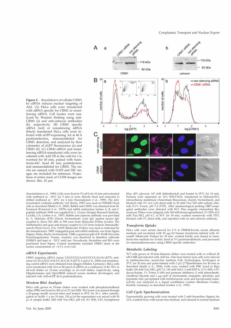
Figure 2. Knockdown of cellular CRM1by siRNA reduces nuclear targeting ofAd2. (A) HeLa cells were transfectedwith siRNA specific for CRM1 or nonsi-lencing siRNA. Cell lysates were ana-lyzed by Western blotting using anti-CRM1 (a) and anti-calnexin antibodies(b), respectively. (B) CRM1 specificsiRNA (red) or nonsilencing siRNA(black) transfected HeLa cells were in-fected with eGFP-expressing Ad at 46 hposttransfection, immunolabeled forCRM1 detection, and analyzed by flowcytometry of eGFP fluorescence (a) andCRM1 (b). (C) CRM1-siRNA and nonsi-lencing siRNA-transfected cells were in-cubated with Ad2-TR in the cold for 1 h,warmed for 40 min, pulsed with trans-ferrin-647, fixed 80 min postinfection,and immunolabeled for CRM1. The nu-clei are stained with DAPI and DIC im-ages are included for reference. Projec-tions of entire stack of CLSM images areshown. Bar, 10 #m.
Cytoplasmic Transport and Nuclear Export
Vol. 16, June 2005 3001

for 12 h 10 min (adopted from Fang et al., 1998). Cells were treated with 20 nMLMB for 20 min and infected with Ad2-TR for 1 h, fixed in PHEMO, andprocessed for immunofluorescence as described above.
Ad5-luc and Ad5-eGFP MeasurementsTriplicate sets of cells were grown on 24-well dishes in DMEM in the presenceof 7% clone III, treated with drugs for 20 min, incubated with Ad5-luc on icefor 60 min, and warmed for 4 h. Cells were washed with PBS and lysed in 400#l of lysis buffer (Luciferase Assay System, Promega, Madison, WI). Lysate, 20#l, was mixed with 50 #l luciferase substrate and chemiluminescence wasmeasured as described (Mabit et al., 2002). In parallel dishes the cell numberswere determined by crystal violet staining and spectrophotometry at 590 nm.For eGFP measurements, HeLa cells grown in six-well dishes were infectedwith 2 #g of Ad5-eGFP 46 h posttransfection of siRNA. Cells were washedwith PBS and detached from the dish by trypsinization 5 h postinfection. Cellswere permeabilized with 0.25% Triton X-100, blocked with 10% goat serumcells, and immunolabeled with a primary CRM1-specific antibody (Fornerodet al., 1997b), followed by a Cy5-conjugated secondary antibody (JacksonImmunoResearch). Samples were analyzed by flow cytometry in a BeckmanCoulter FC500 Cytometer equipped with a 488-nm Argon laser and a 638-nmsolid state laser.
Microscopy of Infected CellsCells grown on 12-mm coverslips were treated with drugs for 20 min andincubated with 0.2–0.5 #g Ad2-TR or 50 #g Ad2 per 0.25 ml cold RPMI-bovine serum albumin binding medium for 45–60 min in the presence orabsence of drugs. Unbound virus was washed off and cells were incubated inDMEM-bovine serum albumin with or without drug at 37°C for indicatedtimes, fixed in pFA (or as otherwise indicated), quenched with 25 mM NH4Clin PBS at room temperature for 5 min, and permeabilized with 0.5% TritonX-100 in PBS for 5 min. Samples were mounted in 3 #l mounting medium(DAKO, Carpinteria, CA) and analyzed by confocal laser scanning micros-copy on a Leica SP2 microscope (Heerbrugg, Switzerland; 63" oil-immersionobjectives) using UV excitation 351 and 364 nm, FITC 468 nm, TR 568 nm, Cy5647 nm and long-pass emission filters in sequential recording modes at0.5-#m section thickness. Fluorescence in situ hybridizations were carried outas described (Greber et al., 1997). Quantification of the subcellular localizationof Ad2-TR was performed as described (Nakano and Greber, 2000). Electronmicroscopy and Ad2 particle quantifications were carried out as described(Nakano et al., 2000).
RESULTS
CRM1-mediated Nuclear Export Required for AdenovirusTransductionNuclear export is a key process in viral infections and cellcommunication, e.g., directly involved in HIV genomic RNAassembly in the cytoplasm (for reviews, see Komeili andO’Shea, 2000; Cullen, 2003). We examined if nuclear exportwas required for adenovirus infection of cultured epithelialcells. HeLa or TC7 cells were treated with different concen-trations of LMB, which covalently binds to CRM1 and sup-presses CRM1 binding to hydrophobic NESs (Fukuda et al.,
1997; Ossareh-Nazari et al., 1997; Wolff et al., 1997; Koster etal., 2003). We also used RJA, another specific inhibitor ofCRM1 (Koster et al., 2003). Cells were treated with inhibitorsfor 30 min, inoculated with luciferase expressing transgenicadenovirus (Ad-luc) in the presence or absence of the drugs,and scored for luciferase activity 4 h postinfection. In bothcell types, we measured a dose-dependent inhibition of lu-ciferase activity with maximal effects of 20–50-fold at con-centrations larger than 7.5 nM LMB and 2 ng"ml$1 RJA,respectively (Figure 1, A–D). Nuclear export of the indicatorprotein 2xNES-eGFP was strongly suppressed in TC7 andHeLa cells (unpublished data) using, for example, 20 nMLMB or 10 ng"ml$1 RJA, indicating that the drugs wereeffective as expected (see Figure 3). Interestingly, at lowerdoses of LMB (&1 nM) and RJA (0.1–0.3 ng"ml$1), we ob-served an increased Ad-luc expression in both cell types, inthe range of 0.1–2-fold compared with no-drug–treated cells(Figure 1, A–D). LMB or RJA concentrations blocking nu-clear export, however, inhibited Ad-luc expression (Figure 1,A and C), ranging from 4.4- to 77-fold, depending on thecells tested (Figure 1E). The strongest effects were obtainedin cells that expressed the highest levels of luciferase, i.e.,human embryonic retinoblast 911 cells, followed by KB,HeLa, A549, COSN, and TC7 cells. The weakest LMB inhi-bitions were in the range of 4–10-fold in cells that werepoorly infected with Ad5-luc, i.e., CV1 cells, the primaryhuman foreskin fibroblasts (2N), and HUVECs. To controlfor cell integrity, we assessed the metabolic state of theLMB-treated cells. A dose of 20 nM LMB for 5 h did notinduce any cytopathic effects indicated by viability/cytotox-icity live cell assays (unpublished data) and metabolic incor-poration of [35S]methionine (Figure 1F), confirming the se-lectivity of the drugs.
We next tested if the inhibition of adenovirus transductionby LMB and RJA was specifically due to CRM1 inactivation.HeLa cells were treated with CRM1 specific siRNAs for 46 h,infected with GFP-expressing adenovirus (Ad5-eGFP) for5 h and analyzed for eGFP and CRM1 expression by flowcytometry (Figure 2). The CRM1 levels were reduced by&40% in the CRM1 siRNA-treated cells as indicated byWestern blotting and flow cytometry (Figure 2, A and B),and eGFP expression was significantly lower than that in thecontrol siRNA-treated cells, whereas the number of nonin-fected cells increased (Figure 2B, left peak). We observed nosigns of toxicity, and this was confirmed by immunofluores-cence analyses of cells labeled with transferrin-Alexa647-
Figure 3. LMB and RJA inhibit the nu-clear export of 2xNES-eGFP and nucleartargeting of adenovirus. TC7 cells trans-fected with plasmid DNA encoding2xNES-eGFP for 24 h were treated or nottreated with 20 nM LMB or 10 ng"ml$1
RJA, incubated with Ad2-TR in the cold,and warmed for 0 or 80 min. Entirestacks of CLSM images are shown for2xNES-eGFP (NES-GFP, green) andAd2-TR (red), and sections across thecell center for DAPI stainings of DNA.Bar, 10 #m.
S. Strunze et al.
Molecular Biology of the Cell3002
and Texas Red-labeled Ad2 (Ad2-TR, Figure 2C). The CRM1siRNA-treated cells had significantly less CRM1 that con-trols treated with nonsilencing siRNA. Both populationsreadily internalized transferrin and Ad2-TR, as indicated byperinuclear endosomes and cytoplasmic viral puncta. Strik-ingly, the nuclear accumulation of adenovirus was stronglyreduced in cells treated with CRM1-specific siRNA, similarto the LMB-treated cells (Figure 1). We also noticed thatreduced nuclear targeting of adenovirus was only observedin cells with significant CRM1 silencing effects and thatprolonged treatments with the CRM1 siRNAs reduced cellviability, consistent with previous observations (Lund et al.,2004). Together, this data indicated a specific requirement ofCRM1 for nuclear targeting and infection of adenovirus.
LMB Arrests Microtubule-dependent CytoplasmicTransport of Adenovirus at the MTOC
Because LMB or RJA blocked CRM1 more efficiently thanthe CRM1 siRNAs, we used short treatments of TC7 cellswith LMB to knock down CRM1 and investigate the natureof the CRM1 block on adenovirus entry. In both control andCRM1-inhibited cells, Ad2-TR was found to be evenly dis-tributed across the cells 0 min after warming, whereas viruswas enriched only at the nucleus of the control cells 80 minpostinfection (Figure 3). The incoming Ad2-TR was clus-tered in the LMB- or RJA-treated cells predominantly at asingle spot near the nucleus. The efficacy of LMB and RJA inthese cells was controlled by transfection of the nuclear
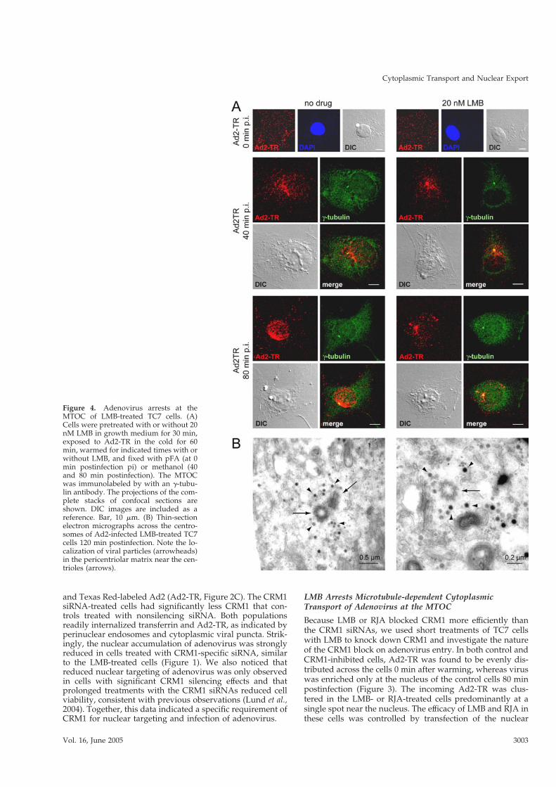
Figure 4. Adenovirus arrests at theMTOC of LMB-treated TC7 cells. (A)Cells were pretreated with or without 20nM LMB in growth medium for 30 min,exposed to Ad2-TR in the cold for 60min, warmed for indicated times with orwithout LMB, and fixed with pFA (at 0min postinfection pi) or methanol (40and 80 min postinfection). The MTOCwas immunolabeled by with an $-tubu-lin antibody. The projections of the com-plete stacks of confocal sections areshown. DIC images are included as areference. Bar, 10 #m. (B) Thin-sectionelectron micrographs across the centro-somes of Ad2-infected LMB-treated TC7cells 120 min postinfection. Note the lo-calization of viral particles (arrowheads)in the pericentriolar matrix near the cen-trioles (arrows).
Cytoplasmic Transport and Nuclear Export
Vol. 16, June 2005 3003

export sequence bearing GFP (2xNES-eGFP) indicator pro-tein showing a clear reduction of NES-GFP signal in thecytoplasm of LMB- or RJA-treated cells (Figure 3). Thesereductions were not complete but were identical to a previ-ous report reflecting the nature of the NES-eGFP construct(Saydam et al., 2001). The enrichment of incoming adenovi-rus at the perinuclear punctum was time-dependent, al-ready visible at 40 min and increased at 80 min postinfection(Figure 4). This effect was also confirmed by subcellularquantification of Ad2-TR fluorescence (unpublished data).Subsequent immunolabeling and EM experiments identifiedthe cytoplasmic puncta of the arrested viruses. An antibodyagainst $-tubulin, an integral component of the centrosome(Joshi et al., 1992) strongly colocalized with LMB-arrestedAd2-TR (Figure 4A). High-resolution thin section EM of
LMB-treated cells infected with Ad2 for 120 min confirmedthat virus particles were enriched in a pericentriolar region,near two centrioles (Figure 4B). A similar centrosomal local-ization of incoming adenovirus was found in LMB-treatedA549, 911, COSN, CV1, normal fibroblasts, and HUVECs(see Figure 1E) and rat kangaroo PtK2 and Vero cells usinglight microscopy (unpublished data). The adenovirus en-richment at the centrosome was completely blocked in no-codazole-treated cells lacking MTs (Figure 5A), indicatingthat LMB intercepted the incoming virus on MTs. This effectwas specific for cytosolic virus, because LMB did not affectthe formation of the MT network after cold-induced depo-lymerization, or the trafficking of transferrin (see Figure 2) orendosomal Ad2 mutant ts1 (unpublished data; Greber et al.,1996). Accordingly, Ad2-TR moved bidirectionally to and
Figure 5. The MTOC arrest of adenovi-rus in LMB-treated TC7 cells requiresMTs and also occurs in mitotic cells. (A)TC7 cells were treated with or withoutLMB (20 nM) and nocodazole (noc, 20#M), infected with Ad2-TR for 80 min,fixed with PHEMO fix, and immuno-stained for %-tubulin (green) and DAPI(blue). DIC images are included as a ref-erence. Bar, 10 #m. (B) Synchronized mi-totic cells treated with or without LMBwere infected with Ad2-TR (red) for 60min, fixed with PHEMO fix, and stainedfor %-tubulin (green) and DNA (DAPI,blue). Whole-cell CLSM analyses areshown. Note the enrichment of Ad2-TRat spindle pole bodies of LMB-treatedmitotic cells. Bar, 10 #m.
S. Strunze et al.
Molecular Biology of the Cell3004

from the MTOC in both LMB and control TC7 cells (seeSupplementary Movies S1 and S1). LMB-treated cells did,however, not contain any viruses on their nuclei at 30 minpostinfection, unlike control cells. This effect was due toinhibition of a cellular target rather than the virus proper,because a centrosomal arrest of Ad2 was not observed if theparticles but not the cells were treated with LMB (unpub-lished data).
We next analyzed mitotic TC7 cells either treated or nottreated with LMB. TC7 cells were synchronized by a thymi-dine block into S-phase, released and treated with LMB for20 min, and infected with Ad2-TR for 1 h. The LMB-treatedcells enriched cytosolic adenovirus particles at both spindlepoles, unlike the control cells (Figure 5B). Importantly, theLMB treated cells showed no obvious cytotoxicity or mitoticdefects and had normal spindle poles as judged by lightmicroscopy. In the untreated mitotic cells, the viruses weredispersed throughout the cytoplasm not colocalizing withMTs or the spindle poles (Figure 5B and unpublished data).This stands in contrast to interphase TC7 cells where morethan 95% of Ad2-TR colocalized with MTs 40 min postinfec-tion (Stidwill and Greber, 2000). The reason for the defect ofMT colocalization of adenovirus in mitotic cells is not due toan irreversible impairment of the viruses by mitotic factors,because Ad2-TR readily engaged with MTs after the cellshad completed mitosis (unpublished data). The data showthat MTs of normal mitotic cells fail to transport adenovi-ruses to the spindle poles, consistent with the notion thatCRM1 or a CRM1-associated factor blocks viral attachmentto MTs in the mitotic cytoplasm, including the spindle MTsand the poles. Interestingly, CRM1 immunoreactivity wasfound throughout the cytosol and on the spindle of mitoticTC7 cells in both control and LMB-treated cells (unpub-lished data), suggesting that the mitotic localization ofCRM1 is independent of its NES-binding function. Nonethe-less, the inhibition of mitotic CRM1 by LMB blocked viraltransport at the spindle poles.
CRM1 Is Required for Ad2 Disassembly at the NPC andNuclear ImportInfection requires capsid disassembly and disassembly inturn depends on the association of capsids with the NPC(Trotman et al., 2001). We therefore characterized the sub-cellular localizations of Ad2 in interphase cells lacking func-tional CRM1 in three different cell lines, TC7, A549, andHeLa cells, 60 min postinfection. Quantitative EM analysesindicated that LMB strongly reduced the number of virusparticles at the nuclear membrane of all cell types, but didnot significantly affect the number of cytosolic-, endosomal-,and plasma membrane–associated particles, indicating thatLMB did not affect virus entry into the cytosol (Figure 6). Toassess the status of the cytosolic capsids that failed to asso-ciate with the NPC, we analyzed capsid disassembly in bothTC7 and HeLa cells using the disassembly-specific anti-hexon antibody R70 (Baum et al., 1972; Trotman et al., 2001).The results show that the MTOC-arrested Ad2-TR capsidswere largely devoid of R70 epitopes in the LMB-treated TC7cells but were strongly R70-positive in the control cells (Fig-ure 7A). Likewise, LMB strongly inhibited the R70 stainingof the randomly dispersed cytosolic Ad2 in HeLa cells (Fig-ure 7B). These results were confirmed by immunostainingsof the viral DNA-associated protein VII, which is protectedin the intact capsid and not accessible to antibodies byimmunofluorescence analyses, unless detached from thecapsid (Greber et al., 1997). LMB strongly blocked the ap-pearance of protein VII in the nucleus of infected TC7 andHeLa cells (Figure 7). This result was further confirmed byfluorescence in situ hybridizations monitoring the incomingviral genome. In LMB-treated TC7 cells the viral DNA wasabsent from the nucleoplasm but enriched in a perinuclearpunctum and smaller cytoplasmic dots reinforcing the no-tion that CRM1 is necessary for targeting functional adeno-viruses from the MTs to the NPC and presenting them to thedisassembly machinery.
DISCUSSION
Viral invasion of the nucleus depends on the coordination ofmany host functions. This facilitates the propagation of, forexample, adenoviruses in almost all vertebrates (Davison etal., 2003). Of the more than 50 human adenovirus serotypes,the respiratory viruses Ad2 and Ad5 cause severe infectionsin immunocompromised patients (Horwitz, 2001). In modi-fied forms these vectors are widely used for gene transfer(Russell, 2000). They enter epithelial cells by clathrin-mediatedendocytosis, activate macropinocytosis, escape to the cytosol inan integrin-dependent manner, and efficiently deliver theirDNA genome into the nucleus (Meier et al., 2002; Imelli et al.,2004). Cytosolic particles are transported on preferentially sta-ble MTs by the dynein/dynactin motor complex toward theMTOC, often located near the nucleus (Suomalainen et al., 1999;Leopold et al., 2000; Giannakakou et al., 2002). The first virusesreaching the nucleus are those trafficking near the MTOC,suggesting that this region is critical to hand over the virusesfrom the MTs to the nuclear membrane. It is unknown howviruses are detached from MTs.
Here we provide evidence linking two previously uncon-nected pathways, nuclear export and cytoplasmic transporton microtubules. Our in vivo results provide a molecularexplanation for the observation that enucleated human epi-thelial cells arrested Ad5 particles at the MTOC (Bailey et al.,2003). Using CRM1-specific drugs and siRNAs, we showthat the export factor CRM1 acts as a positional indicator ofthe nucleus for the incoming adenovirus. If CRM1 function
Figure 6. Inhibition of nuclear envelope targeting of adenovirus inTC7, A549, and HeLa cells. Quantitative subcellular analyses of Ad2by thin-section EM in LMB-treated and control TC7, A549, andHeLa cells. Viral particles (v.p.) were counted at the nuclear mem-brane (A), in the cytosol (B), endosomes (C), and at the plasmamembrane (D). Results indicate the means and SE from the numberof analyzed cells and v.p.
Cytoplasmic Transport and Nuclear Export
Vol. 16, June 2005 3005

is blocked, the detachment of adenoviruses from the MTsfails and viral binding to the nuclear membrane is pre-vented. In normal cells, CRM1 predominantly localizes tothe nucleus and functions to export NES-containing proteinsfrom the nucleus to the cytoplasm (reviewed in Cullen, 2003;Greber and Fornerod, 2005). It is potently inhibited by thebacterial polyketides LMB and RJA. These inhibitors arehighly specific because 1) an Schizosaccharomyces pombestrain expressing an LMB-resistant CRM1 was found to beinsensitive to LMB and 2) LMB bound specifically to CRM1in the low nanomolar range (Neville et al., 1997; Kudo et al.,1999; Koster et al., 2003), exactly matching our experimentalconditions for blocking CRM1. Both LMB and RJA blockedthe transport of incoming adenoviruses at the MTOC inmany different cell types or apparently randomly in theHeLa cytoplasm. A similar nuclear transport block was es-tablished in HeLa cells by siRNA-mediated knock down ofCRM1. In the absence of functional CRM1, the adenovirusesfailed to detach from the MTs and the MTOC, did not bindto NPCs, and did not disassemble and import the DNA intothe nucleus, which is normally mediated by the NPC andassociated proteins (Saphire et al., 2000; Trotman et al., 2001).Thus, LMB and RJA are novel viral entry inhibitors, estab-
lishing a previously unknown postentry block of infections.They are among a small number of compounds restrictingviral entry (McKinlay et al., 1992; Greber et al., 1994).
Host restriction factors are part of innate immunity reac-tions against incoming retroviruses and are responsible forthe narrow host range of these viruses (Goff, 2004). Restric-tions are targeted against the capsid protein before reversetranscription. HIV, for example, overcomes these restrictionsby incorporating cyclophilin A, a modulator of the viralsensitivity to innate immunity factors (Towers et al., 2003). Inaddition, viruses that are targeted to the MTOC by thedynein/dynactin motor complex, such as the adenovirusand HIV could be subject to another postentry host restric-tion point, degradation by the aggresomal pathway thatcollects misfolded or otherwise abnormal proteins (Kopito,2000). By recruiting CRM1 adenovirus could escape fromthis restriction and target to the NPC for uncoating. WhetherCRM1 acts as a gatekeeper or is mobilized to the virus is notknown. We can envision several modes how CRM1 over-comes an entry block. Normally, CRM1 mediates nuclearexport of a large variety of leucine-rich export sequencecontaining substrates by forming a complex with the GTP-bound form of the small GTPase Ran in the nucleus (Petosa
Figure 7. LMB inhibits adenovirus cap-sid disassembly and nuclear import ofprotein VII and viral DNA. LMB-treatedand control TC7 (A) and HeLa (B) cellswere infected with Ad2 for 150 min,fixed, and immunostained for the disas-sembled capsid protein hexon and theDNA-associated core protein VII. Themerged stacks of the entire set of CLSMsections are shown. Bar, 10 #m. (C) LMBblocks Ad2 DNA import into the nu-cleus. TC7 cells were infected with Ad2in the presence or absence of LMB for180 min, processed for in situ hybridiza-tion, and immunostained for laminsA/B/C. Single CLSM sections across acentral plane of the cells are shown in-cluding DIC. Bar, 10 #m.
S. Strunze et al.
Molecular Biology of the Cell3006

et al., 2004). It is possible that either CRM1 alone, a short-lived CRM1-NES-Ran:GTP complex or an unknown NES-containing nuclear protein interact with adenovirus. IfCRM1 alone interacted with adenovirus this interactioncould be of supraphysiological affinity independent of Ran(Engelsma et al., 2004). In this case, CRM1 would stay boundto the capsid until disassembly at the NPC had occurred. Acytosolic CRM1-NES-Ran:GTP complex on the other handwould be a regulated MT dislodging trigger, because it hasa limited life time owing to the hydrolysis of GTP activatedby the cytoplasmic RanGAP protein (Bednenko et al., 2003;Quimby and Dasso, 2003). The chromatin-bound GTP/GDPexchange factor RCC1 in turn activates the complex byloading Ran:GTP and establishes a gradient across the nu-clear membrane in intact cells and also around mitotic chro-mosomes in cell-free systems (Kalab et al., 2002; Macara,2002; Weis, 2002). This regulated scenario is attractive be-cause it would account for our observation that adenovi-ruses were prevented from association with both spindleand astral MTs of mitotic cells. This block was relieved byLMB leading to virus accumulation at the spindle poles. Infact, experiments with Xenopus egg extracts have suggestedthat CRM1 is in a complex with Ran:GTP proximal to mitoticchromatin (Kalab et al., 2002), although it is not known if aRan:GTP gradient exists around chromosomes of epithelialcells (Gorlich et al., 2003). Regardless, even if Ran:GTP wererandomly distributed in mitotic TC7 cells, it could accountfor dislodging adenovirus particles from MTs. On exit of thecells from mitosis, CRM1 would be retrieved to the newlyformed nucleus together with Ran:GTP and the cytosolicadenoviruses could be transported to the nucleus usingmicrotubules, as observed in our studies. The scenario that aNES-containing cargo alone unloads the incoming adenovi-rus from the MTs is perhaps less likely because in mitoticcells with mixed nuclear and cytoplasmic contents, LMBwas very effective at clustering viruses at the spindle poles,very similar to MTOC clustering in interphase cells. Thissuggests that the export function of CRM1 is not needed fordislodging adenovirus from MTs.
There is yet another possibility to carry CRM1 to perinu-clear MTs of interphase cells, namely via the nucleoporinsRanBP2 or Nup214/CAN, as suggested by the observationof RanBP2 antigens at the MTOC (Salina et al., 2003). Nucleo-porin extensions might be possible via extended conforma-tions of the FG domains, which could amount to somehundred nanometers. Regardless, the requirement of CRM1for nuclear targeting of adenovirus is strong, because adeno-viruses are retained at the MTOC even if the MTOC ispositioned in immediate vicinity to the nuclear membrane.Interestingly, CRM1 has been found at low concentrations atthe MTOC and it was postulated to be required to controlcentriole synthesis (Forgues et al., 2003). It is thus feasiblethat CRM1 is part of a centrosomal network, including ki-nases, phosphatases, scaffolding proteins, and nuclear enve-lope proteins. Indeed, a small fraction of Ran is tightlyassociated with the centrosome throughout the cell cycle(Zimmerman and Doxsey, 2000; Keryer et al., 2003). Othercentrosomal proteins such as centrin and pericentrin shuttlebetween the nucleus and the centrosome in an LMB-depen-dent manner. This suggests that centrosomal activities canbe regulated by CRM1-dependent nuclear export involvingspecific cargo proteins and Ran. It is thus possible that viralinfectivity is both positively and negatively regulated bycentrosomal or cytosolic CRM1, because very low concen-trations of LMB or RJA increased Ad5 gene expressions.Future experiments will investigate how CRM1 interactswith cytosolic adenovirus or affects an active transport sys-
tem carrying perinuclear viruses. This will be crucial toaddress the issues of innate immunity and host restrictionsas well as turnover and immunogenicity of vectors.
ACKNOWLEDGMENTS
We thank B. Wolff (Novartis, Vienna) for generous gift of LMB, S. Guttingerand U. Kutay (ETH Zurich, Switzerland) for CRM1 siRNA and advice, and M.Fornerod, F. Melchior, C. Burckhardt, and D. Puntener for discussions. Thiswork was supported by the Swiss National Science Foundation and theKanton Zurich (U.F.G.).
REFERENCES
Alonso, C., Miskin, J., Hernaez, B., Fernandez-Zapatero, P., Soto, L., Canto, C.,Rodriguez-Crespo, I., Dixon, L., and Escribano, J. M. (2001). African swinefever virus protein p54 interacts with the microtubular motor complexthrough direct binding to light-chain dynein. J. Virol. 75, 9819–9827.
Bailey, C. J., Crystal, R.G., and Leopold, P. L. (2003). Association of adenovi-rus with the microtubule organizing center. J. Virol. 77, 13275–13287.
Baum, S. G., Horwitz, M. S., and J. V. Maizel, J. (1972). Studies of themechanism of enhancement of human adenovirus infection in monkey cellsby simian virus 40. J. Virol. 10, 211–219.
Bednenko, J., Cingolani, G., and Gerace, L. (2003). Nucleocytoplasmic trans-port: navigating the channel. Traffic 4, 127–135.
Bienz, M. (2002). The subcellular destinations of APC proteins. Nat. Rev. Mol.Cell. Biol. 3, 328–338.
Bornens, M. (2002). Centrosome composition and microtubule anchoringmechanisms. Curr. Opin. Cell Biol. 14, 25–34.
Crepieux, P., Kwon, H., Leclerc, N., Spencer, W., Richard, S., Lin, R., andHiscott, J. (1997). I kappaB alpha physically interacts with a cytoskeleton-associated protein through its signal response domain. Mol. Cell. Biol. 17,7375–7385.
Cullen, B. R. (2003). Nuclear mRNA export: insights from virology. TrendsBiochem. Sci. 28, 419–424.
Davison, A. J., Benko, M., and Harrach, B. (2003). Genetic content and evo-lution of adenoviruses. J. Gen. Virol. 84, 2895–2908.
Dohner, K., Wolfstein, A., Prank, U., Echeverri, C., Dujardin, D., Vallee, R.,and Sodeik, B. (2002). Function of dynein and dynactin in herpes simplexvirus capsid transport. Mol. Biol. Cell 13, 2795–2809.
Douglas, M. W., Diefenbach, R. J., Homa, F. L., Miranda-Saksena, M., Rixon,F. J., Vittone, V., Byth, K., and Cunningham, A. L. (2004). Herpes simplexvirus type 1 capsid protein VP26 interacts with dynein light chains RP3 andTctex1 and plays a role in retrograde cellular transport. J. Biol. Chem. 279,28522–28530.
Engelsma, D., Bernad, R., Calafat, J., and Fornerod, M. (2004). Supraphysi-ological nuclear export signals bind CRM1 independently of RanGTP andarrest at Nup358. EMBO J. 23, 3643–3652.
Fallaux, F. J. et al. (1998). New helper cells and matched early region 1-deletedadenovirus vectors prevent generation of replication-competent adenovi-ruses. Hum. Gene Ther. 9, 1909–1917.
Fang, G., Yu, H., and Kirschner, M. W. (1998). Direct binding of CDC20protein family members activates the anaphase-promoting complex in mitosisand G1. Mol. Cell 2, 163–171.
Finke, S., Brzozka, K., and Conzelmann, K. K. (2004). Tracking fluorescence-labeled rabies virus: enhanced green fluorescent protein-tagged phosphopro-tein P supports virus gene expression and formation of infectious particles.J. Virol. 78, 12333–12343.
Forgues, M., Difilippantonio, M. J., Linke, S. P., Ried, T., Nagashima, K.,Feden, J., Valerie, K., Fukasawa, K., and Wang, X. W. (2003). Involvement ofCrm1 in hepatitis B virus X protein-induced aberrant centriole replication andabnormal mitotic spindles. Mol. Cell. Biol. 23, 5282–5292.
Fornerod, M., Ohno, M., Yoshida, M., and Mattaj, I. W. (1997a). CRM1 is anexport receptor for leucine-rich nuclear export signals. Cell 90, 1051–1060.
Fornerod, M., van Deursen, J., van Baal, S., Reynolds, A., Davis, D., Murti,K. G., Fransen, J., and Grosveld, G. (1997b). The human homologue of yeastCRM1 is in a dynamic subcomplex with CAN/Nup214 and a novel nuclearpore component Nup88. EMBO J. 16, 807–816.
Fukuda, M., Asano, S., Nakamura, T., Adachi, M., Yoshida, M., Yanagida, M.,and Nishida, E. (1997). CRM1 is responsible for intracellular transport medi-ated by the nuclear export signal. Nature 390, 308–311.
Cytoplasmic Transport and Nuclear Export
Vol. 16, June 2005 3007

Giannakakou, P., Nakano, M. Y., Nicolaou, K. C., O’Brate, A., Yu, J., Blagosk-lonny, M. V., Greber, U. F., and Fojo, T. (2002). Enhanced microtubule-dependent trafficking and p53 nuclear accumulation by suppression of mi-crotubule dynamics. Proc. Natl. Acad. Sci. USA 99, 10855–10860.
Giannakakou, P., Sackett, D. L., Ward, Y., Webster, K. R., Blagosklonny, M. V.,and Fojo, T. (2000). p53 is associated with cellular microtubules and is trans-ported to the nucleus by dynein. Nat. Cell Biol. 2, 709–717.
Gluzman, Y. (1981). SV40-transformed simian cells support the replication ofearly SV40 mutants. Cell 23, 175–182.
Goff, S. P. (2004). Retrovirus restriction factors. Mol. Cell 16, 849–859.
Gorlich, D., and Kutay, U. (1999). Transport between the cell nucleus and thecytoplasm. Annu. Rev. Cell Dev. Biol. 15, 607–660.
Gorlich, D., Seewald, M. J., and Ribbeck, K. (2003). Characterization of Ran-driven cargo transport and the RanGTPase system by kinetic measurementsand computer simulation. EMBO J. 22, 1088–1100.
Greber, U. F., and Fornerod, M. (2005). Nuclear import in viral infections.Curr. Top. Microbiol. Immunol. 285, 109–138.
Greber, U. F., Nakano, M. Y., and Suomalainen, M. (1998). Adenovirus entryinto cells: a quantitative fluorescence microscopy approach. In: AdenovirusMethods and Protocols, Methods in Molecular Medicine, Vol. 21, ed. W.S.M.Wold, Totowa, NJ: Humana Press, 217–230.
Greber, U. F., Singh, I., and Helenius, A. (1994). Mechanisms of virus uncoat-ing. Trends Microbiol. 2, 52–56.
Greber, U. F., Suomalainen, M., Stidwill, R. P., Boucke, K., Ebersold, M., andHelenius, A. (1997). The role of the nuclear pore complex in adenovirus DNAentry. EMBO J. 16, 5998–6007.
Greber, U. F., Webster, P., Weber, J., and Helenius, A. (1996). The role of theadenovirus protease in virus entry into cells. EMBO J. 15, 1766–1777.
Greber, U. F., Willetts, M., Webster, P., and Helenius, A. (1993). Stepwisedismantling of adenovirus 2 during entry into cells. Cell 75, 477–486.
Grieshaber, S. S., Grieshaber, N. A., and Hackstadt, T. (2003). Chlamydiatrachomatis uses host cell dynein to traffic to the microtubule-organizing centerin a p50 dynamitin-independent process. J. Cell Sci. 116, 3793–3802.
Hemmi, S., Bohni, R., Stark, G., Di Marco, F., and Aguet, M. (1994). A novelmember of the interferon receptor family complements functionality of themurine interferon gamma receptor in human cells. Cell 76, 803–810.
Horwitz, M. S. (2001). Adenoviruses. In: Fields Virology, eds. D. M. Knipe andP. M. Howley, Philadelphia, PA: Raven Press, 2301–2326.
Imelli, N., Meier, O., Boucke, K., Hemmi, S., and Greber, U. F. (2004). Cho-lesterol is required for endocytosis and endosomal escape of adenovirus type2. J. Virology 78, 3089–3098.
Jacob, Y., Badrane, H., Ceccaldi, P. E., and Tordo, N. (2000). Cytoplasmicdynein LC8 interacts with lyssavirus phosphoprotein. J. Virol. 74, 10217–10222.
Joshi, H. C., Palacios, M. J., McNamara, L., and Cleveland, D. W. (1992).Gamma-tubulin is a centrosomal protein required for cell cycle-dependentmicrotubule nucleation. Nature 356, 80–83.
Jouvenet, N., Monaghan, P., Way, M., and Wileman, T. (2004). Transport ofAfrican swine fever virus from assembly sites to the plasma membrane isdependent on microtubules and conventional kinesin. J. Virol. 78, 7990–8001.
Kalab, P., Weis, K., and Heald, R. (2002). Visualization of a Ran-GTP gradientin interphase and mitotic Xenopus egg extracts. Science 295, 2452–2456.
Kelkar, S. A., Pfister, K. K., Crystal, R. G., and Leopold, P. L. (2004). Cyto-plasmic dynein mediates adenovirus binding to microtubules. J. Virol. 78,10122–10132.
Keryer, G., Di Fiore, B., Celati, C., Lechtreck, K. F., Mogensen, M., Delouvee,A., Lavia, P., Bornens, M., and Tassin, A. M. (2003). Part of Ran is associatedwith AKAP450 at the centrosome: involvement in microtubule-organizingactivity. Mol. Biol. Cell 14, 4260–4271.
Komeili, A., and O’Shea, E. K. (2000). Nuclear transport and transcription.Curr. Opin. Cell Biol. 12, 355–360.
Kopito, R. R. (2000). Aggresomes, inclusion bodies and protein aggregation.Trends Cell Biol. 10, 524–530.
Koster, M., Lykke-Andersen, S., Elnakady, Y.A., Gerth, K., Washausen, P.,Hofle, G., Sasse, F., Kjems, J., and Hauser, H. (2003). Ratjadones inhibitnuclear export by blocking CRM1/exportin 1. Exp. Cell Res. 286, 321–331.
Kreis, T. E. (1987). Microtubules containing detyrosinated tubulin are lessdynamic. EMBO J. 6, 2597–2606.
Kudo, N., Taoka, H., Toda, T., Yoshida, M., and Horinouchi, S. (1999). A novelnuclear export signal sensitive to oxidative stress in the fission yeast tran-scription factor Pap1. J. Biol. Chem. 274, 15151–15158.
Lakadamyali, M., Rust, M. J., Babcock, H. P., and Zhuang, X. (2003). Visual-izing infection of individual influenza viruses. Proc. Natl. Acad. Sci. USA 100,9280–9285.
Leopold, P. L., Kreitzer, G., Miyazawa, N., Rempel, S., Pfister, K. K., Rodri-guez-Boulan, E., and Crystal, R. G. (2000). Dynein- and microtubule-mediatedtranslocation of adenovirus serotype 5 occurs after endosomal lysis. Hum.Gene. Ther. 11, 151–165.
Luby-Phelps, K. (2000). Cytoarchitecture and physical properties of cyto-plasm: volume, viscosity, diffusion, intracellular surface area. Int. Rev. Cytol.192, 189–221.
Lund, E., Guttinger, S., Calado, A., Dahlberg, J. E., and Kutay, U. (2004).Nuclear export of microRNA precursors. Science 303, 95–98.
Mabit, H., Nakano, M. Y., Prank, U., Saam, B., Dohner, K., Sodeik, B., andGreber, U. F. (2002). Intact microtubules support Adenovirus and Herpessimplex virus infections. J. Virol. 76, 9962–9971.
Macara, I. G. (2002). Why FRET about Ran? Dev. Cell 2, 379–380.
Mahajan, R., Delphin, C., Guan, T., Gerace, L., and Melchior, F. (1997). A smallubiquitin-related polypeptide involved in targeting RanGAP1 to nuclear porecomplex protein RanBP2. Cell 88, 97–107.
Martin-Fernandez, M., Longshaw, S. V., Kirby, I., Santis, G., Tobin, M. J.,Clarke, D. T., and Jones, G. R. (2004). Adenovirus Type-5 entry and disas-sembly followed in living cells by FRET, fluorescence anisotropy, and FLIM.Biophys. J. 87, 1316–1327.
McDonald, D., Vodicka, M. A., Lucero, G., Svitkina, T. M., Borisy, G. G.,Emerman, M., and Hope, T. J. (2002). Visualization of the intracellular behav-ior of HIV in living cells. J. Cell Biol. 159, 441–452.
McKinlay, M. A., Pevear, D. C., and Rossmann, M. G. (1992). Treatment of thepicornavirus common cold by inhibitors of viral uncoating and attachment.Annu. Rev. Microbiol. 46, 635–654.
Meier, O., Boucke, K., Vig, S., Keller, S., Stidwill, R. P., Hemmi, S., and Greber,U. F. (2002). Adenovirus triggers macropinocytosis and endosomal leakagetogether with its clathrin mediated uptake. J. Cell Biol. 158, 1119–1131.
Nakano, M. Y., Boucke, K., Suomalainen, M., Stidwill, R. P., and Greber, U. F.(2000). The first step of adenovirus type 2 disassembly occurs at the cellsurface, independently of endocytosis and escape to the cytosol. J. Virol. 74,7085–7095.
Nakano, M. Y., and Greber, U. F. (2000). Quantitative microscopy of fluores-cent adenovirus entry. J. Struct. Biol. 129, 57–68.
Neville, M., Stutz, F., Lee, L., Davis, L. I., and Rosbash, M. (1997). Theimportin-beta family member Crm1p bridges the interaction between Rev andthe nuclear pore complex during nuclear export. Curr. Biol. 7, 767–775.
Ossareh-Nazari, B., Bachelerie, F., and Dargemont, C. (1997). Evidence for arole of CMR1 in signal-mediated nuclear protein export. Science 278, 141–144.
Petit, C., Giron, M. L., Tobaly-Tapiero, J., Bittoun, P., Real, E., Jacob, Y., Tordo,N., De The, H., and Saib, A. (2003). Targeting of incoming retroviral Gag to thecentrosome involves a direct interaction with the dynein light chain 8. J. CellSci. 116, 3433–3442.
Petosa, C., Schoehn, G., Askjaer, P., Bauer, U., Moulin, M., Steuerwald, U.,Soler-Lopez, M., Baudin, F., Mattaj, I. W., and Muller, C. W. (2004). Architec-ture of CRM1/Exportin1 suggests how cooperativity is achieved duringformation of a nuclear export complex. Mol. Cell 16, 761–775.
Ploubidou, A., and Way, M. (2001). Viral transport and the cytoskeleton. Curr.Opin. Cell Biol. 13, 97–105.
Puthalakath, H., Huang, D. C., O’Reilly, L. A., King, S. M., and Strasser, A.(1999). The proapoptotic activity of the Bcl-2 family member Bim is regulatedby interaction with the dynein motor complex. Mol. Cell 3, 287–296.
Quimby, B. B., and Dasso, M. (2003). The small GTPase Ran: interpreting thesigns. Curr. Opin. Cell Biol. 15, 338–344.
Raux, H., Flamand, A., and Blondel, D. (2000). Interaction of the rabies virusP protein with the LC8 dynein light chain. J. Virol. 74, 10212–10216.
Russell, W. C. (2000). Update on adenovirus and its vectors. J. Gen. Virol. 81,2573–2604.
Salina, D., Enarson, P., Rattner, J. B., and Burke, B. (2003). Nup358 integratesnuclear envelope breakdown with kinetochore assembly. J. Cell Biol. 162,991–1001.
Saphire, A.C.S., Guan, T. L., Schirmer, E. C., Nemerow, G. R., and Gerace, L.(2000). Nuclear import of adenovirus DNA in vitro involves the nuclearprotein import pathway and hsc70. J. Biol. Chem. 275, 4298–4304.
S. Strunze et al.
Molecular Biology of the Cell3008

Saxton, W. M. (2001). Microtubules, motors, and mRNA localization mecha-nisms: watching fluorescent messages move. Cell 107, 707–710.
Saydam, N., Georgiev, O., Nakano, M. Y., Greber, U. F., and Schaffner, W.(2001). Nucleo-cytoplasmic trafficking of metal-regulatory transcription factor1 is regulated by diverse stress signals. J. Biol. Chem. 276, 25487–25495.
Sfakianos, J. N., LaCasse, R. A., and Hunter, E. (2003). The M-PMV cytoplas-mic targeting-retention signal directs nascent Gag polypeptides to a pericen-triolar region of the cell. Traffic 4, 660–670.
Smith, G. A., and Enquist, L. W. (2002). Break ins and break outs: viralinteractions with the cytoskeleton of mammalian cells. Annu. Rev. Cell Dev.Biol. 18, 135–161.
Sodeik, B. (2000). Mechanisms of viral transport in the cytoplasm. TrendsMicrobiol. 8, 465–472.
Stidwill, R. S., and Greber, U. F. (2000). Intracellular virus trafficking revealsphysiological characteristics of the cytoskeleton. News Physiol. Sci. 15, 67–71.
Suikkanen, S., Aaltonen, T., Nevalainen, M., Valilehto, O., Lindholm, L.,Vuento, M., and Vihinen-Ranta, M. (2003). Exploitation of microtubule cy-toskeleton and dynein during parvoviral traffic toward the nucleus. J. Virol.77, 10270–10279.
Suomalainen, M., Nakano, M. Y., Boucke, K., Keller, S., and Greber, U. F.(2001). Adenovirus-activated PKA and p38/MAPK pathways boost microtu-bule-mediated nuclear targeting of virus. EMBO J. 20, 1310–1319.
Suomalainen, M., Nakano, M. Y., Boucke, K., Keller, S., Stidwill, R. P., andGreber, U. F. (1999). Microtubule-dependent minus and plus end-directedmotilities are competing processes for nuclear targeting of adenovirus. J. CellBiol. 144, 657–672.
Towers, G. J., Hatziioannou, T., Cowan, S., Goff, S. P., Luban, J., and Bieniasz,P. D. (2003). Cyclophilin A modulates the sensitivity of HIV-1 to host restric-tion factors. Nat. Med. 9, 1138–1143.
Trotman, L. C., Mosberger, N., Fornerod, M., Stidwill, R. P., and Greber, U. F.(2001). Import of adenovirus DNA involves the nuclear pore complex recep-tor CAN/Nup214 and histone H1. Nat. Cell Biol. 3, 1092–1100.
Verhey, K. J., and Rapoport, T. A. (2001). Kinesin carries the signal. Trends.Biochem. Sci. 26, 545–550.
Weis, K. (2002). Nucleocytoplasmic transport: cargo trafficking across theborder. Curr. Opin. Cell Biol. 14, 328–335.
Wolff, B., Sanglier, J. J., and Wang, Y. (1997). Leptomycin B is an inhibitor ofnuclear export: inhibition of nucleo-cytoplasmic translocation of the humanimmunodeficiency virus type 1 (HIV-1) Rev protein and Rev-dependentmRNA. Chem. Biol. 4, 139–147.
Zimmerman, W., and Doxsey, S. J. (2000). Construction of centrosomes andspindle poles by molecular motor-driven assembly of protein particles. Traffic1, 927–934.
Cytoplasmic Transport and Nuclear Export
Vol. 16, June 2005 3009