update 03/10/13- 97 days to go! simply… lecture … · •classical triad 1.shock 2.pulsatile...
TRANSCRIPT

SIMPLY…(all of) SurgeryMr. Simon Fleming MBBS., MRCS. M.Sc, FRSA, MAcadMEd, MFSTEd MASE(RACS) AFHEA
Trauma & Orthopaedics Registrar
Immediate Past President - British Orthopaedic Trainees Association (BOTA)
International Conference in Residency Education (ICRE) Chief Resident
Vice Chair – Academy Trainee Doctors’ Group (ATDG)
PhD Candidate; Medical Education


Objectives
• To cover common surgical emergencies that may occur• During finals• During your life as an FY1
• To reassure you that if I can pass finals, anyone can

Remember…
• Regardless of what kind of doctor you become, you will still have to see sick surgical patients…
...so this stuff matters

First Things First
• Don’t take notes
• Ask questions – please!
• Don’t…take…notes…


How NOT to Revise…

Cases1. 68 Male; with a sore tummy
2. 42 Male; drunk off his face
3. 19 Female; rock hard abs
4. 47 Female; yellow and puking
5. 50 Male; I…can’t…poo
6. 87 Female; PICKLES!
7. 19 Male; I totes have leg pain. Lol
8. 72 Male; My back hurts
9. 65 Male; cold feet
The Acute Abdomen
• Condition• Non-specific abdominal pain 35%
• Acute appendicitis 17%
• Intestinal obstruction 15%
• Urological causes 6%
• Gallstone disease 5%
• Colonic diverticular disease 4%
• Abdominal trauma 3%
• Abdominal malignancy 3%
• Perforated peptic ulcer 3%
• Pancreatitis 2%
• Ruptured AAA <1%
• Inflammatory bowel disease <1%
• Gastroenteritis <1%
The Acute Abdomen• History
• Site
• Severity
• Radiation• RUQ back
• ?biliary colic/cholecystitis
• RUQ shoulder tip = diaphragmatic irritiation• ?cholecystitis/perf DU/subphrenic collection
• Central back • ?pancreatitis/aorta
• Central epigastric -> RIF • ?appendix
• Loin groin • ?renal colic/aorta

The Acute Abdomen• How Long Does the Pain Last?
• Colic = “comes and goes” (spasm in a hollow viscus)
• Character
• Pain is subjective…
• Precipitating/Relieving Factors
• Peritonitis = lying still
• Colic = Moving about
• Food = ?PUD/colic
• ETOH = ?pancreatitis/gastritis/ulcer

• Don’t forget Associated Symptoms• Dysphagia, jaundice, urinary symptoms etc etc etc
• ANY WOMAN OF CHILDBEARING AGE IS HAVING AN ECTOPIC TILL PROVEN OTHERWISE!

Case 1 - 68♂ with “a sore tummy”
• 68 male• Brought in by ambulance
• Patient complaining of• 7/10 abdominal pain
• Radiates from his back, to his side, down into his groin
• Has been there for a few hours but he is “feeling pretty rough”

History
• What else are you going to ask in the history?
• Have you got a differential in your head yet?
• If not – what else do you want to know?

History….

Examination
• Grey• Clammy
• ABC approach!• What does this actually mean?

Examination Cont
• A –• Maintaining, telling you about his lovely grandchildren and
the war and bloody new age music
• B –• Sats; 90% on air• RR 28• Lung fields - clear

Examination Cont
• C –• HR 127
• BP 105/70
• Abdomen• Painful, central and flank. RT “kidney pain”• Pulsatile vs expansile mass• Good bowel sounds

Investigations
• What do you want and why??
• Least invasive most invasive
• Cheapest most expensive
• Dullest most awesome and shiny

Investigations
• Urine• Blood +• Protein +
• FBC• U&E• LFT• Clotting• G&S vs XM• ABG

ABG – a quickie!

WHAT NOW??

CT Scan vs USS

So what has he got then???

Now what….
• Access
• Fluids – “permissive hypotension”
• Call. For. Help
• Who? When?

Ruptured Abdominal Aortic Aneurysm (AAA)
Sometimes referred to as “the only true surgical emergency”
100% mortality in 3 days

AAA
• An AAA is an increase in aortic diameter by greater than 50% of normal
• Usually regarded as aortic diameter of greater than 3 cm diameter
• More prevalent in elderly men
• Male : female ratio is 4:1
• Mortality of emergency operation is greater than 50% • Mortality of elective surgery is less than 5%

AAA
• 5 year risk of rupture: • 5.0 – 5.9 cm = 25%
• 6.0 – 6.9 cm = 35%
• More than 7 cm = 75%
• Overall only 15% aneurysms ever rupture

AAA
• History/Examination
75% ASYMPTOMATIC!!! Bugger!
• Classical Triad1. Shock2. Pulsatile abdominal mass3. Mild/severe abdominal pain
Can radiate to back, flank, groin.
Remember – an old man with his first presentation of ‘renal colic’ ain’t got renal colic!

AAA• Management
• ABCDE
• If you think you are looking at a AAACall. For. Help.
• “Make a decision, make an incision”
Open Vs EVAR
• Endovascular aneurysm repair
• Introduced into clinical practice with few clinical trials over the past 10 years
• Exact role unclear and medium and late-complications only recently recognised
• Endovascular repair may be associated with: • Reduced physiological stress• Reduced morbidity• Reduced mortality
http://pathways.nice.org.uk/pathways/aortic-aneurysms

Medical Management of AAA
• Smoking Cessation- Single most important modifiable risk factor
• Exercise Therapy- Evidence suggests may benefit small aneurysms
• Beta Blockers- May decrease the rate of expansion? Important cardiovascular effects thus use advocated.
• ACE inhibitors- Evidence is mixed, however, implicated in less aneurysm rupture.
• Statins - associated with reduced aneurysm expansion rates.

1 Down…

Case 2 - 42♂ drunk off his face
• 42 year old male
• Brought himself into A&E
• Smells strongly of alcohol
• Is also completely battered
History
• Not great. Something about “cider” and “stealing his beer”
• You catch a bit about “bloody sore gut”
• Your spidey sense tingles and…

Examination
• Jaundice
• Distended abdomen ?discriminate between ascites and fat?
• ABC approach…
Examination Cont
• A –• Maintaining, just.
• B –• Sats; 99% on air• RR 30• Lung fields – wheeze throughout

Examination Cont
• C –• HR 135• BP 90/55• Abdomen
• Painful. Mostly in the epigastrum.• Guarding• Hypoactive bowel sounds
• D –• AVPU vs GCS
• E –• T 39.2• BM 14

On closer look….
Cullen’s Grey Turner's sign

Investigations
• What do you want and why??
• Least invasive most invasive• Cheapest most expensive• Dullest most awesome

Investigations
• FBC, U&E, LFT, albumin, magnesium, glucose• CRP• Amylase• G&S vs XM• ABG• Erect CXR/AXR• ECG• Urine dip
• ?USS• ?Contrast enhanced CT

Blood tests• Amylase and lipase
• Plasma level peak within 24 hours
• t1/2 of amylase << lipase
Sensitivity Specificity
Amylase 67-100 85-98
Lipase 82-100 86-100

Investigation Results
• WCC - 14• Serum amylase – 5000• CRP – 245• LFTs – all raised, inc ALT (203)• ABG – lactate 5.2. Metabolic acidosis picture.

Diagnosis?
• Acute Pancreatitis
• Chronic Pancreatitis
• Necrotising Pancreatitis
• ….not pancreatitis?

Management
• Pain control – opioids?
• Bowel rest and fluid resus
• Nutritional support
• Antibiotics
• ?Surgery• ?ERCP

Pancreatitis• History;
Establish the (likely) Cause! (GET SMASH’D)
G - Gallstones. (more likely in women)E - Ethanol (more likely in men)T - TraumaS - SteroidsM - Mumps (and other viruses like EBV, CMV)) A - Autoimmune (SLE, PAN)S - Scorpion sting and Snake bitesH - Hypercalcaemia, hypothermia, hyperlipidaemiaE - ERCPD - Drugs (NSAIDs, diuretics, azathioprine)

Trivia
• What is the name of the scorpion that causes pancreatitis?
• Tityus Trinitatis▫ (Found in Central/ South America and the Caribbean)

Pancreatitis
• Examination
• Signs and Symptoms
• Local• Epigastric pain -> back• Acute abdomen• Jaundice• Ascites• Grey Turners/Cullen’s sign
• Systemic• Shock• Respiratory/Renal failure
Cullens and Grey Turners respectively
Pancreatitis
• Investigations• FBC, U&E, LFT, albumin, magnesium, glucose• Amylase• ABG• Erect CXR/AXR• ECG• Urine dip• USS
• ?Contrast enhanced CT


Pancreatitis Scoring SystemsModified Glasgow Score
PO2 <8kPaAge >55Neutrophils >15x109
Ca <2mmol/LRaised Ur >16mmol/LEnzymes(LDH) >600IU/LAlbumin <32g/LSugars >10mmol/L
A score more than 3 = SEVERE
Ransons Criteria
On admissionAge > 55WCC > 16LDH > 600 U/lAST >120 U/lGlucose > 10 mmol/l
Within 48 hours Haematocrit fall >10%Urea rise >0.9 mmol/lCalcium < 2 mmolpO2 < 60 mmHgBase deficit > 4mmol/LFluid sequestration > 6L = 1 point
Mortality; <3 risk <1%, >3 = 18%, >5 = 40%, >7 = ~100%

Pancreatitis Scoring
• APACHE II• Acute Physiology and Chronic Health Evaluation
• Multivariate scoring system
• Measure objective parameters - vital signs and biochemical analysis
• Accounts for pre-morbid state and age
• Can be used throughout course of illness
• as good as Ranson or Glasgow at 24 and 48 hours
Score of >8 = severe!

Pancreatitis• Management
• ABCDE• Fluid resuscitation with both colloid and crystalloid• Correction of hypoxia with an increased inspired oxygen or ventilation• Adequate analgesia - opiate or epidural
• Requires full supportive therapy – often on ITU or HDU
• Urinary catheter, CVP line (possibly arterial line)
Pancreatitis
Management Cont
• Regular assessment of bloods
• Conflicting evidence that antibiotics useful in severe pancreatitis• IV Cefuroxime 750mg TDS.
• “Urgent therapeutic ERCP should be performed in patients with acute pancreatitis of suspected or proven gall stone aetiology who satisfy the criteria for predicted or actual severe pancreatitis, or when there is cholangitis, jaundice, or a dilated common bile duct.”
• http://www.bsg.org.uk/images/stories/docs/clinical/guidelines/pancreatic/pancreatic.pdf


Case 3 - 19♀ with rock hard abs
• 19 year old female• University student
• Self presented to A&E with abdominal pain• Says her stomach feels sore around her belly button
piercing• Is pretty pleased because she hasn’t eaten in a day or
so…

History cont…
• Normally fit and well
• Any other questions ?
• What are your concerns?
Examination
• Seems to be doing her best to stay still
• Intermittently retching
• ABC approach…

Examination Cont
• A –• Maintaining.
• B –• Sats; 97% on air• RR 22• Lung fields clear
Examination Cont• C –
• HR 140• BP 105/60• Abdomen
• Central abdominal pain.• Specifically tender over the right iliac fossa• Percussion vs rebound?• ?Rovsing's sign
• pain in right iliac fossa on palpation of the left iliac fossa• D –
• Alert
• E –• T 37.8• BM 8

Investigations
• What do you want and why??
• Least invasive most invasive• Cheapest most expensive• Dullest most awesome

Investigations
• FBC, U&E, LFT, albumin, magnesium, glucose• CRP• ?Amylase• G&S vs XM• ABG• ?Erect CXR/AXR• ECG• Urine dip• ?USS

Management
• Intravenous fluids and analgesia should be given. NBM
• Do opioids mask peritonitis?
• In cases of diagnostic doubt a period of 'active observation' is useful
• Antibiotics – pre or post op?
• ‘Diagnostic’ laparoscopy should be considered particularly in young women
• When to operate?

Appendicitis
• Right iliac fossa pain accounts for about half of all cases of acute abdominal pain
• Causes of right iliac fossa pain• Appendicitis• Urinary tract infection• Non-specific abdominal pain• Pelvic inflammatory disease• Renal colic• Ectopic pregnancy• Constipation

Appendicitis
• About 10% of the population will develop acute appendicitis
• Appendicitis is more common in men
• Appendicectomy is performed more often in women
• At 10-20% appendicectomies a normal appendix is removed
• The risk of perforation is: • Less than 10 years old = 50%• 10-50 years old = 10%• Over 50 years old = 30%

Scoring System

Appendicitis
• Appendicitis is essentially a clinical diagnosis
• The following may be useful:•
• Urinalysis may exclude urinary tract infection
• Pregnancy test to exclude ectopic pregnancy
• A normal white cell count does not exclude appendicitis
• Ultrasound may be helpful in the assessment of an appendix mass or abscess
• Scoring systems and computer-aided diagnosis my be helpful


Case 4 – 47♀yellow and puking
• 47 year old female
• Self presented to A&E with “I’m a bit…yellow” and constant abdominal pain
• Feels generally unwell.

History
• Sudden epigastric pain, radiating to back. • This lasted for a day or so, nearly continuously
• Vomited a lot.
• Now feeling feverish, with much worse abdominal pain

Examination
• Fat• Says she has lost weight
since “her band”
• Clammy
• Yellow sclera
• ABC approach

Examination Cont
• A –• Maintaining.
• B –• Sats; 94% on air• RR 24• Lung fields clear

Examination Cont
• C –• HR 140• BP 105/60• Abdomen
• Central and RUQ abdominal pain.• Percussion vs rebound?• ?Murphy’s sign
• inspiration is inhibited by pain on palpitation
• D –• AVPU vs GCS
• E –• T 38.2• BM 19
Investigations
• What do you want and why??
• Least invasive most invasive• Cheapest most expensive• Dullest most awesome

Investigations
• FBC, U&E, LFT• CRP• ?Amylase• G&S vs XM• ABG• ?Erect CXR/AXR• ECG• Urine dip• ?USS

Ultrasound
Management
• Intravenous fluids and analgesia should be given. NBM
• PR NSAIDs?
• Antibiotics – pre or post op? PO or IV?
• When to operate?
• Which operation?

Biliary Colic ± Cholecystitis
• Three types of stones • Cholesterol stones (15%)
• Mixed stones (80%)
• Pigment stones (5%)
• A stone can cause distention of the gallbladder outlet/duct system.
• This is biliary colic

Biliary Colic ± Cholecystitis
• Biliary colic can resolve • when the stone is passed,
• or falls back into the gallbladder.
• May lead to a chemical or bacterial inflammation of the gallbladder…
• This is acute cholecystitis

Cholecystitis
• History and Clinical Features
• Biliary colic
• Constant pain (usually greater than 12 hours duration) in right upper quadrant
• Dyspepsia, fat intolerance
• Fever, tachycardia, rigors
• Tenderness in right upper quadrant
• Murphy's sign - guarding in right upper quadrant on deep inspiration

Cholecystitis
• Investigations
• FBC, U&E, LFT, clotting, CRP
• 10% of gallstones are radio-opaque, so AXR may not be helpful
• Ultrasound is the initial investigation of choice
• Diagnostic features on ultrasound include • Presence of gallstones• Distended thick-walled gallbladder• Pericholecystic fluid• Murphy's sign demonstrated with ultrasound
probe
Cholecystitis• Initial Management
• ABCDE
• Pain relief is very important!
• Patients can have clear fluids only until the USS is done. No milky drinks at all
• Patients with suspected cholecystitis need IV antibiotics.
• Patients with biliary colic DO NOT.

• Continuing Management
• Fat free diet can be introduced after the USS. Go back to fluids if the pain is worse.
• Controversy;
• Day-case laparoscopic cholecystectomy for elective planned procedure
• Early laparoscopic cholecystectomy (to be carried out within 1 week of diagnosis) with acute cholecystitis.
• If there is a hx of pancreatitis then lap chole within 3 weeks
http://www.nice.org.uk/guidance/cg188/resources/gallstone-disease-diagnosis-and-initial-management-35109819418309
Surgery
• Laparoscopic cholecystectomy is the preferred procedure.
• Early surgery (within seven days of the onset of symptoms) appears to be safe and shortens hospital stay.
• Postoperative complications are rare but do occur. • Most significant is injury to the bile duct
• 0.2% in both open and laparoscopic surgery.
• Percutaneous cholecystotomy is useful for patients who are unfit for cholecystectomy
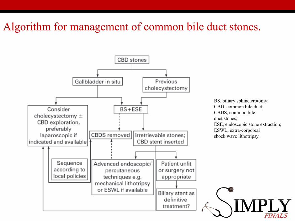
Algorithm for management of common bile duct stones.
BS, biliary sphincterotomy; CBD, common bile duct; CBDS, common bileduct stones; ESE, endoscopic stone extraction; ESWL, extra-corporealshock wave lithotripsy.


Case 5 - 50♂ I…cant…poo…
• 50 year old male
• Intermittent abdominal pain• Has been sick a few times• BNO for 5 days
• Has a history of previous hernia repair

Examination
• Distended abdomen
• Looks pretty uncomfortable
• ABC approach…

Examination Cont
• A –• Maintaining.
• B –• Sats; 98% on air• RR 26• Lung fields - clear

Examination Cont
• C –• HR 115• BP 115/75• Abdomen
• Painful throughout abdomen.• Distended• Hyperactive bowel sounds• Noted to have not PUed either for ???time• PR empty rectum
• D –• AVPU vs GCS
• E –• T 37.2• BM 10

Investigations
• What do you want and why??
• Least invasive most invasive• Cheapest most expensive• Dullest most awesome

Investigations
• FBC, U&E, LFT, albumin, magnesium, glucose• CRP• Amylase• G&S vs XM• ABG• Erect CXR/AXR• ECG• Urine dip• ?USS• ?Contrast enhanced CT

1) Presence of an air-fluid differential height in the same small-bowel loop
2) Presence of a mean level width greater than 25 mm


Management
• Aggressive fluid resuscitation + NBM
• Administration of analgesia and antiemetic
• Administration of antibiotics.• (Antibiotics are used to cover against gram-negative and
anaerobic organisms.)
• Placement of a nasogastric (NG) tube for suctioning GI contents and preventing aspiration.

Small Bowel Obstruction
• Small bowel obstruction accounts for 5% of acute surgical admissions and 80% of bowel obstruction
• In UK the commonest causes are: • Adhesions (60%)
• Strangulated hernia (20%)
• Malignancy (5%)
• Volvulus (5%)

Small Bowel Obstruction
Fluid and gas accumulate behind obstruction, causing proximal bowel to dilate
Peristaltic activity increases (COLICKY PAIN)
Inhibition of motor active (protective)
Increased intraluminal pressure/vascular occlusion leads to strangulation
Venous compromise leads to oedema, which cases arterial compression, ischaemia and necrosis, which may then perforate.

Small Bowel Obstruction• History
• Colicky central abdominal pain• Constant sharp if strangulation, ischaemia or if bowel perforates
• Vomiting - early in high obstruction
• Diarrhea - early or post-obstruction after it has resolved
• Absolute constipation - late feature of small bowel obstruction
• Previous surgery, radiotherapy
• History of malignancy
Small Bowel Obstruction• Examination
• Dehydration -> tachycardia, hypotension and oliguria
• Features of peritonism indicate strangulation or perforation
• Fever and tachycardia together - late, associated with strangulation or perforation
• Abdominal distention
• Hyperactive 'tinkling' bowel sounds- early
• Absent bowel sounds - late.
• PR – Helps differentiate between mechanical and functional• Mechanical, often empty, collapsed rectum (though masses may be palpable)

Small Bowel Obstruction
• Investigations
• FBC, U&E, Amylase, XM blood
• ABG
• AXR and Erect CXR

• AXR• Bowel loops with bands which
traverse the gut
• Central bowel loops
• ‘Pathological’ if diameter more than 5cm
• CT• most cases
• especially on first presentation with confirmed AXR finding

Small Bowel Obstruction• Management
• ABCDE
• May require more than 5 litres of intravenous crystalloid
• Adequacy of resuscitation should be judged by urine output or central venous pressure
• Surgery in under resuscitated patient is associated with increased mortality
• NGT in all patients!
• If obstruction presumed to be due to adhesions and there are no features of peritonism
• Conservative management (drip and suck) for up to 48 hours is often safe
• Requires regular clinical review
• If features of peritonism or systemic toxicity present
• Need to consider early operation
• Exact procedure will depend on underlying cause
Indications for surgery
Absolute Generalised peritonitisLocalised peritonitisVisceral perforationIrreducible hernia
Relative Palpable mass lesion'Virgin' abdomenFailure to improve
Trial of conservatism Incomplete obstructionPrevious surgeryAdvanced malignancyDiagnostic doubt


Case 6 - 87♀ PICKLES!
• 87 year old female• Nursing home resident
• Found on floor; can only have been there for 3-4 mins
• Shouting PICKLES a lot
• Smells strongly of urine

History
• Ambulance crew report she was left for a few minutes and then a carer found her on the floor
• No collateral history available. Clearly demented.
• AMTS – 0/10

Examination
• Pleasantly confused
• Left leg noticeably short and externally rotated
• Patient comfortable
• ABC approach

Examination Cont
• A –• Maintaining.
• B –• Sats; 94% on air• RR 16• Lung fields – bibasal creps

Examination Cont
• C –• HR 95• BP 140/80• Abdomen
• NAD
• D –• AVPU vs GCS
• E –• T 37.1• BM 10

Investigations
• What do you want and why??
• Least invasive most invasive• Cheapest most expensive• Dullest most awesome
Investigations
• FBC, U&E, LFT, clotting
• G&S vs XM
• CXR
• Pelvic x-ray + lateral hip
• ECG
• Urine dip

Management
• IV access and commence intravenous fluids if indicated.
• Ensure analgesia (including opiates) is adequate
• NSAIDs are not recommended.
• Early assessment of treatable comorbidities
• Follow local protocol for NoF fractures
• What op? Consent?

Hemiarthroplasty

Neck of Femur Fractures
• Refers to fractures of the proximal femur
• 70,000 hip fractures occur per year in the UK
• Total cost of care is >£2billion• 10% mortality at 1 month• 40% of patients with a hip fracture die within a year
• 50% of survivors are less independent than before the injury
• Most morbidity is related to coexisting medical conditions
• Main risk factors are female sex and osteoporosis

Classify


Normal Xray

AO Oceania
Classification
- Displaced vs undisplaced

Subcapital Fracture

NICE Guidance Saves Lives and Makes £

Take home Message for NoFs
• MDT inc Ortho-Geri, Ortho etc
• X-ray CT MRI
• Painkillers; reassess need
• Do not delay surgery
• Get them walking
• Don’t give them a DVT


Case 7 – 19♂ I totes have leg pain bruv. Lolz
19 year old male
Tells you he “fell” jumping a 2m high fence…taking kittens...to the orphange...
Leg is excutiatingly painful
Demanding morphine
Keeps swearing at everyone.

History
• Normally fit and well
• Smokes marijuana, cigarettes and drinks “socially”
• Unemployed

Examination
• Loud. Angry. Swearing
• Leg noticeably deformed
• Closed, neurovascularly intact
• ABC approach

Examination Cont
• A –• Maintaining.
• B –• Sats; 99% on air• RR 16• Lung fields – clear

Examination Cont
• C –• HR 70• BP 125/80• Abdomen
• NAD
• D –• AVPU vs GCS
• E –• T 37.1• BM 5

Investigations
• What do you want and why??
• Least invasive most invasive• Cheapest most expensive• Dullest most awesome

X-ray

Investigations
• Do you need bloods?
• Do you need any other x-rays

Management
• Analgesia
• Immobilisation
• NBM
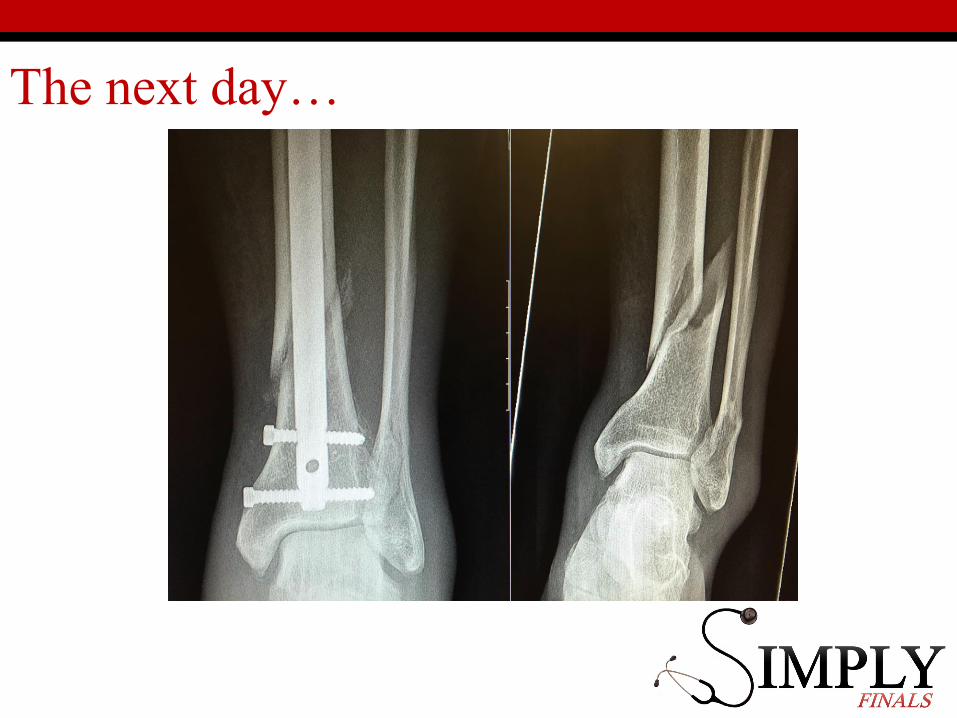
The next day…


The next night…
• Patient is screaming
• Demanding morphine
• Threatening to leave

What are you worried about?

Examination and Management
• Cast/dressings off
• Pain +++
• Pain on passive stretch
• A lot more screaming

What now?
• You are the FY1
• What do you do?


Compartment Syndrome
• Elevated tissue pressure within a closed fascial space
• Reduces tissue perfusion
• Results in cell death
• Pathogenesis
• Too much inflow (edema, hemorrhage)
• Decreased outflow (venous obstruction, tight dressing/cast)

Compartment Syndrome
• Pain out of proportion
• Palpably tense compartment
• Pain with passive stretch
• Paresthesia/hypoesthesia
• Paralysis
• Pulselessness/pallor
• Perishing cold

Compartment Syndrome
• Remove cast or dressing
• Place at level of heart(DO NOT ELEVATE to optimize perfusion)
• Call for help
• Role of bedside pressure measurements
https://www.boa.ac.uk/wp-content/uploads/2014/12/BOAST-10.pdf

Case 8 - 72 ♂ I have back pain!
• 72 year old male
• Retired lawyer (he made sure you knew that)
• Comes into A&E with back pain
• Unhappy because GP is shut and over the counter meds don’t work and he can’t wee
• Smells slightly of urine.

History
• Hurts all the time but wakes him up at night
• Hasn’t been to the toilet in 18 hours. Keeps mentioning this
• Any other questions you want to ask?• Pain• Urology

History – Red flags
• Weight loss
• Pain at rest
• Fever/rigor
• Bladder/bowel dysfunction
• Any others?

Examination
• Can’t get comfy
• Distended abdomen
Examination Cont
• A –• Maintaining.
• B –• Sats; 99% on air• RR 16• Lung fields – clear

Examination Cont
• C –• HR 99• BP 120/80• Abdomen
• Distended. • PR ???
• D –• Neuro exam…
• E –• T 37.1• BM 4

A Terrifying Diagram!

Investigations
• What do you want and why??
• Least invasive most invasive• Cheapest most expensive• Dullest most awesome

Investigations
• FBC, U&E, LFT, clotting, PSA
• G&S
• CXR
• Do you x-ray the spine?
• ECG
• Urine dip +/- bladder scan
• What other imaging?

•Radiographic
•AP and lateral of involved area off spine
•CT scan •helpful to identify metastatic lesions to the spine
•MRI •useful to show neurologic compromise of the spine

Management
• IV access and commence intravenous fluids if indicated.
• Ensure analgesia (including opiates) is adequate
• Who else needs to see him?
• What might be going on?

Prognosis
• thyroid: 48 months
• prostate: 40 months
• breast: 24 months
• kidney: variable but may be as short as 6 months
• lung: 6 months
•

Quick Catheter Chat
• Sterile technique
• Lubricant in urethra and on catheter
• “Penis up to the sky” + cough
• DO NOT INFLATE UNTIL URINE SEEN

Case 9 - 65 ♂ Cold feet
• 65 year old male
• Sudden onset left foot pain
• Walks with stick
• You saw them smoking outside the hospital when you came into work

History
• 1hr history – severe pain on movement
• Normally only walk 100yards before pain
• MI, HTN, Chole, diabetes
• Anything else?

Examination
• Cold
• Mottled & blanching
• Absent pulses distal to popliteal
• Painful ++++
• Motor and sensation intact

Examination Cont
• A –• Maintaining.
• B –• Sats; 89% on air• RR 16• Lung fields – bibasal creps

Examination Cont
• C –• HR 116• BP 165/90• Abdomen
• NAD
• D –• alert
• E –• T 37.1• BM 10

Investigations
• What do you want and why??
• Least invasive most invasive• Cheapest most expensive• Dullest most awesome

Investigations
• Hand-held Doppler ultrasound
• FBC• U&E/LFT/Clotting• ESR • Glucose• Lipids.• Thrombophilia screen.
• ECG

Management
• IV access and commence intravenous fluids if indicated.
• Ensure analgesia (including opiates) is adequate
• Embolic vs Thrombotic
• Revascularise• Reconstruct• Remove

Embolic vs Thrombotic• Embolic
• surgical embolectomy or local intra-arterial thrombolysis:
• If embolectomy fails• on-table angiogram + bypass
graft or intraoperative thrombolysis
• If successful, anticoagulation with heparin
• Thrombotic
• intra-arterial thrombolysis, angioplasty or bypass surgery
• If acute arterial emboli or thrombosis, immediate unfractionated heparin
• Followed by long-term warfarin for embolism.

Limb ischaemia
• Acute (on chronic)
• Emobilic (thrombotic)▫ No claudication
▫ Sudden onset (sec/min)
▫ Recent MI/AF/AAA
▫ Embolectomy
▫ Thrombolysis
▫ Emergency recon
▫ Amputation (10-20% mort)
• Chronic
• Thrombotic▫ Claudication
▫ Gradual onset (hrs)
▫ Chronic vascular disease
▫ Thrombolysis
▫ Angioplasty
▫ Emergency recon
▫ Amputation
