update on lung cancer overview classification ...labmed.ucsf.edu/uploads/488/237_jones...update on...
TRANSCRIPT
5/25/2013
1
Update on Lung Cancer Classification
Kirk D. Jones, MDUCSF Dept. of Pathology
Overview
• International Multidisciplinary Classification of Adenocarcinoma– Categorization– Grading– Invasion
• Molecular Testing Guidelines– CAP/IASLC/AMP– Recent biomarkers
Classification of Lung Adenocarcinoma
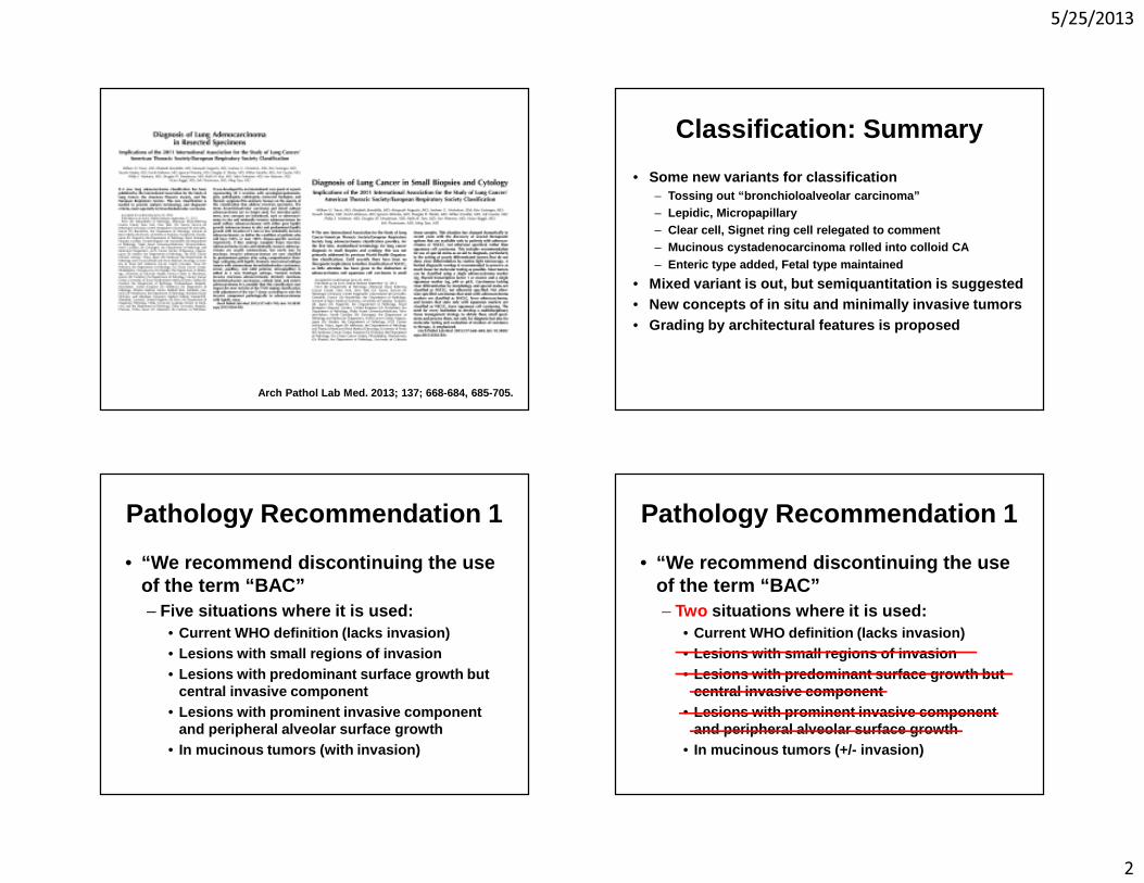
• International multidisciplinary effort by: – IASLC: Int’l Assoc. for the Study of Lung Cancer– ATS: American Thoracic Society– ERS: European Respiratory Society
• Representatives from thoracic oncology, pulmonology, radiology, molecular biology, thoracic surgery, and pathology
Journal of Thoracic Oncology 2011; 6: 244-285
5/25/2013
2
Arch Pathol Lab Med. 2013; 137; 668-684, 685-705.
Classification: Summary
• Some new variants for classification– Tossing out “bronchioloalveolar carcinoma” – Lepidic, Micropapillary– Clear cell, Signet ring cell relegated to comment– Mucinous cystadenocarcinoma rolled into colloid CA– Enteric type added, Fetal type maintained
• Mixed variant is out, but semiquantitation is sugges ted• New concepts of in situ and minimally invasive tumo rs• Grading by architectural features is proposed
Pathology Recommendation 1
• “We recommend discontinuing the use of the term “BAC”– Five situations where it is used:
• Current WHO definition (lacks invasion)• Lesions with small regions of invasion• Lesions with predominant surface growth but
central invasive component• Lesions with prominent invasive component
and peripheral alveolar surface growth• In mucinous tumors (with invasion)
Pathology Recommendation 1
• “We recommend discontinuing the use of the term “BAC”– Two situations where it is used:
• Current WHO definition (lacks invasion)• Lesions with small regions of invasion• Lesions with predominant surface growth but
central invasive component• Lesions with prominent invasive component
and peripheral alveolar surface growth• In mucinous tumors (+/- invasion)
5/25/2013
3
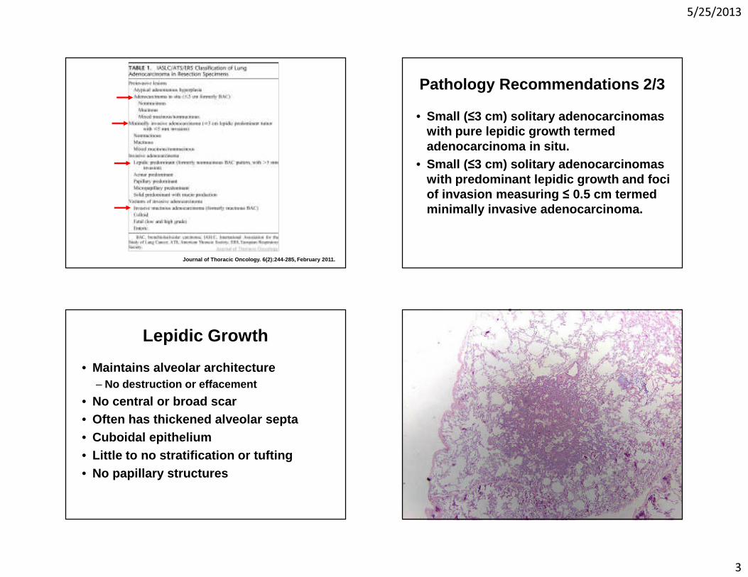
Journal of Thoracic Oncology. 6(2):244-285, Februar y 2011.
Pathology Recommendations 2/3
• Small ( ≤3 cm) solitary adenocarcinomas with pure lepidic growth termed adenocarcinoma in situ.
• Small ( ≤3 cm) solitary adenocarcinomas with predominant lepidic growth and foci of invasion measuring ≤ 0.5 cm termed minimally invasive adenocarcinoma.
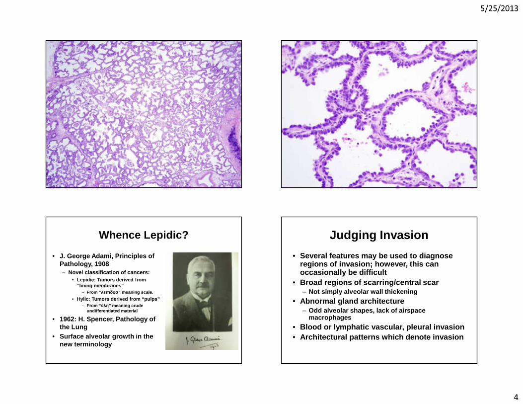
Lepidic Growth
• Maintains alveolar architecture– No destruction or effacement
• No central or broad scar• Often has thickened alveolar septa• Cuboidal epithelium• Little to no stratification or tufting• No papillary structures
5/25/2013
4
Whence Lepidic?
• J. George Adami, Principles of Pathology, 1908– Novel classification of cancers:
• Lepidic: Tumors derived from “lining membranes”
– From “ λεπιδοσ” meaning scale.
• Hylic: Tumors derived from “pulps”– From “ ύλη” meaning crude
undifferentiated material
• 1962: H. Spencer, Pathology of the Lung
• Surface alveolar growth in the new terminology
Judging Invasion
• Several features may be used to diagnose regions of invasion; however, this can occasionally be difficult
• Broad regions of scarring/central scar– Not simply alveolar wall thickening
• Abnormal gland architecture– Odd alveolar shapes, lack of airspace
macrophages
• Blood or lymphatic vascular, pleural invasion• Architectural patterns which denote invasion
5/25/2013
5
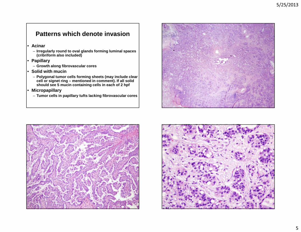
Patterns which denote invasion
• Acinar– Irregularly round to oval glands forming luminal sp aces
(cribriform also included)
• Papillary– Growth along fibrovascular cores
• Solid with mucin– Polygonal tumor cells forming sheets (may include c lear
cell or signet ring – mentioned in comment). If all solid should see 5 mucin containing cells in each of 2 hpf
• Micropapillary– Tumor cells in papillary tufts lacking fibrovascula r cores
5/25/2013
6
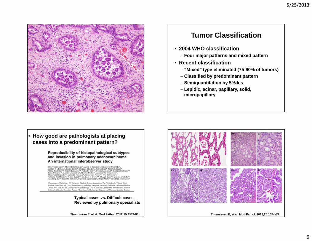
Tumor Classification
• 2004 WHO classification– Four major patterns and mixed pattern
• Recent classification– “Mixed” type eliminated (75-90% of tumors)– Classified by predominant pattern– Semiquantitation by 5%iles– Lepidic, acinar, papillary, solid,
micropapillary
• How good are pathologists at placing cases into a predominant pattern?
Typical cases vs. Difficult casesReviewed by pulmonary specialists
Thunnissen E, et al. Mod Pathol. 2012;25:1574-83. Thunnissen E, et al. Mod Pathol. 2012;25:1574-83.
5/25/2013
7
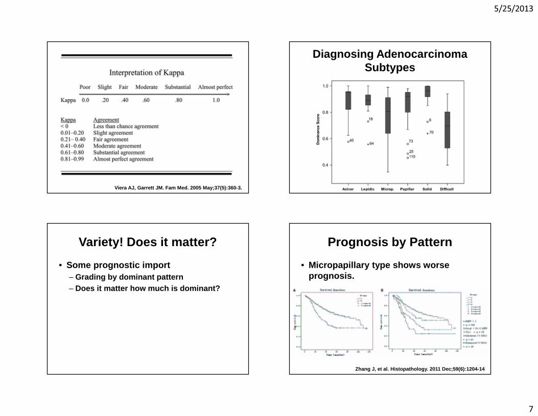
Viera AJ, Garrett JM. Fam Med. 2005 May;37(5):360-3.
Diagnosing Adenocarcinoma Subtypes
Variety! Does it matter?
• Some prognostic import– Grading by dominant pattern– Does it matter how much is dominant?
Prognosis by Pattern
• Micropapillary type shows worse prognosis.
Zhang J, et al. Histopathology. 2011 Dec;59(6):1204 -14
5/25/2013
8
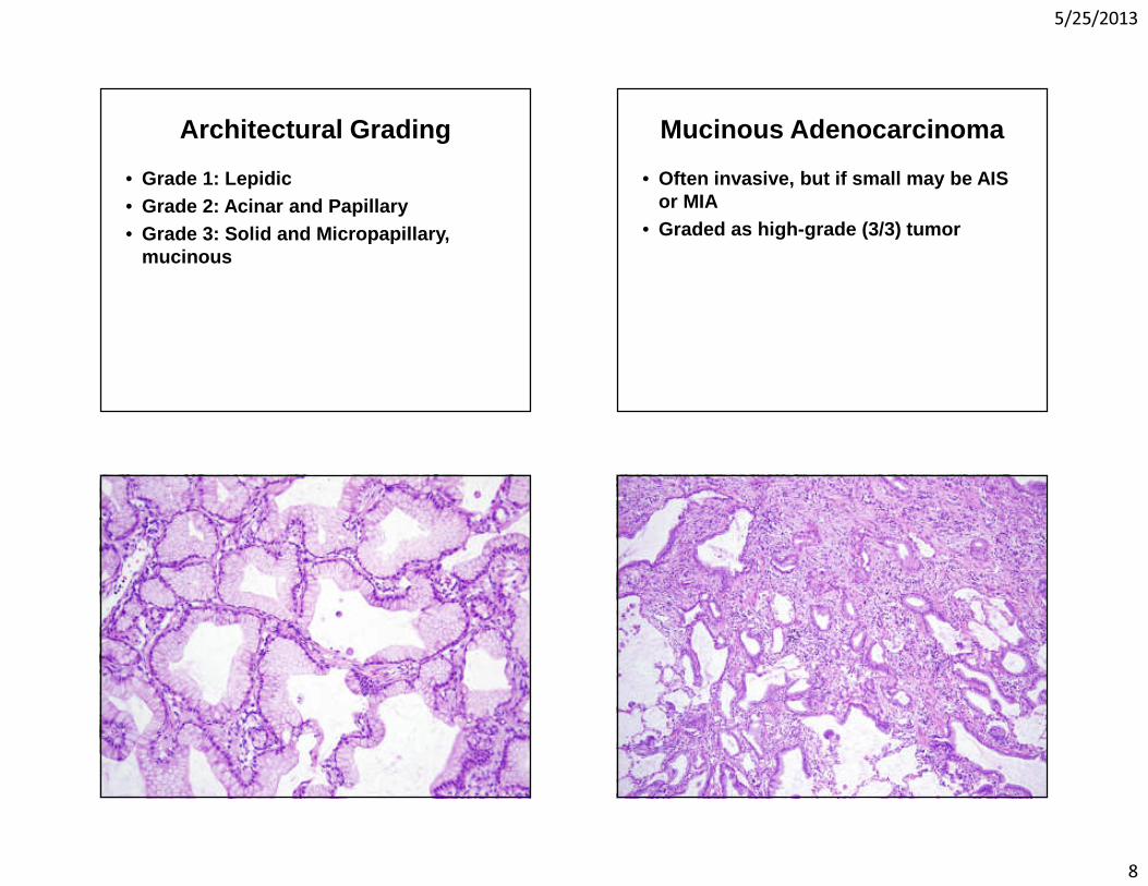
Architectural Grading
• Grade 1: Lepidic• Grade 2: Acinar and Papillary • Grade 3: Solid and Micropapillary,
mucinous
Mucinous Adenocarcinoma
• Often invasive, but if small may be AIS or MIA
• Graded as high-grade (3/3) tumor
5/25/2013
9
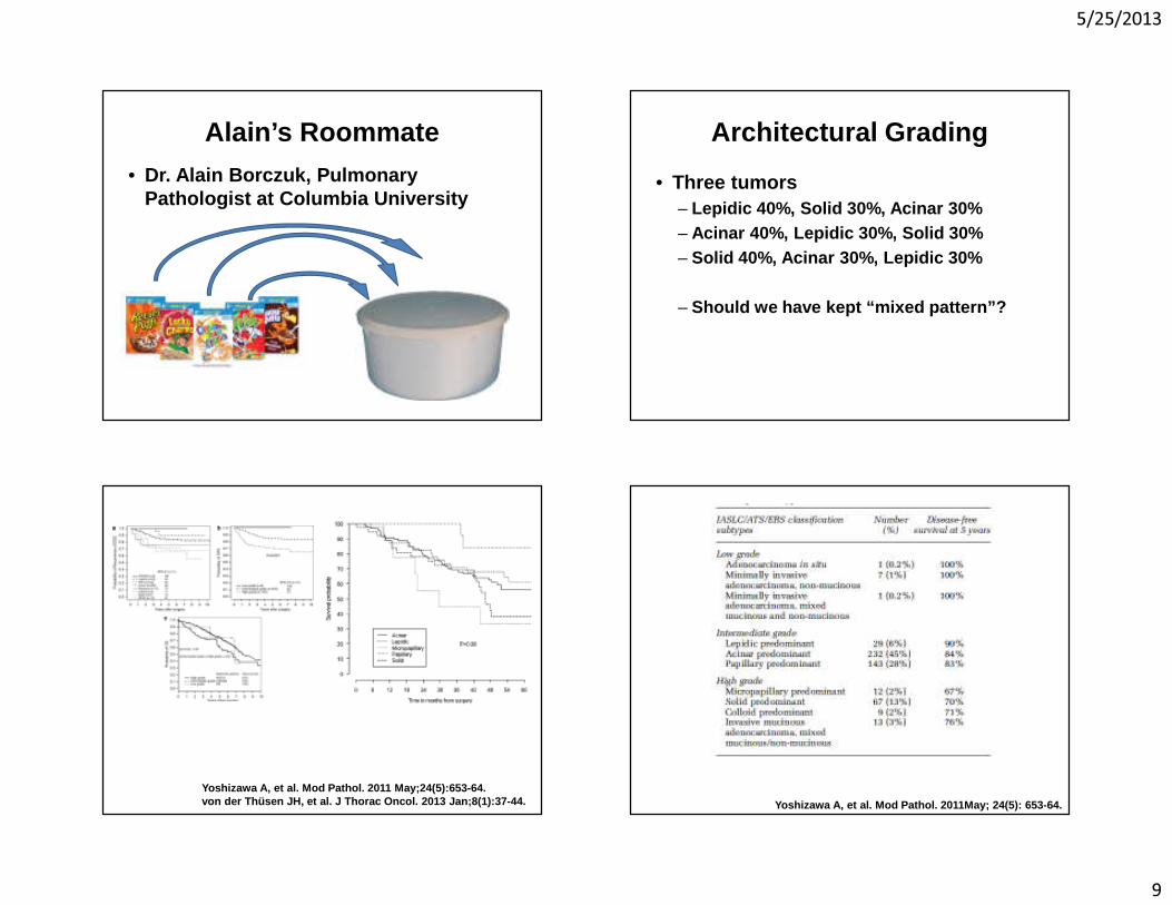
Alain’s Roommate
• Dr. Alain Borczuk, Pulmonary Pathologist at Columbia University
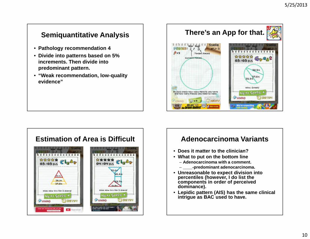
Architectural Grading
• Three tumors– Lepidic 40%, Solid 30%, Acinar 30%– Acinar 40%, Lepidic 30%, Solid 30%– Solid 40%, Acinar 30%, Lepidic 30%
– Should we have kept “mixed pattern”?
Yoshizawa A, et al. Mod Pathol. 2011 May;24(5):653-64 .von der Thüsen JH, et al. J Thorac Oncol. 2013 Jan;8( 1):37-44. Yoshizawa A, et al. Mod Pathol. 2011May; 24(5): 653- 64.
5/25/2013
10
Semiquantitative Analysis
• Pathology recommendation 4• Divide into patterns based on 5%
increments. Then divide into predominant pattern.
• “Weak recommendation, low-quality evidence”
There’s an App for that.
Estimation of Area is Difficult Adenocarcinoma Variants
• Does it matter to the clinician? • What to put on the bottom line
– Adenocarcinoma with a comment. – ____-predominant adenocarcinoma.
• Unreasonable to expect division into percentiles (however, I do list the components in order of perceived dominance).
• Lepidic pattern (AIS) has the same clinical intrigue as BAC used to have.
5/25/2013
11
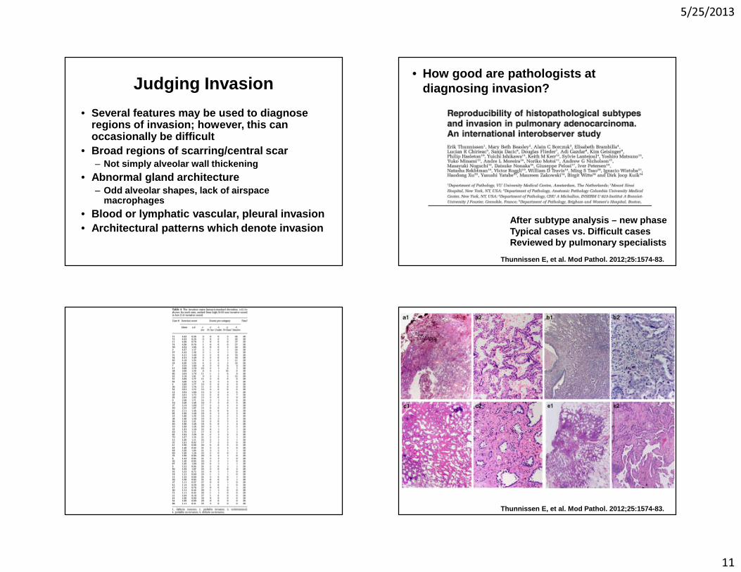
Judging Invasion
• Several features may be used to diagnose regions of invasion; however, this can occasionally be difficult
• Broad regions of scarring/central scar– Not simply alveolar wall thickening
• Abnormal gland architecture– Odd alveolar shapes, lack of airspace
macrophages
• Blood or lymphatic vascular, pleural invasion• Architectural patterns which denote invasion
• How good are pathologists at diagnosing invasion?
After subtype analysis – new phaseTypical cases vs. Difficult casesReviewed by pulmonary specialists
Thunnissen E, et al. Mod Pathol. 2012;25:1574-83.
Thunnissen E, et al. Mod Pathol. 2012;25:1574-83.
5/25/2013
12
How Well Do Pathologists Agree on Invasion?
• Complete agreement in 6 of 64 cases, when probable and definite combined.
• Only two cases with complete agreement (definite invasion).
• Kappa for easy cases = 0.55 (moderate)• Kappa for difficult cases = 0.08 (slight)
How Well Do Pathologists Agree on Invasion?
• The pathologists tended to split into those who favored invasion and those who favored non-invasion– If broke into two groups, K= 0.16 (slight)
• Lack of clear criteria
5/25/2013
13
Assessment of Invasion
• Likely not too many cases that have true non-invasion.
• Correlate with radiology (should be pure ground glass opacity in most cases).
• These criteria are only currently applied to tumors 3 cm or less in diameter, so the only change would be in T1 lesions.
Diagnosis of Small Biopsies
• Endobronchial, transbronchial, core, and aspiration biopsies.
• The main thrust of this paper is “Don’t waste tissue!”
Travis et al. Arch Pathol Lab Med. 2013; 137: 668-68 4.
Long Strange Trip
• From subtyping to lumping– 1993: No clinical import.
• Back to OCD subtyping– 2009: Driven by differences in chemo
• Back to chillax subtyping– 2011: Driven by need to conserve tissue
5/25/2013
14
Thorax 1993; 48: 1135-1139.
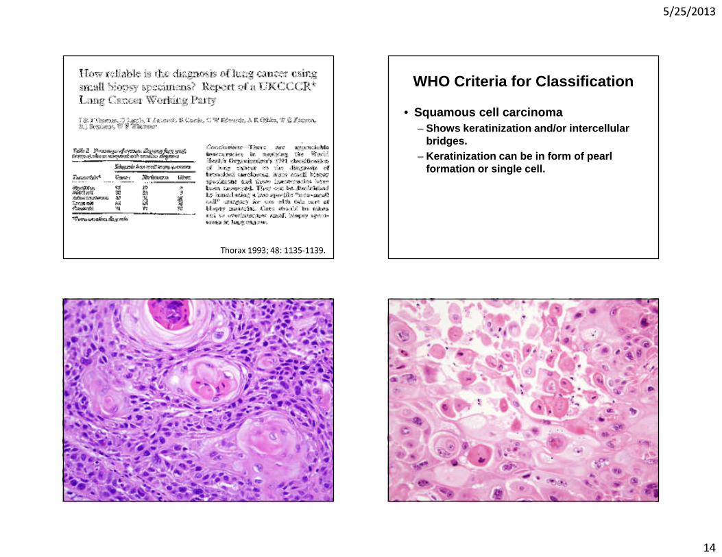
WHO Criteria for Classification
• Squamous cell carcinoma– Shows keratinization and/or intercellular
bridges. – Keratinization can be in form of pearl
formation or single cell.
5/25/2013
15
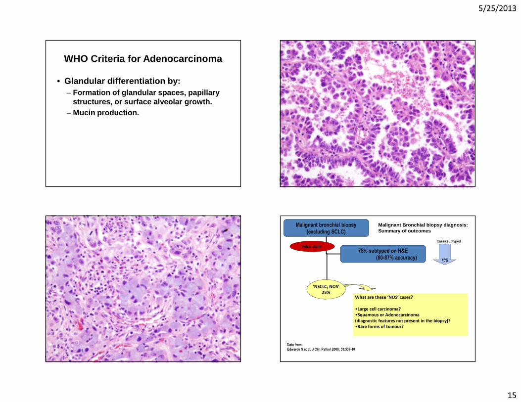
WHO Criteria for Adenocarcinoma
• Glandular differentiation by: – Formation of glandular spaces, papillary
structures, or surface alveolar growth. – Mucin production.
Malignant bronchial biopsy(excluding SCLC)
75% subtyped on H&E(80-87% accuracy)
H&E stain
Data from:Edwards S et al, J Clin Pathol 2000; 53:537-40
Malignant Bronchial biopsy diagnosis:Summary of outcomes
75%
Cases subtyped
‘NSCLC, NOS’25%
What are these ‘NOS’ cases?
•Large cell carcinoma?•Squamous or Adenocarcinoma(diagnostic features not present in the biopsy)?•Rare forms of tumour?
5/25/2013
16
Immunohistochemical Panels
• Can an IHC panel increase diagnostic accuracy in the differentiation of NSCLC?
Immunohistochemical Panels
• Ring et al, Mod Pathol. 2009 Aug; 22(8): 1032-43. – TRIM29, CEACAM5, SLC7A5, MUC1, and CK5/6. – Marketed by Clarient as Pulmotype.
• Loo et al, J Thorac Oncol. 2010 Apr; 5(4): 442-7. – TTF-1, AB/PAS, p63
• Nicholson, et al. J Thorac Oncol. 2010 Apr; 5(4): 4 36-41. – TTF-1, p63, CK5/6, PAS-D
• Terry, et al. Am J Surg Pathol. 2010 Dec;34(12):180 5-11. – p63, TTF-1, CK5/6, CK7, Napsin A, and mucicarmine
• Righi, et al. Cancer. 2011 Jan 18. – TTF-1, desmocollin-3, p63, Napsin A
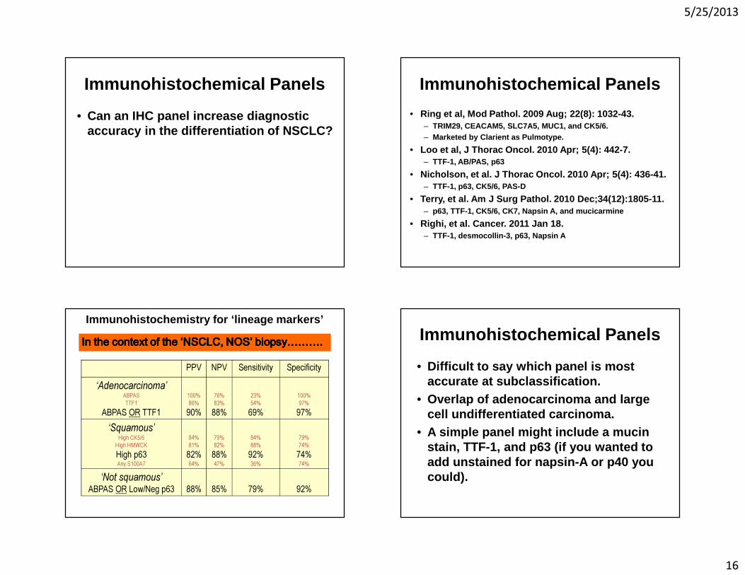
Immunohistochemistry for ‘lineage markers’
In the context of the ‘NSCLC, NOS’ biopsy……….In the context of the ‘NSCLC, NOS’ biopsy……….In the context of the ‘NSCLC, NOS’ biopsy……….In the context of the ‘NSCLC, NOS’ biopsy……….PPV NPV Sensitivity Specificity
‘Adenocarcinoma’ABPASTTF1
ABPAS OR TTF1100%86%90%
76%83%88%
23%54%69%
100%97%97%
‘Squamous’High CK5/6
High HMWCKHigh p63Any S100A7
84%81%82%64%
79%82%88%47%
84%88%92%36%
79%74%74%74%
‘Not squamous’ABPAS OR Low/Neg p63 88% 85% 79% 92%
Immunohistochemical Panels
• Difficult to say which panel is most accurate at subclassification.
• Overlap of adenocarcinoma and large cell undifferentiated carcinoma.
• A simple panel might include a mucinstain, TTF-1, and p63 (if you wanted to add unstained for napsin-A or p40 you could).
5/25/2013
17
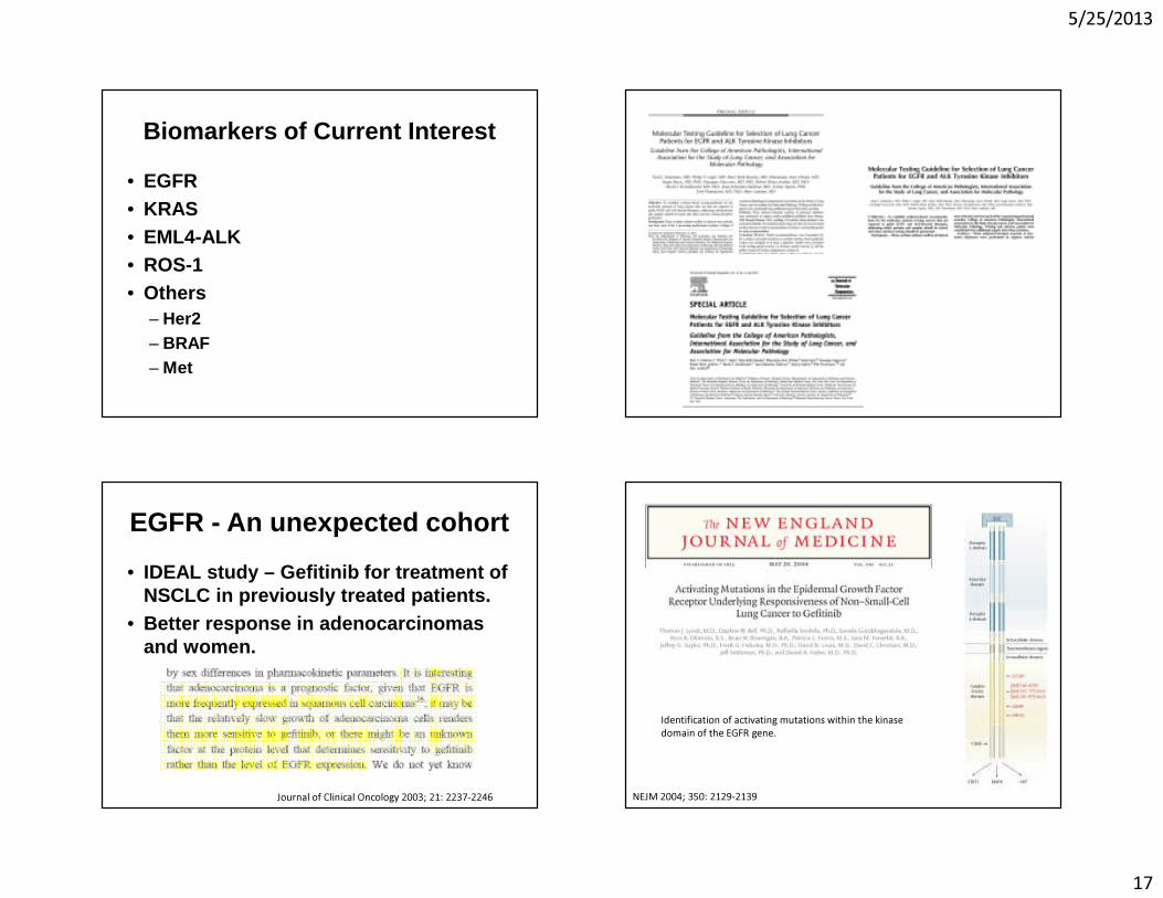
Biomarkers of Current Interest
• EGFR• KRAS• EML4-ALK• ROS-1• Others
– Her2– BRAF– Met
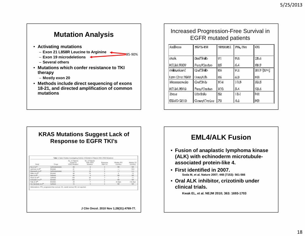
EGFR - An unexpected cohort
• IDEAL study – Gefitinib for treatment of NSCLC in previously treated patients.
• Better response in adenocarcinomas and women.
Journal of Clinical Oncology 2003; 21: 2237-2246
Identification of activating mutations within the kinase domain of the EGFR gene.
NEJM 2004; 350: 2129-2139
5/25/2013
18
Mutation Analysis
• Activating mutations– Exon 21 L858R Leucine to Arginine– Exon 19 microdeletions– Several others
• Mutations which confer resistance to TKI therapy – Mostly exon 20
• Methods include direct sequencing of exons 18-21, and directed amplification of common mutations
85-90%
Increased Progression-Free Survival in EGFR mutated patients
KRAS Mutations Suggest Lack of Response to EGFR TKI’s
J Clin Oncol. 2010 Nov 1;28(31):4769-77.
EML4/ALK Fusion
• Fusion of anaplastic lymphoma kinase (ALK) with echinoderm microtubule-associated protein-like 4.
• First identified in 2007. Soda M, et al. Nature 2007; 448 (7153): 561-566
• Oral ALK inhibitor, crizotinib under clinical trials.
Kwak EL, et al. NEJM 2010; 363: 1693-1703

5/25/2013
19
EML4/ALK Variants• While the breakpoint to the ALK gene is
uniform (2p23), the breakpoint in the EML4 gene varies.
Eur J Cancer. 2010 Jul;46(10):1773-80.
EML4/ALK Testing
• FISH is often used and shows an increased distance in positive cases.
• IHC using typical markers shows some false negatives.
• RT-PCR can be used, but the primers are specific to the different variants.
ROS1 Fusion Protein
Same group that co-discovered ALK mutations back in 2007 also discovered ROS1 mutation in lung cancer
Rikova K, et al. Cell. 2007 Dec14;131(6):1190-203.
• ROS1 testing• Fusion protein• Test with breakapart
FISH assay.• Another tumor that
shows over-representation of non-smokers and young patients.
• 1-2% of adenocarcinomas
5/25/2013
20
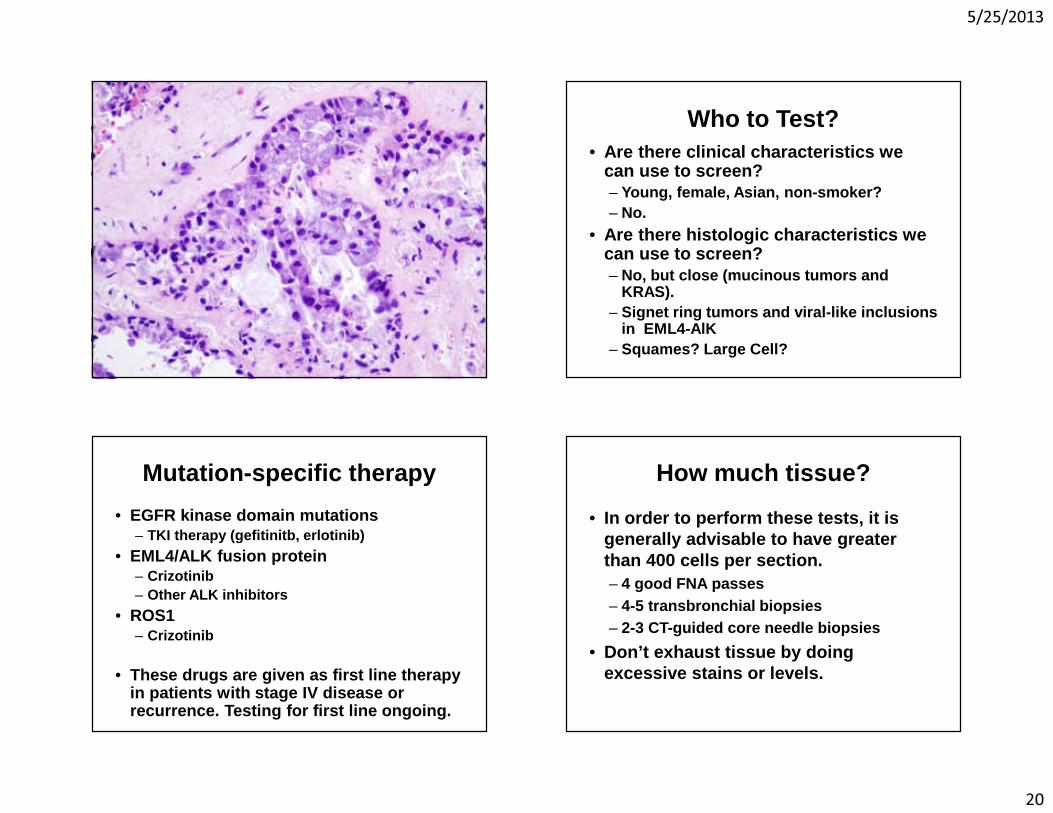
Who to Test?• Are there clinical characteristics we
can use to screen?– Young, female, Asian, non-smoker?– No.
• Are there histologic characteristics we can use to screen? – No, but close (mucinous tumors and
KRAS).– Signet ring tumors and viral-like inclusions
in EML4-AlK– Squames? Large Cell?
Mutation-specific therapy
• EGFR kinase domain mutations– TKI therapy (gefitinitb, erlotinib)
• EML4/ALK fusion protein– Crizotinib– Other ALK inhibitors
• ROS1– Crizotinib
• These drugs are given as first line therapy in patients with stage IV disease or recurrence. Testing for first line ongoing.
How much tissue?
• In order to perform these tests, it is generally advisable to have greater than 400 cells per section. – 4 good FNA passes– 4-5 transbronchial biopsies– 2-3 CT-guided core needle biopsies
• Don’t exhaust tissue by doing excessive stains or levels.
5/25/2013
21
Take Home Messages
• AIS and MIA, as defined, have a better prognosis when compared to invasive tumors of the same size.
• They are rare lesions, and invasion is difficult to assess due to lack of good criteria. Correlate with imaging.
• Histologic subtypes have prognostic significance. – Lepidic– Acinar and Papillary– Solid, Micropapillary, and Mucinous
Take Home MessageAdenocarcinoma vs. Squame
• Use H&E criteria (WHO criteria)– Adenocarcinoma as discussed previously– Squamous cell with:
• Keratinization (single cell or pearl)• Intercellular bridges
• If cannot classify, do 2 stains– TTF-1 (or napsin-A)– P63 (or p40)
• Resist up front levels
Biomarker Analysis
• Make sure you obtain enough tissue. • Make sure you use tissue wisely. • Algorithmic testing may become
difficult as more markers are discovered. – KRAS first in smokers?
• Testing for individual genes may become cheaper or obsolete as cost for these tests decreases.
<BIOMARKERS>
xkcd.com
Thank you.