update on the etiology of tooth resorption in domestic cats · species, hypercementosis has been...
TRANSCRIPT

Vet Clin Small Anim
35 (2005) 913–942
Update on the Etiology of ToothResorption in Domestic Cats
Alexander M. Reiter, Dipl Tzt, Dr Med Veta,*,John R. Lewis, VMDa, Ayako Okuda, DVM, PhDb,c
aDepartment of Clinical Studies, School of Veterinary Medicine,
University of Pennsylvania, 3900 Delancey Street, Philadelphia, PA 19104–6010, USAbDepartment of Anatomy, School of Veterinary Medicine,
Azabu University, Fuchinobe, JapancVettec Dentistry, Tokyo, Japan
Feline odontoclastic resorptive lesions (FORL) were first recognized andhistologically differentiated from caries in the 1920s [1,2]. Some anecdotalreports describing caries-like lesions at the cervical region of feline teethfollowed in the 1950s and 1960s, until two microscopic studies in the 1970sagain revealed that FORL were not caries but a type of tooth resorption[3,4]. A recent study showed that cats with FORL have a significantly lowerurine specific gravity and significantly higher serum concentration of 25-hydroxyvitamin D (25OHD) compared with cats without FORL [5],indicating that multiple tooth resorption in domestic cats could be themanifestation of some systemic insult rather than a local cause. In thisarticle, the histologic and radiographic appearance of FORL and certainother peculiarities of feline teeth are reviewed. An attempt is then made tocompare these findings with changes of the periodontium induced byadministration of excess vitamin D or vitamin D metabolites in experi-mental animals.
Histologic and radiographic features of feline odontoclastic
resorptive lesions
Tooth resorption is caused by odontoclasts. Their precursors derive fromhematopoietic stem cells of bone marrow or spleen and migrate from bloodvessels of the alveolar bone or periodontal ligament toward the external root
* Corresponding author.
E-mail address: [email protected] (A.M. Reiter).
0195-5616/05/$ - see front matter � 2005 Elsevier Inc. All rights reserved.
doi:10.1016/j.cvsm.2005.03.006 vetsmall.theclinics.com
914 REITER et al
surface, where mononuclear cells fuse with other cells to becomemultinucleated mature odontoclasts [6,7]. One important fact to understandis that FORL develop anywhere on the root surface and not just close to thecementoenamel junction [8]. Resorption of enamel as the initial event is onlyrarely observed [9]. Resorption may also start on the same tooth at variousroot surfaces simultaneously, progressing from cementum coronally intocrown dentin as well as apically into root dentin. As the resorptionprogresses into crown dentin, the enamel often becomes undermined anda pink discoloration may be observed at the crown surface [10].
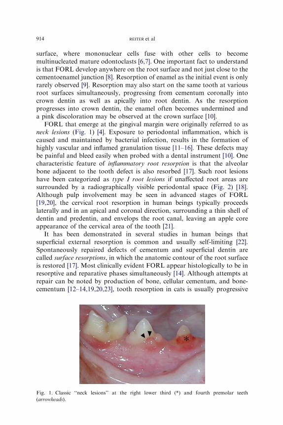
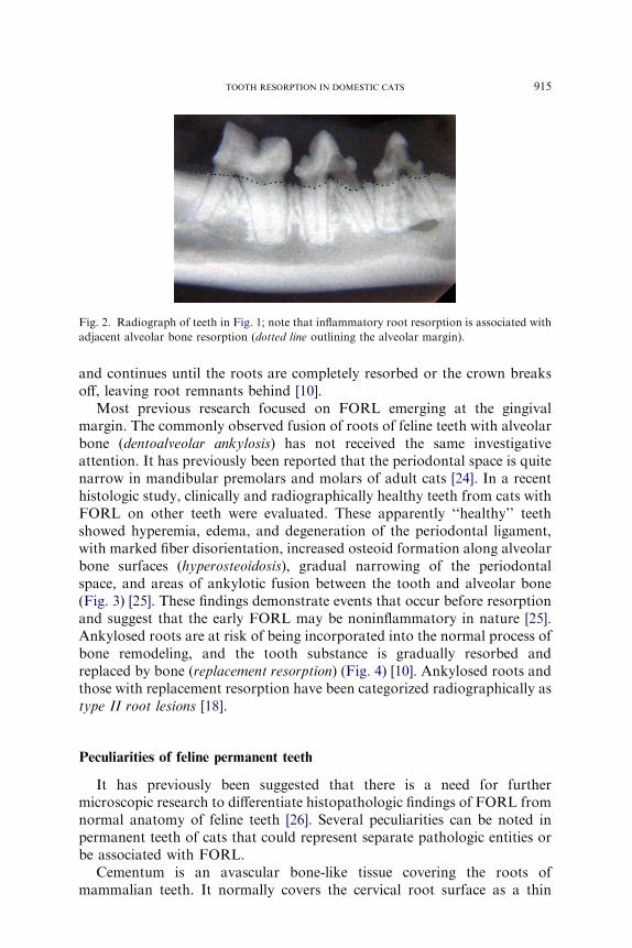
FORL that emerge at the gingival margin were originally referred to asneck lesions (Fig. 1) [4]. Exposure to periodontal inflammation, which iscaused and maintained by bacterial infection, results in the formation ofhighly vascular and inflamed granulation tissue [11–16]. These defects maybe painful and bleed easily when probed with a dental instrument [10]. Onecharacteristic feature of inflammatory root resorption is that the alveolarbone adjacent to the tooth defect is also resorbed [17]. Such root lesionshave been categorized as type I root lesions if unaffected root areas aresurrounded by a radiographically visible periodontal space (Fig. 2) [18].Although pulp involvement may be seen in advanced stages of FORL[19,20], the cervical root resorption in human beings typically proceedslaterally and in an apical and coronal direction, surrounding a thin shell ofdentin and predentin, and envelops the root canal, leaving an apple coreappearance of the cervical area of the tooth [21].
It has been demonstrated in several studies in human beings thatsuperficial external resorption is common and usually self-limiting [22].Spontaneously repaired defects of cementum and superficial dentin arecalled surface resorptions, in which the anatomic contour of the root surfaceis restored [17]. Most clinically evident FORL appear histologically to be inresorptive and reparative phases simultaneously [14]. Although attempts atrepair can be noted by production of bone, cellular cementum, and bone-cementum [12–14,19,20,23], tooth resorption in cats is usually progressive
Fig. 1. Classic ‘‘neck lesions’’ at the right lower third (*) and fourth premolar teeth
(arrowheads).
915TOOTH RESORPTION IN DOMESTIC CATS
and continues until the roots are completely resorbed or the crown breaksoff, leaving root remnants behind [10].
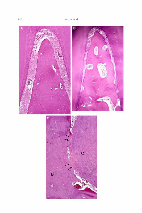
Most previous research focused on FORL emerging at the gingivalmargin. The commonly observed fusion of roots of feline teeth with alveolarbone (dentoalveolar ankylosis) has not received the same investigativeattention. It has previously been reported that the periodontal space is quitenarrow in mandibular premolars and molars of adult cats [24]. In a recenthistologic study, clinically and radiographically healthy teeth from cats withFORL on other teeth were evaluated. These apparently ‘‘healthy’’ teethshowed hyperemia, edema, and degeneration of the periodontal ligament,with marked fiber disorientation, increased osteoid formation along alveolarbone surfaces (hyperosteoidosis), gradual narrowing of the periodontalspace, and areas of ankylotic fusion between the tooth and alveolar bone(Fig. 3) [25]. These findings demonstrate events that occur before resorptionand suggest that the early FORL may be noninflammatory in nature [25].Ankylosed roots are at risk of being incorporated into the normal process ofbone remodeling, and the tooth substance is gradually resorbed andreplaced by bone (replacement resorption) (Fig. 4) [10]. Ankylosed roots andthose with replacement resorption have been categorized radiographically astype II root lesions [18].
Peculiarities of feline permanent teeth
It has previously been suggested that there is a need for furthermicroscopic research to differentiate histopathologic findings of FORL fromnormal anatomy of feline teeth [26]. Several peculiarities can be noted inpermanent teeth of cats that could represent separate pathologic entities orbe associated with FORL.
Cementum is an avascular bone-like tissue covering the roots ofmammalian teeth. It normally covers the cervical root surface as a thin
Fig. 2. Radiograph of teeth in Fig. 1; note that inflammatory root resorption is associated with
adjacent alveolar bone resorption (dotted line outlining the alveolar margin).
916 REITER et al

917TOOTH RESORPTION IN DOMESTIC CATS
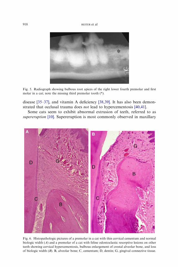
layer that gradually becomes wider apically. Two types of cementum(acellular and cellular) are usually recognized, which can be furthersubdivided depending on the presence and origin of collagen fibers(afibrillar, intrinsic, or extrinsic). Cementum formation beyond physiologicdeposition is called hypercementosis and can commonly be observed in teethof cats with FORL [12]. In one study, hypercementosis was demonstrated inall investigated feline teeth [14]. Excessive amounts of cellular cementum aredeposited particularly at apical and midroot surfaces, sometimes causingbulbous root apices (Fig. 5), whereas an abnormal thickening of acellularcementum can be found on cervical root surfaces (Fig. 6) [25]. In otherspecies, hypercementosis has been observed in unerupted, hypofunctional,and extruding teeth without opposing antagonists [27–30] and in certainconditions, such as hyperthyroidism [31], hyperpituitarism [32–34], Paget’s
Fig. 3. Histopathologic pictures of a feline premolar tooth with a normal furcation area (A) and
a premolar tooth of a cat with feline odontoclastic resorptive lesions on other teeth showing
degeneration of the periodontal ligament, narrowing of the periodontal space, and
dentoalveolar ankylosis (B). Close-up of apical area of tooth root showing periodontal
ligament degeneration and two areas of ankylotic fusion (arrows) between cementum (C) and
alveolar bone (B).
:
Fig. 4. Radiograph of dentoalveolar ankylosis and root replacement resorption of mandibular
canine teeth (dotted line outlining original root contour); also note the bulbous enlargement of
crestal alveolar bone (arrowheads).
918 REITER et al
disease [35–37], and vitamin A deficiency [38,39]. It has also been demon-strated that occlusal trauma does not lead to hypercementosis [40,41].
Some cats seem to exhibit abnormal extrusion of teeth, referred to assupereruption [10]. Supereruption is most commonly observed in maxillary
Fig. 5. Radiograph showing bulbous root apices of the right lower fourth premolar and first
molar in a cat; note the missing third premolar tooth (*).
Fig. 6. Histopathologic pictures of a premolar in a cat with thin cervical cementum and normal
biologic width (A) and a premolar of a cat with feline odontoclastic resorptive lesions on other
teeth showing cervical hypercementosis, bulbous enlargement of crestal alveolar bone, and loss
of biologic width (B). B, alveolar bone; C, cementum; D, dentin; G, gingival connective tissue.
919TOOTH RESORPTION IN DOMESTIC CATS
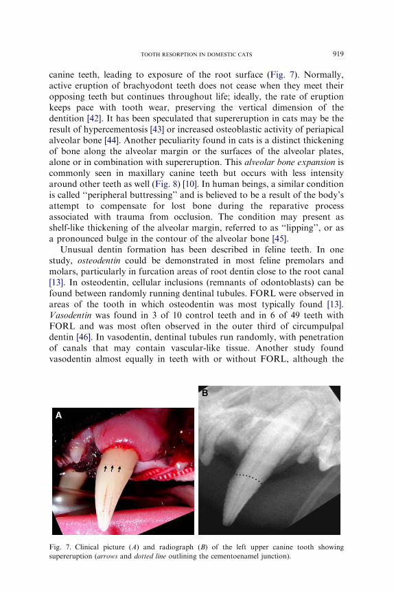
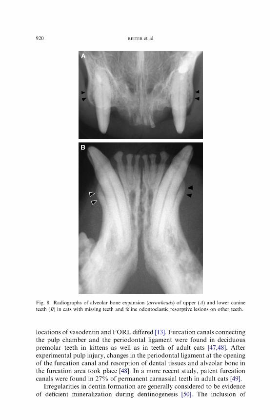
canine teeth, leading to exposure of the root surface (Fig. 7). Normally,active eruption of brachyodont teeth does not cease when they meet theiropposing teeth but continues throughout life; ideally, the rate of eruptionkeeps pace with tooth wear, preserving the vertical dimension of thedentition [42]. It has been speculated that supereruption in cats may be theresult of hypercementosis [43] or increased osteoblastic activity of periapicalalveolar bone [44]. Another peculiarity found in cats is a distinct thickeningof bone along the alveolar margin or the surfaces of the alveolar plates,alone or in combination with supereruption. This alveolar bone expansion iscommonly seen in maxillary canine teeth but occurs with less intensityaround other teeth as well (Fig. 8) [10]. In human beings, a similar conditionis called ‘‘peripheral buttressing’’ and is believed to be a result of the body’sattempt to compensate for lost bone during the reparative processassociated with trauma from occlusion. The condition may present asshelf-like thickening of the alveolar margin, referred to as ‘‘lipping’’, or asa pronounced bulge in the contour of the alveolar bone [45].
Unusual dentin formation has been described in feline teeth. In onestudy, osteodentin could be demonstrated in most feline premolars andmolars, particularly in furcation areas of root dentin close to the root canal[13]. In osteodentin, cellular inclusions (remnants of odontoblasts) can befound between randomly running dentinal tubules. FORL were observed inareas of the tooth in which osteodentin was most typically found [13].Vasodentin was found in 3 of 10 control teeth and in 6 of 49 teeth withFORL and was most often observed in the outer third of circumpulpaldentin [46]. In vasodentin, dentinal tubules run randomly, with penetrationof canals that may contain vascular-like tissue. Another study foundvasodentin almost equally in teeth with or without FORL, although the
Fig. 7. Clinical picture (A) and radiograph (B) of the left upper canine tooth showing
supereruption (arrows and dotted line outlining the cementoenamel junction).
920 REITER et al
locations of vasodentin and FORL differed [13]. Furcation canals connectingthe pulp chamber and the periodontal ligament were found in deciduouspremolar teeth in kittens as well as in teeth of adult cats [47,48]. Afterexperimental pulp injury, changes in the periodontal ligament at the openingof the furcation canal and resorption of dental tissues and alveolar bone inthe furcation area took place [48]. In a more recent study, patent furcationcanals were found in 27% of permanent carnassial teeth in adult cats [49].
Irregularities in dentin formation are generally considered to be evidenceof deficient mineralization during dentinogenesis [50]. The inclusion of
Fig. 8. Radiographs of alveolar bone expansion (arrowheads) of upper (A) and lower canine
teeth (B) in cats with missing teeth and feline odontoclastic resorptive lesions on other teeth.
921TOOTH RESORPTION IN DOMESTIC CATS
odontoblasts or pulp tissue into dentin may also be attributable to times ofrapid mineralization of newly formed dentin matrix, however. This view issupported by the observation that the layer of predentin appeared extremelythin or was not present in teeth of cats with FORL [51].
Increased vitamin D activity in cats with feline odontoclastic resorptive
lesions
Although FORL may have occurred more than 800 years ago [52], retro-spective studies of zoologic collections of feline skulls showed a lowprevalenceof FORL before the 1960s [53,54]. It was suggested that the increased prev-alence of FORL might be associated with aspects of domestication, such asaltered feeding practices, vaccination, and neutering programs [10].
Unlike bone that undergoes resorption and apposition as part ofa continual remodeling process, the roots of permanent teeth are normallynot resorbed because of resorption-inhibiting characteristics of unmineral-ized layers on external and internal root surfaces (eg, periodontal ligament,cementoblasts and cementoid, odontoblasts and predentin) [10,17]. Odon-toclasts may be attracted only to, or can attach only to, mineralized tissue. Ithas been postulated that removal or mineralization of the organic matrix ofthe root covering would make it possible for odontoclasts to recognize themineral component [10,17].
Measurement of biochemical markers of bone turnover, bone alkalinephosphatase (BAP) and deoxypyridinoline (DPD), did not show significantdifferences between cats with and without FORL [55]. It has recently beendemonstrated that cats with FORL expressed a significantly higher meanserum concentration of 25OHD compared with cats without FORL, how-ever [5]. Furthermore, the mean serum concentrations of blood urea nitro-gen and phosphorus were significantly higher and the mean urine specificgravity and mean calcium-phosphorus ratio were significantly lower in catswith FORL compared with cats without FORL [5]. Although the meanvalues of renal parameters remained within physiologic range, the resultssuggest the possibility of gradual impairment of renal function in cats withFORL. Using a human radioimmunoassay not yet validated for use in cats,calcitonin was significantly more often detected in blood sera of cats withFORL, which may be an expression of protective secretion during times oftransient mild hypercalcemia [5]. It was also demonstrated that cats withFORL vomited significantly more often than cats without FORL [5,56].
The diet represents the only source of vitamin D in cats because they areunable to produce vitamin D in the skin [57]. Based on feeding studies in the1950s, the National Research Council proposed a minimum vitamin Drequirement for growing kittens of 500 IU/kg of dietary dry matter [58].Later studies demonstrated that kittens given a diet with vitamin D3 perkilogram of dry matter at a rate of 250 or 125 IU did not show clinical signs
922 REITER et al
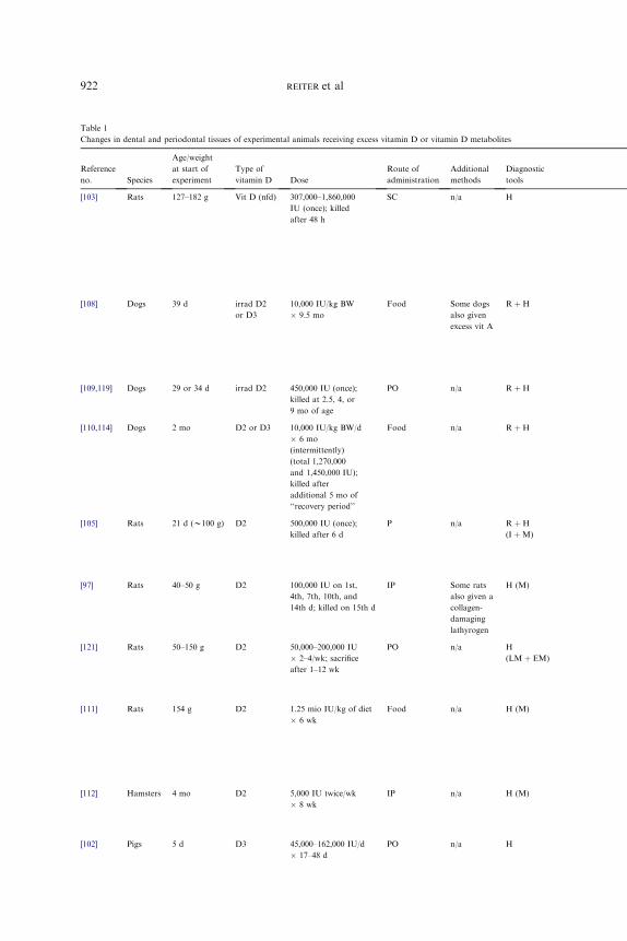
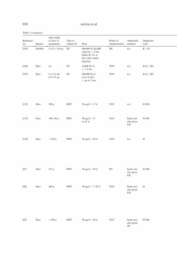
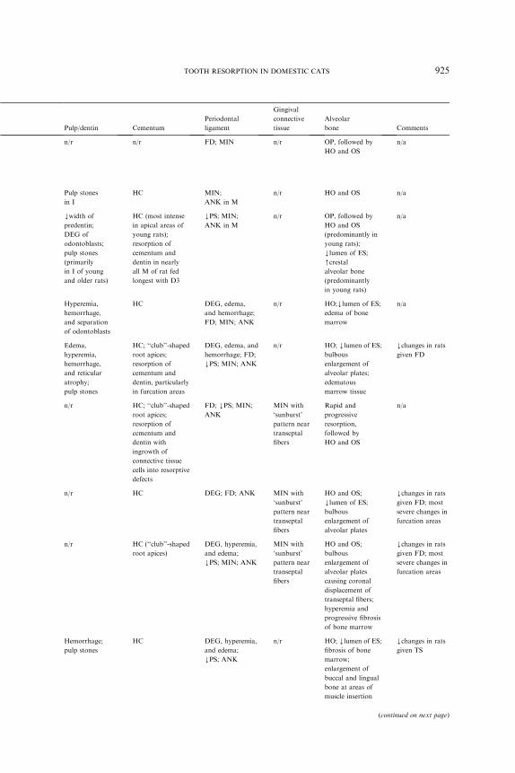
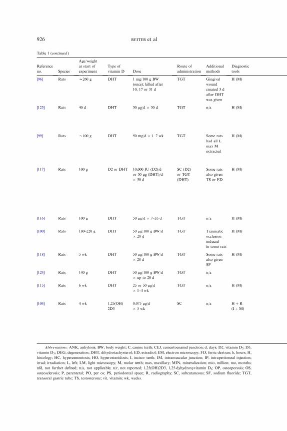
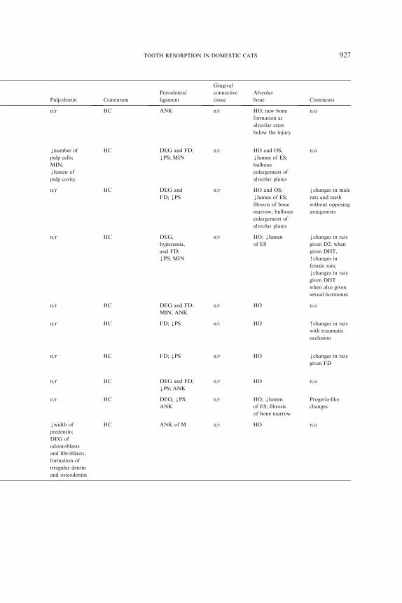
Table 1
Changes in dental and periodontal tissues of experimental animals receiving excess vitamin D or vitamin D metabolites
Reference
no. Species
Age/weight
at start of
experiment
Type of
vitamin D Dose
Route of
administration
Additional
methods
Diagnostic
tools
[103] Rats 127–182 g Vit D (nfd) 307,000–1,860,000
IU (once); killed
after 48 h
SC n/a H
[108] Dogs 39 d irrad D2
or D3
10,000 IU/kg BW
� 9.5 mo
Food Some dogs
also given
excess vit A
R þ H
[109,119] Dogs 29 or 34 d irrad D2 450,000 IU (once);
killed at 2.5, 4, or
9 mo of age
PO n/a R þ H
[110,114] Dogs 2 mo D2 or D3 10,000 IU/kg BW/d
� 6 mo
(intermittently)
(total 1,270,000
and 1,450,000 IU);
killed after
additional 5 mo of
‘‘recovery period’’
Food n/a R þ H
[105] Rats 21 d (w100 g) D2 500,000 IU (once);
killed after 6 d
P n/a R þ H
(I þM)
[97] Rats 40–50 g D2 100,000 IU on 1st,
4th, 7th, 10th, and
14th d; killed on 15th d
IP Some rats
also given a
collagen-
damaging
lathyrogen
H (M)
[121] Rats 50–150 g D2 50,000–200,000 IU
� 2–4/wk; sacrifice
after 1–12 wk
PO n/a H
(LM þ EM)
[111] Rats 154 g D2 1.25 mio IU/kg of diet
� 6 wk
Food n/a H (M)
[112] Hamsters 4 mo D2 5,000 IU twice/wk
� 8 wk
IP n/a H (M)
[102] Pigs 5 d D3 45,000–162,000 IU/d
� 17–48 d
PO n/a H

923TOOTH RESORPTION IN DOMESTIC CATS
Pulp/dentin Cementum
Periodontal
ligament
Gingival
connective
tissue
Alveolar
bone Comments
Calciotraumatic
line on inner edge
of dentin,
followed by
hypomineralized
layer, wide
hypermineralized
zone, and Ywidth
of predentin
n/r n/r n/r n/r Formation of
dentin proceeded
at same rate as
that of control
rats but MIN
was accelerated
DEG; pulp stones
in permanent
C þM
HC MIN; ANK MIN Initial OP,
followed by
OS with Ylumen
of ES in younger
dogs; less OS
in older dogs
Ychanges in dogs
given vit D from
tuna or halibut
liver oil than
irrad D2;
Ychanges in dogs
given excess vit A
DEG; MIN HC; resorption MIN n/r OP n/a
Pulp stones HC Development of
granulation tissue
in furcation and
interdental areas;
MIN; ANK
MIN Increased
vascularity;
granulation
tissue formation;
[periodontitis
in dog given D3
OP was
predominant
Hemorrhage,
odontoblast
DEG, accelerated
dentin formation,
MIN in M
n/r n/r n/r n/r n/a
n/r n/r MIN MIN n/r [changes in rats
given the
lathyrogen
n/r Intracellular
MIN of
cementoblast-like
cells; HC
DEG; MIN of
fibers close to
cemental surface
(‘‘sunburst’’
pattern)
MIN OP followed,
by HO and OS;
alveolar crest
raised to CEJ
n/a
n/r HC FD; YPS;
MIN; ANK
MIN with
‘sunburst’
pattern near
transeptal
fibers
OP followed,
by HO and OS;
alveolar crest
raised to CEJ;
marrow spaces
filled with young
connective tissue
n/a
n/r Cemental
spurs
YPS; MIN;
ANK
n/r Thinning of
cortical bone
and endosteal
resorption,
followed by HO
n/a
DEG and
hyperemia; MIN;
osteodentin
formation
Resorption of
cementum and
dentin with
pulp exposure
Hyperemia;
MIN; ANK
n/r OP, followed
by HO
n/a
(continued on next page)
924 REITER et al
Table 1 (continued )
Reference
no. Species
Age/weight
at start of
experiment
Type of
vitamin D Dose
Route of
administration
Additional
methods
Diagnostic
tools
[101] Rabbits 15 d (w150 g) D3 600,000 IU/kg BW
once/wk � 4 wk;
killed 30, 45, or
60 d after initial
injection
IM n/a R þ H
[106] Rats n/r D3 10,000 IU/d
� 1–4 wk
TGT n/a H (I þM)
[107] Rats 8 or 12 wk
(35–271 g)
D3 200,000 IU/d
(on 6 d/wk)
� up to 2 mo
TGT n/a H (I þM)
[122] Rats 100 g DHT 50 mg/d � 17 d TGT n/a H (M)
[123] Rats 140–150 g DHT 50 mg/d � 31
or 62 d
TGT Some rats
also given
FD
H (M)
[120] Rats w220 g DHT 50 mg/d � 50 d TGT n/a H
[91] Rats 215 g DHT 50 mg/d � 50 d PO Some rats
also given
FD
H (M)
[98] Rats 200 g DHT 50 mg/d � 7–50 d TGT Some rats
also given
FD
H
[95] Rats w100 g DHT 50 mg/d � 40 d TGT Some rats
also given
TS
H (M)
925TOOTH RESORPTION IN DOMESTIC CATS
Pulp/dentin Cementum
Periodontal
ligament
Gingival
connective
tissue
Alveolar
bone Comments
n/r n/r FD; MIN n/r OP, followed by
HO and OS
n/a
Pulp stones
in I
HC MIN;
ANK in M
n/r HO and OS n/a
Ywidth of
predentin;
DEG of
odontoblasts;
pulp stones
(primarily
in I of young
and older rats)
HC (most intense
in apical areas of
young rats);
resorption of
cementum and
dentin in nearly
all M of rat fed
longest with D3
YPS; MIN;
ANK in M
n/r OP, followed by
HO and OS
(predominantly in
young rats);
Ylumen of ES;
[crestal
alveolar bone
(predominantly
in young rats)
n/a
Hyperemia,
hemorrhage,
and separation
of odontoblasts
HC DEG, edema,
and hemorrhage;
FD; MIN; ANK
n/r HO;Ylumen of ES;
edema of bone
marrow
n/a
Edema,
hyperemia,
hemorrhage,
and reticular
atrophy;
pulp stones
HC; ‘‘club’’-shaped
root apices;
resorption of
cementum and
dentin, particularly
in furcation areas
DEG, edema, and
hemorrhage; FD;
YPS; MIN; ANK
n/r HO; Ylumen of ES;
bulbous
enlargement of
alveolar plates;
edematous
marrow tissue
Ychanges in rats
given FD
n/r HC; ‘‘club’’-shaped
root apices;
resorption of
cementum and
dentin with
ingrowth of
connective tissue
cells into resorptive
defects
FD; YPS; MIN;
ANK
MIN with
‘sunburst’
pattern near
transeptal
fibers
Rapid and
progressive
resorption,
followed by
HO and OS
n/a
n/r HC DEG; FD; ANK MIN with
‘sunburst’
pattern near
transeptal
fibers
HO and OS;
Ylumen of ES;
bulbous
enlargement of
alveolar plates
Ychanges in rats
given FD; most
severe changes in
furcation areas
n/r HC (‘‘club’’-shaped
root apices)
DEG, hyperemia,
and edema;
YPS; MIN; ANK
MIN with
‘sunburst’
pattern near
transeptal
fibers
HO and OS;
bulbous
enlargement of
alveolar plates
causing coronal
displacement of
transeptal fibers;
hyperemia and
progressive fibrosis
of bone marrow
Ychanges in rats
given FD; most
severe changes in
furcation areas
Hemorrhage;
pulp stones
HC DEG, hyperemia,
and edema;
YPS; ANK
n/r HO; Ylumen of ES;
fibrosis of bone
marrow;
enlargement of
buccal and lingual
bone at areas of
muscle insertion
Ychanges in rats
given TS
(continued on next page)
926 REITER et al
Table 1 (continued )
Reference
no. Species
Age/weight
at start of
experiment
Type of
vitamin D Dose
Route of
administration
Additional
methods
Diagnostic
tools
[96] Rats w260 g DHT 1 mg/100 g BW
(once); killed after
10, 17 or 31 d
TGT Gingival
wound
created 3 d
after DHT
was given
H (M)
[125] Rats 40 d DHT 50 mg/d � 50 d TGT n/a H (M)
[99] Rats w100 g DHT 50 mg/d � 1–7 wk TGT Some rats
had all L
max M
extracted
H (M)
[117] Rats 100 g D2 or DHT 10,000 IU (D2)/d
or 50 mg (DHT)/d
� 50 d
SC (D2)
or TGT
(DHT)
Some rats
also given
TS or ED
H (M)
[116] Rats 100 g DHT 50 mg/d � 7–35 d TGT n/a H (M)
[100] Rats 180–220 g DHT 50 mg/100 g BW/d
� 28 d
TGT Traumatic
occlusion
induced
in some rats
H (M)
[118] Rats 5 wk DHT 50 mg/100 g BW/d
� 28 d
TGT Some rats
also given
SF
H (M)
[124] Rats 140 g DHT 50 mg/100 g BW/d
� up to 20 d
TGT n/a
[115] Rats 6 wk DHT 25 or 50 mg/d
� 1–4 wk
TGT n/a H (M)
[104] Rats 4 wk 1,25(OH)
2D3
0.075 mg/d
� 5 wk
SC n/a H þ R
(I þM)
Abbreviations: ANK, ankylosis; BW, body weight; C, canine teeth; CEJ, cementoenamel junction; d, days; D2, vitamin D2; D3,
vitamin D3; DEG, degeneration; DHT, dihydrotachysterol; ED, estradiol; EM, electron microscopy; FD, ferric dextran; h, hours; H,
histology; HC, hypercementosis; HO, hyperosteoidosis; I, incisor teeth; IM, intramuscular junction; IP, intraperitoneal injection;
irrad; irradiation; L, left; LM, light microscopy; M, molar teeth; max, maxillary; MIN, mineralization; mio, million; mo, months;
nfd, not further defined; n/a, not applicable; n/r, not reported; 1,25(OH)2D3, 1,25-dyhydroxyvitamin D3; OP, osteoporosis; OS,
osteosclerosis; P, parenteral; PO, per os; PS, periodontal space; R, radiography; SC, subcutaneous; SF, sodium fluoride; TGT,
transoral gastric tube; TS, testosterone; vit, vitamin; wk, weeks.
927TOOTH RESORPTION IN DOMESTIC CATS
Pulp/dentin Cementum
Periodontal
ligament
Gingival
connective
tissue
Alveolar
bone Comments
n/r HC ANK n/r HO; new bone
formation at
alveolar crest
below the injury
n/a
Ynumber of
pulp cells;
MIN;
Ylumen of
pulp cavity
HC DEG and FD;
YPS; MIN
n/r HO and OS;
Ylumen of ES;
bulbous
enlargement of
alveolar plates
n/a
n/r HC DEG and
FD; YPS
n/r HO and OS;
Ylumen of ES;
fibrosis of bone
marrow; bulbous
enlargement of
alveolar plates
Ychanges in male
rats and teeth
without opposing
antagonists
n/r HC DEG,
hyperemia,
and FD;
YPS; MIN
n/r HO; Ylumen
of ES
Ychanges in rats
given D2; when
given DHT,
[changes in
female rats;
Ychanges in rats
given DHT
when also given
sexual hormones
n/r HC DEG and FD;
MIN; ANK
n/r HO n/a
n/r HC FD; YPS n/r HO [changes in rats
with traumatic
occlusion
n/r HC FD; YPS n/r HO Ychanges in rats
given FD
n/r HC DEG and FD;
YPS; ANK
n/r HO n/a
n/r HC DEG; YPS;
ANK
n/r HO; Ylumen
of ES; fibrosis
of bone marrow
Progeria-like
changes
Ywidth of
predentin;
DEG of
odontoblasts
and fibroblasts;
formation of
irregular dentin
and osteodentin
HC ANK of M n/r HO n/a
928 REITER et al
of vitamin D deficiency [59,60]. Furthermore, it was found that one third ofcommercial cat foods contained vitamin D3 in excess of current maximalallowances (O10,000 IU/kg of dietary dry matter), and a direct linearrelation was demonstrated between 25OHD serum concentrations anddietary intake of vitamin D [61]. Therefore, higher 25OHD serumconcentrations in cats with FORL suggest that they had ingested largeramounts of vitamin D or vitamin D metabolites compared with cats withoutFORL [5]. Three separate incidences of fatal hypervitaminosis D werereported in cats in Japan after consumption of commercial cat foodsprepared from fish [62–64]. Clinical, laboratory, and histopathologicfindings in these cats included vomiting, hypercalcemia, hyperphosphate-mia, azotemia, proteinuria, calciuria, phosphaturia, decreased urine specificgravity, and mineralization of various body tissues, particularly the kidneysand walls of large blood vessels [62]. One may speculate as to whether thereis indeed a predisposition to impairment of renal function in cats withFORL, because results of experimental studies on cats fed diets high invitamin D3 (15,000–33,840 IU/kg of dry matter) were contradictory, rangingfrom no evidence of detrimental effects on feline health to a high prevalenceof renal dysfunction and mortality [65].
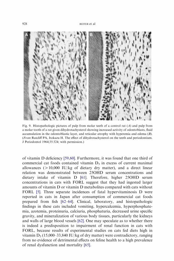
Fig. 9. Histopathologic pictures of pulp from molar teeth of a control rat (A) and pulp from
a molar tooth of a rat given dihydrotachysterol showing increased activity of odontoblasts, fluid
accumulation in the odontoblastic layer, and reticular atrophy with hyperemia and edema (B).
(From Ratcliff PA, Itokazu H. The effect of dihydrotachysterol on the teeth and periodontium.
J Periodontol 1964;35:324; with permission.)
929TOOTH RESORPTION IN DOMESTIC CATS
Vitamin D and vitamin D metabolites are important regulators ofosteoclastic bone resorption [66]. Serum calcium concentration is main-tained within a normal range through the primary action of 1,25-dihydroxy-vitamin D3 [1,25(OH)2D3], which increases intestinal absorption of dietarycalcium and recruits hematopoietic stem cells to become osteoclasts.Osteoclasts, in turn, mobilize calcium stores from bone into the circulation.Osteoclasts do not possess receptors for 1,25(OH)2D3, however [66].Receptors for 1,25(OH)2D3 are located on osteoblasts that produce factorsstimulating osteoclasts, such as receptor activator of nuclear factor-kBligand (RANKL), which plays an important role in osteoclastogenesis [67]and osteoclast activation [68].
Role of local trauma
The occlusal stress (tooth flexure) theory was created in an attempt toexplain noncarious cervical lesions, an overall term for tooth wear (notresorption) at the cervical portion of human teeth [69–71]. Repeatedcompressive and tensile forces attributable to tooth flexure during
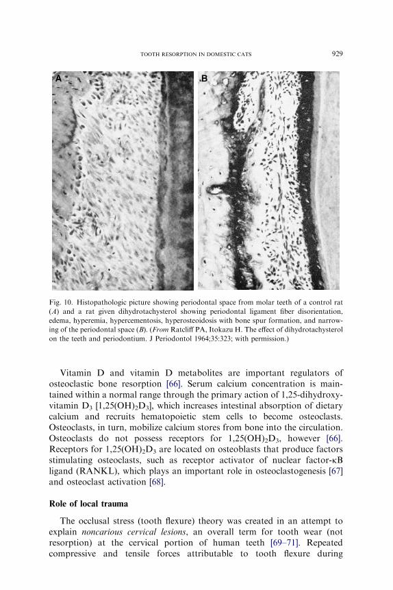
Fig. 10. Histopathologic picture showing periodontal space from molar teeth of a control rat
(A) and a rat given dihydrotachysterol showing periodontal ligament fiber disorientation,
edema, hyperemia, hypercementosis, hyperosteoidosis with bone spur formation, and narrow-
ing of the periodontal space (B). (From Ratcliff PA, Itokazu H. The effect of dihydrotachysterol
on the teeth and periodontium. J Periodontol 1964;35:323; with permission.)

930 REITER et al
mastication and malocclusion may disrupt the bonds between enamel rodsand between enamel and dentin, resulting in abfraction of enamel, exposureof dentin, and cervical hypersensitivity [72,73]. Although FORL are clearlyresorptive in nature and develop on any tooth and any root surface (not juston those exposed to occlusal or shearing forces), occlusal stress caused byeating large dry kibbles has been suggested to be associated with FORL[18,74,75]. A different approach for a possible role of occlusal stress in thedevelopment of FORL is presented in this article.
Surface resorption of cementum and superficial dentin may develop inresponse to normal masticatory stress [76] and excessive occlusal force [77–80]. Apical root resorption has been linked with bruxism in human beings,although the apical defect in that case report could also have resulted fromendodontic disease [81]. Traumatic occlusion from maloccluding teeth maycause resorption of roots in rats and people, with the apical area beingaffected most often [22,82–86]. Root resorption has been demonstrated afterexperimental intrusion of teeth in people [87] and long-standing occlusaltrauma in dogs and monkeys [88,89]. Subsequent repairs could eventuallyresult in ankylosis [90].
Fig. 11. Histopathologic pictures of cervical portion from teeth of a control dog (A) and a dog
given excessive amounts of vitamin D showing abnormal thickening of cervical cementum (B).
(From Becks H. Dangerous effects of vitamin D overdosage on dental and paradental
structures. J Am Dent Assoc 1942;29:1960; with permission.)

931TOOTH RESORPTION IN DOMESTIC CATS
Calciphylaxis is a condition of induced local or systemic hypersensitivityin which tissues respond to appropriate challenging agents with precipitous,sometimes evanescent, local mineralization of various tissues and organs[91,92]. Substances that predispose the organism to calciphylaxis are knownas sensitizers. Sensitizers are systemically administered agents that promotemineralization of tissues and include vitamin D and vitamin D metabolites,parathyroid hormone, and sodium acetylsulfathiazole among many othercalcium salts and phosphates [91]. Agents that precipitate the calciphylaxisphenomenon are known as challengers. Challengers may be direct orindirect. Direct challengers include mechanical trauma and various chemicalagents (eg, salts of iron, chromium, aluminum, zinc, manganese, cesium)that cause mineralization at the site of application and may elicit some formof systemic calciphylaxis when administered intravenously or intraperito-neally. Indirect challengers have little or no effect at the site of applicationand produce diverse systemic syndromes of mineralization and sclerosis [91].
Experiments in dihydrotachysterol (DHT)-sensitized rats indicated thatfunctional stress and topical trauma can produce local calcium deposits invarious parts of the body [91,93,94]. In rats given DHT, enlargement of
Fig. 12. Histopathologic pictures of furcation area from teeth of a control dog (A) and a dog
given excessive amounts of vitamin D showing hypercementosis, hyperosteoidosis, and
narrowing of the periodontal space (B). (From Becks H. Dangerous effects of vitamin D
overdosage on dental and paradental structures. J Am Dent Assoc 1942;29:1951; with
permission.)
932 REITER et al
buccal and lingual bone occurred most notably at muscle insertions [95].Alveolar bone formation at the site of a gingival injury took place morerapidly and was more evident in experimentally injured than noninjured ratsthat also received DHT [96]. Similarly, mineralization of the periodontalligament and gingival connective tissue was enhanced when a collagen-damaging agent was given to rats receiving intraperitoneal injections ofvitamin D2 [97]. In rats given DHT, degeneration of the periodontalligament, hypercementosis, hyperosteoidosis, narrowing of the periodontalspace, and ankylosis were markedly more pronounced in furcation areas[91,98] and teeth that were in occlusion [99] or subjected to traumaticocclusion [100]. Daily masticatory stress could be the reason why chronicincreased vitamin D intake manifests sooner and is more pronounced inperiodontal tissues compared with other soft tissues, and FORL maytherefore occur before or without obvious signs of vitamin D–inducedsystemic disease.
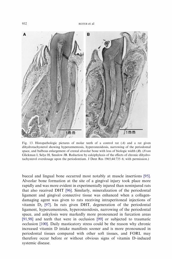
Fig. 13. Histopathologic pictures of molar teeth of a control rat (A) and a rat given
dihydrotachysterol showing hypercementosis, hyperosteoidosis, narrowing of the periodontal
space, and bulbous enlargement of crestal alveolar bone with loss of biologic width (B). (From
Glickman I, Selye H, Smulow JB. Reduction by calciphylaxis of the effects of chronic dihydro-
tachysterol overdosage upon the periodontium. J Dent Res 1965;44:735–6; with permission.)

933TOOTH RESORPTION IN DOMESTIC CATS
Experimental studies with vitamin D and vitamin D metabolites
Numerous reports describe the effects of excess vitamin D and vitamin Dmetabolites on the pulp-dentin complex and periodontium in experimentalanimals (rodents, lagomorphs, pigs, and dogs) (Table 1).
In the pulp-dentin complex, pulpal hyperemia and degeneration,decreased width of the predentin layer, and formation of osteodentin and
Fig. 14. Histopathologic pictures of interdental area from teeth of a control rat (A) and a rat given
dihydrotachysterol showing hypercementosis, hyperosteoidosis, edematous degeneration of the
periodontal ligament, narrowing of the periodontal space, bulbous enlargement of crestal alveolar
bone, coronal displacement of transeptal fibers, and reduction of biologic width (B). (From
Glickman I, Selye H, Smulow JB. Reduction by calciphylaxis of the effects of chronic dihydro-
tachysterol overdosage upon the periodontium. J Dent Res 1965;44:738; with permission.)
934 REITER et al
935TOOTH RESORPTION IN DOMESTIC CATS
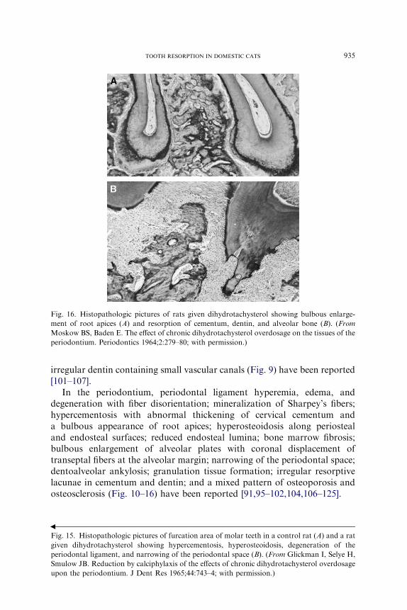
irregular dentin containing small vascular canals (Fig. 9) have been reported[101–107].
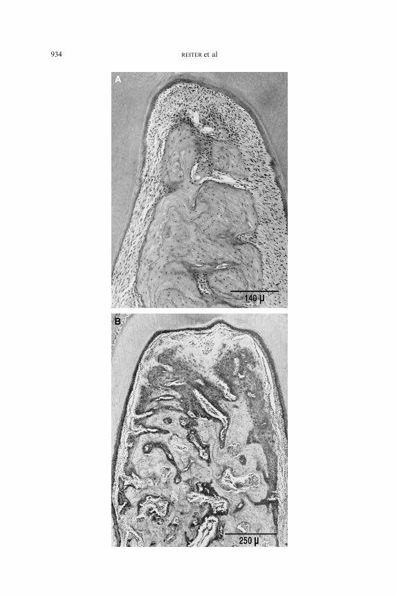
In the periodontium, periodontal ligament hyperemia, edema, anddegeneration with fiber disorientation; mineralization of Sharpey’s fibers;hypercementosis with abnormal thickening of cervical cementum anda bulbous appearance of root apices; hyperosteoidosis along periostealand endosteal surfaces; reduced endosteal lumina; bone marrow fibrosis;bulbous enlargement of alveolar plates with coronal displacement oftranseptal fibers at the alveolar margin; narrowing of the periodontal space;dentoalveolar ankylosis; granulation tissue formation; irregular resorptivelacunae in cementum and dentin; and a mixed pattern of osteoporosis andosteosclerosis (Fig. 10–16) have been reported [91,95–102,104,106–125].
Fig. 16. Histopathologic pictures of rats given dihydrotachysterol showing bulbous enlarge-
ment of root apices (A) and resorption of cementum, dentin, and alveolar bone (B). (From
Moskow BS, Baden E. The effect of chronic dihydrotachysterol overdosage on the tissues of the
periodontium. Periodontics 1964;2:279–80; with permission.)
Fig. 15. Histopathologic pictures of furcation area of molar teeth in a control rat (A) and a rat
given dihydrotachysterol showing hypercementosis, hyperosteoidosis, degeneration of the
periodontal ligament, and narrowing of the periodontal space (B). (From Glickman I, Selye H,
Smulow JB. Reduction by calciphylaxis of the effects of chronic dihydrotachysterol overdosage
upon the periodontium. J Dent Res 1965;44:743–4; with permission.)
:
936 REITER et al
Extrapolating these findings to the domestic cat should be done withcaution, however, because the results of these experimental studies are notuniform. Furthermore, the ages, sizes, and species of animals; the characterof their diets; the varying forms, quantities, and routes of administrationof vitamin D and vitamin D metabolites; and the duration of the experi-ments differed. Nevertheless, there are distinct similarities between thechanges in dental and periodontal tissues induced by administration ofexcess vitamin D and vitamin D metabolites in experimental animals andradiographic and microscopic features that can be found in teeth from catswith FORL (eg, thin predentin layer, irregular dentin formation,periodontal ligament degeneration and fiber disorientation, hypercemen-tosis, hyperosteoidosis, thickening of crestal alveolar bone, narrowing ofthe periodontal space, dentoalveolar ankylosis, root resorption, mixedpattern of osteoporosis and osteosclerosis). Vitamin D–induced thickeningof cervical cementum and abnormal apposition of osteoid at the alveolarcrest and other periosteal surfaces causing bulbous enlargement of alveolarplates and coronal displacement of transeptal fibers could result in re-duction of the biologic width (the dimension of space occupied byjunctional epithelium and gingival connective tissue) and loss of gingivalattachment. Supereruption of teeth in cats with increased vitamin Dactivity may actually be an attempt to maintain or re-establish normalbiologic width.
Certain findings are worthy of additional discussion, including (a)differences in effects of vitamin D and vitamin D metabolites betweencontinuously growing and continuously erupting teeth and between youngand adult animals and (b) apparent alleviation of the detrimental effects ofvitamin D and vitamin D metabolites by concurrent administration of otheragents. In rats, pulpal mineralization and pulp stones occurred morecommonly in incisors than in molars and more commonly in younger thanin older animals [107], which may be an indication that vitamin D activity ismore influential on ‘‘young’’ or continuously renewing tissue. Althoughpulpal mineralization has not been reported in permanent teeth of cats withFORL, pulp stones have been demonstrated in experimental vitamin Dstudies in puppies [108,110,114]. Young animals (dogs and rats) showedinitial alveolar bone resorption and osteoporosis followed by hyper-osteoidosis and osteosclerosis with a narrowing of endosteal spaces,whereas alveolar bone resorption and osteoporosis were predominant inadult or older animals [107,108]. Studies investigating the appearance ofalveolar bone in younger and older FORL-affected cats have not yet beenconducted. Effects of vitamin D or vitamin D metabolites were less severe orcould be reduced in animals given various amounts of vitamin A [108,114],sexual hormones [95,117], ferric dextran [91,98,123], or sodium fluoride[118], in addition to excess administration of vitamin D or vitamin Dmetabolites. This may be of interest when considering future research thatfocuses on prevention of FORL.
937TOOTH RESORPTION IN DOMESTIC CATS
Summary
The following conclusions can be drawn:
1. Cats depend on dietary vitamin D intake because they are not able toproduce vitamin D in the skin.
2. Some commercial cat foods contain vitamin D concentrations in excessof current maximal allowances.
3. Cats with FORL have significantly higher serum concentrations of25OHD compared with cats without FORL, indicating that cats withFORL must have ingested higher concentrations of dietary vitamin D.
4. Cats with FORL have significantly decreased urine specific gravitycompared with cats without FORL.
5. Experimental studies on laboratory animals have shown that excessadministration of vitamin D or vitamin D metabolites causes changes todental and periodontal tissues that resemble many characteristics ofteeth from cats with FORL.
6. Clinical and experimental studies have shown that excess administrationof vitamin D or vitamin D metabolites can lead to soft tissue mineral-ization and various degrees of renal disease.
Dietary intake of excess vitamin D over several years may lead toperiodontal ligament degeneration, narrowing of the periodontal space,dentoalveolar ankylosis, and root replacement resorption. If such a processoccurs close to the gingival margin, an inflammatory component may jointhe disease. Further histologic and experimental studies are required todetermine the role of daily masticatory stresses on the development ofFORL and to verify relations between FORL, vitamin D, and renalinsufficiency.
References
[1] Hopewell-Smith A. The process of osteolysis and odontolysis, or so-called ‘‘absorption’’ of
calcified tissues: a new and original investigation. Dental Cosmos 1930;72:1036–48.
[2] Reiter AM. Feline ‘‘odontolysis’’ in the 1920s: the forgotten histopathological study of
feline odontoclastic resorptive lesions (FORL). J Vet Dent 1998;15:35–41.
[3] Kerebel B, Daculsi G. Histologie et histopathologie dentaires du chat. Sci Rech Odonto-
stomatol 1971;7(5):29–32.
[4] Schneck GW, Osborn JW. Neck lesions in the teeth of cats. Vet Rec 1976;99:100.
[5] Reiter AM. The role of calciotropic factors in the etiology of feline odontoclastic resorptive
lesions (FORL) [thesis]. Vienna: University of Veterinary Medicine Vienna; 2004.
[6] Reiter AM. Biology of alveolar bone and tooth resorbing cells. In: Proceedings of the 12th
Annual Veterinary Dental Forum. New Orleans, 1998. p. 225–7.
[7] Sahara N, Toyoki A, Ashizawa Y, et al. Cytodifferentiation of the odontoclast prior to the
shedding of human deciduous teeth: an ultrastructural and cytochemical study. Anat Rec
1996;244:33–49.
[8] Harvey CE, Orsini P, McLahan C, et al. Mapping the radiographic central point of feline
dental resorptive lesions. J Vet Dent 2004;21:15–21.
938 REITER et al
[9] Eriksen T, Koch R, Nautrup CP. Microradiography of the feline marginal periodontium
with a microfocal high-resolution x-ray system. Scand J Dent Res 1994;102:284–9.
[10] Reiter AM, Mendoza K. Feline odontoclastic resorptive lesions. An unsolved enigma in
veterinary dentistry. Vet Clin N Am Small Anim Pract 2002;32:791–837.
[11] Keinath G. Tierzahnheilkunde bei Katzen unter besonderer Beruecksichtigung der
Aetiologie und Therapie von ‘‘Neck-Lesions’’ [thesis]. Munich: Munich University,
Faculty of Human Medicine; 1997.
[12] Ohba S, Kiba H, Kuwabara M, et al. A histopathological study of neck lesions in feline
teeth. J Am Anim Hosp Assoc 1993;29:216–20.
[13] Okuda A, Harvey CE. Etiopathogenesis of feline dental resorptive lesions. Vet Clin N Am
Small Anim Pract 1992;22:1385–404.
[14] Reichart PA, Durr U-M, Triadan H, et al. Periodontal disease in the domestic cat. A
histopathologic study. J Periodont Res 1984;19:67–75.
[15] Roes F. Pathogenese, Diagnostik und Therapie bei ‘‘neck lesions’’ der Katze unter
Verwendung von Glas-Ionomer-Zementen [thesis]. Berlin: Free University of Berlin,
Faculty of Veterinary Medicine; 1996.
[16] Steinberg S. Histologische Untersuchungen zu Fruehveraenderungen der Felinen Odonto-
klastischen Resorptiven Laesionen (FORL) an klinisch gesunden Zaehnen [thesis]. Berline:
Free University of Berlin, Faculty of Veterinary Medicine; 2002.
[17] Trope M, Chivian N. Root resorption. In: Cohen S, Burns RC, editors. Pathways of the
pulp. 6th edition. St. Louis: Mosby-Year Book; 1994. p. 486–512.
[18] DuPont GA, DeBowes LJ. Comparison of periodontitis and root replacement in cat teeth
with resorptive lesions. J Vet Dent 2002;19:71–5.
[19] Berger M, Schawalder P, Stich H, et al. ‘‘Neck Lesion’’ bei Grosskatzen; Untersuchungen
beim Leoparden (Panthera pardus). Kleintierpraxis 1995;40:537–49.
[20] Schlup D, Stich H. Epidemiologische und morphologische Untersuchungen am
Katzengebiß. II. Mitteilung: Morphologische Untersuchungen der ‘‘neck lesions’’.
Kleintierpraxis 1982;27:179–88.
[21] George DI,Miller RL. Idiopathic resorption of teeth. A report of three cases. Am JOrthod
1986;89:13–20.
[22] Henry JL, Weinmann JP. The pattern of resorption and repair of human cementum. J Am
Dent Assoc 1951;42:270–90.
[23] Shigeyama Y, Grove TK, Strayhorn C, et al. Expression of adhesion molecules during
tooth resorption in feline teeth: a model system for aggressive osteoclastic activity. J Dent
Res 1996;75:1650–7.
[24] Forsberg A, Lagergren C, Lonnerblad T. The periodontal tissue of mandibular premolars
and molars in some mammals. A comparative anatomical study. Svensk Tandlakare-
Tidskrift 1969;62(Suppl 1):1–54.
[25] Gorrel C, Larsson A. Feline odontoclastic resorptive lesions: unveiling the early lesion.
J Small Anim Pract 2002;43:482–8.
[26] Reiter AM. Etiopathology of feline odontoclastic resorptive lesions (FORL). In:
Proceedings of the 12th Annual Veterinary Dental Forum. New Orleans, 1998. p.
228–34.
[27] Blackwood HJJ. Resorption of enamel and dentine in the unerupted tooth. Oral Surg Oral
Med Oral Pathol Oral Radiol Endod 1958;11:79–85.
[28] Boyle PE. Cementumdchanges with age, function, and infection. In: Kronfeld’s histo-
pathology of the teeth and their surrounding structures. Philadelphia: Lea & Febiger; 1955.
p. 258–72.
[29] Stafne EC, Austin LT. Resorption of embedded teeth. J Am Dent Assoc 1945;32:1003–9.
[30] Zemsky JL. Hypercementosis in relation to unerupted and malposed teeth. A preliminary
report. J Dent Res 1931;11:159–74.
[31] Kupfer IJ. Correlation of hypercementosis with toxic goiter. A preliminary report. J Dent
Res 1951;30:734–6.
939TOOTH RESORPTION IN DOMESTIC CATS
[32] Farmer ED, Lawton FE. The effects of endocrine disorders on the jaws and teeth. In:
Stones’ oral and dental diseases. Aetiology, histopathology, clinical features and treatment.
5th edition. Baltimore: Williams & Wilkins; 1966. p. 30–48.
[33] Gardner BS, Goldstein H. The significance of hypercementosis. Dent Cosmos 1931;73:
1065–9.
[34] Schour I, Massler M. Endocrines and dentistry. J Am Dent Assoc 1943;30:595–603.
[35] Rushton MA. The dental tissues in osteitis deformans. Guys Hosp Rep 1938;88:163–71.
[36] Smith NHH. Monostotic Paget’s disease of the mandible presenting with progressive
resorption of the teeth. Oral Surg Oral Med Oral Pathol Oral Radiol Endod 1978;46:
246–53.
[37] Stafne EC. Paget’s disease involving the maxilla and the mandible: report of a case. J Oral
Surg 1946;4:114–5.
[38] Farmer ED, Lawton FE. Cementum: abnormalities associated with its formation. In:
Stones’ oral and dental diseases. Aetiology, histopathology, clinical features and treatment.
5th edition. Baltimore: Williams & Wilkins; 1966. p. 447–67.
[39] King JD. Dietary deficiency, nerve lesions and the dental tissues. J Physiol 1937;88:62–77.
[40] Kellner E. Das Verhaeltnis der Zement- und Periodontalbreiten zur funktionellen
Beanspruchung der Zaehne. Z Stomatol 1931;29:44–62.
[41] Kronfeld R. Die Zementhyperplasien an nicht funktionierenden Zaehnen. Z Stomatol
1927;25:1218–27.
[42] Itoiz MA, Carranza FA. The gingiva. In: NewmanMG, Takei HH, Carranza FA, editors.
Carranza’s clinical periodontology. 9th edition. Philadelphia: WB Saunders; 2002.
p. 16–35.
[43] Black GV. The periosteum and peridental membranes. Dent Rev 1887;1:233–43, 353–65.
[44] Lyon KF. Subgingival odontoclastic resorptive lesions. Classification, treatment and
results in 58 cats. Vet Clin N Am Small Anim Pract 1992;22:1417–32.
[45] Carranza FA, Camargo PM. Periodontal response to external forces. In: Newman MG,
Takei HH, Carranza FA, editors. Carranza’s clinical periodontology. 9th edition.
Philadelphia: WB Saunders; 2002. p. 371–83.
[46] LukmanK, Pavlica Z, Juntes P. Prevalence patterns and histological survey of feline dental
resorptive lesions. In: Proceedings of the 8th Annual Scientific Meeting of the British
Veterinary Dental Association. Birmingham, 1996.
[47] Boling LR. Blood vessels of the dental pulp. Anat Rec 1942;82:25–37.
[48] Winter GB, Kramer IRH. Changes in periodontal membrane and bone following
experimental pulpal injury in deciduous molar teeth in kittens. Arch Oral Biol 1965;10:
279–89.
[49] Negro VB, Hernandez SZ, Maresca BM, et al. Furcation canals of the maxillary fourth
premolar and the mandibular first molar teeth in cats. J Vet Dent 2004;21:10–4.
[50] Okuda A, Habata I. Lacunae or tubular structures in dentin of feline teeth. J Anim Res
Found 1993;2(1):7–14.
[51] Okuda A, Harvey CE. Histopathological findings of features of odontoclastic resorptive
lesions in cat teeth with periodontitis. In: Proceedings of the 5th Annual Veterinary Dental
Forum. New Orleans, 1991. p. 141–4.
[52] Berger M, Stich H, Schaffner T, et al. Testimony from a silent late medieval witnessdwhat
can it tell us about FORL? In: Proceedings of the 13th European Congress of Veterinary
Dentistry. Krakow, 2004. p. 17–8 (plus addendum).
[53] Dobbertin F. Zur Pathologie der Zahn- und Zahnbetterkrankungen bei Felis silvestris
forma catus [thesis]. Hamburg: Hamburg University, Faculty of Human Medicine; 1993.
[54] Harvey CE, AlstonWE. Dental diseases in cat skulls acquired before 1960. In: Proceedings
of the 4th Annual Veterinary Dental Forum. Las Vegas, 1992. p. 41–3.
[55] DeLaurier A, Jackson B, Ingham K, et al. Biochemical markers of bone turnover in the
domestic cat: relationships with age and feline osteoclastic resorptive lesions. J Nutr 2002;
132(Suppl):1742S–4S.
940 REITER et al
[56] Clarke DE, Cameron A. Feline dental resorptive lesions in domestic and feral cats and the
possible link with diet. In: Proceedings of the 5th World Veterinary Dental Congress.
Birmingham, 1997. p. 33–4.
[57] How KL, Hazewinkel AW, Mol JA. Dietary vitamin D dependance of cat and dog due to
inadequate cutaneous synthesis of vitamin D. Gen Comp Endocrinol 1994;96:12–8.
[58] National Research Council. Nutrient requirements of cats. Washington, DC: National
Academy Press; 1986. p. 4, 15, 16, 23, 56.
[59] Morris JG, EarleKE.Growing kittens require less dietary calcium than current allowances.
J Nutr 1999;129:1698–704.
[60] Morris JG, Earle KE, Anderson PA. Plasma 25-hydroxyvitamin D in growing kittens is
related to dietary intake of cholecalciferol. J Nutr 1999;129:909–12.
[61] Morris JG. Vitamin D synthesis by kittens. Vet Clin Nutr 1996;3(3):88–92.
[62] Haruna A, Kawai K, Takaba T, et al. Dietary calcinosis in the cat. J Anim Clin Res Found
1992;1(1):9–16.
[63] Morita T, Awakura T, Shimada A, et al. Vitamin D toxicosis in cats: natural outbreak and
experimental study. J Vet Med Sci 1995;57:831–7.
[64] Sato R, Yamagishi H, Naito Y, et al. Feline vitamin D toxicosis caused by commercially
available cat food. J Jpn Vet Med Assoc 1993;46:577–81.
[65] Sih TR, Morris JG, Hickman MA. Chronic ingestion of high concentrations of
cholecalciferol in cats. Am J Vet Res 2001;62:1500–6.
[66] Holick MF. Vitamin D: photobiology, metabolism, mechanism of action, and clinical
applications. In: Favus MJ, editor. Primer on the metabolic bone diseases and disorders of
mineral metabolism. 4th edition. Philadelphia: Lippincott Williams & Wilkins; 1999.
p. 92–8.
[67] Itonaga I, Sabokbar A, Murray DW, et al. Effect of osteoprotegerin and osteoprotegerin
ligand on osteoclast formation by arthroplasty membrane derived macrophages. Ann
Rheum Dis 2000;59:26–31.
[68] Burgess TL, Qian Y-X, Kaufman S, et al. The ligand for osteoprotegerin (OPGL) directly
activates mature osteoclasts. J Cell Biol 1999;145:527–38.
[69] BraemM,Lambrechts P, VanherleG. Stress-induced cervical lesions. J ProsthetDent 1992;
67:718–22.
[70] LeeWC, EakleWS. Possible role of tensile stress in the etiology of cervical erosive lesions of
teeth. J Prosthet Dent 1984;52:374–80.
[71] LeeWC, EakleWS. Stress-induced cervical lesions: review of advances in the past 10 years.
J Prosthet Dent 1996;75:487–94.
[72] Bevenius J, L’Estrange P, Karlsson S, et al. Idiopathic cervical lesions: in vivo investigation
by oral microendoscopy and scanning electron microscopy. A pilot study. J Oral Rehabil
1993;20:1–9.
[73] Goel VK, Khera SC, Ralston JL, et al. Stresses at the dentinoenamel junction of human
teeth. J Prosthet Dent 1991;66:451–9.
[74] Burke FJT, JohnstonN,Wiggs RB, et al. An alternative hypothesis from veterinary science
for the pathogenesis of noncarious cervical lesions. Quintessence Int 2000;31:475–82.
[75] Johnston N. Acquired feline oral cavity disease. Part 2: feline odontoclastic resorptive
lesions. In Pract 2000;22:188–97.
[76] Orban B. Resorption and repair on the surface of the root. J Am Dent Assoc 1928;15:
1768–77.
[77] Chipps HD. Two cases of root resorption. Dental Cosmos 1928;70:461–2.
[78] Coolidge ED. The reaction of cementum in the presence of injury and infection. J AmDent
Assoc 1931;18:499–525.
[79] Wood P, Rees JS. An unusual case of furcation external resorption. Int Endod J 2000;33:
530–3.
[80] Yusof WZ, Ghazali MN. Multiple external root resorption. J Am Dent Assoc 1989;118:
453–5.
941TOOTH RESORPTION IN DOMESTIC CATS
[81] Rawlinson A. Treatment of root and alveolar bone resorption associated with bruxism.
Br Dent J 1991;170:445–7.
[82] Harris EF, Robinson QC, Woods MA. An analysis of causes of apical root resorption in
patients not treated orthodontically. Quintessence Int 1993;24:417–28.
[83] Itoiz ME, Carranza FA, Cabrini RL. Histologic and histometric study of experimental
occlusal trauma in rats. J Periodontol 1963;34:305–14.
[84] KameyamaY. Histopathologic and autoradiographic studies of the changes of the rat peri-
odontium in experimental traumaticocclusion.BullTokyoMedDentUniv 1968;15:339–57.
[85] Orban B. Tissue changes in traumatic occlusion. J Am Dent Assoc 1928;15:2090–106.
[86] Ramfjord SP, Kohler CA. Periodontal reaction to functional occlusal stress. J Periodontol
1959;30:95–112.
[87] Mjor IA, Stenvik A.Microradiography and histology of decalcified human teeth following
experimental intrusion; with emphasis on resorption. Arch Oral Biol 1969;14:1355–64.
[88] Bhaskar SN, Orban B. Experimental occlusal trauma. J Periodontol 1955;26:270–84.
[89] LefkowitzW,Waugh LM. Experimental depression of teeth. Am J OrthodOral Surg 1945;
31:21–36.
[90] Boyle PE. Tooth resorption. In: Kronfeld’s histopathology of the teeth and their
surrounding structures. Philadelphia: Lea & Febiger; 1955. p. 273–96.
[91] Glickman I, Selye H, Smulow JB. Reduction by calciphylaxis of the effects of chronic
dihydrotachysterol overdose upon the periodontium. J Dent Res 1965;44:734–49.
[92] Sindelka Z. Die Kalziphylaxie im Zahnmark. Stoma (Heidelb) 1968;21:101–9.
[93] SelyeH,Grasso S, PadmanabhanN. Topical injury as ameans of producing calcification at
predetermined points with dihydrotachysterol (DHT). Proc Zool Soc 1960;13(1):1–3.
[94] Selye H, Jean P, Veilleux R. Role of local trauma in the production of cutaneous calcinosis
by dihydrotachysterol. Proc Soc Exp Biol Med 1960;104:409–11.
[95] Ratcliff PA, Krajewski J. The influence of methyl testosterone on dihydrotachysterol
intoxication as it affects the periodontium. J Oral Ther Pharmacol 1966;2:353–61.
[96] Stahl SS, Cohen C, Epstein B. The responses of injured and noninjured rat periodontal
tissues to a single administration of dihydrotachysterol. Oral Surg Oral Med Oral Pathol
Oral Radiol Endod 1967;23:531–7.
[97] Shoshan S, Pisanti S, Sciaky I. The effect of hypervitaminosis D on the periodontal
membrane collagen in lathyritic rats. J Periodont Res 1967;2:121–6.
[98] Moskow BS, Baden E, Zengo A. The effects of dihydrotachysterol and ferric dextran upon
the periodontium in the rat. Arch Oral Biol 1966;11:1017–26.
[99] Kojima M. Experimental study on the physio-pathological changes of periodontal tissues.
Bull Stomatol Kyoto Univ 1969;9:101–40.
[100] Takano K, Watanabe Y, Fuzihashi H, et al. The histological changes of alveolar bone,
cementum and periodontal fiber due to experimental progeria-like syndrome of the rat.
J Jpn Assoc Periodontol 1981;23:357–66.
[101] Cai JJ. Effect of vitamin D overdosage on the tooth and bone development of rabbits. Chin
J Stomatol 1992;27:296–9, 319.
[102] DaemmrichK. Experimentelle D3-Hypervitaminose bei Ferkeln. Zentrabl VeterinarmedA
1963;10:322–49.
[103] Irving TJ, Weinmann JP, Schour I, et al. Experimental studies in calcification. VIII. The
effect of large doses of calciferol on the dentin of the incisor in normal and nephrectomized
rats. J Dent Res 1949;28:362–8.
[104] Pitaru S, Blauschild N, Noff D, et al. The effect of toxic doses of 1,25-dihydroxychole-
calciferol on dental tissues in the rat. Arch Oral Biol 1982;27:915–23.
[105] Tempestini O. Nuove ricerche sugli effetti sperimentali della somministrazione di una alta
dose di vitamina D2 nei tessuti dentali e paradentali del ratto. Riv Ital Stomatol 1952;7:
373–410.
[106] Weinmann J. Experimentelle Untersuchung ueber dieWirkung grosser Dosen Vigantol auf
Knochen und Zaehne. Klin Wochenschr 1929;8:841–2.
942 REITER et al
[107] Weinmann J. Untersuchungen an Knochen und Zaehnen der Ratte bei Verfuetterung
grosser Dosen D-Vitamin. Dtsch Mschr Zahnheilk 1933;51:577–603, 625–54.
[108] Becks H. Dangerous effects of vitamin D overdosage on dental and paradental structures.
J Am Dent Assoc 1942;29:1947–68.
[109] Becks H, Collins DA, Axelrod HE. The effects of a single massive dose of vitamin D2 (D-
stoss therapy) on oral and other tissues of young dogs. Am J Orthod Oral Surg 1946;32:
452–62.
[110] Becks H, Collins DA, Freytag RM. Changes in oral structures of the dog persisting after
chronic overdoses of vitamin D. Am J Orthod Oral Surg 1946;32:463–71.
[111] Bernick S, Ershoff BH, Lal JB. Effects of hypervitaminosis D on bones and teeth of rats. Int
J Vitam Nutr Res 1971;41:480–9.
[112] Fahmy H, Rogers WE, Mitchell DF, et al. Effects of hypervitaminosis D on the
periodontium of the hamster. J Dent Res 1961;40:870–7.
[113] Harris LJ, Innes JRM. Mode of action of vitamin D. Biochem J 1931;25:367–90.
[114] Hendricks JB, Morgan AF, Freytag RM. Chronic moderate hypervitaminosis D in young
dogs. Am J Physiol 1947;149:319–32.
[115] Hirukawa T. Effect of dihydrotachysterol on the periodontal tissues in rats. Aichi-Gakuin
J Dent Sci 1990;28:367–87.
[116] Kondo M. The changes of parodontal tissues of rat, caused by the administration of
dihydrotachysterol. Shigaku 1971;59:219–45.
[117] Mabuchi H. Experimental study on the effect of sexual hormones and calcification
enhancing substances on growth of the alveolar bone. Bull Stomatol Kyoto Univ 1970;10:
31–60.
[118] Miwa Y, Watanabe Y. The effect of fluoride on the periodontal tissues of dihydrotachys-
terol administered rats. Bull Josai Dent Univ 1985;14:343–54.
[119] Morgan AF, Axelrod HE, GroodyM. The effect of a single massive dose of vitamin D2 on
young dogs. Am J Physiol 1947;149:333–9.
[120] MoskowBS, BadenE. The effect of chronic dihydrotachysterol overdosage on the tissues of
the periodontium. Periodontics 1964;2:277–83.
[121] Nomura H. Histopathological study of experimental hypervitaminosis D2 on the
periodontium of the rat. Shikwa Gakuho 1969;69:539–93.
[122] Ratcliff PA, Itokazu H. The effect of dihydrotachysterol on the teeth and periodontium.
J Periodontol 1964;35:320–5.
[123] Ratcliff PA, ItokazuH. The effect of dihydrotachysterol and ferric dextran on the teeth and
periodontium of the rat. J Oral Ther Pharmacol 1964;1:7–22.
[124] Takano K, Watanabe Y. The histological study on osteoid- and cementoid-like tissues of
rats treated with dihydrotachysterol. Bull Josai Dent Univ 1987;16:307–22.
[125] Terai Y. Studies on experimental stimulation of ossification in the paradental tissues of rats
and blood level of Ca, P and alkaline phosphatase in them. Bull StomatolKyotoUniv 1968;
8(4):191–244.