update series 33 - nutrition foundation of...
TRANSCRIPT
JANUARY 2007
Nutrition Foundation of India Building, C-13 Qutab Institutional Area, New Delhi-110 016Tel: 91-11-26857814, 26962615 Fax: 91-11-26857814
E-mail: [email protected]; [email protected] Website: www.crnssindia.org
UPDATE SERIES 33
The Possible Beneficial Role of Probiotics in Sexually Transmitted Infections
Recent Advances in Nutritional Management in Children with Chronic Kidney Diseases

Edited by: Sarath Gopalan, Executive Director, CRNSS and Sakshi Bhushan (Assistant Editor), CRNSSDesigned and produced by Media Workshop India Pvt Ltd
Wish you all a very happy 2007!We launch this year with the publication of the 33rd issue of the CRNSS
Update Series “Nutrition in Disease Management.”The first article is a review from the well-known National Institute of Nutri-
tion, Hyderabad informing the reader about yet another of the several possible beneficial effects attributed to probiotics-their utility in both prevention and treatment of sexually transmitted infections in females. It discusses in detail the beneficial effects of specific strains of probiotics in this clinical setting.
The second article is a review providing the reader with the latest informa-tion regarding nutritional management in chronic kidney disease in children. The corresponding author is a Consultant Pediatric Nephrologist at the pres-tigious Fortis Hospital, Vasant Kunj, New Delhi.
We would appreciate your inputs and suggestions from the standpoint of increasing variety of the topics covered in the Update Series issues so as to arouse greater interest in the reader.
Dr Sarath Gopalan Executive Director, CRNSS
and Editor
To Our Readers

1
The Possible Beneficial Role of Probiotics in Sexually Transmitted
Infections
Genital tract infections including sexually transmitted infections (STI) are a major public health problem in women in the reproductive age group in India. According to the National AIDS Control Organization (NACO) the annual incidence rate of STI is around 5 % among the adult population, with an estimated 40 million new cases occurring annually in the country1. In developing countries, STI and their complica-tions rank in the top five disease categories for which adults seek health care and ranks second amongst maternal factors as causes of morbidity and mortality2.
The presence of an untreated STI enhances both acquisition and transmission of HIV2. Thus, STI treatment is an important HIV prevention strategy in a general population.The association between abnormal vaginal microflora and increased risk for sexually transmitted infections and a higher rate of preterm labor indicate the need to better understand and manage urogenital health3,4,5. The concept of probiotics arose from the realization that humans are inhabited with microbes from birth and that these organisms play a vital role in preventing diseases. probiotic strains have already been shown to effectively prevent diarrhea. Here we discuss the use of probiotics in maintaining reproductive health in women and as a treatment option for recurrent bacterial vaginosis, and sexually transmitted infections.
VAGINAL MICROFLORA AND REPRODUCTIVE HEALTH IN WOMEN
The healthy vagina has a number of natural protective factors against STD/HIV in-fection and related diseases. Under the influence of lactic acid, the vagina maintains a low (acidic) pH of approximately 4.0. This acidic condition prevents pathogenic bacterial multiplication in the vagina and promotes the formation of squamous epithelial cells, which covers the fragile columnar cells of the cervical canal. Apart from hydrogen peroxide, other bactericidal products in the vagina include defensins, antibodies, nonspecific cytokines and inflammatory responses.
Hemalatha R, MDAssistant Director, HOD MicrobiologyNational Institute of Nutrition, Hyderabad

2
The microorganisms that colonize vaginal mucosa play a major role in the maintenance of reproductive health of women. Healthy vaginal vault is predomi-nantly colonized by lactobacilli species. Under normal conditions, L.crispatus and L.jensenii are the most common species of vaginal lactobacilli. The vaginal flora of healthy women is dominated by different species of lactobacilli, but they co exist with a multitude of other species, including potential pathogens6. A disturbance in this balance with reduction in lactobacilli flora could result in infections. Presence of lactobacilli in human vagina is influenced by oestrogen production, which is menstrual cycle and age dependent7. Thus, even in healthy women the vaginal micro flora constantly fluctuates at many time points during the menstrual cycle without any symptoms.
Vaginal colonization with these strains is associated with lower incidence of bacterial vaginosis (BV), and loss of this normal flora is associated with increase in genital and urinary tract infections5,8,9,10. Moreover, recent studies have demon-strated a significant association between low vaginal lactobacilli and HIV infection, particularly among women with bacterial vaginosis3.
In women with abnormal flora or bacterial vaginosis, the normal vaginal Lac-tobacillus flora is replaced by an abundance of cocco-bacilli, primarily consisting of the facultative anaerobes Gardnerella vaginalis, Mobilincus and Bacteroides. This condition is characterised by a fishy-smelling vaginal discharge and typical clue cells on microscopic examination of Gram-stained specimens and is typi-cally devoid of vaginal leukocytes and causes no inflammation8,11,12,13. In cases of aerobic vaginitis, the lactobacilli are replaced by bacteria such as Escherichia coli and Group B streptococci (GBS). This condition is accompanied by severe vaginal leukocytosis associated with foul smelling sticky discharge.
The diagnostic criteria established by Amsel is a very useful tool, which is based on elevated pH (>4.5), homogenous white discharge, a fishy odor after addition of 10% KOH (sniff terb) and presence of clue cells (G.vaginalis adhered to exfoliated epitherlial cells) 12.
BV may lead to many complications, such as increased risk of miscarriage, premature rupture of membranes (PROM), chorioamnionitis and premature deliver-ies14-18. Several different mechanisms have been described for these complications. It is hypothesized that BV microbes might ascend the upper genital tract and cause upper genital tract infections or cause infection of fetal membranes in pregnant women. On the other hand, BV may stimulate inflammatory response and produc-tion of prostaglandin and cytokines that result in abortion or premature delivery. In addition, elevated pH and presence of polyamines increase the infectious potential of pathogens and also reduce the ability of the host to mount an effective immune response, thus facilitating ascending infection19,20.
BV also increases the susceptibility of women to HIV infection3,21,22. Data have indicated that Micoplasma hominis in BV is a potential source for a soluble factor with HIV-inducing activity (HIF)23 that can enhance HIV transcription, replication

3
and increase in the viral load, potentially contributing to increased HIV transmis-sion. Many patients have altered cytokine response and extensively degraded IgA response 24,25. This altered immune response in the cervicovaginal secretion may further increase the risk of STIs and HIV infection in women with BV. Bacterial vaginosis (BV) is the most common lower reproductive tract infection in women of childbearing age. In a study conducted at NIN, 57% of the women attending a local government hospital were positive for BV25. BV organisms have been shown to be sexually transmissible11.
Adequate treatment methods (oral and intravaginal administration) are available for bacterial vaginosis and anaerobic vaginitis. Initially 80 percent of the patients may be cured, but up to 80% recurrence happens within one year26. For vaginitis with disrupted vaginal flora (Aerobic Vaginitis), there is no uniform, efficient treat-ment method available, and the disease is usually enduring for many months to years. Oral penicillins and macrolides cause short-term relief, only to recur after antibiotic treatment is discontinued.
Bacterial vaginosis and aerobic vaginitis involve a severe disruption of the Lactobacillus flora. Therefore, the re-introduction of vaginal Lactobacillus may be a reasonable alternative to prevent recurrence of these conditions27. Lactobacilli are believed to offer resistance against infection by pathogens. It has been proven that the loss of the Lactobacillus flora is linked to increased transmission risk of sexually transmitted diseases, especially HIV, and to pregnancy complications such as pre-term birth3,23.
Preliminary evidence suggests that a disrupted vaginal microflora can be restored to its physiological equilibrium by the use of probiotics. However, it is far from clear which type of probiotic product is to be used, and the ideal way of applying the products in order to prevent recurrences.
PROBIOTICS AND REPRODUCTIVE HEALTH IN WOMENThe term “probiotics” has been defined as microorganisms that confer health
benefits to the host. In 1915, Newman D for the first time used Lactobacilli for treat-ing cystitis28. Later in 1920 it was Schroder who showed the importance of right acid/alkaline balance and hydrogen peroxide production to keep the pathogenic bacteria away from causing disease. However, there are no reports until in 1970, when a correlation was observed between low vaginal lactobacilli and recurrent urinary tract infection29. In 1990’s many experiments were conducted to study the efficacy, viability and colonization of probiotics in human health.
Strains such as L. rhammosus GR-I, L. fermentum RC-14 and B-54 inhibit the adhesion and growth of urinary, vaginal, and intestinal pathogens and produce hydrogen peroxide, which is a potent anti-bacterial agent30. In addition, these commensals signal the host to defend itself against pathogens, for example, by priming macrophages, leukocytes, cytokines, by inducing mucus production, or by suppressing the expression of virulence factors. L. fermentum RC-14 has been

4
shown to prevent Staphylococcus aureus sepsis in animals by producing a collagen binding protein and biosurfactants. In a study conducted at NIN, the immunoglobulin levels in the cervicovaginal secretions were higher, among women with BV, which was associated with higher degradation of IgA and IgM, indicating impaired mu-cosal immunity and increased susceptibity to STIs25. Thus the association between BV and increased risk of sexually transmitted diseases could be due to host cell damage, immunosuppression etc, that increase viral and bacterial penetration of vaginal cells resulting in upper genital tract infections, fetal membrane infection leading to various complications.
Lactobacillus strains are able to interfere with genitourinary pathogens by differ-ent mechanisms, including competitive inhibition of pathogens from the cell surface, production of H2O2 and bacteriocins, competition for nutrients and stimulation of immune system30,31. The ability of lactobacilli to modulate the host’s defenses through cytokines, antibodies, mucus barriers, blockage of pathogen receptor and growth inhibition, act as barrier to successful colonization and proliferation of pathogenic bacteria.
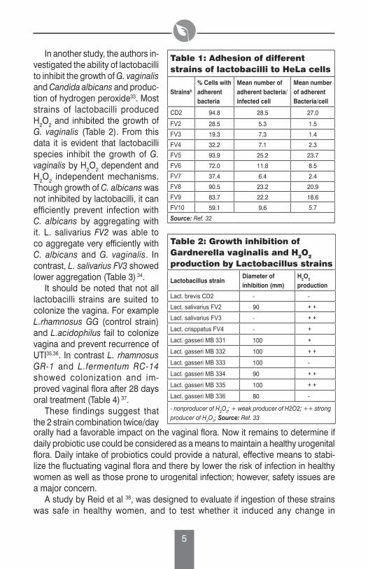
Adherence of lactobacilli to human epithelium is an important prerequisite for successful colonization. A study by Maggi et al, 32 indicate that most strains of lactobacilli adhere to Hela cells, exhibiting different degrees in their attachment to cell membranes. The adherence of the bacteria varied from an over all mean of 5 to 29 bacteria per cell, and 19-95% of cells with adherent bacteria. L. salivarius and L. crispatus were very poorly adhesive, where as L. brevis and 3 strains of L. gasseri were highly adhesive (19-27 bacteria/cell) (Table 1). However, lactobacilli strains from yogurt do not adhere well to vaginal epithelial cells. Most strains of lactobacilli preparations (L. brevis, L. salivarius, L.gasseri) have been reported to be viable for 12 to 18 months of storage at 4oC (Figure 1) 32.
Freeze-dried powder Fast-release tablet Slow-release tablet
1.2E+10
1.0E+10
8.0E+09
6.0E+09
4.0E+09
2.0E+09
0.0E+00Viab
ility
(cfu
/g o
f fre
eze-
drie
d po
wde
r)
Source: Ref. 32
Figure1: Strain viability of the L brevis + L salivarius + L gasseri freeze-dried mixture and fast or slow-release tablets during 1 year storage
5
In another study, the authors in-vestigated the ability of lactobacilli to inhibit the growth of G. vaginalis and Candida albicans and produc-tion of hydrogen peroxide33. Most strains of lactobacilli produced H2O2 and inhibited the growth of G. vaginalis (Table 2). From this data it is evident that lactobacilli species inhibit the growth of G. vaginalis by H2O2 dependent and H2O2 independent mechanisms. Though growth of C. albicans was not inhibited by lactobacilli, it can efficiently prevent infection with C. albicans by aggregating with it. L. salivarius FV2 was able to co aggregate very efficiently with C. albicans and G. vaginalis. In contrast, L. salivarius FV3 showed lower aggregation (Table 3) 34.
It should be noted that not all lactobacilli strains are suited to colonize the vagina. For example L.rhamnosus GG (control strain) and L.acidophilus fail to colonize vagina and prevent recurrence of UTI35,36. In contrast L. rhamnosus GR-1 and L.fermentum RC-14 showed colonization and im-proved vaginal flora after 28 days oral treatment (Table 4) 37.
These findings suggest that the 2 strain combination twice/day
Table 1: Adhesion of different strains of lactobacilli to HeLa cells
Strainsb
% Cells with adherent bacteria
Mean number of adherent bacteria/infected cell
Mean number of adherentBacteria/cell
CD2 94.8 28.5 27.0
FV2 28.5 5.3 1.5
FV3 19.3 7.3 1.4
FV4 32.2 7.1 2.3
FV5 93.9 25.2 23.7
FV6 72.0 11.8 8.5
FV7 37.4 6.4 2.4
FV8 90.5 23.2 20.9
FV9 83.7 22.2 18.6
FV10 59.1 9.6 5.7Source: Ref. 32
Table 2: Growth inhibition of Gardnerella vaginalis and H2O2 production by Lactobacillus strains
Lactobacillus strainDiameter of inhibition (mm)
H2O2 production
Lact. brevis CD2 - -Lact. salivarius FV2 90 + +
Lact. salivarius FV3 - + +
Lact. crisppatus FV4 - +
Lact. gasseri MB 331 100 +
Lact. gasseri MB 332 100 + +
Lact. gasseri MB 333 100 -
Lact. gasseri MB 334 90 + +
Lact. gasseri MB 335 100 + +
Lact. gasseri MB 336 80 -
- nonproducer of H2O2; + weak producer of H2O2; ++ strong producer of H2O2; Source: Ref. 33
orally had a favorable impact on the vaginal flora. Now it remains to determine if daily probiotic use could be considered as a means to maintain a healthy urogenital flora. Daily intake of probiotics could provide a natural, effective means to stabi-lize the fluctuating vaginal flora and there by lower the risk of infection in healthy women as well as those prone to urogenital infection; however, safety issues are a major concern.
A study by Reid et al 38, was designed to evaluate if ingestion of these strains was safe in healthy women, and to test whether it induced any change in

6
pathogen load in the vagina. The study demonstrated that probiotics L.rhamosus GR-1 and L.fermentum RC-14 could be taken orally on a daily basis for 2 months without any side effects. The therapy resulted in improvement in the vaginal flora in terms of increased lactobacilli and decreased yeast and coliforms counts. This could possibly explain the decreased incidence of recur-rent UTI reported by Raz et al39. The two lactobacilli strain (GR-1 & RC-14) can inhibit the growth of Candida in vitro, so the reduced vaginal yeast counts could be due in part to this inhibition33. Fewer yeast emerging from the rectum and ascending into the vagina or alternatively, the lactobacilli dis-placed the yeast in the vagina creating unsuitable environment for the yeast colonization.
Some strains like L. brevis, L. salivarius, L. plantarum pro-duce sphingomylinase an enzyme which converts sphingomylin to ceramide, a molecule essential for host cell membrane integrity. In ad-dition to sphingomylinase, L. brevis produces arginine diminase as well. Arginine diminase has been shown to modulate local and sys-temic immune responses40,41. Both
Table 3: Co-aggregation between lactobacilli and vaginal pathogens
Lactobacillus strain
Co-aggregation score forCandida albicans
Gardnerella vaginalis
Lact. brevis CD2 1 0Lact. salivarius FV2 3 3Lact. salivarius FV3 1 2Lact. crisppatus FV4 1 1Lact. gasseri MB 331 0 0Lact. gasseri MB 332 2 2Lact. gasseri MB 333 3 3Lact. gasseri MB 334 0 0Lact. gasseri MB 335 2 3Lact. gasseri MB 336 1 2Source: Ref. 34
Table 4: Healthy Vaginal flora after treatment with L. strains compared to control strain
group 1 group 2 group 3 group 4
Before treatment 40% 50% 27% 44%
At the end of treatment 60% 82% 45% 38%
Two weeks after treatment 56% 90% 30% 33%
% of women with healthy floragroup 1 = 8 x 10⁸ GR –I/RC-14, group 3 = 6 x 10⁹ of GR –I/RC-14,group 2 = 8 x 10⁸ Twice/day; GR–I/RC-14 group4 = GG 1010/day (control)Group 2 vs Control p = 0.017, Group 1&3 vs Control p = non significantSource: Ref. 37
sphingomylinase and arginine diminase have been shown to inhibit infection and proliferation of HIV42,43. These data suggest testing the hypothesis that promotion of vaginal colonization with lactobacilli might be a potential intervention to reduce the risk of acquiring HIV-1 infection.
Recent advances in our understanding of antimicrobial properties of lactobacilli and their safety in human use have raised further interest for the use of probiotics in maintaining vaginal health, which is important to reduce transmission of STIs including HIV infection.

7
There have been suggestions that suitable strains of lactobacilli preparations could be inserted once a day for 1 to 3 consecutive days in women highly prone to recurrence of urinary tract/lower genital infections as a means to restore their commensal flora. This could be followed by daily oral ingestion or monthly vaginal insertion. Though, there has been no requirement to demonstrate safety or potency before marketing probiotic preperation as they are regulated as dietary supple-mentation rather than pharmaceutical products, there are concerns with regard to probotic use in particular populations.
Rautio et al reported liver abcess and pneumonia 4 months after commencing daily vactobacillus GG supplementation in a 74 year old diabetic women44. The infective and probotic strains were identical by PEGF of chromosomal DNA restric-tion fragments. In another study Mackay et al reported L. rhamnosus endocarditis after dental extraction in a 67-year-old man with mitral regurgitation who was tak-ing probotics supplementation daily45. The strains were identical by biochemical analysis and mass spectrometry. Many cases of bacteremia in premature infants after consuming Lactobacillus G and fungemia in those taking saccharomyces boulandi have been reported46,47,48,49.
However, all cases of probiotic complications have occurred in patients with diabetes, immune compromised status or debilitation, and thus far no reports have described sepsis related to probiotic dose in otherwise healthy persons.
There is a body of evidence that supports the safety of probiotics, particularly lactobacillus strains. Lactobacillus strains have been reported to be safe in adults and children infected with HIV50,51 . Probiotics have been widely used in food pro-cessing for many years, & overall have an excellent safety records52,53.
REFERENCES1. National AIDS Control Organization: Country Scenario 1997-98. Government of India, New Delhi:
Ministry of Health and Family Welfare, 1998.2. World Health Organization: DALYs and Reproductive Heatlh: Report of an informal consultation,
27-28 April 1998. WHO/RHT/98.28, Geneva: WHO, 1999.3. Martin, HL, Richardson BA, Nyange PM, Lavreys H, Hillier SL, Chohan B, Mandaliya K, Ndinya-
Achola JO, Bwayo J, and Kreiss J: Vaginal lactobacilli, microbial flora, and risk of HIV type 1 and sexually transmitted disease acquisition. Journal of Infectious Disease, 180: 1863-1868,1999.
4. Hillier SL, Krohn MA, Klebanoff SK, Eschenbach DA: The relationship of hydrogen peroxide-produc-ing lactobacilli to bacterial vaginosis and genital microflora in pregnant women. Obstet and Gynecol, 79: 369-373, 1992.
4a. Hillier SL, Krohn MA, Rabe LK, Klebanoff SK, Eschenbach DA: The normal vaginal flora, H2O2-producing lactobacilli, and bacterial vaginosis in pregnant women. Clin Infect Dis, 16: S273-S281, 1993.
5. Klebanoff SK, Hillier SL, Eschenbach DA and Waltersdorph AM: Control of the microbial flora of the vagina by H2O2-generating lactobacilli. Journal of Infectious Diseases, 164: 94-100, 1991.
6. Domingue PAG, Sadhu K, Costerton JW, Baarlet K, Chow AW: The human vagina normal flora con-sidered as an in situ tissue-associated adherent biofilm, Genitorin. Medicine, 67: 226-231,1991.
7. Keane FEA, Ison CA and Taylor-Rabionson D.: A longitudinal study of vaginal flora over a menstrual cycle. International Journal of STD and AIDS, 8: 489-494, 1997.
8. Antonio MA, Hawes SE, Hillier SL: The identification of vaginal lactobacillus species and the de-

8
mographic and microbiologic characteristics of women colonized by these species. J Infect Dis, 180:1950-1956, 1999.
9. Redondo-Lopez V, Cook RL and Sobel JD: Emerging role of lactobacilli in the control and mainte-nance of the vaginal bacterial microflora. Reviews of Infectious Diseases, 12: 856-872, 1990.
10. Hawes SE, Hillier SL, Benedetti J, Stevens CE, Koutsky LA, Holner-Hanssen P and Holmes KK: Hydrogen peroxide producing lactobacilli and acquisition of vaginal infections. Journal of Infectious Diseases, 174: 1058-1063, 1996.
11. Sobel JD: Vaginitis. N. Eng J Med, 337: 1896-1903, 199712. Amsel R, Totten PA, Spiegel CA, Chen KS, Eschenbach DA, Holmes KK: Nonspecific vaginitis-
diagnostic criteria and microbial and epidemiologic associations. Am J Med, 74: 14-22,1983.13. Nugent RP, Krohn MA, Hillieer SL.: Reliability of diagnosing bacterial vaginosis is improved by a
standardized method of Gram stain interpretation. J Clin Microbiol, 30: 642-648, 1992.14. Ralph SG, Rutherford AJ, Wilson JD.: Influence of bacterial vaginosis on conception and miscarriage
in the first trimester; a cohort study. Br Med J, 319: 220-223, 1999.15. Hay PE, Lamont RF, Taylor-Robinson D, Morgan DJ, Ison C, Pearson J.: Abnormal bacterial colo-
nization of the genital tract and subsequent preterm delivery and late miscarriage. Br Med J, 308: 295-298, 1994.
16. Oleen-Burket MA, Hillier SL.: Pregnancy complications associated with bacterial vaginosis and their estimated costs. Infect Dis Obstet Gynecol, 33:149-167, 1995.
17. Hillier SL, Martius J, Krohn M, Kiviat N, Holmes KK, Eschenbach DA: A case-control study of chorio-amniotic infection and histologic chorioamniotis in prematurity. N Eng J Med, 319: 972-978,1988.
18. Eschenbach DA: Bacterial vaginosis: emphasis on upper genital tract complications. Obstet Gynecol Clin Borth Am, 16: 593-601, 1989.
19. Holmes KK, Chen KCS, Lipinski CM, Eschenbach DA.: Vaginal redox potential in bacterial vaginosis (nonspecific vaginitis). J Infect Dis, 152:379-382, 1985.
20. Briselden AM, Monela BJ, Stevens CE, Hillier SL,: Sialdases (neuraminidases) in bacterial vaginosis and bacterial vaginosis associated microflora. J Clin Microbiol, 30: 663-666, 1992.
21. Cohen RC, Duerr A, Pruithithada N, et al.: Bacterial vaginosis and HIV seroprevalence among female commercial sex workers in Chiang Mai, Thailand. AIDS, 9:1093-1097, 1995.
22. Sewankambo N, Gray RH, Wawer MJ et al.: HIV-1 infection associated with abnormal vaginal flora morphology and bacterial vaginosis. Lancet, 350: 546-550, 1997.
23. Al-Harthi I, Roebuck KA, Olinger GG, et al: Bacterial vaginosis-associated microflora isolated from the female genital tract activates HIV-1 expression. J Acquir Immune Defic Syndr, 21:194-202, 1999.
24. Cauci S, Monte R, Driussi S, Lauzafame P, Quadrifoglio F.: Impairment of the mucosal immune system:IgA and IgM. Cleavage detected invaginal washings of a subgroup of patients with bacterial vaginosis. J Infect Dis, 178: 1698-1706, 1998.
25. Yasgidgara MR, Sreeramulu D, Venu L, Hemalatha R, Prasanna TK.: Local immunity in Indian women with bacterial vaginosis. Journal of Reproductive Immunology, 70:1133-147, 2006.
26. Joeeroet MR, Schmid GP.: Bacterial Vaginosis: review of treatment options and potential clinical implications for therapy. Clin Infect Dis, 20 (suppl 1): 572-579, 1995.
27. Sanularo G, Tieluigi M, Coccia B, Mahtroiaco P, Simone C: Micro ecology, bacterial vaginosis and probiotics- perspectives for bacteriotherapy. Medical Hypothesis, 56 (4): 421-430, 2001.
28. Newman D.: The treatment of cystitis by intraverical injection of lactic bacillyus cultures. Lancet, 14:330-332, 1915.
29. Bruce AW, Chadwick P, Hassan A, Van Cott GF: Recurrent urethritis in women. Can Med Assoc J, 108:973-976, 1973.
30. Reid G, Cook RL and Bruce AW: Examination of strains of lactobacilli for properties that may influ-ence bacterial interference in the urinary tract. Journal of Urology, 138: 330-335, 1987.
31. Charteris WP, Kelly PM, Morelli L and Collins JK.: The role and therapeutic potential of Lactobacillus species in female urogenital tract infection. Microecology and Therapy, 2226: 59-96, 1997.
9
32. Lauretta Maggi, Paola Mastromarino, Stezania Macchia, Patricia Brigidi, Franco Pirovano, Diego Matteucci,, Ubaldo Conte.: Technological and biological evaluation of tablets containing different strains of lactobacilli for viginal administration. European Journal of Pharmaceutics and Biopharma-ceutics, 50: 389-395, 2000.
33. Mastromarino P, Brigidi P, Macchia S, Maggi L, Pivovano F, Tririchieri, U.Conte and Matteuzzi D : Characterization and selection of vaginal lactobacillus strains for the preparation of vaginal tablets. Journal of Applied Microbiol, 93: 884-893, 2002.
34. Reid G, Mc Groarty AA, Gil Domingue PA, Chow AW, Bruce AW, Eisen A and Costerton JW.: Coag-gregation of urogenital bacteria in vitro and in vivo. Current Microbiology, 20 : 47-52, 1990.
35. Baerhem A, Larsen E and Digranes A.: Vaginal applied flora of lactobacilli in the prophylaxis of recurrent lower urinary tract infections in women. Scand J Prim Health Care, 12: 239-243, 1994.
36. Kontiokari T, Sundgvist K, Nutrimen MP, T Koskela M, and Bhan M.: Radomised trial of cremberry-Imgonberry surce and lactobe, GG drink for the prevention of urinary tract infections in women. Br Med J, 522:1-4, 2001.
37. Reid G, Beurman Dee, Heinemann C, Bruce AW: Probiotic lactobacillus dose required to restore and maintain a normal vaginal flora. FEMS Immunology and Medical Microbiology, 32:37-41,2001.
38. Reid G, Duane C, Julie Erb, Barbara K, Dee Beuerman, Russ P, Andrew WB.: Oral use of lactobacillus rhamnosus GR-1 and L.Yermentum RC-14 significantly alters vaginal flora. Randomized, placebo-con-trolled trial in 64 healthy women. FEMS Immunology and Medical Microbiology, 35:131-134, 2003.
39. Raz R. and Stamm WF: A controlled tiral of intravaginal estriol in postmenopausal women with recurrent urinary tract infections. N Engl J Med, 329: 753- 756, 1993..
40. Gong F, Von Recklinghausen G et al.: Arginine deaminase inhibits cell proliferation by arresting cell cycle and inducing apoptosis. Biochem Biophys Res Common, 261:10-14, 1999.
41. Degnan BA, Palmer JM, Robson T et al.: Inhibition of human peripheral blood mononuclear cell proliferation by streptococcus pyogenes cell extracts is associated with arginine deaminase activity. Infect Immun, 66: 3050-3058, 1998.
42. Finnegan CM, Rawat SS, Puria Wang J, Ruscetti FW, Bluementhal R.: Ceramide, a target for anti-vertoviral therapy. Medical Sciences POVAS 101: 15452-15457, 2004.
43. Kubo et al.: Journal of General Virology, 87:1589-1593, 2006 44. Rautio M, Jousimies-Somer H, Kauna H, et al.: Liver abscess due to a Lactobacillus rhammosus
strain indistringuishable from L.rhammasus strain GG. Clin Infect Dis, 28:1159-60, 1999.45. Mackay AD, Taylor MB, Kibbler CC, Hamilton-Miller JM.: Lactobacillus rndocarditis caused by a
probiotic organism. Clin Microbiol Infect, 5:290-2, 1999.46. Kunz AN, Noel JM, Fairchok MP.: Two cases of lactobacillus bacteremia during probiotic treatment
of short gut syndrome. J Pediatr Gastroenterol Nutr , 38:457-8, 2004.47. De Groote MA, Frank DN, Dowell E, Glode MP, Pace NR.: Lactobacillus rhammosus GG bacteremia
associated with probiotic use in a child with short gut syndrome. Pediatr Infect Dis J, 24:278-80, 2005.48. Lhern T, Monet C, Nougiere B, et al.: Seven cases of fungemia with Saccharomyces boulardii in
critically ill patients. Intensive Care Med, 28:797-801, 2002.49. Bassetti S, Frei R, Zimmerli W,: Fungemia with Saccharomyces cerevisiae after treatment with Sac-
charomyces boulardii. Am J Med, 105: 71-2, 1998.50. Wolf BW, Wheeler KB, Ataya DG, Garleb KA.: Safety and tolerance of Lactobacillus reuteri supple-
mentation to a population infected with the human immunodeficiency virus. Food Chem Toxicol, 36:1085-94, 1998.
51. Salminen MK, Tynkkynen S, Rautelin H, et al. The efficacy and safety of probiotic Lactobacillus rhammosus GG on prolonged, noninfectious diarrhea in HIV patients on antiretroviral therapy: a randomized, placebo-controlled, crossover study. HIV Clin Trials, 5:183-91, 2004.
52. Ishibashi N, Yamazaki S.: Probiotics and safety. Am J Clin Nutr, 73 (suppl): 465S-70S, 2001.53. Borriello SP, Hammes WP, Holzapfel W, et al.: Safety of probiotics that contain lactobacilli or bifido-
bacteria. Clin Infect Dis, 36:775-80, 2003.

10
INTRODUCTION Dietary management is of paramount importance in children with chronic kidney
diseases (CKD). These patients have an altered metabolic milieu in view of deranged kidney function. The dietary requirement varies depending on the cause of chronic kidney disease as well as the fact that the child is requiring dialysis or not. These children require long periods of dietary restrictions. The challenge to the pediatri-cian and the dietician is to make the diet interesting and palatable, so as to ensure compliance. The goal is to optimize the growth and development of the children in this setting and not only to add years to life but also life to years.
NUTRITIONAL PROBLEMS IN CKDAs glomerular filteration rate (GFR) gradually declines, patients become progres-
sively uremic which causes anorexia, nausea, vomiting and this leads to decreased food intake. This progressively leads to muscle breakdown with loss of body mass, fat and weight. Growth retardation is quite common in paediatric patients with chronic kidney diseases. This is especially evident when it occurs during the acute growth phase. The main reasons of growth failure could be one or more of these- inadequate intake of nutrients due to uremia per se, hormonal imbalances, indiscreminatory dietary restrictions, altered taste sensitivity, psychological factors,
Dr. Sanjeev GulatiMD,DNB(Paediatrics)DM,DNB (Nephrology)FIPN,FIAP,FRCPC Senior Consultant NephrologyFortis HospitalsNew Delhi [email protected]: 98716-00885
Archana Sinha MSc
Recent Advances in Nutritional Management in Children with
Chronic Kidney Diseases

11
dialysate losses of protein and amino acids, infections and inflammatory condi-tions, continuation of dietary restrictions of pre dialysis period, growth inhibitors and renal oesteodystrphy.
The main objectives of dietary management of paediatric patients with CKD are: To optimize nutritional status. To maintain fluid and electrolyte balance. To minimize uremic symptoms. To promote growth and development. To assist in the conservation of Residual Renal Function (RRF) - this may slow
the progression to End Stage Kidney Disease (ESKD)
CHILDREN WITH CHRONIC KIDNEY DISEASE (CKD) ON CONSERVATIVE TREATMENT
These children have a GFR >10-15 ml/min/1.73m2 and are not on any dialysis support. The urine output would vary from patients to patients depending upon the underlying cause of CKD. Children with chronic interstitial nephritis (dysplastic kidney, reflex nephropathy, hereditary nephronopthesis) usually have significantly preserved urine output. In contrast patients with chronic glomerulonephritis (Fo-cal Segmental Glomerulosclerosis, Mesangiocapillary glomerulonephritis) are oliguric.
Poor energy intakes are common in CKD due to anorexia, altered taste acuity, hospitalization, dietary restriction and psychological factors1. Growth failure is an important issue in long-term management of paediatric patients with CKD. Children have relatively higher energy and protein requirement per kg of their weight as compared to adults because they have proportionally greater lean body mass2.
Energy: Energy requirement should meet at least the recommended dietary allowance (RDA) for normal children of same height for age. If PEM is present it needs to be increased further to improve weight gain and linear growth. Calorie intake in the diet should be enough to enhance the efficiency of protein (protein sparing effect) and prevent the patient from lapsing into a catabolic state. When use of chronological age does not account for the growth, height for age should be the basis for energy estimation3. Supplementation can be used as per requirement (enteral or parenteral nutrition as needed).
Protein: The diet should have 1.1-1.2 g/kg/day protein with 60-70% protein from high biological value origin (so that the child can have all essential amino acids (EAA) in adequate amounts). Protein is required to maintain positive nitrogen bal-ance for growth and maintain body protein turn over. It is important to remember that indiscriminatory protein restriction leads to malnutrition and stunted growth. Dietary intake of calories and protein is already low in these patients due to uremic symptoms per se. So, undue restrictions not only make the dietary prescription unacceptable and unpalatable but also limit the intake of important nutritients like

12
calcium, iron and zinc2. For efficient utilization of protein, particular attention should be made to ensure adequate calorie intake so as to prevent protein catabolism. High biological value (HBV) proteins are of utmost importance as they are beneficial in promoting muscle anabolism and decreasing muscle wasting.
Phosphorus and Calcium: Uremia is associated with accumulation of phospho-rus mainly in the form of phosphate and reduced intestinal absorption of calcium caused either by deficiency of vitamin D or resistance to the action of Vitamin D. The goal of dietary management is the achievement of normal serum calcium and phosphorus levels, reduction of secondary hyperparathyroidism, and restora-tion of normal Vitamin D activity. As glomerular filtration rate (GFR) progressively declines, excretion of phosphate also decreases and hence serum phosphorus level increases. So, care must be taken for: (1) dietary phosphorus restriction (2) regular phosphate binders with the meals. Intake recommended for paediatric CKD patients are5:
1 – 3 Years 500-600 mg/day
4 - 10 Years 500-600 mg/day
11 –18 Years 800-1000 mg/day
High phosphorus will affect the growth in children and if levels are high over a long period of time, it may cause renal ostoedystrophy. Besides prolonged eleva-tion of serum calcium and phosphorus levels lead to vascular calcification. The daily calcium requirement is about 80-100 mg/kg/day.
Foods rich in phosphorus are dry fruits, dry beans (lima beans, kidney beans), dry peas, milk & milk products, cakes, pastries, nuts (all types), peanut butter, fish fingers, sausages, organ meat ,egg yolk, processed meat, bran (all type) ,choco-late, toffee, bournvita, horlicks and instant coffee . Intake of phosphate binders like calcium carbonate with meals helps in binding phosphorus in the gut, thereby decreasing phosphorus absorption.
Potassium: The requirement of potassium required should be individualized depending upon the serum potassium levels. A daily intake of 20meq/day of K can be given. A close watch should be kept on the K levels and modifications to be made accordingly. Hyperkalemia may occur due to excessive intake of high K foods [fruit juices, dry fruits, pulses and legumes, nuts, chocolates, cocoa, brown sugar, legumes, certain fruits (like musambi, strawberry, water melon, musk melon, bael fruit, pomegranate, green leafy vegetables, lotus-stem] as well as catabolism. Special attention should be given if the child is anuric. Leaching of pulses and vegetables should be suggested if the child is hyperkalemic. It is important that the child has a daily bowel movement as GI route accounts for upto 30% of potassium excretion in patients with chronic renal failure.
Sodium and Fluid: “NAS” (No Added Salt) with restriction of salty snacks is recommended. If child is hypertensive and edematous, further restriction of salt
13
and fluid is emphasized. However the exceptions are diseases in which sodium is lost in the urine (salt losing nephropathies)6. Allowances of salt depends on the presence of oedema, hypertension, and administration of sodium containing medications. Usually it should be <2400mg/d.
Fluid: Fluid needs to be restricted especially if oedema and hypertension is present. The thumb rule is to permit the fluid equal to previous day’s urine output plus 400ml/m2. An oliguric child requires 30-35 ml per 100KCal expended per day. Use approximately 100Kcal/kg for first 10kg, 50 Kcal/kg for next 10kg and 20Kcal /kg for remaining weight.
NUTRITIONAL REQUIREMENT OF PAEDIATRIC PATIENTS UNDERGOING CONTINUOUS AMBULATORY PERITONEAL DIALYSIS (CAPD)
Nutritional status of the paediatric patients at the start of the dialysis therapy and prior to initiation of dialysis affects the short term as well as long-term outcome. In poorly nourished children growth retardation is of greater concern (unlike in adults where mortality). CAPD is one of the preferred modes of RRT (Renal Replacement Therapy) as the child can be managed at home. In CAPD, there are fewer dietary restrictions, with a more flexible and liberal diet as the waste products and fluid are continuously removed.
Energy: The initial prescribed energy intake for children should be as per Rec-ommended Dietary Allowance (RDA) level for chronological age. Modifications should be made depending upon the child’s response7. Energy recommendations based on height for rage should be used as a basis for energy intake goals only if the patient does not gain weight appropriately with consistent calorie intake at the RDA for chronological age7.
The calories absorbed from the Dialysate glucose concentration should also be added to the total calorie intake. About 70% of infused glucose is available as absorbed carbohydrate.
Protein: The protein requirement should be high to compensate for the reported losses of protein (especially albumin) and amino acids. The losses in young chil-dren are usually higher because of larger peritoneal surface area. Protein intake needs to be increased during the episodes of peritonitis inflammation or other intercurrent illness. The diet should have at least protein intake based on RDA for chronological age plus an amount anticipated to compensate for the losses in the peritoneal Dialysate. More than 50% of the protein should be from HBV. Beside adequate intake of calories should be ensured so as to prevent protein catabolism.
Appropriate supplements should be used if oral intake of calorie and protein alone are insufficient. Enteral /parenteral nutrition should be used as needed. Factors which are likely to decrease protein intake are peritonitis, infections, or inflammatory conditions, (which itself require a much higher intake than required),
14
intercurrent illness, abdominal fullness due to constant glucose load from the dialysate, boredom for protein rich foods (especially those who are on CAPD for a long time), anorexia, nausea and vomiting due to uremia per se.
For increasing the efficacy of protein: Ensure adequate energy intake Increase use of proteins with HBV Take measures to prevent infections, inflammation and increased catabolism. Ingestion of protein rich foods before others. Small frequent meals with high protein sources. Foods should be palatable and acceptable in spite of restrictions.
Calcium and Phosphorus: Hyperphosphataemia may occur due to excessive intake of high phosphorus foods as high protein foods are also rich in phosphorus. As a result, it is often quite difficult to restrict dietary phosphorus without unduly restricting or decreasing protein intake. Hence, use of oral phosphate binders is necessary to control hyperphosphataemia. However, the dietary phosphorus should be cautiously limited especially the use of dry beans, nuts, dry fruits, excessive intake of milk & milk products. The restriction of phosphorus should be around 600-1000 mg/d depending on age and serum values.
Potassium: Hyperkalemia is generally not the usual feature of peritoneal di-alysis patients, still foods very high in K needs to be restricted especially if serum potassium is high. Modifications can be made accordingly depending on the serum values.
Fat: Excessive fat from animal origin should be avoided to prevent dyslipidae-mia. Vegetable oils rich in PUFA and MUFA (soyabean oil, corn oil, cottonseed oil, mustard oil etc) should be preferred to saturated fat containing oils like ghee and dalda etc.
Salt and Fluid: These are to be restricted if hypertension and oedema is present “NAS” is prescribed to maintain fluid and BP. Fluid intake should be according to urine output + ultrafiltration. Due allowances should be made for insensible losses especially in hot summer days. If diarrhoea and vomiting are present, allowances should be made accordingly.
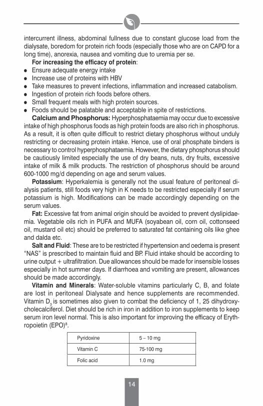
Vitamin and Minerals: Water-soluble vitamins particularly C, B, and folate are lost in peritoneal Dialysate and hence supplements are recommended. Vitamin D3 is sometimes also given to combat the deficiency of 1, 25 dihydroxy-cholecalciferol. Diet should be rich in iron in addition to iron supplements to keep serum iron level normal. This is also important for improving the efficacy of Eryth-ropoietin (EPO)8.
Pyridoxine 5 – 10 mg
Vitamin C 75-100 mg
Folic acid 1.0 mg
15
NUTRITIONAL REQUIREMENT OF THE PAEDIATRIC PATIENTS ON HEMODIALYSIS (HD)
The nutritional requirement of paediatric patients on hemodialysis (HD) is somewhat restricted and regulated to minimize fluctuations in the predialysis blood chemistry levels and intradialytic weight gain. Many of the drugs prescribed are also of importance from drug-nutrient interaction point of view e.g. phosphate binders, sodium supplements diuretics, etc. Similarly, fluid balance and dry weight is also relevant to the dietary prescription9.
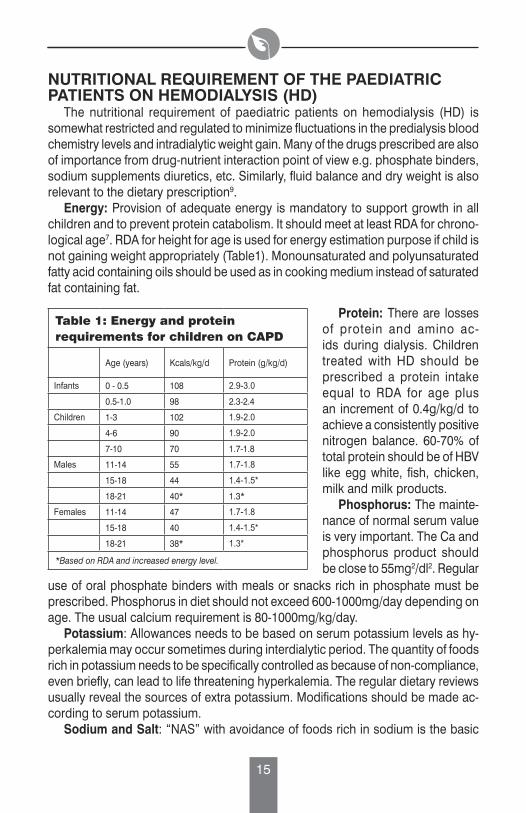
Energy: Provision of adequate energy is mandatory to support growth in all children and to prevent protein catabolism. It should meet at least RDA for chrono-logical age7. RDA for height for age is used for energy estimation purpose if child is not gaining weight appropriately (Table1). Monounsaturated and polyunsaturated fatty acid containing oils should be used as in cooking medium instead of saturated fat containing fat.
Table 1: Energy and protein requirements for children on CAPD
Age (years) Kcals/kg/d Protein (g/kg/d)
Infants 0 - 0.5 108 2.9-3.0
0.5-1.0 98 2.3-2.4
Children 1-3 102 1.9-2.0
4-6 90 1.9-2.0
7-10 70 1.7-1.8
Males 11-14 55 1.7-1.8
15-18 44 1.4-1.5*
18-21 40* 1.3*
Females 11-14 47 1.7-1.8
15-18 40 1.4-1.5*
18-21 38* 1.3*
*Based on RDA and increased energy level.
Protein: There are losses of protein and amino ac-ids during dialysis. Children treated with HD should be prescribed a protein intake equal to RDA for age plus an increment of 0.4g/kg/d to achieve a consistently positive nitrogen balance. 60-70% of total protein should be of HBV like egg white, fish, chicken, milk and milk products.
Phosphorus: The mainte-nance of normal serum value is very important. The Ca and phosphorus product should be close to 55mg2/dl2. Regular
use of oral phosphate binders with meals or snacks rich in phosphate must be prescribed. Phosphorus in diet should not exceed 600-1000mg/day depending on age. The usual calcium requirement is 80-1000mg/kg/day.
Potassium: Allowances needs to be based on serum potassium levels as hy-perkalemia may occur sometimes during interdialytic period. The quantity of foods rich in potassium needs to be specifically controlled as because of non-compliance, even briefly, can lead to life threatening hyperkalemia. The regular dietary reviews usually reveal the sources of extra potassium. Modifications should be made ac-cording to serum potassium.
Sodium and Salt: “NAS” with avoidance of foods rich in sodium is the basic

16
principle to prevent intradialytic weight gain as excess sodium intake is most likely to lead to peripheral or/and pulmonary edema. Similarly fluid intake should be based on insensible fluid loss plus previous day urine output. Restriction of fluid and salt is advocated especially if oedema is present.
Vitamins and Minerals: There are losses of folate and other water soluble vitamins during dialysis and hence needs to be supplemented.
Dietary restrictions for pediatric patients on HD should be done on individual basis and minimized so as to optimize the nutrient intake and maintain the nutritional status. The diet should be palatable, has variety in spite of restrictions through careful meal planning.
RENAL TRANSPLANTATIONThe ultimate goal for children with ESKD is a successful renal transplant to re-
store normal renal physiology and metabolic function without the aid of dialysis and dietary manipulation. Appetite improves after renal transplantation but care should be taken to prevent excessive weight gain in long term. The emphasis should be on well-balanced healthy diet. Patients are susceptible to hyperlipidaemia following transplantation. Hence use of monounsaturated and polyunsaturated fats instead of saturated fats should be advocated. Excess use of fat and fatty and fast foods should be avoided. A significant number of these children are also hypertensive due to imunosuppressive therapy and partly due to the underlying disease. Hence diet plus exercise is beneficial and “NAS” should be encouraged.
REFERENCES1. Grupe WE.: Nutritional consideration in the Management of infant and Children with Renal Disease.
in Textbook of Gastroenterology and Nutrition in infancy. Second Edition, Ed.E. Lebenthal, Reven Press, 230-235, 1989.
2. Raymond NG, Dwyer JT, Nevins P& Kurtin P.: An approach to protein restriction in children with renal insuffiency. Pediatr Nephrol, l4:145-151, 1990.
3. Shapiro AC, Bandini LG and Kurtin PS.: Estimating energy requirements for children with renal disease. A comparison of methods. Journal of the American Dietetic Association, 92 (5): 571-573, 1992.
4. Diet and Nutrition guide for patients with renal diseases and related disorders. Vidya N. Acharya, Swati Piramal (eds) 3rd Edition; 57-64, 2001.
5. Gillit D, Stover J, Spinozzi N.: A clinical guide to nutrition care in end stage renal failure. American Dietetic Association, 224-248, 1987.
6. Solhaug MJ, Adelman RD and Chan JCM.: Hypertension in child with chronic renal insufficiency or undergoing Dialysis. Child Nephrol Urol, 12:133-138, 1992.
7. National kidney Foundation and DOQI: Clinical Practice Guidelines for Nutrition in Chronic Renal Failure - Pediatric guidelines. Am J Kidney Dis, 35(6) :S112-117, 2000.
8. Varsha J.: Nutrition in Renal disorders. Indian J Perit Dial, 2:35-53, 1999. 9. Shaw V., and Lawson M.: Clinical Pediatric Dietetics Shaw. V and Lawson M (eds). Oxford Blackwell
publications, London;125-142, 1994.
Dr S. Padmavati PresidentDirector, National Heart Institute, New Delhi
Dr C. Gopalan MemberPresident, Nutrition Foundation of India, New Delhi
Dr Prema Ramachandran MemberDirector, Nutrition Foundation of India, New Delhi
Dr Kamala Krishnaswamy MemberSenior Emeritus Scientist, National Institute of Nutrition, Hyderabad
Dr S. Janaki MemberConsultant Neurologist, New Delhi
Dr S. Ramji MemberProfessor, Department of Paediatrics, Maulana Azad Medical College, New Delhi
Ms Malini Seshadri TreasurerFreelance Writer and Company Secretary, Chennai
Mr Rakesh Bhargava MemberManaging Director & CEO, Fresenius Kabi India Limited
Dr Sarath Gopalan Executive DirectorConsultant, Clinical Nutrition and Paediatric Gastroenterologist Pushpawati Singhania Research Institute, New Delhi