upper extremity orthoses syllabus
TRANSCRIPT
Upper Extremity Orthoses
Determining splint design and fabricating hand splints are extremely
important aspects in providing optimal care for persons with upper
extremity injuries and functional deficits. Splint fabrication is a
combination of science and art.
Definition of splint:
Mosby's Medical Dictionary (1994) defines a splint as:
Splint is an orthopedic device for immobilization, control, or support of
any part of the body.
Orthosis is a force system designed to control, correct, or compensate for
a bone deformity, deformity forces, or forces absent from the body.
Today in the health care field, these terms are often used synonymously.
Technically, the term splint refers to a temporary device that is part of a
treatment program, whereas the term orthosis refers to permanent device
to replace or substitute for loss of muscle function.
Splints and orthoses not only immobilize but also mobilize, position and
protect a joint or a specific body part. Design and fabrication of splints
ranges from simple to complex, depending on the goals established for a
particular condition.
Splint design must be based on scientific principles, and splint fabrication
requires creative problem solving because each person and splint may be
different.
1

Splint categorization:
According to the American Society of Hand Therapists (ASHT- 1992)
splints are classified as:
1 – Mobilization
2 – Immobilization
3 – Restrictive
1 – Mobilization splints:
Mobilization splints are designed to move or mobilize primary and
secondary joints.
2 – Immobilization splints:
Immobilization splints are designed to immobilize primary and secondary
joints.
3 – Restrictive splints:
Restrictive splints, limit a specific aspect of joint range of motion for the
primary joints.
In the past, splints have been categorized as static or dynamic. This
classification system has its problems and controversies.
A – Static splints:
Static splints have no moveable parts, in addition, static splints place
tissues in a stress – free position to enhance healing and minimize
friction.
B – Dynamic splints:
2
Dynamic splints have one or more moveable parts and are synonymous
with splints that employ elastic, spring and wire as well as with multipart
splints .
Splinting purposes:
Static and dynamic splints may serve a variety of purposes. Occasionally
a static or dynamic splint may accomplish the same therapeutic goal. At
other times, a static or dynamic splint is preferable in relationship to a
therapeutic outcome.
Static splints:
The primary purposes of static splints are four fold:
A – To immobilize
B – To help prevent further deformity
C – To prevent a soft – tissue contracture
D – To substitute for lost motor function
Rest:
Static splints provide rest to soft – tissue structures, a joint or several
joints. A common static splint is a resting hand splint. Therapists
commonly use splints that immobilize with such diagnoses as rheumatoid
arthritis, carpal tunnel syndrome, fractures, and soft – tissue repairs .
Static splints also provide support for joint laxity and ligament injury.
Prevention of further deformity:
Although controversial, static (immobilization) splints may prevent
further deformity by maintaining stretch on soft tissue to increase range
of motion and help align and stabilize joints. An example of this type of
splint is an ulnar deviation splint. Therapists commonly use this splint to
3
help prevent further ulnar deviation deformity for persons who have
rheumatoid arthritis.
Prevention of soft – tissue contractures:
Static splint can also help prevent soft – tissue contracture by maintaining
joints in their most functional positions and controlling the formation a
modeling of scars. An example of a splint for a person who has hand
burns.
Substitution for lost motor function:
A static splint may be helpful when motor function is lost, for example, a
person with an ulnar nerve injury may receive a splint to keep the hand in
a more functional position in light of lost ulnar nerve motor innervation.
Dynamic splints:
Dynamic splints (mobilization) have many purposes including:
1 – To substitute for loss of motor function
2 – To correct an existing deformity
3 – To provide controlled motion
4 – To aid in fracture alignment and wound healing
Substitution for lost motor function:
An example of a splint that substitutes for loss of motor function is the
tenodesis splint. This splint helps persons with spinal cord injuries at the
cervical level of six or seven grasp objects using a tenodesis grasp
pattern.
Correction of a deformity:
4

Therapists may use dynamic splints to correct contractures by applying
force to position the joint end range with the soft tissue at a maximum
length. An example of this may be proximal interphalangeal (PIP) joint
flexion mobilization splints that assist the PIP joint to gradually regain
maximal flexion.
Control of motion:
An example of a splint that provides controlled motion is a dorsal
blocking splint with dynamic traction for flexor tendon repair. A dynamic
flexion splint protects the healing flexor tendon from stress while
allowing limited glide to prevent adherence of the tendon.
Aid in fracture alignment and wound healing:
Dynamic splints aid in fracture alignment and wound healing. An
example of the need for dynamic splinting is the treatment of intra –
articular finger fractures. By providing constant traction at various joint
angles, the splint allows the fracture to heal while maintaining joint
motion. This decreases soft – tissue adherence, which is a frequent
complication of immobilized fractures.
Splint designs:
The purpose of a splint helps to indicate the type of splint design a
therapist uses. Splinting designs can be classified as:
a – static, b – drop – out, c – articulated, d – dynamic, e – static –
progressive, f – serial static.
Static splint:
A static splint can maintain a position to held anatomical structures at the
end of available range of motion, thus exerting a mobilizing effect on a
5
joint. For example to increase extension of a stiff wrist, a splint might be
fabricated to position the wrist in maximum tolerated extension. Because
the splint positions the shortened wrist flexors at maximum length and
holds them there, the tissue remodels in a lengthened form.
Drop – out splint:
A drop – out splint allows motion in one direction while blocking motion
in another. This type of splint may help a person regain lost range of
motion while preventing a prior poor posture.
For example, a splint may be designed to enhance wrist extension while
blocking wrist flexion.
Articulated splints:
Articulated splints have a minimum of two static components, which
allows for movement in one plane. For example, a splint may be designed
to limit motion at the ends of a movement arc so that the person uses a
specific range of motion.
Dynamic splints:
Usually dynamic splints have self – adjusting or elastic components,
which may include wire, rubber bands, or springs. For example, a splint
that uses a dynamic tension force to straighten an index finger with a
proximal interphalangeal joint (PIP) flexion contracture may be
fabricated .
Static – progressive splints:
They are often categorized as dynamic splints. They can be distinguished
from dynamic splints by the use of inelastic components such as hook –
and – loop tapes, outrigger line, progressive hinges, turnbuckles and
6
screws. The use of inelastic components allows the patient to adjust the
line of tension to prevent overstressing of tissue.
Serial static splints:
They require the remolding of a static splint. Johnson (1996) points out
those circumferential splints that are nonremoveable require no
cooperation from those who wear them, except to leave them on. For
example, a wrist splint may be remolded as a person gains more
extension movement .
7
Anatomical and biomechanical principles of splinting
Arches of the hand:
To have a strong functional grasp, the hand uses the following three
arches:
1 – The longitudinal arch
2 – The distal transverse arch
3 – The proximal transverse arch
Because of their functional significance, these arches require care during
the splinting process for their preservation. The therapist should never
splint a hand in a flat position because doing so compromises function
and creates deformity.
Especially in case of muscle atrophy (as with a tendon or nerve injury),
the splint should maintain integrity and mobility of the arches.
The proximal transverse arch:
The proximal transverse arch is fixed and consists of the distal row of
carpal bones. It is a rigid arch acting as a stable pivot point for the wrist
and long finger flexor muscles. The transverse carpal ligament and the
bones of the proximal transverse arch form the carpal tunnel. The
transverse carpal ligament provides mechanical advantage to the finger
flexor tendons by serving as a pulley.
8
The distal transverse arch:
The distal transverse arch, which deepens with flexion of the fingers, is
mobile and passes through the metacarpal heads. A splint must allow for
the functional movement of the distal arch to maintain or increase normal
hand function.
The longitudinal arch:
The longitudinal arch allows the distal interphalangeal (DIP), the
proximal interphalangeal (PIP), and the metacarpophalangeal (MCP)
joints to flex. This arch follows the longitudinal axes of each finger.
Because of the mobility of their base, the first, fourth and fifth
metacarpals move in relationship to the shape and size of an object placed
in the palm .
Grasp is the result of holding an object against the portion of the hand
that the second and third digits provide. The flattening and cupping
motions of palm allow the hand to pickup and handle objects of various
sizes.
Anatomical landmarks of the hand:
Creases of the hand:
The creases of the hand are critical landmarks for splint – pattern making
and molding. Therefore, knowledge of the creases and their functional
implications is important.
Three flexion creases are located on the palmar surface of digits II
through V, and additional creases are located on the palmar surface of the
hand and wrist.
9
The three primary palmar creases are:
1 – The distal palmar crease
2 – The proximal palmar crease
3 – The thenar crease
The distal palmar crease:
The distal palmar crease extends transversely from the fifth MCP joint to
a point midway between the third and second MCP joints. This crease is
the landmark for the distal edge of the palmar portion of a splint intended
to immobilize the wrist while allowing motion of the MCPs .
By positioning the splint proximal to this crease, the therapist makes full
MCP joint flexion possible.
The proximal palmar crease:
Below the distal palmar crease is the proximal palmar crease, which is
used as a guide during splint fabrication. A splint must be proximal to the
proximal palmar crease at the index finger or the MCP joint will not be
free to move into flexion.
The thenar crease:
The thenar crease begins at the proximal palmar crease and curves around
the base of the thenar eminence. To allow thumb motion this crease
should define the limit of the splint's edge .
10
If the splint extends beyond the thenar crease toward the thumb, thumb
opposition and palmar abduction of the carpometacarpal (CMC) joint are
inhibited.
Wrist creases:
The two palmar (or volar) wrist creases are the distal and proximal wrist
creases. The distal wrist crease extends from the pisiform bone to the
tubercle of the trapezium and forms a line that separates the proximal and
distal rows of the carpal bones.
The proximal wrist crease corresponds to the radiocarpal joint and
delineates the proximal border of the carpal bones, which articulates with
the distal radius. The distal and proximal wrist creases assist in locating
the axis of the wrist motion.
The creases are close to but not always directly over bony joints. When
splinting to immobilize a particular joint, the therapist must be sure to
include the corresponding joint flexion crease within the splint so as to
provide adequate support for immobilization .
Conversely when attempting to mobilize a specific joint, the therapist
must not incorporate the corresponding flexion crease in the splint to
allow for full range of motion. When one is working with persons who
have moderate to severe edema, the creases may dissipate. Creases may
also dissipate with disuse associated with paralysis or disuse resulting
from pain, stiffness or psychological problems.
Grasp and prehension patterns:
The normal hand can perform many prehensile patterns in which the
thumb is a crucial factor.
11
Even though hand movements are extremely complex, they can be
categorized into several basic prehensile and grasp patterns, including the
following:
1 – Fingertip prehension
2 – Palmar prehension
3 – Lateral prehension
4 – Cylindrical grasp
5 – Spherical grasp
6 – Hook grasp
7 – Intrinsic plus grasp
Therapists should keep in mind that finger prehensile movements require
less strength than grasp movements. The grasp and prehension that may
be provided by hand splinting are determined by the muscles that are
functioning, potential and present deformities and how the hand is to be
used .
Fingertip prehension:
Fingertip prehension is the contact of the pad of the index or middle
finger with the pad of thumb. This movement, which patients use to
pickup small objects such as beads and pins, is the weakest of the pinch
patterns and requires fine motor coordination .
A splint to facilitate the fingertip prehension for a person with arthritis
may include a static splint to block (stabilize) the thumb IP joint in slight
flexion.
Palmar prehension:
Palmar prehension also known as the tripod or three-jaw chuck pinch is
the contact of the thumb pad with the pads of the middle and index
fingers .
12
People use palmar pinch for holding pencils and picking up small
spherical objects. Splints to facilitate palmar prehension include thumb
spica splints that position the thumb in palmar abduction, which may be
hand or forearm based.
Lateral prehension:
Lateral prehension, the strongest of the pinch patterns is the contact
between the thumb pad and the lateral aspect of the index finger. Patients
typically use this pattern for holding keys.
Splints that position the hand for lateral prehension include thumb spica
splints that place the thumb in slight radial abduction.
Cylindrical grasp:
Cylindrical grasp is used for holding cylindrical – shaped objects such as
soda cans, panhandles and cylindrical tools. The objects rests against the
palm of the hand, and the adducted fingers flex around the object to
maintain a grasp.
Splinting to encourage such motions as thumb opposition or finger and
thumb joint flexion may contribute to a person's ability to regain
cylindrical grasp.
Spherical grasp:
Spherical grasp is used to hold round objects such as tennis balls and
baseballs. The object rests against the palm of the hand, and the abducted
fire digits flex around the object. Splinting to enhance spherical grasp
13
may include splints addressing such motions as finger and thumb
abduction.
Hook grasp:
Hook grasp, which is accomplished with the finger only involves the
carrying of such items a briefcase and suitcases by the handles. The PIPs
and DIPs flex around the object, and the thumb often remains passive in
this type of grasp .
With ulnar and median nerve damage this position may be avoided, rather
than encouraged. However, for PIP and DIP joints lacking flexion, a
therapist may fabricate dynamic flexion splints to gain range of motion in
these joints.
Intrinsic plus grip:
This grip is characterized by MCP flexion, PIP, and DIP extension. The
thumb is positioned in palmar abduction for opposition with the third and
forth fingers. This grasp is holding flat objects such as books, trays or
sandwiches. The interinsic plus grip is not present with ulnar and median
nerve injuries. A therapist may facilitate the grasp by using a figure – of –
eight splints.
Normally, the interossei extend the PIP and DIP joints when the MCP
joint is in extension. The dorsal interossei produce finger abduction and
the palmar interossei produce finger adduction. Functionally, the first
dorsal interossei is a strong abductor of the index finger, which assists in
properly positioning the hand for pinching .
14
Research shows the interossei are active during grasp and power grip in
addition to pinch.
With function of the interossei and lumbericals, a person is able to place
the hand in an intrinsic plus position. An intrinsic plus position is
established when the MCP joints are flexed and the PIP joints are fully
extended.
Pressure systems
There are three types of pressure system:
1 – Three – point force system
2 – Four – point force system
3 – Circumferential total contact pressure
Supportive systems involve a series of forces and counter forces, which
are known as pressure systems. The basic pressure system for an orthosis
is the three – point force system.
Three point force system:
The system consist of a principal force acting in one direction and two
counter forces acting in the opposite direction located proximal and distal
15
to the principal force. For example, trunk flexion is controlled by an
anteriorly directed force from the mid portion of posterior uprights in a
LSO(principal force) and posteriorly directed counter forces applied by
the sternal plate and supra public plate.
Four-point force system:
Some orthoses exert a four – point force system. The parapodium a
THKAFO applies posteriorly directed forces from the chest band and the
anterior leg bands, and applies anteriorly directed forces from the
dorsolumbar band and the back of the shoe supports.
Circumferential pressure:
In a few instances, the orthosis surrounds the body segment, applying
circumferential total contact pressure. For example, the patient recovering
from third – degree burns to the arm may be fitted with an elastic sleeve
that distributes pressure over the greatest area.
Three-point pressure system:
Most splints use a three – point pressure system to affect a joint motion.
A three – point pressure system consists of three individual linear forces
in which the middle force is directed in an opposite direction from the
other two forces.
16
Three – point pressure system in splints are used for different purposes.
For example, a splint affecting extension or flexion of a joint exerts
forces in one plane or unidirectionally .
Three – point systems can be applied to multiple directions. In other
words, a splint may immobilize one joint while mobilizing a near joint.
An example of a multiple – directional three – point pressure system is a
circumferential wrist splint.
Mechanical advantage:
Splints incorporate lever system, which incorporate forces, resistance
axes of motion and moment arms. Splints serving as levers use a
proximal input force (Fi), two moment arms and an axis or fulcrum to
move a distal output force. Similar to a teeter – totter, the force side of a
splint lever equals the resistance side of the lever and the sum of the
proximal (Fi) and the distal (Fo) forces equals the magnitude (Fm) of the
middle opposing force, (Fm = Fo + Fi) .
The system's balance is defined as:
(Fi x di = Fo x do)
In this equation, (Fi) is the input force and (di) is the input distance or the
proximal force moment arm. (Fo) is the resistance or output force, and
(do) is the output distance or the resistance moment arm .
Mechanical advantage is defined as (di / do). Mechanical advantage
principles can be applied and adjusted when one is designing a splint. For
example, when designing a volar – based wrist cock – up splint,
increasing the length of the forearm trough will decrease force on the
proximal anterior forearm.
This results in a more comfortable splint for the patient. Application of
this construct involves consideration of the anatomical segment length in
17
designing the splint. The length of a splint's forearm trough should be
approximately two-thirds the length of the forearm.
Persons wearing volar – based splint should be able to flex their elbows
without interference with full motion. The width of the thumb or forearm
trough should be one-half the circumference of the thumb or forearm.
The muscle bulk of an extremity gradually increases more proximal to the
body, and the splint trough should widen proportionately in the proximal
area. When making a splint pattern, the therapist attempts to maintain
one-half the circumference of the thumb or forearm for a correct fit.
Pressure and stress
Stress:
Stress is the measure of force per unit area. Surface stress is called
pressure. Greater force over a small area causes high stress and high
pressure.
Skin and soft tissue can be damaged by force or pressure in four ways:
1 – Degree
2 – Duration
3 – Repetition
4 – Direction
Degree and duration of stresses:
Generally, low stress can be tolerated for longer periods of time, while
high stress over long periods of time will cause damage.
A therapist should remember that generally, the tissue that least tolerates
pressure is the skin. Skin becomes ischemic as load increases.
Low stress can be damaging if it is continuous and eventually causes
capillary damage and, finally, ischemia .
18
The effects of continuous, low force from constricting circumferential
bandages and splints and their straps can be damaging at times. However,
if a system can be devised to distribute pressure over a larger area of skin,
a higher load can be exerted on a ligament, adhesion tendon, or muscle.
Such a splint, system may include a longer trough or a circumferential
component.
Repetitive stress:
If a stress is repetitively applied in moderate amounts, it can lead to
inflammation and skin breakdown. An example of a repetitive stress may
be seen in a person wearing a dynamic flexion splint that has rubber band
traction. If the person continually flexes the finger against the tension, the
tissue may become inflamed after some time.
If inflammation or redness occurs, the therapist must realize persons with
traumatic hand injuries or pathology may not be able to tolerate repetitive
amounts of stress that a normal person could tolerate. Poor tolerance is
usually a result of damaged vascular and lymph structures.
High stress may quickly result in tissue damaged. High stress can be
applied to the skin from any object, such as a splint or bandage.
The smaller or sharper the object, the greater the amount of stress that is
produced. High stress should be avoided at all times. For example, if a
dynamic splint is applying too much stress to a joint, circulation may be
restricted and cause tissue damage.
Direction of stress:
The direction of stress or force on the skin and soft tissue is considered
during splinting.
There are three directions of force to consider:
19
1 – Tension
2 – Compression
3 – Shear
1 – Tension:
Tension occurs when forces on an object are applied opposite each other.
2 – Compression:
Compression stress results from forces pressing inwardly on an object.
3 – Shear:
The third type of stress is shear, which is the horizontal sliding of one
plane of the material over another.
Shear force occurs when parallel forces are applied in an equal and
opposite direction across opposite faces of a structure. Research suggests
that shear stress is the most damaging to skin.
Therapists must be smart in recognizing and knowing how to use the
stress of splints in such a way as to not create soft – tissue damage.
Generally, therapists avoid excessive stress or pressure from splints by
employing wide troughs placed far from the fulcrum of movement while
using an appropriate amount of tension on structures.
To determine the appropriate amount of tension on structures, the splint's
tension should be sufficient to take the joint to a comfortable joint end
range. This means the tension in the splint should bring the joint just to
the maximum comfortable position (flexion, extension, deviation or
rotation) that is tolerable. This should be a position the client can tolerate
for long periods of time.
20
The patient may need to work up to long periods, but goal is usually at
least 4 hours per day. Ideally, the 4 hours is continuous, but it can be
broken up as necessary. Patients can be asked to try to wear their splints
to improve passive range of motion (PROM) during sleep.
The concepts of stress are considered when splinting. Splints and straps
apply external forces on tissues that in turn, affect forces or stresses
exerted internally. The formula for pressure is as follows:
Total forcePressure----------------------------------- =
Area of force application
Ideally, splints should be contoured and cover a large surface area to
decrease pressure and the risk of pressure sores .
Straps should also be as wide as possible to distribute pressure
appropriately and to prevent restriction of circulation or trapping of
edema.
Thermoplastic splints can cause pressure points over areas with minimal
soft tissue or over bony prominences. To avoid this risk, the therapist
should use a splint design that is wider and longer.
A larger design is more comfortable because it decreases the force
concentrated on the hand and arm by increasing the surface area of the
splint's force application.
Continuous well – distributed pressure is the goal of a splint, but pressure
over any bony prominence should be nonexistent. Therapists should be
cautious of pressure over bony prominences, such as the radial and ulnar
styloids and the dorsal – aspect MCPs and the PIPs.
21

Therapists can use heater guns to alleviate pressure exerted by the splint.
This is done by heating the plastic in problem areas and pushing the
plastic away from the bony prominence.
Another technique to avoid pressure on bony prominences is to splint
over padding, gel pads, or elastomer positioned over bony prominences .
A frequent mistake in splinting occurs when a pad is placed over the
localized pressure area after the splint is formed. Therapists should keep
in mind that padding takes up space, reducing the circumference
measurement of the splint and increasing the pressure over an area.
Planning must be done before application of the thermoplastic material.
The splint's design must accommodate the thickness of the padding.
Contour:
When flat, thermoplastic materials are more flexible and can be bent.
Curving and contouring thermoplastic material to an underlying surface
will change the mechanical properties of the material.
Contoured thermoplastic material is stronger and is better able to handle
externally applied forces.
22
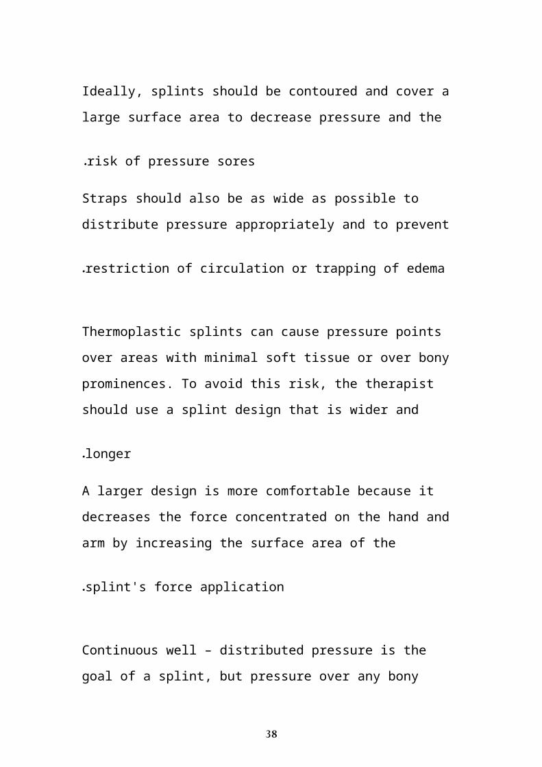
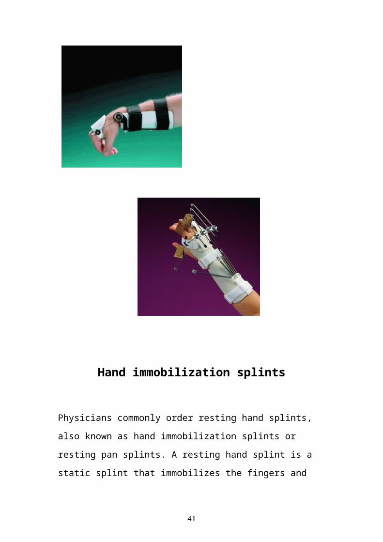
Hand immobilization splints
Physicians commonly order resting hand splints, also known as hand
immobilization splints or resting pan splints. A resting hand splint is a
static splint that immobilizes the fingers and wrist. The thumb may or
may not be immobilize by the splint.
Purpose of the resting hand splint:
The resting hand splint has three purposes:
23
1 – To immobilize
2 – To position in functional alignment
3 – To delay further deformity
When inflammation and pain are present in the joint and surrounding
structures, become swollen and result in improper hand alignment.
Resting hand splint components:
The resting hand splint has four main components:
1 – The forearm trough
2 – The pan
3 – The thumb trough
4 – The C – Bar
Forearm trough:
Forearm troughs can be volarly or dorsally based. The volarly based
forearm trough at the proximal portion of the splint supports the weight of
the forearm. Dorsally based forearm troughs are located on the dorsum of
the forearm.
When a great amount of forearm support is desired, a volarly based
forearm trough is the best design .
When the volar surface of the forearm must be avoided because of sutures
sores or skin rashes a dorsally based troughs design is frequently used.
Dorsally based troughs can be a helpful design for applying a resting
hand splint to a person with hyper tonicity.
The pan:
The pan of the splint supports the finger and the palm. The therapist must
conform the pan to the arches of the hand, thus helping to maintain such
hand functions as grasping and cupping motions.
24
The pan should be wide enough to house the width of the index, middle,
ring and little fingers when they are in a slightly abducted position.
The thumb trough:
The thumb trough supports the thumb and should extend approximately
1/2 inch beyond the end of the thumb. This extension allows the entire
thumb to rest in the trough. The CMC joint should be position in 40 to 45
degrees of palmar abduction.
The C – bar:
The C – bar keeps the web space of the thumb positioned in palmar
abduction. If the web space tightens, it inhibits cylindrical grasp and
prevents the thumb from fully opposing the other digits.
Generally, two types of positioning are accomplished by a resting hand
splint: a functional position and an antideformity (intrinsic plus) position.
Functional position:
To rest the wrist and hand joints, the resting hand splint positions the
hand in functional or mid joint position. One functional position places
the wrist in 20 – 30 degrees of extension, the thumb in 45 degrees of
palmar abduction, the MCP joints in 35 – 45 degrees of flexion and all
the PIP and DIP joints in slight flexion.
Antideformity position:
The antideformity position is often used to place the hand in such a
fashion as to maintain a tension/distraction of anatomical structures to
avoid contracture and promote function. The antideformity position
25

places the wrist in 30 – 40 degrees of extension, the thumb in 40 – 45
degrees of palmar abduction, the thumb IP joint in full extension, the
MCPs at 70 – 90 degrees of flexion and the PIPs and DIPs in full
extension.
Diagnostic indications:
Several diagnostic categories may warrant the provision of resting hand
splint. Persons who require resting hand splints commonly have arthritis,
post operative Dupuytren's contracture release, burn injuries to the hand,
tendonitis, hemiplegic hand and tenosynovitis .
The resting hand splint maintains the hand in functional or antideformity
position, preserves a balance between extrinsic and intrinsic muscles, and
provides localized rest to the tissues of the fingers, thumb and wrist.
Rheumatoid arthritis:
Therapists often provide resting hand splints for people with rheumatoid
arthritis (RA) during periods of acute inflammation and pain, and when
these people do not use their hand for activities but require support and
immobilization.
The biomechanical principle for splinting acutely inflamed joint is to
reduce pain by relieving stress; however, it may not additionally prevent
deformity. Typical joint placement for a person with rheumatoid arthritis
positions the wrist in 10 degrees of extension, thumb in palmar abduction,
the MCP joints in 35 – 45 degrees of flexion, and all DIP and PIP joints
in slight flexion.
Hand burns:
26
For persons who have hand burns, therapists do not splint in the
functional position. Instead, the therapist places the hand in the intrinsic
plus or antideformity position.
In general, the goal of splinting in the antideformity position is to prevent
deformity by keeping structures whose length allows motion from
shortening. These structures are the collateral ligaments of the MCPs, the
volar plates of the IPs, and the wrist capsule and ligaments .
The dorsal skin of the hand will maintain its length in the antideformity
position. The thumb web space is also unsafe to remodeling in a
shortened form in the presence of inflammation and in a situation in
which tension of the structure is absent.
The antideformity position for a palmar or circumferential burn places the
wrist in 30 – 40 degrees of extension and 0 degree or neutral position for
a dorsal hand burn.
For dorsal and volar burns, the therapist should flex the MCPs into 70-90
degrees, fully extend the PIP and DIP joints, and palmarly abducted the
thumb to the index and middle fingers with the thumb IP joint extended.
After a burn injury, the thumb web space is at risk for developing an
adduction contracture; therefore, palmar abduction of the thumb is the
position of choice for the thumb CMC joint .
Stages of burn recovery should be considered with splinting. The phases
of recovery are:
Emergency, acute, skin grafting and rehabilitation.
Dupuytren's disease:
Dupuytren's disease is characterized by the formation of finger flexion
contracture with a thickened band of palmar fascia. Nodules develop in
the distal palmar crease, usually in line with the fingers.
27
Slowly the condition matures into a longitudinal cord that is readily
distinguishable from a tendon. In addition, pain and decreased ROM are
the primary symptoms that often lead to impaired functional performance.
When a Dupuytren's contracture is apparent, stretching or splinting joint
in extension does not delay the progression of the contracture. Surgery is
performed to correct joint contractures and to prevent recurrence of the
disease .
Although surgery dose not cure the disease, it is often indicated in the
presence of painful nodules, uncomfortable induration (Any pathological
hardening or thickening of tissue), and MCP, PIP or DIP joint
contractures.
Therapy and splinting begin immediately after surgery. Postoperative
splinting may include a resting hand splint or a dorsal forearm based
static extension splint.
When a resting hand splint is used, the wrist is placed in a neutral or
slightly extended position. The MCP, PIP and DIP joints are splinted in
full extension.
Hand crushes injury:
To splint a crushed hand the therapist can position the wrist in 0 – 30
degrees of extension, the MCPs in 60 – 80 degrees of flexion, the PIPs
and DIPs in full extension and the thumb in palmar abduction and
extension. Splinting a crushed hand into this position provides rest to the
injured tissue and decrease pain, edema and inflammation.
Wrist immobilization splints:
28
One of the most common wrist splints is a wrist immobilization splint or
a wrist cock – up splint.
This type of splint usually maintains the wrist in either a neutral or a
slightly extended position. A wrist immobilization splint immobilizes the
wrist while allowing full MCP flexion and thumb mobility .
Therapists can fabricate wrist immobilization splints to provide volar,
dorsal, ulnar, or circumferential forearm, wrist and hand support.
They can also use wrist immobilization splints as based for dynamic and
static progressive splinting.
Diagnostic indications:
The clinical indications for the wrist immobilization splint vary according
to the diagnoses. Application of this splint usually decreases wrist pain or
inflammation, provides support, enhances digital function, prevents wrist
deformity, and minimizes tension on involved structures.
Wrist splinting for carpal tunnel syndrome:
For CTS, splinting the wrist as close as possible to 0 degree (neutral)
helps avoid added pressure on the median nerve. One must be careful
when applying prefabricated wrist immobilization splints for this
condition because some splints place the wrist in a functional position of
20 – 30 degrees of extension. Therefore, if it is possible to adjust the wrist
angle of splints, it should be modified to a neutral position.
Splinting is usually more effective if applied during the early phases of
symptom onset or within 3 months of symptom onset. When conservative
measures are ineffective, surgery is an option.
The goal of wrist splinting after carpal tunnel release surgery is to
minimize pressure on the median nerve, provide support during stressful
29
activities to maintain gains from exercise and to rest the extremity during
the immediate healing phase.
Wrist splinting for radial nerve injuries:
Radial nerve injuries occur from fractures of the humeral shaft, fractures
and dislocation of the elbow, or compression of the nerve. The classic
picture of a radial nerve injury is a wrist drop position whereby the wrist
and MCP joints are unable to actively extend.
If the wrist is involved, sometimes a physician may order a wrist splint to
place the wrist in a more functional position. Commonly 30 degrees of
extension is considered a position of function .
Wrist splinting for tendonitis and tenosynovitis:
Tendonitis (inflammation of the tendon) and tenosynovitis (inflammation
of the tendon and its surrounding synovial sheath) are painful conditions
that benefit from conservative management, including wrist splinting.
Tendinitis or tenosynovitis can occur in many of the muscles on the volar
and dorsal surfaces of the forearm.
Tendinitis or tenosynovitis often leads to substitution patterns and muscle
imbalance. Resting the hand in a splint helps to relieve tension of the
muscle tendon unit and eliminate friction at the insertion of the muscles.
Generally, for many of these conditions, such as flexor carpi radialis
(FCR) tenosynovitis, it is recommended that the person's wrist be splinted
at neutral to rest the tendons.
Wrist extensor tendinitis can be splinted in 20 – 30 degrees of wrist
extension as this normal resting position provides a balance between the
flexors and extensors.
Wrist splinting for fractures:
30

The initial goal of rehabilitation after a fracture of the distal radius is to
regain functional wrist extension. To achieve this goal splinting of wrist
in slight extension is beneficial while the person is receiving therapy.
Sometimes serial static splinting may be necessary to regain PROM. It is
best to fabricate a custom splint because prefabricated splints may not fit
comfortably and may block range of motion of finger flexion and thumb
motions.
The therapist should fabricate a well – designed dorsal or volar splint. If
the person needs more support, a circumferential wrist splint may be
considered. Circumferential splinting is highly supportive and very
comfortable. It tends to limit forearm rotation more than a volar or dorsal
wrist splint.
Wrist splinting for sprains:
A person who has a grade I sprain, or stretching of the ligament with
minimal disruption of the fibers, or a mild grade II sprain, with tearing of
the ligament fibers may benefit from wearing a wrist immobilization
splint.
With grade I sprains, the person will likely wear the splint for 3 weeks .
For grade II sprains, 6 weeks of wear may be indicated. This wrist splint
will help rest the hand during the acute healing phase and remove stress
from the healing tendon.
Features of the wrist immobilization splint:
The static wrist immobilization splint maintains the wrist in proper
alignment and provides support for the hand by stabilizing the wrist.
When serving as a base for a dynamic splint, a wrist immobilization
splint properly positions the wrist and provides an attachment site for the
outrigger.
31
Wrist splint components are:
1 – Forearm trough
2 – Metacarpal bar (palmar bar)
3 – Hypothenar – bar
Hypothenar – bar:
The hypothenar bar helps to place the hand in neutral resting position by
preventing extreme ulnar deviation. This bar should not inhibit the
motions of the ring and little fingers.
Metacarpal bar )palmar bar(:
The Metacarpal bar (palmar bar) supports the transverse MP arch. When
supporting the palmar surface of the hand, the MP – bar is sometimes
called a palmar bar .
With a volar wrist splint, this bar must be position proximal to the distal
palmar crease and distal to the thenar crease to ensure full MCP flexion.
On a dorsal wrist splint, this bar should be position slightly proximal to
the MP heads and angle it to point to the triquetrum or distal ulnar bone.
Thumb immobilization splints
A commonly prescribed splint in clinical practice is the thumb
immobilization splint. Other names for this splint are the thumb spica
splint, the short or long opponens splint and the thumb gauntlet splint.
The purpose of this splint is to immobilize, protect, rest and position one
or all of the thumb carpometacarpal (CMC), metacarpophalangeal (MCP)
and interphalangeal (IP) joints while allowing the other digits to be free.
32
Some thumb immobilization splints include the wrist and are forearm
based.
For example, the thumb immobilization splint that includes the wrist
provides people who have tendonitis with rest, support and protection of
the thumb.
With the resulting muscle imbalance from a median nerve injury the
therapist may apply a hand – based thumb immobilization splint to keep
the thumb web space adequately open.
The therapist also uses a thumb immobilization splint that includes the
wrist to splint postoperatively for control of motion in persons with
rheumatoid arthritis after joint arthrodesis or replacement.
In addition, the thumb immobilization splint can position the thumb
before surgery and provide support and positioning after traumatic thumb
injuries, such as sprains, joint dislocations, ligament injuries and scaphoid
fractures.
Frequently a hand – based thumb immobilization splint is applied to
persons with gamekeeper's thumb, which involves the ulnar collateral
ligament of the thumb MCP joint.
The thumb is essential for hand functions because of its overall
importance to grip, pinch and fine manipulation.
Features of the thumb spica splint:
The thumb immobilization splint is a static splint because it has no
moveable parts. The splint has numerous design variations:
It can be volar, dorsal or radial gutter.
The splint may be hand based or wrist based, depending on the person's
diagnosis and the associated pain at the wrist.
The splint components fabricated in the final product will vary according
to the thumb joints that are included.
33
Central to most thumb immobilization splints are:
The opponens bar
The C – bar
The thumb post
The opponens bar and C – bar position the thumb, usually in some degree
of palmar abduction.
The thumb post, which is an extension of the C – bar, immobilizes the
MCP only or both the MCP and IP joints.
The position of the thumb in a splint varies from palmar abduction to
radial abduction, depending on the person's diagnosis.
With some conditions, such as arthritis, the therapist can assist
prehension by stabilizing the thumb CMC joint in palmar abduction and
opposition. Certain diagnostic protocols, such as those for extensor
pollicis longus (EPL) repairs, tendon transfers for thumb extension, and
extensor tenolysis of the thumb, require the thumb to have an extension
and radial abducted position.
The thumb immobilization splint may do one of the following:
1 – Stabilize only the CMC joint
2 – Include the CMC and MCP joints
3 – Encompass the CMC, MCP and IP joints
The physician's order may specify which thumb joints to immobilize in
the splint. Certain diagnostic protocols, such as those for thumb
replantations, tendon transfers, and tendon repairs, often require the
inclusion of the IP joint in the splint.
34
Diagnostic indications:
Therapists fabricate thumb spica splints in general and specialized hand
therapy practices.
Specific diagnostic conditions that require a thumb spica splint include
but are not limited to the following:
Scaphoid fractures
Stable fractures of the proximal phalanx of the first metacarpal
Tendon transfers
Radial or ulnar collateral ligament strains
Repair of MCP joint collateral ligaments
Rheumatoid arthritis
Osteoarthritis
De Quervain's tenosynovitis
Median nerve injury
MCP joint dislocations
Capsular tightness of the MCP and IP joints after trauma
Posttraumatic adduction contracture
Extrinsic flexor or extensor muscle contracture
Flexor pollicis longus repair
Uncomplicated EPL repairs
Congenital adduction deformity of the thumb
The therapist should also be aware that the thumb palmar abduction may
be uncomfortable for some patients. Therefore, the thumb positioned
midway between radial and palmar abduction.
Splinting for de Quervain's tenosynovitis:
De Quervain's tenosynovitis that results from repetitive thumb motions
and wrist ulnar deviations is a form of tenosynovitis affecting the
35
abductor pollicis longus (APL) and the extensor pollicis brevis (EPB) in
the first dorsal compartment.
De Quervain's may be recognized by pain over the radial styloids, edema
in the first dorsal compartment, and positive results from the Finkelstein's
test.
Finkelstein's test:
Finkelstein's test is performed by stabilizing the forearm and instructing
the person to clasp the fingers over the flexed thumb, the examiner
passively deviates the wrist toward the ulnar side, with this test the person
may complain over the abductor pollicis longus tendon.
During the acute phase of this condition, conservative therapeutic
management involves immobilization of the thumb and wrist. This splint
may cover the volar or dorsal forearm or the radial aspect of the forearm
and hand.
Therapist should positions the wrist in 15 degrees of extension, neutral
wrist deviation, 40 degrees of palmar abduction of the thumb CMC joint,
and 10 degrees of flexion in the MCP joint .
Usually the therapist allows the IP joint to the free for functional
activities and includes the joint in the splint, causing even more pain.
This splint is worn continuously, with removal for hygiene and exercise.
Splinting for rheumatoid arthritis and osteoarthritis:
Rheumatoid arthritis often affects the thumb joints, particularly the MCP
and CMC joints. Splinting can reduce pain, slow deformity and stabilize
the thumb joints.
The disease includes three stages; each stage has a different splinting
approach, even though the therapist may apply the same thumb
immobilization splint.
36
First stage:
The first stage involves an inflammatory process. The goal of splinting at
this stage is to rest the joints and reduce inflammation.
The person wears the thumb spica splint continuously during periods of
inflammation and periodically thereafter for pain control as necessary .
Second stage:
When the disease progresses in the second stage, the hand requires
mechanical support because the joints are less stable and are painful with
use.
The person wears a thumb spica splint for support while doing daily
activities and perhaps at night for pain relief .
Third stage:
In the third stage, pain is usually not a factor, but the joints may be
grossly deformed and unstable. Instead of surgical stabilization, a thumb
splint may provide support to increase function during certain activities.
At this stage, splinting is rarely helpful for the person at night.
One approach to splinting is to immobilize the thumb in a long thumb
spica splint with the wrist in 20 – 30 degrees of extension, the CMC joint
in 45 degrees of palmar abduction (if tolerated) and the MCP joint in 0-5
degrees of flexion.
Some persons with rheumatoid arthritis affecting the CMC joint benefit
from a hand – based thumb spica splint. Positioning the thumb in enough
palmar abduction for functional activities is important.
On the other hand, some therapists stabilize the thumb CMC joint alone
with a short splint that is properly molded and positioned. Padding the
splint for comfort to prevent skin irritation may be necessary on a person
37
who has rheumatoid arthritis because of the person may have fragile
(breakable) skin.
CMC joint osteoarthritis is a common thumb condition especially among
women over 40 years old. Pain from osteoarthritis at the base of the
thumb interferes with the person's ability to engage in normal function
activities .
Fabrication a hand – based thumb spica or short opponens splint with the
primary therapeutic goal of restricting the mobility of thumb joints to
decrease pain and inflammation. The splint stabilizes the CMC and MCP
joints in the maximal amount of palmar abduction.
Splinting for ulnar collateral ligament injury:
Injury to the ulnar collateral ligament (UCL) a condition also known as
gamekeeper's thumb or skier's thumb is a common injury that can occur
at the extent of ligamental tear.
Therapist may uses a short opponens splint or a hand – based thumb spica
splint to positioning the first CMC joint in 25 – 30 degrees of abduction
(palmar abduction) and MP joint in neutral position. The purpose of this
splint is to provide rest and protection during the healing phase.
Foot orthoses
A functional foot orthosis (FO) is designed to redistribute weight-bearing
pressure over the plantar surface of the foot .
Foot orthoses functions are as follows:
Relief to painful areas
Reduced stress to proximal joint structures
38
Equalization of a limb discrepancy
Support to arches of the foot
Limiting motion at specific joints
Foot orthoses are available in prefabricated, custom – fitted and custom
designs, each accommodating variable results relative to fit, function,
comfort, sizing, cost, adjustability, personal acceptance/satisfaction and
effectiveness.
Biomechanical function:
Most FOs are biplanar designs, addressing joint deviations and providing
support in the sagittal (mid foot depression) and coronal planes (hind foot
and forefoot varus or valgus).
More involved FO designs, such as the University of California
Biomechanics Laboratory (UCBL) foot orthosis offer triplanar support
with additional control for transverse plane deviations (forefoot abduction
or adduction). A complete individual evaluation procedure is the basis for
design and selection of the most appropriate foot orthosis.
General indications and contraindications:
Prescription criteria for foot orthoses are based on three main principles:
1 – Accommodation
2 – Support
3 – Correction
1 – Accommodative foot orthoses:
39
These orthoses are designed to provide additional padding, relief of
pressure and protection for insensate or deformed feet.
2 – Supportive orthoses:
These orthoses are fabricated from semi rigid materials, offering a greater
degree of alignment control.
3 – Corrective orthoses:
These orthoses tend to be manufactured from rigid materials and require
extensive fitting procedures to obtain maximum joint alignment/
correction and acceptance.
Design options:
Foot orthoses are measured in a variety of ways. Foam impressions, hand
casting or computerized mapping of the foot are the first steps in
obtaining and accurate impression of the person's foot. Material selection
offers the greatest variety of design options.
Open – and closed – cell foams, leather, cork, plastics, laminates and
other materials serve to create a flexible, semi rigid or rigid orthotic
design.
Joint alignment is altered via posting, where additional material is placed
in a particular area of the plantar surface of the orthosis to limit range of
motion or tilt the foot into the correct alignment. Maximum joint
alignment is achieved in designs offering triplanar control.
Specific applications:
Pronated conditions are effectively treated with custom orthotic design.
Runners often experience an excessive and sustained pronation moment,
which may involve excessive joint range of motion or sustained pronation
throughout the stance phase.
40
A foot orthosis is helpful in reducing the hind foot valgus deviation,
depression of the longitudinal arch, and excessive forefoot flexibility and
supination.
Semi rigid orthosis:
Semi rigid orthosis allows a normal pronation moment to occur from heel
strike through the loading response, and then encourage the foot to
become rigid for improved forward progression and propulsion.
Diabetic and insensate feet:
Diabetic and insensate feet require daily inspection to prevent pressure
sores and ulcerations from the stresses incurred during normal daily
activities. Ulcerations develop in areas of excessive pressure when joint
deviations and bony prominences are hypersensitive to skin breakdown.
Accommodative orthoses can be effectively designed to provide
sufficient padding, redistribution of weight – bearing pressures and allow
slow and gradual healing to occur.
Orthotic interventions
Once the shoe has been selected, it may be modified to improve the
patient's comfort and function. Alteration of foot function and alignment
may be accomplished by both shoe modifications may be internal (placed
within the shoe) or can be secured to the exterior of the shoe and foot
orthoses. Though these terms overlap somewhat, the former refers to
internal or external changes or additions to the structure of the shoe,
while the latter refers to removable appliances that are placed within the
shoe. Both are used to relieve pain and improve balance and junction in
standing and walking .
41
They accomplish these objectives because they help to :
1 - Transfer forces from sensitive to tolerant areas
2 - Reduce friction and shear on sensitive areas
3 - Modify weight transfer patterns
4 - Correct flexible deformities
5 - Accommodate fixed deformities
6 - Limit motion of painful, inflamed or unstable joints.
Inserts and internal modifications:
Internal modifications are glued to the inner sole. Because they lie
closer to the foot, they are mechanically more effective than external
ones. However, since they reduce the space available within the shoe
for the foot, their use must be considered when selecting the size of
shoe.
The most common foot orthosis is an insert, which can be placed in many
shoes assuming that the shoes were all made on the same last. An
alternate foot orthosis is an internal shoe modification that is
biomechanically identical to the insert but cannot be removed and
transferred to another shoe.
Neither the insert nor the internal shoe modification is visible when the
shoe is worn. The advantage of the internal modification is that it
guarantees that the patient wears the appropriate shoes when using the
modification.
A three – quarter insert terminates just proximal to the metatarsal heads
without crowding the forefoot.
42
A full – length insert terminates at the distal end of the toe box, thereby
preventing any slippage of the insert. The insert must fit snugly in the
patient's shoe.
Heel spurs, Achilles' tendon contracture, and hind foot malalignment are
some hind foot disorders that can be managed with internal heel
modifications. Heel orthoses may benefit patients with leg length
reduction, postural instability and other disorders that originate outside
the foot.
Internal heel orthoses:
These are often worn to reduce discomfort associated with heel spurs. A
resilient (elastic), tapered cushion absorbs shock at heel contact and
transfers load to the forefoot. A heel orthosis with a wall that surrounds
the medial, posterior and lateral portions of the anatomic heel will reduce
irritating sliding in the shoe. The cushion usually has a concave relief
near the spur to minimize pressure.
Hind and mid foot orthoses:
Malalignment of the hind and mid foot may result in either pes planus
(pes valgus, pes planovalgus) in which the foot is abnormally pronated or
pes varus, in which the foot is excessively supinated. The malalignment
interferes with the smooth transition of weight from the heel to the toes
during the stance phase of gait and may result in fatigue or pain.
Reducing foot malalignment also decreases patellofemoral pain and
discomfort from plantar fasciitis.
When the foot remains in a pronated posture during the stance phase, the
patient is tending to experience hyper mobility with consequent overuse
of the foot muscles that results in fatigue. Foot orthoses for pes planus
should apply an upward and laterally directed force to the talus and
43
medially directed forces to the calcaneus and forefoot to counteract the
abnormal foot alignment.
Inserts whether mass – produced or custom – made are formed of many
different materials, remaining from rigid plastic to semirigid cork,
molded leather and plastic to relatively elastic plastics. The degree of
optimal firmness is determined by the patient's weight, activity and the
extent of deformity. The greater the load on the foot, the firmer the
orthosis.
Medial heel wedge:
Medial heel wedge (post) within the shoe may suffice, assuming that the
shoe counter fits snugly. A shoe with a long medial counter is sometimes
prescribed for children with flexible flatfoot; the medial border of the
counter terminates at the first metatarsophalangeal joint.
Another way to support the longitudinal arch in the presence of flexible
deformity is an insert that has a medial vertical contour.
Such orthoses are sometimes called arch supports. Arch support is a
misleading term because normally one does not bear weight under the
longitudinal arch. Weight should be borne under the calcaneal
tuberosities in the hind foot and the first and fifth metatarsal heads in the
forefoot.
Medial Longitudinal Arch Support )Scaphoid Pad, Navicular Pad,
Cookie(:
44
The support is a rubber, cork, or plastic foam wedge fixed to the sock
lining and the lower portion of the upper. The modification extends from
a point 1 cm (1/2 in.) posterior to the first metatarsal head to the anterior
tubercle of the calcaneus, with the apex, 0.5-1 cm
(1/4-1/2 in.) in height, located between the
sustentaculum tali (talonavicular joint) and the
navicular (scaphoid) tuberosity.
UCBL insert:
A particularly effective custom – made orthosis for flexible hyper –
pronation is the UCBL insert. The orthosis holds the foot in the position
of maximum correction. The UCBL improves foot alignment when the
orthosis is worn but does not appear to have long – term effect. It is not
intended for a rigid foot.
The UCBL (University of California Biomechanics Laboratory) insert is
a rigid plastic orthosis molded over a modified model of the mid- and
hind foot. The foot is cast with maximum manual correction and the
positive plaster model is then modified so that the contours of the
finished insert will maintain the control of foot position. The insert
encompasses the heel and extends forward, terminating at a point just
posterior to the metatarsal heads. It has
posterior and sidewalls, covering the heel and
both sides of the foot, to a level just below the
malleoli. The UCBL insert is a very effective
longitudinal arch support because it helps
maintain the optimal position of the calcaneus
45
relative to the talus, and stabilizes the intertarsal and tarsometatarsal
joints.
Metatarsal Pad:
This domed pad, usually made of sponge rubber, is designed to
increase load on the metatarsal shafts with consequent reduction of load
on the metatarsal heads. To accomplish this goal, the apex of the dome
is located posterior to the heads and is approximately 0.2-0.5 cm (1/8-
1/4 in.) in height. From the apex, the pad gradually feathers posteriorly
and feathers more quickly anteriorly. From side to side, the width and
feathering depend on how many metatarsal heads require relief. If all
five heads are involved, the side-to-side feathering will be slight and the
device is then often called an internal metatarsal bar. In some instances
of too little weight bearing under the first and fifth metatarsal heads and
painful weight bearing under the middle heads, the metatarsal pacts may
have flanges extending forward beyond the first and fifth heads, thus
assuming a horseshoe, "U", shape. Other individuals may require a
Norton's extension (a flange extending distal to the first metatarsal
head) or a sesamoid pad (a medial extension terminating proximal to the
first metatarsal head).
46
Toe Crest:
A 1 cm (1/2 in.) wide crescent-shaped resilient pad may be placed behind
the second through fourth toes.
The crest fills the void under each proximal phalanx, transferring stress
from the toes to the hollow (sulcus) beneath the toes and resisting any
tendency of the foot to slide forward.
Insoles:
The contour and consistency of the sole have a marked effect on the
wearer's comfort, stability and walking area. The insole influences
pressure distribution and shock absorption.
Flat inserts made of resilient plastic, such as closed –cell polyethylene
foam, open cell foam, viscoelastic polymer, silicone gel, or closed –
cell neoprene sponge reduce high pressure concentrations when the
foot is not markedly deformed. Resilient insoles are especially
important for patients with diabetic neuropathy and with poor balance.
Molded inserts conform to the plantar contour and are more effective
in the presence of severe deformity.
47
EXTERNAL SHOE MODIFICATIONS
They do not reduce space inside the shoe. However, they are visible and
may diminish from the cosmetic appeal of the shoe. External
modifications may be applied to the heel and/or the sole of the shoe.
Heel Modifications
Cushion Heel :
The cushion heel (often called a SACH heel) is a wedge of sponge
rubber, interposed between the outer sole and a
thinned regular heel. At and just after heel strike,
the soft rubber absorbs impact, reducing stress on
the heel and ankle, as well as diminishing the
moment of force tending to flex the knee. The range of plantar flexion
required to bring the sole into contact with the floor is also reduced.
Heel Flare :
The plantar surface of the heel is extended outward on the medial,
lateral, or both sides. Compared to a regular heel, the increased distance
from the subtalar axis to the border of the flare results in a
corresponding increase in the moment of force generated
around the subtalar axis when the flared heel is in contact
with the ground. This increased moment tends to produce or to resist
eversion or inversion of the hind foot, depending on whether the flare
projects laterally or medially. A flare on both medial and lateral aspects
helps provide greater stability of the heel with respect to the subtalar axis.
48
Heel Wedge :
A wedge of firm material, usually leather, is incorporated in the medial
or lateral portion of the heel. The outer border of the wedge is 0.2-0.5 cm
(1/8-1/4 in.) high. (For adult)
A medial wedge is designed to rotate the hind foot in the
direction of inversion while a lateral wedge does the
opposite. Wedges also serve to accommodate a fixed
deformity. When a wedge is used, the opposing heel
counter must be sufficiently firm to resist any tendency of the hind foot to
slide along the inclination created by the wedge.
Extended )Thomas( Heel :
In this prefabricated firm rubber heel, the medial portion of the breast
projects anteriorly approximately 1 cm (1/2 in.). The heel may
also include a 0.5 cm (1/4 in.) medial wedge to aid in
supporting the medial longitudinal arch. Less frequently, the
lateral portion of the breast projects anteriorly, thus providing
support under the lateral aspect of the mid-foot. In this case, it is called a
reverse extended (reverse Thomas) heel.
Heel elevation :
A layer of firm material is added to the heel as compensation for leg
length discrepancy or fixed equinus deformity. The height of the
elevation is determined by placing temporary lift blocks under the heel
(and sole if necessary) to align the pelvis. A heel elevation less than 0.5
cm (1/4 in.) may be placed inside the shoe.
49
Sole modifications:
Rocker Bar :
A rocker bar consists of firm material with skived anterior and posterior
edges placed across the sole. The rounded apex of the bar is positioned
just posterior and parallel to a line from the first to the fifth metatarsal
heads, and projects approximately 0.5-1 cm (1/4-1/2
in.) below the level of the sole (This configuration
reduces time spent on the heads ,thereby alleviating
metatarsalgia) . Anterior and posterior to the apex, the
bar feathers to nothing near the toe and the shank.
Essentially, the rocker bar shifts the rollover point posterior to the
metatarsal heads, thus transferring pressure from the heads to the
metatarsal shafts. In addition, ankle motions during stance and plantar-
flexor force during push-off are reduced because the rollover point is
closer to the heel and the foot is slightly dorsiflexed (helpful for the
patients who have weak ankle planter flexors).
The rocker also diminishes the need for full ankle excursion, thus making
the sole useful for the patient who has hind foot pain or restriction of
motion or who wears an AFO that restricts dorsiflexion.
Metatarsal Bar :
The generic term "metatarsal bar" includes a number of designs known
variously as Denver, Mayo, Hauser, Jones, and flush bars, among others.
Although differing in details, all consist of a firm material, usually
leather, attached across the sole of the shoe posterior to the metatarsal
50
heads. Their basic function is to transfer pressure from the heads to the
metatarsal shafts, in which regard they are similar to the rocker bar.
They differ, however, in that their plantar surfaces are much flatter than
the rounded contour of the rocker bar and thus provide a broader area of
contact with the floor but less assistance to rollover (indicated for patients
complaining of metatarsalgia).
Sole Wedge:
A wedge of firm material, usually leather, is incorporated on the lateral
(or medial) half of the sole. The outer border of the wedge is usually 0.2-
0.5 cm (1/8-1/4 in.) thick. A lateral sole wedge tends to evert or pronate
the forefoot; a medial wedge does the opposite. Sole wedges are also used
to accommodate fixed deformity of the forefoot.
Sole Flare :
Extending the medial, lateral, or both borders of the sole creates a
broader bearing surface for greater stability and resistance
to inversion and/or eversion of the foot.
Steel sole Bar :
51
Spring steel may be inserted between the inner and outer soles of a
welted shoe. The bar is approximately 0.2 cm (1/8 in.) thick, 1.5 cm (3/4
in.) wide, and extends from a point 1 cm (1/2 in.) anterior to the posterior
edge of the heel counter to terminate 1 cm posterior to the anterior edge
of the toe box. Since the bar prevents motion of the anterior portion of
the sole, it helps reduce stress at the phalanges and metatarsals. It is
ordinarily used with a rocker bar to assist rollover.
Combination Heel and sole modification :
In some instances, modifications to both the heel and sole are required.
Examples are a cushion heel and rocker bar; heel and sole wedges on the
same shoe; and elevations under both the heel and sole. The sole eleva-
tion is usually necessary when the heel elevation is higher than 0.5 cm
(1/2 in.) to prevent the foot from assuming an equinus attitude. Combined
modifications may have separate heel and sole sections or a shank filler
may be incorporated in the space between the heel and sole components.
For the patient with hemi paresis, a 1cm heel and toe lift in the shoe on
the nonparetic side facilitates paretic foot clearance during swing phase
and improves weight-bearing symmetry during stance on the paretic side.
Pediatric foot orthoses:
Two orthoses for the nonambulatory infant are the Denis Brown splint
and the hinged shoe.
Denis Brown splint:
The Denis Brown splint is a metal bar with attachments to the shoe soles.
The device resists any tendency of the baby to adduct or internally rotate
52
the hips. One version of the Denis Brown splint has a linkage that enables
the child to flex and extend the legs at the hips .
The splint is prescribed to maintain correction of talipes equinovarus in
infants. The splint also used to treat internal tibial torsion. Although its
efficacy has been challenged. The effect is dissipated at the hip, knee and
ankle joint, rather than at the tibia.
Hinged shoe:
A shoe with a hinge at the midfoot can be used to treat metatarsus
adductus. The shoe secures the forefoot and hind foot independently; the
clinician adjusts the degree of rotation with screw mechanism at the
hinge.
53
54