upper limb salvage with endovascular treatment …...the rate of limb salvage following...
TRANSCRIPT
CASE REPORT AND REVIEW
Vascular Disease Management® March 2015 36
Upper Limb Salvage With Endovascular Treatment of Acute Axillary Artery Occlusion Secondary to Proximal Humeral Fracture
Acute upper limb ischemia (AULI) is much
less commonly encountered and clinically
recognized than acute ischemia of the lower
limb.1 According to a previous report, the early com-
mencement of revascularization is crucial for limb
salvage in patients with AULI.2 Sudden axillosubcla-
vian artery occlusion is one cause of AULI. How-
ever, it rarely occurs as a complication of proximal
humeral fracture.3 Although there are some reports
on endovascular treatment (EVT) for the manage-
ment of axillosubclavian artery injuries, such as lacer-
ations, pseudoaneurysms, and arteriovenous fistulas,4
there are only a few reports on its application in acute
axillo-subclavian arterial occlusions.5,6 We present a
case demonstrating the value of immediate EVT in
the management of AULI secondary to acute axillary
artery occlusion caused by a closed humeral fracture.
CASE PRESENTATIONAn 82-year-old Japanese male presented with severe
pain in the left shoulder and arm, with discoloration
of hand and fingers, 2 hours after falling at home. His
medical history included osteoporosis and prostate can-
cer that were well controlled with medical treatment.
On admission, his vital signs were stable. On physical
examination performed approximately 2 hours after
Tsuyoshi Isawa, MD1; Kenji Suzuki, MD1; Hideki Abe, MD2
From 1Sendai Kousei Hospital, Sendai, Japan, and 2Sanyudo Hospital, Yonezawa, Japan.
ABSTRACT: Acute axillary artery occlusion associated with proximal humeral fracture is rare.
Traditionally, axillary artery complications associated with humeral fractures are managed with open
surgery. However, open vascular repair presents a considerable challenge to even the most skilled
surgeons. Endovascular treatment (EVT) offers an alternative to surgical management. We describe
the case of an 82-year-old Japanese male with acute upper limb ischemia (AULI) secondary to acute
axillary artery occlusion caused by a proximal humeral fracture. He was successfully treated with
EVT. Unless there is vessel transection, EVT is feasible and offers a minimally invasive and prompt
therapy for AULI resulting from axillary artery occlusion.
VASCULAR DISEASE MANAGEMENT 2015;12(3):E36-E43 Key words: endovascular management, acute upper limb ischemia, proximal humeral fracture
Copyri
ght H
MP Com
munica
tions
CASE REPORT AND REVIEW
Vascular Disease Management® March 2015 37
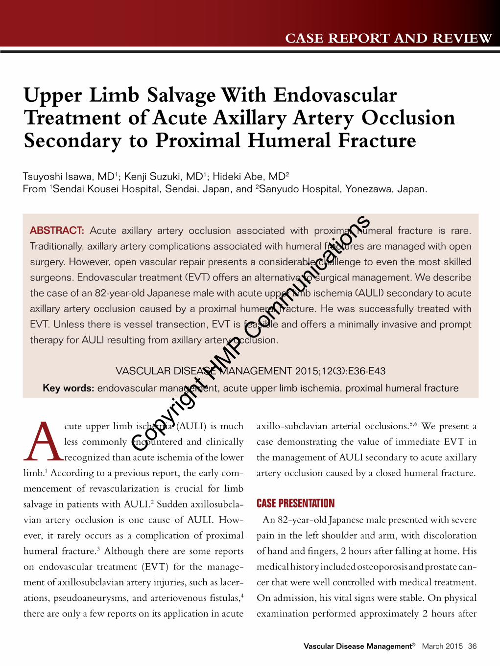
Figure 1. Radiograph showing proximal fracture of the left humerus before (A) and after closed reduction (B).
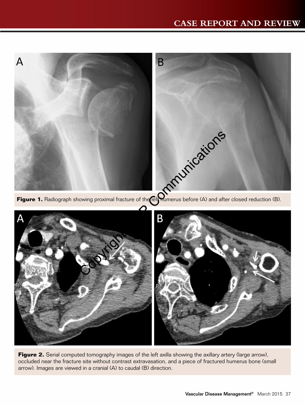
Figure 2. Serial computed tomography images of the left axilla showing the axillary artery (large arrow), occluded near the fracture site without contrast extravasation, and a piece of fractured humerus bone (small arrow). Images are viewed in a cranial (A) to caudal (B) direction.
Copyri
ght H
MP Com
munica
tions
CASE REPORT AND REVIEW
Vascular Disease Management® March 2015 38
the fall there were no brachial, radial, or ulnar pulses
in the left arm. There were no open wounds. The
hand and forearm were pale and cool, but the skin of
the fingers was dark, indicating critical ischemia. In
spite of mild hypoesthesia, there was no weakness in
his hand and fingers. His hemoglobin was 9.6 g/dL
and the estimated glomerular filtration rate was 71.9
mL/min/1.73 m2. The serum creatine kinase level was
123 IU/L. An initial radiography of the left shoulder
revealed a three-part fracture of the left proximal hu-
merus with severe medial displacement, according to
Neer classification. To restore blood flow, he under-
went immediate closed reduction under interscalene
brachial plexus block (Figure 1). However, the distal
pulses were still not palpable and the fingers remained
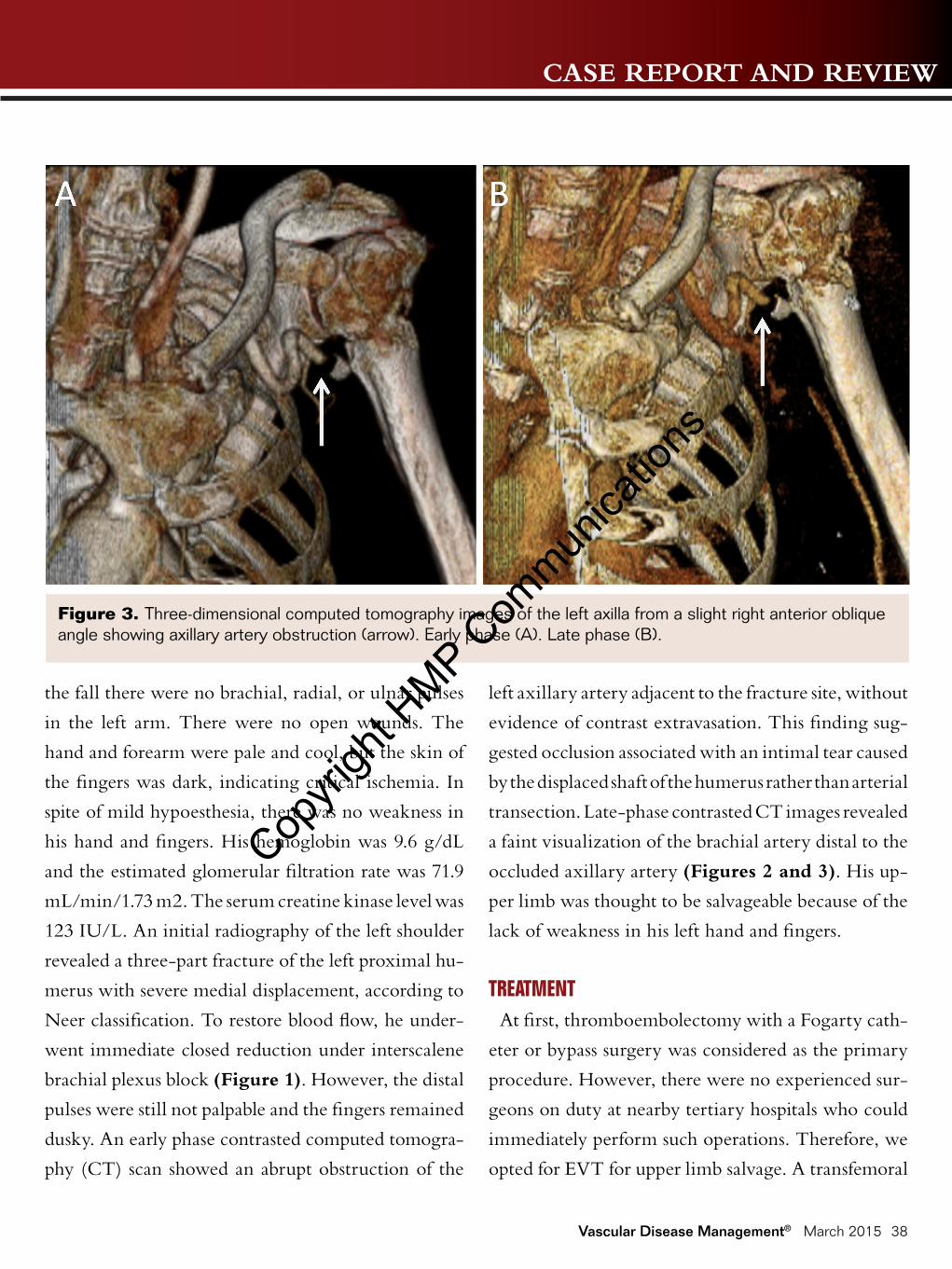
dusky. An early phase contrasted computed tomogra-
phy (CT) scan showed an abrupt obstruction of the
left axillary artery adjacent to the fracture site, without
evidence of contrast extravasation. This finding sug-
gested occlusion associated with an intimal tear caused
by the displaced shaft of the humerus rather than arterial
transection. Late-phase contrasted CT images revealed
a faint visualization of the brachial artery distal to the
occluded axillary artery (Figures 2 and 3). His up-
per limb was thought to be salvageable because of the
lack of weakness in his left hand and fingers.
TREATMENTAt first, thromboembolectomy with a Fogarty cath-
eter or bypass surgery was considered as the primary
procedure. However, there were no experienced sur-
geons on duty at nearby tertiary hospitals who could
immediately perform such operations. Therefore, we
opted for EVT for upper limb salvage. A transfemoral
Figure 3. Three-dimensional computed tomography images of the left axilla from a slight right anterior oblique angle showing axillary artery obstruction (arrow). Early phase (A). Late phase (B).
Copyri
ght H
MP Com
munica
tions
CASE REPORT AND REVIEW
Vascular Disease Management® March 2015 39
approach under local anesthesia was used, with place-
ment of a 6 Fr sheath into the right common femoral
artery. Following intravenous heparin (5,000 U), se-
lective angiography was performed. The axillary artery
was occluded proximally without the visualization of
distal vessels. A 6 Fr JR4 Launcher guiding catheter
(Medtronic) was placed in the left subclavian artery
with the aid of a 0.035˝ angled soft guidewire. Next, a
5 Fr JR4 Trail diagnostic catheter (Fukuda Denshi) was
introduced using the mother-child technique. With
the support of the 5 Fr diagnostic catheter antegradely
advanced to a point just proximal to the occluded part
of the artery, the guidewire was successfully passed
through the occlusion and reached the brachial ar-
tery. The delivery of a Thrombuster II 6 Fr aspiration
catheter (Kaneka Medix) from a femoral artery was
not possible because of the tortuous axillosubclavian
artery. Therefore, we tried to gain access through the
brachial artery. The brachial artery provides a more
direct, shorter, and less tortuous approach for the treat-
ment of axillary artery lesions.
Although several attempts at brachial artery puncture
were unsuccessful using a 0.035˝ antegrade guidewire
as a landmark to obtain retrograde access, we achieved
radial access under fluoroscopic guidance and intro-
duced a 6 Fr long sheath. A 0.018˝ Thruway guidewire
(Boston Scientific) was passed through the lesion in
retrograde fashion, advanced into the guiding catheter
in the left subclavian artery, and retrieved with a Goose-
neck snare (ev3) to gain sufficient backup force. Next,
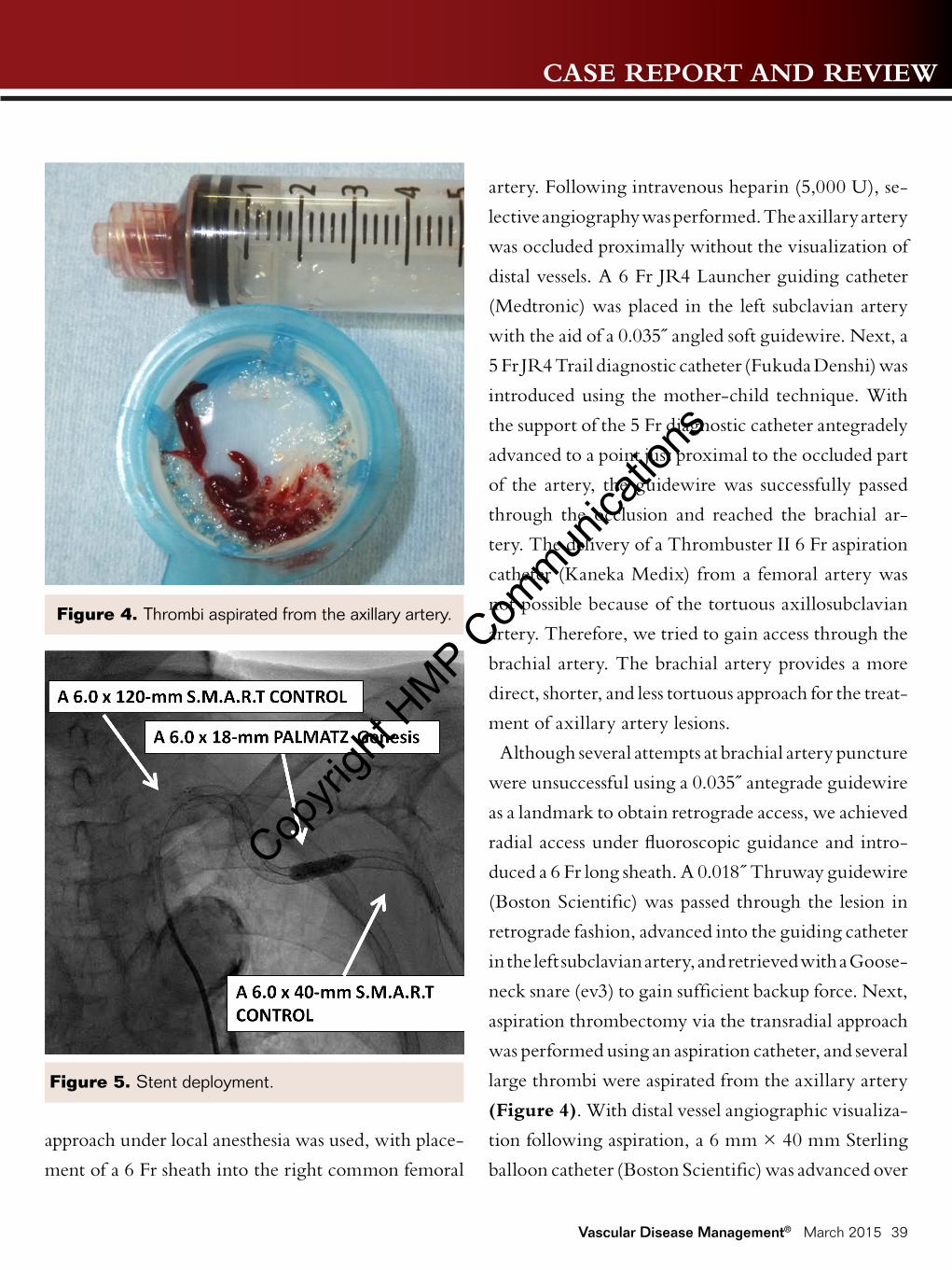
aspiration thrombectomy via the transradial approach
was performed using an aspiration catheter, and several
large thrombi were aspirated from the axillary artery
(Figure 4). With distal vessel angiographic visualiza-
tion following aspiration, a 6 mm × 40 mm Sterling
balloon catheter (Boston Scientific) was advanced over
Figure 4. Thrombi aspirated from the axillary artery.
Figure 5. Stent deployment.
Copyri
ght H
MP Com
munica
tions
CASE REPORT AND REVIEW
Vascular Disease Management® March 2015 40
the guidewire. The balloon was inflated at 8 atmo-
spheres for 60 seconds. After the balloon dilatation,
the distal blood flow was only partially restored and
the cyanosis of the upper limb did not resolve.
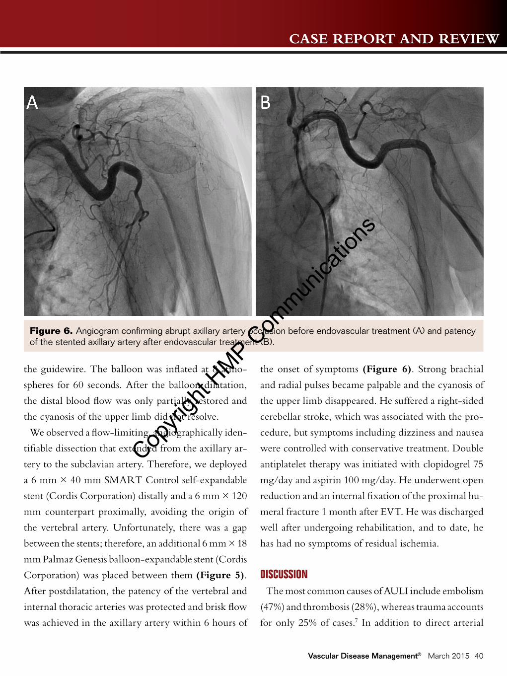
We observed a flow-limiting, angiographically iden-
tifiable dissection that extended from the axillary ar-
tery to the subclavian artery. Therefore, we deployed
a 6 mm × 40 mm SMART Control self-expandable
stent (Cordis Corporation) distally and a 6 mm × 120
mm counterpart proximally, avoiding the origin of
the vertebral artery. Unfortunately, there was a gap
between the stents; therefore, an additional 6 mm × 18
mm Palmaz Genesis balloon-expandable stent (Cordis
Corporation) was placed between them (Figure 5).
After postdilatation, the patency of the vertebral and
internal thoracic arteries was protected and brisk flow
was achieved in the axillary artery within 6 hours of
the onset of symptoms (Figure 6). Strong brachial
and radial pulses became palpable and the cyanosis of
the upper limb disappeared. He suffered a right-sided
cerebellar stroke, which was associated with the pro-
cedure, but symptoms including dizziness and nausea
were controlled with conservative treatment. Double
antiplatelet therapy was initiated with clopidogrel 75
mg/day and aspirin 100 mg/day. He underwent open
reduction and an internal fixation of the proximal hu-
meral fracture 1 month after EVT. He was discharged
well after undergoing rehabilitation, and to date, he
has had no symptoms of residual ischemia.
DISCUSSIONThe most common causes of AULI include embolism
(47%) and thrombosis (28%), whereas trauma accounts
for only 25% of cases.7 In addition to direct arterial
Figure 6. Angiogram confirming abrupt axillary artery occlusion before endovascular treatment (A) and patency of the stented axillary artery after endovascular treatment (B).
Copyri
ght H
MP Com
munica
tions
CASE REPORT AND REVIEW
Vascular Disease Management® March 2015 41
trauma, remote arterial trauma can cause AULI when
traction on a blood vessel causes an injury to the ves-
sel at a site distant from the evident bony or soft tissue
damage.8 In particular, the violent overstretching of
an artery under hyperabduction can precipitate acute
arterial injuries, which can involve a total or partial
rupture of all arterial layers or intimal damage only,
causing lumen occlusion.3,9 Although axillary artery
injury sometimes occurs with shoulder dislocation and
clavicle fractures, it is rarely concomitant with proxi-
mal humeral fractures.10
Although open surgery has become the mainstay of
primary treatment for acute upper limb ischemia re-
lated to trauma, the procedure is technically demand-
ing, even for experienced operators. Furthermore, it
often takes time to obtain reperfusion because of the
anatomical complexity of the arteries that are adjacent
to the clavicle and brachial plexus. In contrast, throm-
boembolectomy with a Fogarty catheter has gained
widespread acceptance as first-line treatment for the
management of nontraumatic AULI. The rate of limb
salvage following thromboembolectomy has been re-
ported as 98%.11
EVT is seldom indicated, and consequently has been
rarely used as the concurrent treatment of both trau-
matic and nontraumatic AULI. To our knowledge,
there are few case reports on EVT for occluded axil-
losubclavian arteries associated with blunt trauma, in-
cluding clavicular and scapular fracture.5,6 However, no
reports on EVT for an occluded axillary artery related
to proximal humeral fracture have been published.
This may be because the number of cases is small. The
humerus is more distant from the axillary artery than
the clavicle and scapula, and fractures of the humerus
rarely compromise the axillary artery.
The occurrence of AULI represents an emergency
indication for surgical intervention to prevent gan-
grene of the affected extremity. If the arm is to be
successfully salvaged in patients with AULI, revascu-
larization should be achieved within 6 hours of the
onset of symptoms.2 Therefore, an immediate access
to vascular surgeons capable of accomplishing complex
operations is crucial. If the restoration of blood flow is
delayed, there is a risk of acute renal failure secondary to
myoglobinemia. However, the immediate availability
of skilled vascular surgeons may be limited, particu-
larly in rural areas in Japan. In this setting, EVT is a
viable alternative to surgery. In the present case, EVT
was used to prevent critical ischemia and successfully
salvage the upper limb of the patient.
In general, EVT is feasible unless there is hemody-
namic instability, vessel transection, or the absence of
an adequate proximal vascular fixation site.4 One ma-
jor advantage of EVT over open surgical repair is that
general anesthesia is not required, enabling faster revas-
cularization and minimizing blood loss. Furthermore,
EVT can be immediately performed after diagnostic
angiography, in which vascular access remote from the
local injury is obtained. Kim et al described the efficacy
of percutaneous aspiration thromboembolectomy us-
ing a 6 Fr or 7 Fr guiding catheter via a transbrachial
approach with placement of a 7 Fr sheath.12 Successful
recanalization was achieved in all 11 patients enrolled
in that study, although no cases of concomitant trauma
such as fractures were included. In our case, although
we made several attempts under fluoroscopic guidance,
we were unable to obtain brachial access. Although
ultrasound-guided retrograde arterial access in the
Copyri
ght H
MP Com
munica
tions
CASE REPORT AND REVIEW
Vascular Disease Management® March 2015 42
brachial artery would have made the procedure faster,
we were inexperienced in the procedure. Therefore,
we opted for a more familiar procedure, radial access
under fluoroscopic guidance. Brachial access usually
allows the use of a 7 Fr sheath. Instead, we obtained
radial access and selected a 6-Fr sheath because a 7-Fr
one was too large to insert into his radial artery. Despite
use of a 6 Fr aspiration catheter, by repeating aspira-
tion and stent placement, a large amount of thrombus
was aspirated and acceptable reperfusion was achieved
within the critical period for limb salvage.
Although 5- to 10-month patency has been reported
in a few cases,13 the long-term results of deploying
stents in the axillary artery have not yet been well
investigated. In the present case we had no alterna-
tive because upper limb ischemia persisted even after
balloon dilatation and the aspiration of the thrombus.
Long-term follow-up is required to confirm stent
patency in this patient.
We believe that, for choosing the type of stent for
axillary artery, the following factors are important:
acceptable flexibility and sufficient radial strength.
SMART Control stents demonstrate these factors.
Therefore, we used these stents to cover the axillary
artery lesion. In general, the axillary artery and super-
ficial femoral artery are similar because they have high
mobility. We speculate that the clinical effectiveness
of the SMART Control stents for both axillary artery
and superficial femoral artery lesions may be similar.
The evidence of the long-term result of the SMART
Control stent for superficial femoral artery lesions has
been established.14 As for a balloon-expandable stent
(Palmaz Genesis), although it was not suitable for ax-
illary artery lesions because of the lack of flexibility,
we had to use it for covering a short gap between the
two SMART Control stents. From our experiences, a
balloon-expandable stent does not increase the likeli-
hood of stent strut fracture if it is short.
There are potential procedure-related complica-
tions associated with the use of EVT for acute axil-
losubclavian artery occlusion secondary to trauma.
The risk of distal embolization during recanalization
of an occlusion must be considered. The placement of
the stent near the origin of the vertebral artery should
be avoided to prevent vertebrobasilar embolization.
Consideration must also be given to the possibility
that traversing the occlusive lesions with a guidewire
may cause bleeding because the occlusion may be
the result of partial or complete arterial transection.
Therefore, a CT must be performed before the pro-
cedure to determine whether there is a hematoma
around the artery or a hemothorax. Either of these
findings suggests the transection of the artery, in
which case open surgical repair is a preferred treat-
ment option.
CONCLUSIONIn the management of AULI, treatment should be
initiated as early as possible after the onset of isch-
emic symptoms. EVT offers a minimally invasive and
prompt alternative to open surgery or thromboembo-
lectomy with a Fogarty catheter for AULI resulting
from an occluded axillary artery. n
Editor’s note: Disclosure: The authors have completed
and returned the ICMJE Form for Disclosure of Potential
Conflicts of Interest The authors report no disclosures related
to the content herein.
Copyri
ght H
MP Com
munica
tions
CASE REPORT AND REVIEW
Vascular Disease Management® March 2015 43
Manuscript received September 8, 2014; provisional ac-
ceptance given November 3, 2014; final manuscript accepted
December 5, 2014.
Address for correspondence: Tsuyoshi Isawa, MD, Sendai
Kousei Hospital, Cardiology, 4-15 Hirose-machi, Sendai,
Miyagi, Japan. Email: [email protected]
REFERENCES1. Eyers P, Earnshaw JJ. Acute non-traumatic arm isch-
aemia. Br J Surg. 1998;85(10):1340-1346.2. Miller HH, Welch CS. Quantitative studies on
the time factor in arterial injuries. Ann Surg. 1949;130:428-438.
3. Modi CS, Nnene CO, Godsiff SP, Esler CN. Axil-lary artery injury secondary to displaced proximal humeral fractures: a report of two cases. J Orthop Surg. 2008;16(2):243-246.
4. DuBose JJ, Rajani R, Gilani R, et al. Endovas-cular management of axillo-subclavian arterial in-jury: a review of published experience. Injury. 2012;43(11):1785-1792.
5. IIkay E, Rahman A, Ozdemir H, et al. Endovascular stent management of acute traumatic subclavian artery occlusion by intimal flap. Eur J Vasc Endovasc Surg Ex-tra. 2003;6:32-35.
6. Molloy S, Jacob S, Buckenham T, Taylor RS. Percu-taneous repair of an acute traumatic subclavian artery
occlusion. Eur J Vasc Endovasc Surg. 2001;21(1):75-76.7. Turner EJH, Loh A, Howard A. Systematic review of
the operative and non-operative management of acute upper limb ischemia. J Vasc Nurs. 2012;30(3):71-76.
8. Quraishy MS, Cawthorn SJ, Giddings AEB. Criti-cal ischaemia of the upper limb. J R Soc Med. 1992;85(5):269-273.
9. Theodorides T, de Keizer C. Injuries of the axillary artery caused by fractures of the neck of the humerus. Injury. 1976;8(2):120-123.
10. Yagubyan M, Panneton JM. Axillary artery injury from humeral neck fracture: a rare but disabling trau-matic event. Vasc Endovasc Surg. 2004;38(2):175-184.
11. Hernandez-Richter T, Angele MK, Helmberger T, et al. Acute ischemia of the upper extremity: long-term results following thrombembolectomy with the Fogarty catheter. Langenbecks Arch Surg. 2001;386(4):261-266.
12. Kim SK, Kwak HS, Chung GH, Han YM. Acute upper limb ischemia due to cardiac origin thrombo-embolism: the usefulness of percutaneous aspiration thromboembolectomy via a transbrachial approach. Korean J Radiol. 2011;12(5):595-601.
13. Vijayvergiya R, Yadav M, Grover A. Percutaneous endovascular management of atherosclerotic axillary artery stenosis: report of 2 cases and review of litera-ture. World J Cardiol. 2011;3(5):165-168.
14. Suzuki K, Iida O, Soga Y, et al. Long-term results of the S.M.A.R.T. ControlTM stent for superfi-cial femoral artery lesions, J-SMART registry. Circ J. 2011;75(4):939-944.
Copyri
ght H
MP Com
munica
tions