uroporphyrinogen decarboxylation as a benchmark for … · uroporphyrinogen decarboxylation as a...
TRANSCRIPT
Uroporphyrinogen decarboxylation as a benchmarkfor the catalytic proficiency of enzymesCharles A. Lewis, Jr. and Richard Wolfenden1
Department of Biochemistry and Biophysics, University of North Carolina, Chapel Hill, NC 27599
Contributed by Richard Wolfenden, October 1, 2008 (sent for review September 3, 2008)
The magnitude of an enzyme’s affinity for the altered substrate inthe transition state exceeds its affinity for the substrate in theground state by a factor matching the rate enhancement that theenzyme produces. Particularly remarkable are those enzymes thatact as simple protein catalysts, without the assistance of metals orother cofactors. To determine the extent to which one suchenzyme, human uroporphyrinogen decarboxylase, enhances therate of substrate decarboxylation, we examined the rate of spon-taneous decarboxylation of pyrrolyl-3-acetate. Extrapolation offirst-order rate constants measured at elevated temperatures in-dicates that this reaction proceeds with a half-life of 2.3 � 109 yearsat 25 °C in the absence of enzyme. This enzyme shows no signif-icant homology with orotidine 5�-monophosphate decarboxylase(ODCase), another cofactorless enzyme that catalyzes a very slowreaction. It is proposed that, in both cases, a protonated basicresidue (Arg-37 in the case of human UroD; Lys-93 in the case of yeastODCase) furnishes a counterion that helps the scissile carboxylategroup of the substrate leave water and enter a relatively nonpolarenvironment, stabilizes the incipient carbanion generated by thedeparture of CO2, and supplies the proton that takes its place.
decarboxylase � catalysis � porphyrin � coproporphyrinogen � evolution
The catalytic power of an enzyme can be judged from the rateenhancement that it produces. In general, enzymes act on
their substrates at somewhat similar rates, with kcat valuesranging between 50 and 5,000 s�1. However, the rate constantsof the same reactions in the absence of a catalyst vary over arange of at least 15 orders of magnitude. And the rate enhance-ments produced by enzymes vary over a similarly broad range(�1015-fold), indicating the magnitude of the increase in theenzyme’s affinity for the substrate as it passes from the groundstate to the transition state (1). Particularly remarkable are thosecases in which the enzyme acts as a simple protein catalyst,without the assistance of metals or other cofactors. Such areaction, the decarboxylation of orotidine 5�-phosphate, wasfound earlier to proceed in the absence of enzyme with ahalf-time of 7.8 � 107 years in the absence of enzyme (2).
Here, we describe another reaction, involving a very differentsubstrate (uroporphyrinogen, Uro’gen), that proceeds with ahalf-life of 2.3 � 109 years at 25 °C in the absence of enzyme. Theenzymes catalyzing these reactions act as pure protein catalysts.Their amino acid sequences show no significant homology.
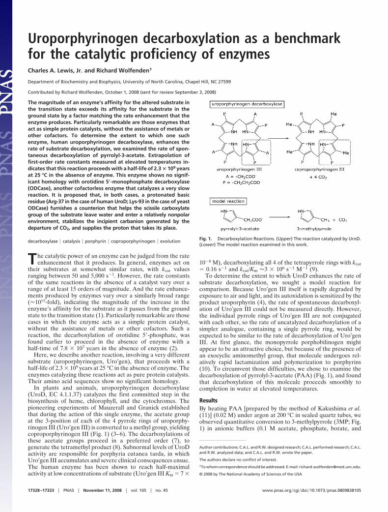
In plants and animals, uroporphyrinogen decarboxylase(UroD, EC 4.1.1.37) catalyzes the first committed step in thebiosynthesis of heme, chlorophyll, and the cytochromes. Thepioneering experiments of Mauzerall and Granick establishedthat during the action of this single enzyme, the acetate groupat the 3-position of each of the 4 pyrrole rings of uroporphy-rinogen III (Uro’gen III) is converted to a methyl group, yieldingcoproporphyrinogen III (Fig. 1) (3–6). The decarboxylations ofthese acetate groups proceed in a preferred order (7), togenerate the tetramethyl product (8). Subnormal levels of UroDactivity are responsible for porphyria cutanea tarda, in whichUro’gen III accumulates and severe clinical consequences ensue.The human enzyme has been shown to reach half-maximalactivity at low concentrations of substrate (Uro’gen III Km � 7 �
10�8 M), decarboxylating all 4 of the tetrapyrrole rings with kcat� 0.16 s�1 and kcat/Km �3 � 106 s�1 M�1 (9).
To determine the extent to which UroD enhances the rate ofsubstrate decarboxylation, we sought a model reaction forcomparison. Because Uro’gen III itself is rapidly degraded byexposure to air and light, and its autoxidation is sensitized by theproduct uroporphyrin (4), the rate of spontaneous decarboxyl-ation of Uro’gen III could not be measured directly. However,the individual pyrrole rings of Uro’gen III are not conjugatedwith each other, so the rate of uncatalyzed decarboxylation of asimpler analogue, containing a single pyrrole ring, would beexpected to be similar to the rate of decarboxylation of Uro’genIII. At first glance, the monopyrrole porphobilinogen mightappear to be an attractive choice, but because of the presence ofan exocyclic aminomethyl group, that molecule undergoes rel-atively rapid lactamization and polymerization to porphyrins(10). To circumvent those difficulties, we chose to examine thedecarboxylation of pyrrolyl-3-acetate (PAA) (Fig. 1), and foundthat decarboxylation of this molecule proceeds smoothly tocompletion in water at elevated temperatures.
ResultsBy heating PAA [prepared by the method of Kakushima et al.(11)] (0.02 M) under argon at 200 °C in sealed quartz tubes, weobserved quantitative conversion to 3-methylpyrrole (3MP; Fig.1) in anionic buffers (0.1 M acetate, phosphate, borate, and
Author contributions: C.A.L. and R.W. designed research; C.A.L. performed research; C.A.L.and R.W. analyzed data; and C.A.L. and R.W. wrote the paper.
The authors declare no conflict of interest.
1To whom correspondence should be addressed. E-mail: richard�[email protected].
© 2008 by The National Academy of Sciences of the USA
Fig. 1. Decarboxylation Reactions. (Upper) The reaction catalyzed by UroD.(Lower) The model reaction examined in this work.
17328–17333 � PNAS � November 11, 2008 � vol. 105 � no. 45 www.pnas.org�cgi�doi�10.1073�pnas.0809838105
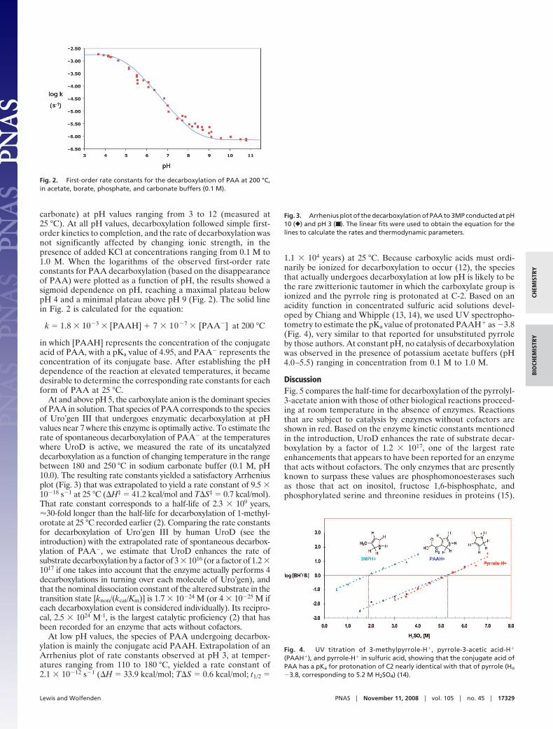
carbonate) at pH values ranging from 3 to 12 (measured at25 °C). At all pH values, decarboxylation followed simple first-order kinetics to completion, and the rate of decarboxylation wasnot significantly affected by changing ionic strength, in thepresence of added KCl at concentrations ranging from 0.1 M to1.0 M. When the logarithms of the observed first-order rateconstants for PAA decarboxylation (based on the disappearanceof PAA) were plotted as a function of pH, the results showed asigmoid dependence on pH, reaching a maximal plateau belowpH 4 and a minimal plateau above pH 9 (Fig. 2). The solid linein Fig. 2 is calculated for the equation:
k � 1.8 � 10�3 � [PAAH] � 7 � 10�7 � [PAA�] at 200 °C
in which [PAAH] represents the concentration of the conjugateacid of PAA, with a pKa value of 4.95, and PAA� represents theconcentration of its conjugate base. After establishing the pHdependence of the reaction at elevated temperatures, it becamedesirable to determine the corresponding rate constants for eachform of PAA at 25 °C.
At and above pH 5, the carboxylate anion is the dominant speciesof PAA in solution. That species of PAA corresponds to the speciesof Uro’gen III that undergoes enzymatic decarboxylation at pHvalues near 7 where this enzyme is optimally active. To estimate therate of spontaneous decarboxylation of PAA� at the temperatureswhere UroD is active, we measured the rate of its uncatalyzeddecarboxylation as a function of changing temperature in the rangebetween 180 and 250 °C in sodium carbonate buffer (0.1 M, pH10.0). The resulting rate constants yielded a satisfactory Arrheniusplot (Fig. 3) that was extrapolated to yield a rate constant of 9.5 �10�18 s�1 at 25 °C (�H‡ � 41.2 kcal/mol and T�S‡ � 0.7 kcal/mol).That rate constant corresponds to a half-life of 2.3 � 109 years,�30-fold longer than the half-life for decarboxylation of 1-methyl-orotate at 25 °C recorded earlier (2). Comparing the rate constantsfor decarboxylation of Uro’gen III by human UroD (see theintroduction) with the extrapolated rate of spontaneous decarbox-ylation of PAA�, we estimate that UroD enhances the rate ofsubstrate decarboxylation by a factor of 3 � 1016 (or a factor of 1.2 �1017 if one takes into account that the enzyme actually performs 4decarboxylations in turning over each molecule of Uro’gen), andthat the nominal dissociation constant of the altered substrate in thetransition state [knon/(kcat/Km)] is 1.7 � 10�24 M (or 4 � 10�25 M ifeach decarboxylation event is considered individually). Its recipro-cal, 2.5 � 1024 M-1, is the largest catalytic proficiency (2) that hasbeen recorded for an enzyme that acts without cofactors.
At low pH values, the species of PAA undergoing decarbox-ylation is mainly the conjugate acid PAAH. Extrapolation of anArrhenius plot of rate constants observed at pH 3, at temper-atures ranging from 110 to 180 °C, yielded a rate constant of2.1 � 10�12 s�1 (�H � 33.9 kcal/mol; T�S � 0.6 kcal/mol; t1/2 �
1.1 � 104 years) at 25 °C. Because carboxylic acids must ordi-narily be ionized for decarboxylation to occur (12), the speciesthat actually undergoes decarboxylation at low pH is likely to bethe rare zwitterionic tautomer in which the carboxylate group isionized and the pyrrole ring is protonated at C-2. Based on anacidity function in concentrated sulfuric acid solutions devel-oped by Chiang and Whipple (13, 14), we used UV spectropho-tometry to estimate the pKa value of protonated PAAH� as �3.8(Fig. 4), very similar to that reported for unsubstituted pyrroleby those authors. At constant pH, no catalysis of decarboxylationwas observed in the presence of potassium acetate buffers (pH4.0–5.5) ranging in concentration from 0.1 M to 1.0 M.
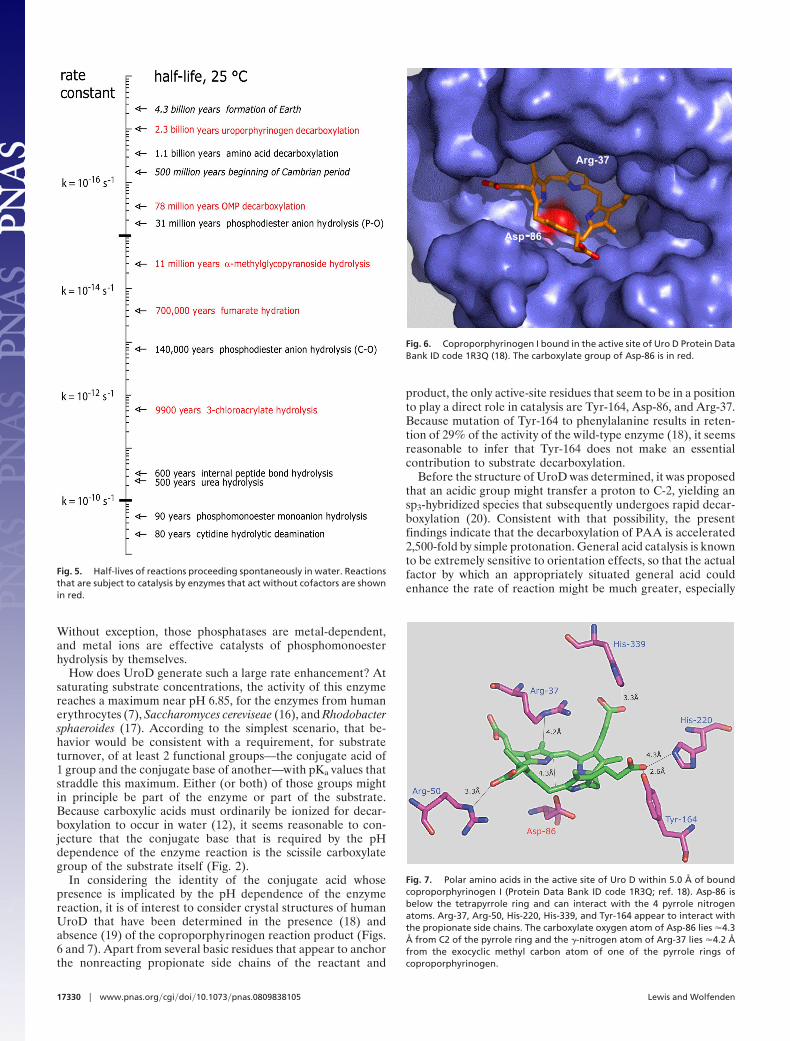
DiscussionFig. 5 compares the half-time for decarboxylation of the pyrrolyl-3-acetate anion with those of other biological reactions proceed-ing at room temperature in the absence of enzymes. Reactionsthat are subject to catalysis by enzymes without cofactors areshown in red. Based on the enzyme kinetic constants mentionedin the introduction, UroD enhances the rate of substrate decar-boxylation by a factor of 1.2 � 1017, one of the largest rateenhancements that appears to have been reported for an enzymethat acts without cofactors. The only enzymes that are presentlyknown to surpass these values are phosphomonoesterases suchas those that act on inositol, fructose 1,6-bisphosphate, andphosphorylated serine and threonine residues in proteins (15).
Fig. 2. First-order rate constants for the decarboxylation of PAA at 200 °C,in acetate, borate, phosphate, and carbonate buffers (0.1 M).
Fig. 3. Arrhenius plot of the decarboxylation of PAA to 3MP conducted at pH10 (}) and pH 3 (■ ). The linear fits were used to obtain the equation for thelines to calculate the rates and thermodynamic parameters.
Fig. 4. UV titration of 3-methylpyrrole-H�, pyrrole-3-acetic acid-H�
(PAAH�), and pyrrole-H� in sulfuric acid, showing that the conjugate acid ofPAA has a pKa for protonation of C2 nearly identical with that of pyrrole (Ho
�3.8, corresponding to 5.2 M H2SO4) (14).
Lewis and Wolfenden PNAS � November 11, 2008 � vol. 105 � no. 45 � 17329
CHEM
ISTR
YBI
OCH
EMIS
TRY
Without exception, those phosphatases are metal-dependent,and metal ions are effective catalysts of phosphomonoesterhydrolysis by themselves.
How does UroD generate such a large rate enhancement? Atsaturating substrate concentrations, the activity of this enzymereaches a maximum near pH 6.85, for the enzymes from humanerythrocytes (7), Saccharomyces cereviseae (16), and Rhodobactersphaeroides (17). According to the simplest scenario, that be-havior would be consistent with a requirement, for substrateturnover, of at least 2 functional groups—the conjugate acid of1 group and the conjugate base of another—with pKa values thatstraddle this maximum. Either (or both) of those groups mightin principle be part of the enzyme or part of the substrate.Because carboxylic acids must ordinarily be ionized for decar-boxylation to occur in water (12), it seems reasonable to con-jecture that the conjugate base that is required by the pHdependence of the enzyme reaction is the scissile carboxylategroup of the substrate itself (Fig. 2).
In considering the identity of the conjugate acid whosepresence is implicated by the pH dependence of the enzymereaction, it is of interest to consider crystal structures of humanUroD that have been determined in the presence (18) andabsence (19) of the coproporphyrinogen reaction product (Figs.6 and 7). Apart from several basic residues that appear to anchorthe nonreacting propionate side chains of the reactant and
product, the only active-site residues that seem to be in a positionto play a direct role in catalysis are Tyr-164, Asp-86, and Arg-37.Because mutation of Tyr-164 to phenylalanine results in reten-tion of 29% of the activity of the wild-type enzyme (18), it seemsreasonable to infer that Tyr-164 does not make an essentialcontribution to substrate decarboxylation.
Before the structure of UroD was determined, it was proposedthat an acidic group might transfer a proton to C-2, yielding ansp3-hybridized species that subsequently undergoes rapid decar-boxylation (20). Consistent with that possibility, the presentfindings indicate that the decarboxylation of PAA is accelerated2,500-fold by simple protonation. General acid catalysis is knownto be extremely sensitive to orientation effects, so that the actualfactor by which an appropriately situated general acid couldenhance the rate of reaction might be much greater, especially
Fig. 5. Half-lives of reactions proceeding spontaneously in water. Reactionsthat are subject to catalysis by enzymes that act without cofactors are shownin red.
Arg-37
Asp-86
Fig. 6. Coproporphyrinogen I bound in the active site of Uro D Protein DataBank ID code 1R3Q (18). The carboxylate group of Asp-86 is in red.
Fig. 7. Polar amino acids in the active site of Uro D within 5.0 Å of boundcoproporphyrinogen I (Protein Data Bank ID code 1R3Q; ref. 18). Asp-86 isbelow the tetrapyrrole ring and can interact with the 4 pyrrole nitrogenatoms. Arg-37, Arg-50, His-220, His-339, and Tyr-164 appear to interact withthe propionate side chains. The carboxylate oxygen atom of Asp-86 lies �4.3Å from C2 of the pyrrole ring and the �-nitrogen atom of Arg-37 lies �4.2 Åfrom the exocyclic methyl carbon atom of one of the pyrrole rings ofcoproporphyrinogen.
17330 � www.pnas.org�cgi�doi�10.1073�pnas.0809838105 Lewis and Wolfenden
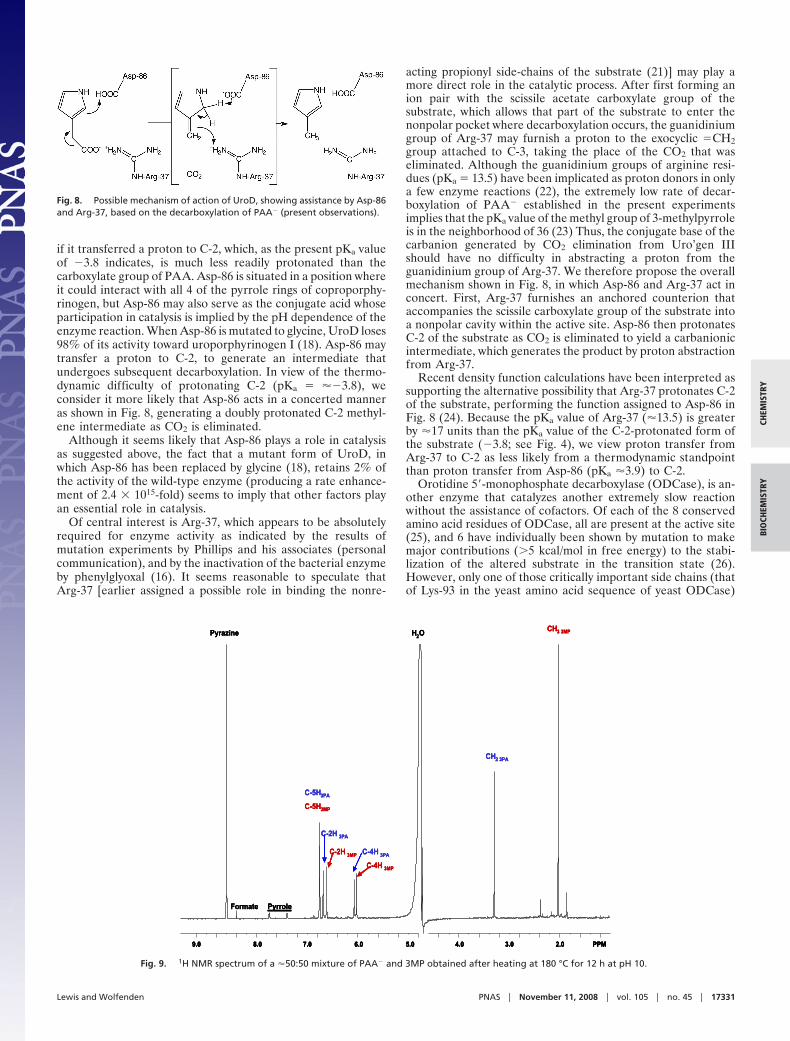
if it transferred a proton to C-2, which, as the present pKa valueof �3.8 indicates, is much less readily protonated than thecarboxylate group of PAA. Asp-86 is situated in a position whereit could interact with all 4 of the pyrrole rings of coproporphy-rinogen, but Asp-86 may also serve as the conjugate acid whoseparticipation in catalysis is implied by the pH dependence of theenzyme reaction. When Asp-86 is mutated to glycine, UroD loses98% of its activity toward uroporphyrinogen I (18). Asp-86 maytransfer a proton to C-2, to generate an intermediate thatundergoes subsequent decarboxylation. In view of the thermo-dynamic difficulty of protonating C-2 (pKa � ��3.8), weconsider it more likely that Asp-86 acts in a concerted manneras shown in Fig. 8, generating a doubly protonated C-2 methyl-ene intermediate as CO2 is eliminated.
Although it seems likely that Asp-86 plays a role in catalysisas suggested above, the fact that a mutant form of UroD, inwhich Asp-86 has been replaced by glycine (18), retains 2% ofthe activity of the wild-type enzyme (producing a rate enhance-ment of 2.4 � 1015-fold) seems to imply that other factors playan essential role in catalysis.
Of central interest is Arg-37, which appears to be absolutelyrequired for enzyme activity as indicated by the results ofmutation experiments by Phillips and his associates (personalcommunication), and by the inactivation of the bacterial enzymeby phenylglyoxal (16). It seems reasonable to speculate thatArg-37 [earlier assigned a possible role in binding the nonre-
acting propionyl side-chains of the substrate (21)] may play amore direct role in the catalytic process. After first forming anion pair with the scissile acetate carboxylate group of thesubstrate, which allows that part of the substrate to enter thenonpolar pocket where decarboxylation occurs, the guanidiniumgroup of Arg-37 may furnish a proton to the exocyclic �CH2group attached to C-3, taking the place of the CO2 that waseliminated. Although the guanidinium groups of arginine resi-dues (pKa � 13.5) have been implicated as proton donors in onlya few enzyme reactions (22), the extremely low rate of decar-boxylation of PAA� established in the present experimentsimplies that the pKa value of the methyl group of 3-methylpyrroleis in the neighborhood of 36 (23) Thus, the conjugate base of thecarbanion generated by CO2 elimination from Uro’gen IIIshould have no difficulty in abstracting a proton from theguanidinium group of Arg-37. We therefore propose the overallmechanism shown in Fig. 8, in which Asp-86 and Arg-37 act inconcert. First, Arg-37 furnishes an anchored counterion thataccompanies the scissile carboxylate group of the substrate intoa nonpolar cavity within the active site. Asp-86 then protonatesC-2 of the substrate as CO2 is eliminated to yield a carbanionicintermediate, which generates the product by proton abstractionfrom Arg-37.
Recent density function calculations have been interpreted assupporting the alternative possibility that Arg-37 protonates C-2of the substrate, performing the function assigned to Asp-86 inFig. 8 (24). Because the pKa value of Arg-37 (�13.5) is greaterby �17 units than the pKa value of the C-2-protonated form ofthe substrate (�3.8; see Fig. 4), we view proton transfer fromArg-37 to C-2 as less likely from a thermodynamic standpointthan proton transfer from Asp-86 (pKa �3.9) to C-2.
Orotidine 5�-monophosphate decarboxylase (ODCase), is an-other enzyme that catalyzes another extremely slow reactionwithout the assistance of cofactors. Of each of the 8 conservedamino acid residues of ODCase, all are present at the active site(25), and 6 have individually been shown by mutation to makemajor contributions (�5 kcal/mol in free energy) to the stabi-lization of the altered substrate in the transition state (26).However, only one of those critically important side chains (thatof Lys-93 in the yeast amino acid sequence of yeast ODCase)
PPM8.0 6.0 4.0 2.05.0 3.07.09.0
Pyrazine
C-5H3PA
C-5H3MP
C-2H 3PA
C-2H 3MP C-4H 3PA
C-4H 3MP
CH2 3PA
CH3 3MPH2O
PyrroleFormate
PPM8.0 6.0 4.0 2.05.0 3.07.09.0
Pyrazine
C-5H3PA
C-5H3MP
C-2H 3PA
C-2H 3MP C-4H 3PA
C-4H 3MP
CH2 3PA
CH3 3MPH2O
PyrroleFormate
PPM8.0 6.0 4.0 2.05.0 3.07.09.0 PPM8.0 6.0 4.0 2.05.0 3.07.09.0
Pyrazine
C-5H3PA
C-5H3MP
C-2H 3PA
C-2H 3MP C-4H 3PA
C-4H 3MP
CH2 3PA
CH3 3MPH2O
PyrroleFormate
Fig. 9. 1H NMR spectrum of a �50:50 mixture of PAA� and 3MP obtained after heating at 180 °C for 12 h at pH 10.
Fig. 8. Possible mechanism of action of UroD, showing assistance by Asp-86and Arg-37, based on the decarboxylation of PAA� (present observations).
Lewis and Wolfenden PNAS � November 11, 2008 � vol. 105 � no. 45 � 17331
CHEM
ISTR
YBI
OCH
EMIS
TRY
makes close contact with the pyrimidine ring in such a way thatit might be expected to stabilize a carbanion at the positionwhere decarboxylation occurs. Other active-site residues ofODCase are believed to play a supporting role in orienting thesubstrate and organizing the closure of loops of the active sitearound the altered substrate in the transition state (26–28).
Interestingly, comparison of amino acid sequences reveals nosignificant homology (8%) between UroD and ODCase. Not-withstanding that lack of homology, we infer that the evolutionof these 2 extraordinarily proficient enzymes, UroD andODCase, may have arrived at a common strategy of catalysis, inwhich unusually large rate enhancements are largely achievedthrough the action of a single nitrogenous side chain. In itsprotonated form, that basic residue (Arg-37 in the case of humanUroD; Lys-93 in the case of yeast ODCase) furnishes a coun-terion that helps the scissile carboxylate group of the substrateleave water and enter a relatively nonpolar environment, stabi-lizes the incipient carbanion generated by the departure of CO2,and supplies the proton that takes its place. Preliminary exper-iments (C.A.L. and R.W., unpublished data) indicate that thedecarboxylation of 1-cyclohexylorotate, like that of benzisox-azole-3-carboxylic acid (29), is greatly accelerated by transfer tononpolar solvents in the presence of amine counterions.
Because heme proteins play a central role in the metabolismof modern organisms, it is natural to inquire how evolutionovercame the kinetic obstacle raised by the 2.3-billion-yearhalf-life for uncatalyzed decarboxylation of uroporphyrinogen.A primitive decarboxylase that accelerated uroporphyrinogendecarboxylation by a factor of 1 million would have reduced thehalf-life of the reaction to 2.3 thousand years, offering little orno selective advantage to the host organism. But the potentialbiological usefulness of decarboxylated tetrapyrroles would havebecome apparent only with the appearance of an oxidizingatmosphere. Before that stage in evolution, the binding ofoxygen by heme proteins was probably unnecessary, and iron-sulfur proteins were presumably capable (at least in principle) ofperforming many of the redox functions of the cytochromes (30).And the biosynthesis of siroheme, which is present in modernnitrite and sulfite reductases and may have served other func-tions in ancient organisms, proceeds directly from uroporphy-rinogen and requires no decarboxylation (31). Taking thesealternatives into consideration, it seems probable that the en-zymes involved in uroporphyrinogen metabolism arose by mu-tation from ancient proteins with other functions, many of whichmay have existed before oxygen first appeared in the atmosphereand decarboxylation became necessary for the first time.
Materials and MethodsPAA was prepared by the method of Kakushima et al. (11). Other chemicalswere purchased from Sigma-Aldrich. Typical reaction mixtures contained PAA(2 � 10�3 M) in sodium acetate-d3, potassium phosphate, sodium borate, andsodium carbonate buffers (0.1 M) ranging in pH from 3 to 12, measured at25 °C under argon. Samples were sealed in quartz tubes under vacuum andheated in convection ovens (Barnstead/Thermolyne no. 47900) maintained at110–250 2 °C, as indicated by ASTM thermometers. After cooling, thecontents were diluted with D2O containing added pyrazine (5 � 10�4 M) as aproton integration standard (4H, � � 8.60 ppm). 1H NMR spectra were ac-quired using a Varian Inova Unity 500 MHz system equipped with a cold probeand operated by Solaris 9 software. Spectra of PAA and 3MP (�1 � 10�3 M)were readily obtained in 4 or 8 transients, with a 60-s pulse delay (Fig. 9). Themethylene protons of PAA and methyl protons of 3MP gave distinct singletresonances at 3.29–3.54 ppm (depending on pH) and 2.04 ppm, respectively.Integrated intensities using all protons for both species were consistent exceptbelow pH 5.5, where exchange of the C-2 and C-4 protons with D2O becamesignificant during the time required for NMR analysis. For that reason, theintegrated intensities of the methylene protons of PAA and the methylprotons of 3MP, which underwent no significant exchange, were used forkinetic analysis.
Effects of pH on the rate of decarboxylation of PAA were examined at200 °C, showing quantitative conversion of PAA to 3MP at rates that ap-proached a constant high value below pH 4.0 and a constant low valuebetween pH 9 and 10.5 (Fig. 2). The effects of temperature on the rate ofdecarboxylation were then examined at pH 3.0 for uncharged PAAH, and atpH 10.0 for PAA�. Times of heating were adjusted so that the reaction hadproceeded to between 15% and 85% completion. Samples were examinedbefore and after reaction to establish that no significant change in pH hadoccurred during the course of reaction. Experiments testing for potentialacetate catalysis were performed by using sodium acetate-d3 buffers at 0.1and 1.0 M at the temperature range from 110 °C to 180 °C. Effects of salt onthe uncatalyzed decarboxylation reaction were studied by comparing rates atpH 3.0 (sodium acetate-d3) and pH 10.0 (sodium carbonate) in the absence andpresence of 0.33 M or 1.0 M KCl at suitable temperatures and incubation times.
Equilibria of C-2 protonation of pyrrole derivatives in strong acid wereexamined by using the acidity function developed by Chiang and Whipple (13,14) by recording UV spectra in the presence of varying concentrations ofsulfuric acid from 0.5 to 7.5 M. The reversibility of the changes observedshowed that no decomposition occurred during the time (�3 min) requiredfor these experiments. When log [BH�]/[B] was plotted as a function of theconcentration of sulfuric acid, the half-titration point {at which log ([BH�]/[B]) � 0} was found to be 5.2 M for PAA, 5.3 M for pyrrole, and 1.8 M for 3MP(Fig. 4). Thus, the pKa value of the conjugate acid PAAH� is similar to the valuereported for pyrrole-H� (��3.8), and lower than the value reported for3-methylpyrrole-H� (�1.0) by Chiang and Whipple (14).
ACKNOWLEDGMENTS. We thank Prof. David Mauzerall (Rockefeller Univer-sity) for helpful discussions of the chemical behavior of pyrrole derivatives,and Prof. John Phillips (University of Utah) for acquainting us with the resultsof mutation experiments on human UroD, and for pointing out that each ofthe 4 decarboxylation events catalyzed by UroD can be considered to proceedat least 4 times as rapidly as the overall decarboxylation of Uro’gen. This workwas supported by National Institutes of Health Grant GM-18325.
1. Wolfenden R (2006) Degrees of difficulty of water-consuming reactions in the absenceof enzymes. Chem Rev 106:3379–3396.
2. Radzicka A, Wolfenden R (1995) A proficient enzyme. Science 267:90–93.3. Granick S (1958) Porphyrin biosynthesis in erythrocytes. I. Formation of �-aminolevu-
linic acid in erythrocytes. J Biol Chem 232:1101–1117.4. Granick S, Mauzerall D (1958) Porphyrin biosynthesis in erythrocytes. II. Enzymes
converting �-aminolevulinic acid to coproporphyrinogen. J Biol Chem 232:1119 –1140.
5. Mauzerall D, Granick S (1958) Porphyrin biosynthesis in erythrocytes. III. Uroporphy-rinogen and its decarboxylase. J Biol Chem 232:1141–1162.
6. Mauzerall D (1960) The condensation of pophobilinogen to uroporphyrinogen. J AmChem Soc 82:2605–2609.
7. Luo J, Lim CK (1993) Order of uroprophyrinogen III decarboxylation on incubation ofporphobilinogen and uroporphyrinogen III with erythrocyte uroporphyrinogen de-carboxylase. Biochem J 289:529–532.
8. Jackson AH, Sancovich HA, Ferrmola AM, et al. (1976) Macrocyclic intermediates in thebiosynthesis of porphyrins. Philos Trans R Soc Lond Ser B 273:191–206.
9. deVerneuil H, Sassa S, Kappas A (1983) Purification and properties of uroporphyrino-gen decarboxylase from human erythrocytes. J Biol Chem 258:2454–2460.
10. Shemin, D (1957) Biosynthesis of protoporphyrin. Methods Enzymol 4:643–651.11. Kakushima M, Hamel P, Frenette R, Rokach J (1983) Regioselective synthesis of acylpyr-
roles. J Org Chem 48:3214–3219.
12. Brown BR (1951) The mechanism of thermal decarboxylation. Q Rev Chem Soc 5:131–146.
13. Whipple EB, Chiang Y, Hinman RL (1963) The conjugate acids of 2,5-dimethylpyrrole.J Am Chem Soc 85:26–30.
14. Chiang Y, Whipple EB (1963) The protonation of pyrroles. J Am Chem Soc 85:2763–2767.
15. Lad C, Williams NH, Wolfenden, R (2003) The rate of hydrolysis of phosphomonoesterdianions and the catalytic proficiencies of protein and inositol phosphatases. Proc NatlAcad Sci USA 100:5607–5610.
16. Felix F, Brouillet N (1990) Purification and properties of uroporphyrinogen decarbox-ylase from S. cerevisiae. Eur J Biochem 188:393–403.
17. Jones M, Jordan PM (1993) Purification and Properties of the uroporphyrinogendecarboxylase from Rhodobacter sphaeroides. Biochem J 293:703–712.
18. Phillips JD, Whitby FG, Kushner JP, Hill CP (2003) Structural basis for tetrapyrrolecoordination by uroporphyrinogen decarboxylase. EMBO J 22:6225–6233.
19. Whitby FG, Phillips JD, Kushner JP, Hill CP (1998) Crystal structure of human uropor-phyrinogen decarboxylase. EMBO J 17:2463–2471.
20. Akhtar M (1994) The modification of acetate and propionate side chains during thebiosynthesis of haem and chlorophylls: Mechanistic and stereochemical studies. CIBAFound Symp 180:131–151.
21. Martins BM, Grimm B, Mock H-P, Huber R, Messerschmidt A (2001) Crystal structure andsubstrate binding modeling of the uroporphyrinogen-III decarboxylase from Nicotinatabacum. J Biol Chem 276:44108–44116.
17332 � www.pnas.org�cgi�doi�10.1073�pnas.0809838105 Lewis and Wolfenden
22. Guillen Schlippe YV, Hedstrom L (2005) A twisted base? The role of arginine inenzyme-catalyzed proton abstractions. Arch Biochem Biophys 433:266–278.
23. Callahan BP, Wolfenden R (2004) Charge development in the transition state fordecarboxylations in water: Spontaneous and acetone-catalyzed decarboxylation ofaminomalonate. J Am Chem Soc 126:4514–4515.
24. Silva PJ, Ramos MJ (2005) Density-functional study of mechanisms for the cofactor-freedecarboxylation performed by uroporphyrinogen III decarboxylase. J Phys Chem109:18195–18200.
25. Traut WT, Temple BRS (2000) The chemistry of the reaction determines the invariantamino acids during the evolution and divergence of orotidine 5�-monophosphatedecarboxylase. J Biol Chem 275:28675–28681.
26. Miller BG, Butterfoss GL, Short SA, Wolfenden R (2001) Role of enzyme-ribofuranosylcontacts in the ground state and transition state for orotidine 5�-phosphate decar-boxylase: A role for substrate destabilization? Biochemistry 40:6227–6232.
27. Miller BG, Hassell AM, Wolfenden R, Milburn MV, Short SA (2000) Anatomy of a proficientenzyme: The structure of orotidine 5�-monophosphate decarboxylase in the presence andabsence of a potential transition state analog. Proc Natl Acad Sci USA 97:2011–2016.
28. Barnett SA, Amyes TL, Wood BM, Gerlt JA, Richard, J P (2008) Dissecting the total transitionstate stabilization provided by amino acid side chains at orotidine 5�-monophosphatedecarboxylase: A two-part substrate approach. Biochemistry 47:7785–7787.
29. Kemp DS, Paul KG (1975) The physical organic chemistry of benzisoxazole. III. Themechanism and the effects of solvents on rates of decarboxylation of benzisoxazole-3-carboxylic acids. J Am Chem Soc 97:7305–7312.
30. Frausto daSilva JJR, Williams RJP (1991) The Biol Chem of the Elements: The Inorg Chemof Life (Clarendon Press, Oxford), pp 319–342.
31. Akutsu H, Park J-S, Sano S (1993) L-Methionine Methyl is specifically incorporated intothe C-2 and C-7 positions of the porphyrin of cytochrome c3 in a strictly anaerobicbacterium Desulfovibrio vulgaris. J Am Chem Soc 115:12185–12186.
Lewis and Wolfenden PNAS � November 11, 2008 � vol. 105 � no. 45 � 17333
CHEM
ISTR
YBI
OCH
EMIS
TRY