urs ziegler [email protected] zentrum für mikroskopie und ... · advanced image processing using...
TRANSCRIPT

Advanced Image Processing using Imaris (25. 11. 2008)
Urs Ziegler [email protected]
Zentrum für Mikroskopie und Bildanalyse

Imaris – integrated modules
• Open and Save data
• Contrast and BrightnessDisplay Adjustments
• Navigation through dataSliceSectionEasy 3D
• Snapshots
• Surpass
• Isosurface
• Spot detection
• Filament tracing
• Contour
• Statistics and Measurements
• Animations
Todays seminar

Surface rendering: Isosurface
Identification of voxelsbelonging to an object. The criteria for the identification is the grey value of the voxel: threshold value.
grey level < threshold value = background
grey level > threshold value = object
surface voxel = voxel belonging to an object while some of its neighbors do not.

Isosurface
Data displayed as a volume (MIP or Blend) with intensities adjusted that only structures of interest are visible
→ Threshold in isosurface rendering
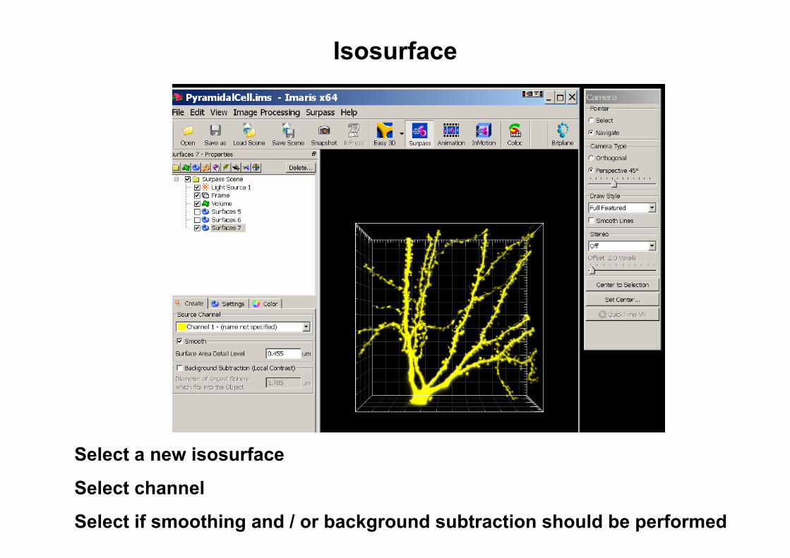
Isosurface
Select a new isosurface
Select channel
Select if smoothing and / or background subtraction should be performed
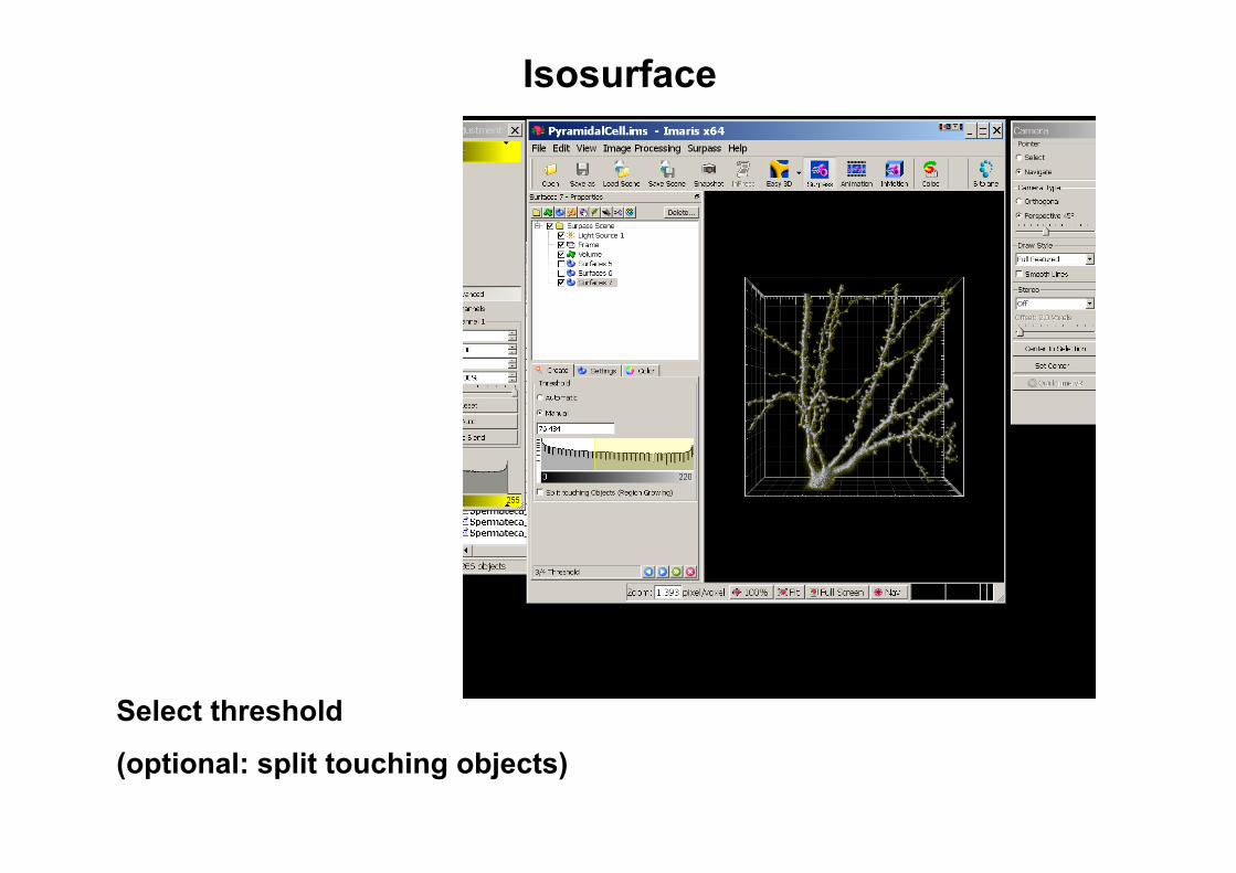
Isosurface
Select threshold
(optional: split touching objects)

Spot detection
Data displayed as a volume (MIP or Blend) with intensities adjusted that only structures of interest are visible
→ Measure diameter of structures in slice mode

Spot detection
Filter detected spots according to selected criteria (Quality ~intensity at center)
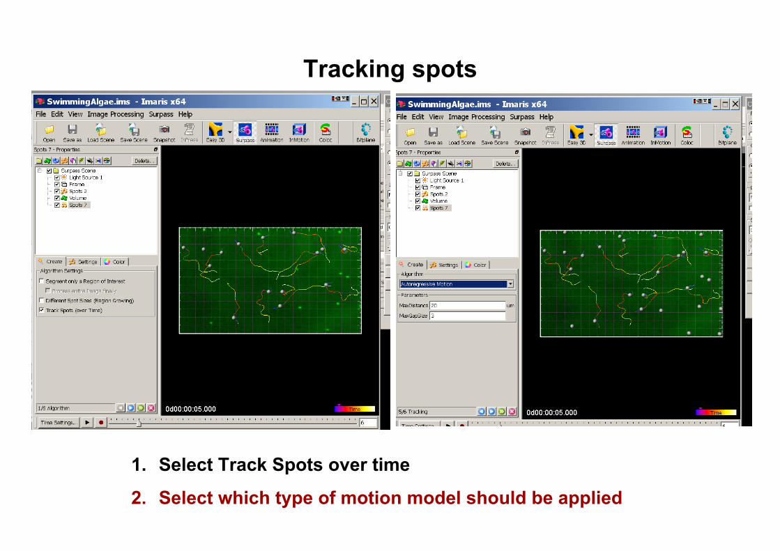
Tracking spots
1. Select Track Spots over time
2. Select which type of motion model should be applied

Spot trackingMotion models
Brownian Motion
• This algorithm models the motion of each Spot as a Brownian motion. This model is appropriate, if your Spots actually perform a Brownian motion.
Autoregressive Motion
• This algorithm models the motion of each Spot as an autoregressive process. This model is appropriate if your Spots perform any kind of continuous motion.
Filter detected tracks according to selected criteria

Filtering, statistics and counting
Example: Chlamydia infected cells
• Counting cells based on DNA stained nuclei
• Evaluation of infection based on the size of inclusion (fluorescently labeled Chlamydia)
• Attention: DNA found in nuclei and Chlamydia
• 1. Detect nuclei via spot function
Spots are only detected in the nuclei because of filtering for low intensity in channel 2 (green labeled Chlamydia)

Filtering, statistics and counting
Example: Chlamydia infected cells
• Counting cells based on DNA stained nuclei
• Evaluation of infection based on the size of inclusion (fluorescently labeled Chlamydia)
• Attention: DNA found in nuclei and Chlamydia
• 2. Detect inclusions via isosurface
Isosurfaces are filtered for size (>100um, green labeled).

Filtering, statistics and counting
Example: Chlamydia infected cells
• Counting cells based on DNA stained nuclei
• Evaluation of infection based on the size of inclusion (fluorescently labeled Chlamydia)
• Attention: DNA found in nuclei and Chlamydia
• View or export values / statistic
Individual objects can be selected

Animation
Add a keyframe Number of frames
Animation can be designed using keyframes which store scenes shown in the display. Frames between keyframes are interpolated.
Keyframes (blue, white if selected)
Save animation (format: uncompressed avi for ease of use)

Questions and Outlook
Next lunch seminar: Tuesday, 20th of January 2009
Advanced Image Processing: Spot Detection
Animation Generation
If time: starting Deconvolution
Questions and input: www.zmb.uzh.ch or [email protected]

Imaris
• 7 floating licenses bought by several research groups (2005)
• License server maintained by the computing center:[email protected]
• Maintenance of Imaris paid by the Center for Microsocpy and Image Analysis
• Download of Imaris and coordination for acquisiton of new modulesand licenses at the University via the Center for Microsocpy and Image Analysis: :