us of the neonatal urogenital tract - legeforeningen.no · applicable to the pediatric urogenital...
TRANSCRIPT

Modern methods and approaches in pediatric
uroradiology
Michael Riccabona Dept. of Radiology
University Hospital Graz, Austria

To revisit standard & modern imaging methods applicable to the pediatric urogenital tract
To describe new insights & imaging techniques
do these impacted imaging approach & task?
To learn about new imaging options how have they impacted imaging algorithms?
To discover new potential of modern imaging at reduced invasiveness
less radiation burden
Objectives

Objectives
To revisit established standards & methods
To describe new insights a& imaging techniques
To learn about new imaging options
To discover new potential of modern imaging
To give typical examples & suggestions
procedural recommendations & imaging algorithm based on ESPR uroradiology task force
for common paediatric nephro-urologic queries

Aim & task of imaging/paediatric uroradiology assess prenatally suspected finidings
screening?
diagnose clinically manifest conditions
follow-up diseases
impact on management & prognosis
Basic considerations

Basic considerations
Aim & task of imaging assess prenatally suspected changes
diagnose clinically manifest conditions
follow-up diseases
impact on management & prognosis
less important (= “relative” indication): reassurance of parents & relieve of doctors
"forensic imaging“ – but often requested
Consider: growing economic & legal pressure

First:
Imaging methods
all modern imaging approaches need solid base
=
fundamental established tools & rules need to be respected, performed properly and adapted
towards today’s standards & knowledge

US = mainstay of uroradiology dedicated equipment needs
high resolution & linear Tdx high frame rate ... modern US methods helpful
HI & compounding, HR ...
Imaging methods = requisites

US = mainstay of pediatric uroradiology equipment needs knowledgeable & engaged radiologist
training in pediatric US, comprehensive exam proper timing
Imaging requisites: US
day 3
3 weeks

Imaging requisites: US
Mainstay of uroradiology equipment needs knowledgeable radiologist
investigation technique post void US, volumes assessment ...
dorsal approach, documentation ...

Other requisites for good US results: hydration enough time standardised investigation & report
heating, place for assisting persons
pacifier, swaddling facility ....
ergonomic setting
warmed US jelly
information, clear query old images & reports?
present situation (medication, query ...)
Imaging requisites: US

ESUR / ESPR procedural recommendation: pediatric urosonography
well hydrated, full bladder, adequate equipment, transducer, training …
urinary bladder: size (volume), shape, ostium, wall, bladder neck include distal ureter & retrovesical space/inner genitalia, urachus? …
optional: CDS for urine inflow, perineal US, scrotal US …
post void evaluation bladder: residual volume, bladder neck, shape & configuration
kidneys: dilatation of pelvo-caliceal system / ureter changed?
optional: (a)CDS & duplex-Doppler …
kidneys: lateral and/or dorsal, longitudinal & axial sections parenchyma? pelvo-caliceal system? standardised measurements in 3 dimensions & volume calculation if dilated: max. axial pelvis & calix, narrowest parenchymal width, + UPJ
optional: contrast-enhanced urosonography, 3DUS …
Pediatr Radiol 2008:38
Additional: abdominal US survey recommended

Improves US capabilities invasive/irradiating exams
helps to tailor further imaging
e.g. perineal US, m-mode
unconventional approach or use document ureteral peristalsis
visualise urethra during voiding
sonogenitography
Modern US

Modern US
Improves US capabilities
e.g. extended field of view applications overview & improved measurements
sometimes essential, e.g. renal transplant comprehensive & conspicuous illustration

Improves US capabilities
e.g. Doppler techniques - many applications information on perfusion
sometimes essential
spectral analysis …
Modern US

Improves US capabilities
e.g. Doppler techniques
information on perfusion
CDS valuable too, e.g.,
twinkling stone, ureteric jet
additional vessels …
Modern US

Modern US
Improves US capabilities
e.g. aCDS sensitive for focal lesions
can visualise aPN many other applications
trauma, infarction ...

Modern US
Improves US capabilities
e.g. "B-flow" non-Doppler flow depiction
no angel dependency lower MI

Improves US capabilities
e.g. ce-VUS reliable & sensitive VUR detection
may visualize intra-renal VUR
potential to visualize urethra
no radiation
girls, screening, follow-up
Modern US

ce-VUS
Fill bladder with NaCl + US-CM assess bladder, retrovesical space + kidneys
try to look at urethra during voiding perineally
Darge K. EJR 2002

Install US contrast medium, e.g., SonoVue ®, 0.5-1.0% of bladder volume slow, US monitoring, fractional administration
ESPR / ESUR procedural recommendation
No diet restriction or enema, urine analysis; AB as in VCUG …
Catheterism: feeding tube, 4-8 french, or suprapubic puncture anaesthetic lubricant or coated plaste
Bladder filling with NaCl (only from plastic containers)
During/after voiding: US of bladder & kidneys supine ± prone, laying or sitting or standing
Assess urethra during voiding – perineal US
Peri-/ post-contrast US of bladder + kidneys: continuous, alternating US modalities: fundamental, HI, CDS, contrast specific methods
alternate scans of right & left side during & after filling
VUR diagnosis: echogenic micro-bubbles in ureters or renal pelves
Standard US of bladder & kidneys (supine, ± prone)
Pediatr Radiol 2008: 38
ce-VUS

e.g. ce-US = intravenous/intra-cavitary CM reliable & sensitive
enhances US potential
BUT: not licensed for pediatric use
native kidney lesions
transplant complications
drain position …
Modern US

e.g. US genitography in ambiguous genitalia, suspected malformation
fill vagina with NaCl enhances genital US
perineal approach may add US-CM, + fluoro …
Modern US

Modern US
e.g. 3D/4DUS anatomic analysis ... accurate volume calculation
even in HN
comprehensive documentation
Modern US
e.g. 3D/4DUS anatomic analysis, volume calculation
comprehensive documentation collecting system rendering
conspicuous visualisation
virtual cystoscopy

Imaging requisite: VCUG
VCUG = still essential in uroradiology
BUT: adequate equipment & technique some rules (also for genitography):
pulsed fluoroscopy short screening last image hold no blind films

VCUG
VCUG = still essential in uroradiology BUT: adequate equipment & technique some rules: make utmost use of it
modified protocol = functional assessment

Pediatr Radiol 2008:38
No diet restriction or enema, urine analysis, potentially antibiotics …
catheterism: feeding tube, 4-8 french or suprapubic puncture latex precaution: neuro tube defect, bladder exstrophy …
Bladder filling with radiopaque contrast gravity drip = bottle 30-40 cm above table, watch dripping, AB?
after voiding: ap view of bladder & renal fossae assess contrast drainage form kidney if refluxed
when voiding: remove catheter, unless cyclic VCUG = 3 fillings, 1st y(s) female: 1-2 spots of distended urethra (slightly oblique) male: 1-3 spots during voiding (ap & high oblique / lateral) include renal fossae during voiding, if VUR => spot film of kidney
fluoroscopy: if signs of increased bladder pressure, imminent voiding, urge … bilateral oblique views of distal ureters, include catheter document VUR, include kidney (spot film, intra-renal reflux)
Note: VUR staging, AB-prophylaxis? …
fluoroscopic view of renal fossae & bladder, initial + early filling
VCUG
ESPR / ESUR procedural recommendation

Huge potential BUT: radiation! pediatric protocol? indication?
General rule, particularly in infants: try to avoid CT, use alternatives
extensive use of dedicated US, MRU ...
Imaging method: CT
Disclosure: Graz = Toshiba center for pediatric CT

If CT necessary - never just try liberal use of immobilisation & sedation
high radiation risk (Brenner 2001 etc ...)
particularly in infants
accept some image noise don’t use large detectors, no over-ranging ....
individually adapt protocol - dose, CM, technique
- spiral, increment, timing ...
- corrected for weight, age & query
- never use adult protocols
- avoid multiphase CT
CT

Indications: Emergencies
severe & multiple trauma acute hemorrhage
infarction malformation
CT

Indications: Emergencies
severe multiple trauma acute hemorrhage infraction, malformation … collecting system injury
split bolus technique?
CT

Indications: Emergencies
Severe, multiple trauma acute hemorrhage infraction, malformation … collecting system injury associated injury
spine & bone …
…
CT

Indications: Emergencies
Tumor assessment no MRI available
calcifications?
part of (lung) staging ...
DDx …
CT

Indications: Emergencies, tumors (?)
CTA – renal artery stenosis CT-DSA?
80-100 kV?
flow?
bolus tacking?
CT

Indications: Emergencies, tumors, CTA
Rare others UTI complications
stone CT
DDx (no MRI) …
CT

Some rules for adapting protocols age, weight, query … - look at entire imaging chain
CT
referral diagnosis
CT protocol
image reconstruction (Kernel) increment
image transfer
visual perception
scout view, scan length, shielding, FOV
exposure settings (kV, mAs, sSlice thickness, pitch...)
type (single Slice, helical, volume)
AEC and iterative reconstruction
display device
Sorantin, Pediatr Radiol. 2008 CM amount, timing, phases …

Some rules for adapting protocols
kV & mA:
mAs – linear relation 2x mAs = 2x dose
kV – exponential relation 2x kV = 4x dose
CM: 2,5 – 1 ml/kg (300mg/ml), flow? …
ALARA US before planning CT?!
usually 1 phase sufficient
timing critical
NOTE: resolution? small increment = dose ...
CT - How
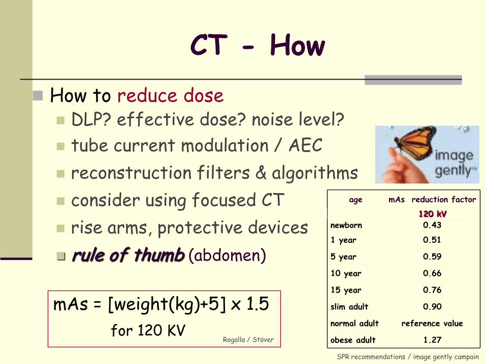
How to reduce dose DLP? effective dose? noise level?
tube current modulation / AEC
reconstruction filters & algorithms
consider using focused CT
rise arms, protective devices
rule of thumb (abdomen)
age mAs reduction factor
120 kV
newborn 0.43
1 year 0.51
5 year 0.59
10 year 0.66
15 year 0.76
slim adult 0.90
normal adult reference value
obese adult 1.27
SPR recommendations / image gently campain
mAs = [weight(kg)+5] x 1.5
for 120 KV Rogalla / Stöver
CT - How

Pediatr Radiol 2010:40
Uro-CT in children
ESPR / ESUR procedural recommendation
Indications severe urinary tract trauma, complicated/equivocal urolithiasis & infection, tumour & DD, renovascular disease
NOTE: only in case high level US (+ KUB) not conclusive, always consider alternatively MRU, if available
Preparation avoid pain, decrease anxiety, local protection device, generous immobilisation & sedation for CM administration - previous line placement, measure creatinine, hydration
NOTE: age dependent different normal creatinine values in infants & children
Contrast application - 2,5-1,5 ml/kg (weight dependent); generally 2 ml/kg, injection speed 1 - 2ml / sec (if power injector applicable)
age adapted injection rate & scan delay time, hand or - depends on: location/size/type of IV access, child size/weight, underlying disease & query
Protocols NOTE: always use age-/weight-adapted paediatric settings, restrict acquisition area keep age corrected effective dose < 2mSv tailor protocols to query (according to clinical indication & result of previous US) avoid multi-phase acquisitions = perform CT study according to query / clinical indication, including one (“or rarely more”) of the following:
unenhanced, arterial, nephrographic, and excretory phase
Nephrolitiasis • unenhanced scan
• consider to further reduce mAs
Trauma • arterial phase: suspected vessel
injury • nephrographic phase: often
sufficient, always informative • urographic phase: suspected
injury of collecting system
Tumour & DD, infections • nephrographic phase: usually
sufficient, mandatory • urographic phase: in selected
cases to assess involvement or pathology of collecting system
Renovascular disease/ vascular malformation
• arterial phase / CTA

= the future method of pediatric uroradiology T2-MRU = "T2 MR-urogram"
anatomic display, cysts ...
availability?
Modern imaging: MRI & MRU

= the future method T2-MRU = "T2 MR-urogram"
ce-T1 MRU, dynamic/diuretic
MRU

Practically has replaced IVU non ionizing, dynamic information assess collecting system + parenchyma + genitalia
MRU

Indications obstruction & malformation inflammation, scars, complications cysts & tumors
MRU

Indications obstruction & malformation inflammation, scars, complications cysts & tumors helpful established tool for DDx
MRU

Modern MRU allows to quantify function & drainage
various methods, diuretic stimulation ...
MRU
Rohrschneider, Radiology 2002 ,
Grattan-Smith Pediatr Radiol 2008,
Images from Riccabona, EJR 2008 … BUT: needs sedation & Gd

Modern MRU & MRI allows to quantify function & drainage
various methods, diuretic stimulation ...
HR imaging
MRA & MRV (also pre-transplant …)
"one stop shop" imaging
MRU

Modern MRU allows to quantify function & drainage
various methods, diuretic stimulation ...
HR imaging, MRA future potential by new applications
perfusion imaging MR-spectroscopy DWI BOLD non-enhanced MRA (ASL …) new contrast agents MR-VCUG
MRU

Then:
modern applications
adapt imaging algorithm to impact =
imaging must have impact on patient management (= efficacy)
sometimes less (e.g., detailed US, but performed properly) is more
properly select when to do what and how

= use of imaging today
+ imaging algorithms
affect many & common queries in infancy
based on new tasks for imaging
Modern applications

imaging in infants with fetally detected HN
e.g.,

Aim of imaging detect malformations & urinary tract conditions
before they cause renal damage
If disease present differentiate entity - obstructive versus refluxing uropathy
obstruction common with high grade HN
grade disease need for treatment
follow-up?
timing of investigations?
Imaging in infants with fetal HN

Aim of imaging detect malformations
If disease present differentiate obstructive vs. refluxing uropathy grade disease
define need for further imaging different for severe vs. minimal abnormalities
different for obstruction or VUR or others
comparison with prenatal US essential
standardised HN grading
Imaging in infants with fetal HN

HN 0 HN I HN II HN III HN IV HN V
HN 0 = collecting system not or minimally visible, considered normal
HN I = just renal pelvis visible, axial diameter < 5-7 mm, considered normal
HN II = axial pelvis diameter < 5/7-10 mm, some calices with normal fornices visible
HN III = marked dilatation of calices, pelvis > 10 mm, rounded papilla & fornices without parenchymal narrowing
HN IV = gross dilatation of collecting system + narrowing of parenchyma
HN V = used in some places to communicate extreme HN with only thin, membrane-like residual renal parenchymal rim
Pediatr Radiol 2008; 38
US: HN grading system Derived from Hofmann & fetal SFU classification (Fernbach et al)

Aim of imaging detect malformations
If disease present differentiate obstructive vs. refluxing uropathy grade disease & define further imaging
Essential question Whom to image how & when?
not too invasive not missing important conditions
without diagnostic overkill = economic approach
in the light of new knowledge & treatment concepts
Imaging in infants with fetal HN

abnormal: pelvis 7(10) mm + dilated calices (HN III/IV), other anomalies normal
abnormal
US at 1 mo
US at 3 mo
pelvis 10 mm otherwise
normal, HN II
pelvis > 10 mm other changes, HN > II
Stop follow-up further morphological & functional
evaluation: Scintigraphy, (IVU), MRU …
US: 1st US around day 5
normal
Stop follow-up
abnormal
normal
pelvis 10 mm (HN III) other malformation, “extended criteria”
VCUG or ce-VUS in girls
Pediatr Radiol 2008; 38
if therapeutic consequence
Postnatal imaging in newborns with fetally diagnosed moderate (= mild to moderate) HN
new adapted imaging algorithm

Imaging in infants with high grade HN
Different than for low degree HN findings: obstructive uropathy/high grade VUR
beware of PUV (in boys) earlier imaging mandatory
VCUG

Imaging in infants with high grade HN
Different from low degree HN obstructive uropathy, PUV …
Consider DDx duplex systems
obstructive/refluxing moiety
cystic disease PCKD, MCDK cystic tumour ...
Imaging in infants with high grade HN
Different from low degree HN obstructive uropathy, PUV …
Consider DDx duplex systems, cystic disease
complex malformation creative imaging approach

Imaging in infants with high grade HN
Different from low HN obstructive uropathy, PUV
Consider DDx cystic disease
duplex systems
complex malformations
different imaging needs than in low grade HN different entities & risks
But: also consider immaturity & therapeutic options

early US + VCUG
PUV obstructive uropathy high grade VUR
UPJO, MU *5 drainage renal function? +isotopes*2, MRU*3
US follow-up 6 mo: isotopes*2, MRU*3?
others *4
as indicated *5
*1 (US) genitography: in patients with single kidney, MCDK, ectopic kidney, suspected genital anomaly …
*2 MAG3: better than DMSA in dilated systems and neonates, DMSA usually after > 3-6 months, not before 6 weeks; + open bladder catheter to avoid VUR induced errors
*3 MRU: complex anatomy, function, obstructive component ... *4 e.g.: MCDK, cystic dysplasia, duplex or horseshoe kidney, other malformation, non-obstructive HN, cysts/cystic Tu …
*5 see respective algorithm
Postnatal imaging in newborns with fetally diagnosed
high grade HN (= gross dilatation = HN ≥ IV)
VCUG. in all boys particularly if ureter dilated
ce-VUS in girls, potentially delayed
+ narrowed or dysplastic parenchyma, dilated ureter particularly if bilateral or single kidney *1
Pediatr Radiol 2009:39

Imaging in infants with fetal HN
The only clinically important question = which kidney needs treatment?
which only needs monitoring, which nothing?
reliable prospective assessment? deterioration without surgery?
function, growth ...
approach & treatment varies with condition
MU / ureterocele ectopic insertion
PUV, UPJO ...

Imaging in infants with fetal HN
The only clinically important question Which kidney needs treatment? And then: Which imaging should be used?
Imaging options: US (incl. diuretic US, CDS / DDS ...)
IVU (outdated)
MAG 3 (split function + drainage)
MRU (anatomy + function)
at present no reliable a-priori pro-futuro assessment = serial investigations using various methods necessary
to monitor disease, intervene in deterioration

US (+ DDS/CDS)
US follow-up hydration! CDS! diuretic US (?)
mild (HN <3 ) x1
<6 weeks old
VCUG (ce-VUS?)
VUR/PUV x1
no VUR
MAG3 (T+20) x4 or (quantitative) MRU or IVU (pre-op., if no MRU)
obstructive low function OP (>3-6 mo)
equivocal - also, if <3 mo + obstructive & normal function
>6 weeks old
potentially diuretic US(?)
deteriorationx2 clinical symptomsx3
non-obstructive normal function
VUR?
others x1
MAG3 (T-15), follow-up (after 3-6 mo) …
Imaging algorithm in infants with suspected obstructive uropathy
x2 Proposed imaging criteria for deterioration: - on MAG 3: decreased (split) renal function & drainage, contra-lateral hypertrophy
on US increasing dilatation, decreasing parenchymal width, echotecture, contra-lateral hypertrophy
decreased vascularisation (on aCDS), asymmetrically elevated RI (on PW-DDS), reduced peristalsis (in MU) or ureteric jet (asymmetrically in unilateral disease) x3 Clinical criteria for deterioration: pain, infection, haematuria, (kidney) growth failure, hypertension
clinical symptoms x3
x1 as appropriate, see respective algorithm
HN ≥3
x4 assess drainage pattern and (split renal) function , Pediatr Radiol 2009 :39
UTI
UTI

Imaging in other urinary tract conditions & symptoms
based on modern imaging potential
Other examples

Imaging in urolithiasis
Do we need to apply all modern options? CT for all?
urolithiasis
nephrocalcinosis
haematuria

Imaging in urolithiasis
Do we need to apply all modern options? CT for all?
US + KUB / IVU?

Imaging algorithm for infants & children with suspected urolithiasis
US (gray scale + DDS/CDS)
x1 or KUB (+ adapted IVU, particularly if low-dose CT unavailable)
positive
negative + low clinical suspicion
x2 potentially contrast-enhanced CT, if other DD or complication; MRU in selected cases
inconclusive US US non-diagnostic
mismatch US & clinics
negative + high clinical suspicion
Stop
uro-CT x1 (ultra-)low dose + unenhanced x2
only 2nd signs + no stone visible, or before intervention, if therapeutically necessary
+ KUB (± IVU) before intervention / lithotripsy potentially + KUB for confirmation, for treatment needs …
follow-up after treatment
Pediatr Radiol 2009, 39

Imaging in other conditions
What about tumors? US + CT or MRI?
depends on entity = age matters!
+ symptoms ..

Imaging in other conditions
What about tumors? US + CT or MRI?
increasingly shifted towards US + MRI use modern MR tools: MRA, DWI, WBMRI …
lung MRI?

Summary & conclusion
US = mainstay of uroradiology in infants provided proper equipment & application
VCUG = indicated more restrictively proper technique essential, partially ce-VUS
IVU & CT = hardly used in infants except CT for severe trauma, …
MRU = the ideal one stop shop imaging in obstructive uropathy
complex malformations
tumor, complicated infection …

Summary & conclusion
Procedural recommendations exist help to standardise high quality imaging
Imaging algorithms exist suggestions for optimal use of imaging methods
Individual responsibility remains adapt to patient & local needs / options
Remember: Aim of imaging = diagnose disease & maintain renal function
help prevent harm to the kidney

Any questions -
Yes, please, ... ??


Imaging algorithm for hematuria in infants
abscess, pyohydronephrosis, severe haemorrhage … intervention *1
US + DDS/(a)FDS
US normal
*1 DD: Tu, haemorrhage, complicated UTI e.g., XPN, Tbc, abscess ... => MRI/CT
diagnosis evident => further imaging as appropriate, if necessary
Clinical & laboratory evaluation * = presumable diagnosis in most situations
*2 see respective dedicated imaging algorithm
potentially further imaging depends on clinical course & query*3
+ basic imaging:
UTI *2
UPJO, VUR, MU *2
Tumour*3
Trauma*2,3
Vascular*2,3.
Urolithiasis*2
GN & nephropathies*3
Bladder-, urethral- , ureteral pathology *3
US inconclusive mismatch US & clinics
*3 proceed to next imaging step (CT, Angiography, VCUG …)
* clinical & laboratory evaluation: variation (day time, position, activity …)? vaginal/rectal discharge? duration? recurrent? undulating symptoms? pain or colic? trauma? fever? dysuria? age (foreign body …)?, blood pressure? microscopic or macroscopic haematuia? family history? urinalysis (erythrocyte morphology, isolated hematuria? …), renal function? blood count / CRP …
Note: malignancy much rarer than in adults
Stop imaging
Pediatr Raiol 2009; 39

inconclusive US or no signs of RVD diagnosis evident
RAS
DDS criteria: PSV >180-200 cm/sec
note: age variations
RAR >3.5, δ-RI >0.05 Acceleration time >80msec, Tardus-Parvus pattern distally
other causes
further imaging as appropriate *
DSA with simultaneous PTA (potentially + renal vein sampling)
stage 1 hypertension or BP well controlled on 1-2 drugs
stage 2 hypertension, BP not controlled by 2 or more drugs,
stage 1 age <3y or high clinical suspicion**
CTA or Captopril renography ****/MRA ***
RVD normal
clinical follow-up DSA + renal vein sampling potentially simultaneous PTA
high clinical suspicion**
* Further imaging as appropriately indicated (see existing recommendations …)
** High clinical suspicion: history of renal trauma or radiation, umbilical artery catheterization, renal vascular thrombosis, bruit over renal arteries, high renin levels, presence of disease associated with renovascular pathology (e.g., neurofibromatosis, Williams’ syndrome, tuberous sclerosis …)
*** MRA: potentially & increasingly ce-MRU for large vessels & infarcted areas, non-enhanced MRA techniques?
Abbreviations: BC = blood count, BP = blood pressure, BUN = blood urea nitrogen, CDS = color Doppler sonography, cm/sec = centimeters per second, CTA = CT-angiography, DSA = digital subtraction angiography, DDS = spectral duplex Doppler, MRA = MR angiography, PSV = peak, systolic velocity, PTA = percutaneous transluminal angioplasty, RAR = renal aortic ratio, RAS = renal artery stenosis, δ-RI = Resistive Index difference, RVD = renovascular disease, US = ultrasound, y = year
Imaging algorithm in infants with potential reno-vascular hypertension
renal US + CDS & DDS
Basic clinical & laboratory evaluation BP measured both arms + one leg, repeated BP measurements, 24 hour BP monitoring (age related chart), retinal examination, echocardiography chest X-rays. BUN, creatinine, eloctrolytes, urinalysis, urine culture, BC
validate indication
**** Captopril scintigraphy: potentially prior to PTA for function, particularly in doubtful situations or neonates until old enough for PTA, note: local variations
Pediatr Radiol 2011; 41
suspected reno-vascular hypertension

„Anatomic“ paediatric MR-Urography (MRU) INDICATION Always previous US (+ reflux study, if indicated = VCUG, VUS, or RNC)
Queries: e.g. malformation, obstructive uropathy, complicated infection, tumour, post-traumatic, cystic disease, transplant ...
PREPARATION: General: Place line in advance, creatinine for CM-studies (GFR calculation - NSF), mock unit / visit to magnet Hydration: NaCl or Ringer’s solution (20 ml/kg for 1 hour, max. 1000 ml), empty bladder before entering the magnet Sedation: priority to immobilization (feed & wrap), or no (minimal) sedation. Deep sedation only if necessary Bladder catheter: deeply sedated patients who cannot empty the bladder (particularly after Furosemide)
potentially also in high grade VUR with dynamic queries Polyethlene catheter without balloon, urine bag, below level of MR table
Diuresis: Furosemide 1 mg/kg IV (max. 20 mg), 15 min before beginning of morphologic investigation timing may vary in dynamic-diuretic functional protocols (F -20, F -15, F 0, F +10, F +15, F +20)
MRU examination*1: Positioning: Supine position with arms above the head
SCOUT: Sagital important for correct oblique coronal plane, FOV: from above both diaphragms to below symphysis
potentially SSFP axial & coronal (+ sagital)
Heavily T2-weighted sequences coronal (e.g.,T2-3D TSE fs or 2D-thin & -thick slice [3D-UROGRAM], HASTE/RARE/PACE, …)?
T2-IR sequence, non-enhanced T1-weighted & GRE sequence NOTE: 3 slices anterior + posterior of kidneys for GRE; adjust FOV
CM-Application - cyclic Gd compounds*2 iv. in first year of life (renal immaturity …) & bilateral uropathy, or GFR
Repeated serial coronal T1-3D sGRE fs (for functional assessment continuously for 3-5min) NOTE: subtraction helpful - particularly for MRA, if achievable; for MRA use motor pump & flow of 1 ml/sec
T1 axial & coronal (fs), + sagital if needed
Final coronal T1-3D GRE fs; or additional delayed imaging up to 20(-30) min p.i. potentially changing to prone position or post void scan (when delay in CM washout)
*1 functional MRU not yet standardised and not addressed • Furosemide timing, contrast dose & application may need adaptation
for various queries tailored protocols are essential • e.g., MRA, diffusion, additional sagital acquisition
*2 non-cyclic compounds can be used in older children according to approval • Gd-dose as recommended by manufacturer
Pediatr Radiol 2010; 40