usage of karyomapping for preimplantation genetic diagnosis (pgd) of complex single gene disorders...
TRANSCRIPT
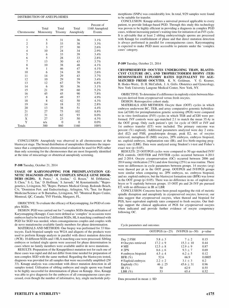
DISTRIBUTION OF ANEUPLOIDIES
ChromosomeTotal
MonosomyTotal
TrisomyTotal
Aneuploidy
Percent of1160 Aneuploid
Events
1 5 31 36 3.1%2 5 33 38 3.3%3 3 27 30 2.6%4 10 24 34 2.9%5 6 33 39 3.4%6 5 28 33 2.8%7 13 30 43 3.7%8 10 38 48 4.1%9 1 46 47 4.1%10 6 24 30 2.6%11 14 29 43 3.7%12 10 29 39 3.4%13 12 67 79 6.8%14 6 30 36 3.1%15 21 39 60 5.2%16 45 45 90 7.8%17 8 49 57 4.9%18 8 42 50 4.3%19 14 18 32 2.8%20 13 38 51 4.4%21 27 75 102 8.8%22 31 62 93 8.0%X 27 23 50 4.3%Y n/a 0 0 0.0%
Totals 300 860 1160 100%
CONCLUSION: Aneuploidy was observed in all chromosomes at theblastocyst stage. The broad distribution of aneuploidies illustrates the impor-tance that a comprehensive chromosomal evaluation be used for PGS ratherthan only screening for the chromosomes that are most frequently identifiedat the time of miscarriage or obstetrical aneuploidy screening.
Cycle parameters and outcomes
OOT/PGS (n¼25) IVF/PGS (n¼50) p-value
Day 2 FSH 5.8 � 3 7 � 2 0.15# Oocytes retrieved 17.2 � 9 15.3 � 10 0.44# MII 12.5 � 8 12.9 � 9 0.87# 2PN 8.6 � 6 9.3 � 7 0.69# embryos biopsied 4.5 � 4 6.2 � 5 0.12BFR (%) 52.6 66.9 0.0005# Euploid embryos 2.2 � 2 3.1 � 3 0.2Euploidy (%) 46 � 25 45.6 � 31 0.96IR (%) 50 62.9 0.55LBR (%) 53 65.4 0.52
Data presented in mean � SD
P-108 Tuesday, October 21, 2014
USAGE OF KARYOMAPPING FOR PREIMPLANTATION GE-NETIC DIAGNOSIS (PGD) OF COMPLEX SINGLE GENE DISOR-DERS (SGDS). R. Prates,a A. Jordan,a G. Rosen,b M. DiMattina,c
S. Chen,d S.-L. Tan,e D. Hoffman,f S. Jaroudi,a M. Konstantinidis.a aRepro-genetics, Livingston, NJ; bRepro. Partners Medical Group, Redondo Beach,CA; cDominion Fert. and Endocrinology, Arlington, VA; dInst. for ReproMedicine/Science at St Barnabas, Livingston, NJ; eMontreal Repro Centre,Montreal, QC, Canada; fIVF Florida, Margaret, FL.
OBJECTIVE: To evaluate the efficacy of Karyomapping, for PGD of com-plex SGDs.
DESIGN: PGD was carried out for 7 complex SGDs through utilization ofKaryomapping (Kmapp). Cases were defined as ‘complex’ in occasions wereembryos had to be tested for 2 different SGDs, HLAmatching combinedwithPGD for SGD was needed, when consanguineous couples and couples withde novo mutations/no available family members for phase determination.
MATERIALS AND METHODS: One biopsy was performed for 33 blas-tocysts. Each biopsied sample was WGA and aliquots of the products wereused to perform Kmapp analysis in parallel with direct mutation detection( PCR). 10 different SGDs and 1 HLAmatching case were processed. Siblingembryos or isolated single sperm were assessed for phase determination incases where no family members were available and/or de novo mutations.
RESULTS: Preparation of the Kmapp/direct mutation test for each one ofthese cases was rapid and did not differ from time needed for preparation ofnon-complex SGD with the same method. Regarding the blastocysts tested,diagnosis was provided for all samples that were successfully amplified (29/33). Kmapp analysis was concordant with direct mutation detection in allsamples tested. Utilisation of sibling embryos and single sperm was foundto be highly successful for determination of phase on Kmapp. Also, Kmappwas able to give diagnosis for the embryos of all consanguineous cases pro-cessed, even though the number of informative, key, single nucleotide poly-
FERTILITY & STERILITY�
morphisms (SNPs) was considerably low. In total, 9/29 samples were foundto be suitable for transfer.CONCLUSION: Kmapp utilizes a universal protocol applicable to every
patient, to provide linkage-based PGD. Through this study this technologywas proven to be highly efficient in providing a diagnosis in complex PGDcases, without increasing patient’s waiting time for initiation of an IVF cycle.It is advisable that at least 2 sibling embryos/single sperms are processedwith Kmapp for establishment of phase and that direct mutation detectionis always performed in parallel for consanguineous cases. Karyomappingis expected to make PGD more accessible to patients under the ‘complexcases’ category.
P-109 Tuesday, October 21, 2014
CRYOPRESERVED OOCYTES UNDERGOING THAW, BLASTO-CYST CULTURE (BC), AND TROPHECTODERM BIOPSY (TEB)DEMONSTRATE EUPLOIDY RATES EQUIVALENT TO AGE-MATCHED FRESH OOCYTES. K. N. Goldman, Y. G. Kramer,B. Hodes-Wertz, D. H. McCulloh, J. A. Grifo. Obstetrics and Gynecology,New York University Langone Medical Center, New York, NY.
OBJECTIVE: To determine if a difference in euploidy exists between blas-tocysts derived from cryopreserved versus fresh oocytes.DESIGN: Retrospective cohort study.MATERIALS AND METHODS: Oocyte thaw (OOT) cycles in which
embryos underwent BC, TEB, and array comparative genomic hybridiza-tion (aCGH) for preimplantation genetic screening (PGS) were comparedto in vitro fertilization (IVF) cycles in which TEB and aCGH were per-formed. IVF controls were age-matched 2:1 to match the mean 35.4y inthe OOT group. Only each patient’s (pt) 1st cycle of OOT or IVF and1st embryo transfer (ET) were included. The primary outcome waspercent (%) euploidy. Additional parameters analyzed were day 2 estra-diol (E2) and FSH, gonadotropin dosage, peak E2, no. of oocytesretrieved, metaphase-II (MII) oocytes, 2PN embryos, embryos biopsied,no. euploid embryos, implantation rate (IR), and live birth-ongoing preg-nancy rate (LBR). Data were analyzed using Student’s t-test and Fisher’sexact test (p<0.05).RESULTS: 25 OOT/PGS cycles were compared to 50 age-matched IVF/
PGS controls. OOT/TEB and IVF/TEB cycles occurred between 4-2011and 2-2014. Oocyte cryopreservation (OC) occurred between 2006 and2014 using vitrification (75%) and slow freezing (25%) as was routine. Therewere no differences in cycle parameters between groups. 14 oocytes (avg)were thawed per pt in the OOT group. OOT/PGS and IVF/PGS groupswere similar when comparing no. 2PN embryos, no. embryos biopsied,and no. euploid embryos, but the blastocyst formation rate (BFR) was lowerin the OOT group (p<0.05). There was no difference in no. of euploid em-bryos or % euploidy between groups. 15 OOT pts and 26 IVF pts pursuedET, with no difference in IR or LBRCONCLUSION: Concerns have been posed regarding the risk of meiotic
spindle disruption and aneuploidy in cryopreserved oocytes; however, ourdata suggest that cryopreserved oocytes, when thawed and biopsied forPGS, have equivalent euploidy rates compared to fresh oocytes. Our find-ings support the clinical application of PGS for cryopreserved oocyteswhen indicated and provide further evidence of oocyte competencyfollowing OC.
e175