:usda united states common fungal diseases of russian forests · most common fungal diseases of...
TRANSCRIPT
:USDA United States ~~ Department of ' Agriculture
Forest Service
Northeastern Research Station
General Technical Report NE-279
Common Fungal Diseases of Russian Forests
Evgeny P. Kuz'michev Ella S. Sokolova Elena G. Kulikova

Abstract
Describes common fungal diseases of Russian forests, including diagnostic signs and symptoms, pathogen biology, damage caused by the disease, and methods of control. The fungal diseases are divided into two groups: those that are the most common in Russian forests and those that are found only in Russia. Within each group, diseases are subdivided by plant organ attacked, i.e. fruit, seeds, leaves, needles, roots, stems, and branches.
The Authors
EVGENY P. KUZ'MICHEV is Corresponding Member of the Russian Academy of Agricultural Sciences, Professor (Ecology and Forest Pathology), Doctor of Biology. He was Deputy Chief, Federal Forest Service of Russia, when this project was initiated; he is currently Deputy Rector, International Independent University on Ecology and Politology, Moscow.
ELLA S. SOKOLOVA is Associate Professor (Forest Pathology) and Researcher, Department of Ecology and Forest Protection, Moscow State Forest University.
ELENA G. KULIKOVA was Associate Professor (Forest Protection), Department of Ecology and Forest Protection, Moscow State Forest University, when this project was initiatied; she is currently Division Head, Department of International Cooperation, Ministry of Natural Resources of the Russian Federation, Moscow.
Cover Photo
Basidiocarps of Ganoderma applanatum on the trunk of an aspen (Populus tremula).
This publication/database reports research involving pesticides. It does not contain recommendations for their use, nor does it imply that the uses discussed here have been registered. All uses of pesticides
must be registered by appropriate State and/or Federal, agencies before they can be recommended.
CAUTION: Pesticides can be injurious to humans, domestic animals, desirable plants, and fish or other wildlife-if they are not handled or applied properly. Use all pesticides selectively and carefully. Follow recommended practices for the disposal of surplus pesticides and pesticide containers.
Manuscript received for publication 11 January 2000
Published by:
USDA FOREST SERVICE 11 CAMPUS BLVD SUITE 200 NEWTOWN SQUARE PA 19073-3294
June 2001
For additional copies:
USDA Forest Service Publications Distribution 359 Main Road Delaware, OH 43015-8640 Fax: (7 40)368-0152
Visit our homepage at: http://www.fs.fed.us/ne
l

Contents
-~ .. . Introduction .......................................... ':: ................................................................................................. , .... 1
Most Common Fungal Diseases of Russian Forests ..................................................................... 2
Diseases of Fruits and Seeds ...................................................................... -:: ............................ 2 Birch Seed Mummification ...................................................................................................................... 2 Fruit Deformation .................................................................................................................................... 2 Fruit Spots .............................................................................................................................................. 4 Molds ...................................................................................................................................................... 6 Seed and Fruit Rots ............................................................................................................................... 9
Diseases of Needles and Leaves ............................................................................................ 11 Needle Diseases .................................................................................................................................. 11
Lophodermium Needle Casts .......................................................................................................... 11 Snow Blight ...................................................................................................................................... 15 Meria Needle Blight ......................................................................................................................... 16 Brown Felt Blight ............................................................................................................................. 18 Rhizosphaera Needle Casts ............................................................................................................ 18 Needle Rusts of Pine ....................................................................................................................... 20
Leaf Diseases ....................................................................................................................................... 22 Powdery Mildews ............................................................................................................................. 22 Leaf Spots ...................................................... : .. .............................................................................. 23 Leaf Rusts ....................................................................................................................................... 35
Diseases Of Roots, Stems, And Branches ............................................................................... 36 Diseases in Tree Nurseries and Young Forests ................................................................................... 36
Damping - off ................................................................................................................................... 36 Diseases of Forest Stands ................................................................................................................... 38
Wilts ................................................................................................................................................. 38 Verticillium Wilt ............................................................................................................................ 38 Dutch Elm Disease ..................................................................................................................... 39 Oak Wilt ......................................................................... · ............................................................. 41
Dieback and Canker Diseases ........................................................................................................ 42 Cenangium Dieback of Pine ....................................................................................................... 42 Nectria Canker and Dieback ....................................................................................................... 44 Cytospora Canker ....................................................................................................................... 45 Dothichiza Canker of Poplar ....................................................................................................... 46 Clithris Canker and Dieback of Oaks .......................................................................................... 48 Nummularia Canker .................................................................................................................... 48 Black Naemospora Canker ......................................................................................................... 49 Thyrostroma Canker and Dieback .............................................................................................. 50 Ascocalyx Scleroderris Shoot Canker ........................................................................................ 52 European Larch Canker .............................................................................................................. 53 Lachnellula Canker of Siberian Pine Understory ........................................................................ 54 Stem Rust of Pine ....................................................................................................................... 55 Broom Rust of Fir ........................................................................................................................ 56 Perennial Nectria Canker ............................................................................................................ 58 Black Hypoxylon Canker ............................................................................................................. 60 Cytophoma Canker of Ash .......................................................................................................... 60
Wood-Decaying Diseases ............................................................................................................... 62 Annosum Root and Butt Rot ....................................................................................................... 62 Armillaria Root Rot ...................................................................................................................... 65 Butt and Trunk Rot of Conifers ................................................................................................... 67 Trunk and Limb Rot of Hardwoods ............................................................................................. 75

Fungal Dise~ses that Occur Only in Russian Forests ................................................................. 89
Diseases of Fruits and Seeds .......... .' ....................................................................................... 89 Thecopsora Rust of Spruce Cones ...................................................................................................... 89 Acorn Mummification Deformity ........................................................................................................... 89
Diseases of Needles and Leaves ....................................................................... '..:-................... 91 Needle Diseases .................................................................................................................................. 91
Hypodermella Needle Cast of Pine Hosts ....................................................................................... 91 . Chrysomyxa Rust of Spruce ............................................................................................................ 91
Leaf Diseases ....................................................................................................................................... 93 Powdery Mildew of Siberian Pea Tree ............................................................................................. 93 Other Powdery Mildews ................................................................................................................... 94 Orange Leaf Spot of Padus ............................................................................................................. 95 Red Spot of Ussurian Plum ............................................................................................................. 96 Foliage Anthracnoses, Spots, and Blights ....................................................................................... 96 Leaf Rusts ..................................................................................................................................... 108 Taphrina Diseases: Leaf Blisters, Leaf and Shoot Deformation .................................................... 110
Diseases of Roots, Stems, and Branches ............................................................................... 111 Diseases in Tree Nurseries and Young Forests ................................................................................. 111
"Infectious Damping'.' of Coniferous Seedlings .............................................................................. 111 Sclerophoma Disease of Pine Shoots ........................................................................................... 112 Pine Shoot Rust ............................................................................................................................. 113 Chrysomyxa Rust of Spruce Shoots and Needles ........................................................................ 114
Diseases of Forest Stands .... , ............................................................................................................ 116 Dieback and Canker Diseases ...................................................................................................... 116
Black Cytospora Canker of Poplar ............................................................................................. 116 Biatorella Canker ...................................................................................................................... 117 Pitch Blister Rust Canker .......................................................................................................... 119 Endoxylina Canker of Ash ......................................................................................................... 120 Cankers and Diebacks .............................................................................................................. 122
Wood-Decaying Diseases ............................................................................................................. 128 Ganoderma Butt Rot of Beech .................................................................................................. 129 Vuillemenia Decay .................................................................................................................... 129 Trunk and Limb Rots ................................................................................................................. 130
Acknowledgment ............................................................................................................ 133
Appendix A Pathogens That Affect Trees and Shrubs in Russia ....................................................................... 134
Appendix B Host Trees, Shrubs, and Herbs Listed in this Report ..................................................................... 137
Introduction
Contacts in forestry between Russia and the United States range from programs that exchange information and/or scientists to those associated with bilateral economic relations. Forest products and raw materials routinely cross the borders of the two countries, and a variety of phytopathogenic microorganisms can accompany them. Accidental introductions of forest insects and diseases can be unpredictable and result in serious damage to forested ecosystems. In the United States, the devastation caused by epiphytotics such as Dutch elm disease and Endothia cankers of chestnut has been well documented. These pathogens were introduced to North America from Europe. Less well known is the damage caused by blister rust of cork pine ( Pinus strobus) caused by Cronartium ribicola Ditr., which was transported across Russia on host-plants. Today, this disease affects many five-needle pine species (e.g., P. sibirica and P. strobus), including endemic species of pine.
This publication is part of an effort to determine whether there are differences between pathogenic microflora of Russian and U.S. forests. Accordingly, we present descriptions of the most common fungal diseases of forest plants in Russia, including pathogenic fungi that are found only in Russian forests. Because there was no list of pathogenic fungi that affect forest plants in Russia, we had to summarize data from numerous, albeit highly regarded, sources. Also, not all fungi are fully described because some species have not been investigated completely. Nor is the area of distribution for certain fungi precise, as many species have been reported only in a single publication. Still other fungi are scattered throughout Russia. Among the fungal species not found in the United States are dangerous pathogens that could pose a serious threat to U.S. forest ecosystems as well as species whose role in Russia is insignificant.
Although many of the major fungi of Russia described in this report are familiar to the American forestry community, U.S. scientists and research foresters can gain an awareness of the current situation in Russian forests (most research papers on fungal diseases of forest trees are not available to forestry groups outside of Russia). Because the distribution of some diseases differs in different countries, causes can range from resistance of host-plants and environmental peculiarities to intraspecific, strain diversity of fungi and their properties.
Most Common Fungal Diseases of Russian Forests
Diseases of Fruits and Seeds
Seed quality exerts considerable influence on the health of seedlings in both artificial and natural forest regeneration. Fungal diseases reduce the quality and yield of fruits and thus seedling production in different regions of Russia. Fruit and seed pathogens differ significantly with respect to virulence and host specialization, development characteristics, and symptoms. Some diseases that develop during the summer change the shape, color, structure, or size of seeds and fruits. They are easily recognized when the latter are harvested. In the case of seed infection that occurs after maturity and dispersal, it is impossible to determine external disease symptoms during harvest. As a result, diseased seeds that are stored with healthy ones can serve as the source of inoculum for infection of healthy seeds.
Fungal infection of fruits and seeds can be internal, damaging the embryo and cotyledons, or external on the seedcoat. In the latter case, the seeds are not infected but the superficial mycelium can infect the germinating seedling. Fungal diseases cause partial losses of fruits and seeds. Sometimes an entire crop is lost. Most of these diseases reduce a seed's ability to germinate. Some diseases reduce germination power during storage or cause heavy damage and seed loss on growing trees. Other diseases delay the growth and development of seedlings.
Birch Seed Mummification
Class/Order: Ascomycetes, Helotiales
Pathogen Sclerotinia betulae Woron.
Hosts Species of birch (Betula)
Diagnosis Sclerotia form as a black, horseshoe-shaped rim on the boundary between the seed's achene and wing. Affected achenes are dark. Germinating sclerotia produce funnel-shaped apothecia 1-4 mm in diameter, with tiny stalks 3-15 mm long. The outside surface of the apothecia is brown and the base of the stalk is covered with dark-brown hairs. A dirty-white or brown-yellow asci layer (hymenium) forms on the inside surface of the apothecia. Asci are cylindrical, 130 x 5-6 µ. Ascospores are oval, colorless, with a verrucose cover, 10-12 x 4.5 µ.
Biology During the flowering period in spring, ascospores mature in apothecia on autumn-infected seeds. Ascospores are windborne to catkins and infect young seeds. Mycelium penetrates the seed tissues and then the wing; sclerotia are formed here. Apothecia form on sclerotia the following spring. The' disease occurs primarily in birch stands. Single trees and groups of birches growing in open areas are rarely affected significantly, an important factor when harvesting seeds.
Damage Reduces germination power, sometimes reducing seedling yields by 90-100 percent
Distribution European part of Russia, Urals, Siberia, Far East
Fruit Deformation
The form, color, and anatomical structure of fruits are affected. Seeds of affected fruits fail to develop or remain underdeveloped.
Class/Order: Ascomycetes, Taphrinales
Pathogen Taphrina alni-incanae (Kuhn.) Magn.
2

Hosts Species of alder (A/nus), including European (A. glutinosa), speckled (A. incana), and Manchurian (A. hirsuta) · ' '
Diagnosis Flowers and seed scales of young fruits become elongate, about 2 cm long, and vary in form. Asci with ascospores develop on these deformed parts.
Damage Reduces seed production
Distribution European part of Russia, Urals, Siberia, Far East
••• Pathogen Taphrina johansonii Sad.
Hosts Species of poplar (Popu/us), including gray (P. canescens), and Bolle's (P. pyramidalis), and European aspen (P. tremu/a)
Diagnosis Seeds enlarge to several times normal size. A yellow-orange layer of asci forms on the surface of affected seeds. Every ascus has 8 spores, but sometim.es ascospores form buds and fill the sac.
Damage Reduces seed production
Distribution European part of Russia, Urals, Siberia, Far East
••• Pathogen Taphrina rhisophorus Sad.
Host White poplar (Popu/us alba)
Diagnosis The seeds enlarge to several times normal size. A waxy, golden-yellow layer of asci with ascospores forms on the surface of affected seeds. Asci are elongate, clavate, thin on the base, 120-160 x 22 µ. Ascospores are globose, colorless, 4 µ.
Damage Reduces seed production
Distribution Middle and southern areas of European part of Russia, southwestern Siberia
••• Pathogen Taphrina pruni Fckl.
Hosts Species of cherry (Padus, Prunus)
Diagnosis The wall of the ovary enlarges but the embryo fails to develop, and an elongate, sac-like or pocketlike, hollow fruit develops. A waxy, gray layer of asci with ascospores forms on the surface of affected fruits. Asci are cylindrical, 40-60 x 8-15 µ.
3
Damage Reduces seed production
Distribution European part of Russia, Urals, Siberia, Far East
Fruit Spots
Spots occur primarily on seed wings of maple (Acer, and ash (Fraxinus), and on fruits of nut-bearing trees (Jug/ans). They rarely form on the seeds and fruits of other species. Some affect other plant organs, including leaves. Under favorable conditions for fungal development, these diseases can significantly reduce the germination power of seeds and infect germinating seeds and seedlings. The following are the most common spot diseases of seeds and fruits.
Class/Order: Deuteromycetes, Hyphomycetales
Pathogen Cercospora acerina Hart.
Hosts Species of maple (Acer,
Diagnosis Dark-brown or dark-red, small, coalesced spots form on the seed wings. Clusters of conidiophores with conidia form on the spots. Conidia are reversely clavate, pointed on top, brown-olive, 45-180 x 5-8 µ.
Damage Reduces germination power of seeds and kills leaves
Distribution European part of Russia
••• Pathogen Heterosporium fraxini Ferci. et Winde.
Hosts Species of ash (Fraxinus)
Diagnosis Gray spots form on the seed wings. Conidiophores form on spots as small, black tufts. Conidia are elliptic-elongate, thorny, 2- or 4-celled, yellow, 5-6 x 1.7 µ.
Damage Reduces germination power of seeds
Distribution European part of Russia
Class/Order: Deuteromycetes, Melanconiales
Pathogen Cylindrosporium platanoides (Allesch.) Died.
Host Norway maple (Acer platanoides)
Diagnosis Dark-brown, elongate spots form on the seed wings. Sporodochia form on the spots. Conidia are thread-like, 4-celled, pale green, 28-80 x 5-3 µ.
4
Damage Reduces germination power of seeds and affects seedling leaves
Distribution European part of Russia
Pathogen G/oeosporium fagiWest.
Host European beech (Fagus sylvatica)
Diagnosis
•••
Circular or irregular, brown or green spots with dark borders and light centers form on the nuts. Sporodochia are brown and appear as concentric circles. Conidia are 1-celled, colorless. There are two types of conidia: macroconidia are oval or widely spindle-like, 10-16 x 4-5 µ; microconidia are elongate-oval, 4-6 x 1.5-2.0 µ.
Damage Reduces germination power of seeds; seedlings are infected and killed
Distribution Southeastern area of European part of Russia
••• Pathogen Marssonina juglandis (Lib.) P. Magn.
Host Persian walnut (Jug/ans regia)
Diagnosis Brown or gray-brown spots of various shapes and dimensions form on the fruits. Black, dotted, convex sporodochia form on the spots as concentric circles. There are two types of conidia: macroconidia are stick-like with one opaque septa 16-30x 3-4.5 µ; microconidia are stick-like, straight or slightly curved, 6-12 X 1-1.5 µ.
Damage Immature fruits drop; leaves, petioles, and young shoots are infected.
Distribution Southern area of European part of Russia
Class/Order: Deuteromycetes, Sphaeropsidales
Pathogen Phyllosticta aceris Sacc.
Host English field maple (Acer campestre)
Diagnosis Small, round, yellow (later white) spots with a dark border form on seed wings. Pycnidia are globose, black, about 120 µ in diameter, and imbedded in wing tissue but later break through tissues. Conidia are egg-shaped, elongate, colorless, 5-7 x 2.5-3 µ.
Damage Reduces germination power of seeds; leaves also are infected.
Distribution European part of Russia
5
Pathogen Phoma samarorum Desm.
Hosts Species of maple (Acer)
Diagnosis Pycnidia form on the surface of seed wings and are imbedded in the tissue. Pycnidiatops form on the wing as small, brown hillocks. Conidia are oval-elongate, colorless, 1-celled, 5-7 x 2-3 µ.
Damage Immature seeds drop
Distribution European part of Russia
Molds
Seed molds are caused by saprophytic fungi and rarely by facultative parasites. Seeds and fruits of nearly all tree and shrub species are affected. A characteristic external symptom of molds is superficial mycelium on infected tissues of seeds and fruits. Infection occurs during storage under high moisture conditions. Initially, the mycelium of mold fungi develops superficially and does not influence seed germination power. However, it can destroy the seedcoat and penetrate interior tissues. Infection of interior tissue reduces germination power and often destroys the embryo. Affet:ted seeds rot and are useless for sowing.
Class/Order: Deuteromycetes, Hyphomycetales
Pathogen Penici/lium expansum (Link.) Thom., and P. italicum Pers.
Hosts Primarily species of birch (Betula), oak (Quercus), beech (Fagus), and chestnut (Castanea sativa)
Diagnosis Bright-brown or red, sharply outlined, and gradually coalesced spots form on the surface of seeds. Green or blue powdery mycelium forms on the spots. The seed tissue becomes friable and brown. Conidiophores form coremia. The upper part of the coremia resembles a brush. Conidia are elliptical, green, connected in chains, 3 x 3.4 µ.
Damage Reduces germination power and kills seeds
Distribution Throughout Russia
••• Pathogen Trichothecium roseum Link.
Hosts Primarily species of maple (Acer), birch (Betula), ash (Fraxinus), oak (Quercus), spruce (Picea), pine (Pinus), and larch (Larix)
Diagnosis Dark-brown or nearly black, sharply outlined spots form on the surface of infected seeds. Pink (rosy) powdery mycelium with conidia develops on the spots. Conidia are pear-like, 2-celled, 12-18 x 8-10 µ.
Damage Reduces germination power
6
Distribution Throughout Russia
Pathogen Fusarium spp.
Hosts
•••
Primarily species of fir (Abies), larch (Larix), spruce (Picea), pine (Pinus), and oak (Ouercus)
Diagnosis Rosy or crimson mycelium forms on the seed surface. Infected tissues of pulpy seeds tum red. The embryo can die. There are two types of conidia: microconidia are oval, cylindrical-oval or ellipsoid, usually 1-celled, sometimes 2-celled, colorless, numerous, 4-12 x 3-8 µ; macroconidia are multicelled, fusiform and slightly curved, 10-60 x 2-5 µ.
Damage Reduces germination power and causes seed and seedling rot and damping-off
Distribution Throughout Russia
••• Pathogen Botrytis cinerea Pers.
Hosts Primarily species of fir (Abies), larch (Larix), spruce (Picea), pine (Pinus), elm (U/mus), rose (Rosa), oak (Quercus), and chestnut (Castanea)
Diagnosis A thin, downy, dark-gray web of mycelium that consists of hyphae and conidiophores forms on the seeds. Seeds eventually decay, and compact black sclerotia form on them. The conidiophores produce clusters of conidia which are egg-shaped or round, 1-celled, colorless or smoky, 9-12 x 5-10 µ.
Damage Reduces germination power and causes seed and seedling rot and damping-off
Distribution Throughout Russia
••• Pathogen Alternaria tenuis Nees.
Hosts Primarily species of fir (Abies), larch (Larix), spruce (Picea), pine (Pinus), birch (Betula), elm ( U/mus), oak (Quercus), chestnut (Castanea), and Siberian pear tree (Caragana arborescens)
Diagnosis A dark-brown or olive-black thin mycelium and conidia form on seeds and fruits. Conidia are single or connected in clusters or chains, reversely clavate, with 1-9 transverse septa and 1 or more longitudinal septa, and dark-olive or olive-brown, 7-130 x 6-22.5 µ.
Damage Reduces germination power and causes seed and seedling rot and damping-off
Distribution Throughout Russia
••• Pathogen C/adosporium herbarum Link.
7
Hosts Primarily species of fir (Abies), spruce (Picea), pine (Pinus), oak (Quercus), and ash (Fraxinus)
Diagnosis A dark-olive, velvety, turf-like mycelial web forms on seeds and fruits. Conidiophores are single or in clusters, with septa. Egg-shaped or elliptical conidia are 1-celled; cylindrical conidia are 2- or 3-celled, olive-brown, 12-28 x 6-7 µ.
Damage Reduces germination power of seeds and causes mold of needles and leaves
Distribution Throughout Russia
••• Pathogen Asp!:Jrgillus niger Link.
Hosts Primarily species of beech (Fagus), oak (Quercus), spruce (Picea), and pine (Pinus)
Diagnosis Round spots with a black, turf-like mycelial web form on seeds and fruits. Conidiophores are numerous, straight, brown. Conidial heads are round, 20-50 µ in diameter. Conidia are oval, 1-celled, olive-brown, 2.5-5 µ, connected in chains.
Damage Reduces germination power
Distribution Throughout Russia
Class/Order: Zygomycetes, Mucorales
Pathogen Mucorspp.
Hosts Species of oak ( Quercus), beech (Fagus), and Persian walnut (Jug/ans regia)
Diagnosis Gray or gray-white, downy mycelium forms on seeds and fruits. The surface of mycelium is covered with distinct, dark-brown, spherical sporangia heads.
Damage Delays seed germination
Distribution Throughout Russia
••• Pathogen Rhizopus nigricans Ehr.
Hosts Primarily species of oak (Quercus), apple (Ma/us), mulberry,( Morus alba), blackberry (Rubus), and Persian walnut (Jug/ans regia)
Diagnosis White or gray, downy, mycelium forms on seeds and fruits. Numerous bead-like black sporangia form on the mycelium. Spores are ellipsoid, angular, dark, 8-14 x 6-11 µ.
8
Damage Delays seed germination
Distribution Throughout Russia
Pathogen Thamnidium elegans Link.
Hosts
•••
Primarily Siberian pea tree (Caragana arborescens), spindle tree (Euonymus), elderberry (Sambucus), lime (Tilia), and honeysuckle (Lonicera)
Diagnosis Sparse, white, yellow, or gray mycelium forms on seeds and fruits. Sporangia are spherical, with a colorless cover. Spores are colorless, elliptical, 8-10 x 6-8 µ.
Damage Delays seed germination
Distribution Throughout Russia
Seed and Fruit Rots
The rots most often damage fruits with excess moisture and nutrients. They distort seeds and fruit tissue structure and later decompose the tissue. Seed and fruit infection occurs during their harvest, transport, and especially, storage under high moisture and poor ventilation conditions.
Class/Order: Deuteromycetes, Sphaeropsidales
Pathogen Phomopsis quercella ( Sacc.) Died.
Hosts Species of oak ( Quercus)
Diagnosis Dark, initially gray, spots form on cotyledon surfaces. Later, the spots enlarge and spread over the entire cotyledon. In high humidity, luxurious white pellicles develop on the cotyledons. Black pycnidia, 1 .5 mm in diameter, develop within the mycelial mat. The seedcoat becomes erumpent and then bursts. Mature pycnidia produce orange conidial masses. There are two kinds of conidia: fusiform, with sharp ends and 2 oil drops, colorless, 7-11 x 1.5 -2 µ, and thread-like, curved, hook-like, colorless, 22-66 X 0.2-0.7 µ.
Damage Kills acorns in storage and withers germinating seedlings.
Distribution European part of Russia, southern Urals, Far East
••• Pathogen Cytospora intermedia Sacc.
Hosts Species of oak ( Quercus)
Diagnosis Dark-brown, sharply outlined spots with white pellicles form on cotyledons. Later, the pellicles turn dark, enlarge, and cover the entire cotyledon. Black stromata with pycnidia form on the mycelium and
9
arise on the surfaces of acorns through cracks in the seedcoat. Pycnidia produce horn-like conidial masses. Conidia are cylindrical, slightly curved, colorless, 1-celled, 5-6 x 1.5 µ.
Damage Reduces germination power and causes seedling mortality.
Distribution European part of Russia, southern Urals, Far East
Control Observation • Monitor seed production plantations for appearance and distribution of seed and fruit disease to determine species, levels of damage, and dynamics of disease development. • Collect and analyze seeds and fruits twice a year according to periods of pathogen development. • Inspect seeds and fruits before sowing, check for fungal or bacterial infection, and apply seed treatments.
Cultural • Harvest seeds and fruits only from special seed plantations to maintain healthy seeds with high genetic and germination qualities. • Select quality healthy stands for seed plantations to maintain tree species ecotypes and forms that are the most resistant to diseases and abiotic factors. . , • Avoid seed and fruit injury during harvest, transport, extraction, and storage of seeds. • Store seeds and fruit at optimal temperature, moisture, and ventilation conditions.
Chemical • Disinfect instruments and scales before and after every seedlot harvest. • Disinfect storage areas with sulfur fumigation before storing new harvests of seeds and fruits. • Use specific chemicals for specific diseases.
Class/Order: Deuteromycetes, Melanconiales
Pathogen Gloeosporium quercinum West.
Hosts Species of oak ( Quercus)
Diagnosis Gray-brown, dark-brown or nearly black, irregularly shaped, sharply outlined spots form on cotyledons. The spots become thicker and enlarged. Affected cotyledons are covered by black spots and become dry. Under humid conditions, yellow pellicles form on affected parts of acorns. Small yellow-brown cushion-like sporodochia develop on them in concentric circles. The conidial mass is white and slimy. There are two kinds of conidia: elongate-oval, colorless, 8-17 x 3.5-7.5 µ, and cylindrical or wedgelike, colorless, 4-8 x 1.5-2 µ.
Damage Reduces acorn germination power and causes leaf spot
Distribution European part of Russia, southern Urals, Far East
Class/Order: Basidiomycetes, Aphyllophorales
Pathogen Schizophyllum commune Fr.
Hosts Species of oak ( Quercus)
10

Contents
-~ .. . Introduction .......................................... ':: ................................................................................................. , .... 1
Most Common Fungal Diseases of Russian Forests ..................................................................... 2
Diseases of Fruits and Seeds ...................................................................... -:: ............................ 2 Birch Seed Mummification ...................................................................................................................... 2 Fruit Deformation .................................................................................................................................... 2 Fruit Spots .............................................................................................................................................. 4 Molds ...................................................................................................................................................... 6 Seed and Fruit Rots ............................................................................................................................... 9
Diseases of Needles and Leaves ............................................................................................ 11 Needle Diseases .................................................................................................................................. 11
Lophodermium Needle Casts .......................................................................................................... 11 Snow Blight ...................................................................................................................................... 15 Meria Needle Blight ......................................................................................................................... 16 Brown Felt Blight ............................................................................................................................. 18 Rhizosphaera Needle Casts ............................................................................................................ 18 Needle Rusts of Pine ....................................................................................................................... 20
Leaf Diseases ....................................................................................................................................... 22 Powdery Mildews ............................................................................................................................. 22 Leaf Spots ...................................................... : .. .............................................................................. 23 Leaf Rusts ....................................................................................................................................... 35
Diseases Of Roots, Stems, And Branches ............................................................................... 36 Diseases in Tree Nurseries and Young Forests ................................................................................... 36
Damping - off ................................................................................................................................... 36 Diseases of Forest Stands ................................................................................................................... 38
Wilts ................................................................................................................................................. 38 Verticillium Wilt ............................................................................................................................ 38 Dutch Elm Disease ..................................................................................................................... 39 Oak Wilt ......................................................................... · ............................................................. 41
Dieback and Canker Diseases ........................................................................................................ 42 Cenangium Dieback of Pine ....................................................................................................... 42 Nectria Canker and Dieback ....................................................................................................... 44 Cytospora Canker ....................................................................................................................... 45 Dothichiza Canker of Poplar ....................................................................................................... 46 Clithris Canker and Dieback of Oaks .......................................................................................... 48 Nummularia Canker .................................................................................................................... 48 Black Naemospora Canker ......................................................................................................... 49 Thyrostroma Canker and Dieback .............................................................................................. 50 Ascocalyx Scleroderris Shoot Canker ........................................................................................ 52 European Larch Canker .............................................................................................................. 53 Lachnellula Canker of Siberian Pine Understory ........................................................................ 54 Stem Rust of Pine ....................................................................................................................... 55 Broom Rust of Fir ........................................................................................................................ 56 Perennial Nectria Canker ............................................................................................................ 58 Black Hypoxylon Canker ............................................................................................................. 60 Cytophoma Canker of Ash .......................................................................................................... 60
Wood-Decaying Diseases ............................................................................................................... 62 Annosum Root and Butt Rot ....................................................................................................... 62 Armillaria Root Rot ...................................................................................................................... 65 Butt and Trunk Rot of Conifers ................................................................................................... 67 Trunk and Limb Rot of Hardwoods ............................................................................................. 75

Fungal Dise~ses that Occur Only in Russian Forests ................................................................. 89
Diseases of Fruits and Seeds .......... .' ....................................................................................... 89 Thecopsora Rust of Spruce Cones ...................................................................................................... 89 Acorn Mummification Deformity ........................................................................................................... 89
Diseases of Needles and Leaves ....................................................................... '..:-................... 91 Needle Diseases .................................................................................................................................. 91
Hypodermella Needle Cast of Pine Hosts ....................................................................................... 91 . Chrysomyxa Rust of Spruce ............................................................................................................ 91
Leaf Diseases ....................................................................................................................................... 93 Powdery Mildew of Siberian Pea Tree ............................................................................................. 93 Other Powdery Mildews ................................................................................................................... 94 Orange Leaf Spot of Padus ............................................................................................................. 95 Red Spot of Ussurian Plum ............................................................................................................. 96 Foliage Anthracnoses, Spots, and Blights ....................................................................................... 96 Leaf Rusts ..................................................................................................................................... 108 Taphrina Diseases: Leaf Blisters, Leaf and Shoot Deformation .................................................... 110
Diseases of Roots, Stems, and Branches ............................................................................... 111 Diseases in Tree Nurseries and Young Forests ................................................................................. 111
"Infectious Damping'.' of Coniferous Seedlings .............................................................................. 111 Sclerophoma Disease of Pine Shoots ........................................................................................... 112 Pine Shoot Rust ............................................................................................................................. 113 Chrysomyxa Rust of Spruce Shoots and Needles ........................................................................ 114
Diseases of Forest Stands .... , ............................................................................................................ 116 Dieback and Canker Diseases ...................................................................................................... 116
Black Cytospora Canker of Poplar ............................................................................................. 116 Biatorella Canker ...................................................................................................................... 117 Pitch Blister Rust Canker .......................................................................................................... 119 Endoxylina Canker of Ash ......................................................................................................... 120 Cankers and Diebacks .............................................................................................................. 122
Wood-Decaying Diseases ............................................................................................................. 128 Ganoderma Butt Rot of Beech .................................................................................................. 129 Vuillemenia Decay .................................................................................................................... 129 Trunk and Limb Rots ................................................................................................................. 130
Acknowledgment ............................................................................................................ 133
Appendix A Pathogens That Affect Trees and Shrubs in Russia ....................................................................... 134
Appendix B Host Trees, Shrubs, and Herbs Listed in this Report ..................................................................... 137

Diagnosis Brown spots covered by compact, yellow mycelium fol/m on cotyledons. The seedcoat bursts from mycelial growth and fungal fruiting bodies form that l:Mlve a lateral stalk, a pale-gray, wavy surface, and a pale-brown, gill-bearing hymenophore. The tissues of affected acorns are destroyed.
Damage Reduces germination power
Distribution European part of Russia, southern Urals, Far East
••• Pathogen Stereum hirsutum (Willd: Fr) S.F. Gray
Hosts Species of oak ( Quercus)
Diagnosis Cotyledons turn brown and lose their structure. Yellow, chamois-like pellicles develop between the seedcoat and cotyledon surface. Fruiting bodies form on the outer seed surface as thin, hairy, leatherlike pileuses with a gray upper part and a smooth, yellow hymenophore.
Damage Reduces germination power
Distribution European part of Russia, southern Urals, Far East
Class/Order: Ascomycetes, Microascales
Pathogen Ceratocystis roboris Georg. et Teod. and C. valachicum Georg. et Teod.
Hosts Species of oak ( Quercus)
Diagnosis Black spots develop near the base of acorns; cotyledons become soft and the outer seedcoat turns black. Qonidia form, more often in coremia, on affected acorns. Perithecia develop after the acorns die. Perithecia are pear-like, black, with long necks.
Damage Reduces germination power and withers seedlings
Distribution Southern area of European part of Russia
Diseases of Needles and Le~ves
Needle Diseases Lophodermium Needle Casts
Class/Order: Ascomycetes, Phacidales
Pathogen Lophodermium seditiosum Mint., Stal., et Mill. and L. Rinastri Chev.
Hosts Primarily species of pine, including Scots (P. sylvestris), white (P. strobus), jack (P. banksiana), Siberian (P. sibirica), and mountain (P. pumila)
11
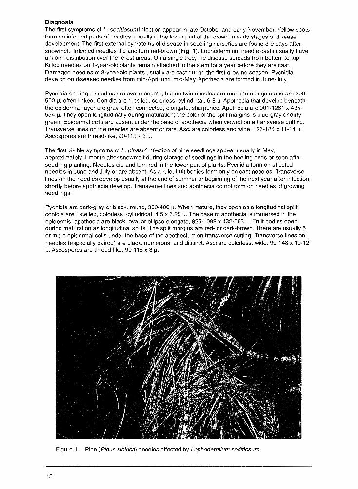
Diagnosis The first symptoms of L. seditiosum infection appear in late October and early November. Yellow spots form on infected parts of needles, usually in the lower part of the crown in early stages of disease development. The first external symptoms of disease in seedling nurseries are found 3-9 days after snowmelt. Infected needles die and turn red-brown (Fig. 1 ). Lophodermium needle casts usually have uniform distribution over the forest areas. On a single tree, the disease spreads from bottom to top. Killed needles on 1-year-old plants remain attached to the stem for a year before they are cast. Damaged needles of 3-year-old plants usually are cast during the first growing season. Pycnidia develop on diseased needles from mid-April until mid-May. Apothecia are formed in June-July.
Pycnidia on single needles are oval-elongate, but on twin needles are round to elongate and are 300-500 µ, often linked. Conidia are 1-celled, colorless, cylindrical, 6-8 µ. Apothecia that develop beneath the epidermal layer are gray, often connected, elongate, sharpened. Apothecia are 901-1281 x 435-554 µ. They open longitudinally during maturation; the color of the split margins is blue-gray or dirtygreen. Epidermal cells are absent under the base of apothecia when viewed on a transverse cutting. Transverse lines on the needles are absent or rare. Asci are colorless and wide, 126-184 x 11-14 µ. Ascospores are thread-like, 90-115 x 3 µ.
The first visible symptoms of L. pinastri infection of pine seedlings appear usually in May, approximately 1 month after snowmelt during storage of seedlings in the heeling beds or soon after seedling planting. Needles die and turn red in the lower part of plants. Pycnidia form on affected needles in June and July or are absent. As a rule, fruit bodies form only on cast needles. Transverse lines on the needles develop usually at the end of summer or beginning of the next year after infection, shortly before apothecia develop. Transverse lines and apothecia do not form on needles of growing seedlings.
Pycnidia are dark-gray or black, round, 300-400 µ. When mature, they open as a longitudinal split; conidia are 1-celled, colorless, cylindrical, 4.5 x 6.25 µ. The base of apothecia is immersed in the epidermis; apothecia are black, oval or ellipse-elongate, 825-1099 x 432-563 µ. Fruit bodies open during maturation as longitudinal splits. The split margins are red- or dark-brown. There are usually 5 or more epidermal cells under the base of the apothecium on transverse cutting. Transverse lines on needles (especially paired) are black, numerous, and distinct. Asci are colorless, wide, 90-148 x 10-12 µ. Ascospores are thread-like, 90-115 x 3 µ.
Figure 1.-Pine (Pinus sibirica) needles affected by Lophodermium seditiosum.
12
Biology Lophodermium needle casts of seedlings in nurseries and green-houses, saplings, understory trees, and forest plantations less than 5 years old are caused primarily by L seditiosum. Occasionally, L pinastri is found on seedlings in nurseries and greenhouses. Understory trees and forest plantations 6-14 years old are damaged by both species of fungi at the same time. Pines more than 15 years old are infected only by L pinastri.
Sources of L seditiosum inoculum are diseased plants in nurseries, pine plantations, and understory trees. Both healthy and weak plants are affected. Ascospores mature and spread in June and July. Needle penetration occurs from late July to late September or early October. Intensity of disease development depends on the quantity of precipitation from June to August and air temperature during July and August; however, moisture is the primary factor in disease development.
Pine plantations and understory 15 years old and older are the sources of L pinastri inoculum. Apothecia form on dead needles on the ground during the year after they drop. The most active period for ascospore release and needle penetration is July and August, but these can occur in May. Seedlings subjected to infection may have been weakened by unfavorable growing conditions, infection by other diseases, and mechanical injuries.
Damage Nursery seedlings and forest plantations less than 5 years old sustain the greatest damage. Damage to pine seedlings in Russian nurseries ranges from 30-100 percent. Needle casts reduce productivity of pine plantations as well as the health of standard planting material in the nursery.
Distribution Throughout Russia
Control Observation Survey for the appearance and distribution of disease in spring just after snowmelt and again during the second half of summer and early autumn.
Cultural • Choose location of new nurseries carefully, e.g., at least 200 m from existing pine forests or plantations. • Rotate pine with other conifers in nursery beds at intervals of 2 years or more. • Select seed sources from resistant plantations. • Use appropriate and agrotechnical methods for establishing and maintaining seedlings.
Chemical • Protect 1-3-year-old seedlings with systemic fungicides (BAYMEB, Benomyl, Bavistin, Daconil, Benlate, Topsin M) or protective fungicides (Zineb, sulphur) .
••• Pathogen Lophodermium macrosporum (Hart.) Rehm.
Host Norway spruce ( Picea abies)
Diagnosis Needles on previous-year shoots turn brown in May. Long, black apothecia form on lower surface of needles during summer. Asci are mace-shaped, 100 x 15-21 µ.
Damage Weakens and can kill seedlings in understory and forest plantations
Distribution European part of Russia, Urals, Siberia
13
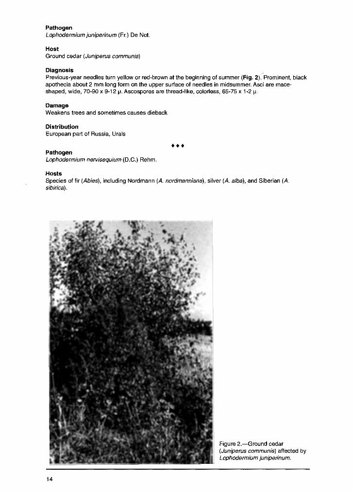
Pathogen Lophodermium juniperinum (Fr.) De Not.
Host Ground cedar (Juniperus communis)
Diagnosis -Previous-year needles turn yellow or red-brown at the beginning of summer (Fig. 2). Prominent, black apothecia about 2 mm long form on the upper surface of needles in midsummer. Asci are maceshaped, wide, 70-90 x 9-12 µ. Ascospores are thread-like, colorless, 65-75 x 1-2 µ.
Damage Weakens trees and sometimes causes dieback
Distribution European part of Russia, Urals
••• Pathogen Lophodermium nervisequium (D.C.) Rehm.
Hosts Species of fir (Abies), including Nordmann (A. nordmanniana), silver (A. alba), and Siberian (A. sibirica).
14
Figure 2.-Ground cedar (Juniperus communis) affected by Lophodermium juniperinum.
Diagnosis Needles turn brown at the end of summer. Pycnidia form on the lower surface of needles as tiny black marks. Elliptical black apothecia, 1-1.5 mm long, form in needles later. Asci are mace-shaped, 70-100 x 15-20 µ. Ascospores are oblong-clavate, slightly curved, colorless, 1-celled, 50-60 x 2-2.5 µ.
Damage Weakens trees in young stands and can cause partial dieback
Distribution Southeastern area of European part of Russia, Urals, Siberia
Snow Blight
Pathogen Phacidium infestans Karst.
Hosts Scotch pine (Pinus sylvestris), Siberian stone pine (P. sibirica), mountain pine (P. pumila), Norway spruce (Picea abies), Siberian black spruce (P. obovata), and ground cedar (Juniperus communis)
Diagnosis The first symptoms of disease appear in January when temperature inside the snow layer ranges from 0-5°C. The foliage is pale-green, and an ephemeral and cobwebby mycelium forms on the needles at this time. In February, needles have a marbled appearance with specific alternation of green, yellow, and brown spots. Active development of exterior mycelium begins in March when the temperature inside the snow layer reaches to +0.5°C. Needles beneath the snow are killed.
Mycelium spreads from diseased needles and during the period of snowmelt develops into white and gray pellicles, which are important diagnostic features. The pellicles are short-lived and only dirty-white pieces of mycelium remain on the seedlings and soil. Diseased needles die during the first several days after snowmelt and become bright-red or orange. At this time groups of infected seedlings are clearly noticeable in comparison with the green color of healthy plants. Soon after snowmelt, diseased needle color lightens and primordia of apothecia develop on them. The apothecia are small, dark blurred spots. During summer, infected needles become gray, and apothecia resemble dark hillocks. At the beginning of autumn, needles turn a distinct ashy color. Fragile apothecia open and burst through the needle epidermis, forming star-shaped openings. The gray-pink round hymenial layer is visible at this time. Asci are widely clavate with a distinct thick cover and they contain ellipsoid, rarely egg-shaped ascospores. Asci are 72-140 x 12-25 µ; spores are 11-28 x 5-9 µ.
Biology Ascospores mature and spread after the apothecia open. The most active period occurs under warm, moist conditions after heavy precipitation and higher than mean annual air temperature. Spore dispersal usually is from late September through October. Major spore dissemination occurs in midOctober. The most favorable conditions for this process and needle infection are periods of snow followed by snow melt. Spore spread ends before permanent snow cover is established. Typical midOctober weather patterns, alternating rain and snowfall, result in heavy precipitation and high humidity. Spore loading on the needles is well established when snow cover is permanent. Ascospores germinate on and penetrate needles. Fungal development inside leaf tissue begins after snow cover is established. The most important factor in this process is the temperature under the snow cover. Conditions for pathogen development are most favorable when high snow cover lies on unfrozen soil that remains slushy during the winter and the temperature inside the snow layer is 0° and above.
The saprophytic phase of fungus development begins after snowmelt and continues through the vegetative growth period. Fungal fruit bodies form and mature throughout the year. The parasitic phase of the fungus begins with apothecia opening and continues up to snowmelt. In Russia, depending on climate conditions, there are three types of fungal development during the parasitic phase: European, Siberian, and Intermediate. In the European type, the spores spread in autumn and disease develops during the winter. The European type develops in regions with wet autumns, relatively mild winters, and high snow cover. With the Siberian type, sporulation, penetration, and infection of healthy needles occur simultaneously in the snow layer cavities and in the snow "greenhouses" that have relatively high humidity and temperature during spring snowmelt. The
15

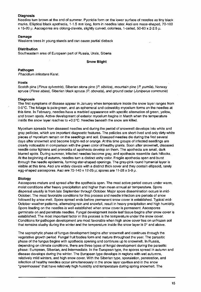
Figure 3.-Pine (Pinus sylvestris) understory in cutting area showing symptoms of snow blight (Phacidium infestans).
Siberian type occurs under conditions of continental climate with cold dry autumns, harsh winters, and warm springs. The Intermediate type is similar to the other types depending on weather changes.
Damage Damages seedlings in nurseries, forest plantations, and pine understory. Snow blight is most prevalent on planted and natural seedlings, and on pine plantation understory during winters with a deep snow layer. Annual mortality of nursery seedlings ranges from 25-40 percent. Snow blight also hinders successful artificial regeneration in clearcut areas where annual mortality ranges from 50-60 percent (Fig. 3).
Distribution European part of Russia, Urals, Siberia, Far East
Control Observation • Survey for snow-blight loci during the first 2 3 days after snowmelt; mycelial patches are visible a1 this time; repeat observations in September-October.
Prevention and Cultural • Establish new tree nurseries at least 200 meters from adjacfm1 forest stands and plantations. • During spring, collect and bum all diseased seedlings. • Do not use snow blight-infected seedlings for reforestation.
Chemical • In nurseries and pine plantations, apply protective fungicides (colloid sulfur) and systemic fungicides (Benornyl, Benlate, Daconil, Bavistin, Derosa!, BAYMEB).
Meria Needle Blight
Class/Order: Deuteromycetes, Hyphomycetales
Pathogen Meria laricis Vuill.
16
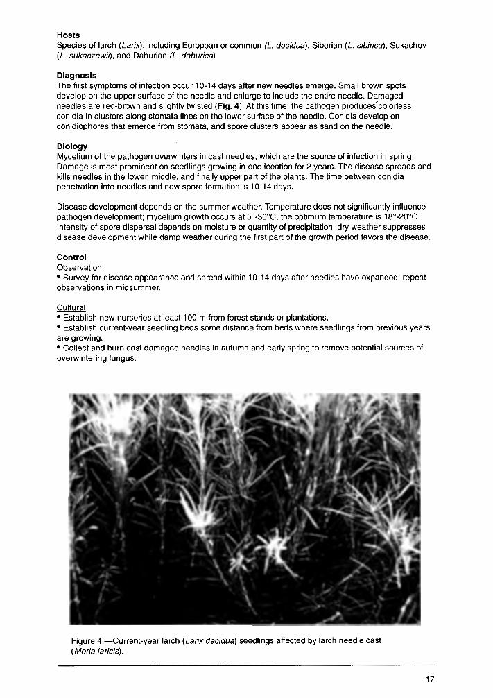
Hosts Species of larch (Larix), including European or common (L. decidua), Siberian (L sibirica), Sukachev (L. sukaczewit), and Dahurian (L. dahurica)
Diagnosis The first symptoms of infection occur 10-14 days after new needles emerge. Small brown spots develop on the upper surface of the needle and enlarge to include the entire needle. Damaged needles are red-brown and slightly twisted (Fig. 4). At this time, the pathogen produces colorless conidia in clusters along stomata lines on the lower surface of the needle. Conidia develop on conidiophores that emerge from stomata, and spore clusters appear as sand on the needle.
Biology Mycelium of the pathogen overwinters in cast needles, which are the source of infection in spring. Damage is most prominent on seedlings growing in one location for 2 years. The disease spreads and kills needles in the lower, middle, and finally upper part of the plants. The time between conidia penetration into needles and new spore formation is 10-14 days.
Disease development depends on the summer weather. Temperature does not significantly influence pathogen development; mycelium growth occurs at 5°-30°C; the optimum temperature is 18°-20°C. Intensity of spore dispersal depends on moisture or quantity of precipitation; dry weather suppresses disease development while damp weather during the first part of the growth period favors the disease.
Control Observation • Survey for disease appearance and spread within 10-14 days after needles have expanded; repeat observations in midsummer.
Cultural • Establish new nurseries at least 100 m from forest stands or plantations. • Establish current-year seedling beds some distance from beds where seedlings from previous years are growing. • Collect and burn cast damaged needles in autumn and early spring to remove potential sources of overwintering fungus.
Figure 4.-Current-year larch (Larix decidua) seedlings affected by larch needle cast (Meria laricis).
17
• Use greenhouses to protect seedlings from primary infection.
Chemical • Apply fungicides to fallen needles (e.g., colloid sulfur) to exterminate the inoculum source. • Apply preventive fungicides on foliage of seedlings and young plantations during the growing season. • Use systemic fungicides (BAYMEB, Topsin M, Daconil, Bavistin, and Derosal) an(!protective fungicides (colloid sulfur, Zineb, Poliram, Policarbacin, Metiram).
Brown Felt Blight
Class/Order: Ascomycetes, Sphaeriales
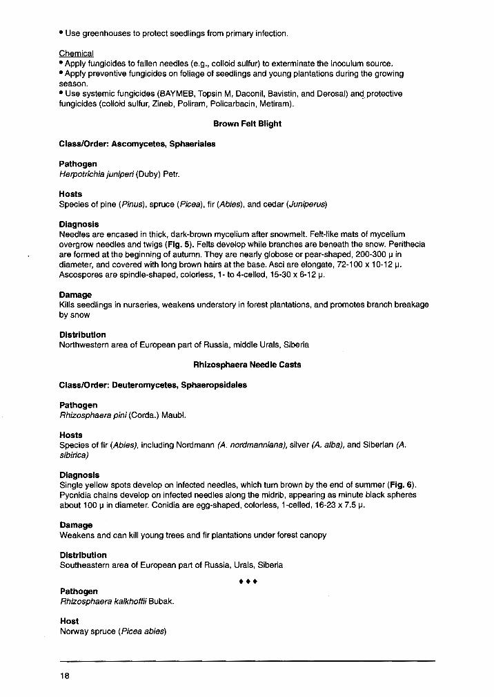
Pathogen Herpotrichia juniperi (Duby) Petr.
Hosts Species of pine (Pinus), spruce (Picea), fir (Abies), and cedar (Juniperus)
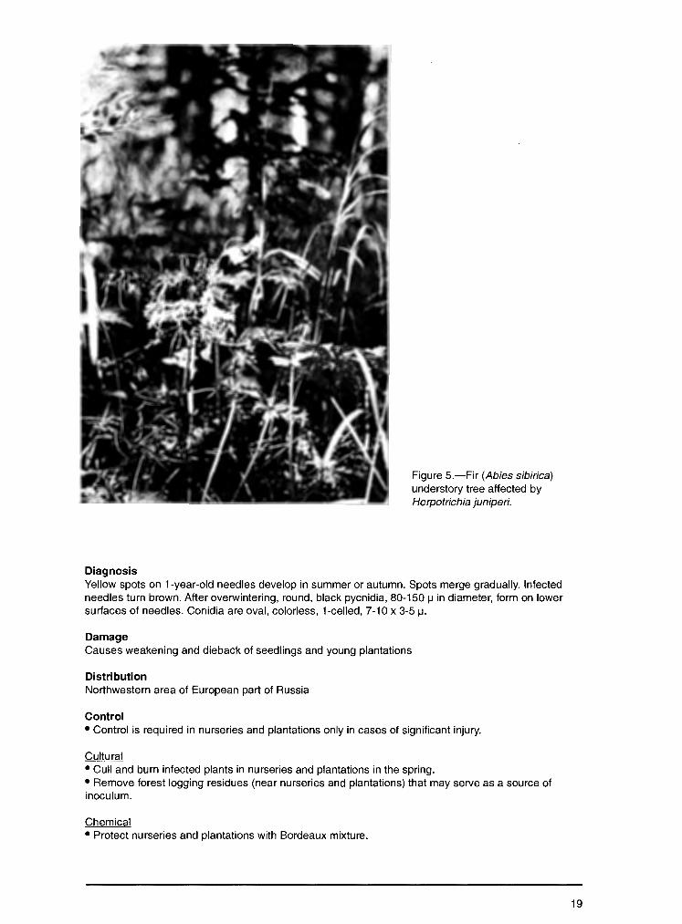
Diagnosis Needles are encased in thick, dark-brown mycelium after snowmelt. Felt-like mats of mycelium overgrow needles and twigs (Fig. 5). Felts develop while branches are beneath the snow. Perithecia are formed at the beginning of autumn. They are nearly globose or pear-shaped, 200-300 µ in diameter, and covered with long brown hairs at the base. Asci are elongate, 72-100 x 10-12 µ. Ascospores are spindle-shaped, colorless, 1- to 4-celled, 15-30 x 6-12 µ.
Damage Kills seedlings in nurseries, weakens understory in forest plantations, and promotes branch breakage by snow
Distribution Northwestern area of European part of Russia, middle Urals, Siberia
Rhizosphaera Needle Casts
Class/Order: Deuteromycetes, Sphaeropsidales
Pathogen Rhizosphaera pini (Corda.) Maubl.
Hosts Species of fir (Abies), including Nordmann (A. nordmanniana), silver (A. alba), and Siberian (A. sibirica)
Diagnosis Single yellow spots develop on infected needles, which turn brown by the end of summer (Fig. 6). Pycnidia chains develop on infected needles along the midrib, appearing as minute black spheres about 100 µ in diameter. Conidia are egg-shaped, colorless, 1-celled, 16-23 x 7.5 µ.
Damage Weakens and can kill young trees and fir plantations under forest canopy
Distribution Southeastern area of European part of Russia, Urals, Siberia
••• Pathogen Rhizosphaera kalkhoffii Bubak.
Host Norway spruce (Picea abies)
18
Diagnosis
Figure 5.-Fir (Abies sibirica) understory tree affected by Herpotrichia juniperi.
Yellow spots on 1-year-old needles develop in summer or autumn. Spots merge gradually. Infected needles turn brown. After overwintering, round, black pycnidia, 80-150 µ in diameter, form on lower surfaces of needles. Conidia are oval, colorless, 1-celled, 7-10 x 3-5 µ.
Damage Causes weakening and dieback of seedlings and young plantations
Distribution Northwestern area of European part of Russia
Control • Control is required in nurseries and plantations only in cases of significant injury.
Cultural • Cull and burn infected plants in nurseries and plantations in the spring. • Remove forest logging residues (near nurseries and plantations) that may serve as a source of inoculum.
Chemical • Protect nurseries and plantations with Bordeaux mixture.
19
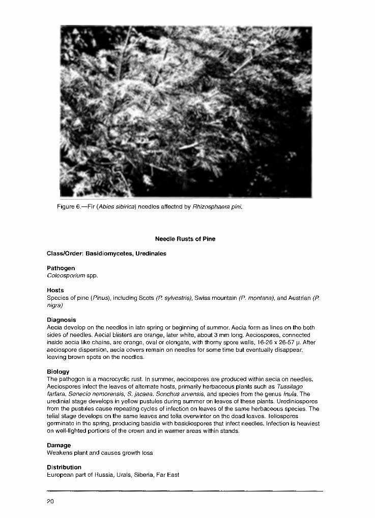
Figure 6.-Fir (Abies sibirica) needles affected by Rhizosphaera pini.
Needle Rusts of Pine
Class/Order: Basidiomycetes, Uredinales
Pathogen Coleosporium spp.
Hosts Species of pine (Pinus), including Scots (P sy/vestris), Swiss mountain (P. montana), and Austrian (P nigra)
Diagnosis Aecia develop on the needles in late spring or beginning of summer. Aecia form as lines on the both sides of needles. Aecial blisters are orange, later white, about 3 mm long. Aeciospores, connected inside aecia like chains, are orange, oval or elongate, with thorny spore walls, 16-26 x 26-57 µ. After aeciospore dispersion, aecia covers remain on needles for some time but eventually disappear, leaving brown spots on the needles.
Biology The pathogen is a macrocyclic rust. In summer, aeciospores are produced within aecia on needles. Aeciospores infect the leaves of alternate hosts, primarily herbaceous plants such as Tussi/ago farfara, Senecio nemorensis, S. jacaea, Sonchus arvensis, and species from the genus /nu/a. The uredinial stage develops in yellow pustules during summer on leaves of these plants. Urediniospores from the pustules cause repeating cycles of infection on leaves of the same herbaceous species. The telial stage develops on the same leaves and telia overwinter on the dead leaves. Teliospores germinate in the spring, producing basidia with basidiospores that infect needles. Infection is heaviest on well-lighted portions of the crown and in warmer areas within stands.
Damage Weakens plant and causes growth loss
Distribution European part of Russia, Urals, Siberia, Far East
20
Control Prevention • Remove alternate host plants.
Chemical • Apply Bordeaux mixture to foliage.
• •• Pathogen Chrysomyxa ledi (Alb. et Schw.) de Bary.
Hosts Norway spruce (Picea abies) and Siberian black spruce (P. obovata)
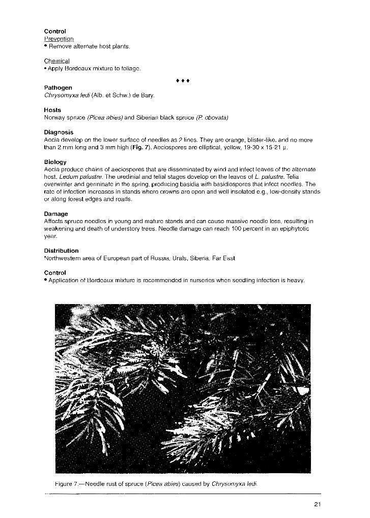
Diagnosis Aecia develop on the lower surface of needles as 2 lines. They are orange, blister-like, and no more than 2 mm long and 3 mm high (Fig. 7). Aeciospores are elliptical, yellow, 19-30 x 15-21 µ.
Biology Aecia produce chains of aeciospores that are disseminated by wind and infect leaves of the alternate host, Ledum pa/ustre. The uredinial and telial stages develop on the leaves of L. palustre. Telia overwinter and germinate in the spring, producing basidia with basidiospores that infect needles. The rate of infection increases in stands where crowns are open and well insolated e.g., low-density stands or along forest edges and roads.
Damage Affects spruce needles in young and mature stands and can cause massive needle loss, resulting in weakening and death of understory trees. Needle damage can reach 100 percent in an epiphytotic year.
Distribution Northwestern area of European part of Russia, Urals, Siberia, Far East
Control • Application of Bordeaux mixture is recommended in nurseries when seedling infection is heavy.
Figure ?.~Needle rust of spruce (Picea abies) caused by Chrysomyxa ledi.
21
Leaf Diseases Powdery Mildews
Class/Order: Ascomycetes/Erysiphales
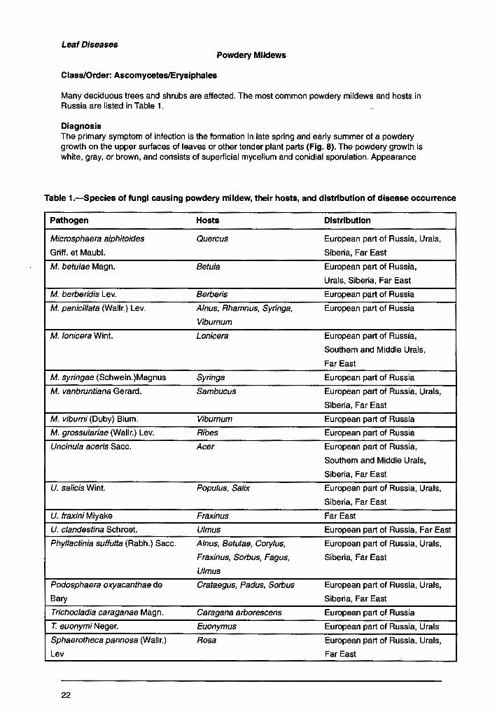
Many deciduous trees and shrubs are affected. The most common powdery mildews and hosts in Russia are listed in Table 1.
Diagnosis The primary symptom of infection is the formation in late spring and early summer of a powdery growth on the upper surfaces of leaves or other tender plant parts (Fig. 8). The powdery growth is white, gray, or brown, and consists of superficial mycelium and conidial sporulation. Appearance
Table 1.-Species of fungi causing powdery mildew, their hosts, and distribution of disease occurrence
Pathogen Hosts Distribution
Microsphaera alphitoides Quercus European part of Russia, Urals,
Griff. et Maubl. Siberia, Far East
M. betulae Magn. Betula European part of Russia,
Urals, Siberia, Far East
M. berberidis Lev. Berberis European part of Russia
M. penicillata (Wallr.) Lev. A/nus, Rhamnus, Syringa, European part of Russia
Viburnum
M. lonicera Wint. Lonicera European part of Russia,
Southern and Middle Urals,
Far East
M. syringae (Schwein.)Magnus Syringa European part of Russia
M. vanbruntiana Gerard. Sambucus European part of Russia, Urals,
Siberia, Far East
M. viburni (Duby} Blum. Viburnum European part of Russia
M. grossulariae (Wallr.) Lev. Ribes European part of Russia
Uncinula aceris Sacc. Acer European part of Russia,
Southern and Middle Urals,
Siberia, Far East
U. salicis Wint. Populus, Salix European part of Russia, Urals,
Siberia, Far East
U. fraxini Miyake Fraxinus Far East
U. clandestina Schroet. Ulmus European part of Russia, Far East
Phyllactinia suffulta (Rabh.) Sacc. A/nus, Betulae, Cory/us, European part of Russia, Urals,
Fraxinus, Sorbus, Fagus, Siberia, Far East
Ulmus
Podosphaera oxyacanthae de Crataegus, Padus, Sorbus European part of Russia, Urals,
Bary Siberia, Far East
Trichocladia caraganae Magn. Caragana arborescens European part of Russia
T. euonymi Neger. Euonymus European part of Russia, Urals
Sphaerotheca pannosa (Wallr.) Rosa European part of Russia, Urals,
Lev Far East
22
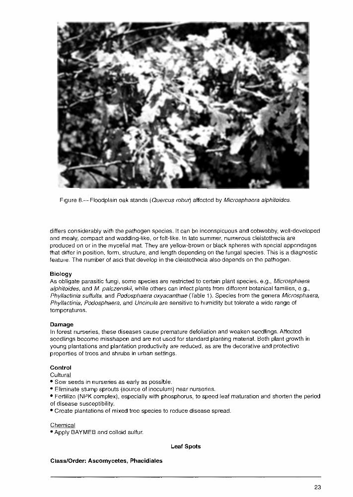
Figure 8.-Floodplain oak stands (Quercus robur) affected by Microsphaera alphitoides.
differs considerably with the pathogen species. It can be inconspicuous and cobwebby, well-developed and mealy, compact and wadding-like, or felt-like. In late summer, numerous cleistothecia are produced on or in the mycelial mat. They are yellow-brown or black spheres with special appendages that differ in position, form, structure, and length depending on the fungal species. This is a diagnostic feature. The number of asci that develop in the cleistothecia also depends on the pathogen.
Biology As obligate parasitic fungi, some species are restricted to certain plant species, e.g., Microsphaera a/phitoides, and M. palczenskii, while others can infect plants from different botanical families, e.g., Phyllactinia suffulta, and Podosphaera oxyacanthae (Table 1 ). Species from the genera Microsphaera, Phyllactinia, Podosphaera, and Uncinula are sensitive to humidity but tolerate a wide range of temperatures.
Damage In forest nurseries, these diseases cause premature defoliation and weaken seedlings. Affected seedlings become misshapen and are not used for standard planting material. Both plant growth in young plantations and plantation productivity are reduced, as are the decorative and protective properties of trees and shrubs in urban settings.
Control Cultural • Sow seeds in nurseries as early as possible. • Eliminate stump sprouts (source of inoculum) near nurseries. • Fertilize (NPK complex), especially with phosphorus, to speed leaf maturation and shorten the period of disease susceptibility. • Create plantations of mixed tree species to reduce disease spread.
Chemical • Apply BAYMEB and colloid sulfur.
Leaf Spots
Class/Order: Ascomycetes, Phacidiales
23

Pathogen Rhytisma acerinum (Pers.) Fr. (anamorph: Melasmia acerina Zev.)
Hosts Species of maple (Acer)
Diagnosis -Spots develop on leaves in summer. The infected tissue turns yellow and numerous small, black stromata develop within the spots on upper leaf surfaces. Large convex, black, shiny, stromata coalesce with small ones within the spot. Stromata are surrounded by yellow-green margins. Conidia are produced on the stromata. After overwintering, apothecia develop in the stromata and asci with ascospores mature during the summer. Asci are mace-shaped on a pedicle, 120-130 x 9-10 µ. Ascospores are sticky, thread-like, 52 x 80 x 1.5-3 µ.
Distribution European part of Russia, Urals, Far East
••• Pathogen Rhytisma punctatum (Pers.) Fr. (anamorph: Melasmia punctata Sacc.)
Hosts Species of maple (Acer)
Diagnosis Small, yellow spots develop on leaves in summer, and conidophores and spores develop on them. Later, small, dotted, shiny stromata develop within the spots. Apothecia form within the stroma. Asci are wide, 120-130 x 9-10 µ. Ascospores are thread-like, colorless, 60-80 x 1.5-3 µ.
Distribution European part of Russia, Urals, Far East
••• Pathogen Rhytisma salicinum (Pers.) Fr. (anamorph: Melasmia salicina Lev.)
Hosts Species of willow ( Salix)
Diagnosis Yellow spots develop on leaves in summer. Later, a convex, black, shiny stroma, 0.5-2 cm in diameter, forms (Fig. 9). Apothecia form, ripen during spring, and emerge from the stroma. The hymenium is yellow. Asci are wide, 120-150 x 10-15 µ. Ascospores are colorless, thread-like, 60-100 x 1.5-3 µ.
Distribution European part of Russia, Urals, Siberia, Far East
Class/Order: Ascomycetes, Dothideales
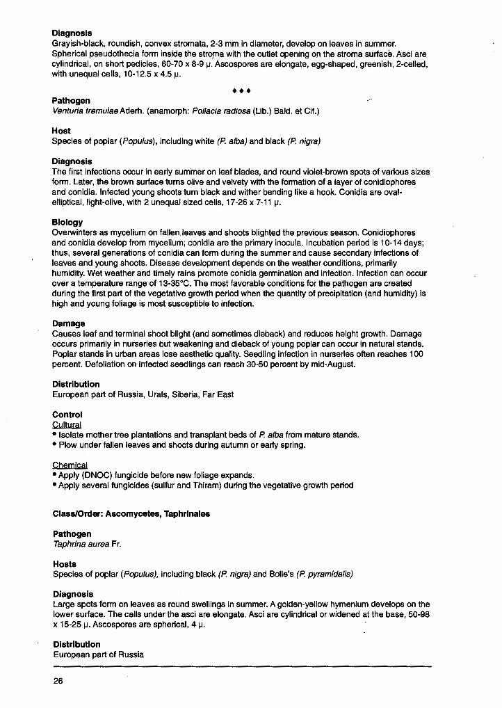
Pathogen Dothidella betulina (Fr.) Sacc.
Hosts Species of birch (Betula)
Diagnosis In summer, numerous small, black, convex stromata develop on the upper leaf surfaces (Fig. 10). They are round or irregular. Spherical pseudothecia with several loculi form after the leaves drop. Asci in loculi are cylindrical, 38-70 x 10-12.5 µ. Ascospores are elliptical, 2-celled with unequal cells, greenish, 10-14 x 5 µ.
Distribution European part of Russia, Urals, Far East
24
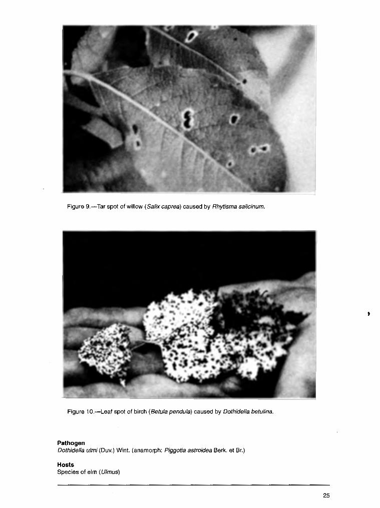
Figure 9.-Tar spot of willow (Salix caprea) caused by Rhytisma sa/icinum.
Figure 10.-Leaf spot of birch (Betu/a pendula) caused by Dothidella betulina.
Pathogen Dothidel/a ulmi (Duv.) Wint. (anamorph: Piggotia astroidea Berk. et Br.)
Hosts Species of elm (U/mus)
25
Diagnosis Grayish-black, roundish, convex stromata, 2-3 mm in diameter, develop on leaves in summer. Spherical pseudothecia form inside the strorn.a with the outlet opening on the stroma surface. Asci are cylindrical, on short pedicles, 60-70 x 8-9 µ. Ascospores are elongate, egg-shaped, greenish, 2-celled, with unequal cells, 10-12.5 x 4.5 µ.
• •• Pathogen Venturia tremulaeAderh. (anamorph: Pollacia radiosa (Lib.) Bald. et Cit.)
Host Species of poplar (Popu/us), including white (P. alba) and black (P. nigra)
Diagnosis The first infections occur in early summer on leaf blades, and round violet-brown spots of various sizes form. Later, the brown surface turns olive and velvety with the formation of a layer of conidiophores and conidia. Infected young shoots turn black and wither bending like a hook. Conidia are ovalelliptical, light-olive, with 2 unequal sized cells, 17-26 x 7-11 µ.
Biology Overwinters as mycelium on fallen leaves and shoots blighted the previous season. Conidiophores and conidia develop from mycelium; conidia are the primary inocula. Incubation period is 10-14 days; thus, several generations of conidia can form during the summer and cause secondary infections of leaves and young shoots. Disease development depends on the weather conditions, primarily humidity. Wet weather and timely rains promote conidia germination and infection. Infection can occur over a temperature range of 13-35°C. The most favorable conditions for the pathogen are created during the first part of the vegetative growth period when the quantity of precipitation (and humidity) is high and young foliage is most susceptible to infection.
Damage Causes leaf and terminal shoot blight (and sometimes dieback) and reduces height growth. Damage occurs primarily in nurseries but weakening and dieback of young poplar can occur in natural stands. Poplar stands in urban areas lose aesthetic quality. Seedling infection in nurseries often reaches 100 percent. Defoliation on infected seedlings can reach 30-50 percent by mid-August.
Distribution European part of Russia, Urals, Siberia, Far East
Control Cultural • Isolate mother tree plantations and transplant beds of P. alba from mature stands. • Plow under fallen leaves and shoots during autumn or early spring.
Chemical • Apply (DNOC) fungicide before new foliage expands. • Apply several fungicides (sulfur and Thiram) during the vegetative growth period
Class/Order: Ascomycetes, Taphrlnales
Pathogen Taphrina aurea Fr.
Hosts Species of poplar (Popu/us), including black (P. nigra) and Bolle's (P. pyramids/is)
Diagnosis Large spots form on leaves as round swellings in summer. A golden-yellow hymenium develops on the lower surface. The cells under the asci are elongate. Asci are cylindrical or widened at the base, 50-98 x 15-25 µ. Ascospores are spherical, 4 µ.
Distribution European part of Russia
26
Pathogen Taphrina polyspora Johans.
I
Host Tartarian maple (Acer tatarica)
Diagnosis Infected areas on the leaf are swollen and become irregularly shaped brown spots. Later, tney turn black and burst. The hymenium develops on the upper surface of spots. There are no cells under the asci. Asci are wide, cylindrical, rounded at the top, 33-47 x 12-17 µ. Ascospores are spherical, 3-4 µ.
Distribution European part of Russia
••• Pathogen Taphrina ulmi Johans.
Host European white elm (Ulmus laevis)
Diagnosis . Infected areas on leaves are slightly swollen, irregularly shaped spots. They are initially yellow and
later turn gray-brown or black. The hymeniun develops on the upper surface of spots. The cells under asci are wide. Asci vary from cylindrical to egg-shaped, rounded at the top, 12-20 x 8-10 µ. Ascospores are spherical, 3.5 µ.
Distribution European part of Russia
Class/Order: Deuteromycetes, Hyphomycetales
Pathogen Cercospora coryli Mont.
Hosts Species of hazel ( Cory/us)
Diagnosis Small, irregularly shaped, red-brown spots with a light center form on leaves. They may coalesce. The pathogen sporulates on the spots. Conidia are cylindrical, light-olive, 2- to 3-celled, 30-45 x 3-4.5 µ.
Distribution European part of Russia
••• Pathogen Cercospora fraxini Sacc.
·Hosts Species of ash (Fraxinus), including European (F. excelsior) and green (F. viridis)
Diagnosis Irregularly shaped or angular, brown or nearly black spots, 2-4 mm in diameter, form on leaves. Clusters of conidiophores develop on the lower surface of leaves. Conidia are spindle-shaped, pale olive, and 1-celled at the beginning of development; later, they become 2- to 6-celled, 26-65 x 4.4-6.5 µ.
Distribution European part of Russia
27
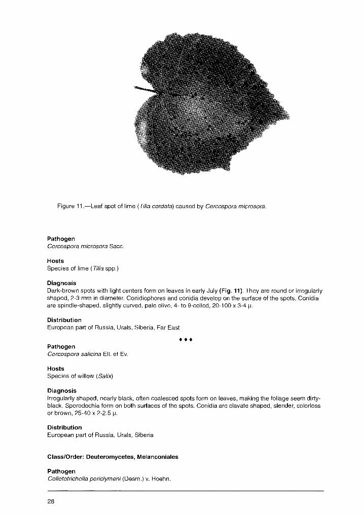
Figure 11.-Leaf spot of lime ( Tilia cordata) caused by Cercospora microsora.
Pathogen Cercospora microsora Sacc.
Hosts Species of lime (Tilia spp.)
Diagnosis Dark-brown spots with light centers form on leaves in early July (Fig. 11). They are round or irregularly shaped, 2-3 mm in diameter. Conidiophores and conidia develop on the surface of the spots. Conidia are spindle-shaped, slightly cuNed, pale olive, 4- to 9-celled, 20-100 x 3-4 µ.
Distribution European part of Russia, Urals, Siberia, Far East
••• Pathogen Cercospora salicina Ell. et Ev.
Hosts Species of willow ( Salix)
Diagnosis Irregularly shaped, nearly black, often coalesced spots form on leaves, making the foliage seem dirtyblack. Sporodochia form on both surfaces of the spots. Conidia are clavate shaped, slender, colorless or brown, 25-40 x 2-2.5 µ.
Distribution European part of Russia, Urals, Siberia
Class/Order: Deuteromycetes, Melanconiales
Pathogen Colletotrichella periclymeni (Desm.) v. Hoehn.
28
Hosts _ ~pecies of honeysuckle (Lonicera)
Diagnosis Spots are round, often coalesced, olive-brown with light centers, often with a dark narrow border. Sporodochia are formed on the upper surface of spots. Conidia are elongate spindle- or sickle-shaped, colorless, 17-31 x 6.5-10 µ.
Distribution European part of Russia, Urals, Siberia
••• Pathogen Coryneum fo/iicola Fckl.
Hosts Species of hawthorne ( Crataegus)
Diagnosis Spots are round or irregularly shaped when coalesced, and red-brown or brown. Sporodochia are formed on upper surface of spots. Conidia, formed on slender pedicles, are elongate-elliptical, 4-celled, olive, 12-18 x 5.5-7 µ.
Distribution European part of Russia, Urals, Siberia
••• Pathogen Cylindrosporium ulmi (Fr.) Vassil.
Hosts Species of elm ( Ulmus)
Diagnosis Spots are small, round or irregularly shaped, and may be coalesced. They are yellow initially and then turn brown. Sporodochia form on lower surface of spots and are slightly convex and yellow-brown. Conidia are colorless, cylindrical, widened at the top or spindle-shaped, curved or straight, 2- to 5-celled, 28-60 x 3.5-8 µ.
Distribution European part of Russia, Urals, Siberia, Far East
••• Pathogen Gloeosporium coryli (Desm.) Sacc.
Hosts Species of hazel ( Cory/us)
Diagnosis Spots are round, red- to dark-brown. Small, dark, flat, cushion-like sporodochia (acervuli) are formed on both surfaces of spots. Conidia are cylindrical, with spherical ends, colorless, 1-celled, 7-15 x 5-7 µ.
Distribution European part of Russia, Urals, Siberia
••• Pathogen Gloeosporium betulinum West. (syn. Discula betulina West. Arx)
Hosts Species of birch (Betula)
29
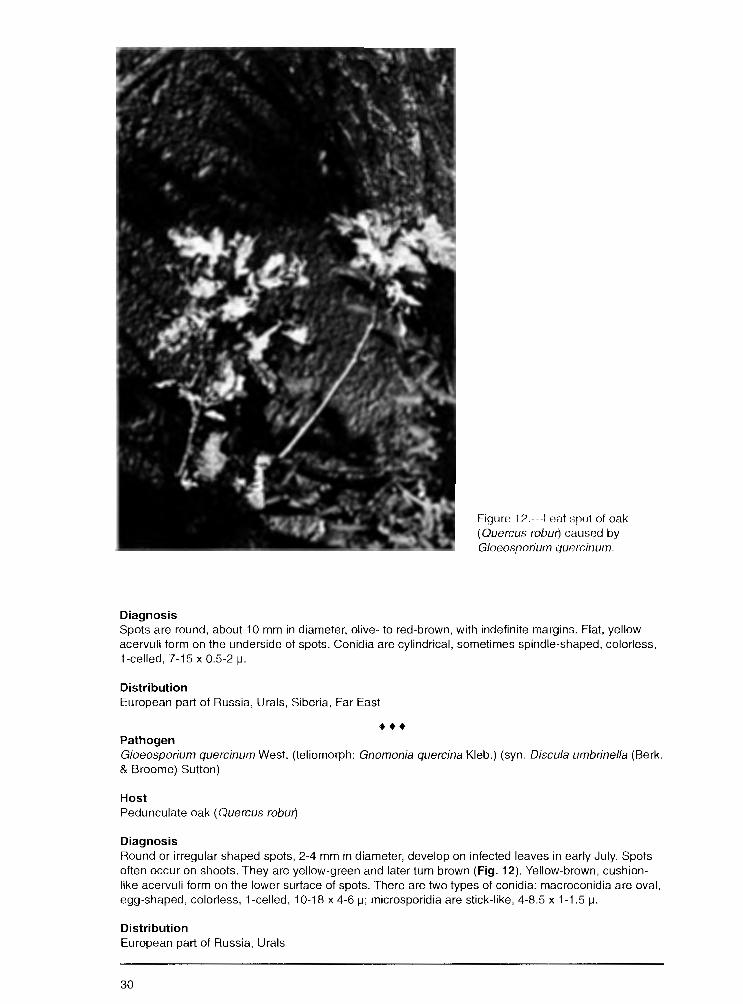
Diagnosis
Figure 12.---Leat :;pot ot oak ( Quercus robur) causmJ by Gloeosporium quercinwn
Spots are round, about 10 mm in diameter, olive- to red-brown, with indefinite margins. Flat, yellow acervuli form on the underside of spots. Conidia are cylindrical, sometimes spindle-shaped, colorless, 1-celled, 7-15 x 05-2 µ.
Distribution European part of Russia, Urals, Siberia, Far East
••• Pathogen G/oeosporium quercinum West (teliomorph: Gnomonia quercina Kleb.) (syn. Discula umbrinel/a (Berk. & Broome) Sutton)
Host Pedunculate oak ( Quercus robur)
Diagnosis F1ound or irregular shaped spots, 2-4 mm in diameter, develop on infected leaves in early July. Spots often occur on shoots. They are yellow-green and later turn brown (Fig. 12). Yellow-brown, cushionlike acervuli form on the lower surface of spots. There are two types of conidia: macroconidia are oval, egg-shaped, colorless, 1-celled, 10-18 x 4-6 µ; microsporidia are stick-like, 4-8.5 x 1-1 .5 µ.
Distribution European part of Russia, Urals
30
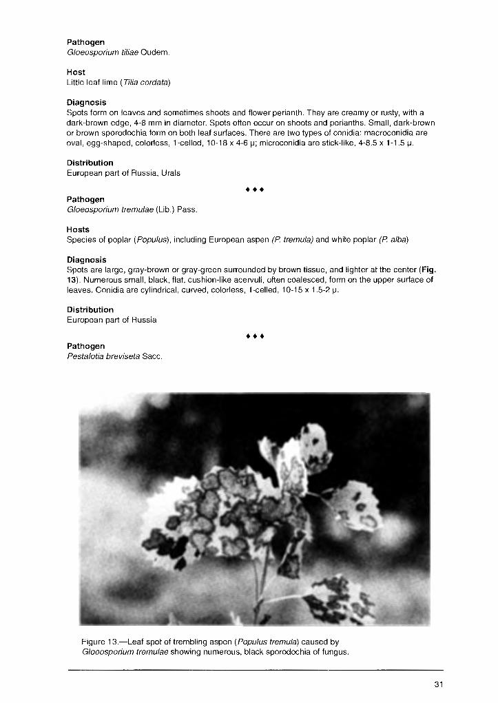
Pathogen Gloeosporium tiliae Oudem.
Host Little leaf lime ( Tilia cordata)
Diagnosis Spots form on leaves and sometimes shoots and flower perianth. They are creamy or rusty, with a dark-brown edge, 4-8 mm in diameter. Spots often occur on shoots and perianths. Small, dark-brown or brown sporodochia form on both leaf surfaces. There are two types of conidia: macroconidia are oval, egg-shaped, colorless, 1-celled, 10-18 x 4-6 µ; microconidia are stick-like, 4-8.5 x 1-1.5 µ.
Distribution European part of Russia, Urals
••• Pathogen G/oeosporium tremulae (Lib.) Pass.
Hosts Species of poplar (Populus), including European aspen (P tremula) and white poplar (P alba)
Diagnosis Spots are large, gray-brown or gray-green surrounded by brown tissue, and lighter at the center (Fig. 13). Numerous small, black, flat, cushion-like acervuli, often coalesced, form on the upper surface of leaves. Conidia are cylindrical, curved, colorless, 1-celled, 10-15 x 1.5-2 µ.
Distribution European part of Russia
••• Pathogen Pestalotia breviseta Sacc.
Figure 13.-Leaf spot of trembling aspen (Popu/us tremula) caused by G/oeosporium tremulae showing numerous, black sporodochia of fungus.
31
Hosts Species of apple (Ma/us) and pear (Pyrus)
Diagnosis Leaf spots are gr~y and vary in shape and size. Cushion-like sporodochia form on the upper surfaces of black spots in clusters. Conidia are spindle-shaped, 5 celled, with 3 appendages, 8-10 x 1 µ.
Distribution European part of Russia
••• Pathogen Marssonina populi Kleb.
Hosts Species of poplar (Populi.Js), especially balsam poplar hybrid (P. tacamahaca)
Diagnosis Spots appear on leaves from late May to early June. Spots are brown, round, about 4-5 mm in diameter, and gradually coalesce. They can cover the entire leaf surface. The central part of the spot is dotted with yellow-white sporodochia. Conidia are egg-shaped, elongate to pear-shaped, sometime curved. They are 1-celled initially but later 2-celled with the upper cell wider than the basal cell, colorless, 15-29 x 5-10 µ.
Biology Source of inoculum in the spring is fallen infected leaves. Conidia from sporodochia initiate the repeating cycle of disease during summer. Pathogen incubation is about 3-5 days, so incidence of infected trees and degree of crown damage increase quickly. Primary infection occurs on lower branches nearest the conidia on overwintering fallen leaves. Infection spreads to the middle and upper branches of the crown. Wet weather (or high humidity) and moderate temperatures (13-18°C) intensify disease development.
Damage Causes premature defoliation. By midsummer, susceptible species of poplar lose 50-80 percent of their foliage. Successive seasons of infection and leaf loss can cause a decrease in growth and vigor. Damage is most severe in nurseries.
Distribution European part of Russia, Urals, Siberia, Far East
Control Cultural • Isolate newly created poplar nurseries and plantations from mature stands. • Gather and burn infected fallen leaves in autumn.
Chemical • Apply fungicide (DNOC) to fallen leaves in spring before or soon after new leaves emerge. • Apply Bordeaux mixture to foliage during growth stages.
Class/Order: Deuteromycetes, Sphaeropsidales
Pathogen Asteroma padi Grev.
Host Bird cherry (Padus avium)
Diagnosis Large spots form on both surfaces of leaf. Brown-violet spots form among branching, fan-like hyphae. Brown pycnidia are inconspicuous, embedded in the leaf tissue in groups. Conidia are elongate, eggshaped, colorless, 1-celled, 12-18 x 3-4 µ.
32

Distribution European part of Russia, Urals, Siberia
Pathogen Asteroma tiliae Rud.
Host Little leaf lime ( Tilia cordata)
Diagnosis
•••
Large spots, brown-violet or nearly black, form on the upper surface of leaf. Heavily branched mycelial fans form on the surface of the spot.
Distribution European part of Russia, Urals, Siberia
••• Pathogen Phyllosticta coryli West.
Hosts Species of hazel ( Cory/us)
Diagnosis Spots are large, irregular shaped, brown with lighter brown center. Pycnidia form on both surfaces of spots embedded in leaf tissue. They later break through the tissue. Pycnidia are spherical with a flat top, 100-150 µ in diameter. Conidia are colorless, ellipsoidal, with 2 drops of oil, 1-celled, 7-8 x 2-3.5 µ.
Distribution European part of Russia
••• Pathogen Phyllosticta fraxini Ell. et Mart.
Host European ash (Fraxinus excelsiot,
Diagnosis Spots are round, 5-10 mm in diameter, brown, surrounded by dark-red margins. Brown pycnidia about 150 µ in diameter form on the upper surface of leaf. Conidia are elongate, elliptical, colorless, with 1-2 drops of oil, 7-9 x 2.5-3 µ.
Distribution European part of Russia
••• Pathogen Phyllosticta populina Sacc.
Hosts Species of poplar (Popu/us)
Diagnosis Leaf spots are angular, white, with a black border, 1-3 mm in diameter. Pycnidia form on the upper surface of spots, are spherical, and black. Conidia are egg-shaped or elliptical, 1-celled, olive, 6-8 x 3-3.5 µ.
Distribution European part of Russia, Urals, Siberia
33
Pathogen Septoria betulae Pass. non (Lib.) West.
Hosts Species of birch (Betula)
Diagnosis ~
Spots form on both surfaces of leaf. They are irregularly-shaped or round, brown or light-brown, and coalesce. Pycnidia about 100 µ in diameter form on the lower surface of leaf. Conidia are thread-like, colorless, with 1 to several septa, 30-60 x 1.5-2 µ.
Distribution European part of Russia, Urals, Siberia
••• Pathogen Septoria populi Desm.
Hosts Species of poplar ( Populus)
Diagnosis White necrotic spots are 2-3 mm in diameter, surrounded by dark margins. Pycnidia develop on dead tissues as black dots, 112-240 µ in diameter. Conidia are cylindrical, curved, colorless, 2-celled, 30-45 X 3 µ.
Distribution European part of Russia, Urals, Siberia
••• Pathogen Septoria quercicola (Desm.) Sacc.
Hosts Species of oak ( Quercus)
Diagnosis Small white spots, 1-3 mm in diameter, form on both surfaces of leaves, and are surrounded by wide dark margins. Pycnidia are scattered and black. Conidia are thread-like, curved, colorless, 40-60 x 1.5 µ.
Distribution European part of Russia
••• Pathogen Septoria xy/ostei Sacc. et West.
Host Honeysuckle (Lonicera tatarica)
Diagnosis Spots form on both surfaces of leaves. They are gray-white, surrounded by black margins. Black pycnidia, about 130 µ in diameter, are embedded in the leaf tissue. Conidia are thread-like, curved, colorless, 40-60 x 1.5 µ.
Distribution European part of Russia, Urals, Si.beria
Damage Heavy infection causes premature yellowing of foliage and defoliation. Infections in successive years weaken trees. Leaf spots cause considerably more damage in nurseries. Damage can reach 40-100 percent. In urban areas, decorative properties of trees and stands can be reduced.
34
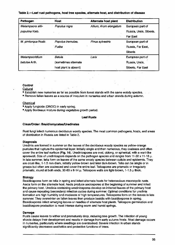
Table 2.-Leaf rust pathogens, host tree species, alternate host, and distribution of disease
Pathogen Host - Alternate host plant Distribution
Melampsora a/Iii- Populus nigra Allium, Arum e/ongatum European part of
populina Kleb. Russia, Urals, Siberia,
M. pinitorqua Rostr.
Melampsoridium
betulae Arth.
Control Cultural
Popu/us tremu/ae,
P.alba
Betu/a
(sometimes alternate
host plant is absent)
Far l:ast
Pinus sylvestris European part of
Russia, Far East,
Siberia
Larix European part of
Russia, Urals,
Siberia, Far East
• Establish new nurseries as far as possible from forest stands with the same woody species. • Remove fallen leaves as a source of inoculum in nurseries and urban stands during autumn.
Chemical • Apply fungicide (DNOC) in early spring. • Apply Bordeaux mixture during vegetative growth period.
Leaf Rusts
Class/Order: Basidiomycetes/Uredinales
Rust fungi infect numerous deciduous woody species. The most common pathogens, hosts, and areas of distribution in Russia are listed in Table 2.
Diagnosis Uredinia are formed in summer on the leaves of the deciduous woody species as yellow-orange pustules that rupture the epidermal layer. Initially single and then numerous, they coalesce and often cover the entire leaf surface (Fig. 14). Urediniospores are oval, oblong, or spherical, with a wart-like sporewall. Size of urediniospore depends on the pathogen species and ranges from 11-30 x 11-18 µ. In late summer, telia form on leaves of the same woody species between cuticle and epidermis. They are crust-like, 1-1.5 mm diam, initially yellow-brown and later dark-brown. Talia can be single or in groups but often are coalesced and cover the entire leaf. Teliospores are prismatic or irregularly prismatic, round at both ends, 30-60 x 6-14 µ. Teliospore walls are light-brown, 1-1.5 µ thick.
Biology Basidiospores form on telia in spring and infect alternate hosts for heteroecious macrocyclic rusts. Aecia form on the alternate host. Aecia produce aeciospores at the beginning of summer and infect the primary host. Uredinia containing urediniospores develop on infected leaves of the primary host and cause repeating (secondary) infection cycles during summer. Optimal conditions for uredinia formation are high humidity and moderate or high temperatures. Teliospores form on the leaves in late summer. They overwinter on fallen leaves then produce basidia with basidiospores in spring. Basidiospores infect emerging leaves or needles of alternate host plants. Teliospore germination and basidiospore production is most intense during warm and humid springs.
Damage Rusts cause leaves to wither and prematurely drop, reducing tree growth. The infection of young shoots delays their development and results in damage from early autumn frosts. Most damage occurs in nurseries, particularly where seedlings are overstocked. Severe infection in urban stands significantly decreases aesthetics and protective functions of trees.
35

Contents
-~ .. . Introduction .......................................... ':: ................................................................................................. , .... 1
Most Common Fungal Diseases of Russian Forests ..................................................................... 2
Diseases of Fruits and Seeds ...................................................................... -:: ............................ 2 Birch Seed Mummification ...................................................................................................................... 2 Fruit Deformation .................................................................................................................................... 2 Fruit Spots .............................................................................................................................................. 4 Molds ...................................................................................................................................................... 6 Seed and Fruit Rots ............................................................................................................................... 9
Diseases of Needles and Leaves ............................................................................................ 11 Needle Diseases .................................................................................................................................. 11
Lophodermium Needle Casts .......................................................................................................... 11 Snow Blight ...................................................................................................................................... 15 Meria Needle Blight ......................................................................................................................... 16 Brown Felt Blight ............................................................................................................................. 18 Rhizosphaera Needle Casts ............................................................................................................ 18 Needle Rusts of Pine ....................................................................................................................... 20
Leaf Diseases ....................................................................................................................................... 22 Powdery Mildews ............................................................................................................................. 22 Leaf Spots ...................................................... : .. .............................................................................. 23 Leaf Rusts ....................................................................................................................................... 35
Diseases Of Roots, Stems, And Branches ............................................................................... 36 Diseases in Tree Nurseries and Young Forests ................................................................................... 36
Damping - off ................................................................................................................................... 36 Diseases of Forest Stands ................................................................................................................... 38
Wilts ................................................................................................................................................. 38 Verticillium Wilt ............................................................................................................................ 38 Dutch Elm Disease ..................................................................................................................... 39 Oak Wilt ......................................................................... · ............................................................. 41
Dieback and Canker Diseases ........................................................................................................ 42 Cenangium Dieback of Pine ....................................................................................................... 42 Nectria Canker and Dieback ....................................................................................................... 44 Cytospora Canker ....................................................................................................................... 45 Dothichiza Canker of Poplar ....................................................................................................... 46 Clithris Canker and Dieback of Oaks .......................................................................................... 48 Nummularia Canker .................................................................................................................... 48 Black Naemospora Canker ......................................................................................................... 49 Thyrostroma Canker and Dieback .............................................................................................. 50 Ascocalyx Scleroderris Shoot Canker ........................................................................................ 52 European Larch Canker .............................................................................................................. 53 Lachnellula Canker of Siberian Pine Understory ........................................................................ 54 Stem Rust of Pine ....................................................................................................................... 55 Broom Rust of Fir ........................................................................................................................ 56 Perennial Nectria Canker ............................................................................................................ 58 Black Hypoxylon Canker ............................................................................................................. 60 Cytophoma Canker of Ash .......................................................................................................... 60
Wood-Decaying Diseases ............................................................................................................... 62 Annosum Root and Butt Rot ....................................................................................................... 62 Armillaria Root Rot ...................................................................................................................... 65 Butt and Trunk Rot of Conifers ................................................................................................... 67 Trunk and Limb Rot of Hardwoods ............................................................................................. 75

Fungal Dise~ses that Occur Only in Russian Forests ................................................................. 89
Diseases of Fruits and Seeds .......... .' ....................................................................................... 89 Thecopsora Rust of Spruce Cones ...................................................................................................... 89 Acorn Mummification Deformity ........................................................................................................... 89
Diseases of Needles and Leaves ....................................................................... '..:-................... 91 Needle Diseases .................................................................................................................................. 91
Hypodermella Needle Cast of Pine Hosts ....................................................................................... 91 . Chrysomyxa Rust of Spruce ............................................................................................................ 91
Leaf Diseases ....................................................................................................................................... 93 Powdery Mildew of Siberian Pea Tree ............................................................................................. 93 Other Powdery Mildews ................................................................................................................... 94 Orange Leaf Spot of Padus ............................................................................................................. 95 Red Spot of Ussurian Plum ............................................................................................................. 96 Foliage Anthracnoses, Spots, and Blights ....................................................................................... 96 Leaf Rusts ..................................................................................................................................... 108 Taphrina Diseases: Leaf Blisters, Leaf and Shoot Deformation .................................................... 110
Diseases of Roots, Stems, and Branches ............................................................................... 111 Diseases in Tree Nurseries and Young Forests ................................................................................. 111
"Infectious Damping'.' of Coniferous Seedlings .............................................................................. 111 Sclerophoma Disease of Pine Shoots ........................................................................................... 112 Pine Shoot Rust ............................................................................................................................. 113 Chrysomyxa Rust of Spruce Shoots and Needles ........................................................................ 114
Diseases of Forest Stands .... , ............................................................................................................ 116 Dieback and Canker Diseases ...................................................................................................... 116
Black Cytospora Canker of Poplar ............................................................................................. 116 Biatorella Canker ...................................................................................................................... 117 Pitch Blister Rust Canker .......................................................................................................... 119 Endoxylina Canker of Ash ......................................................................................................... 120 Cankers and Diebacks .............................................................................................................. 122
Wood-Decaying Diseases ............................................................................................................. 128 Ganoderma Butt Rot of Beech .................................................................................................. 129 Vuillemenia Decay .................................................................................................................... 129 Trunk and Limb Rots ................................................................................................................. 130
Acknowledgment ............................................................................................................ 133
Appendix A Pathogens That Affect Trees and Shrubs in Russia ....................................................................... 134
Appendix B Host Trees, Shrubs, and Herbs Listed in this Report ..................................................................... 137
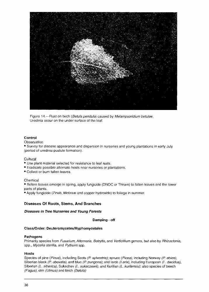
Figure 14.-Rust on birch (Betula pendu/a) caused by Melampsoridium betulae. Uredinia occur on the under surface of the leaf.
Control Observation • Survey for disease appearance and dispersion in nurseries and young plantations in early July (period of uredinia pustule formation).
Cultural • Use plant material selected for resistance to leaf rusts. • Eradicate possible alternate hosts near nurseries or plantations. •Collector burn fallen leaves.
Chemical • Before leaves emerge in spring, apply fungicide (DNOC or Thiram) to fallen leaves and the lower parts of plants. • Apply fungicide (Zineb, Metiram and copper hydroxide) to foliage in summer.
Diseases Of Roots, Stems, And Branches
Diseases in Tree Nurseries and Young Forests
Damping - off
Class/Order: Deuteromycetes/Hyphomycetales
Pathogens Primarily species from Fusarium, Alternaria, Botrytis, and Verticil/ium genera, but also by Rhizoctonia, spp., Mycelia sterilia, and Pythuim spp.
Hosts Species of pine (Pinus), including Scots (P. sylvestris); spruce (Picea), including Norway (P. abies), Siberian black (P. obovata), and blue (P. pungens); and larch (Larix), including European (L. decidua), Siberian (L. siberica), Sukachev (L. sukaczewii), and Kurilian (L. kurilensis); also species of beech (Fagus), elm (Ulmus) and birch (Betula)
36
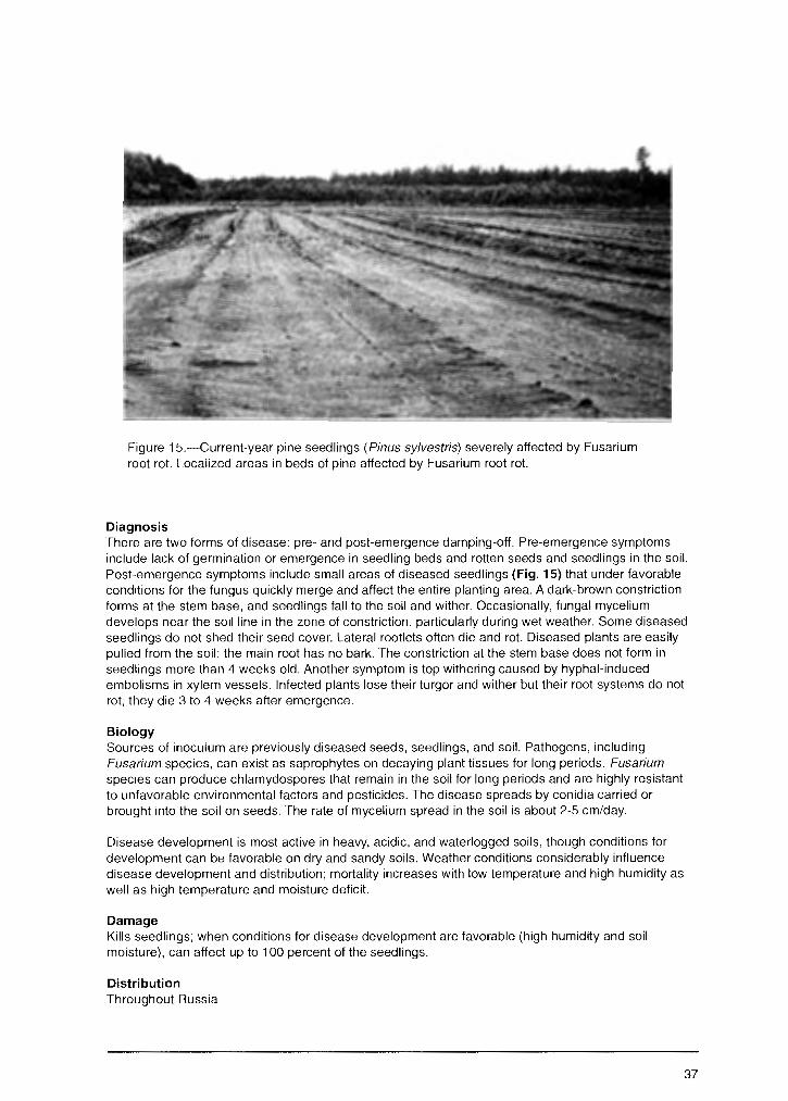
Figure 15.-Current-year pine seedlings (Pinus sy/vestris) severely affected by Fusarium root rot. Localized areas in beds of pine affected by Fusarium root rot.
Diagnosis There are two forms of disease: pre- and post-emergence damping-off. Pre-emergence symptoms include lack of germination or emergence in seedling beds and rotten seeds and seedlings in the soil. Post-emergence symptoms include small areas of diseased seedlings (Fig. 15) that under favorable conditions for the fungus quickly merge and affect the entire planting area. A dark-brown constriction forms at the stem base, and seedlings fall to the soil and wither. Occasionally, fungal mycelium develops near the soil line in the zone of constriction, particularly during wet weather. Some diseased seedlings do not shed their seed cover. Lateral rootlets often die and rot. Diseased plants are easily pulled from the soil; the main root has no bark. The constriction at the stem base does not form in seedlings more than 4 weeks old. Another symptom is top withering caused by hyphal-induced embolisms in xylem vessels. Infected plants lose their turgor and wither but their root systems do not rot; they die 3 to 4 weeks after emergence.
Biology Sources of inoculum are previously diseased seeds, seedlings, and soil. Pathogens, including Fusarium species, can exist as saprophytes on decaying plant tissues for long periods. Fusarium species can produce chlamydospores that remain in the soil for long periods and are highly resistant to unfavorable environmental factors and pesticides. The disease spreads by conidia carried or brought into the soil on seeds. The rate of mycelium spread in the soil is about 2-5 cm/day.
Disease development is most active in heavy, acidic, and waterlogged soils, though conditions for development can be favorable on dry and sandy soils. Weather conditions considerably influence disease development and distribution; mortality increases with low temperature and high humidity as well as high temperature and moisture deficit.
Damage Kills seedlings; when conditions for disease development are favorable (high humidity and soil moisture), can affect up to 100 percent of the seedlings.
Distribution Throughout Russia
37
Control Cultural • Select flat areas with light soils when establishing nurseries. • Avoid sites formerly used to grow agricultural plants, e.g., potatoes, corn, or melons, due to the potential for previous Fusarium infection. • Sow seeds at optimal time, observe rules of sowing, and use only certified seeds. • Care for seeding beds after seedlings emerge and prevent weed growth and drying 9f soils.
Chemical • Use chemical seed dressing where necessary. Dust seeds with fungicides (Bavistin, Benomyl, BAYMEB, Topsin-M, Daconil, Vitavax, Thiram, and Tersan). Fungicide treatment combined with micronutrient fertilizer (ZnS0
4, CoS0
4, CuS0
4, KMn0
4) is the most effective treatment.
• Apply fungicides (Thiram, Tersan, Karbation, or Vapam) to the soil where infection is heavy. • Use fungicides in irrigation water in disease loci in nursery beds.
Biological • Apply antagonistic antibiotics (Trichothecin, Phytobacteriomycin, or Phytolavin).
Diseases of Forest Stands
Witts Vertlcillium WIit
Class/Order: Deuteromycetes/Hyphomycetales
Pathogen Verticillium dahliae Kleb. and II. albo- atrum Ake. et Berth.
Hosts Species of maple (Acer), particularly Norway (Acer platanoides), and birch (Betula), poplar (Popu/us), lime (Tilia), and elm (U/mus)
Diagnosis Affected trees fail to foliate in spring or die suddenly in midsummer (Fig. 16) in the acute form of disease development. In the chronic form, several branches die and then later the entire crown dies. Numerous epicormic shoots form on the stem but they soon die. Bark sloughs from dead trees. The wood of infected roots and stem initially has green stripes but later turns dark-green. An important diagnostic feature in the conidial stage is the conidiophores, which typically are branched vertically with colorless, ellipsoid conidia, 2-5 µ, on them. Microsclerotia and chlamydospores develop later. In natural conditions, the fungus sporulates rarely, however, spores develop rapidly in the laboratory (in moisture chambers).
Biology Infection is initiated by conidia at the base of the stem or branch. Occasionally, trees are infected through damaged stems and branches. The mycelium penetrates the tree and spreads in the vascular system. From the lower part of the stem, mycelium grows upward and penetrates branches. When the fungus attacks, most of the roots (or the entire root system) are infected. The tree usually dies before mycelium reaches the stem. Sources of inoculum are infected wood, bark residues, and dead roots. The pathogen survives by forming microsclerotia and chlamydospores. These structures survive in the soil on dead debris for several years. lnoculum potential increases in soils where agricultural species such as potato (Solanum tuberosum), tomato (Lycopersicum esculentum), and sunflower (Helianthus annuus) have grown.
Damage Kills seedlings in nurseries and artificially and naturally regenerated maple stands 1 to 2 years old. Dieback of trees in forest plantations and natural stands is possible during first 4 years of growth.
Distribution European part of Russia, particularly the southwestern region
38

Figure 16.-Dieback of naturally regenerated maple (Acer platanoides) caused by Verticil/ium dahliae.
Control Cultural • Do not establish forest nurseries and plantations in areas where S. tuberosum, L. esculentum, and H. annuus have grown. • In nurseries, remove diseased plants and their entire root system. • Selectively remove infected trees or clearcut in disease loci in affected natural stands. • Remove and destroy all felling residue.
Dutch Elm Disease
Class/Order: Ascomycetes, Microsporales
Pathogen Ophiostoma ulmi (Buism.) Nannf. (Syn. Ceratocystis ulmi (Buism.) Mor. (anamorph: Graphium ulmi Schwarz.)
Hosts Species of elm (Ulmus), including table top (U. glabra), Ohyo (U. /aciniata), European white (U. laevis), smooth leaf (U. carpinifolia), Japanese (U. japonica), Siberian (U. pumila), and Androssowi (U. androssowii)
39
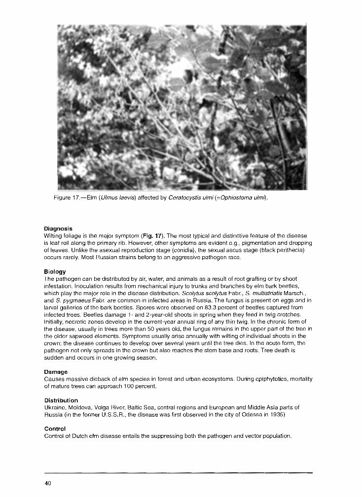
Figure 17.-Elm ( U/mus /aevis) affected by Ceratocystis ulmi (=Ophiostoma ulm1).
Diagnosis Wilting foliage is the major symptom (Fig. 17). The most typical and distinctive feature of the disease is leaf roll along the primary rib. However, other symptoms are evident e.g., pigmentation and dropping of leaves. Unlike the asexual reproduction stage (conidia), the sexual ascus stage (black perithecia) occurs rarely. Most Russian strains belong to an aggressive pathogen race.
Biology The pathogen can be distributed by air, water, and animals as a result of root grafting or by shoot infestation. Inoculation results from mechanical injury to trunks and branches by elm bark beetles, which play the major role in the disease distribution. Scolytus scolytus Fabr., S. multistriatis Marsch., and S. pygmaeus Fabr. are common in infected areas in Russia. The fungus is present on eggs and in larval galleries of the bark beetles. Spores were observed on 83.3 percent of beetles captured from infected trees. Beetles damage 1- and 2-year-old shoots in spring when they feed in twig crotches. Initially, necrotic zones develop in the current-year annual ring of any thin twig. In the chronic form of the disease, usually in trees more than 50 years old, the fungus remains in the upper part of the tree in the older sapwood elements. Symptoms usually arise annually with wilting of individual shoots in the crown; the disease continues to develop over several years until the tree dies. In the acute form, the pathogen not only spreads in the crown but also reaches the stem base and roots. Tree death is sudden and occurs in one growing season.
Damage Causes massive dieback of elm species in forest and urban ecosystems. During epiphytotics, mortality of mature trees can approach 100 percent.
Distribution Ukraine, Moldova, Volga River, Baltic Sea, central regions and European and Middle Asia parts of Russia (in the former U.S.S.R., the disease was first observed in the city of Odessa in 1935}
Control Control of Dutch elm disease entails the suppressing both the pathogen and vector population.
40
Observation • Survey elm stands in June and July for tree condition, presence of symptoms, and abundance of elm bark beetles.
Cultural • Create mixed stands with a variety of deciduous species. • Use disease-resistant planting material. -• Conduct selective and clear sanitation cutting and remove trees with disease symptoms (October through April) to reduce the abundance of overwintering bark beetles. • Destroy infected wood and debark healthy portions of cut trees and branches.
Class/Order: Ascoymycetes, Microascales
Pathogen
Oak Wilt
Species of Ceratocystis (C. roboris Georg. et Teod., C. valachicum Georg. et Teod.) and species of Fusarium and Verticillium
Hosts Species of oak (Quercus)
Diagnosis The disease has both rapid and chronic development forms. The r~pid form is characterized by leaves wilting at the top of the tree and branches. The disease quickly spreads to the entire crown. In this case, leaves curl, turn yellow or bronze, and fall 3 to 6 weeks after the first symptoms appear. If infection occurs at the end of summer, leaves turn tan and some fall while others remain on branches throughout the winter. Mycelial masses form under the bark of dead trees and arise from bark cracks. In the chronic form, wilting begins on several separate branches. Leaves are smaller than normal, turn yellow or tan, and drop. Often, buds on diseased branches fail to grow in spring, and/or leaves do not reach normal size and fall prematurely. Affected trees are characterized by sparse foliage and top dieback. Heavily weakened and dying trees produce adventitious sprouts. Morphological changes in the crown are not specific for the chronic form. Similar symptoms can be caused.by other diseases as well as chemical, weather, and other adverse environmental factors.
The pathogen develops within the water-conducting xylem vessels, and causes dieback of adjacent parenchyma and extensive tyloses formation. Characteristic symptoms of this disease are evident when branches and trunks are cut; tan or dark-olive spots and streaks and dots form in the sapwood or the entire sapwood turns brown.
Biology Infection usually occurs by spores, though infection by mycelium can occur through grafted roots. Spores are distributed by water, wind, and on seeds and tools, but most commonly by insects. The most active vectors are insects such as Scolitus intricatus, Plagionotus detritis, P. arcuatus, and Agrilus angustulus. Oak wilt usually develops in previously weakened stands. The percentage of diseased trees increases with tree age. The disease is prevalent in low-density stands. Oak dieback occurs in both mountain and floodplain stands but is more severe in the latter.
Damage Weakens and kills oak stands (Fig. 18). Depending on age, nearly 75 percent of the stands can be affected. The death rate can reach 70 percent.
Distribution European part of Russia
Control Cultural • Conduct sanitation cuts to remove and destroy infected trees, including trees infested by oak bark beetles; spray stumps with Fundasol or Nitraphen. • Establish seed plantations in healthy and disease-resistant stands. • Examine acorns for infection before seeding.
41
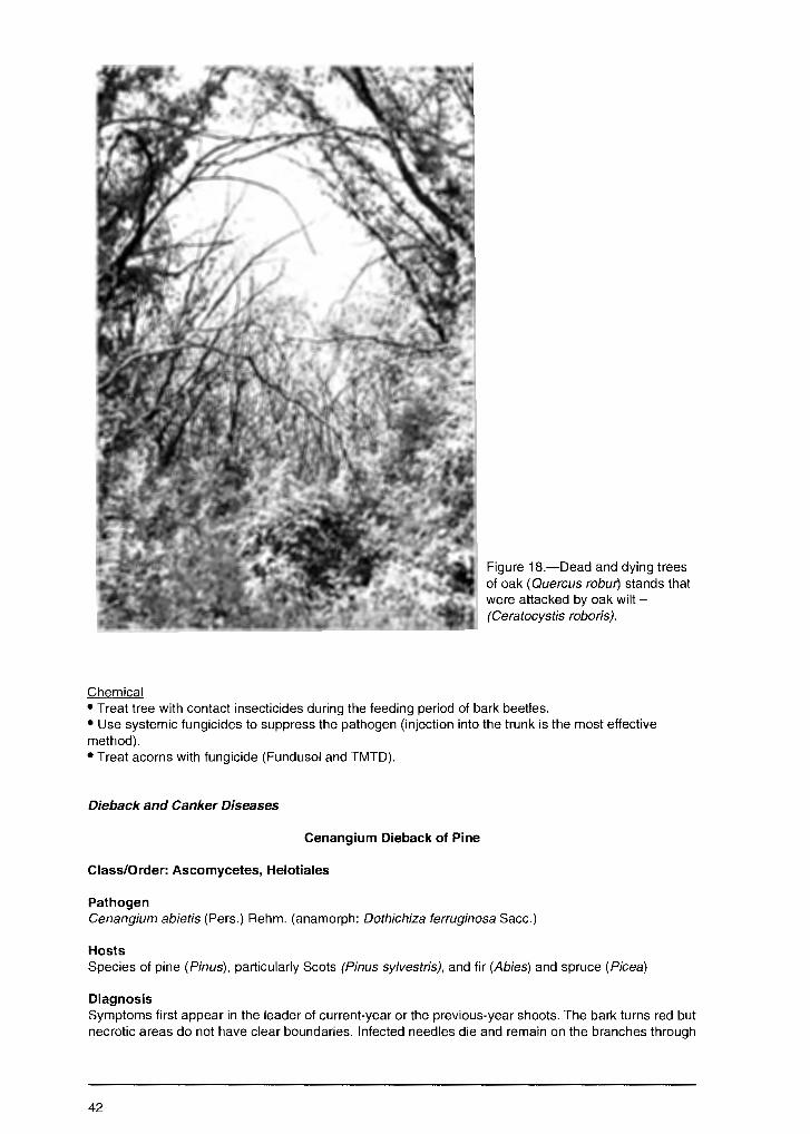
Chemical
Figure 18.-Dead and dying trees of oak ( Que re us robur') stands that were attacked by oak wilt -(Ceratocystis roboris).
• Treat tree with contact insecticides during the feeding period of bark beetles. • Use systemic fungicides to suppress the pathogen (injection into the trunk is the most effective method). • Treat acorns with fungicide (Fundusol and TMTD).
Dieback and Canker Diseases
Cenangium Dieback of Pine
Class/Order: Ascomycetes, Helotiales
Pathogen Cenangium abietis (Pers.) Rehm. (anamorph: Dothichiza ferruginosa Sacc.)
Hosts Species of pine (Pinus), particularly Scots (Pinus sylvestris), and fir (Abies) and spruce (Picea)
Diagnosis Symptoms first appear in the leader of current-year or the previous-year shoots. The bark turns red but necrotic areas do not have clear boundaries. Infected needles die and remain on the branches through
42

the growing season (Fig. 19). The conidial stage develops on infected branches and needles during the entire growing season. Small, black pycnidia-like tubercles develop in bark splits. Apothecia develop on dead branches and stems 1 or 2 years after infection. In wet conditions, apothecia are saucer-like with a green-yellow hymenial layer. In dry weather, apothecia resemble dark-brown, nearly black rough tubercles, 1-3 mm in diameter, raised from the bark splits. Conidia are colorless, oval or ovoid, 8-12 x 4 µ. Asci are clavate, 60-80 x 10-12 µ. Ascospores are colorless, ellipsoid or ovoid, with 1 or 2 oil drops, 10-12 x 5-7 µ.
Biology The sources of inoculum are diseased plants and fallen needles. New infections on healthy treees are initiated by conidia and ascospores. High humidity promotes sporulation and infection. The fungus penetrates tissues of needles and bark injured by various agents, including insects. Many factors that result in poor growing conditions and weakened plants promote disease development. These include unfavorable climate conditions (drought and others), changes in water regime, insect injury (from Aradus cinnamomeus Panz. and Ellopia fasciaria L.), and industrial pollution.
Damage The fungus usually occurs as a saprotroph on pine trees killed by various agents. More rarely, the disease develops as a epiphytotic in nurseries and pine plantations, causing dieback. Plant damage can range from 50-60 percent. In nurseries, seedlings can die if the epiphytotic continues for several years.
Distribution European part of Russia, Siberia, Far East
Control Cultural • Remove infected trees to prevent disease spread. • Remove and burn dead trees and branches as sources of inoculum. • Create mixed hardwood/pine plantations as more biologically sustainable; hardwood species reduce the possibility of pine infection.
Figure 19.-Cenangium dieback of pine (Pinus sy/vestris) caused by Cenangium abietis.
43
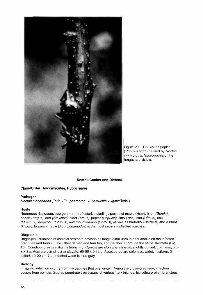
Nectria Canker and Dieback
Class/Order: Ascomycetes, Hypocreales
Pathogen
Figure 20.-Canker on poplar (Populus nigra) caused by Nectria cinnabarina. Sporodochia of the fungus are visible.
Nectria cinnabarina (Tode.) Fr. (anamorph: Tubercu/aria vulgaris Tode.)
Hosts Numerous deciduous tree genera are affected, including species of maple (Acer), birch (Betula), beech (Fagus), ash (Fraxinus), alder (A/nus), poplar (Populus), lime (Tilia), elm (U/mus), oak (Quercus), dogwood (Camus), and mountain-ash (Sorbus), as well as barberry (Berberis) and currant (Ribes). Bosnian maple (Acer platanoides) is the most severely affected species.
Diagnosis Bright-pink cushions of conidial stromata develop as longitudinal lines in bark cracks on the infected branches and trunks. Later, they darken and turn tan, and perithecia form on the same stromata (Fig. 20). Conidiophores are slightly branched. Conidia are elongate-ellipsoid, slightly curved, colorless, 5.5-8 x 3 µ. Asci are cylindrical or clavate, 60-90 x 9-12 µ. Ascospores are colorless, widely fusiform, 2-celled, 12-20 x 4-7 µ. Infected wood is blue gray.
Biology In spring, infection occurs from ascospores that overwinter. During the growing season, infection occurs from conidia. Spores penetrate tree tissues at various bark injuries, including broken branches.
44
The mycelium spreads in the bark, cambium, and sapwood, and penetrates the trachids where it plugs vessels and causes rapid dieback.
Damage The fungus often is reported as a saprophyte but it can be pathogenic in nurseries and young forest plantations. Infected seedlings in nurseries can reach 20 percent. Severe disease reduces vigor and causes dieback in nurseries. The canker significantly reduces productivity of mature treeJ.
Distribution European part of Russia, Urals, Siberia, Far East
Control Observation • Survey stands during the summer when conidial sporodochia are visible.
Cultural • Protect trees from environmental factors that can weaken them. • Remove infected, dying, and dead trees in forest nurseries and urban stands; prune infected branches. • Prevent or reduce mechanical injuries that serve as entry points for the pathogen.
Cytospora Canker
Class/Order: Deuteromycetes, Sphaeropsidales
Pathogen Cytospora chrysosperma (Pers.) Fr. (teliomorph: Valsa sordida Nits.)
Hosts Species of poplar (Populus) and willow (Salix)
Diagnosis The pathogen develops in bark tissue and kills it. Thin, smooth bark that is affected is red-brown; the color of thick bark does not change. Dark-gray or brown conidial stromata form in the bark. Numerous pycnida develop in the stromata and raise the bark surface as small tubercles. Mass sporulation occurs in May and early June, as well as in late August and September. Spore masses exude from pycnidia and congeal as golden-yellow or orange horns (cirris). Pycnidia can be 1.8 mm in diameter, chambered with a common opening. Conidia are colorless, allantoid, 3-5 x 1 µ.
Biology Sources of inoculum are infected cuttings, seedlings, and infected trees in stands. Conidia are spread by rainwater or insects and infect healthy trees. The pathogen penetrates tree tissues though mechanical injuries and cracks at the base of branches. As the fungus grows, it secretes toxins that kills the bark. Mycelium then develops in dead bark. Pockets (loci) of the disease develop usually in stands predisposed to infection by unfavorable environmental conditions, e.g., droughts, flooding, winter damage, and unfavorable soil. Disease develops just after foliation and later in midsummer. Death may occur during one growing season or over several years. In the case of rapid disease development, affected trees do not foliate in the spring or die shortly after foliating. In chronic disease development, single branches die gradually in the crown; epicormic shoots develop on the stems but also are infected and die.
Damage Kills weakened trees. Damage is most severe in nurseries, stool-beds, and forest plantations.
Distribution Throughout Russia
Control Cultural • Cull diseased plant material in nurseries, forest plantations, and stands. • Select resistant species and clones.
45
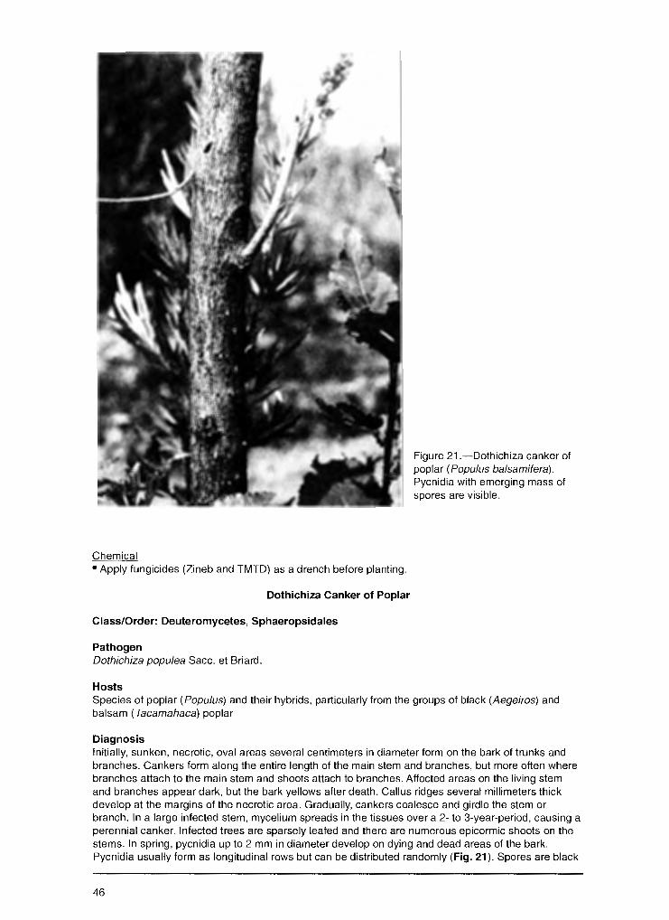
Chemical • Apply fungicides (Zineb and TMTD) as a drench before planting.
Dothichiza Canker of Poplar
Class/Order: Deuteromycetes, Sphaeropsidales
Pathogen Dothichiza popu/ea Sacc. et Briard.
Hosts
Figure 21.-Dothichiza canker of poplar (Populus balsamifera). Pycnidia with emerging mass of spores are visible.
Species of poplar (Populus) and their hybrids, particularly from the groups of black (Aegeiros) and balsam ( Tacamahaca) poplar
Diagnosis Initially, sunken, necrotic, oval areas several centimeters in diameter form on the bark of trunks and branches. Cankers form along the entire length of the main stem and branches, but more often where branches attach to the main stem and shoots attach to branches. Affected areas on the living stem and branches appear dark, but the bark yellows after death. Callus ridges several millimeters thick develop at the margins of the necrotic area. Gradually, cankers coalesce and girdle the stem or branch. In a large infected stem, mycelium spreads in the tissues over a 2- to 3-year-period, causing a perennial canker. Infected trees are sparsely leafed and there are numerous epicormic shoots on the stems. In spring, pycnidia up to 2 mm in diameter develop on dying and dead areas of the bark. Pycnidia usually form as longitudinal rows but can be distributed randomly (Fig. 21 ). Spores are black
46
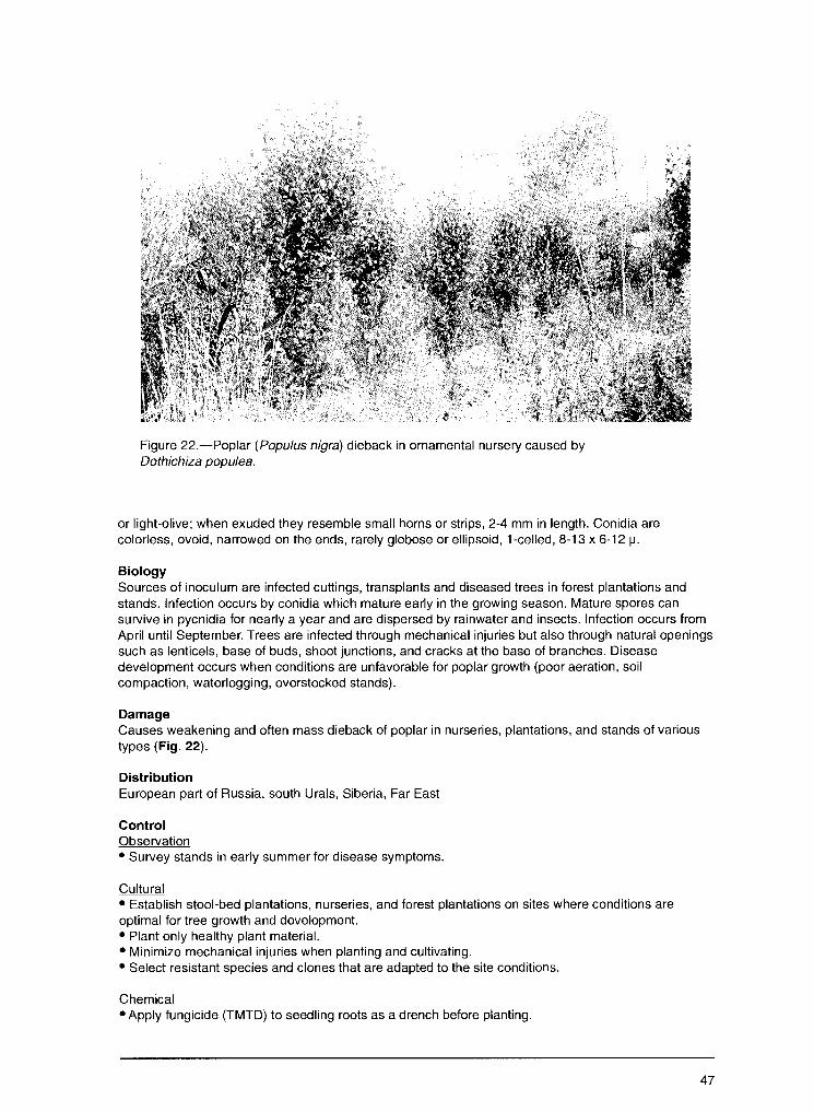
Figure 22.-Poplar (Populus nigra) dieback in ornamental nursery caused by Dothichiza popu/ea.
or light-olive; when exuded they resemble small horns or strips, 2-4 mm in length. Conidia are colorless, ovoid, narrowed on the ends, rarely globose or ellipsoid, 1-celled, 8-13 x 6-12 µ.
Biology Sources of inoculum are infected cuttings, transplants and diseased trees in forest plantations and stands. Infection occurs by conidia which mature early in the growing season. Mature spores can survive in pycnidia for nearly a year and are dispersed by rainwater and insects. Infection occurs from April until September. Trees are infected through mechanical injuries but also through natural openings such as lenticels, base of buds, shoot junctions, and cracks at the base of branches. Disease development occurs when conditions are unfavorable for poplar growth (poor aeration, soil compaction, waterlogging, overstocked stands).
Damage Causes weakening and often mass dieback of poplar in nurseries, plantations, and stands of various types (Fig. 22).
Distribution European part of Russia, south Urals, Siberia, Far East
Control Observation • Survey stands in early summer for disease symptoms.
Cultural • Establish stool-bed plantations, nurseries, and forest plantations on sites where conditions are optimal for tree growth and development. • Plant only healthy plant material. • Minimize mechanical injuries when planting and cultivating. • Select resistant species and clones that are adapted to the site conditions.
Chemical • Apply fungicide (TMTD) to seedling roots as a drench before planting.
47

Clithris Canker and Dieback of Oaks
Clas$/Order: Ascomycetes, Phacidlales
Pathogen Clithris quercina (Pers.) Rehm. (anamorph: Cytospora quercefla Sacc.)
Hosts Species of oak (Quercus), particularly peduncate (Q. robur)
Diagnosis The bark of infected stems and branches turns red and later yellow-white upon death. Necrotic areas are sharply delimited from healthy bark. Necrosis can be elongate or circumferential. Pycnidia develop initially on the dead bark as small gray-white tubercles (hillocks). Later, numerous apothecia 5 mm long form in the dead bark. They are gray-white or dark-gray and curved. In humid conditions, apothecia open as relatively wide longitudinal splits uncovering a gray, jelly-like hymenial layer. Asci are clavate, with a stalk, 125-150 x 9-10 µ. The ascospores are thread-like, colorless, initially 1-celled but later multicelled, 90 x 1-1.5 µ.
Biology Healthy branches and stems are infected from ascospores in early summer through injuries to bark. Trees weakened by poor growing conditions, e.g., repeated droughts, changing water regimes, or frost injury, are highly susceptible.
Damage In the forest the fungus acts as a saprophyte, occurring on the lower dying branches and promoting the process of branch shedding. However, in the steppe zone it acts as a pathogen affecting plantations under 25 years, reducing vigor, and sometimes killing trees. Damaged plants range from 10-40 percent. The fungus causes sapwood decay as well as bark necrosis.
Distribution European part of Russia
Control Cultural • Create optimal conditions for growth (soil moisture, fertilization, spacing) in nurseries and plantations. • Remove damaged trees and felling debris after thinning. • Minimize mechanical injury during tending operations.
Nummularia Canker
Class/Order: Ascomycetes, Sphaeriales
Pathogen Nummularia bulliardii Tul.
Hosts Species of oak (Quercus) and beech (Fagus)
Diagnosis Symptoms are visible several years after infection. Thick, up to 3 mm, oval, cushion-like stromata form in infected bark and over entire length of the stem. Stromata are 1-6 cm wide and 2-40 cm long. Often, stromata are coalesced, girdling and extending up stems to a length of 1 m. Initially, stromata are brown but later turn black and coal-like. Numerous perithecia develop within the stroma. Perithecia openings appear as pointed hillocks on the entire stroma surface. Perithecia in stromata occur as a single layer; they are ovoid or sack-like, 0.5 mm high. Asci are elongate, cylindrical, on a short stalk, 100-210 x 10 µ. Ascospores are dark-brown, ellipsoid or globose, 1-celled, 12-14 x 6-10 µ. Decayed sapwood appears light-yellow in the main stem and branches.
Biology The source of inoculum is diseased trees and felling residues with well-developed stromata. Tree infection occurs from ascospores that penetrate into tree tissues through bark cracks or mechanical
48
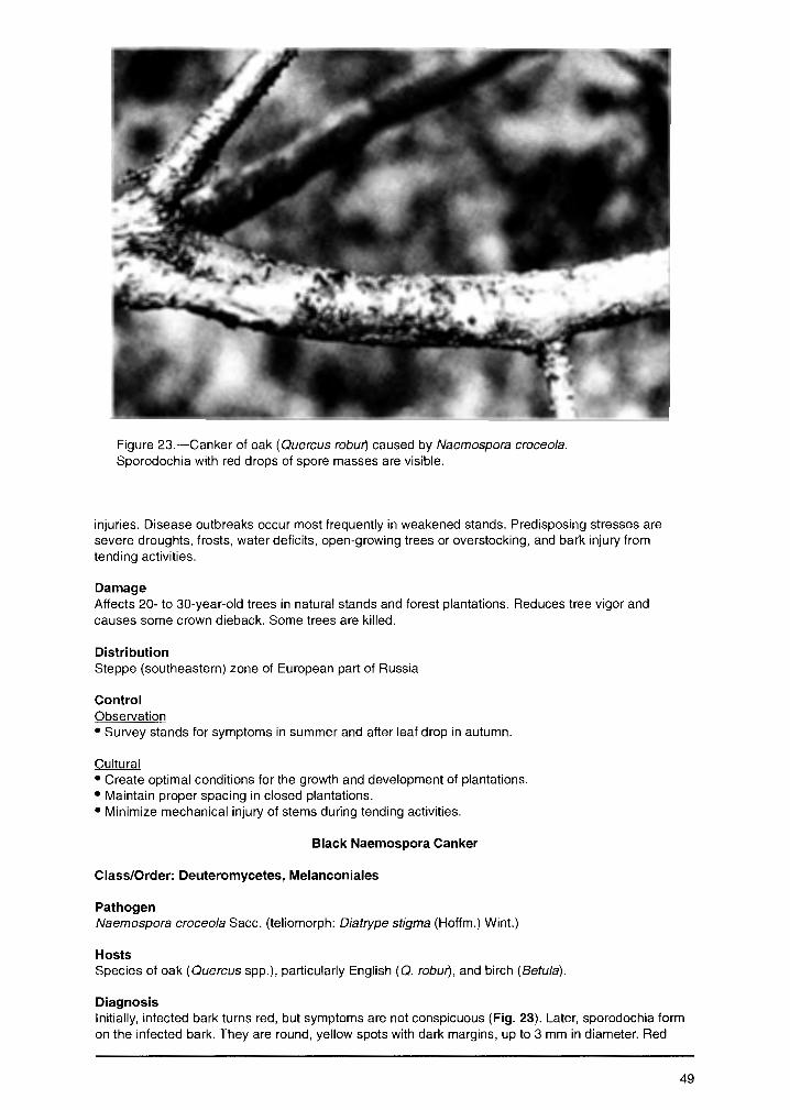
Figure 23.-Canker of oak (Quercus robur} caused by Naemospora croceo/a. Sporodochia with red drops of spore masses are visible.
injuries. Disease outbreaks occur most frequently in weakened stands. Predisposing stresses are severe droughts, frosts, water deficits, open-growing trees or overstocking, and bark injury from tending activities.
Damage Affects 20- to 30-year-old trees in natural stands and forest plantations. Reduces tree vigor and causes some crown dieback. Some trees are killed.
Distribution Steppe (southeastern) zone of European part of Russia
Control Observation • Survey stands for symptoms in summer and after leaf drop in autumn.
Cultural • Create optimal conditions for the growth and development of plantations. • Maintain proper spacing in closed plantations. • Minimize mechanical injury of stems during tending activities.
Black Naemospora Canker
Class/Order: Deuteromycetes, Melanconiales
Pathogen Naemospora croceo/a Sacc. (teliomorph: Diatrype stigma (Hoffm.) Wint.)
Hosts Species of oak (Quercus spp.), particularly English (Q. robur}, and birch (Betula).
Diagnosis Initially, infected bark turns red, but symptoms are not conspicuous (Fig. 23). Later, sporodochia form on the infected bark. They are round, yellow spots with dark margins, up to 3 mm in diameter. Red
49
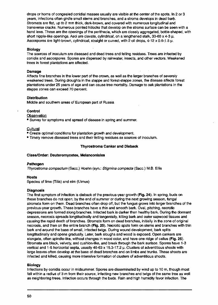
drops or horns of congealed conidial masses usually are visible at the center of the spots. In 2 or 3 years, infections often girdle small stems and branches, and a stroma develops in dead bar~. Stromata are flat, up tb 2 mm thick, dark-brawn, and covered with numerous longitudinal and transverse cracks. Numerous pointed hillocks that develop on the stroma surface can be seen with a hand lens. These are the openings of the perithecia, which are closely aggregated, bottle-shaped, with short nipple-like openings. Asci are clavate, cylindrical, on a lengthened stalk, 30-60 x 4-3 µ. Ascospores are light-brown, cylindrical, straight or curved, with 2 oil drops, 4-12 x 0.6-J .5 µ.
Biology The sources of inoculum are diseased and dead trees and felling residues. Trees are infected by conidia and ascospores. Spores are dispersed by rainwater, insects, and other vectors. Weakened trees in forest plantations are affected.
Damage Affects fine branches in the lower part of the crown, as well as the larger branches of severely weakened trees. During droughts in the steppe and forest-steppe zones, the disease affects forest plantations under 25 years of age and can cause tree mortality. Damage to oak plantations in the steppe zones can exceed 70 percent.
Distribution Middle and southern areas of European part of Russia
Control Observation • Survey for symptoms and spread of disease in spring and summer.
Cultural • Create optimal conditions for plantation growth and development. • Timely remove diseased trees and their felling residues as sources of inoculum.
Thyrostroma Canker and Dieback
Class/Order: Deuteromycetes, Melanconiales
Pathogen Thyrostroma compactum (Sacc.) Hoehn (syn.: Stigmina compacta (Sacc.) M.B. Ellis
Hosts Species of lime (Tilia) and elm (Ulmus)
Diagnosis The first symptom of infection is dieback of the previous-year growth (Fig. 24). In spring, buds on these branches do not open; by the end of summer or during the next growing season, fungal stromata form on them. Dead branches often drop off, but the fungus grows into larger branches of the previous-year growth. These branches have a thin and smooth bark. Oval, pitching, necrotic depressions are formed along branches. Infected bark is darker than healthy bark. During the dormant season, necrosis spreads longtitudinally and tangentally, killing bark and outer sapwood tissues and causing the rapid death of branches. Stromata form on dead branches, initially in the zone of original necrosis, and then on the entire branch (Fig. 25). Necrotic spots form on stems and branches with thin bark and around the base of small, infected twigs. During wound development, bark splits longtitudinally and opens gradually. Later, bark sloughs and wood is exposed. Open cankers are elongate, often spindle-like, without changes in wood color, and have one ridge of callus (Fig. 25). Stromata are black, velvety, and cushion-like, and break through the bark surface. Spores have 1-3 vertical and 1-5 horizontal septa, usually 46-63 x 15.3-17 .2 µ. Clusters of adventitious shoots with large leaves often develop at the base of dead branches and on limbs and trunks. These shoots are infected and killed, causing more intensive formation of clusters of adventitious shoots.
Biology Infections by conidia occur in midsummer. Spores are disseminated by wind up to 10 m, though most fall within a radi1.1s of 3 m from their source, infecting new branches and twigs of the same tree as well as neighboring trees. Infection occurs through the buds. Rain and high humidity favor infection. The
50

Figure 24.-Elm (Ulmus pumila) dieback caused by Thyrostroma compactum (=Stigmina compacta).
Figure 25.-Lesion on branches of lime (Tilia cordata) caused by Thyrostroma compactum.
pathogen cannot colonize actively growing tissues in the tree; colonization occurs only during the dormant season. The pathogen tolerates low temperatures as growth occurs even at -2°C. Spores deposited on vulnerable twigs and branches remain viable for up to 7 months.
Damage Causes cankers, resulting in branch dieback (Figs. 24-25).
Distribution European part of Russia
Control Cultural • Use canker-free, healthy plant material to establish plantations. • Prune infected branches at early stages of disease development to reduce inoculum. • Establish quarantine areas to prevent the spread of diseased plant material to regions where the disease does not occur.
51

Ascocalyx Scleroderris Shoot Canker
Class/Order: Ascomycetes, Helotiales
Pathogen Ascocalyx abietina (Lagerb.) Schlapfer. ( Gremmeniella abietina (Lagerb.) Moretel., Crumenula abietina Lagerbe., Sc/eroderris lagerbergii Gremm.) (anamorph: Brunchorstia pines (l:Sarst.) Hohnk.)
Hosts Species of pine (Pinus), spruce, (Picea), fir (Abies) and larch (Larix)
Diagnosis Pinus species are affected more often than other species. Symptoms of the disease vary with plant age. Initial symptoms of pine infection in nurseries occur several days after snowmelt. The fungus penetrates into the needles and upper buds and affects the cambium of young branches and stems. Initially, needles are green but turn red at the base where they attach to the stem. Paired needles of the 2 to 3-year-old seedlings hang like an umbrella and fall at the slightest touch. In the nurseries this disease is called the "umbrella disease." Following snowmelt, needles of affected seedling in nurseries are flattened along the stem. They turn brown but remain attached to the stem for a long time. The upper bud and stem cambium die and bark sloughs away easily if the seedling is pulled from the soil. In disease centers in young pine stands more than 5 years old, needles hang flaglike. Upper shoots are short and thick, with shortened or thickened needles. Necrotic and sometimes girdling lesions develop on the stem and branches and kill upper shoots on plants with a stem diameter larger than 1 cm. Canker wounds sometimes form.
During summer, mature pycnidia form on needle bases and in the bark of dying and dead seedlings. Pycnidia resemble rough black warts, about 2 mm in diameter. Spores are colorless, curved, usually 3-or 4-celled, but can be 4- or 6-celled, 16-32 x 2-3.5 m. The sexual stage occurs rarely. Apothecia are black-brown, 1-1.8 mm in diameter (Fig. 26). Asci are colorless, clavate, 95-110-x?.5-10 m. The ascospores are oval, with 2 9r 3 septa, 15-24 x 4-4.8 m. In pine stands 15 to 20 years old, the tips of upper shoots usually die with characteristic symptoms of needles thinning and falling off. Pycnidia often form on these shoots. Similar symptoms can occur in middle-age, mature, and overmature stands.
Biology Infection occurs by conidia and ascospores that are spread by wind or insects. Conidia dispersal occurs throughout summer; ascospore dispersal occurs from mid-July to September. Various factors cause tree weakening and reduced resistance to this disease, resulting in canker development. Unfavorable climate and soil conditions, hard frosts, and cold and rainy vegetative periods delay shoot development and disturb nutrient balance. Severe disease development occurs in low-lying areas where frequent fogs and autumn and spring frosts occur. Resistance to the disease is reduced in seedling beds and high-density plantings. Pine species planted offsite are highly susceptible to this disease.
Damage Primarily kills seedlings in nurseries; mortality ranges from 10-60 percent. Mature trees are noticeably weakened and many are killed.
Distribution European part of Russia, and Siberia
Control Observation • Survey nurseries for 10-14 days after snowmelt. • Survey forest plantations during the summer.
Cultural • Use indigenous seeds sources to increase seedling resistance to the disease. • Remove infected plants from bordering forest plantations or stands. • Select sites with flat topography for forest plantations; avoid cold, wet, shady areas. • Use healthy indigenous planting material. • Control the condition and density of plantings during the first 2 or 3 years after planting.
52
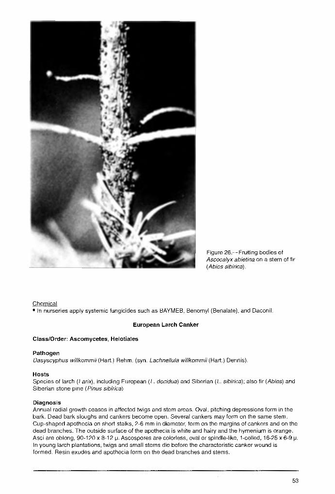
Chemical
Figure 26.-Fruiting bodies of Ascocalyx abietina on a stem of fir (Abies sibirica).
• In nurseries apply systemic fungicides such as BAYMEB, Benomyl (Benalate), and Daconil.
European Larch Canker
Class/Order: Ascomycetes, Helotiales
Pathogen Dasyscyphus willkommii (Hart.) Rehm. (syn. Lachnellu/a willkommii (Hart.) Dennis).
Hosts Species of larch (Larix), including European (L. decidua) and Siberian (L. sibirica); also fir (Abies) and Siberian stone pine (Pinus sibirica)
Diagnosis Annual radial growth ceases in affected twigs and stem areas. Oval, pitching depressions form in the bark. Dead bark sloughs and cankers become open. Several cankers may form on the same stem. Cup-shaped apothecia on short stalks, 2-6 mm in diameter, form on the margins of cankers and on the dead branches. The outside surface of the apothecia is white and hairy and the hymenium is orange. Asci are oblong, 90-120 x 8-12 µ. Ascospores are colorless, oval or spindle-like, 1-celled, 16-25 x 6-9 µ. In young larch plantations, twigs and small stems die before the characteristic canker wound is formed. Resin exudes and apothecia form on the dead branches and stems.
53
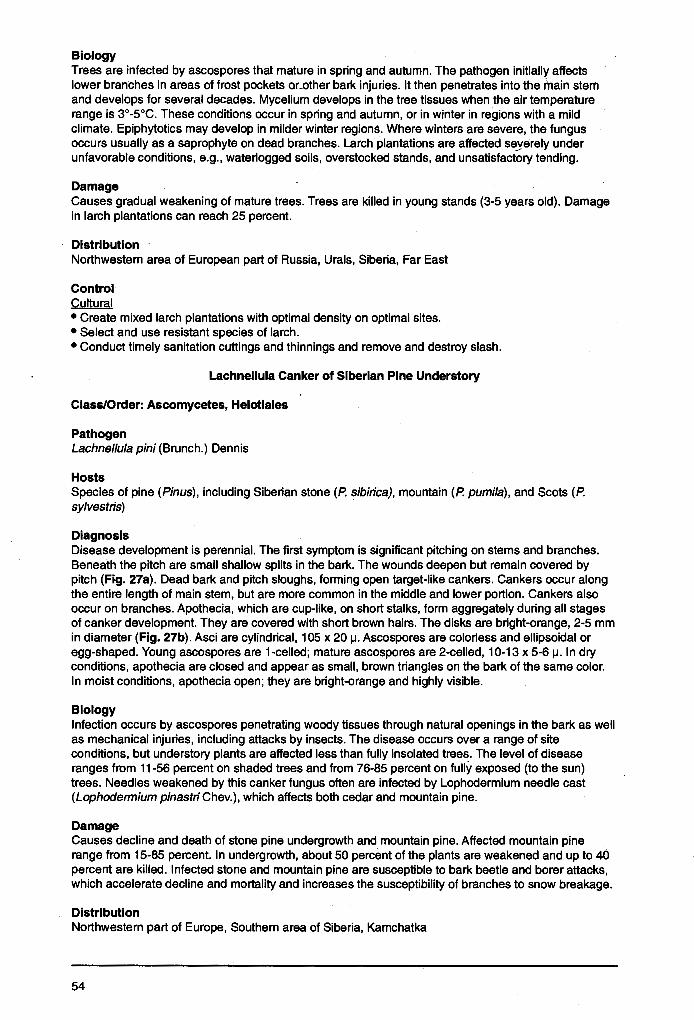
Biology Trees are infected by ascospores that mature in spring and autumn. The pathogen initially affects lower branches in areas of frost pockets or_other bark injuries. It then penetrates into the l'llain stem and develops for several decades. Mycelium develops in the tree tissues when the air temperature range is 3°-5°C. These conditions occur in spring and autumn, or in winter in regions with a mild climate. Epiphytotics may develop in milder winter regions. Where winters are severe, the fungus occurs usually as a saprophyte on dead branches. Larch plantations are affected se_yerely under unfavorable conditions, e.g., waterlogged soils, overstocked stands, and unsatisfactory tending.
Damage Causes gradual weakening of mature trees. Trees are killed in young stands (3-5 years old). Damage in larch plantations can reach 25 percent.
Distribution Northwestern area of European part of Russia, Urals, Siberia, Far East
Control Cultural • Create mixed larch plantations with optimal density on optimal sites. • Select and use resistant species of larch. • Conduct timely sanitation cuttings and thinnings and remove and destroy slash.
Lachnellula Canker of Siberian Pine Understory
Class/Order: Ascomycetes, Helotiales
Pathogen Lachnel/ula pini (Brunch.) Dennis
Hosts Species of pine (Pinus), including Siberian stone (P. sibirica), mountain (P. pumila), and Scots (P. sylvestris)
Diagnosis Disease development is perennial. The first symptom is significant pitching on stems and branches. Beneath the pitch are small shallow splits in the bark. The wounds deepen but remain covered by pitch (Fig. 27a). Dead bark and pitch sloughs, forming open target-like cankers. Cankers occur along the entire length of main stem, but are more common in the middle and lower portion. Cankers also occur on branches. Apothecia, which are cup-like, on short stalks, form aggregately during all stages of canker development. They are covered with short brown hairs. The disks are bright-orange, 2-5 mm in diameter (Fig. 27b). Asci are cylindrical, 105 x 20 µ. Ascospores are colorless and ellipsoidal or egg-shaped. Young ascospores are 1-celled; mature ascospores are 2-celled, 10-13 x 5-6 µ. In dry conditions, apothecia are closed and appear as small, brown triangles on the bark of the same color. In moist conditions, apothecia open; they are bright-orange and highly visible.
Biology Infection occurs by ascospores penetrating woody tissues through natural openings in the bark as well as mechanical injuries, including attacks by insects. The disease occurs over a range of site conditions, but understory plants are affected less than fully insolated trees. The level of disease ranges from 11-56 percent on shaded trees and from 76-85 percent on fully exposed (to the sun) trees. Needles weakened by this canker fungus often are infected by Lophodermium needle cast (Lophodermium pinastri Chev.), which affects both cedar and mountain pine.
Damage Causes decline and death of stone pine undergrowth and mountain pine. Affected mountain pine range from 15-85 percent. In undergrowth, about 50 percent of the plants are weakened and up to 40 percent are killed. Infected stone and mountain pine are susceptible to bark beetle and borer attacks, which accelerate decline and mortality and increases the susceptibility of branches to snow breakage.
Distribution Northwestern part of Europe, Southern area of Siberia, Kamchatka
54
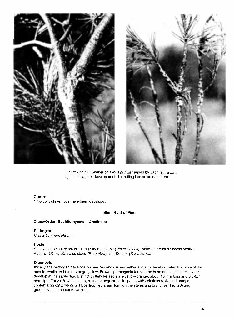
Control
Figure 27a,b.-Canker on Pinus pumi/a caused by Lachnellu/a pini: a) initial stage of development; b) fruiting bodies on dead tree.
• No control methods have been developed.
Stem Rust of Pine
Class/Order: Basidiomycetes, Uredinales
Pathogen Cronartium ribicola Ditr.
Hosts Species of pine (Pinus) including Siberian stone (Pinus sibirica), white (P strobus); occasionally, Austrian (P nigra), Swiss stone (P cembra), and Korean (P koraiensis)
Diagnosis Initially, the pathogen develops on needles and causes yellow spots to develop. Later, the base of the needle swells and turns orange-yellow. Brown spermogonia form at the base of needles; aecia later develop at the same site. Distinct blister-like aecia are yellow-orange, about 10 mm long and 0.5-0.7 mm high. They release smooth, round or angular aeciospores with colorless walls and orange contents, 22-29 x 18-22 µ. Hypertrophied areas form on the stems and branches (Fig. 28) and gradually become open cankers.
55
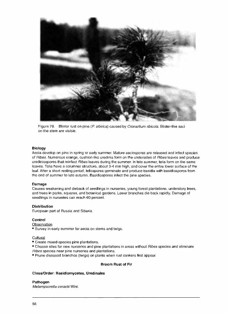
Figure 28.-Blister rust on pine (P sibirica) caused by Cronartium ribicola. Blister-like asci on the stem are visible.
Biology Aecia develop on pine in spring or early summer. Mature aeciospores are released and infect species of Ribes. Numerous orange, cushion-like uredinia form on the undersides of Ribes leaves and produce urediniospores that reinfect Ribes leaves during the summer. In late summer, telia form on the same leaves. Telia have a columnar structure, about 3-4 mm high, and cover the entire lower surface of the leaf. After a short resting period, teliospores germinate and produce basidia with basidiospores from the end of summer to late autumn. Basidiospores infect the pine species.
Damage Causes weakening and dieback of seedlings in nurseries, young forest plantations, understory trees, and trees in parks, squares, and botanical gardens. Lower branches die back rapidly. Damage of seedlings in nurseries can reach 60 percent.
Distribution European part of Russia and Siberia
Control Observation • Survey in early summer for aecia on stems and twigs.
Cultural • Create mixed-species pine plantations. • Choose sites for new nurseries and pine plantations in areas without Ribes species and eliminate Ribes species near pine nurseries and plantations. • Prune diseased branches (twigs) on plants when rust cankers first appear.
Broom Rust of Fir
Class/Order: Basidiomycetes, Uredinales
Pathogen Melampsorella cerastii Wint.
56
Hosts
Figure 29.-Broom rust of fir (Abies alba), caused by Melampsorella cerastii. Stem swelling is visible.
Species of fir (Abies) including silver (A. alba), Siberian (A. sibirica), balsam (A. balsamea), concolor (A. concolor), Nordmann (A. nordmanniana) and Sachalin (A. sachalinensis).
Diagnosis The pathogen causes galls or spindle-shaped swellings to form on branches (Fig. 29). Longitudinal cracks develop on these swellings and gradually widen and deepen. Damaged bark later sloughs off. Several galls may form along the stem. Numerous "witches'-brooms" are produced (Fig. 30). Spermogonia and aecia develop along the primary rib on needles of the broom. Aecia are round or cylindrical, orange, small, cushion-like. Aeciospores are globose or elliptical, orange, with colorless walls, 16-30 x 14-20 µ. Damaged needles of witches' -brooms usually drop in winter.
Biology In spring, needles are infected by basidiospores formed from telia produced on the previous-year leaves of chickweed species Stelliaria and Cerastium. Initially, the fungus damages young shoots and twigs and causes slight spindle-shaped swellings. The next-year buds on the diseased twig produce numerous short shoots called witches'-brooms. From the infected twigs, mycelium penetrates into the stele, develops for several years, and causes galls to develop. The galls grow slowly, can become large, and encircle the stem. Depending on the region, in mid- to late summer, aecia develop on the needles or on short shoots of witches'-brooms. The orange aeciospores are wind dispersed to the leaves of the alternate host plant, where uredinia and telia develop. Telia overwinter on the dead leaves of the alternate host and teliospores germinate in spring, producing basidia and basidiospores.
57
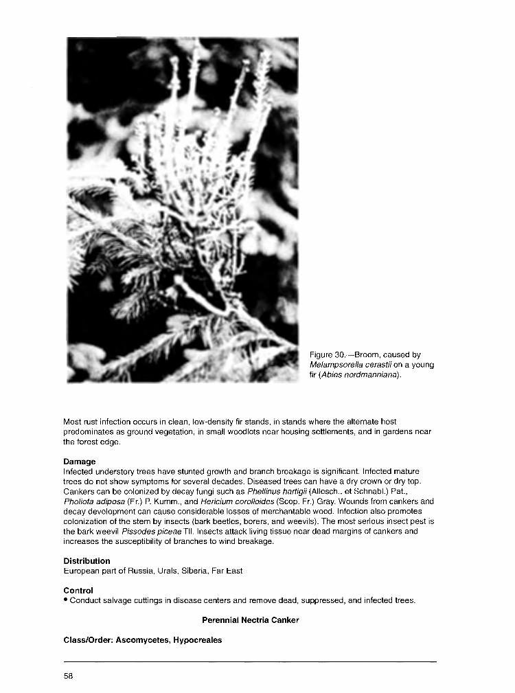
Figure 30.-Broom, caused by Melampsorella cerastii on a young fir (Abies nordmanniana).
Most rust infection occurs in clean, low-density fir stands, in stands where the alternate host predominates as ground vegetation, in small woodlots near housing settlements, and in gardens near the forest edge.
Damage Infected understory trees have stunted growth and branch breakage is significant. Infected mature trees do not show symptoms for several decades. Diseased trees can have a dry crown or dry top. Cankers can be colonized by decay fungi such as Phellinus hartigii (Allesch .. et Schnabl.) Pat., Pho/iota adiposa (Fr.) P. Kumm., and Hericium corolloides (Scop. Fr.) Gray. Wounds from cankers and decay development can cause considerable losses of merchantable wood. Infection also promotes colonization of the stem by insects (bark beetles, borers, and weevils). The most serious insect pest is the bark weevil Pissodes piceae TII. Insects attack living tissue near dead margins of cankers and increases the susceptibility of branches to wind breakage.
Distribution European part of Russia, Urals, Siberia, Far East
Control • Conduct salvage cuttings in disease centers and remove dead, suppressed, and infected trees.
Perennial Nectria Canker
Class/Order: Ascomycetes, Hypocreales
58

Pathogen
Figure 31.-Cankers caused by Nectria gal/igena on the trunk of a maple (Acer platanoides).
Nectria species, including N. galligena Bres., N. ditissima Tul., and N. coccinea (Pers.: Fr) Fr.
Hosts Species of maple (Acer}, beech (Fagus), oak (Quercus), apple (Ma/us), pear (Pyrus), hornbeam (Carpinus), poplar (Popu/us), and plum (Prunus)
Diagnosis Cankers form on stems and branches; several may occur on the same tree. Initially, cankers are closed but later the bark cracks and sloughs off, exposing the wood. Infection occurs annually, giving the wood a "target-like" appearance (Fig. 31 ). White, cushion-like sporodochia of the conidial stage form between the bark cracks. Dark-red perithecia develop singly or in groups at the same site. Conidia are colorless, cylindrical, straight, with several septa, 54-62 x 5-6 µ. Asci are clavate. Ascospores are colorless, ellipsoid, 2-celled, 15-21 x 6-8 µ.
Biology Conidia develop in spring and autumn during moist periods. Maturation and dispersion of ascospores may occur throughout the year. Ascospores overwinter and do not lose germinative power and viability. Ascospore discharge occurs after rain and fog. Spores penetrate into tree tissues at the juncture of dead branches with the stem or in cracks in dead bark. Sunscald, frost, mechanical damage, and insect injury can kill the bark. Long summers and mild winters with high levels of precipitation favor disease development.
59

Damage Weakens but rarely kills trees. Fruit orchards suffer extensive damage. In beech understory, weakness and stem breakage occur near the canker: In addition, the cankers are colonized by wood-decay fungi.
Distribution European part of Russia, Urals, Siberia, Far East
Control Cultural • Conduct timely sanitation cuttings and thinnings and remove and destroy felling residues. • Create mixed stands and maintain optimal stand density. • Minimize mechanical injuries during tending activities.
Black Hypoxylon Canker
Class/Order: Ascomycetes, Sphaeropsidales
Pathogen Hypoxylon pruinatum (Kl.) Cke.
Hosts European aspen (Populus tremula), white poplar (P. alba), and poplar hybrids from the Aegeiros and Tacamahaca selections
Diagnosis Affects the bark, cambium, and wood of stems and branches. Initially, bark in lesion areas is slightly red. Later, it becomes blistered and cracked, and white liquid flows from the cracks. Cankers form gradually. They are irregularly elongate and extend along trunks and branches a length of 1-1.5 m. The conidial structures develops in bark in 1-1.5 years. They are dark-brown, blister-like pillars, up to 1 mm tall. Conidiophores with conidia form on them. In about 3 years after infection, black, smeared stromata develop inside the bark. Stromata, which are gray-black, multiangular, and up to 1 cm in diameter, contain groups of perithecia (Fig. 32). Perithecia are globose or bottle-like, completely immersed in the stromata. Asci are oblong, with stalks, 50-62 x 4-6 µ. Ascospores are elliptic, black,~ -celled, 10 x 6 µ.
Biology Infection is initiated by both conidia and ascospores. Conidia form from May to August. Ascospores mature and actively discharge in high humidity during late autumn and early spring. The fungus infects tree tissues through injuries to the bark, including those from insects. Poplar species and hybrids differ in degree of resistance, which depends on bark moisture, chemical composition of cell sap, and physical properties of the bark. Disease outbreaks occur in a range of forest site conditions.
Damage Weakens trees and causes dieback in natural stands, plantations, and urban street plantings. Diseased trees in stands can reach 40 percent. The disease also promotes extensive wind breakage of branches.
Distribution European part of Russia, Urals, Siberia
Control Cultural • Conduct timely sanitation cuttings and remove and destroy felling residue.
Cytophoma Canker of Ash
Class/Order: Deuteromycetes, Sphaeropsidales
Pathogen Cytophoma pulchella (Sacc.) Guthn.
60
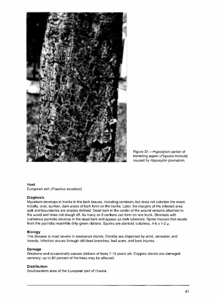
Figure 32.-Hypoxylon canker of trembling aspen (Populus tremu/a) caused by Hypoxylon pruinatum.
Host European ash (Fraxinus excelsior')
Diagnosis Mycelium develops in trunks in the bark tissues, including cambium, but does not colonize the wood. Initially, oval, sunken, dark areas of bark form on the trunks. Later, the margins of the infected area split and boundaries are sharply defined. Dead bark in the center of the wound remains attached to the wood and does not slough off. As many as 8 cankers can form on one trunk. Stromata with numerous pycnidia develop in the dead bark and appear as dark tubercles. Spore masses that exude from the pycnidia resemble dirty-green ribbons. Spores are alantoid, colorless, 4-6 x 1-2 µ.
Biology The disease is most severe in weakened stands. Conidia are dispersed by wind, rainwater, and insects. Infection occurs through old dead branches, leaf scars, and bark injuries.
Damage Weakens and occasionally causes dieback of trees 7-15 years old. Coppice stands are damaged severely; up to 80 percent of the trees may be affected.
Distribution Southeastern area of the European part of Russia
61

Contents
-~ .. . Introduction .......................................... ':: ................................................................................................. , .... 1
Most Common Fungal Diseases of Russian Forests ..................................................................... 2
Diseases of Fruits and Seeds ...................................................................... -:: ............................ 2 Birch Seed Mummification ...................................................................................................................... 2 Fruit Deformation .................................................................................................................................... 2 Fruit Spots .............................................................................................................................................. 4 Molds ...................................................................................................................................................... 6 Seed and Fruit Rots ............................................................................................................................... 9
Diseases of Needles and Leaves ............................................................................................ 11 Needle Diseases .................................................................................................................................. 11
Lophodermium Needle Casts .......................................................................................................... 11 Snow Blight ...................................................................................................................................... 15 Meria Needle Blight ......................................................................................................................... 16 Brown Felt Blight ............................................................................................................................. 18 Rhizosphaera Needle Casts ............................................................................................................ 18 Needle Rusts of Pine ....................................................................................................................... 20
Leaf Diseases ....................................................................................................................................... 22 Powdery Mildews ............................................................................................................................. 22 Leaf Spots ...................................................... : .. .............................................................................. 23 Leaf Rusts ....................................................................................................................................... 35
Diseases Of Roots, Stems, And Branches ............................................................................... 36 Diseases in Tree Nurseries and Young Forests ................................................................................... 36
Damping - off ................................................................................................................................... 36 Diseases of Forest Stands ................................................................................................................... 38
Wilts ................................................................................................................................................. 38 Verticillium Wilt ............................................................................................................................ 38 Dutch Elm Disease ..................................................................................................................... 39 Oak Wilt ......................................................................... · ............................................................. 41
Dieback and Canker Diseases ........................................................................................................ 42 Cenangium Dieback of Pine ....................................................................................................... 42 Nectria Canker and Dieback ....................................................................................................... 44 Cytospora Canker ....................................................................................................................... 45 Dothichiza Canker of Poplar ....................................................................................................... 46 Clithris Canker and Dieback of Oaks .......................................................................................... 48 Nummularia Canker .................................................................................................................... 48 Black Naemospora Canker ......................................................................................................... 49 Thyrostroma Canker and Dieback .............................................................................................. 50 Ascocalyx Scleroderris Shoot Canker ........................................................................................ 52 European Larch Canker .............................................................................................................. 53 Lachnellula Canker of Siberian Pine Understory ........................................................................ 54 Stem Rust of Pine ....................................................................................................................... 55 Broom Rust of Fir ........................................................................................................................ 56 Perennial Nectria Canker ............................................................................................................ 58 Black Hypoxylon Canker ............................................................................................................. 60 Cytophoma Canker of Ash .......................................................................................................... 60
Wood-Decaying Diseases ............................................................................................................... 62 Annosum Root and Butt Rot ....................................................................................................... 62 Armillaria Root Rot ...................................................................................................................... 65 Butt and Trunk Rot of Conifers ................................................................................................... 67 Trunk and Limb Rot of Hardwoods ............................................................................................. 75

Fungal Dise~ses that Occur Only in Russian Forests ................................................................. 89
Diseases of Fruits and Seeds .......... .' ....................................................................................... 89 Thecopsora Rust of Spruce Cones ...................................................................................................... 89 Acorn Mummification Deformity ........................................................................................................... 89
Diseases of Needles and Leaves ....................................................................... '..:-................... 91 Needle Diseases .................................................................................................................................. 91
Hypodermella Needle Cast of Pine Hosts ....................................................................................... 91 . Chrysomyxa Rust of Spruce ............................................................................................................ 91
Leaf Diseases ....................................................................................................................................... 93 Powdery Mildew of Siberian Pea Tree ............................................................................................. 93 Other Powdery Mildews ................................................................................................................... 94 Orange Leaf Spot of Padus ............................................................................................................. 95 Red Spot of Ussurian Plum ............................................................................................................. 96 Foliage Anthracnoses, Spots, and Blights ....................................................................................... 96 Leaf Rusts ..................................................................................................................................... 108 Taphrina Diseases: Leaf Blisters, Leaf and Shoot Deformation .................................................... 110
Diseases of Roots, Stems, and Branches ............................................................................... 111 Diseases in Tree Nurseries and Young Forests ................................................................................. 111
"Infectious Damping'.' of Coniferous Seedlings .............................................................................. 111 Sclerophoma Disease of Pine Shoots ........................................................................................... 112 Pine Shoot Rust ............................................................................................................................. 113 Chrysomyxa Rust of Spruce Shoots and Needles ........................................................................ 114
Diseases of Forest Stands .... , ............................................................................................................ 116 Dieback and Canker Diseases ...................................................................................................... 116
Black Cytospora Canker of Poplar ............................................................................................. 116 Biatorella Canker ...................................................................................................................... 117 Pitch Blister Rust Canker .......................................................................................................... 119 Endoxylina Canker of Ash ......................................................................................................... 120 Cankers and Diebacks .............................................................................................................. 122
Wood-Decaying Diseases ............................................................................................................. 128 Ganoderma Butt Rot of Beech .................................................................................................. 129 Vuillemenia Decay .................................................................................................................... 129 Trunk and Limb Rots ................................................................................................................. 130
Acknowledgment ............................................................................................................ 133
Appendix A Pathogens That Affect Trees and Shrubs in Russia ....................................................................... 134
Appendix B Host Trees, Shrubs, and Herbs Listed in this Report ..................................................................... 137
Control Observation • Survey stands during summer to locate ne.w and developing,disease centers.
Cultural • Timely remove affected dying and dead trees. • Destroy felling residue as a source of inoculum. • Create optimal conditions for plantation growth. '
Wood-Decaying Diseases
Annosum Root and Butt Rot
Class/Order: Basldiomycetes, Aphyllophorales
Pathogen Heterobasidion annosum (Fr.) Bref .. (syn. Fomitopsis annosa (Fr.) Karst.)
Hosts Species of fir (Abies), spruce (Picea), pine (Pinus), larch (Larix), arborvitae (Thuja), beech (Fagus), and birch (Betula)
Diagnosis In pine stands, existing disease centers are characterized by weakened and dying trees, previous-year dead trees, and tipped and windthrown trees. Group mortality and blowdown occur and grassy glades develop in the opening (Fig. 33). Disease centers in grassy glades have relatively distinctive boundaries. These boundaries enlarge each year and new dying trees appear on the edges. Individual grassy glades coalesce and the stand becomes an open forest. By contrast, infected fir and spruce remain alive for a long time even though considerable decay develops in their roots and stems. Symptoms of infection are visible and include a reduction in radial and terminal growth, thin-crowned trees with dull green and brown shade-needles, and shoot deformities. In spruce stands, the disease often Is concealed until tree mortality occurs as a result of blowdown. Conspicuous disease centers
·and grassy glades do not form in spruce stands as they do in pine stands (Fig. 34).
Disease characteristics and symptoms vary with different hosts. In pine, decay develops in the root system. In the first stage of infection, abundant resin exudes from the affected resin vessels in the roots. Resin-soaked root wood is a red-orange, sometimes lilac, becomes crystalline, and has a turpentine order. Resin accumulates under the bark on roots of diseased trees, then exudes to the surface where soil and other debris become entrapped, forming hard resin galls on the roots. As decay develops, the resin gradually disappears and the infected wood turns uniformally yellow. Small, white flecks of cellulose may be scattered throughout the wood. The final stage is characterized by the formation of numerous small pockets, and the wood becomes bastlike, stringy, and crumbles.
In fir and spruce, the fungus initially spreads in roots and then extends up the stem causing heart rot, which is bordered by a lilac-gray ring. The fungus grows about 3 to 4 m up the trunk but can reach 10 m. Initial decay is characterized by the gray~violet color of the wood, which later turns red-brown. In the final stage, wood is typically mottled, with relatively large white cellulose pockets and typical black specks. The decayed wood has a pocket-laminated structure and crumbles easily when dry. The most reliable sign is the presence of the basidiocarps on the roots and root-stem base. They form usually in shady places, on the lower surfaces of decaying roots of windthrown trees, sometimes at the base of dead tree stumps, and on the partially decayed butts. Basidiocarps differ in shape and size. They are thin and perennial (Figs. 35-36). The surface is brown, with a light-brown margin and concentric furrows. The hymenophore is initially white and later yellow with a silky shine. Pores are tiny, round or angular, sometimes sloped.
Biology Initial infection occurs by basidiospores and conidia of the fungus. Basidiospores develop in basidiocarps; conidia develop on the mycelium on the surface of decaying parts of infected stumps or roots. The spores are dispersed by air, water, and animals (primarily insects). Spores are transported to roots and, especially on mechanically wounded roots, infect them. Mycelium develops in roots and decay begins. The pathogen develops in both living and dead wood on colonized roots, stumps, and
62

Figure 33.-Annosum root rot (Heterobasidion annosum). Dead and dying trees are in a 25-year-old plantation of pine (Pinus sylvestris).
Figure 34.-Disease gap caused by Heterobasidion annosum in a spruce (Picea abies) stand.
woody debris in forest litter. If spores settle on freshly cut stumps (e.g., after thinnings), they germinate and mycelium spreads in stump wood and extends to the roots. The disease also spreads by secondary infection of living-tree roots by direct mycelial transfer at contacts of healthy and diseased roots. Tree infection also is initiated through dead rootlets or dead ends of deep roots.
The disease occurs in nearly every forest site except swamps; bog (sphagnum) and lichen (cladina) pine stands are rarely infected. Stands of various age are affected. Initial symptoms may be visible in 15- to 20-year-old stands. Seedlings of conifers that develop in disease centers also are subjected to potential infection and death. Homogenious conifer stands suffer the most damage, especially forest plantations established on former agricultural soil, waste grounds, or areas previously affected by the fungus. The disease is rare in natural pine stands. However, spruce and fir are damaged severely both in forest plantations and in natural stands. Mixed coniferous-hardwood stands are more resistant to the disease. Excessive stand density with closely interlaced and grafted roots promotes rapid infection, development, and fungal spread. In suburban forests and parks, recreation use causes root injury, soil compaction, and poor soil aeration, predisposing trees to infection and disease development.
Damage Causes massive decline and death of stands and significant changes in stocking. In spruce and fir, significant decay may extend up the trunk from the roots and the value of the butt log is significantly reduced. Losses of merchantable volume may reach 50 percent in spruce and 75 percent in fir. Weakening and decline of infected trees usually results in bark beetle and wood-borer infestations.
63
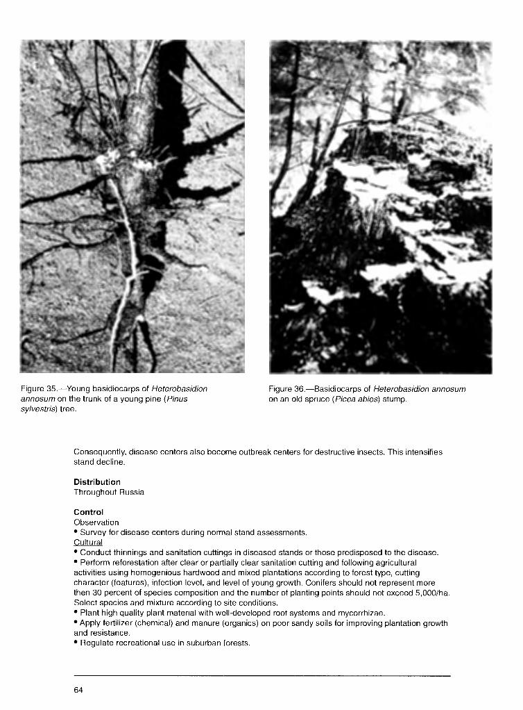
Figure 35.-Young basidiocarps of Heterobasidion annosum on the trunk of a young pine (Pinus sylvestris) tree.
Figure 36.-Basidiocarps of Heterobasidion annosum on an old spruce (Picea abies) stump.
Consequently, disease centers also become outbreak centers for destructive insects. This intensifies stand decline.
Distribution Throughout Russia
Control Observation • Survey for disease centers during normal stand assessments. Cultural • Conduct thinnings and sanitation cuttings in diseased stands or those predisposed to the disease. • Perform reforestation after clear or partially clear sanitation cutting and following agricultural activities using homogenious hardwood and mixed plantations according to forest type, cutting character (features), infection level, and level of young growth. Conifers should not represent more then 30 percent of species composition and the number of planting points should not exceed 5,000/ha. Select species and mixture according to site conditions. • Plant high quality plant material with well-developed root systems and mycorrhizae. • Apply fertilizer (chemical) and manure (organics) on poor sandy soils for improving plantation growth and resistance. • Regulate recreational use in suburban forests.
64
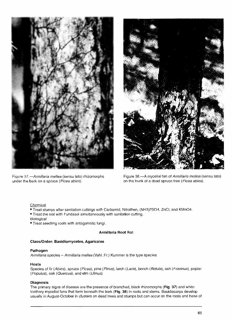
Figure 37.--Armillaria me/lea (sensu lato) rhizomorphs under the bark on a spruce (Picea abies).
Figure 38.-A mycelial fan of Armillaria me/lea (sensu lato) on the trunk of a dead spruce tree (Picea abies).
Chemical • Treat stumps after sanitation cuttings with Carbamid, Nitrathen, (NH3)2S04, ZnCI, and KMn04. • Treat the soil with Fundasol simultaneously with sanitation cutting. Biological • Treat seedling roots with antagonistic fungi.
Armillaria Root Rot
Class/Order: Basidiomycetes, Agaricales
Pathogen Armillaria species - Armillaria me/lea (Vahl.:Fr.) Kummer is the type species
Hosts Species of fir (Abies), spruce (Picea), pine (Pinus), larch (Larix), beech (Betu/a), ash (Fraxinus), poplar (Popu/us), oak (Quercus), and elm (U/mus)
Diagnosis The primary signs of disease are the presence of branched, black rhizomorphs (Fig. 37) and white leathery mycelial fans that form beneath the bark (Fig. 38) in roots and stems. Basidiocarps develop usually in August-October in clusters on dead trees and stumps but can occur on the roots and base of
65

Figure 39.-Basidiocarps of Armillaria me/lea (sensu Jato) on the trunk of a birch (Betula pendula).
trunks of affected live trees (Fig. 39). The basidiocarp, which can reach 15 cm in diameter depending on species, is initially convex and later flat with a hillock in the center, yellow-brown or gray-brown, with numerous dark (or the same color) scales. The internal tissue is white and friable, with a pleasant odor. The hymenophore gills are slightly descending and initially white, later darkening. The stipe is central, cylindrical, 10-15 cm long and 1-1.5 cm in diameter, with small scales, white or light-brown and darker on the base, and a white thick fluffy-silk ring (annulus) under the cap. Decayed wood is white or light-yellow, fiberous, with characteristic sinuous black lines. Decay can extend 1-1.5 m up the trunk.
Biology Basidiospores are dispersed by wind, rainfall, and animals, and germinate and infect stumps and roots of trees. Infection also occurs from root contact with rhizomorphs of the fungus or direct mycelial transfer from diseased to healthy roots. Stands are predisposed to this disease by abiotic and biotic stressors of temperature and moisture extremes, wind, soil compaction, air pollution, and defoliation by frost (abiotic factors), and defoliation by insects and fungi, attacks by sucking insects, and other foliar and stem diseases (biotic factors).
Damage Causes dieback in a range of stand ages and types. Often develops into an epyphytotic, causing heavy mortality in stands of single-species conifers and in oak and birch stands. The volume of merchantable wood can be reduced by decay.
Distribution Throughout Russia except mountain highlands, deserts, and tundra sites
66
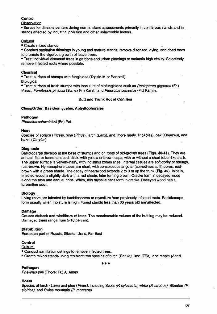
Control Observation • Survey for disease centers during normal-stand assessments primarily in coniferous stands and in stands affected by industrial pollution and other unfavorable factors.
Cultural • Create mixed stands. • Conduct sanitation thinnings in young and mature stands; remove diseased, dying, and dead trees to promote the vigorous growth of leave trees. • Treat individual diseased trees in gardens and urban plantings to maintain high vitality. Selectively remove infected roots where possible.
Chemical • Treat surface of stumps with fungicides (Topsin-M or Benomil). Biological • Treat surface of fresh stumps with inoculum of biofungicides such as Peniophora gigantea (Fr.) Mass., Fomitopsis pinicola (Sw. ex Fr.) Karst., and Pleurotus ostreatus (Fr.) Kamm.
Butt and Trunk Rot of Conifers
Class/Order: Basidiomycetes, Aphyllophorales
Pathogen Phaeo/us schweinitzii (Fr.) Pat.
Host Species of spruce (Picea), pine (Pinus), larch (Larix), and, more rarely, fir (Abies), oak (Quercus), and hazel ( Cory/us)
Diagnosis Basidiocarps develop at the base of stumps and on roots of old-growth trees (Figs. 40-41). They are annual, flat or funnel-shaped, thick, with yellow or brown caps, with or without a short tuber-like stalk. The upper surface is velvety-hairy, with indistinct zones lines. Internal tissues are soft-corky or spongy, rust-brown. Hymenophore tubes are short, with conspicuous angular (sometimes split) pores, rustbrown with a green shade. The decay of heartwood extends 2 to 3 m up the trunk (Fig. 42). Initially, infected wood is slightly dark with a red shade, later turning brown. Cracks form in decayed wood along the rays and annual rings. White, thin mycelial fans form in cracks. Decayed wood has a turpentine odor.
Biology Living roots are infected by basidiospores or mycelium from previously infected roots. Basidiocarps form usually when moisture is high. Forest stands less than 60 years old are·attected.
Damage Causes dieback and windthrow of trees. The merchantable volume of the butt log may be reduced. Damaged trees range from 5-10 percent.
Distribution European part of Russia, Siberia, Urals, Far East
Control Cultural • Conduct santitation cuttings to remove infected trees. • Create mixed stands using resistant tree species of birch (Betula), lime (Tilia), and maple (Acer) .
••• Pathogen Phellinus pini (Thore: Fr.) A. Ames
Hosts Species of larch (Larix) and pine (Pinus), including Scots (P. sy/vestris), white (P. strobus), Siberian (P. sibirica), and Swiss mountain (P. montana)
67
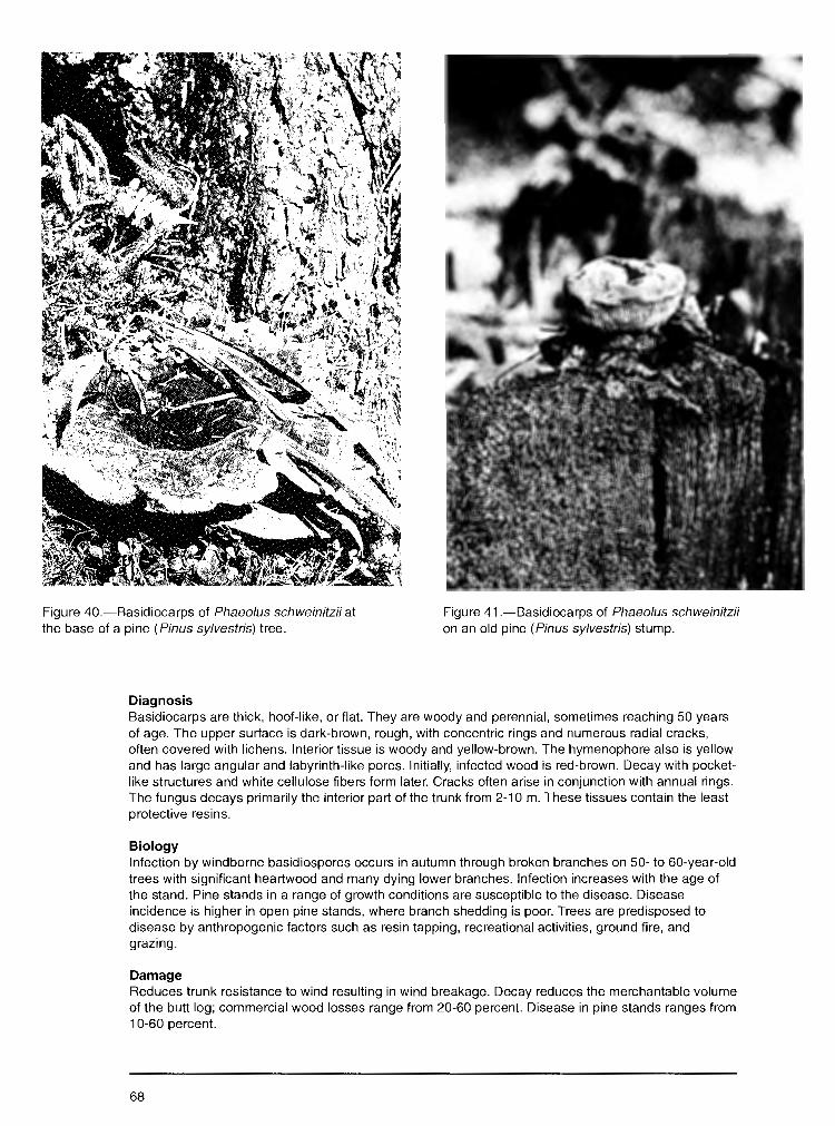
Figure 40.-Basidiocarps of Phaeo/us schweinitzii at the base of a pine (Pinus sylvestris) tree.
Diagnosis
Figure 41.-Basidiocarps of Phaeo/us schweinitzii on an old pine (Pinus sylvestris) stump.
Basidiocarps are thick, hoof-like, or flat. They are woody and perennial, sometimes reaching 50 years of age. The upper surface is dark-brown, rough, with concentric rings and numerous radial cracks, often covered with lichens. Interior tissue is woody and yellow-brown. The hymenophore also is yellow and has large angular and labyrinth-like pores. Initially, infected wood is red-brown. Decay with pocketlike structures and white cellulose fibers form later. Cracks often arise in conjunction with annual rings. The fungus decays primarily the interior part of the trunk from 2-10 m. These tissues contain the least protective resins.
Biology Infection by windborne basidiospores occurs in autumn through broken branches on 50- to 60-year-old trees with significant heartwood and many dying lower branches. Infection increases with the age of the stand. Pine stands in a range of growth conditions are susceptible to the disease. Disease incidence is higher in open pine stands, where branch shedding is poor. Trees are predisposed to disease by anthropogenic factors such as resin tapping, recreational activities, ground fire, and grazing.
Damage Reduces trunk resistance to wind resulting in wind breakage. Decay reduces the merchantable volume of the butt log; commercial wood losses range from 20-60 percent. Disease in pine stands ranges from 1 0-60 percent.
68
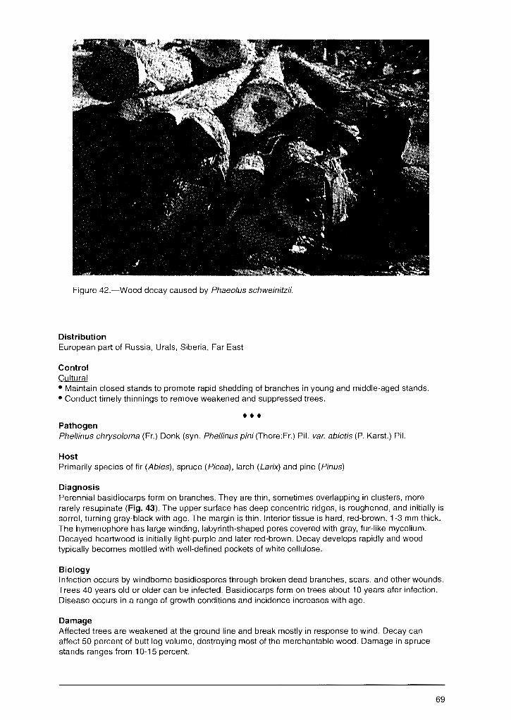
Figure 42.-Wood decay caused by Phaeo/us schweinitzii.
Distribution European part of Russia, Urals, Siberia, Far East
Control Cultural • Maintain closed stands to promote rapid shedding of branches in young and middle-aged stands. • Conduct timely thinnings to remove weakened and suppressed trees .
••• Pathogen Phellinus chryso/oma (Fr.) Donk (syn. Phellinus pini (Thore:Fr.) Pil. var. abietis (P. Karst.) Pil.
Host Primarily species of fir (Abies), spruce (Picea), larch (Larix) and pine (Pinus)
Diagnosis Perennial basidiocarps form on branches. They are thin, sometimes overlapping in clusters, more rarely resupinate (Fig. 43). The upper surface has deep concentric ridges, is roughened, and initially is sorrel, turning gray-black with age. The margin is thin. Interior tissue is hard, red-brown, 1-3 mm thick. The hymenophore has large winding, labyrinth-shaped pores covered with gray, fur-like mycelium. Decayed heartwood is initially light-purple and later red-brown. Decay develops rapidly and wood typically becomes mottled with well-defined pockets of white cellulose.
Biology Infection occurs by windborne basidiospores through broken dead branches, scars, and other wounds. Trees 40 years old or older can be infected. Basidiocarps form on trees about 10 years afer infection. Disease occurs in a range of growth conditions and incidence increases with age.
Damage Affected trees are weakened at the ground line and break mostly in response to wind. Decay can affect 50 percent of butt log volume, destroying most of the merchantable wood. Damage in spruce stands ranges from 10-15 percent.
69
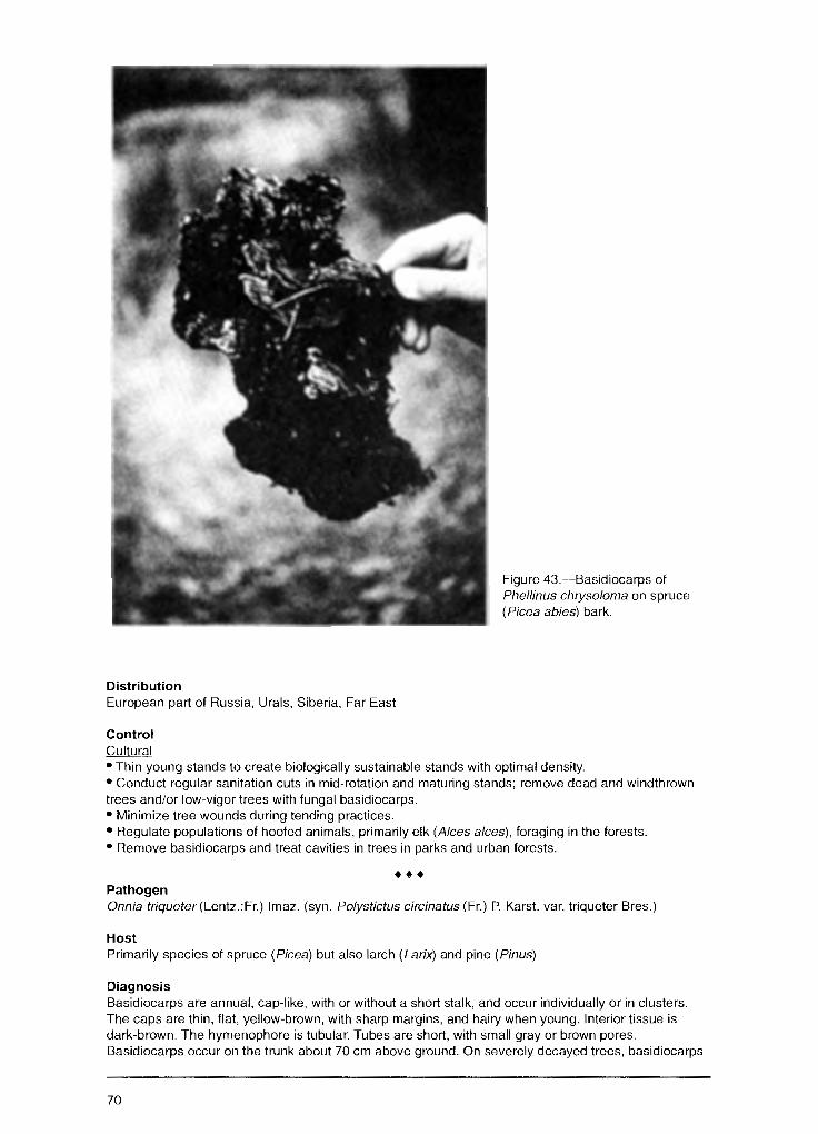
Distribution European part of Russia, Urals, Siberia, Far East
Control Culturg_[
Figure 43.-Basidiocarps of Phellinus chryso/oma on spruce ( Picea abies) bark.
• Thin young stands to create biologically sustainable stands with optimal density. • Conduct regular sanitation cuts in mid-rotation and maturing stands; remove dead and windthrown trees and/or low-vigor trees with fungal basidiocarps. • Minimize tree wounds during tending practices. • Regulate populations of hoofed animals, primarily elk (A/ces alces), foraging in the forests. • Remove basidiocarps and treat cavities in trees in parks and urban forests .
••• Pathogen Onnia triqueter(Lentz.:Fr.) lmaz. (syn. Po/ystictus circinatus (Fr.) P. Karst. var. triqueter Bres.)
Host Primarily species of spruce (Picea) but also larch (Larix) and pine (Pinus)
Diagnosis Basidiocarps are annual, cap-like, with or without a short stalk, and occur individually or in clusters. The caps are thin, flat, yellow-brown, with sharp margins, and hairy when young. Interior tissue is dark-brown. The hymenophore is tubular. Tubes are short, with small gray or brown pores. Basidiocarps occur on the trunk about 70 cm above ground. On severely decayed trees, basidiocarps
70
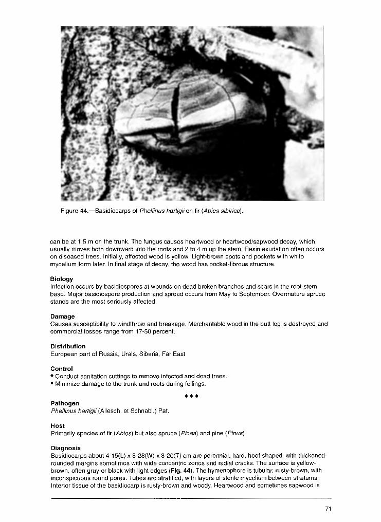
Figure 44.-Basidiocarps of Phellinus hartigii on fir (Abies sibirica).
can be at 1.5 m on the trunk. The fungus causes heartwood or heartwood/sapwood decay, which usually moves both downward into the roots and 2 to 4 m up the stem. Resin exudation often occurs on diseased trees. Initially, affected wood is yellow. Light-brown spots and pockets with white mycelium form later. In final stage of decay, the wood has pocket-fibrous structure.
Biology Infection occurs by basidiospores at wounds on dead broken branches and scars in the root-stem base. Major basidiospore production and spread occurs from May to September. Overmature spruce stands are the most seriously affected.
Damage Causes susceptibility to windthrow and breakage. Merchantable wood in the butt log is destroyed and commercial losses range from 17-50 percent.
Distribution European part of Russia, Urals, Siberia, Far East
Control • Conduct sanitation cuttings to remove infected and dead trees. • Minimize damage to the trunk and roots during tellings .
••• Pathogen Phellinus hartigii (Allesch. et Schnabl.) Pat.
Host Primarily species of fir (Abies) but also spruce (Picea) and pine (Pinus)
Diagnosis Basidiocarps about 4-15(L) x 8-28(W) x 8-20(T) cm are perennial, hard, hoof-shaped, with thickenedrounded margins sometimes with wide concentric zones and radial cracks. The surface is yellowbrown, often gray or black with light edges (Fig. 44). The hymenophore is tubular, rusty-brown, with inconspicuous round pores. Tubes are stratified, with layers of sterile mycelium between stratums. Interior tissue of the basidiocarp is rusty-brown and woody. Heartwood and sometimes sapwood is
71
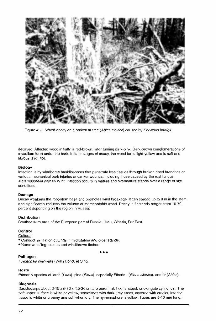
Figure 45.-Wood decay on a broken fir tree (Abies sibirica) caused by Phellinus hartigii.
decayed. Affected wood initially is red-brown, later turning dark-pink. Dark-brown conglomerations of mycelium form under the bark. In later stages of decay, the wood turns light-yellow and is soft and fibrous (Fig. 45).
Biology Infection is by windborne basidiospores that penetrate tree tissues through broken dead branches or various mechanical bark injuries or canker wounds, including those caused by the rust fungus Melampsorel/a cerastiiWint. Infection occurs in mature and overmature stands over a range of site conditions.
Damage Decay weakens the root-stem base and promotes wind breakage. It can spread up to 8 m in the stem and significantly reduces the volume of merchantable wood. Decay in fir stands ranges from 10-70 percent depending on the region in Russia.
Distribution Southeastern area of the European part of Russia, Urals, Siberia, Far East
Control Cultural • Conduct sanitation cuttings in midrotation and older stands. • Remove felling residue and windthrown timber .
••• Pathogen Fomitopsis officinalis (Will.) Bond. et Sing.
Hosts Primarily species of larch (Larix), pine (Pinus), especially Siberian (Pinus sibirica), and fir (Abies)
Diagnosis Basidiocarps about 3-15 x 5-30 x 4.5-26 cm are perennial, hoof-shaped, or elongate cylindrical. The soft upper surface is white or yellow, sometimes with dark-gray areas, covered with cracks. Interior tissue is white or creamy and soft when dry. The hymenophore is yellow. Tubes are 5-10 mm long,
72

with inconspicuous, round or angular pores. Affected wood initially is light-brown but later turns darkbrown. In the final stage of decay, wide cracks associated with annual rings form in the wood. Creamywhite, thick, velvety mycelial sheets develop-in the cracks. The decay is cubical rot. It develops primarily in the heartwood but can colonize sapwood. It can spread 15-20 m upward in the trunk.
Biology Infection occurs by basidiospores that penetrate tree tissues through broken dead branches and various trunk injuries. Trees more than 30 years old are affected. The disease level increases with age. The fungus affects living trees but can continue developing and remain on dead trunks and stumps for extended periods. The disease occurs over a range of site conditions.
Damage Trees are weakened at the root-stem base and break at the ground line during windstorms. Most of the diseased trees have dead tops. Damage in larch stands ranges from 5-10 percent.
Distribution Urals, Siberia, Far East
Control Cultural • Conduct sanitation cuttings to remove weakened, dying, and dead trees .
••• Pathogen Fomitopsis pinicola (Sw. et Fr.) Karst.
Hosts Primarily species of fir (Abies), larch (Larix), spruce (Picea), and pine (Pinus); also birch (Betula), lime (Tilia), alder (A/nus), and European aspen (Popu/us tremula)
Diagnosis Basidiocarps are perennial, hoof-shaped, with a cushion-like or flat bottom. They differ in size but can reach 0.5 m in diameter. The upper surface of the young basidiocarp is yellow-red or rusty but with age turns red-brown to nearly black with a slightly shiny crust. The margin is edged by a yellow, brightred, or dark-cherry band. The hymenophore is yellow-white, tubular, with relatively large, round pores. The interior tissues are light-yellow and corky or woody. Decay begins in the sapwood of the stem and spreads rapidly to the central core where it spreads slowly up the trunk. In the early stage of decay, wood is yellow-brown with a silky sheen; later, white stripes of fungal hyphal form in the wood. In the final stage of decay, wood turns dark-brown with numerous cracks filled with white sheets of mycelium. The decay is a typical cubical rot.
Biology Infections of living trees occur primarily at wounds from mechanical injury. Basidiocarps form several years after infection.
Damage Primarily a saprotroph but can cause significant economic loss in logs left in cutting areas or stored in timber yards. The fungus decomposes wood on the forest floor, accelerating the return of nutrients to the soil. Occasionally, the fungus infects living, weakened trees, predisposing them to wind breakage. Proportion of infected trees in a stand rarely exceeds 3 percent.
Distribution Throughout Russia except deserts, semideserts, and tundra
Control • Prompt removal of harvested wood from the forest.
Class/Order: Basldiomycetes/Agaricales
Pathogen Pho/iota adiposa (Fr.:Fr) Kumm.
73
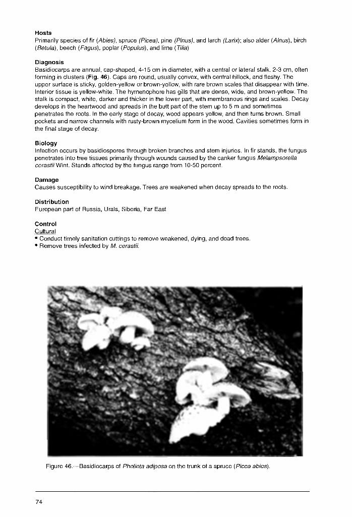
Hosts Primarily species of fir (Abies), spruce (Picea), pine (Pinus), and larch (Larix); also alder (A/nus), birch (Betula), beech (Fagus), poplar (Populus), and lime (Tilia)
Diagnosis Basidiocarps are annual, cap-shaped, 4-15 cm in diameter, with a central or lateral stalk, 2-3 cm, often forming in clusters (Fig. 46). Caps are round, usually convex, with central hillock, and fleshy. The upper surface is sticky, golden-yellow or brown-yellow, with rare brown scales that disappear with time. Interior tissue is yellow-white. The hymenophore has gills that are dense, wide, and brown-yellow. The stalk is compact, white, darker and thicker in the lower part, with membranous rings and scales. Decay develops in the heartwood and spreads in the butt part of the stem up to 5 m and sometimes penetrates the roots. In the early stage of decay, wood appears yellow, and then turns brown. Small pockets and narrow channels with rusty-brown mycelium form in the wood. Cavities sometimes form in the final stage of decay.
Biology Infection occurs by basidiospores through broken branches and stem injuries. In fir stands, the fungus penetrates into tree tissues primarily through wounds caused by the canker fungus Me/ampsorella cerastii Wint. Stands affected by the fungus range from 10-50 percent.
Damage Causes susceptibility to wind breakage. Trees are weakened when decay spreads to the roots.
Distribution European part of Russia, Urals, Siberia, Far East
Control Cultural • Conduct timely sanitation cuttings to remove weakened, dying, and dead trees. • Remove trees infected by M. cerastii.
Figure 46.-Basidiocarps of Pho/iota adiposa on the trunk of a spruce (Picea abies).
74
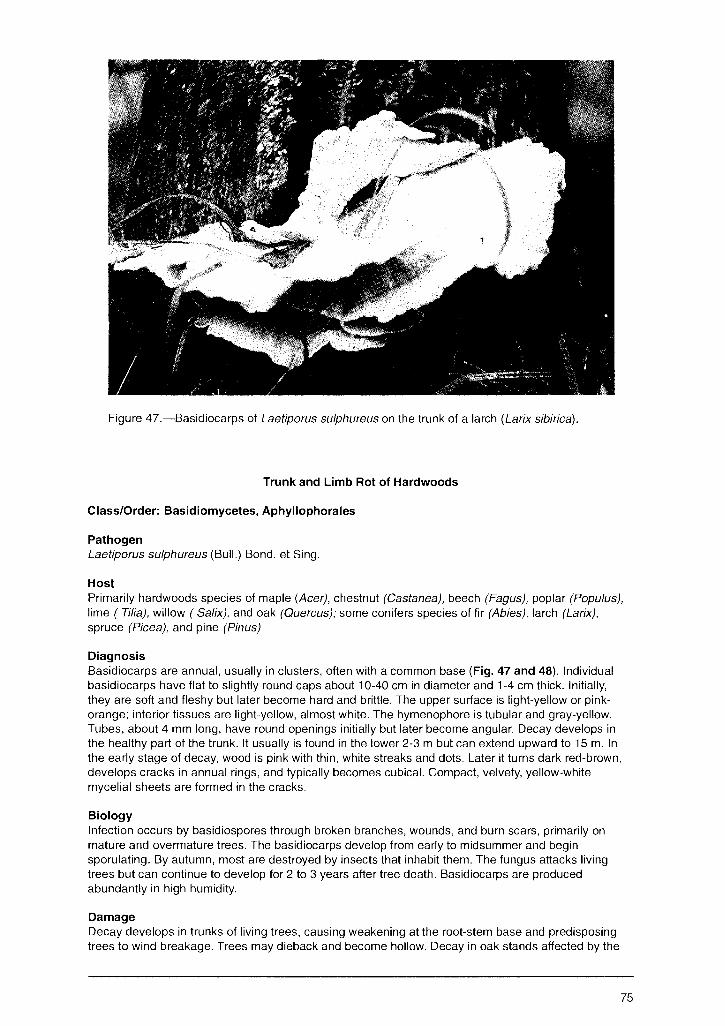
Figure 47.-Basidiocarps of Laetiporus sulphureus on the trunk of a larch (Larix sibirica).
Trunk and Limb Rot of Hardwoods
Class/Order: Basidiomycetes, Aphyllophorales
Pathogen Laetiporus sulphureus (Bull.) Bond. et Sing.
Host Primarily hardwoods species of maple (Acer), chestnut (Castanea), beech (Fagus), poplar (Populus), lime ( Tilia), willow ( Salix), and oak (Quercus); some conifers species of fir (Abies), larch (Larix), spruce (Picea), and pine (Pinus)
Diagnosis Basidiocarps are annual, usually in clusters, often with a common base (Fig. 47 and 48). Individual basidiocarps have flat to slightly round caps about 10-40 cm in diameter and 1-4 cm thick. Initially, they are soft and fleshy but later become hard and brittle. The upper surface is light-yellow or pinkorange; interior tissues are light-yellow, almost white. The hymenophore is tubular and gray-yellow. Tubes, about 4 mm long, have round openings initially but later become angular. Decay develops in the healthy part of the trunk. It usually is found in the lower 2-3 m but can extend upward to 15 m. In the early stage of decay, wood is pink with thin, white streaks and dots. Later it turns dark red-brown, develops cracks in annual rings, and typically becomes cubical. Compact, velvety, yellow-white mycelial sheets are formed in the cracks.
Biology Infection occurs by basidiospores through broken branches, wounds, and burn scars, primarily on mature and overmature trees. The basidiocarps develop from early to midsummer and begin sporulating. By autumn, most are destroyed by insects that inhabit them. The fungus attacks living trees but can continue to develop for 2 to 3 years after tree death. Basidiocarps are produced abundantly in high humidity.
Damage Decay develops in trunks of living trees, causing weakening at the root-stem base and predisposing trees to wind breakage. Trees may dieback and become hollow. Decay in oak stands affected by the
75
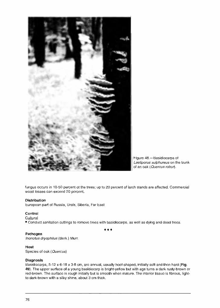
Figure 48.-Basidiocarps of Laetiporus sulphureus on the trunk of an oak ( Quercus robw).
fungus occurs in 10-50 percent ot the trees; up to 20 percent of larch stands are affected. Commercial wood losses can exceed 20 percent.
Distribution European part of Russia, Urals, Siberia, Far East
Control Cultural • Conduct sanitation cuttings to remove trees with basidiocarps, as well as dying and dead trees .
••• Pathogen lnonotus dryophilus (Berk.) Murr.
Host Species of oak ( Quercus)
Diagnosis Basidiocarps, 5-12 x 6-18 x 3-8 cm, are annual, usually hoof-shaped, initially soft and then hard (Fig. 49). The upper surface of a young basidiocarp is bright-yellow but with age turns a dark rusty-brown or red-brown. The surface is rough initially but is smooth when mature. The interior tissue is fibrous, lightto dark-brown with a silky shine, about 3 cm thick.
76
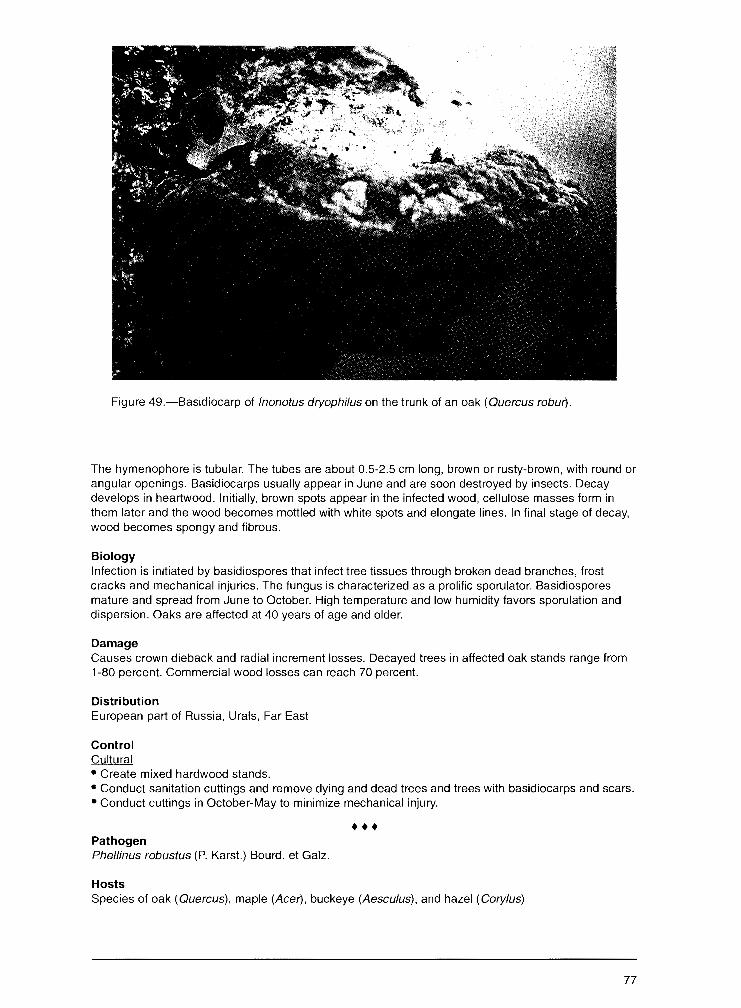
Figure 49.-Basidiocarp of /nonotus dryophilus on the trunk of an oak (Quercus robur).
The hymenophore is tubular. The tubes are about 0.5-2.5 cm long, brown or rusty-brown, with round or angular openings. Basidiocarps usually appear in June and are soon destroyed by insects. Decay develops in heartwood. Initially, brown spots appear in the infected wood, cellulose masses form in them later and the wood becomes mottled with white spots and elongate lines. In final stage of decay, wood becomes spongy and fibrous.
Biology Infection is initiated by basidiospores that infect tree tissues through broken dead branches, frost cracks and mechanical injuries. The fungus is characterized as a prolific sporulator. Basidiospores mature and spread from June to October. High temperature and low humidity favors sporulation and dispersion. Oaks are affected at 40 years of age and older.
Damage Causes crown dieback and radial increment losses. Decayed trees in affected oak stands range from 1-80 percent. Commercial wood losses can reach 70 percent.
Distribution European part of Russia, Urals, Far East
Control Cultural • Create mixed hardwood stands. • Conduct sanitation cuttings and remove dying and dead trees and trees with basidiocarps and scars. • Conduct cuttings in October-May to minimize mechanical injury .
••• Pathogen Phellinus robustus (P. Karst.) Bourd. et Galz.
Hosts Species of oak (Quercus), maple (Acer), buckeye (Aesculus), and hazel (Cory/us)
77
Diagnosis Basidiocarps are perennial, woody, cushion- or hoof-like, with a thick, wide, rusty margin, about 5-25 cm in diameter and ~p to 10 cm thick. The_ypper surface is dark-gray, nearly black, with wide concentric rings and cracks. Interior tissue is rusty-brown, woody, with concentric stripes. The hymenophore is poroid, rusty-yellow. Tubes, lighter in color than surrounding tissues, are about 2-5 mm long, with round openings. Infected wood turns brown and later becomes mottled with lighter flecks. In the final stage of decay, wood is yellow-white with thin, black, parallel lines. Rusty-orange mycelial masses often form in infected wood. The rot is primarily in heartwood but can spread to the sapwood.
Biology Infection is initiated by basidiospores that mature and spread from June to October. High humidity favors sporulation and dispersion. Basidiospores infect tree tissues through broken dead branches, frost cracks, injuries from hoofed animals, e.g., elk, and mechanical injuries. Stand are affected at 20-30 years of age, infection and damage increase with age. Disease levels are highest in coppiced stands.
Damage Causes tree weakening, partial crown decline, and wind breakage. Commercial wood losses range from 30-40 percent. Affected oak in stands range from 5-15 percent.
Distribution European part of Russia, the southen Urals, Western Siberia, Far East
Control Cultural • Create mixed hardwood stands. • Conduct timely thinnings to maintain optimal density. • Conduct sanitation cuttings and remove all trees with large wounds/scars. • Conduct cuttings in November-May when sporulation is scarce or absent.
••• Pathogen Phellinus igniarius (L.: Fr.) Quel.
Hosts Species of maple (Ace1', birch (Betula), hornbeam (Carpinus), buckeye (Aesculus), ash (Fraxinus), willow (Sa/ix), lime (Tt/ia), elm (U/mus) {forms: f. igniarius on willow (Sa/ix), f. sorbi on mountain-ash (Sorbus), f. betulae on birch (Betula), f. resupinatus on birch (Betula), alder (A/nus), and mountainash}
Diagnosis Basidiocarps are perennial, woody, hoof-shaped, with a flat or cushion-like bottom, more rarely resupinate, and 3-25 x 2-16 x 1.5-12 cm (Fig. 50). The upper surface is dark-gray, sometimes nearly black, with concentric rings. The hymenophore is rusty-brown with inconspicuous round pores. Tubes are stratiform, 3-5mm long, and become overgrown by white mycelium with age. Decay develops in heartwood of trunks. Affected wood is initially red-brown but later turns light-yellow and is separated from healthy wood by a thin, dark-brown ring (Fig. 51 ). A characteristic feature of the decay is the presence of winding black lines that spread as concentric circles in the affected part of wood. The extension of decay up the stem can vary but often occurs the entire length.
Biology Infection is initiated by basidiospores. Mass sporulation occurs in early summer. Spores penetrate tree tissues mostly through dead limbs but also through bark scars. Trees are infected when they are 20-30 years old. The disease level increases with age. Timely diagnosis is difficult because the decay often develops with no external symptoms, basidiocarp formation, or signs of tree weakening.
Damage Primarily commercial wood losses that range from 60· 100 percent
Distribution Throughout Russia
78

Figure 50.-Basidiocarp of Phellinus igniarius f. betulae on the trunk of a birch (Betula pendula).
HcJuro 51.----Decay on birch (Betula pendula) caused by Phellinus igniarius.
79
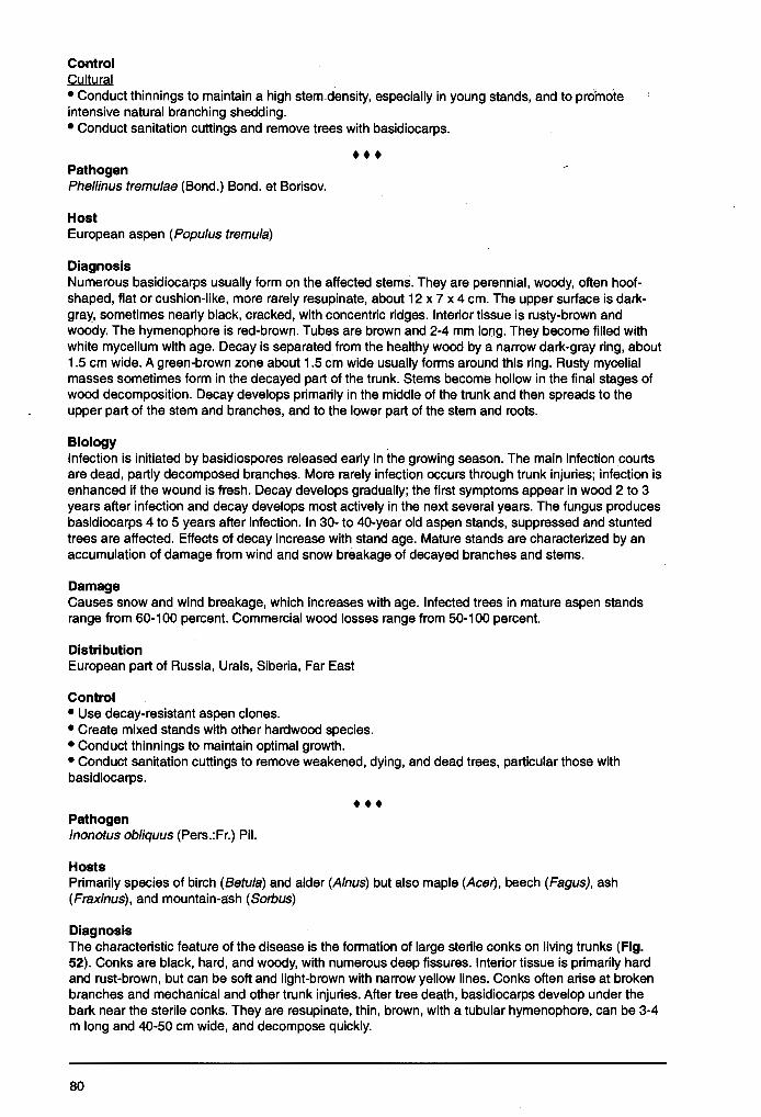
Control Cultural • Conduct thinnings to maintain a high stem density, especially in young stands, and to promote intensive natural branching shedding. • Conduct sanitation cuttings and remove trees with basidiocarps .
••• Pathogen Phellinus tremulae (Bond.) Bond. et Borisov.
Host European aspen (Populus tremula)
Diagnosis Numerous basidiocarps usually form on the affected stems. They are perennial, woody, often hoofshaped, flat or cushion-like, more rarely resupinate, about 12 x 7 x 4 cm. The upper surface is darkgray, sometimes nearly black, cracked, with concentric ridges. Interior tissue is rusty-brown and woody. The hymenophore is red-brown. Tubes are brown and 2-4 mm long. They become filled with white mycelium with age. Decay is separated from the healthy wood by a narrow dark-gray ring, about 1.5 cm wide. A green-brown zone about 1.5 cm wide usually forms around this ring. Rusty mycelial masses sometimes form in the decayed part of the trunk. Stems become hollow in the final stages of wood decomposition. Decay develops primarily in the middle of the trunk and then spreads to the upper part of the stem and branches, and to the lower part of the stem and roots.
Biology Infection is initiated by basidiospores released early in the growing season. The main infection courts are dead, partly decomposed branches. More rarely infection occurs through trunk injuries; infection is enhanced if the wound is fresh. Decay develops gradually; the first symptoms appear in wood 2 to 3 years after infection and decay develops most actively in the next several years. The fungus produces basidiocarps 4 to 5 years after infection. In 30- to 40-year old aspen stands, suppressed and stunted trees are affected. Effects of decay increase with stand age. Mature stands are characterized by an accumulation of damage from wind and snow breakage of decayed branches and stems.
Damage Causes snow and wind breakage, which increases with age. Infected trees in mature aspen stands range from 60-100 percent. Commercial wood losses range from 50-100 percent.
Distribution European part of Russia, Urals, Siberia, Far East
Control • Use decay-resistant aspen clones. • Create mixed stands with other hardwood species. • Conduct thinnings to maintain optimal growth. • Conduct sanitation cuttings to remove weakened, dying, and dead trees, particular those with basidiocarps.
• •• Pathogen lnonotus obliquus (Pers.:Fr.) Pil.
Hosts Primarily species of birch (Betula) and alder (A/nus) but also maple (Ace/}, beech (Fagus), ash (Fraxinus), and mountain-ash (Sorbus)
Diagnosis The characteristic feature of the disease is the formation of large sterile conks on living trunks (Fig. 52). Conks are black, hard, and woody, with numerous deep fissures. Interior tissue is primarily hard and rust-brown, but can be soft and light-brown with narrow yellow lines. Conks often arise at broken branches and mechanical and other trunk injuries. After tree death, basidiocarps develop under the bark near the sterile conks. They are resupinate, thin, brown, with a tubular hymenophore, can be 3-4 m long and 40-50 cm wide, and decompose quickly.
80

Figure 52.-Sterile conk of lnonotus ob/iquus on the trunk of a birch (Betula pendula).
Biology Basidiocarps form under the bark and basidiospores are dispersed. Spores penetrate tree tissues through frost cracks, broken branches, and other bark injuries. Mycelium penetrates through the sapwood to the heardwood where active decay develops. Trees 40 years old and older are affected.
Damage Decay results in significant wind breakage. Affected birch and alder range from 1-5 percent but can reach 20 percent.
Distribution European part of Russia, Urals, Siberia, Far East
Control • Conduct sanitation cuttings and remove obviously infected trees with conks .
••• Pathogen Fames fomentarius (L.:Fr.) Gill.
Hosts Primarily species of maple (Acer}, alder (A/nus), beech (Fagus), ash (Fraxinus), chestnut (Castanea), poplar (Popu/us), oak (Quercus), willow (Sa/ix), lime (Tilia), and birch (Betula)
81
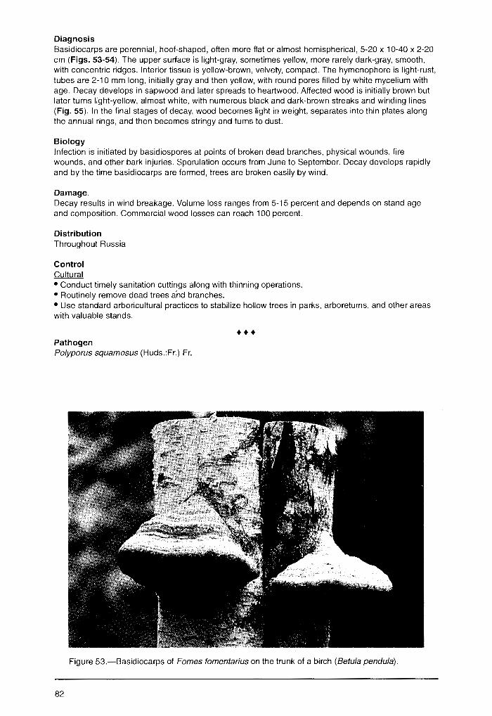
Diagnosis Basidiocarps are perennial, hoof-shaped, often more flat or almost hemispherical, 5-20 x 10-40 x 2-20 cm (Figs. 53-54). The upper surface is light-gray, sometimes yellow, more rarely dark-gray, smooth, with concentric ridges. Interior tissue is yellow-brown, velvety, compact. The hymenophore is light--rust, tubes are 2-10 mm long, initially gray and then yellow, with round pores filled by white mycelium with age. Decay develops in sapwood and later spreads to heartwood. Affected wood is initially brown but later turns light-yellow, almost white, with numerous black and dark-brown streaks and winding lines (Fig. 55). In the final stages of decay, wood becomes light in weight, separates into thin plates along the annual rings, and then becomes stringy and turns to dust.
Biology Infection is initiated by basidiospores at points of broken dead branches, physical wounds, fire wounds, and other bark injuries. Sporulation occurs from June to September. Decay develops rapidly and by the time basidiocarps are formed, trees are broken easily by wind.
Damage. Decay results in wind breakage. Volume loss ranges from 5-15 percent and depends on stand age and composition. Commercial wood losses can reach 100 percent.
Distribution Throughout Russia
Control Cultural • Conduct timely sanitation cuttings along with thinning operations. • Routinely remove dead trees a'nd branches. • Use standard arboricultural practices to stabilize hollow trees in parks, arboretums, and other areas with valuable stands.
• •• Pathogen Polyporus squamosus (Huds.:Fr.) Fr.
Figure 53.-Basidiocarps of Fames fomentarius on the trunk of a birch (Betula pendula).
82

Figure 54.-Basidiocarps of Fames fomentarius on the trunk of a fir (Abies sibirica).
Hosts Primarily species of maple (Acer), birch (Betula), poplar (Populus), oak (Quercus), elm (Ulmus), and beech (Fagus)
Diagnosis Basidiocarps are annual, 5-50 cm in diameter and 5-10 cm thick, with slightly bent margins, usually on a lateral stalk. The upper surface is slightly depressed, yellow, with large brown scales. The stalk is stout, brown, with a black base. Fresh basidiocarp tissue is white and soft, but after drying turns yellow and becomes brittle. The hymenophore is tubular; tubes are about 1-2 mm long, with large angular pores. Decay occurs mainly in heartwood but can spread to sapwood. It usually develops in the lower part of the trunk but can develop in the roots. In the advanced stage of decay, wood becomes white, often with dark lines, with numerous cracks. In the final stage, affected wood separates into plates and crumbles easily.
Biology Infection is initiated by basidiospores from basidiocarps that form in midsummer, particularly during years with heavy precipitation. Sporulation also occurs at this time. Spores penetrate tree tissues through injuries such as frost cracks and mechanical wounds. The disease affects older forest trees and is widespread in urban forests.
Damage Affected trees gradually weaken but few die. Affected trees in stands rarely exceed 5 percent as the fungus mostly decays wood in timber yards.
Distribution European part of Russia, Urals, Far East
Control Cultural • Conduct sanitation cuttings to remove weakened trees. • Minimize mechanical injuries of trunks during cuttings. • Remove basidiocarps from trees in parks and street plantings before sporulation.
83
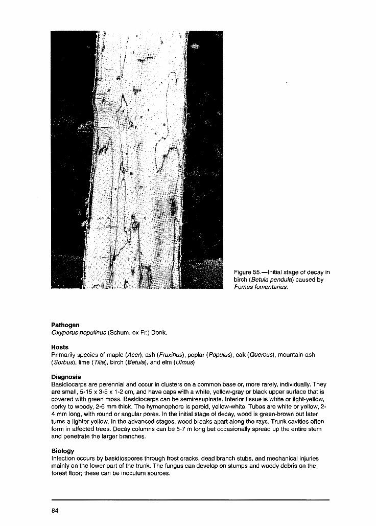
Pathogen Oxyporus popu/inus (Schum. ex Fr.) Donk.
Hosts
Figure 55.-lnitial stage of decay in birch (Betula pendula) caused by Fornes fomentarius.
Primarily species of maple (Ace!), ash (Fraxinus), poplar (Populus), oak (Quercus), mountain-ash (Sorbus), lime (Tilia), birch (Betula), and elm (Ulmus)
Diagnosis Basidiocarps are perennial and occur in clusters on a common base or, more rarely, individually. They are small, 5-15 x 3-5 x 1-2 cm, and have caps with a white, yellow-gray or black upper surface that is covered with green moss. Basidiocarps can be semiresupinate. Interior tissue is white or light-yellow, corky to woody, 2-6 mm thick. The hymenophore is poroid, yellow-white. Tubes are white or yellow, 2-4 mm long, with round or angular pores. In the initial stage of decay, wood is green-brown but later turns a lighter yellow. In the advanced stages, wood breaks apart along the rays. Trunk cavities often form in affected trees. Decay columns can be 5-7 m long but occasionally spread up the entire stem and penetrate the larger branches.
Biology Infection occurs by basidiospores through frost cracks, dead branch stubs, and mechanical injuries mainly on the lower part of the trunk. The fungus can develop on stumps and woody debris on the forest floor; these can be inoculum sources.
84
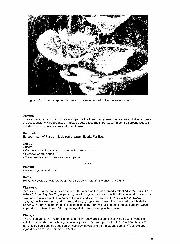
Figure 56.-Basidiocarps of Daeda/ea quercina on an oak (Quercus robw') stump.
Damage Trees are affected in the middle or lower part of the trunk; decay results in cavities and affected trees are susceptible to wind breakage. Infected trees, especially in parks, can reach 80 percent. Decay in the stem base causes commercial wood losses.
Distribution European part of Russia, middle part of Urals, Siberia, Far East
Control Cultural • Conduct sanitation cuttings to remove infected trees. • Remove woody debris. • Treat tree cavities in parks and forest parks .
••• Pathogen Daeda/ea quercina (L.) Fr.
Hosts Primarily species of oak (Quercus) but also beech (Fagus) and chestnut (Castanea)
Diagnosis Basidiocarps are perennial, with flat caps, thickened on the base, broadly attached to the trunk, 4-12 x 6-20 x 2-5 cm (Fig. 56). The upper surface is light-brown or gray, smooth, with concentric zones. The hymenophore is labyrinth-like. Interior tissue is corky when young but woody with age. Decay develops in the lower part of the trunk and spreads upwards at least 3 m. Decayed wood is darkbrown with a gray shade. In the final stages of decay, narrow cracks form along rays and the wood separates into thin plates. Yellow-gray mycelial sheets develop in the cracks.
Biology The fungus primarily invades stumps and freshly cut wood but can infect living trees. Infection is initiated by basidiospores through various injuries in the lower part of trunk. Sprouts can be infected not only by basidiospores but also by mycelium developing on the parent stumps. Weak, old and injured trees are most commonly affected.
85
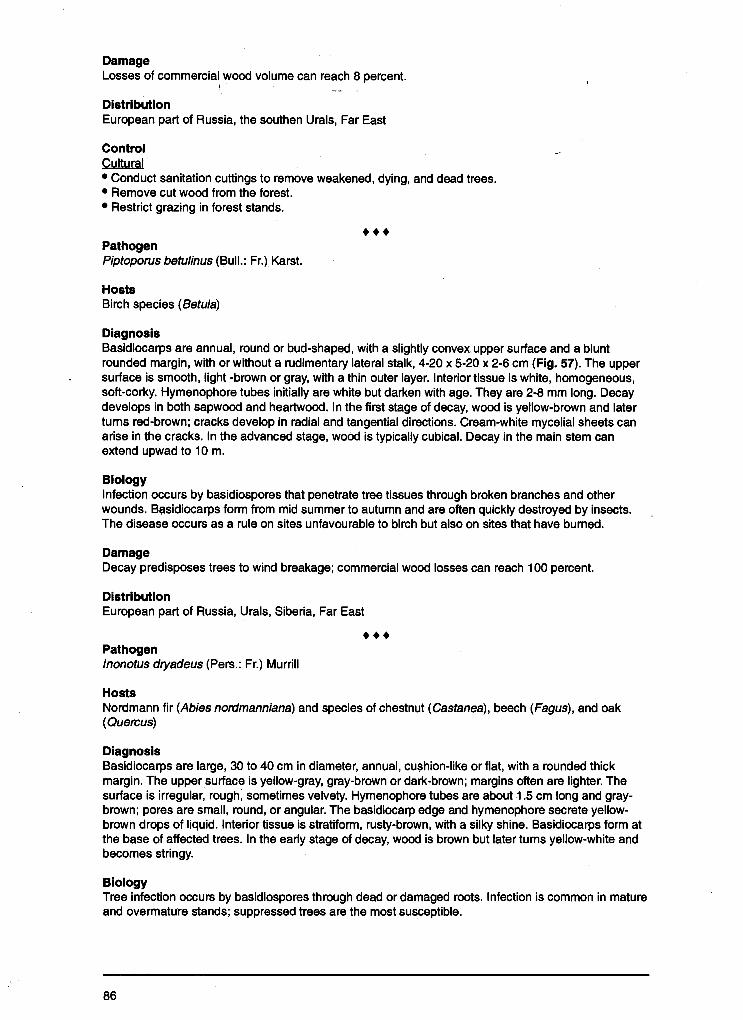
Damage Losses of commercial wood volume can reach 8 percent.
!
Distribution European part of Russia, the southen Urals, Far East
Control Cultural • Conduct sanitation cuttings to remove weakened, dying, and dead trees. • Remove cut wood from the forest. • Restrict grazing in forest stands.
• •• Pathogen Piptoporus betulinus (Bull.: Fr.) Karst.
Hosts Birch species (Betula)
Diagnosis Basidiocarps are annual, round or bud-shaped, with a slightly convex upper surface and a blunt rounded margin, with or without a rudimentary lateral stalk, 4-20 x 5-20 x 2-6 cm (Fig. 57). The upper surface is smooth, light -brown or gray, with a thin outer layer. Interior tissue is white, homogeneous, soft-corky. Hymenophore tubes initially are white but darken with age. They are 2-8 mm long. Decay develops in both sapwood and heartwood. In the first stage of decay, wood is yellow-brown and later turns red-brown; cracks develop in radial and tangential directions. Cream-white mycelial sheets can arise in the cracks. In the advanced stage, wood is typically cubical. Decay in the main stem can extend upwad to 1 O m.
Biology Infection occurs by basidiospores that penetrate tree tissues through broken branches and other wounds. Basidiocarps form from mid summer to autumn and are often quickly destroyed by insects. The disease occurs as a rule on sites unfavourable to birch but also on sites that have burned.
Damage Decay predisposes trees to wind breakage; commercial wood losses can reach 100 percent.
Distribution European part of Russia, Urals, Siberia, Far East
••• Pathogen lnonotus dryadeus (Pers.: Fr.) Murrill
Hosts Nordmann fir (Abies nordmanniana) and species of chestnut (Castanea), beech (Fagus), and oak (Quercus)
Diagnosis Basidiocarps are large, 30 to 40 cm in diameter, annual, cushion-like or flat, with a rounded thick margin. The upper surface is yellow-gray, gray-brown or dark-brown; margins often are lighter. The surface is irregular, rough, sometimes velvety. Hymenophore tubes are about 1.5 cm long and graybrown; pores are small, round, or angular. The basidiocarp edge and hymenophore secrete yellowbrown drops of liquid. Interior tissue is stratiform, rusty-brown, with a silky shine. Basidiocarps form at the base of affected trees. In the early stage of decay, wood is brown but later turns yellow-white and becomes stringy.
Biology Tree infection occurs by basidiospores through dead or damaged roots. Infection is common in mature and overmature stands; suppressed trees are the most susceptible.
86
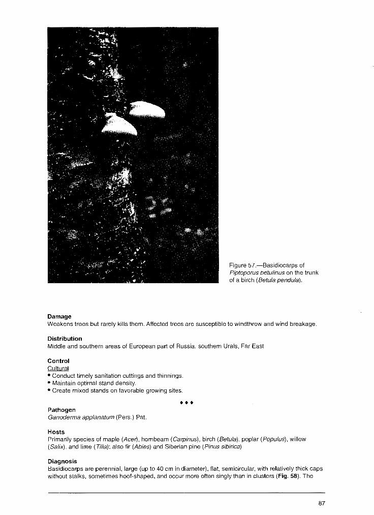
Damage
Figure 57.-Basidiocarps of Piptoporus betulinus on the trunk of a birch (Betula pendu/a).
Weakens trees but rarely kills them. Affected trees are susceptible to windthrow and wind breakage.
Distribution Middle and southern areas of European part of Russia, southern Urals, Far East
Control Cultural • Conduct timely sanitation cuttings and thinnings. • Maintain optimal stand density. • Create mixed stands on favorable growing sites .
••• Pathogen Ganoderma applanatum (Pers.) Pat.
Hosts Primarily species of maple (Acer), hornbeam (Carpinus), birch (Betula), poplar (Popu/us), willow (Salix), and lime (Tilia); also fir (Abies) and Siberian pine (Pinus sibirica)
Diagnosis Basidiocarps are perennial, large (up to 40 cm in diameter), flat, semicircular, with relatively thick caps without stalks, sometimes hoof-shaped, and occur more often singly than in clusters (Fig. 58). The
87

Figure 58.-Basidiocarps of Ganoderma app/anatum on the trunk of an aspen (Populus tremu/a).
upper surface is dull, chocolate-brown, sometimes gray-brown, usually irregular, rough or with concentric ridges, thin, and hard. Interior tissue is corky or velvety, brown, usually soft. The hymenophore, about 0.5-1.5 cm thick, is white initially but then turns brown. Decay begins in the heartwood and is light-yellow, with elongate pockets filled by white mycelium; later, the decay spreads to sapwood. In the final stages, wood turns white and becomes fibrous; tree cavities are common.
Biology The fungus is found primarily on stumps and dead trees but can attack living trees. Trees can be infected through wounds to the roots and base of the trunk. Mycelium spreads in roots and the butt log. Infection occurs predominantly in mixed conifer-deciduous forests.
Damage Causes susceptibility to windthrow and wind breakage. The disease is serious in urban and forest parks and urban plantations.
Distribution European part of Russia, Urals, Far East
Control Cultural • Conduct timely sanitation cuttings to remove weakened, dying, and dead trees. • Treat tree cavities in urban and park settings.
88
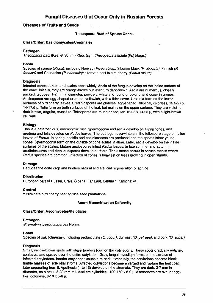
Fungal Diseases that Occur Only in Russian Forests
Diseases Of Fruits;and Seeds
Thecopsora Rust of Spruce Cones
Class/Order: Basidiomycetes/Uredinales
Pathogen Thecopsora padi (Kze. et Schm.) Kleb. (syn. Thecopsora aleolata (Fr.) Magn.)
Hosts Species of spruce (Picea), including Norway (Picea abies,) Siberian black (P. obovata), Finnish (P. fennica) and Caucasian (P. orientalis); alternate host is bird cherry (Padus avium)
Diagnosis Infected cones darken and scales open widely. Aecia of the fungus develop on the inside surface of the cone. Initially, they are orange-brown but later turn dark-brown. Aecia are numerous, closely packed, globose, 1-2 mm in diameter, powdery, white and round or oblong, and occur in groups. Aeciospores are egg-shaped or round, yellowish, with a thick cover. Uredinia form on the lower surfaces of bird cherry leaves. Urediniospores are globose, egg-shaped, elliptical, colorless, 15.5-27 x 14-17.5 µ. Telia form on both surfaces of the leaf, but mainly on the upper surface. They are violet- or dark-brown, angular, crust-like. Teliospores are round or angular, 16-23 x 14-25 µ, with a light-brown cell wall.
Biology This is a heteroecious, macrocyclic rust. Spermogonia and aecia develop on Picea cones, and uredinia and telia develop on Padus leaves. The pathogen overwinters in the teliospore stage on fallen leaves of Padus. In spring, basidia and basidiospores are produced and the spores infect young cones. Spermogonia form on the outside of cone scales in June. Later, aecia develop on the inside surfaces of the scales. Mature aeciospores infect Padus leaves. In late summer and autumn, urediniospores and then teliospores develop on them. The disease occurs in spruce stands where Padus species are common. Infection of cones is heaviest on trees growing in open stands.
Damage Reduces the cone crop and hinders natural and artificial regeneration of spruce.
Distribution European part of Russia, Urals, Siberia, Far East, Sakhalin, Kamchatka
Control • Eliminate bird cherry near spruce seed plantations.
Acorn Mummification Deformity
Class/Order: Ascomycetes/Helotiales
Pathogen Stromatinia pseudotuberosa Rehm.
Hosts Species of oak (Quercus), including pedunculate (Q. robur), durmast (Q. petraea), and cork (Q. suber)
Diagnosis Small, yellow-brown spots with sharp borders form on the cotyledons. These spots gradually enlarge, coalesce, and spread over the entire cotyledon. Gray, fungal mycelium forms on the surface of infected cotyledons. Interior cotyledon tissues turn dark. Eventually, the cotyledons become black, friable masses of sclerotial stroma. Affected cotyledons become enlarged and rupture the fruit coat, later separating from it. Apothecia (1 to 15) develop on the stromata. They are dark, 2-7 mm in diameter, on a stalk, 3-30 mm tall. Asci are cylindrical, 100-150 x 6-9 µ. Ascospores are oval or egglike, colorless, 8-10 x 5-6 µ.
89
l Biology The fungus develops on acorns in natural forest environments as well as on acorns that are stored. In forest stands, acorn infection occurs by ascospores that are disseminated over most of the growing season. A portion of the acorn crop is infected on tne tree, but most infection occurs after acorn drop. When healthy acorns contact previous-year acorns that are diseased, infection occurs by means of mycelium that penetrates acorn tissues through splits in the coat or through tracheal scars where the acorn was attached to the cup. In the forest, apothecia grow from the infected acorns the following year, but the sexual ascospore stage rarely forms. When humidity is high, the conidial stage develops on the uncovered stromata and conidia also can infect acorns. Disease development is most active during acorn storage, especially under improper storage conditions (high humidity-high temperatures). Mycelium develops on affected acorns and spreads rapidly to healthy ones.
Damage Reduces acorn quality. Affected acorns lose germination power partially or completely. In storage, damage can reach 30 percent.
Distribution European part of Russia, southern Urals, Far East
Control Cultural • Harvest acorns shortly after the shedding period. • Collect acorns only from healthy and superior trees. • Avoid mechanical injury during acorn harvesting and transportation. • Dry acorns before storage; maintain temperatures below 5°C and good ventilation. • Periodically sample stored acorns for presence of disease.
Chemical • Apply fungicides (dust) before acorn storage.
Class/Order: Deuteromycetes/Melanconiales
Pathogen Gloeosporium aucuparia Henn.
Host Species of mountain-ash (Sorbus)
Diagnosis Dark-brown or nearly black round spots form on the berries. Sporodochia are aggregated and resemble small, black cushions. Conidia are clavate or cylindrical, colorless, 7-14 x 3.5-4 µ.
Distribution European part of Russia
Class/Order: Deuteromycetes-Hyphomycetales
Pathogen Monilia linhartiana Sacc. (syn. Ovularia necans Pass.)
Host Bird cherry (Padus avium)
Diagnosis Initially small, later coalesced spots, 7-14 x 3.5-4 mm, form on the surface of berries. Gray, cushionlike conidiophores form on the spots in summer. In autumn, mycelium penetrates into the interior tissues; the berries turn black, become wrinkled, and fall or remain on the tree. In spring, apothecia, 3-7 mm in diameter on 2 cm stalks, form on infected berries. Conidia are spherical or lemon-like, 12-18 x 8-15 µ, and occur in chains that are colorless to gray in mass. Asci are cylindrically clavate, 200-260 x 17-19 µ. Ascospores are ellipsoid or egg-shaped, colorless, 17-18 x 9.5 µ.
90
Contents
-~ .. . Introduction .......................................... ':: ................................................................................................. , .... 1
Most Common Fungal Diseases of Russian Forests ..................................................................... 2
Diseases of Fruits and Seeds ...................................................................... -:: ............................ 2 Birch Seed Mummification ...................................................................................................................... 2 Fruit Deformation .................................................................................................................................... 2 Fruit Spots .............................................................................................................................................. 4 Molds ...................................................................................................................................................... 6 Seed and Fruit Rots ............................................................................................................................... 9
Diseases of Needles and Leaves ............................................................................................ 11 Needle Diseases .................................................................................................................................. 11
Lophodermium Needle Casts .......................................................................................................... 11 Snow Blight ...................................................................................................................................... 15 Meria Needle Blight ......................................................................................................................... 16 Brown Felt Blight ............................................................................................................................. 18 Rhizosphaera Needle Casts ............................................................................................................ 18 Needle Rusts of Pine ....................................................................................................................... 20
Leaf Diseases ....................................................................................................................................... 22 Powdery Mildews ............................................................................................................................. 22 Leaf Spots ...................................................... : .. .............................................................................. 23 Leaf Rusts ....................................................................................................................................... 35
Diseases Of Roots, Stems, And Branches ............................................................................... 36 Diseases in Tree Nurseries and Young Forests ................................................................................... 36
Damping - off ................................................................................................................................... 36 Diseases of Forest Stands ................................................................................................................... 38
Wilts ................................................................................................................................................. 38 Verticillium Wilt ............................................................................................................................ 38 Dutch Elm Disease ..................................................................................................................... 39 Oak Wilt ......................................................................... · ............................................................. 41
Dieback and Canker Diseases ........................................................................................................ 42 Cenangium Dieback of Pine ....................................................................................................... 42 Nectria Canker and Dieback ....................................................................................................... 44 Cytospora Canker ....................................................................................................................... 45 Dothichiza Canker of Poplar ....................................................................................................... 46 Clithris Canker and Dieback of Oaks .......................................................................................... 48 Nummularia Canker .................................................................................................................... 48 Black Naemospora Canker ......................................................................................................... 49 Thyrostroma Canker and Dieback .............................................................................................. 50 Ascocalyx Scleroderris Shoot Canker ........................................................................................ 52 European Larch Canker .............................................................................................................. 53 Lachnellula Canker of Siberian Pine Understory ........................................................................ 54 Stem Rust of Pine ....................................................................................................................... 55 Broom Rust of Fir ........................................................................................................................ 56 Perennial Nectria Canker ............................................................................................................ 58 Black Hypoxylon Canker ............................................................................................................. 60 Cytophoma Canker of Ash .......................................................................................................... 60
Wood-Decaying Diseases ............................................................................................................... 62 Annosum Root and Butt Rot ....................................................................................................... 62 Armillaria Root Rot ...................................................................................................................... 65 Butt and Trunk Rot of Conifers ................................................................................................... 67 Trunk and Limb Rot of Hardwoods ............................................................................................. 75
Fungal Dise~ses that Occur Only in Russian Forests ................................................................. 89
Diseases of Fruits and Seeds .......... .' ....................................................................................... 89 Thecopsora Rust of Spruce Cones ...................................................................................................... 89 Acorn Mummification Deformity ........................................................................................................... 89
Diseases of Needles and Leaves ....................................................................... '..:-................... 91 Needle Diseases .................................................................................................................................. 91
Hypodermella Needle Cast of Pine Hosts ....................................................................................... 91 . Chrysomyxa Rust of Spruce ............................................................................................................ 91
Leaf Diseases ....................................................................................................................................... 93 Powdery Mildew of Siberian Pea Tree ............................................................................................. 93 Other Powdery Mildews ................................................................................................................... 94 Orange Leaf Spot of Padus ............................................................................................................. 95 Red Spot of Ussurian Plum ............................................................................................................. 96 Foliage Anthracnoses, Spots, and Blights ....................................................................................... 96 Leaf Rusts ..................................................................................................................................... 108 Taphrina Diseases: Leaf Blisters, Leaf and Shoot Deformation .................................................... 110
Diseases of Roots, Stems, and Branches ............................................................................... 111 Diseases in Tree Nurseries and Young Forests ................................................................................. 111
"Infectious Damping'.' of Coniferous Seedlings .............................................................................. 111 Sclerophoma Disease of Pine Shoots ........................................................................................... 112 Pine Shoot Rust ............................................................................................................................. 113 Chrysomyxa Rust of Spruce Shoots and Needles ........................................................................ 114
Diseases of Forest Stands .... , ............................................................................................................ 116 Dieback and Canker Diseases ...................................................................................................... 116
Black Cytospora Canker of Poplar ............................................................................................. 116 Biatorella Canker ...................................................................................................................... 117 Pitch Blister Rust Canker .......................................................................................................... 119 Endoxylina Canker of Ash ......................................................................................................... 120 Cankers and Diebacks .............................................................................................................. 122
Wood-Decaying Diseases ............................................................................................................. 128 Ganoderma Butt Rot of Beech .................................................................................................. 129 Vuillemenia Decay .................................................................................................................... 129 Trunk and Limb Rots ................................................................................................................. 130
Acknowledgment ............................................................................................................ 133
Appendix A Pathogens That Affect Trees and Shrubs in Russia ....................................................................... 134
Appendix B Host Trees, Shrubs, and Herbs Listed in this Report ..................................................................... 137
Distribution European part of Russia
I
Diseases of Needles and Leaves
Needle Diseases
Hypodermella Needle Cast of Pine Hosts
Class/Order: Ascomycetes/Phacidiales
Pathogen Hypodermella sulcigena Tub. (anamorph: Hendersonia acicola Munch. et. Tub.)
Host Species of pine (Pinus), including Scots (Pinus silvestris) and Siberian (P. sibirica)
Diagnosis In summer, the tips of infected needles turn yellow. A brown band, 2-3 mm wide, separates infected from healthy tissue on the needle. Infected needles ·remain on the trees most of the growing season. Pycnidia form during the summer. On the dead portion of needles, black pycnidia about 140 µ in diameter form. Gray conidia, 3-celled, oval-cylindrical, 11-15 x 4-5 µ, develop in pycnidia. The sexual stage (ascospores) of the pathogen develops the next spring. Apothecia are convex, black, elongate. Asci are cylindrical, 90-100 µ long. Ascospores are clavate, with a thick, jellied enclosure, 42-60 x 12-16 µ.
Biology Ascospores infect pine needles in spring. Ascospore maturation and release, and needle infection occurs during spring and summer when the humidity is high. Temperature is not a critical factor for pathogen development. Trees on borders of plantations, along forest edges, and in understory of open forests are particularly susceptible.
Damage Repeated crown infection causes considerable crown loss that both weakens and reduces the height growth of understory and forest plantation trees less than 5 years old.
Distribution European part of Russia, Urals, Siberia
Control • Apply Bordeaux mixture when annual infection is repeated.
Chrysomyxa Rust of Spruce
Class/Order: Basidiomycetes, Uredinales
Pathogen Chrysomyxa abietis (Wallr.) Unger.
Host Norway spruce (Picea abies)
Diagnosis Cushion-like bright-orange telia, 2-6 mm long, form along the midribs of the needles. Chains of teliospores mature in the telia. Teliospores are oblong, 1-celled, reddish-orange, 18.8-28 x 10.5-17.8 µ.
Biology This microcylic rust has only the telio- and basidiospore stages. No alternate host is known or required for infection of spruce needles. Talia are produced on needles in early summer. They overwinter, germinate in the spring, and produce basidia with basidiospores that infect the newly formed needles. Mycelium penetrates epidermis cells, where telia develop. Damaged needles with telia fall during the next year and basidiospores are formed. The disease occurs mostly in overstocked plantations and on
91
understory trees. Disease development depends primarily on high humidity; the pathogen develops under moderate temperatures. The lower portion of the crown usually is affected but the entire crown can be damaged. The severity of current-year infection indicates the inoculum potential for'disease development the following year.
Damage Causes withering and premature drop of needles on understory and young trees in pl_?ntations, occasionally resulting in death.
Distribution European part of Russia, western Siberia, Far East
Control Chemical • Bordeaux mixture can be used in late spring in heavily infected stands or plantations.
Class/Order: Deuteromycetes/Sphaeropsidales
Pathogen Ceutospora abietina Delacr.
Hosts Norway spruce (Picea abies)
Diagnosis Initially, single brown spots form on needles; later, they coalesce. Stromata form in the spots and are black-olive, oval-oblong, about 0.6 mm in diameter. Stroma have 3 compartments with a common ostiole. Conidia are cylindrical, with rounded tops; they are colorless, 6 x 2.5 µ, and contain 2 oil drops.
Distribution European part of Russia
Class/Order: Basidomycetes/Uredinales
Pathogen Coleosporium pini-pumila Azb.
Host Mountain pine (Pinus pumila)
Diagnosis Yellow spots appear on the both surfaces of the needle. Later, convex, oblong, orange telia form. Teliospores are 1-celled, 45-152 x 20-37.5 µ.
Distribution Urals, Siberia, Far East
••• Pathogen Me/ampsorella symphiti (DC) Bub.
Host Silver fir (Abies alba) (Fig. 59) (alternate host: Symphitum officianale, and S.cordatum)
Diagnosis Spermagonia and aecia develop on the lower surface of needles. Blister-shaped aecia are covered by the peridium. Uredinia are tiny, orange, and develop on the undersides of leaves of the alternate host, Symphitum spp. Telia develop subepidermally on these leaves. Aeciospores are globose, rarely eggshaped, elliptical, with orange contents, a warty wall, 20-40 x 13-29 µ. Urediniospores are globose,
92
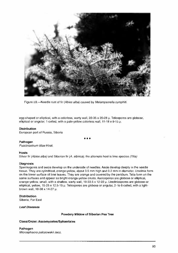
Figure 59.-Needle rust of fir (Abies alba) caused by Melampsore/la symphiti.
egg-shaped or elliptical, with a colorless, warty wall, 22-35 x 20-28 µ. Teliospores are globose, elliptical or angular, 1-celled, with a pale-yellow colorless wall, 11-18 x 9-15 µ.
Distribution European part of Russia, Siberia
••• Pathogen Pucciniastrum ti/iae Hirat.
Hosts Silver fir (Abies alba) and Siberian fir (A. sibirica); the alternate host is lime species (Tilia)
Diagnosis Spermogonia and aecia develop on the underside of needles. Aecia develop deeply in the needle tissue. They are cylindrical, orange-yellow, about 3.5 mm high and 0.2 mm in diameter. Uredinia form on the lower surface of lime leaves. They are orange and covered by the peridium. Telia form on the same surfaces and appear as bright-orange-yellow crusts. Aeciospores are globose or elliptical, orange-yellow, small, with a shallow, warty wall, 19-33.5 x 12-22 µ. Urediniospores are globose or elliptical, yellow, 15-25 x 12.5-15 µ. Teliospores are globose or angular, 2- to 6-celled, with a lightbrown wall, 18-38 x 14-27 µ.
Distribution Siberia, Far East
Leaf Diseases
Powdery Mildew of Siberian Pea Tree
Class/Order: Ascomycetes/Sphaeriales
Pathogen Microsphaera palczewskii Jacz.
93
Host Siberian pea tree ( Caragana arborescens)
' ' Diagnosis White, cobwebby mycelium forms on both surfaces of leaves in early June. Mycelium and conidia develop on the foliage anq become felty and prominent. In late June, numerous cleistothecia form in the mycelial mat and C9"8r the leaf surface. Infected foliage is a gray to blue at this time. Cleistothecia are dark-brown, nearly black, spherical, 90-120 µ in diameter, with oblong, hexangular cells in the peridium. Cleistothecia have 5-10 appendages that are colorless, without partitions, 80-147 x 5-9 µ, with dichotomous.branches on the tips. Four to eight mace-shaped asci form on short pedicles, 47-55 x 8-12 µ. Ascospores are elliptical or egg-shaped, on short pedicles, 14-21 x 8-12 µ.
Biology Asci form in the cleistothecia in summer and overwinter on fallen infected leaves. Asci mature early the following summer. Mature ascospores infect new leaves in the crown. In 2 weeks, superficial mycelium appears on the leaves; 7-10 days later, conidia form that cause secondary infections of young leaves on the middle and upper crown. In late June, the first cleistothecia are produced on the mycelial mats. By late July, numerous cleistothecia are formed. High humidity is the most important factor in disease development. Development occurs at moderate (18°-20°C), or higher (25°-30°C) temperatures. Environmental factors,that delay leaf maturation increase susceptibility to powdery mildew.
Damage Causes complete loss of aesthetic appearance of trees. Damaged leaves wither and drop prematurely. The plants are weakened and lose resistance to low temperatures.
Distribution Middle regions of European part of Russia, Siberia, Far East
Control Observation • Survey for the disease in early June when the mycelium develops.
Chemical • Apply colloid sulfur or BAYMEB when the white tender superficial mycelium appears on leaves. Apply 3-4 treatments at intervals of 10-12 days.
Other Powdery Mildews
Class/Order: Ascomycetes, Sphaeriales
Pathogen Microsphaera divaricata Lev.
Host Buckthorn (Frangula a/nus)
Diagnosis Mycelium forms on the surfaces of leaves, often cobwebby, sometimes in several spots. Cleistothecia occur in groups or individually and have 7-16 appendages, dichotomously branched, 135-330 x 6-9 µ. Asci have short pedicles, 48-60 x 25-40 µ. Asci contain 4-6 ascospores, 18-24 x 9-13.5 µ.
Distribution European part of Russia
••• Pathogen Uncinula clandestina Schroet.
Hosts Species of elm ( Ulmus)
94
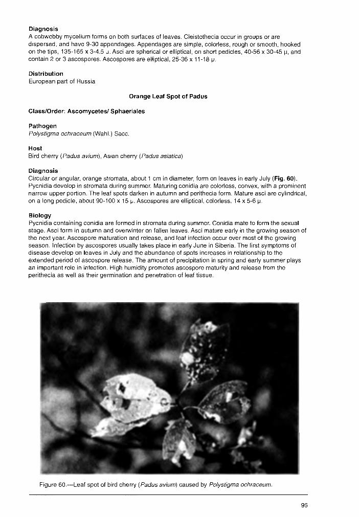
Diagnosis A cobwebby mycelium forms on both surfaces of leaves. Cleistothecia occur in groups or are dispersed, and have 9-30 appendages. Appendages are simple, colorless, rough or smooth, hooked on the tips, 135-165 x 3-4.5 µ. Asci are spherical or elliptical, on short pedicles, 40-56 x 30-45 µ, and contain 2 or 3 ascospores. Ascospores are elliptical, 25-36 x 11-18 µ.
Distribution European part of Russia
Orange Leaf Spot of Padus
Class/Order: Ascomycetes/ Sphaeriales
Pathogen Polystigma ochraceum (Wahl.) Sacc.
Host Bird cherry (Padus avium), Asian cherry (Padus asiatica)
Diagnosis Circular or angular, orange stromata, about 1 cm in diameter, form on leaves in early July (Fig. 60). Pycnidia develop in stromata during summer. Maturing conidia are colorless, convex, with a prominent narrow upper portion. The leaf spots darken in autumn and perithecia form. Mature asci are cylindrical, on a long pedicle, about 90-100 x 15 µ. Ascospores are elliptical, colorless, 14 x 5-6 µ.
Biology Pycnidia containing conidia are formed in stromata during summer. Conidia mate to form the sexual stage. Asci form in autumn and overwinter on fallen leaves. Asci mature early in the growing season of the next year. Ascospore maturation and release, and leaf infection occur over most of the growing season. Infection by ascospores usually takes place in early June in Siberia. The first symptoms of disease develop on leaves in July and the abundance of spots increases in relationship to the extended period of ascospore release. The amount of precipitation in spring and early summer plays an important role in infection. High humidity promotes ascospore maturity and release from the perithecia as well as their germination and penetration of leaf tissue.
Figure 60.-Leaf spot of bird cherry (Padus avium) caused by Polystigma ochraceum.
95
Damage Causes premature withering and dropping of leaves, reduced fruit production in the current year, and partial or complete loss of fruit in the next year.
Distribution Siberia, rarely in European part of Russia
Red Spot of Ussurian Plum
Class/Order: Ascomycetes/Sphaeriales
Pathogen Polystigma ussuriensis (Natal. et Jacz.) A. Proz.
Host Ussurian plum (Prunus ussuriensis)
Diagnosis Small, circular spots, 2-8 mm in diameter, develop on infected leaves. They are pale-yellow initially and later bright-orange. Lower surfaces of spots are convex and covered with numerous, dark dots. These dots are the pores at the top of stromata containing pycnidia. As they age, the spots turn brown and on lower surfaces of leaves, nearly black. Pycnidia are 200-300 x 150-210 µ. Conidia are colorless, thread-like, 15 x 1 µ. Asci are narrow, mace-shaped, 85-90 x 10-11. Ascospores are colorless, elliptical, 8-11 x 3-5 µ.
Biology The pathogen overwinters on fallen leaves in the sexual stage. Ascospores mature in perithecia in late May or early June. Ascospores are disseminated by wind and infect leaves. The first spots appear on leaves in late June and continue to increase; this is related to the long period of pathogen sporulation. Conidia are not involved in foliage infection but mate to form perithecia in autumn. Asci and ascospores do not mature until the next year. The amount of precipitation at the beginning of the growing season influences maturation and release of ascospores and subsequent foliage infection.
Damage Ca~es premature foliage damage and reduces fruit yield. Successive infections weaken or kill the tree.
Distribution Far East, Primorski and Khabarovski regions of Russia
Control Cultural • Collect and burn fallen foliage to eliminate the inoculum source of the overwintering pathogen.
Chemical • Spray three applications of Bordeaux mixture: 1) during.the period of foliage expansion; 2) immediately after flowering; and 3) 10-12 days after the second application when humidity is high at the beginning of the growing season. • Spray with Bordeaux mixture in autumn (before leaf drop) to reduce overwintering inoculum.
Foliage Anthracnoses, Spots, and Blights
Class/Order: Deuteromycetes, Sphaeropsidales
Pathogen Ascochyta borjomi Bond.
Host Siberian pea tree ( Caragana arborescens)
96
Diagnosis Leaf spots are round, 476 mm in diameter, whiteish, surrounded by brown margins. Pycnidia f,orm on the upper surlaces of spots as small, black dots. Conidia are cylindrical, with rounded ends, straight or curved, usually 1-, or 2-celled, colorless, 8-10.5 x 4.5-5 µ.
Distribution Southern area of European part of Russia
••• Pathogen Ascochyta crataegi Fckl.
Hosts Species of hawthorne ( Crataegus)
Diagnosis Spots form on the upper surfaces of leaves, often along the margin. They are round or angular, about 6 mm in diameter, brown, with dark borders. Pycnidia form in these spots as brown dots. Conidia are oblong cylindrical, with rounded ends, 2-celled, colorless, 8-12 x 2-3 µ.
Distribution Southern area of European part of Russia
••• Pathogen Ascochyta elaeagni Sacc.
Hosts Species of oleaster (Elaeagnus)
Diagnosis Spots usually form near the leaf margins and are irregular, creamy, with a narrow brown border. Pycnidia form on upper surfaces of spots as small black dots. Conidia are spindle-like, 2-celled, lightolive, 8-10 x 3.5-4 µ. Mycelium forms on both surfaces of leaves. It has a cobwebby appearance. Cleistothecia occur in groups or are dispersed, with 9-30 appendages that are simple, colorless, rough or smooth, hooked on the tips, 135-165 x 3-4.5 µ. Asci are spherical or widely elliptical, on a short pedicle, 40-56 x 30-45 µ. Ascospores are elliptical, 25-36 x 11-18 µ.
Distribution European part of Russia
••• Pathogen Ascochyta pirico/a Sacc.
Hosts Species of apple (Ma/us) and pear (Pyrus)
Diagnosis Leaf spots are bright-gray-white. Pycnidia form on upper surfaces of spots as small, black dots. Conidia are oblong, olive, 10 x 2 µ.
Distribution European part of Russia
••• Pathogen Ascochyta populina Sacc.
Host White poplar (Populus alba)
97
Diagnosis Leaf spots are brown and vary in size. Pycnidia form on upper surfaces of spots as small brown dots. Conidia are cylindrical, 2-celled, colorless, 8"'1 O x 2 µ. '
Distribution Southwestern area of European part of Russia
••• Pathogen Ascochyts ribesis Sacc. et Fautr.
Host Species of currant (Ribes)
Diagnosis Leaf spots are small, white or pale-brown, with dark borders. Pycnidia form on the upper surfaces of spots as brown dots. Conidia are cylindrical, with rounded ends, 8-12 x 3 m.
Distribution European part of Russia
••• Pathogen Ascochyts ssrmentics Sacc.
Host Honeysuckle (Lonicers tstsrics)
Diagnosis Leaf spots are white with dark borders. Pycnidia form on upper surfaces of spots as dark dots. Conidia are elongate-oval, widened at the top, 2-celled, colorless or smoke-gray, 20-25 x 8 µ.
Distribution Western area of European part of Russia
••• Pathogen Coniothyrium sslicicols Rossi.
Hosts Species of willow (Salix)
Diagnosis Leaf spots are yellow-brown, sometimes with black borders; in dry conditions they are gray-white. Pycnidia form on upper surfaces of spots as black dots. Conidia are egg-shaped, brown, 5-6.5 x 3-3.5 µ.
Distribution European part of Russia
Class/Order: Deuteromycetes/Hyphomycetales
Pathogen Cercospors psdi Bub. et Serebr.
Host Bird cherry (Psdus svium)
Diagnosis Leaf spots are small, irregular, sometimes coalesced, white on upper surfaces and brown on the lower, about 0.5-3 mm in diameter. Conidia are spindle-shaped, cylindrical or elongate and wide at the top, slightly curved, 2- or more-celled, pale-olive, 20-70 x 4-6.5 µ.
98
Distribution European part of Russia, Western Siberia, Far East
••• Pathogen Coniothecium phyllophilum Desm.
Hosts Species of hawthorne ( Crataegus)
Diagnosis Leaf spots are black and small; they occur on both surfaces of leaves. Conidia are spherical or irregularly spherical, smoke-gray, 4-celled, about 12 µ.
Distribution European part of Russia
Class/Order: Deuteromycetes/Melanconlales
Pathogen Cylindrosporium avellanum (B. et Br.) Jbr. et Ach.
Host Hazel ( Cory/us avellana)
Diagnosis Leaf spots are numerous, round or angular, brown, sometimes coalesced. Conidia are cylindrical, straight or curved, 2- to 4-celled, colorless, 25-32 x 2-3 µ.
Distribution European part of Russia
••• Pathogen Cylindrosporium platanoides (Allesch.) Died. (teliomorph: Mycosphaerella latebrosa (Ckl.) Schroet.)
Host Bosnian maple (Acer platanoides)
Diagnosis Leaf spots are small and dark-brown, elongate. Sporodochia are pale, later dark and spherical. Conidia are thread-like or narrow, cylindrical; one end is narrow, straight or slightly curved, 4-celled, colorless, 27-80 x 1.5-3.5 µ.
Distribution Southern area of European part of Russia
••• Pathogen Cylindrosporium pseudoplatani (Rob. et Desm.) Died. (teliomorph: Mycosphaerella pseudoplatani Zer.)
Hosts Species of maple (Acer)
Diagnosis Leaf spots are dark-brown, on both surfaces of leaves, single or coalesced, about 3 mm in diameter. Sporodochia are small, dark, in clusters. Conidia are cylindrical; one or both ends are narrow, straight or slightly curved, 4-celled, colorless, 22-56 x 2-3.5 µ.
Distribution European part of Russia
99

Pathogen Cylindrosporium pr~pinquum (Bub. et Vleug.) Vassil.
Hosts Species of willow ( Salix)
Diagnosis Leaf spots are irregularly shaped, brown or yellowish, sharply delimited or diffuse. Sporodochia form on the lower surfaces of spots; they are numerous, brown or pale-yellow. There are two types of conidia: microconidia are spindle-shaped, straight or slightly curved, colorless, 10-22 x 2-3.5 µ; macroconidia are spindle-shaped or widened at the top, curved, rarely straight, colorless, 4- to 6-celled, 22-80 x 3.5-7.5 µ.
Distribution Southeastern area of European part of Russia
••• Pathogen G/oeosporium acericolum Allesch.
Hosts Species of maple (Ace,)
Diagnosis Leaf spots are gray or brown, coalesced. Sporodochia form on the lower surfaces of spots, are inconspicuous, light-yellow, cushion-like, under the epidermal layer. Conidia are cylindrical, colorless, 1-celled, 6-12 x 2-2.5 µ.
Distribution European part of Russia
••• Pathogen Gloeosporium capreae Allesch.
Hosts Species of willow ( Salix)
Diagnosis Leaf spots are large, gray-brown. Numerous small, black, cushion-like sporodochia form on upper surfaces of spots. Conidia are oblong, straight or curved, colorless, 6-18 x 2-4.5 µ.
Distribution European part of Russia.
• •• Pathogen Gloeosporium perexiguum Sacc.
Hosts Species of mountain-ash ( Sorbus)
Diagnosis Leaf spots are red-brown, small, and eventually drop out of the leaf to form shot holes. Bright-yellow, cushion-like sporodochia form on lower surface of spots. Conidia are elongate, elliptical, 1-celled, colorless, 5-6 x 2-3 µ.
Distribution European part of Russia
••• Pathogen Pestalotia malorum Elenk. et Ohl.
100
Hosts Species of apple (Ma/us)
Diagnosis Leaf spots are gray-brown, roundish, 3-7 mm in diameter, and coalesced. Sporodochia form on the upper surfaces of spots, are black, cushion-like, and inconspicuous. Conidia are spindle-like, 4-celled, with 3-4 appendages, dark-olive, 17 x 6.5 µ.
Distribution European part of Russia
Class/Order: Deuteromycetes/Sphaeropsidales
Pathogen Hendersonia piricolea Sacc.
Hosts Species of pear (Pyrus)
Diagnosis Leaf spots are white-gray on the upper surfaces of leaves. Pycnidia occur as black dots. Conidia are egg-shaped, 3- to 4-celled, brown, 10x5 µ.
Distribution European part of Russia
••• Pathogen Phleospora oxyacanthae (Kze. et Schum.) Wallr. (teliomorph: Mycosphaerella oxyacanthae Jaap.)
Hosts Species of hawthorne ( Crataegus)
Diagnosis Spots are round, form on the both surfaces of leaves, are brown on the upper surface and gray on the lower. Perithecium form on the upper surfaces of spots, and are buried in leaf tissue. Perithecia form under the stroma. Asci are wide at the top, 90-110 x 10 m. Ascospores are cylindrical, sickle-like, with a wide top, yellow-brown, 6- or 7-celled, with oil drops, 66-78 x 4-5 µ.
Distribution Siberia
••• Pathogen Phoma betulae Jacz.
Hosts Species of birch (Betula)
Diagnosis Leaf spots are small, brown, scattered over entire leaf surface. Pycnidia are black and inconspicuous. Perithecia form on upper surfaces of spots buried in leaf tissue. Conidia are elliptical, colorless, 1-celled, 12-14 x 2 µ.
Distribution European part of Russia, Siberia
••• Pathogen Phyllosticta aceris Sacc.
101
Host English fiel.d maple (Acer campestre)
Diagnosis Leaf spots are small, light-brown, later whitish with a dark border. Scattered pycnidia form on the spots as black dots. Conidia are oblong, pointed, colorless, 5-7 x 2.5-3 µ.
Distribution European part of Russia, Siberia
••• Pathogen Phy/losticta aucupariae Thum.
Hosts Species of mountain-ash ( Sorbus)
Diagnosis Leaf spots are gray, with wide, dark-brown borders. Pycnidia form on upper surfaces of spots, scattered or in groups, inconspicous, black. Conidia are elliptical, colorless, 1-celled, 5-8 x 3 µ.
Distribution European part of Russia, Siberia
••• Pathogen Phy/losticta associata Bub.
Host Pedunculate oak ( Querqus robuf)
Diagnosis Leaf spots form on both surfaces of leaves; they are brown and coalesced. Scattered, black pycnidia form on the lower surfaces of spots. Conidia are short and cylindrical, with rounded ends, colorless, 2-4 X 1 µ.
Distribution Southwestern area of European part of Russia
••• Pathogen Phy/losticta be/lunensis Mart.
Hosts Species of elm ( Ulmus)
Diagnosis Leaf spots are large, round, and brown. Pycnidia are small and inconspicous. Conidia are cylindrical, 1-celled, colorless, 2.5-3 x 0.75-1 µ.
Distribution European part of Russia, Urals, Siberia
••• Pathogen Phyllosticta borschzowii Thum.
Host Siberian pea tree ( Caragana arborescens)
Diagnosis Leaf spots form on the lower surface of leaves; they are gray- or light-brown. Pycnidia form on the lower surface of spots, rarely both sides. Conidia are ellyptical, colorless, 3 x 1.5 µ.
102
Distribution European part of Russia
••• Pathogen Phyllosticta cathartici Sacc.
Host European buckthom (Rhamnus cathartics)
Diagnosis Leaf spots are round, light-brown, with a dark border. Pycnidia form on the upper surface of spots; they are black, spherical, and flat. Conidia are oval spindle-shaped, 1-celled, colorless, 10 x 4 µ.
Distribution European part of Russia
••• Pathogen Phyllosticta corylaria Sacc.
Host Hazel ( Cory/us avellana)
Diagnosis Leaf spots are scattered on both surfaces of leaves; they are large, irregularly shaped, yellow-brown, with a brown border. Pycnidia form on the upper surfaces of spots; they are scattered, inconspicuous, spherical, and black. Conidia are oval, 1-celled, light-olive, 4-4.5 x 2 µ.
Distribution European part of Russia
••• Pathogen Phyllosticta globulosa Thum.
Host Pedunculate oak ( Quercus robur')
Diagnosis Leaf spots form on the upper surfaces of leaves, are irregularly shaped, and gray-white. Pycnidia are scattered, spherical, and black. Initially, they are buried in leaf tissue but later erupt. Conidia are spherical, widely egg-shaped, 1-celled, colorless, with 1-2 oil drops, 6 x 9 µ.
Distribution Southwestern area of European part of Russia
••• Pathogen Phyllosticta /acerans Pass.
Hosts Species of elm ( Ulmus)
Diagnosis Leaf spots are round, 0.4-0.6 mm in diameter, gray, and eventually drop out, leaving holes in leaves. Black pycnidia form on the upper surface of spots. Initially, they are buried in leaf tissue but later erupt. Conidia are egg-shaped, 1-celled, colorless, 4-7.5 x 2.5-3 µ.
Distribution European part of Russia, Urals
••• Pathogen Phyllosticta monogyna Allesch.
103
Hosts Species of h~wthorne (.Crataegus)
Diagnosis Leaf spots vary in size, are round and rusty-brown. Pycnidia form on lower surface of spots, are scattered, spherical, and black. Initially, they are buried in leaf tissue but later erupt. Conidia are ovalelongate, 1-celled, colorless, 3-8 x 2.5-3 µ.
Distribution Southwestern area of European part of Russia
••• Pathogen Phyllosticta michai/owskoensis Elenk.
Hosts Species of hawthorne ( Crataegus)
Diagnosis Leaf spots form on the upper surface of leaves, are round or angular, light-brown, with a dark border. Pycnidia form on the upper surface of spots, are scattered or in groups, spherical, and black. Initially, they are buried in leaf tissue but later erupt. Conidia are egg-shaped or cylindrical, with rounded ends, 1-celled, sometimes with 1-2 oil drops, 4-6 x 2.5-3 µ. ·
Distribution European part of Russia, Urals, Siberia
••• Pathogen Phyl/osticta populi-nigrae Allesch.
Hosts Species of poplar (Populus)
Diagnosis Spots are round and initially gray-brown, later white to gray, with a dark border. Pycnidia form on the upper surface of spots, are inconspicuous and spherical; they are initially buried in leaf tissue but later erupt. Conidia are elongate, oval, 1-celled, colorless, 15 x 2.5-3.5 µ.
Distribution Urals, Siberia
••• Pathogen Phyllosticta quercina Thum.
Host Pedunculate oak ( Quercus robur)
Diagnosis Leaf spots are round, red-brown, with diffuse margins. Pycnidia form in the center of the upper surface of spots, are black and spherical. Conidia are egg-shaped or oval, 1-celled, grayish, 3.5-5 x 2.5 µ.
Distribution European part of Russia
••• Pathogen Phyllosticta tambowiensis Bub. et Serebr.
Hosts Species of maple (Acer)
104
Diagnosis. Leaf spots are round or oval, about 1 O mm in diameter, initially gray and later brown. Pycnidia form on the lower surface of spots. They are black, spherical, numerous, and aggregated. Conidia are · cylindrical, 1-celled, colorless, 4-7.5 x 1 µ.
Distribution European part of Russia
••• Pathogen Phyllosticta ulmaria Pass.
Hosts Species of elm ( Ulmus)
Diagnosis Leaf spots are round, white, with a dark-brown border. Pycnidia are black, spherical, and scattered. Conidia are oval, 1-celled, colorless, 3.5-4 x 2.5 to 3 µ.
Distribution European part of Russia, Urals
••• Pathogen Phyllosticta ulmi H.C. Greene
Hosts Species of elm ( Ulmus)
Diagnosis Leaf spots form on both surfaces of leaves, are round, gray, scattered, sometimes coalesced. Pycnidia form on both surfaces of spots, are spherical, black, and scattered. Conidia are egg-shaped, 1-celled, colorless, with 1-2 oil drops, 10 x 5 µ.
Distribution European part of Russia, Urals
••• Pathogen Septoria acerella Sacc. (syn. S. acerina Sacc.)
Hosts Species of maple (Acer)
Diagnosis Spots are white, round or angular, small, and form on upper surfaces of leaves. Pycnidia are black, spherical, and rare. Conidia are cylindrical, curved, 1-celled or without a clear partition, colorless, 20-22 X 2 µ.
Distribution European part of Russia
••• Pathogen Septoria candida (Fckl.) Sacc.
Host White poplar (Populus alba)
Diagnosis Spots are round, white, and form on the upper surface of leaves. Pycnidia are spherical, brown, and dispersed. Conidia are cylindrical, curved, 4-celled, colorless, 28-30 x 2.5 µ.
105
Distribution European part of Russia
. ! t •••
Pathogen Septoria crataegicola Bond. et Tranz.
Host Hawthorne ( Crataegus oxyacantha)
Diagnosis Spots form on both surfaces of leaves and are scattered. Initially, they are yellow-green, later chestnut-brown and gray in the center. Black, spherical pycnidia form on the upper surface of spots. Conidia are wide, cylindrical or spindle-shaped, curved, 2- to 6-celled, colorless or light olive, 45-80 x 3.5-4 µ.
Distribution European part of Russia
••• Pathogen Septoria ebuli Desm. et Rob.
Hosts Species of elderberry ( Sambucus)
Diagnosis Spots form on both surfaces of leaves, initially pale-yellow but later red, with a brown border. Pycnidia form on the upper surface of spots; they are brown and later black. Conidia are thread-like, slightly curved, 2-celled, colorless, 30-50 x 1-1.5 µ.
Distribution European part of Russia
••• Pathogen Septoria frangu/ae Guep.
Host Buckthorn (Rhamnus frangula)
Diagnosis Spots are yellow-brown with light borders and form on the upper surface of leaves. Pycnidia are black, spherical, and raised or flat. Conidia are cylindrical, 1-celled, colorless, 18-25 x 2 µ.
Distribution European part of Russia, Urals
••• Pathogen Septoria pa/lens Sacc.
Hosts Species of plum and cherry (Prunus)
Diagnosis Leaf spots are pale-yellow, later brown. Black, spherical pycnidia form on the upper surface of spots. Conidia are thread-like, straight or curved, 1-celled, colorless or greenish, 8-18 x 0.5-1 µ.
Distribution European part of Russia
106
Pathogen Septoria tiliae Westend.
Hosts Species of lime (Tilia)
Diagnosis Leaf spots are brown, often with a lighter center, scattered, sometimes coalesced. Pycnidia form on both surfaces of spots. Conidia are cylindrical, straight or curved, 3- to 5-celled, colorless, 20-40 x 2-3 µ.
Distribution European part of Russia, Urals, Siberia
••• Pathogen Septoria tremu/ae Pass.
Host European aspen (Popu/us tremu/a)
Diagnosis Brown spots form on both surfaces of leaves, sometimes coalesced. Black pycnidia form on the lower surface of spots, buried in leaf tissue. Conidia are thread-like, twisted, 1-celled, colorless, 20-25 x 1.5 µ.
Distribution European part of Russia, Urals
Class/Order: Deuteromycetes/Hyphomycetales
Pathogen Polystigmina rubra (Desm.) Sacc. (teliomorph: Polistigma rubra Sacc.)
Hosts Species of cherry and plum (Prunus)
Diagnosis Bright-red, shiny, convex, stromata form on the lower surface of leaves. Initially, pycnidia develop inside stromata. Conidia are colorless, hooked, 25-30 x 1-1.5 µ. Perithecia later form near pycnidia. Asci are wide on top and elongate. Ascospores are oval, 1-celled, colorless, 11-13 x 4.5 µ.
Distribution European part of Russia
••• Pathogen Ramu/aria sorbi Karak.
Hosts Species of mountain-ash ( Sorbus)
Diagnosis Leaf spots are red-brown, with diffuse margins, and coalesced. There are two types of conidia: peachlike, colorless or greenish, 2-celled, 21-24 x 4-8 µ; cylindrical, colorless or greenish, 2- to 4-celled, 20-40 X 4-6.µ.
Distribution European part of Russia
••• Pathogen Ramu/aria tiliae Lobik.
107
Hosts Species of lime ( Tilia)
Diagnosis Leaf spots are gray-brown, 0.7-1.2 mm in diameter. Conidiophores form on the lower surface of leaves as small, gray, fur-like spots. Conidia are cylindrical, pointed, colorless, 2- to 4-celled, 23-36 x 2.2-3.8 µ.
Distribution European part of Russia
Class/Order: Ascomycetes/Phacidiales
Pathogen Rhytisma symmetricum Joh. Mull.
Hosts Species of willow ( Salix)
Diagnosis Black, shiny stromata, about 5 mm in diameter, form on both surfaces of leaves. Asci are wide on top, 135-162 x 12-9 µ. Ascospores are thread-like, wide at one end and pointed at the other, colorless, 108 x2.5 µ.
Distribution European part of Russia
••• Pathogen Rhytisma xylostei Naum.
Hosts Species of honeysuckle (Lonicera)
Diagnosis Black, shiny stromata, 5-12 mm in diameter, are formed between leaf veins. Asci are wide at one end, 69-82 x 8-11 µ. Ascospores are 1-celled, colorless, elongate, wide at one end, 50-55 x 2.8 µ.
Distribution Siberia
Leaf Rusts
Class/Order: Basldiomycetes/Uredinales
Pathogen Melampsora larici-caprearum Kleb.
Hosts Willow ( Salix caprea) and species of larch (Larix)
Diagnosis Spermagonia and aecia develop on the needles of larch. Uredinia form on the lower surface of leaves of willow (the alternate host). They are light-orange, powdery, cushion-like. Talia form on the upper surface of willow leaves. They are initially yellow, later dark-brown, 1-1.5 mm long, and coalesce to cover the entire leaf surface. Urediniospores are oval, sherical or angular, with a warty wall, 14-21 x 13-15 µ. Teliospores are prismatic, with a light-brown enclosure, 30-45 x 7-14 µ.
Distribution European part of Russia, Siberia, Far East
108
Pathogen Melampsora /arici-populina Kleb.
Hosts Poplar ( Populus nigra) and species of larch (Larix)
Diagnosis Sperrnagonia and aecia develop on larch needles. Uredinia develop on both surfaces of poplar leaves. They are orange, powdery, cushion-like. Talia form on the upper and rarely on the lower surface of leaves, are light-brown, later turning dark-brown and becoming crust-like. Urediniospores are elongate, with a thorny, warty wall, 30-40 x 13-17 µ. Teliospores are prismatic, with a round top and a thin, light-brown wall, 40-70 x 10 µ.
Distribution European part of Russia, Siberia, Far East
••• Pathogen Melampsora larici-tremulae Kleb.
Hosts Species of poplar ( Popu/us tremula, P. alba) and larch (Larix)
Diagnosis Spermagonia and aecia develop on larch needles. Uredinia form on the lower surface of poplar leaves. They are orange and powdery. Talia develop on the lower surface of poplar leaves. They are small, dark-brown, single or clustered. Urediniospores are oval, oblong, elliptical, sometimes globose, with a thorny wall, 15-22 x 10-15 µ. Teliospores are prismatic, with round ends, 40-50 x 7-12 µ.
Distribution Siberia, Caucasus Mountains
••• Pathogen Pucciniastrum coryli Korn.
Host Hazel ( Cory/us avellana)
Diagnosis This is an autoecious rust. Orange pustules of uredinia form on the upper surface of leaves. Telia form on the lower surface, are crust-like, small, initially yellow but later yellow-brown. Urediniospores are egg-like, elliptical, globose, orange, 17.5-25 x 12.5-17.5 µ. Teliospores are single or in groups, 2- to 8-celled, oblong, with a light-brown wall, 18-30 x 12-5 µ.
Distribution Far East
••• Pathogen Puccinia coronifera Kleb.
Host European buckthorn (Rhamnus cathartics), species of oats (Avena), and fescue grass (Fustuca)
Diagnosis Globose spermogonia develop beneath the epidermis on the upper surface of leaves of Rhamus. Cuplike aecia develop on the lower surface and on leaf petioles. Damaged leaves and shoots are deformed. Uredinia and telia develop on leaves of Avena and Festuca species. Aeciospores are globose or angular-globose, with orange contents and a colorless, warty wall, 16-24 x 12-18 µ.
Distribution European part of Russia, Urals, Siberia
109
Taphrina Diseases: Leaf •listers, Leaf and Shoot Deformation ,,
Class/Order: Ascomycetes-Taphrinales--·
Pathogen Taphrina autumnalis Palm.
Hosts Species of birch (Betula)
Diagnosis Red spots form on both surfaces of leaves. Asci are cylindrical, 44-80 x 14-30 µ. Ascospores are globose, colorless, 3-4 µ.
Distribution European part at Russia
••• Pathogen Taphrina betulae Johans.
Hosts Species of birch (Betula)
Diagnosis Leaf spots are round, swollen, white to yellow. A layer of asci develop on the upper surface of spots; they are cylindrical with round tops, 40 x 8-12 µ. Ascospores are globose or elliptical, 3-5 µ.
Distribution European part of Russia, Urals, Siberia
••• Pathogen Taphrina betulina Rostr.
Hosts Species of birch (Betula)
Diagnosis Infected leaves are curled with a layer of asci on the lower surface. Brooms of numerous, shortened shoots develop from sprouting, latent buds. Asci are cylindrical, 45-55 x 15 µ. Ascospores are globose, colorless, 3-5 µ.
Distribution European part of Russia
••• Pathogen Taphrina crataegi Sad.
Hosts Species of hawthorne ( Crataegus)
Diagnosis Red, swollen spots with a white, waxy surface form on the lower surface of leaves, rarely on calix lobes. Asci are cylindrical, with round. tops, 25-35 x 8 µ. Ascospores are globose, colorless, 4-6 µ.
Distribution European part of Russia
••• Pathogen Taphrina turgida Sad.
110
Hosts Species of birch (Betula)
!
Diagnosis Infected leaves are curled with a gray, waxy layer of asci on the lower surface of leaves. Brooms of numerous, shortened shoots with dwarfed leaves develop. Asci are cylindrical, with blunt, usually concave tops, 45-50 x 15 µ. Ascospores are globose, 4 µ.
Diseases of Roots, Stems, and Branches
Diseases in Tree Nurseries and Young Forests
"Infectious Damping" of Coniferous Seedlings
Class/Order: Ascomycetes/Helotiales and Basidlomycetes/Aphyllophorales
Pathogens Sclerotinia graminearum Elen. and Typhula graminearum Tul.
Hosts Scots pine (Pinus si/vestris), Norway spruce (Picea abies), and species in the following genera: Phleum L. Poa L., Festuca L., Agropyron, A/opecurus L., Dactylis L., Bromus L., Lolium L., Myosotis L., Sel/aria L., Cerastium L., Potentilla L., and Achillea L.
Diagnosis The first symptoms of infection occur immediately after snowmelt. Needles of infected seedlings are covered by a gray-white cobwebby mycelium that disappears after several days. Affected dead needles are red-brown but later turn gray. The tops of infected seedlings die and fall off. Sclerotia form on the stems near the buds, sometimes inside the stem. Sclerotia drop to the ground when seedlings are disturbed. Sclerotia of S. graminearum are dark-brown, almost black, often irregularely shaped, about 6 mm in long. They produce yellow-gray, stalked, saucer-like apothecia, about 7 mm in diameter. Asci are cylindrical, 175-225 x 10-13 µ. Ascospores are colorless, oval, 14-21 x 6.5-9 µ. Sclerotia of T. graminearum are dark-brown, almost black, spherical, about 2 mm in diameter. They produce clavate fruiting bodies about 17 mm long; basidia with basidiospores develop on them.
Biology Infection of conifer seedlings is from spores produced by germinating fruiting bodies from the fallen sclerotia, ascospores of S. graminearum, or basidiospores of T. graminearum. Warm and wet weather in autumn promotes germination of S. graminearum sclerotia. High humidity also is important for germination of T. graminearum sclerotia. Sclerotia can survive 1-2 years. For both fungi, spore discharge and seedling infection continue through the growing.season until temperatures are constantly below 0°c. Disease development occurs in winter under the snow layer. Mycelium develops inside needles until mid-March when growth on the needle surface begins. Healthy seedlings are infected by aerial mycelium during this time. Damaged needles remain alive and green until snowmelt and then die and turn gray-green. Warm winters with numerous thaws and deep snow cover along with long springs with slow snowmelts favor disease development, i.e., small cavities are formed around seedlings under the snow cover. Herbaceous hosts are sources of inoculum for infection of · conifer seedlings.
Damage Growth is reduced and tops dieback in affected seedlings, which become deformed. Heavy infection may kill seedlings. Infection in nurseries fluctuates yearly and ranges from 10-80 percent; mortality ranges from 10-40 percent.
Distribution European part of Russia, Middle Urals, Western Siberia, Far East
Control Prevention • Establish nurseries on level, well-drained soil; avoid low, wet areas.
111
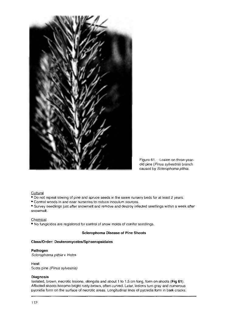
Cultural
Figure 61.-Lesion on three-yearold pine (Pinus sylvestris) branch caused by Sclerophoma pithia.
• Do not repeat sowing of pine and spruce seeds in the same nursery beds for at least 2 years. • Control weeds in and near nurseries to reduce inoculum sources. • Survey seedlings just after snowmelt and remove and destroy infected seedlings within a week after snowmelt.
Chemical • No fungicides are registered for control of snow molds of conifer seedlings.
Sclerophoma Disease of Pine Shoots
Class/Order: Deuteromycetes/Sphaeropsidales
Pathogen Sclerophoma pithia v. Hohn
Host Scots pine (Pinus sylvestris)
Diagnosis Isolated, brown, necrotic lesions, elongate and about 1 to 1.5 cm long, form on shoots (Fig 61 ). Affected shoots become bright rusty-brown, often curved. Later, lesions turn gray and numerous pycnidia form on the surface of necrotic areas. Longitudinal lines of pycnidia form in bark cracks.
112
Pycnidia are spherical, black, about 460 µ in diameter. Conidia are egg-shaped, colorless, 1-celled, 6-7 x 2-2.5 µ. Needles of diseased shoots turn brown and dark bands about 2 mm wide form,on the boundary with the healthy part of needles. +n autumn, pycnidia form on dead ends of needles. Infected shoots are covered by picnidia, turn black, and drop in autumn.
Biology In spring, shoots are infected by conidia from pycnidia on previously damaged shoots. Infection severity depends on seedling age. The number of diseased plants decreases significantly as plantation age increases. In plantations more than 6 years old, only shoots of upper nodes are infected. Environmental conditions also play a role in disease development. High levels of sunlight and low humidity promote moisture deficits in seedlings and stimulate rapid and vigorous fungal development. Cool, moist weather can cause outbreaks to collapse. Attacks by the pine tip moth (Rhuacionia petrova) promote infection of shoots by Sclerophoma.
Damage Affects seedlings in nurseries and in plantations less than 12 years old. Causes seedling death in nurseries and reduces the number of seedlings for standard planting material. Infection in plantations less than 5 years old results in deformed crowns and, with repeated epiphytotics, seedling death and plantation decline.
Distribution European part of Russia
Control Observation • Survey nurseries and pine plantations in the beginning of summer during drought years.
Cultural • Remove and burn infected seedlings in the nurseries. • Maintain row spacing in young pine plantations to allow for ploughing infected shoots into the soil.
Pine Shoot Rust
Class/Order: Basidiomycetes/Uredinales
Pathogen Melampsora pinitorqua Rostr.
Hosts Species of pines (Pinus), including Scots (Pinus sylvestris), Siberian (P. sibirica), and white (P. strobus); the alternate host is poplar (Populus)
Diagnosis Aecia form from May to June on current-year shoots or on needles (Fig. 62). They develop beneath epidermis as oblong, orange swellings. Mature aeciospores burst through the epidermis as a brightyellow, powdery mass. Aeciospores are 1-celled, oval or globose, 15-22 x 11-17 µ. The infected shoots lose turgor and bend. Small pitching wounds remain on the shoot's surface after aeciospore rele.ase. Numerous dormant (latent) buds can grow from below injured shoots and one can replace the dead leader, creating a false whorl.
Biology The spermogonial and aecial stages develop on Pinus species. Uredinial and basidial stages develop on Populus tremula and other Populus species. In June, aeciospores disperse and infect leaves of Popu/us species. In summer, uredinia with urediniospores develop and spores are dispersed by wind to reinfect poplar; this continues until August. In late summer (August to early September), telia form in place of uredinia. Talia are brown or black and crust-like. Teliospores are brown, irregularly prismatical, 20-35 x 7-11 µ. Teliospores overwinter in fallen leaves. In spring, they produce basidia with basidiospores, which forms a golden layer on fallen poplar leaves. Basidiospores disperse in May and infect pine. Wet and warm springs favor the maturity and dispersion of basidiospores, and the infection of pine. The degree of injury also depends on growing conditions and seed origin (provenance). Optimal growing conditions increase pine resistance. The disease can be severe where P.tremula and P.alba grow near pine seedlings or plantations.
113
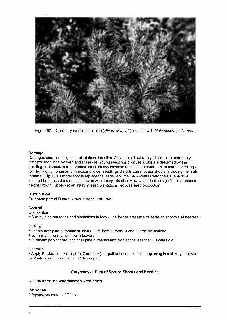
Figure 62.-Current-year shoots of pine (Pinus sylvestris) infected with Melampsora pinitorqua.
Damage Damages pine seedlings and plantations less than 20 years old but rarely affects pine understory. Infected seedlings weaken and some die. Young seedlings (1-2 years old) are deformed by the bending or dieback of the terminal shoot. Heavy infection reduces the number of standard seedlings for planting by 40 percent. Infection of older seedlings distorts current-year shoots, including the main terminal (Fig. 63). Lateral shoots replace the leader and the main stem is deformed. Dieback of infected branches does not occur even with heavy infection. However, infection significantly reduces height growth. Upper crown injury in seed plantations reduces seed production.
Distribution European part of Russia, Urals, Siberia, Far East
Control Observation • Survey pine nurseries and plantations in May-June for the presence of aecia on shoots and needles.
Cultural • Locate new pine nurseries at least 250 m from P tremula and P alba plantations. • Gather and burn fallen poplar leaves. • Eliminate poplar sprouting near pine nurseries and plantations less than 10 years old.
Chemical • Apply Bordeaux mixture (1%), Zineb (1%), or poliram combi 3 times beginning in mid-May, followed by 2 additional applications 6-7 days apart.
Chrysomyxa Rust of Spruce Shoots and Needles
Class/Order: Basidiomycetes/Uredinales
Pathogen Chrysomyxa woroninii Tranz.
114
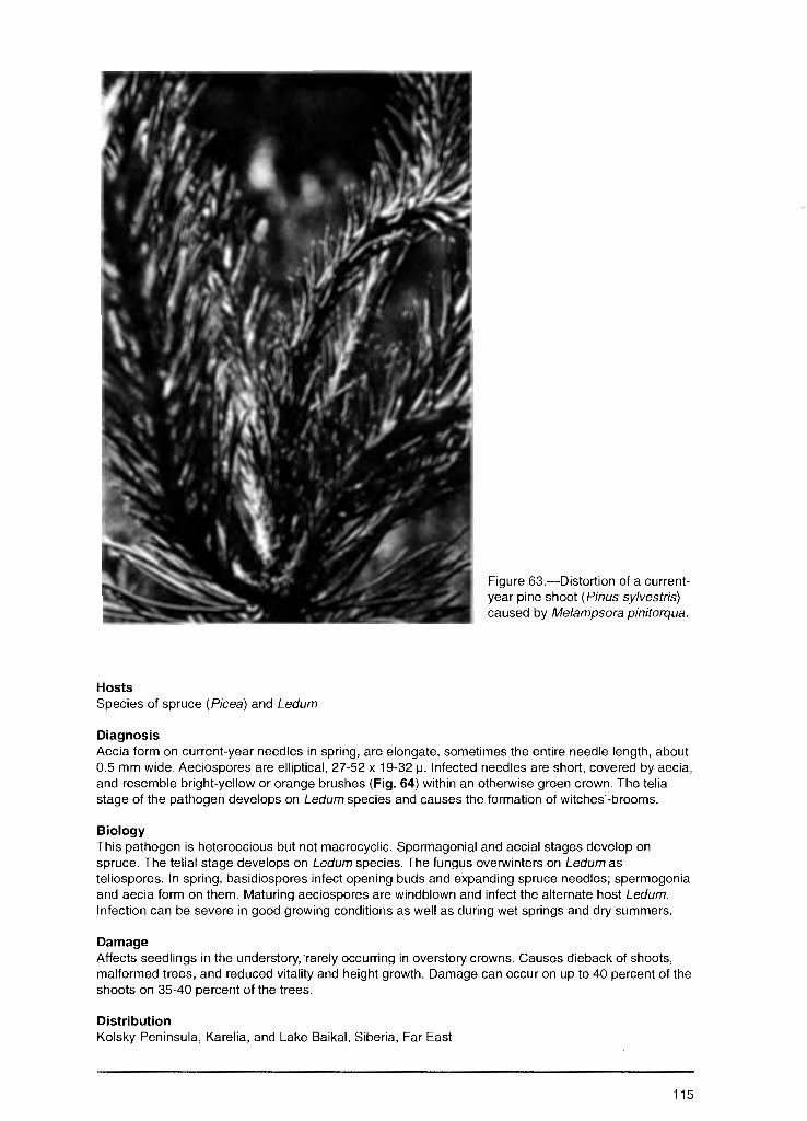
Hosts Species of spruce (Picea) and Ledum
Diagnosis
Figure 63.-Distortion of a currentyear pine shoot (Pinus sylvestris) caused by Melampsora pinitorqua.
Aecia form on current-year needles in spring, are elongate, sometimes the entire needle length, about 0.5 mm wide. Aeciospores are elliptical, 27-52 x 19-32 µ. Infected needles are short, covered by aecia, and resemble bright-yellow or orange brushes (Fig. 64) within an otherwise green crown. The telia stage of the pathogen develops on Ledum species and causes the formation of witches'-brooms.
Biology This pathogen is heteroecious but not macrocyclic. Spermagonial and aecial stages develop on spruce. The telial stage develops on Ledum species. The fungus overwinters on Ledum as teliospores. In spring, basidiospores infect opening buds and expanding spruce needles; spermogonia and aecia form on them. Maturing aeciospores are windblown and infect the alternate host Ledum. Infection can be severe in good growing conditions as well as during wet springs and dry summers.
Damage Affects seedlings in the understory, ·rarely occurring in overstory crowns. Causes dieback of shoots, malformed trees, and reduced vitality and height growth. Damage can occur on up to 40 percent of the shoots on 35-40 percent of the trees.
Distribution Kolsky Peninsula, Karelia, and Lake Baikal, Siberia, Far East
115
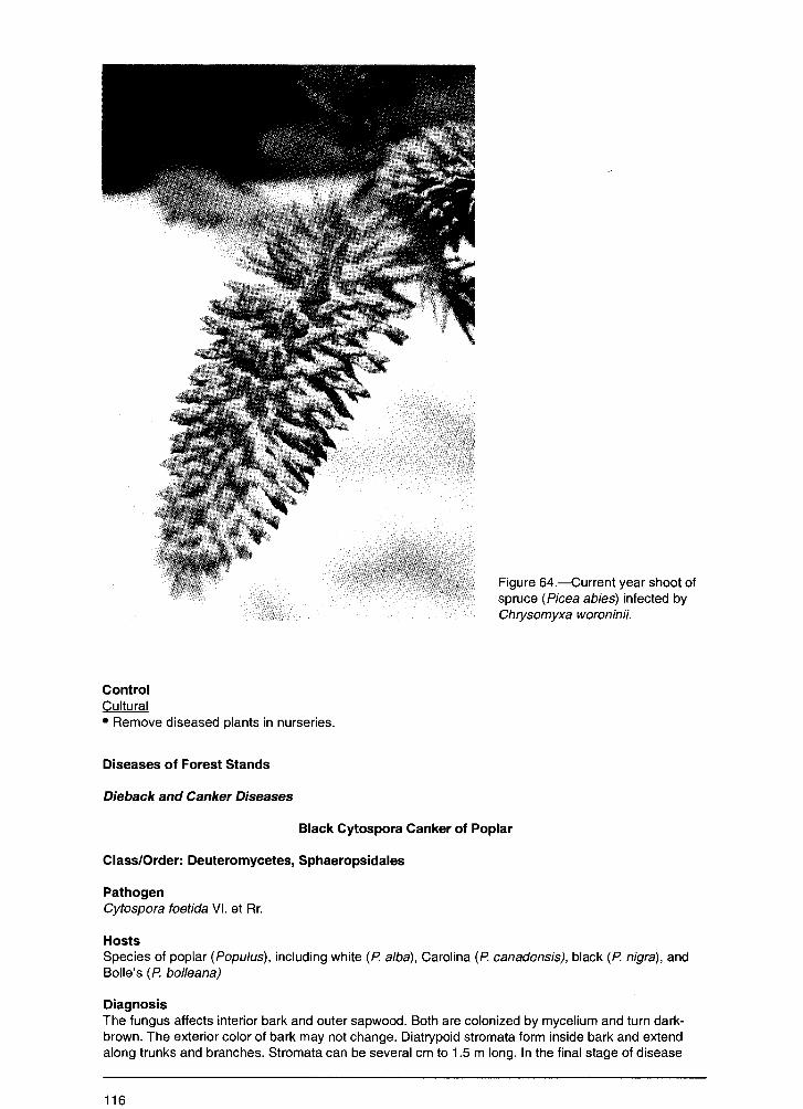
Control Cultural • Remove diseased plants in nurseries.
Diseases of Forest Stands
Dieback and Canker Diseases
Figure 64.-Current year shoot of spruce (Picea abies) infected by Chrysomyxa woroninii.
Black Cytospora Canker of Poplar
Class/Order: Deuteromycetes, Sphaeropsidales
Pathogen Cytospora foetida VI. et Rr.
Hosts Species of poplar (Populus), including white (P. alba), Carolina (P. canadensis), black (P. nigra), and Belle's (P. bol/eana)
Diagnosis The fungus affects interior bark and outer sapwood. Both are colonized by mycelium and turn darkbrown. The exterior color of bark may not change. Diatrypoid stromata form inside bark and extend along trunks and branches. Stromata can be several cm to 1.5 m long. In the final stage of disease
116
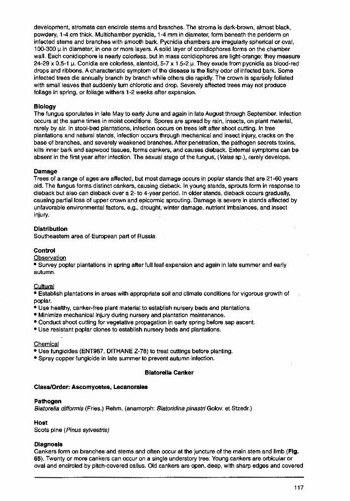
development, stromata can encircle stems and branches. The stroma is dark-brown, almost black, powdery, 1-4 cm thick. Multichamber pycnidia, 1-4 mm in diameter, form beneath the periderm on infected stems and branches with smooth bark. Pycnidia chambers are irregularly spherical or oval, 100-300 µ in diameter, in one or more layers. A solid layer of conidiophores forms on the chamber wall. Each conidiophore is nearly colorless, but in mass conidiophores are light-orange; they measure 24-29 x 0.5-1 µ. Conidia are colorless, alantoid, 5-7 x 1.5-2 µ. They exude from pycnidia as blood-red drops and ribbons. A characteristic symptom of the disease is the fishy odor of infected bark. Some infected trees die annually branch by branch while others die rapidly. The crown is sparsely foliated with small leaves that suddenly turn chlorotic and drop. Severely affected trees may not produce foliage in spring, or foliage withers 1-2 weeks after expansion.
Biology The fungus sporulates in late May to early June and again in late August through September. Infection occurs at the same times in moist conditions. Spores are spread by rain, insects, on plant material, rarely by air. In stool-bed plantations, infection occurs on trees left after shoot cutting. In tree plantations and natural stands, infection occurs through mechanical and insect injury, cracks on the base of branches, and severely weakened branches. After penetration, the pathogen secrets toxins, kills inner bark and sapwood tissues, forms cankers, and causes dieback. External symptoms can be absent in the first year after infection. The sexual stage of the fungus, ( Valsa sp.), rarely develops.
Damage Trees of a range of ages are affected, but most damage occurs in poplar stands that are 21-60 years old. The fungus forms distinct cankers, causing dieback. In young stands, sprouts form in response to dieback but also can dieback over a 2- to 4-year period. In older stands, dieback occurs gradually, causing partial loss of upper crown and epicormic sprouting. Damage is severe in stands affected by unfavorable environmental factors, e.g., drought, winter damage, nutrient imbalances, and insect injury.
Distribution Southeastern area of European part of Russia
Control Observation • Survey poplar plantations in spring after full leaf expansion and again in late summer and early autumn.
Cultural • Establish plantations in areas with appropriate soil and climate conditions for vigorous growth of poplar. • Use healthy, canker-free plant material to establish nursery beds and plantations. • Minimize mechanical injury during nursery and plantation maintenance. • Conduct shoot cutting for vegetative propagation in early spring before sap ascent. • Use resistant poplar clones to establish nursery beds and plantations.
Chemical • Use fungicides (ENT987, DITHANE Z-78) to treat cuttings before planting. • Spray copper fungicide in late summer to prevent autumn infection.
Biatorella Canker
Class/Order: Ascomycetes, Lecanorales
Pathogen Biatorella difformis (Fries.) Rehm. (anamorph: Biatoridina pinastri Golov. et Stzedr.)
Host Scots pine (Pinus sylvestris)
Diagnosis Cankers form on branches and stems and often occur at the juncture of the main stem and limb (Fig. 65). Twenty or more cankers can occur on a single understory tree. Young cankers are orbicular or oval and encircled by pitch-covered callus. Old cankers are open, deep, with sharp edges and covered
117

Figure 65.-Young pine tree ( Pinus sylvestris) affected by the fungus Biatore/la difformis. Note the "target" canker on the trunk.
with pitch. Cankers form yearly concentric rings of dead wood. In mature and overmature forests, cankers develop for several decades. The surface of old cankers, often black, is caused by the nonparasitic fungus Auerobasidium pu/lulans (de Bary) G.Arnaud. The imperfect stage of the canker fungus forms on the surface of wood and appears as a circle of black pycnidia, about 25-3 mm, resting on a brown matrix of mycelium. Bottle-like conidiophores about 6 to 8 µ long form in the pycnidia. Conidia are oval, colorless or light-olive but dark-olive in mass, 1.8-3 µ. Conidia are extruded as a dark, slimy mass. Black, waxy apothecia form very rarely. They are 500-800 µ in diameter and form in the same brown matrix as pycnidia.
Biology Infection and spread from tree to tree occurs by conidia produced in early summer. A second sporulation occurs in autumn when temperatures are lower; most infections occur at this time. Conidia penetrate tree tissues through natural openings but primarily through mechanical injuries, including borer damage. Insects that are important in development of this disease are Eveteria resinella L., Hylobius abictis L., and species of Pissodes. Mycelium spreads from damaged to living tree tissue, affecting the phloem, cambium, and eventually sapwood, where mycelium spreads up and around the stem at a rate of 1-2 cm per year. The disease mostly affects weak and suppressed trees growing offsite. High moisture and low light are predisposing factors in the understory, where infected trees range from 20-80 percent. In plantations growing in full sunlight, infection is 2 percent.
Damage Trees that are 10-80 years old are damaged. Understory trees and forest plantations are affected more than natural mature forests. Infection reduces radial growth in both understory trees and forest plantations. Severe infection on understory trees increases branch breakage by snow, but few trees
118
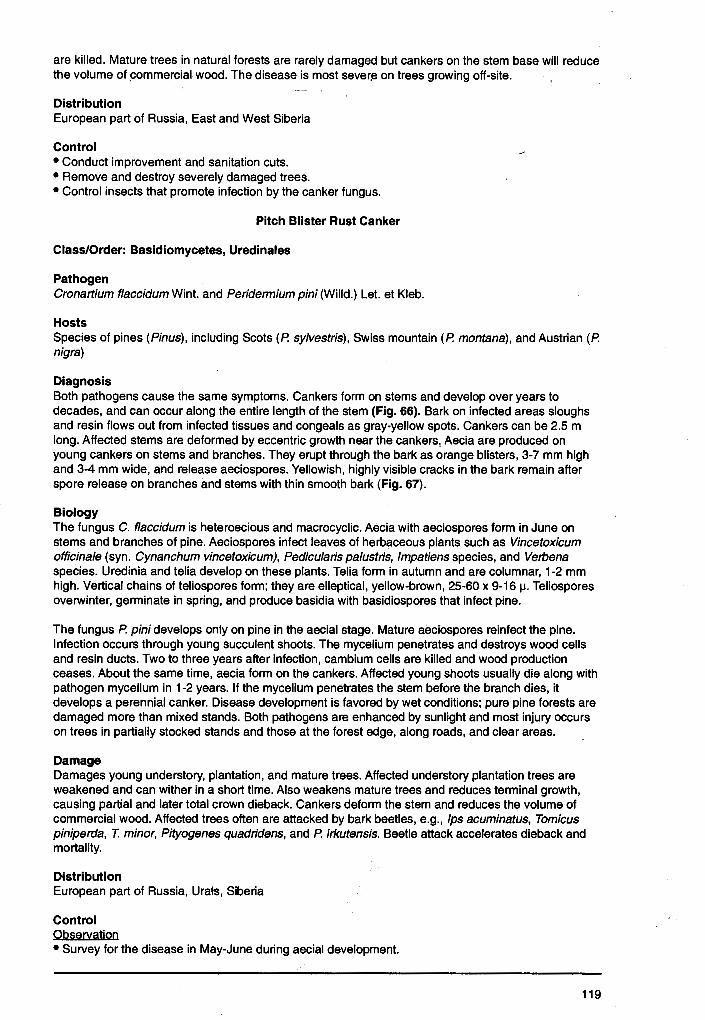
are killed. Mature trees in natural forests are rarely damaged but cankers on the stem base will reduce the volume of commercial wood. The disease is most sever~ on trees growing off-site.
Distribution European part of Russia, East and West Siberia
Control • Conduct improvement and sanitation cuts. • Remove and destroy severely damaged trees. • Control insects that promote infection by the canker fungus.
Pitch Blister Rust Canker
Class/Order: Basidiomycetes, Uredinales
Pathogen Cronartium flaccidum Wint. and Peridermium pini (Willd.) Let. et Kleb.
Hosts Species of pines (Pinus), including Scots (P. sylvestris), Swiss mountain (P. montana), and Austrian (P. nigra)
Diagnosis Both pathogens cause the same symptoms. Cankers form on stems and develop over years to decades, and can occur along the entire length of the stem (Fig. 66). Bark on infected areas sloughs and resin flows out from infected tissues and congeals as gray-yellow spots. Cankers can be 2.5 m long. Affected stems are deformed by eccentric growth near the cankers,, Aecia are produced on young cankers on stems and branches. They erupt through the bark as orange blisters, 3-7 mm high and 3-4 mm wide, and release aeciospores. Yellowish, highly visible cracks in the bark remain after spore release on branches and stems with thin smooth bark (Fig. 67).
Biology The fungus C. flaccidum is heteroecious and macrocyclic. Aecia with aeciospores form in June on stems and branches of pine. Aeciospores infect leaves of herbaceous plants such as Vincetoxicum officinale (syn. Cynanchum vincetoxicum), Pedicu/aris pa/ustris, Impatiens species, and Verbena species. Uredinia and telia develop on these plants. Telia form in autumn and are columnar, 1-2 mm high. Vertical chains of teliospores form; they are elleptical, yellow-brown, 25-60 x 9-16 µ. Teliospores overwinter, germinate in spring, and produce basidia with basidiospores that infect pine.
The fungus P. pini develops only on pine in the aecial stage. Mature aeciospores reinfect the pine. Infection occurs through young succulent shoots. The mycelium penetrates and destroys wood cells and resin ducts. Two to three years after infection, cambium cells are killed and wood production ceases. About the same time, aecia form on the cankers. Affected young shoots usually die along with pathogen mycelium in 1-2 years. If the mycelium penetrates the stem before the branch dies, it develops a perennial canker. Disease development is favored by wet conditions; pure pine forests are damaged more than mixed stands. Both pathogens are enhanced by sunlight and most injury occurs on trees in partially stocked stands and those at the forest edge, along roads, and clear areas.
Damage Damages young understory, plantation, and mature trees. Affected understory plantation trees are weakened and can wither in a short time. Also weakens mature trees and reduces terminal growth, causing partial and later total crown dieback. Cankers deform the stem and reduces the volume of commercial wood. Affected trees often are attacked by bark beetles, e.g., fps acuminatus, Tomicus piniperda, T. minor, Pityogenes quadridens, and P. irkutensis. Beetle attack accelerates dieback and mortality.
Distribution European part of Russia, Urats, Siberia
Control Observation • Survey for the disease in May-June during aecial development.
119
Figure 66.-Pitch blister rust of pine ( Pi nus sylvestris) caused by Cronartium flaccidum and Peridermium pini. The affected pine has a canker on upper portion of the trunk.
• Determine tree condition and canker distribution (location) on the stem, e.g., low, middle, or upper portion of crown. • Determine the degree of canker development around the stem circumference: less than 1/2, more than 1/2 but less than 3/4, more than 3/4 to complete. • Record species and abundance of bark beetles on affected trees.
Cultural • Conduct a 2-stage sanitation cutting at intervals of 3-5 years. Remove heavily weakened, suppressed, and dead trees with below-crown cankers and evidence of attack by bark beetles; cut stag-headed trees in the second stage. • Create mixed-pine stands and forest plantations with high initial planting density.
Endoxylina Canker of Ash
Class/Order: Ascomycetes, Diaporthales
Pathogen Endoxylina stel/ulata Rom. (syn. Valsaria stellulata Rom.) (anamorph: Libertella fraxini Ogan.)
Host European ash ( Fraxinus excelsior L.)
120
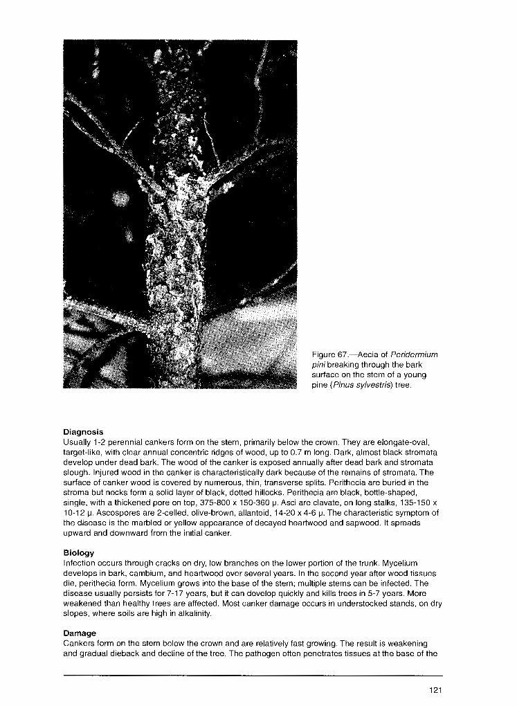
Diagnosis
Figure 67.-Aecia of Peridermium pini breaking through the bark surface on the stem of a young pine (Pinus sylvestris) tree.
Usually 1-2 perennial cankers form on the stem, primarily below the crown. They are elongate-oval, target-like, with clear annual concentric ridges of wood, up to 0.7 m long. Dark, almost black stromata develop under dead bark. The wood of the canker is exposed annually after dead bark and stromata slough. Injured wood in the canker is characteristically dark because of the remains of stromata. The surface of canker wood is covered by numerous, thin, transverse splits. Perithecia are buried in the stroma but necks form a solid layer of black, dotted hillocks. Perithecia are black, bottle-shaped, single, with a thickened pore on top, 375-800 x 150-360 µ. Asci are clavate, on long stalks, 135-150 x 10-12 µ. Ascospores are 2-celled, olive-brown, allantoid, 14-20 x 4-6 µ. The characteristic symptom of the disease is the marbled or yellow appearance of decayed heartwood and sapwood. It spreads upward and downward from the initial canker.
Biology Infection occurs through cracks on dry, low branches on the lower portion of the trunk. Mycelium develops in bark, cambium, and heartwood over several years. In the second year after wood tissues die, perithecia form. Mycelium grows into the base of the stem; multiple stems can be infected. The disease usually persists for 7-17 years, but it can develop quickly and kills trees in 5-7 years. More weakened than healthy trees are affected. Most canker damage occurs in understocked stands, on dry slopes, where soils are high in alkalinity.
Damage Cankers form on the stem below the crown and are relatively fast growing. The result is weakening and gradual dieback and decline of the tree. The pathogen often penetrates tissues at the base of the
121
stem, causing dieback of sprouts. Sapwood and heartwood decay reduces resistance to wind, promoting breakage. Coppice stands are less resistant ta the disease; damage in these sta,nds ranges from 40-60 percent. ·
Distribution Steppe zone of southeastern area of European part of Russia.
Control Cultural • Conduct sanitation cuttings to remove dying and dead trees within disease centers. • Remove or uproot stumps after sanitation cutting to prevent sprout infection.
Cankers and Diebacks
Class/Order: Deuteromycetes/Sphaeropsidales
Pathogen Camarosporium ribicolum Sacc.
Host Golden currant (Ribes aureum)
Diagnosis Necrotic lesions develop on the bark. Pycnidia are dispersed or coalesced in groups in necrotic areas. They are black, spherical, similar to stromata. Conidia are oblong-globose or egg-shaped, brown, with 3-4 transverse and 1-2 longitudinal walls, 12-16 x 6-8 µ.
Distribution European part of Russia
••• Pathogen Coniothyrium inisitivum Sacc.
Hosts Bird cherry (Padus avium), barberry (Berberis vu/garis), and lilac (Syringa vulgaris)
Diagnosis Dark, necrotic lesions form on the bark. Pycnidia develop in the necrotic areas in clusters. They are black and spherical. Initially, they are buried in leaf tissue but later erupt and release conidia. Conidia are elongate-egg-shaped, 1-celled, brown, 4.5 x 2.5 x 4 µ.
Distribution European part of Russia
••• Pathogen Cytospora tumida Lib.
Host Pedunculate oak (Quercus robur)
Diagnosis Necrotic lesions form on the bark; later, black stromata develop that are cushion-like, black, round or angular. Initially, they are buried in leaf tissue but later erupt. Conidia are alantoid, colorless, 6 x 1.5 µ.
Distribution European part of Russia
••• Pathogen Diplodia amphisphaerioides Pass.
122
Host Pedunculate oak (Quercus robut} . :
Diagnosis Black, sphaerical or flat-spherical pycnidia form on the bark of dead, dry branches. Initially, they are buried in tissue but later erupt. Conidia are ellipsoidal, brown, 2-celled, 17.5 x 7.5 µ.
Distribution European part of Russia
••• Pathogen Microdiplodia ascochytula (Sacc.) Allesch. (syn. Diplodia ascochytula Sacc.)
Hosts Species of honeysuckle (Lonicera)
Diagnosis Brown necrotic lesions form on dead areas of bark. Brown pycnidia in groups form on the dead bark and are initially buried in tissue but later erupt. Conidia are spindle-like with blunt ends, 2-celled, olivebrown, 8-12 x 2.5-3 µ.
Distribution European part of Russia
••• Pathogen Diplodia juniperi Westend.
Host Red cedar (Juniperus virginiana)
Diagnosis Bark on branches dies. Black, spherical, single, scattered pycnidia form in the cracks of dead bark. Conidia are oblong-egg-shaped, 2-celled, brown or dark-brown, 18-20 x 8-1 O µ.
Distribution Southwestern areas of European part of Russia
••• Pathogen Diplodina tatarica Allesch.
Host Honeysuckle (Lonicera tatarica)
Diagnosis Bark dies on affected branches. Numerous, black, spherically-flat, dispersed pycnidia form in bark cracks. Conidia are spindle-like, 2-celled, with a small septum, colorless, 8-12 x 2.5-3 µ. ·
Distribution European part of Russia
••• Pathogen Dothiorella robiniae Prill. et Delacr.
Host Black locust (Robinia pseudoacacia)
123
Diagnosis Black, erurnpent str~mata form in the bark of dead; dry branches. Pycnidia, oblong or irregularly shaped, develop in the stroma. Conidia are egg-shaped or elliptical, 1-celled, colorless, with 2-3 oil
1
drops, 10.5-12 x 3.5-4 µ.
Distribution European part of Russia
••• Pathogen Fucicoccum obtusulum Grove
Hosts Species of maple (Ace!)
Diagnosis Stromata form within the bark of dead branches. They are dark-gray, with yellow centers, flat-conical, dispersed. Conidia are spindle-like with blunt ends, straight, 1-celled, colorless, 12-14 x 2-2.5 µ.
Distribution European part of Russia
••• Pathogen Hendersonia pseudoacaciae Ell. et Barth.
Host Black locust (Robinia pseudoacacia)
Diagnosis Black, dispersed pycnidia form in the bark of dead branches. Conidia are oblong-oval, 4- to 8-celled, brown, 15-24 x 6-10 µ.
Distribution European part of Russia
••• Pathogen Phoma aceris-negundinis Arcang.
Host Species of maple (Ace!)
Diagnosis The affected bark surface is rough and light-yellow. Pycnidia form under the epidermis; they are slightly raised, black, and spherical. Conidia are oval, colorless, with 2 drops of oil, 8-3 µ.
Distribution European part of Russia
••• Pathogen Rhabdospora passerinii Sacc.
Hosts Species of maple (Acer), primarily boxelder (A. negunda).
Diagnosis Affected branches and sprouts dieback. Brown-black pycnidia form in the cracks in dead bark in longitudinal lines. Conidia are cylindrical, straight, slightly narrowed on the ends, 4-celled, 30-32 x 2.5 µ.
Distribution European part of Russia
124
Class/Order: Ascomycetes/Helotiales
Pathogen Lachnellula angustispora Raitv.
Hosts Species of fir (Abies) and larch (Larix)
Diagnosis Apothecia form on the bark of dead branches. They have a short stalk, white outside surface, brightorange center, 0.3-3 mm in diameter. Asci are clavate, 101-106 x 9.7-10.2 µ. Ascospores are ellipsoid to spindle-shaped, asymmetrical, colorless, 1-celled, 12.8-14.2 x 5-5.2 µ.
Distribution Middle Urals, Siberia
••• Pathogen Lachnellula flavovirens (Bres.) Dennis
Hosts Species of spruce (Picea), fir (Abies), and larch (Larix)
Diagnosis Apothecia form on the bark of dead branches. They have a short stalk, brown, hairy outside surface, bright-orange center, 1-3 mm in diameter. Asci are clavate, 55-65 x 6.2-6.6 µ. Ascospores are ellipsoid to spindle-shaped, asymmetrical, colorless, 1-celled, 7-7.5 x 3.5-3.8 µ.
Distribution Urals, Siberia, Far East
••• Pathogen Lachnellula fuckelii (Bres. in Rehm.) Dharne.
Hosts Species of spruce (Picea) and pine (Pinus)
Diagnosis Apothecia form on the bark of dead branches. They have a short stalk, snowy-white outside surface, bright-orange center, 0.3 to 3 mm in diameter. Asci are clavate, 113-118 x 9.3-10.5 µ. Ascospores are ellipsoid to spindle-shaped, asymmetrical, 1-celled, colorless, 12.8-13.2 x 6-6.4 µ.
Distribution Middle Urals, Far East
••• Pathogen Lachnellula kamtschatica Raitv.
Hosts Mountain pine (Pinus pumila)
Diagnosis Apothecia form on the bark of dead branches. They have a short stalk, snowy-white outside surface, and a bright-orange center. Ascospores are widely ellipsoid, 1-celled, colorless, 6-7 x 4-5 µ.
Distribution Far East
••• Pathogen Lachnellula minuscula Raitv.
125
Host Species o(fir (Abies)
' j
Diagnosis Apothecia on short stalks form on the bark of dead branches. The outside surface is covered with hairs; the inside disk is bright-orange, 1-2 mm in diameter. Asci are clavate, 44-56 x 4.4-5 µ. Ascospores are ellipsoid to spindle-shaped, 1-celled, colorless, 5.1-5. 7 x 1.8-2.3 µ. -
Distribution Siberia, Far East
••• Pathogen Lachnellula pseudofarinacea (Cronan.) Dennis
Host Species of pine (Pinus)
Diagnosis Apothecia form on the bark of dead branches. The outside surface is covered with snowy-white, dense hairs; the inside disk is bright-orange, 1-1.5 mm in diameter. Asci are clavate, 100-115 x 9-10.5 mm. Ascospores are thread-like, without a septum, colorless, 73-97 x 1.5-2 µ.
Distribution Northern area of European part of Russia, Siberia
Class/Order: Deuteromycetes/Melanconlales
Pathogen Didymosporium profusum Fr.
Host Species of maple (Acer)
Diagnosis Cushion-like, conical structures form in cracks on dead bark. Conidia are oblong or egg-shaped, 2-celled, a smoky color, 10-12 x 8-9 µ.
• •• Pathogen Melanconium desmazierii (Berk.et Br.) Sacc.
Hosts Species of lime ( Tilia)
Diagnosis Black, depressed, cushion-like stromata, slightly erumpent, form under the epidermis of dead branches and stems. Conidia are short, spindle-like, with blunt ends, smoky-blue, 1-celled, with 2 oil drops, 30-35 x 6-10 µ.
Distribution European part of Russia
••• Pathogen Melanconium czerniaiewi Poteb.
Hosts Species of oak ( Quercus)
126

Diagnosis Bark dies on young branches. Black, conical stromata, 1-2 mm in diameter, form under the bar~. Conidia are oval, ellipsoid or pear-like, dark-brown, 1-celled, 18-24 x 11-14 µ. ·
Distribution Southwestern area of European part of Russia
••• Pathogen Septogloeum hartigianum Sacc.
Hosts Species of maple (Ace,}
Diagnosis Affected branches and sprouts dieback and tum brown. Sporodochia form in longitudinal bark cracks. They are cushion-like, oblong, brown, 1-3 mm in diameter. Conidia are oblong-oval, 3-celled, colorless, 4-15 x 2-4 µ.
Distribution European part of Russia
••• Pathogen Septomyxa negundinis Allesch.
Hosts Species of maple (Ace,}
Diagnosis White-yellow or gray necrotic lesions with dark borders form on affected branches. Numerous, aggregated sporodochia form on dead bark. Initially, they are buried in tissue but later erupt. They are cushion-like, red- or dark-brown. Conidia are fusiform, pointed, rarely oval, initially 1-celled, and later 2-celled, colorless, 10-20 x 3-4.5 µ.
Distribution European part of Russia
••• Pathogen Stilbospora angustata Pers. (teliomorph: Pseudovalsa berkeleyi Sacc.)
Hosts Species of hornbeam (Carpinus) and beech (Fagus)
Diagnosis Black, cushion-like, convex, often coalesced sporodochia form under the epidermis of affected bark. Conidia are cylindrical, elongate, 4-celled, olive, often with a sticky jelly cover, 35-50 x 10-14 µ.
Distribution European part of Russia
Class/Order: Ascomycetes/Sphaeriales
Pathogen Leucostoma diatrype Fr.
Hosts Species of alder (A/nus)
127
Diagnosis Black, conical, dispersed stromata with white or gray-white disks form in the bark of dying and dead branches. Each stroma contains 3-7 perithe.cia. Asci are clavate, 80-100 x 8-16 µ. Ascospores are cylindrical, colorless, 16-18 x 3-6 µ.
Distribution European part of Russia
••• Pathogen Nitschkia cupularis (Pers.) Winter (anamorph: Phoma fuckeli Sacc.)
Hosts Species of elm ( Ulmus)
Diagnosis Affected branches, especially sprouts, die. Perithecia form in clusters in dead bark. They are black, round but more often elongate, 0.5-3 mm in diameter, and resemble small hillocks erupting the epidermis. Asci are elongate, clavate, 40-60 x 7-8 µ. Ascospores are short, cylindrical, slightly curved, colorless, with oil drops, 9-10 x 2-3 µ.
Distribution European part of Russia
••• Pathogen Nummularia succenturiata (Tode) Nitschke
Hosts Species of elm (Ulmus)
Diagnosis Affected trees die. Stromata form within dead bark as black, flat disks, 3-5 mm in diameter. Perithecia form on the surface of the stromata. Asci are elongate, clavate, 120-150 x 9 µ. Ascospores are ovoid or fusiform, straight or slightly curved, brown, 16-18 x 5-7 µ.
Distribution European part of Russia
Class/Order: Ascomycetes/Dothideales
Pathogen Cucurbitaria rhamni (Nees.) Fckl. (anamorph: Diplodia rhamni Gaap.
Host Species of buckthorn (Frangula a/nus, Rhamnus cathartica)
Diagnosis Initially, pycnidia form on dead branches. They are single or clustered, black, and spherical. Initially, they are buried in bark tissue but later erupt. Black pseudothecia form in bark cracks. They are spherical, cup-like, concave, black, often in long rows. Conidia are oblong, egg-shaped, dark-brown, 2-celled, 20-25 x 7.5-10 µ. Asci are clavate, elongate, 120-140 x 12 µ. Ascospores are oblong-elliptical, yellow-brown, with 3-6 transverse walls and 1 incomplete londitudinal septum, 16-21 x 7-8 µ.
Distribution European part of Russia
Wood-Decaying Diseases
Class/Order: Basidiomycetes, Aphyllopherales
128
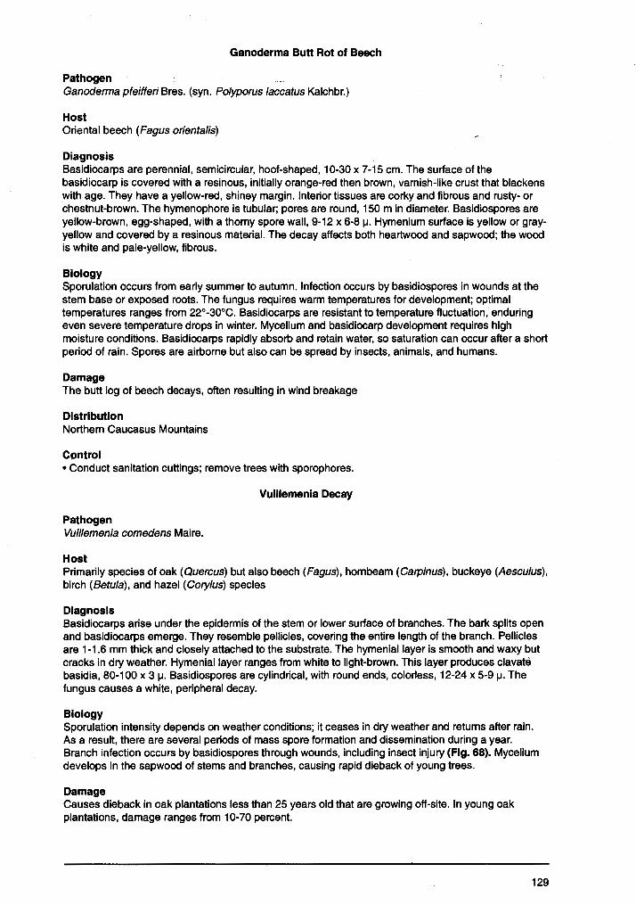
Ganoderma Butt Rot of Beech
Pathogen Ganoderrna pfeifferi Bres. (syn. Po/yporus laccatus Kalchbr.)
Host Oriental beech (Fagus orientalis)
Diagnosis Basidiocarps are perennial, semicircular, hoof-shaped, 10-30 x 7-15 cm. The surface of the basidiocarp is covered with a resinous, initially orange-red then brown, varnish-like crust that blackens with age. They have a yellow-red, shiney margin. Interior tissues are corky and fibrous and rusty- or chestnut-brown. The hymenophore is tubular; pores are round, 150 m in diameter. Basidiospores are yellow-brown, egg-shaped, with a thorny spore wall, 9-12 x 6-8 µ. Hymenium surface is yellow or grayyellow and covered by a resinous material. The decay affects both heartwood and sapwood; the wood is white and pale-yellow, fibrous.
Biology Sporulation occurs from early summer to autumn. Infection occurs by basidiospores in wounds at the stem base or exposed roots. The fungus requires warm temperatures for development; optimal temperatures ranges from 22°-30°C. Basidiocarps are resistant to temperature fluctuation, enduring even severe temperature drops in winter. Mycelium and basidiocarp development requires high moisture conditions. Basidiocarps rapidly absorb and retain water, so saturation can occur after a short period of rain. Spores are airborne but also can be spread by insects, animals, and humans.
Damage The butt log of beech decays, often resulting in wind breakage
Distribution Northern Caucasus Mountains
Cpntrol • Conduct sanitation cuttings; remove trees with sporophores.
Vuillemenia Decay
Pathogen Vuillemenia comedens Maire.
Host Primarily species of oak (Quercus) but also beech (Fagus), hornbeam (Carpinus), buckeye (Aesculus), birch (Betula), and hazel (Cory/us) species
Diagnosis Basidiocarps arise under the epidermis of the stem or lower surface of branches. The bark splits open and basidiocarps emerge. They resemble pellicles, covering the entire length of the branch. Pellicles are 1-1.6 mm thick and closely attached to the substrate. The hymenial layer is smooth and waxy but cracks in dry weather. Hymenial layer ranges from white to light-brown. This layer produces clavate basidia, 80-100 x 3 µ. Basidiospores are cylindrical, with round ends, colorless, 12-24 x 5-9 µ. The fungus causes a white, peripheral decay.
Biology Sporulation intensity depends on weather conditions; it ceases in dry weather and returns after rain. As a result, there are several periods of mass spore formation and dissemination during a year. Branch infection occurs by basidiospores through wounds, including insect injury (Fig. 68). Mycelium develops in the sapwood of stems and branches, causing rapid dieback of young trees.
Damage Causes dieback in oak plantations less than 25 years old that are growing off-site. In young oak plantations, damage ranges from 10-70 percent.
129
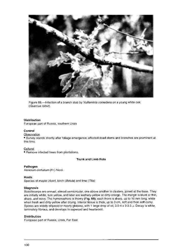
Figure 68.-lnfection of a branch stub by Vuilleminia comedens on a young white oak ( Que re us robw).
Distribution European part of Russia, southern Urals
Control Observation • Survey stands shortly after foliage emergence; affected dead stems and branches are prominent at this time.
Cultural • Remove infected trees from plantations.
Trunk and Limb Rots
Pathogen Hericium cirrhatum (Fr.) Nicol.
Hosts Species of maple (Acer), birch (Betula) and lime (Tilia)
Diagnosis Basidiocarps are annual, almost semicircular, one above another in clusters, joined at the base. They are initially white, turn yellow, and later are leathery-yellow or dirty-orange. The margin is blunt or thin, sharp, and wavy. The hymenophore is thorny (Fig. 69); each thorn is sharp, up to 10 mm long, white when fresh and dirty-yellow after drying. Interior tissue is thick, up to 3 cm, soft and then soft corky. Spores are widely ellipsoid or nearly globose, with 1 large drop of oil, 3.5-4 x 3-3.5 µ. Decay is white, laminately fibrous, and develops in sapwood and heartwood.
Distribution European part of Russia, Urals, Far East
130
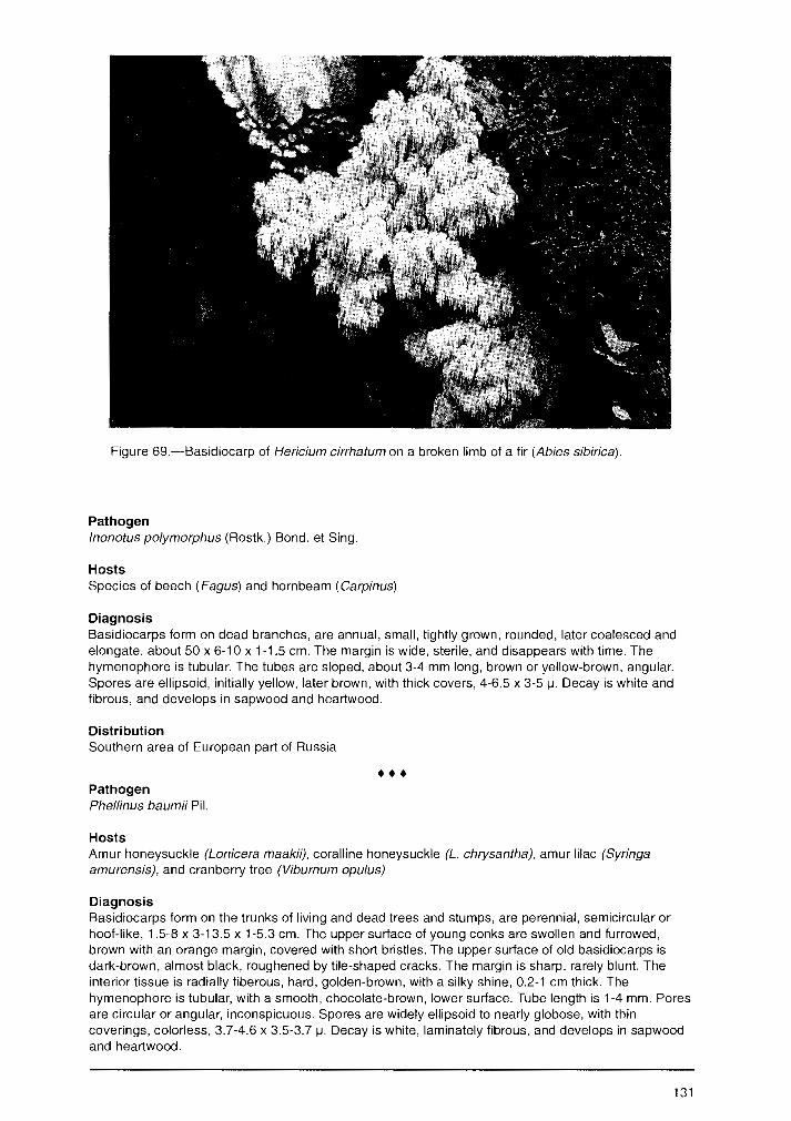
Figure 69.-Basidiocarp of Hericium cirrhatum on a broken limb of a fir (Abies sibirica).
Pathogen lnonotus polymorphus (Rostk.) Bond. et Sing.
Hosts Species of beech (Fagus) and hornbeam (Carpinus)
Diagnosis Basidiocarps form on dead branches, are annual, small, tightly grown, rounded, later coalesced and elongate, about 50 x 6-10 x 1-1.5 cm. The margin is wide, sterile, and disappears with time. The hymenophore is tubular. The tubes are sloped, about 3-4 mm long, brown or yellow-brown, angular. Spores are ellipsoid, initially yellow, later brown, with thick covers, 4-6.5 x 3-5 µ. Decay is white and fibrous, and develops in sapwood and heartwood.
Distribution Southern area of European part of Russia
••• Pathogen Phellinus baumii Pil.
Hosts Amur honeysuckle (Lonicera maakii), coralline honeysuckle (L chrysantha), amur lilac (Syringa amurensis), and cranberry tree (Viburnum opulus)
Diagnosis Basidiocarps form on the trunks of living and dead trees and stumps, are perennial, semicircular or hoof-like, 1.5-8 x 3-13.5 x 1-5.3 cm. The upper surface of young conks are swollen and furrowed, brown with an orange margin, covered with short bristles. The upper surface of old basidiocarps is dark-brown, almost black, roughened by tile-shaped cracks. The margin is sharp, rarely blunt The interior tissue is radially fiberous, hard, golden-brown, with a silky shine, 0.2-1 cm thick. The hymenophore is tubular, with a smooth, chocolate-brown, lower surface. Tube length is 1-4 mm. Pores are circular or angular, inconspicuous. Spores are widely ellipsoid to nearly globose, with thin coverings, colorless, 3.7-4.6 x 3.5-3.7 µ. Decay is white, laminately fibrous, and develops in sapwood and heartwood.
131
Distribution Far East
Pathogen Phellinus microporus (Pil.) Parm.
Hosts
\ ... Species of fir (Abies), spruce (Picea), and pine (Pinus)
Diagnosis Basidiocarps form on the trunks of living and dead trees and roots of windthrown trees, are perennial, attached to the substrate at one point or by the entire base, 2-5.5 x 1.5-4 x 0.2-0.6 cm. The upper surface is brown-yellow, chestnut-brown or black, bristly or velvety, with narrow furrows. The margin is straight, thin, and sharp. Interior tissue is thin, 1-2 mm thick, corky to woody, and brown. Tubes of the hymenophore are stratiform, 2-5 mm long, lighter in color than the trama. The lower surface of the hymenophore is brown. Pores are circularly-angular. Spores are widely ellipsoid, slightly ovoid, with thin covers, colorless but turn yellow with age, 4.5-5.1 x 3.5-4 µ. Decay is brown with white spots, pocket-fibrous, and develops in sapwood and heartwood.
Distribution East Siberia, Far East
••• Pathogen Piptoporus quercinus (Schrad. ex Fr.) Pil.
Hosts Species of oak ( Quercus)
Diagnosis Basidiocarps form on the trunks of living and dead trees, are annual and are elongate, fan- or tongueshaped conks, 4-9 x 6-11 x 1.5-3 cm, with a thickened base on stalks. The upper surface is initially velvety, later rough, initially pale-yellow, later light-chestnut to chestnut-brown. The margin is blunt. The interior tissue is 2-3 cm thick. The young trama initially is soft and wet, then soft-corky, white and pale-cream after drying. The hymenophore is tubular. Tubes are 2-3 mm long, with rounded or angular pores, 0.3-0.5 mm in diameter. The lower surface of the hymenophore shrinks after drying, and the margins turn white to yellowish with age. Spores are fusiform or ovoid, with sharp ends and several oil drops, colorless, 6.5-9 x 3-3.5 µ. Red-brown decay develops in the heartwood, cracks into prismatic chunks, and later crumbles to dust.
Distribution European part of Russia
••• Pathogen Spongipellis litschaueri Lohw.
Hosts Species of oak (Quercus), elm (U/mus), maple (Ace1', poplar (Populus), lime (Ti/ia), and pine (Pinus)
Diagnosis Basidiocarps form on the trunks of living and dead trees, are primarily annual but sometimes 2-5 years old, flat, hoof- shaped, 3-12 x 4-20 x 2-6 cm. They are initially soft, spongy, or fibrous but later become hard. The upper surface is nearly white, later yellow to brown, and becomes soft felty-bristly, bare or rough; the margin is sharp. The interior tissue is white, pale-brown after drying, radially fibrous. Hymenophore tubes are 1-2 cm long, initially white or creamy, later orange with fringy or tattered margins. Pores are irregular, rounded angular, 0.3-2 mm in diameter. The lower surface of the hymenophore is white to cream and turns brown after drying. Spores are widely ellipsoid, nearly globose, colorless, with 1 large oil drop, 5.5-7 x 4.5-6 µ. The decay is white and fibrous; initially, it develops in heartwood but later spreads to sapwood.
132