use of antithrombotic agents in patients with
TRANSCRIPT
J Neurosurg / Volume 118 / January 2013 43
J Neurosurg 118:43–46, 2013©AANS, 2013
LittLe is known about the safety of using anti-thrombotic agents in patients with known ICMs. Since the pathological structure of ICM is a leaky
endothelium, concurrent use of antithrombotics could theoretically pose an increased risk of clinically overt hemorrhage. Only 2 case reports exist regarding anti-thrombotic treatment in patients with known ICM. One of these reports described a patient with known famil-ial ICM who underwent treatment with low-molecular-weight heparin.5 The patient developed hand dysesthesias several days after treatment, and an MRI study revealed a subacute hemorrhage of an ICM. A second case report described a patient with a known ICM who was success-
fully treated with intravenous tissue plasminogen activa-tor for an ischemic stroke without hemorrhage into the cavernous malformation.3
Methods
Patient Selection
The records of all patients seen at our institution between 1989 and 1999 with an MRI diagnosis of ICM were retrospectively identified. We have reported on the entire cohort elsewhere.2 Patients from this cohort who were placed on an antiplatelet agent, anticoagulant, or both, after the diagnosis of ICM was established radio-graphically, were included in this study.
Use of antithrombotic agents in patients with intracerebral cavernous malformations
Clinical articleKelly D. Flemming, m.D.,1 michael J. linK, m.D.,2 Teresa J. h. chrisTianson, B.sc.,3 anD roBerT D. Brown Jr., m.D.1
Departments of 1Neurology and 2Neurosurgery, and 3Division of Biomedical Statistics & Informatics, Mayo Clinic, Rochester, Minnesota
Object. The goal of this study was to determine the risk of using antithrombotic agents in patients with estab-lished intracerebral cavernous malformations (ICMs).
Methods. From a previously described cohort of 292 patients with radiographically defined ICMs, 40 required an antithrombotic after the ICM was diagnosed. Patients underwent follow-up to determine the incidence of hemor-rhage.
Results. The mean age of these 40 patients was 62.4 years; there were 21 male and 19 female patients. Five (12.5%) of the 40 patients initially presented with hemorrhage and 4 (10%) had multiple ICMs. Of these patients, 32 were placed on an antiplatelet agent alone, 6 on an anticoagulant alone, and 2 were placed on both. In patients necessitating any antithrombotic agent, 1 patient developed a prospective hemorrhage over the 258 person-years of follow-up (prospective hemorrhage rate 0.41% per person-year).
Conclusions. Antithrombotics likely do not precipitate hemorrhage in patients with known ICMs. However, caution should be exercised in the use of antithrombotics in patients with ICMs at high risk for hemorrhage. The risks and benefits of antithrombotics in each situation should be carefully weighed against the natural history of ICM.(http://thejns.org/doi/abs/10.3171/2012.8.JNS112050)
Key worDs • cavernous malformation • hemorrhage • antithrombotic agent • aspirin • anticoagulation • vascular disorders
43
Abbreviation used in this paper: ICM = intracerebral cavernous malformation.
See the corresponding editorial in this issue, p 42.

K. D. Flemming et al.
44 J Neurosurg / Volume 118 / January 2013
Data CollectionThe primary investigator retrospectively reviewed
all medical records. Extensive demographic and clinical information was abstracted from medical records on a standardized form and was entered into a computerized database. Demographic information, medical history, ini-tial presentation, and treatment were recorded.
Follow-up information was calculated from the time of first diagnosis to the most recent contact. Follow-up information was obtained between 2000 and 2003 from the medical record if the patient was seen at our institu-tion within 6 months of our review. If the patient did not undergo follow-up at our institution, a mail survey was sent and was followed up by a phone call and ascertain-ment of medical records and images from other medical institutions. Follow-up information obtained included use of antithrombotics (warfarin, antiplatelet agent, or both), whether the surgical intervention had occurred during the follow-up time frame, and whether the patient had suffered a cerebral hemorrhage related to the cavernous malformation. A definitive prospective hemorrhage was defined as a new clinical event (focal deficit, seizure, or severe headache) in association with radiographic evi-dence of acute hemorrhage or autopsy data suggesting acute hemorrhage. A probable prospective hemorrhage was defined as a clinical event with medical records ob-tained from a medical institution outside of our medical record system suggesting intracerebral hemorrhage, but without images for us to personally review. An undocu-mented prospective hemorrhage was defined as an acute clinical event, but without imaging reports or images to personally review. Typically, the latter scenario includ-ed information obtained from the patient only, without verification by medical records. This diagnosis of defini-tive hemorrhage is similar to that described as an overt hemorrhage by Moriarity et al.4 and a Type I lesion by Zabramski et al.7
Radiographic DataInitial brain MRI scans were reviewed by the prima-
ry investigator. The imaging study obtained closest to the time of diagnosis was reviewed in detail. The ICMs were identified as previously described.6,7 The location and di-ameter were recorded for each ICM. Association with a venous angioma or other vascular malformation was also recorded. The type of ICM was also recorded based on the nomenclature by Zabramski and colleagues,7 and multiplicity was noted. When multiple ICMs were pres-ent, the location and size of symptomatic lesions were re-corded. The symptomatic lesion was included in analysis of variables determining future hemorrhage. If there were multiple lesions and no symptomatic lesion, the largest was used.
We received approval from the neurology research committee and institutional review board. Informed con-sent was obtained from all patients participating in the study.
Statistical AnalysisDescriptive statistics including means, standard de-
viations, and frequencies were used for patient character-
istics and presenting symptoms. The Kaplan-Meier meth-od was used to determine survival free from prospective hemorrhage. The prospective hemorrhage rate was de-termined based on the number of definitive and probable hemorrhages during the follow-up period divided by the number of prospective person-years of follow-up (an inci-dence rate); the annual rates presented are the percentage per person-year. Patients were censored from this calcula-tion if they underwent complete surgical removal of the ICM, but not if they underwent Gamma Knife surgery.
ResultsTwo hundred ninety-two patients met the radiograph-
ic diagnosis of ICM (47.3% male). The median follow-up was 7.3 years with a total of 2035 person-years. Seventy-four (25%) of these patients presented with hemorrhage, and 55 (18%) had multiple ICMs. Thirty-two patients had a probable or definitive hemorrhage from the ICM during the follow-up period. The overall risk of hemorrhage of this cohort was 2.25%.2 In those patients in whom the ICM was an incidental finding, the annual risk of hemor-rhage was 0.33% (95% CI 0.04%–1.18%). Prior hemor-rhage predicted future hemorrhage, as did multiplicity.
From this previously reported cohort, 40 patients (21 male and 19 female patients) were placed on an an-tithrombotic agent after the diagnosis of ICM was made (Table 1). The mean age of these 40 patients was 62.4 years, and 258 person-years of follow-up were obtained (median follow-up 6.5 years [range 18 days–11.5 years]). Five (12.5%) of the 40 patients initially presented with hemorrhage and 4 (10%) had multiple ICMs. Of these pa-tients, 32 were placed on an antiplatelet agent alone, 6 on an anticoagulant alone, and 2 were placed on both. In those patients requiring any antithrombotic agent, one developed a prospective hemorrhage (prospective hemor-rhage rate 0.41% per person-year [95% CI 0.01%–2.26%]).
Of the 6 patients on a regimen anticoagulation ther-apy, one had initially presented with hemorrhage. During follow-up, 1 patient had an intraventricular hemorrhage of unclear but doubtful relationship to the cavernous mal-formation, and the remaining patients had no new symp-toms. The patient with an intraventricular hemorrhage was known to have a tiny Type III ICM in the left cerebel-lum that was asymptomatic in 1991. The patient was tak-ing aspirin at the time, but in 1992, was placed on antico-agulation for a mechanical heart valve. The patient died in 1994, with the death certificate listing “intraventricular hemorrhage” as the primary cause of death. Therefore, this hemorrhage is of unclear, but doubtful, relationship to the tiny cerebellar lesion.
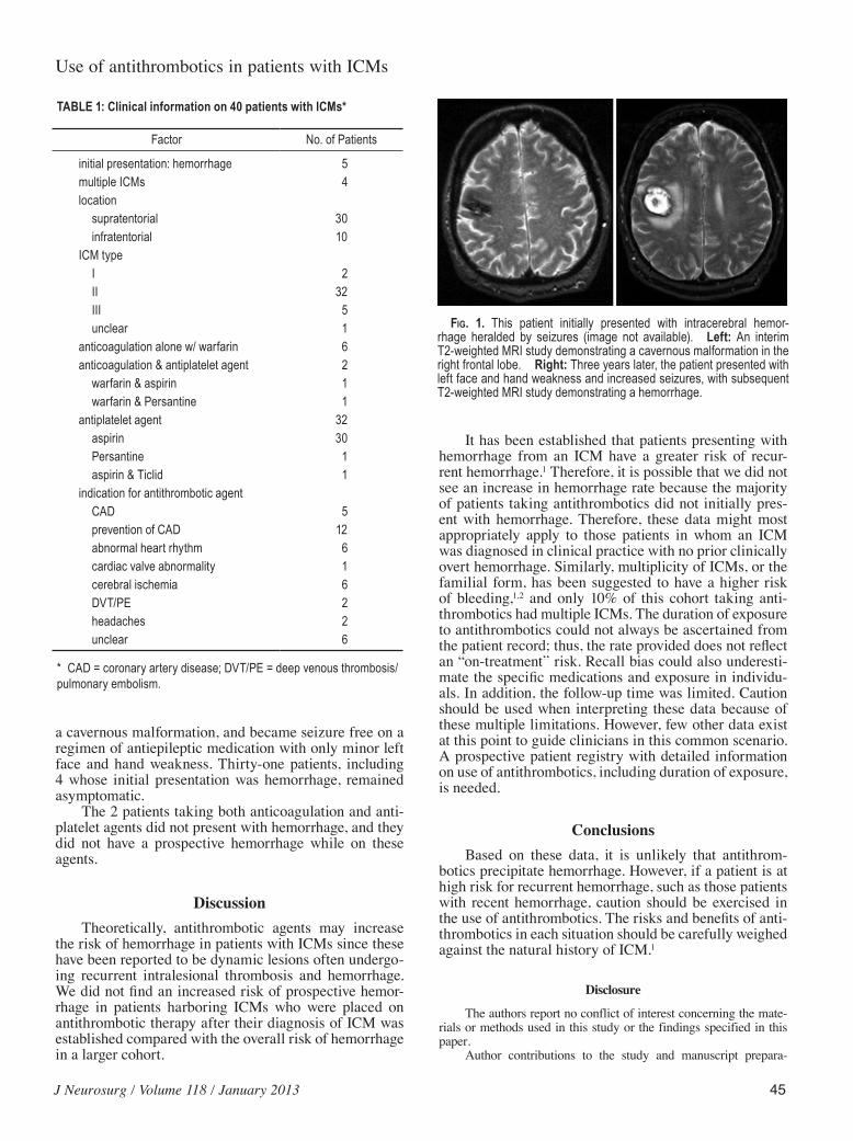
Thirty-two patients were treated with antiplatelet agents after the diagnosis of ICM. The indication for an-tiplatelet agents was most commonly related to coronary artery disease or prevention of such. One of these 32 pa-tients had a prospective symptomatic hemorrhage (Fig. 1). This hemorrhage occurred 3 years after the patient pre-sented with seizures and radiographic evidence of a small cortical hemorrhage. This patient was taking aspirin dur-ing that time for concomitant cardiovascular disease. The patient subsequently underwent surgery, which confirmed
J Neurosurg / Volume 118 / January 2013
Use of antithrombotics in patients with ICMs
45
a cavernous malformation, and became seizure free on a regimen of antiepileptic medication with only minor left face and hand weakness. Thirty-one patients, including 4 whose initial presentation was hemorrhage, remained asymptomatic.
The 2 patients taking both anticoagulation and anti-platelet agents did not present with hemorrhage, and they did not have a prospective hemorrhage while on these agents.
DiscussionTheoretically, antithrombotic agents may increase
the risk of hemorrhage in patients with ICMs since these have been reported to be dynamic lesions often undergo-ing recurrent intralesional thrombosis and hemorrhage. We did not find an increased risk of prospective hemor-rhage in patients harboring ICMs who were placed on antithrombotic therapy after their diagnosis of ICM was established compared with the overall risk of hemorrhage in a larger cohort.
It has been established that patients presenting with hemorrhage from an ICM have a greater risk of recur-rent hemorrhage.1 Therefore, it is possible that we did not see an increase in hemorrhage rate because the majority of patients taking antithrombotics did not initially pres-ent with hemorrhage. Therefore, these data might most appropriately apply to those patients in whom an ICM was diagnosed in clinical practice with no prior clinically overt hemorrhage. Similarly, multiplicity of ICMs, or the familial form, has been suggested to have a higher risk of bleeding,1,2 and only 10% of this cohort taking anti-thrombotics had multiple ICMs. The duration of exposure to antithrombotics could not always be ascertained from the patient record; thus, the rate provided does not reflect an “on-treatment” risk. Recall bias could also underesti-mate the specific medications and exposure in individu-als. In addition, the follow-up time was limited. Caution should be used when interpreting these data because of these multiple limitations. However, few other data exist at this point to guide clinicians in this common scenario. A prospective patient registry with detailed information on use of antithrombotics, including duration of exposure, is needed.
ConclusionsBased on these data, it is unlikely that antithrom-
botics precipitate hemorrhage. However, if a patient is at high risk for recurrent hemorrhage, such as those patients with recent hemorrhage, caution should be exercised in the use of antithrombotics. The risks and benefits of anti-thrombotics in each situation should be carefully weighed against the natural history of ICM.1
Disclosure
The authors report no conflict of interest concerning the mate-rials or methods used in this study or the findings specified in this paper.
Author contributions to the study and manuscript prepara-
TABLE 1: Clinical information on 40 patients with ICMs*
Factor No. of Patients
initial presentation: hemorrhage 5multiple ICMs 4location supratentorial 30 infratentorial 10ICM type I 2 II 32 III 5 unclear 1anticoagulation alone w/ warfarin 6anticoagulation & antiplatelet agent 2 warfarin & aspirin 1 warfarin & Persantine 1antiplatelet agent 32 aspirin 30 Persantine 1 aspirin & Ticlid 1indication for antithrombotic agent CAD 5 prevention of CAD 12 abnormal heart rhythm 6 cardiac valve abnormality 1 cerebral ischemia 6 DVT/PE 2 headaches 2 unclear 6
* CAD = coronary artery disease; DVT/PE = deep venous thrombosis/pulmonary embolism.
Fig. 1. This patient initially presented with intracerebral hemor-rhage heralded by seizures (image not available). Left: An interim T2-weighted MRI study demonstrating a cavernous malformation in the right frontal lobe. Right: Three years later, the patient presented with left face and hand weakness and increased seizures, with subsequent T2-weighted MRI study demonstrating a hemorrhage.
K. D. Flemming et al.
46 J Neurosurg / Volume 118 / January 2013
tion include the following. Conception and design: Flemming. Acquisition of data: Flemming. Analysis and interpretation of data: Flemming, Christianson. Drafting the article: Flemming. Criti-cally revising the article: all authors. Reviewed submitted version of manuscript: all authors. Approved the final version of the manuscript on behalf of all authors: Flemming. Statistical analysis: Flemming, Christianson.
References
1. Flemming KD, Brown RD Jr: The natural history of intra-cranial vascular malformations, in Winn H (ed): Youmans Neurological Surgery, ed 6. Philadelphia: Elsevier Saunders, 2011, Vol 1, pp 4016–4033
2. Flemming KD, Link MJ, Christianson TJH, Brown RD Jr: Prospective hemorrhage risk of intracerebral cavernous mal-formations. Neurology 78:632–636, 2012
3. Henninger N, Ahmad N, Morris JG: Intravenous thrombolysis in a patient with known cavernous malformation: a first case report. Am J Emerg Med 28:117, e1–e3, 2010
4. Moriarity JL, Clatterbuck RE, Rigamonti D: The natural his-
tory of cavernous malformations. Neurosurg Clin N Am 10: 411–417, 1999
5. Pozzati E, Zucchelli M, Marliani AF, Riccioli LA: Bleeding of a familial cerebral cavernous malformation after prophy-lactic anticoagulation therapy. Case report. Neurosurg Focus 21(1): e15, 2006
6. Rigamonti D, Hadley MN, Drayer BP, Johnson PC, Hoenig-Rigamonti K, Knight JT, et al: Cerebral cavernous malforma-tions. Incidence and familial occurrence. N Engl J Med 319: 343–347, 1988
7. Zabramski JM, Wascher TM, Spetzler RF, Johnson B, Golfi-nos J, Drayer BP, et al: The natural history of familial cavern-ous malformations: results of an ongoing study. J Neurosurg 80:422–432, 1994
Manuscript submitted November 14, 2011.Accepted August 23, 2012.Please include this information when citing this paper: published
online September 21, 2012; DOI: 10.3171/2012.8.JNS112050.Address correspondence to: Kelly D. Flemming, M.D., Depart-
ment of Neurology, Mayo Clinic, 200 First Street SW, Rochester, Minnesota 55905. email: [email protected].