use of functional electrical stimulation to improve hand
TRANSCRIPT

Use of Functional Electrical Stimulation to Improve Hand Function with Cervical Spinal Cord
Injury: A Case Study
by
Natalie Musselman
A thesis presented to the Honors College of Middle Tennessee State University in partial
fulfillment of the requirements for graduation from the University Honors College
Fall 2016

Use of Functional Electrical Stimulation to Improve Hand Function with Cervical Spinal Cord
Injury: A Case Study
by
Natalie Musselman
APPROVED:
____________________________
Dr. Sandra Stevens
Health and Human Performance
_____________________________
Dr. Doug Winborn
Chair, Health and Human Performance
___________________________
Dr. David Nelson
Biology
Honors Council Representative
___________________________
Dr. John Vile
Dean, University Honors College

i
Abstract
Spinal cord injuries paralyze many people in the United States each year. The injury
location has an impact on the degree of residual function. For people with injuries to the
cervical region, loss of hand function is typically observed. This inhibits ability to
perform daily activities and results in loss of independence; therefore, restoration of hand
function is an important area of study. Functional electrical stimulation applied to
paralyzed nerves is an intervention implemented to promote neural health and strength.
The NESS H200 is a stimulation device for the hands that is available commercially. This
system was applied daily to upper extremities of a quadriplegic participant. Following
treatment, no increase the grip or pinch force was observed and no change in the
functional assessment was noted. When attempting to increase hand function, the NESS
H200 may not be the most time or cost effective treatment for patients with similar
injuries.

ii
Table of Contents
Page
List of Figures…………………………………………………………………………….iii
List of Tables……………………………………………………………………………..iii
List of Abbreviations……………………………………………………………………..iv
Introduction………………………………………………………………………………..1
Methods……………………………………………………………………………………6
Results……………………………………………………………………………………10
Discussion………………………………………………………………………………..15
References………………………………………………………………………………..21
Appendices……………………………………………………………………………….24
Appendix A: Regions of the Spinal Cord and Spinal Nerves……………………25
Appendix B: IRB Approval……………………………………………………...26
Appendix C: Photo Release……………………………………………………...29

iii
List of Figures Page
Figure 1: Study participant wearing the Bioness device…………………………..5
Figure 2: Proper use of the NESS H200…………………………………………..7
Figure 3: JAMAR Smedley-Type Lightweight Hand Dynamometer……………..8
Figure 4: J-Tech Commander Muscle Tester……………………………………...9
Figure 5: Pre-Intervention Pinch Strength……………………………………….11
Figure 6: Pinch Strength Average by Week……………………………………..12
Figure 7: Pre-Intervention Grip Strength………………………………………...13
Figure 8: Grip Strength Average by Week………………………………………13
Figure 9: Study participant on crutches………………………………………….14
List of Tables
Table 1: Pinch strength force outputs……………………………………………11
Table 2: Grip strength force outputs……………………………………………..12

iv
List of Abbreviations
SCI Spinal Cord Injury
CNS Central Nervous System
AIS The American Spinal Injury Association Impairment Scale
ADL Activities of Daily Living
FES Functional Electrical Stimulation
LMN Lower Motor Neuron
FIM The Functional Independence Measure
QIF The Quadriplegia Index of Function

1
Introduction
People engage in activities every day to which they devote little or no attention.
These tasks such as walking, eating, or even simply wiggling the toes, are all possible
thanks to the body’s nervous system. However, every year in the U.S. more than ten
thousand people experience traumatic spinal cord injury (SCI), are disabled, and in many
cases are unable to perform these daily functions. (1-2)
The nervous system functions to process information that it collects about the
body and its environment and generate an appropriate response that may involve sending
signals to muscles to contract, glands to secrete, or to initiate a wide variety of other
necessary functions. (1) These messages are sent via electrical signals transmitted by
specialized cells of the nervous system called neurons. (1-2) Sensory information from
the body is sent via sensory neurons to the spinal cord, a long and tubular structure,
which then sends this information to the brain. (1-2) Similarly, the spinal cord relays
information from the brain to effectors in the body via motor neurons. (1-2) The spinal
cord also controls the simplest reaction called a reflex in which sensory input or feedback
such as muscle or tendon stretch send signals to motor neurons of the spinal cord. These
neurons then fire, signaling the muscle to contract. Together, the brain and the spinal cord
constitute the central nervous system (CNS). The spinal cord is protected by a spinal
column which consists of protective membranes called meninges, bones called vertebrae,
and by muscular tissue. However, it may still sustain damage that, even when slight, can
manifest itself as severe disability. (1-2) Even minor contusions can lead to what is called

2
the ischemic cascade, where insufficient blood flow leads to a vicious cycle of swelling,
membrane damage, and cell death. (1)
The spinal cord is regionally named and consists of the cervical, thoracic, lumbar,
and sacral regions (Appendix A). (1-2) The cervical spinal cord is located in the neck
region and consists of eight segments and eight sets of spinal nerves that innervate the
arms and neck. (1) These are abbreviated as C1 through C8. (2) Each region of skin
(dermatome), organ, and muscle, connects at a particular level of the spinal cord, which
allows for some degree of specificity when identifying the site of a spinal cord injury, as
the symptoms of SCI depend on both the extent and location of the injury. (1-2) For this
case, injuries sustained to the cervical region, particularly C5 are of interest.
Injuries are further classified by the extent of the damage, where a complete
injury indicates no neurological function preserved below the injury site and an
incomplete injury indicates some preservation of function. (1) The American Spinal
Injury Association Impairment Scale (AIS) classifies injuries based on the evaluation of
muscle strength and skin sensation. (1-2) Grade A is a complete injury where all other
classifications are incomplete. (1-3) B is preservation of sensory but no motor function, C
is preservation of sensory and some weak motor function, D is preservation of sensory
and some stronger motor function of essential muscles, and grade E indicates normal
sensory and motor function. (1-2) The less complete the injury is, the more likely it is to
gain some recovery of function. (2) For those classified as a C, there is a 75 percent
chance that the ability to walk (with or without assistive devices) will be regained. (1)

3
SCI usually results in loss of some or all movement and sensation below the
injury level. (1) This type of injury when located high so all four limbs are paralyzed is
known as tetraplegia (or quadriplegia). (2) Injuries sustained in the middle cervical
regions still allow movement of the head and neck, but the hands, trunk, arms and legs
will be paralyzed and/or numb. (1) Due to paralysis of the chest muscles, breathing may
be difficult. (1) For C5 injuries, movement of the head, neck and shoulders is retained as
is flexion of the elbow. (3) Patients can expect to achieve independent feeding with
assistive devices, can be independent with an appropriate power wheelchair, but will need
maximal assistance for transfers. (3-4)
Evidence suggests, patients with tetraplegia report that the most difficult aspect of
their disability was loss of hand function. (5-6) Reduced dexterity in tetraplegic patients
due to paralysis of the hands and arms can affect activities of daily living (ADL) as well
as limiting vocational prospects. (7) Even a very small increase in hand function can
result in increased ability to perform ADL and increase independence. (5) Though
improving hand function is an area of great importance, the research is not consistent due
to differences in functional ability with various levels of SCI, and it usually involves
small sample sizes. (5) Though the majority of upper extremity function is regained in the
first six months after injury (3) and some studies agree that early rehabilitation is vital to
prevent functional loss, others have been successful with interventions starting in the
chronic phase of recovery. (5) In select patients with cervical spinal injury, surgeries and
functional electrical stimulation have been successful in regaining some function. (5, 8-
10)

4
Functional electrical stimulation (FES) is a technology fairly recently developed
and introduced commercially in 1990 (3) that stemmed from the knowledge that
electricity stimulates muscle contraction. (7) This technology applies electrical currents
to nervous tissue in an attempt to regain control over their functions and is being applied
to patients with SCI. (3, 7, 11) The ultimate goal is to induce changes that promote
muscular and nervous tissue health and allow function after the stimulation has ceased.
(11) The application of FES has been utilized to improve functionality of the upper
extremities but is only appropriate if the lower motor neuron (LMN) is not extensively
damaged. (3-4, 7, 11) C5 level quadriplegia patients typically have some regions of the
LMN intact and make viable candidates for FES. (7) Possible benefits to this type of
intervention include the ability to grasp, hold, and release a variety of objects. (4,7) Such
interventions have also been used to increase muscle size, treat osteoporosis, and control
spasticity (7). The Bioness company provides a commercially available FES device with
surface electrodes called the NESS H200 (Figure 1). (7, 11) The device supports the
wrists and has five electrodes for the extensors and flexors of the thumb and fingers, and
it has a control that stimulates various grasp and release patterns. (7-8, 11) A small scale
study showed ability to perform three ADL (using a telephone, eating with a fork, and
another ADL chosen by the subject) improved while utilizing the device after three
weeks of at home training. (8) A different study concluded that the device is only
effective on a limited subset of patients with C5 SCI. (9)

5
Figure 1: Study participant wearing the Bioness device
The Middle Tennessee State University Exercise Science Department has been
successful in its unique research with spinal cord injuries and aquatic therapy. One
participant has experienced the benefits of this therapy after sustaining spinal cord trauma
of the C5 region during a skiing accident in 2012. His injury is classified as AIS C
(incomplete), but function wise appears like a complete injury, though at the time of
injury, it was noted that the spine was not severed, nor the meninges breached. To date,
he has regained the ability to take steps both while on the underwater treadmill and above
ground with a walker. However, due to the inability to grip, when doing above ground
walking with crutches, his hands must be wrapped in wrist splints and then around the
hand holds of the crutches by another person to provide support. This dependence due to

6
inadequate hand function has slowed the progress that can be made in regards to the
functioning of the lower extremities and in regaining more independence.
Thesis Statement
The purpose of this study is to determine if function and hand strength can be
improved by utilizing the NESS H200 in this case. This will be done by evaluating both
strength and hand function on ADLs as well as changes in mobility based on the ability
of the participant to use crutches. It is hypothesized that the FES will increase the force
output of tested measures of strength by at least ten percent, and consequently, increase
the functional measure score.
Methods
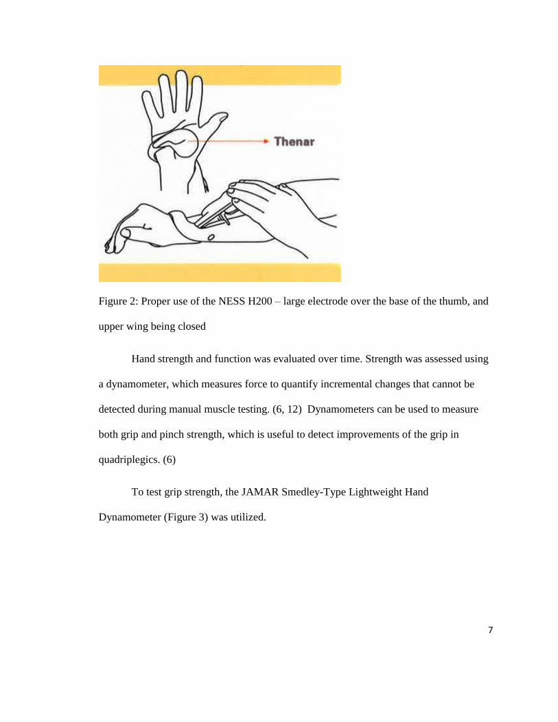
Before the device was used, all metal was removed from the wrist and hand. The
electrodes were wet, and the orthosis (a stabilizer) was put on the wrist, making sure that
the large electrode sat over the base of the thumb. The wing on top of the orthosis was
completely closed (Figure 2). The system was controlled by a wireless unit that after
being turned on allowed the training parameters to be entered. The participant had two
devices, one for each hand, and utilized them with the stimulation intensity setting at
eight, a relatively high intensity, for a duration of forty-five minutes to an hour. The
devices have a personal operating mode and were used once every day at the participant’s
home. The “trigger” button on the device stimulated the grasp and release patterns via
electrical stimulation and could be put in an exercise mode or a function mode for doing
daily activities. The participant used devices consistently and met the criteria established
in the protocol.
7
Figure 2: Proper use of the NESS H200 – large electrode over the base of the thumb, and
upper wing being closed
Hand strength and function was evaluated over time. Strength was assessed using
a dynamometer, which measures force to quantify incremental changes that cannot be
detected during manual muscle testing. (6, 12) Dynamometers can be used to measure
both grip and pinch strength, which is useful to detect improvements of the grip in
quadriplegics. (6)
To test grip strength, the JAMAR Smedley-Type Lightweight Hand
Dynamometer (Figure 3) was utilized.

8
Figure 3: JAMAR Smedley-Type Lightweight Hand Dynamometer
To utilize the equipment, the participant sits in an upright position with the elbow
forming a ninety degree angle and the wrist and forearm relaxed. (13) He then squeezes
the dynamometer as hard as possible, and the amount of force produced is indicated on
the dial. (13) In this case, upper extremity impairments related to tetraplegia limited the
participant’s ability to hold his arm in the standard position recommended for testing with
this instrument. Therefore, support was provided to ensure the participant’s arm position
remained consistent over the course of testing, and the use other muscles to compensate
for lack of hand strength was minimized. The grip force was tested twice each time in
both the left and right hands, and the average was taken to represent the actual force
reading for each hand. For males age 25 to 29, grip strength at the tenth percentile is 90
pounds of pressure in the right and 82 in the left hand. (13) However, even with the
intervention, due to the nature of SCI, we did not expect the participant to reach these
parameters, and were simply looking for changes from the baseline testing. To test pinch

9
strength, the Commander Muscle Tester (Figure 4) was used. The thumb was placed on
one side of the device, and the two adjacent fingers were on the other. (14) Maximum
pinch force was exerted and the output recorded. As with the grip strength test, the pinch
strength was measured twice on each hand and the average of each was taken. Baseline
measurements were taken before the intervention was implemented, and posttest
measurements were taken upon the conclusion of the intervention. Two measurements
were taken in-between to monitor progress.
Figure 4: J-Tech Commander Muscle Tester
Ultimately, the purpose of increased strength in the hand is the ability to apply
any gain to daily tasks and to achieve more independence. There is a wide variety of
measures that have been developed to evaluate function and ADL. (5) One of the most

10
commonly used assessments for SCI is the Functional Independence Measure (FIM).
However, it has been argued that the measure is not sensitive enough to identify changes
in patients with SCI and that some of the tasks are not feasible for a person with
quadriplegia. (15) The Quadriplegic Index of Function (QIF) was developed specifically
for people with tetraplegia and to be a measure more sensitive to change than the FIM.
(6) The measure tests ten different areas such as transfers and grooming, and the ability to
perform certain tasks in these areas is rated from 0 (dependent) to 4 (independent). (16)
Each of the categories is weighted with the final score ranging from 0 to 100. Though
scoring on the QIF is less specific than the FIM, (15) the feeding category of this
assessment is able to assess changes not identified on the FIM. (6) The QIF has a high
correlation with the overall FIM score (6) and was found to be reliable and a viable
option for evaluating improvement in persons with quadriplegia in clinical studies and
when monitoring program outcomes (16); thus it was selected as the ADL measure for
this case study. The QIF was tested once at the start of the intervention and once at the
end. The intervention was concluded in early May, three months from the beginning FES,
which is adequate time to detect changes.
Results
To assess the effectiveness of the intervention, scores before and after FES were
compared as well as those taken mid-intervention. Any trend in the data was compared to
hand strength data taken yearly since 2013. Because a case study’s sample size is one,
statistical measures were not appropriate, and instead pinch and grip strength data was

11
analyzed graphically. When the full effort is being exerted, no more than a ten percent
variation in strength is expected. (13)
Table 1: Pinch strength force outputs and averages in pounds both during and prior to the
intervention.
Date Pinch R (1) Pinch R (2) Pinch R (Avg) Pinch L (1) Pinch L (2) Pinch L (Avg)
Feb. 2013 3.5 3 3.25 2.25 2.25 2.25
Feb. 2014 3 3 3 2.25 2 2.13
Jan. 2015 2 2.25 2.13 1.5 1.5 1.5
Feb. 2016/ Week 0 2 2 2 1 1.5 1.25
Week 6 2 2 2 2 2 2
Week 9 2 2 2 2 1.5 1.75
Week 12 2.5 2 2.25 3 2 2.5
Figure 5: The graph shows pinch force data in pounds each year prior to the start of FES.

12
Figure 6: Changes in the average pounds of pinch force as detected by the J-Tech
Commander Muscle Tester over the 12 week use of the NESS H200. Time zero is
baseline testing done as included in figure 5.
Table 2: Grip strength force outputs and averages in pounds both during and prior to the
intervention.
Date Grip R (1) Grip R (2) Grip R (Avg) Grip L (1) Grip L (2) Grip L (Avg)
Feb. 2013 4.5 3.5 4 4 5 4.5
Feb. 2014 3 3 3 4.5 4.5 4.5
Jan. 2015 2.5 1.5 2 3.5 2.5 3
Feb. 2016/ Week 0 1.5 2.7 2.1 3 5 4
Week 6 5 3 4 5 5 5
Week 9 5.5 5.5 5.5 4.5 4 4.25
Week 12 3.4 3.5 3.45 3 2.5 2.75

13
Figure 7: Yearly changes in generated grip force in pounds prior to the use of the FES
intervention.
Figure 8: Changes in the average maximal grip force from baseline to the conclusion of
the intervention as measured by the dynamometer.

14
Before the intervention with the NESS H200 began, the participant exhibited a
QIF score of 52.83, and at the conclusion of the intervention the score was 56 which is a
six percent change. Furthermore, by May the participant was able to take three steps
while on crutches. From sit to stand, physical assistance was provided, but all steps were
taken independently. Throughout the study, the type and degree of assistance remained
unchanged, and the hands were still wrapped extensively and stabilized with splints to
provide needed grip support (Figure 9).
Figure 9: Study participant standing crutches with the hands wrapped for support

15
In order better to understand what effects the Bioness was exerting, the participant
was asked to report any changes he felt due to the intervention. In order to prevent bias
reporting, he was not told the intention of the FES or given any positive or negative
comments about his reporting. Over the course of the study, the reported effects of the
Bioness stayed consistent. The FES made his hands feel “looser,” which was said in a
positive manner, especially in the hours and days after the device was utilized. When first
utilized, these loosening effects lasted an hour or two after use, but upon use every day
these effects lasted longer. If a treatment was missed, the participant reported increased
tightness of both hands once again. There was no mention of increased feelings of
strength or of a greater ease when doing daily tasks.
Discussion
Prior to the intervention, the yearly strength data taken shows a slight decline in
the pinch strength of both hands (figure 5). As illustrated in Figure 7, the grip strength of
the right hand also exhibits a decline while in the left hand the values remained relatively
constant. The declines could be due to a lack of stimulation from the nervous system,
consequently leading to the atrophy of the muscles responsible for movement in the hand.
However, because the decline is slight and no functional measures were taken, it is
unclear as to whether such a decline would further inhibit the existing function.
If loss of strength was due to a lack of stimulation, the use of appropriate FES
would theoretically slow or reverse this decline. This change in the pattern of strength
loss should reflect greater hand function through the use of tenodesis grasp, which is a
compensatory grip strategy implemented by C5-6 tetraplegics. A person with this level of

16
injury would be able to grip due to flexion of the fingers achieved by the extension of the
wrist. (17) The effectiveness of this grip can be improved by increased support with
adaptive devices, increased range of motion at the wrist, and increased muscular strength.
(18) If, therefore, the NESS H200 was able to stimulate greater strength, then the
tenodesis grip would be improved as well. If grip is weak or a large enough contraction is
not achieved, then adaptive devices must be used to get the desired results. (17) For
example, equipment such as splints can help make joints more stable to prevent injury or
other issues.
Pinch strength of the right hand remained unchanged over the course of the
intervention (figure 6). The amount of force exerted by the left hand pinch instead
showed an increase from the start to the end of the trial. For grip strength, each hand
exhibited opposite trends. Overall the force of grip by the right hand increased slightly
while that of the left hand decreased slightly (figure 8). In both measures of strength,
there appeared to be no consistent trend in the changes that occurred while utilizing the
NESS H200. While recovery from this type injury is expected to consist of fluctuations,
there is no continuity in the overall direction of strength. Neither hand appeared to be
more responsive to FES than the other, as the pinch strength of the left hand exhibited an
increase while grip strength of the right hand exhibited an increase instead. Furthermore,
though the left hand increased its pinch strength, it decreased its grip strength. While pre-
intervention measures consistently show a decline or a stasis of strength, the measures
during the intervention show no such consistency. Therefore, it cannot be concluded
given this protocol that the NESS H200 increases strength of the muscles responsible for

17
hand function. In most cases, however, there was either a slight increase in strength or a
maintenance of strength over the course of the study, which may mean that while utilized
consistently the NESS H200 could prevent the decline that was seen prior to its use. The
overall changes in the negative direction prior to utilization of the NESS H200 were
small, and occurred over the course of a year. Therefore, it is possible that the duration of
testing was too short to show what, just like the grip strength of the left hand, may have
been an eventual decline in function. The study duration, the loss of left hand grip
strength, and the inconsistency of the data trends make it impossible to conclude with
confidence that this NESS H200 may prevent this decline.
One of the functional measurements for the study, the QIF, further supports the
lack of consistency seen in the strength data. The score exhibited only a six percent
increase over the course of three months. This small change, however, is easily attributed
to changes in question interpretation and feelings of the participant. For example, there is
a significant difference in interpreting a wheelchair to vehicle transfer as moving from
the wheelchair seat to the seat of the car instead of simply driving the wheelchair into the
back of the vehicle. Furthermore, fluctuations so small could simply be due to the
circumstances of the participant that particular day, such as how difficult it was to get
dressed and in what type of garment. The QIF is designed to be sensitive to minor
changes in function which causes a greater change in score as opposed to other more
common tests. (16) Therefore, this correspondingly small change indicates that little to no
functional gain was obtained as detectable by this measure. This result is not surprising
given the very small changes in strength seen exhibited in figures 5 through 8.

18
The participant’s description of the effects of the Bioness also seem to support
these results. As stated, there was no mention of increased feelings of strength or ability
to perform ADL. The only reported, detectable change was a temporary increased
looseness of the hands. This was spoken of in a positive manner, and was probably more
comfortable, however, these effects may have been counterintuitive. In order to grasp
with a cervical spinal injury, additional tension in the muscles may have been useful as
there would have been less tension to produce in order to effectively grip something. In
fact, functional tenodesis grip requires some tightness of the finger flexors in order to be
functional. (17) One of the hopeful goals of the intervention was an ability to grasp the
crutches freely while in use, and “looser” hands would not necessarily aid in this goal.
Regardless, the lack of increase in hand strength did not allow a safe grip to be obtained.
While the participant was able to take three steps with the crutches, the hands were
wrapped onto the handles and the wrists were also stabilized with adaptive equipment.
Overall, there were no issues with the protocol for the intervention. The devices
can be safely utilized, and the only issue was the inconvenience and time required to
utilize the device appropriately. Nevertheless, given this particular protocol, the NESS
H200 was not successful at increasing strength of the hand such that functional gain was
achieved. The study is limited in that it only consists of one participant, and because the
nature of SCI is extremely varied, the results are not necessarily applicable to the entire
population. However, this case exhibits a stage of recovery advanced for his injury level,
and hand function began to be a limiting factor in training. In the future when working
with patients with similar injuries, the very costly and time consuming treatment with the

19
Bioness device may not be a prudent course of action since the desired outcome was not
achieved in this case. Furthermore, due to time constraints of the participant’s residence
in the area, the study could not be conducted over a long period of time, nor could further
data be taken after secession of the daily FES. Had the study been conducted longer, the
long term trend in data could have been used to determine if the NESS H200 prevents
decline.
This Bioness Company advertises very different results than those achieved in
this study, even though one of the many populations to which the device is being
marketed are those with incomplete spinal cord injuries of the cervical spine. One of the
advertised intentions of the FES system is to prevent the atrophy of muscles, which was
not necessarily seen in this study. (19) Improving or maintaining range of motion is also
listed as one of the benefits of the device, along with others, all for the purposes of more
easily performing ADL. Furthermore, Bioness claims that the H200 may also reeducate
the muscle so that they can function without the system. The brochure boasts ‘Grasp
onto Life,’ (19: p.1) selling the concepts of freedom and independence, which may be
successful in other situations, but not in this particular population subset whose
restoration of hand function is a significant need. Unfortunately, given the measured
parameters, it seems the company’s claims may be too good to be true, though further
study is needed. It was hypothesized that the Bioness device would increase strength by
at least ten percent, which would result in an improvement in the functional measure
(QIF) score. For this given methodology, the NESS H200 did not consistently result in

20
increases in pound of force generated by grip or pinch, and thus, the QIF score did not
exhibit a substantial percent change.

21
References
1. Selzer ME, Dobkin BH. Spinal cord injury. New York: Demos Health; 2008.
2. Liverman CT, Altevogt BM, Joy JE. Spinal cord injury: progress, promise, and
priorities. Washington DC: National Academies Press; 2005 July.
3. Bryce, TN. Spinal cord injury. New York: Demos Medical; 2010.
4. Formal CS, Cawley MF, Stiens SA. Spinal cord injury rehabilitation. 3.
Functional outcomes. Arch Phys Med Rehabil. 1997 Mar;78(3 Suppl):S59-64.
5. Lu X, Battistuzzo CR, Zoghi M, Galea MP. Effects of training on upper limb
function after cervical spinal cord injury: a systematic review. Clin Rehabil. 2015
Jan;29(1):3-13.
6. Tuijl JH, Janssen-Potten, YJ, Seelen HA. Evaluation of upper extremity motor
function tests in tetraplegics. Spinal Cord. 2002 Feb;40(2):51-64.
7. Ragnarsson KT. Functional electrical stimulation after spinal cord injury: current
use, therapeutic effects, and future directions. Spinal Cord. 2008 Apr;46(4):255-
74.
8. Alon G, McBride K. Persons with C5 or C6 tetraplegia achieve selected
functional gains using a neuroprosthesis. Arch Phys Med Rehabil. 2003
Jan;84(1):119-24.
9. Snoek GJ, IJzerman MJ, in 't Groen FA, Stoffers TS, Zilvold G. Use of the NESS
handmaster to restore handfunction in tetraplegia: clinical experiences in ten
patients. Spinal Cord. 2000 Apr;38(4):244-49.

22
10. Gorman PH, Wuolle KS, Peckham PH, Heydrick D. Patient selection for an upper
extremity neuroprosthesis in tetraplegic individuals. Spinal Cord. 1997
Sep;35(9):569-73.
11. Peckham PH, Knutson JS. Functional Electrical Stimulation for neuromuscular
application. Annu Rev Biomed Eng. 2005;7:327-60.
12. Mulcahey M, Hutchinson D, Kozin S. Assessment of upper limb in tetraplegia:
Considerations in evaluation and outcomes research. J Rehabil Res Dev.
2007;44(1):91-101.
13. Country Technology, Inc. Smedley-Type Lightweight Hand Dynamometer. Gays
Mills, WI: Country Technology, Inc.
14. Livingston T, Bernardi D, and Carroll M. Commander Muscle Tester User’s
Manual. Midvale, UT: JTECH Medical; 2015.
15. Bryden AM, Sinnott KA, Mulcahey MJ. Innovative Strategies for Improving
Upper Extremity Function in Tetraplegia and Considerations in Measuring
Functional Outcomes. Top Spinal Cord Inj Rehabil. 2005;10(4):75-93.
16. Gresham GE, Labi ML, Dittmar SS, Hicks JT, Joyce SZ, Stehlik MA. The
Quadriplegia Index of Function (QIF): sensitivity and reliability demonstrated in a
study of thirty quadriplegic patients. Paraplegia. 1986 Feb;24(1):38-44.
17. Ford J, Duckworth B. Physical management for the quadriplegic patient. 2nd ed.
Philadelphia: Davis; 1987.

23
18. Kohlmeyer KM, Hill JP, Yarkony GM, Jaeger RJ. Electrical stimulation and
biofeedback effect on recovery of Tenodesis grasp: A controlled study . Archives
of Physical Medicine & Rehabilitation . 1996Jul;77(7):702–6.
19. Bioness Inc. NESS H200 Wireless. Valencia, CA: Bioness Inc; 2013.
24
List of Appendices Page
Appendix A: Regions of the spinal cord and spinal nerves……………………25
Appendix B: IRB Approval……………………………………………………26
Appendix C: Photo Release……………………………………………………29

25
Appendix A
Regions of the Spinal Cord and Spinal Nerves
Reference
Liverman CT, Altevogt BM, Joy JE. Spinal cord injury: progress, promise, and priorities.
Washington DC: National Academies Press; 2005 July. Figure 2-2, Functions
controlled by nerves at different levels of the spine. Damage at a particular level
usually impairs the functions controlled by all nerves at lower levels; p. 33.

26
Appendix B
IRB Approval
IRB INSTITUTIONAL REVIEW BOARD
Office of Research Compliance,
010A Sam Ingram Building,
2269 Middle Tennessee Blvd
Murfreesboro, TN 37129
IRBN008 Version 1.0 Revision Date 04/13/2016
IRBN008 - PROTOCOL APPROVAL NOTICE Wednesday, April 1, 2016 Investigator(s): Sandra Stevens (PI), Don W. Morgan Investigator(s’) Email(s): [email protected]; [email protected]
Department: Health and Human Performance
Study Title: The effects of underwater treadmill training on mobility and function in
adults with spinal cord injuries Protocol ID: 15-200
Dear Investigator(s), The above identified research proposal has been reviewed by the MTSU Institutional Review Board (IRB) through the FULL COMMITTEE REVIEW mechanism under 45 CFR part 46. and 21 CFR part 56. This protocol was reviewed by the IRB at a convened meeting which meets the HHS requirements on 4/1/15. The IRB has determined that this study poses minimal risk to the participants or that you have satisfactorily worked to minimize the risks, and you have satisfactorily addressed all of the concerns brought up during the review. A summary of the IRB action and other particulars in regard to this protocol application is tabulated as shown below: IRB Action APPROVED for one year
Date of expiration 4/1/2017 Participant Size 10 (TEN) Participant Pool Adult diagnosed with a SCI, free from progressive medical condition

27
Exceptions NONE Restrictions 1. Signed informed consent for the collection of biological sample(s); (2) Patient records including full name, telephone numbers, street address, email address and photographic information MUST be stored securely in the designated location Comments: This protocol was originally requested through the expedited process. It was referred to the full committee on 3/17/2015 by the primary reviewer in consutation with the secondary reviewer. Subsequently, the protocol approved by the IRB after clarifications and alterations to the protocol . Amendments Date 9/18/2015 Post-approval Amendments 1. Increase in training frequency to three times per week instead of previously approved two times has been granted 2. Change to the testing procedure to use wireless electromyography and electrical stimulation has been approved. Institutional Review Board Office of Compliance Middle Tennessee State University 3. Addition of a resistive exercise (refer to addendum request on file) to the protocol has been approved This protocol can be continued for up to THREE years (4/1/2018) by obtaining a continuation approval prior to 4/1/2017. Refer to the following schedule to plan your annual project reports and be aware that you may not receive a separate reminder to complete your continuing reviews. Failure in obtaining an approval for continuation will automatically result in cancellation of this protocol. Moreover, the completion of this study MUST be notified to the Office of Compliance by filing a final report in order to close-out the protocol. Continuing Review Schedule: Reporting Period Requisition Deadline IRB Comments First year report 3/1/2016 The continuing review was completed through the expedited procedure in accordance with Category #9 sub classification 3: "Continuing review of research previously approved by the IRB at a convened meeting where NO ADDITIONAL RISKS OF THE RESEARCH HAVE BEEN IDENTIFIED" as defined further in 9.3a "the research project as a whole involved no more than minimal risk." Second year report 3/1/2017 INCOMPLETE Final report 3/17/2018 INCOMPLETE The investigator(s) indicated in this notification should read and abide by all of the post-approval conditions imposed with this approval. Refer to the post-approval guidelines posted in the MTSU IRB’s website. Any unanticipated harms to participants or adverse events must be reported to the Office of Compliance at (615) 494-8918 within 48 hours of the incident. Amendments to this protocol must be approved by the IRB. Inclusion of new researchers must also be approved by the Office of Compliance before they begin to work on the project. All of the research-related records, which include signed consent forms, investigator information and other documents related to the study, must be retained by the PI or the faculty advisor (if the PI is a student) at the secure location mentioned in the protocol application. The data storage must be maintained for at least three (3) years after study completion. Subsequently, the researcher may destroy the data in a manner that maintains confidentiality and anonymity. IRB reserves the right to modify, change or cancel the terms of this letter without prior notice. Be advised that IRB also reserves the right to inspect or audit your records if needed. Sincerely, Institutional Review Board Middle Tennessee State University Quick Links:

28
Click here for a detailed list of the post-approval responsibilities. Institutional Review Board Office of Compliance Middle Tennessee State University IRBN008 – Full Committee Protocol Approval Notice Page 3 of 3
29
Appendix C
Photo Release