use of paired serum sample for the diagnosis of …
TRANSCRIPT

USE OF PAIRED SERUM SAMPLE FOR THE
DIAGNOSIS OF Toxoplasma INFECTION IN
SELECTED POPULATION BY USING ELECSYS
TOXO IgG/IgM ASSAY IN HUSM
by
DR. PADMALOSENI THANGARAJAH
Dissertation Submitted in Partial Fulfillment of the
Requirements for the Degree of Master of Pathology
(Microbiology)
SCHOOL OF MEDICAL SCIENCES
UNIVERSITI SAINS MALAYSIA
2018
ii
ACKNOWLEDGEMENT
Bismillah hirrahman nirrahim,
Foremost, I am grateful to Allah s.w.t for his blessings in establishing me to complete
my thesis and entire academic career goals. This thesis becomes a reality with the kind
support and assistance of many individuals. I would like to extend my sincere thanks to
all of them.
I wish to express my sincere gratitude and deep respect to my main supervisor, Prof. Dr.
Zeehaida Mohamed, and co-supervisor Dr. Nabilah Ismail for their experts, valuable
guidance and constant encouragement to me. I also take this opportunity to express my
deepest indebtedness to my main supervisor in providing me consistent invaluable
moral support and motivation to endure all the obstacles throughout my study.
I place on record, my sense of appreciation to Mr. Muhammad Amiruddin Abdullah,
my field supervisor for his knowledgeable expertise in serology and his unlimited
perseverance during my laboratory work and thesis writing. My thanks extends to
everybody at the Medical Microbiology Laboratory (HUSM) for their kind help and
cooperation. I also would like extend my warmest appreciation to all my colleagues
Wan Amani, Norazizah, Faizah and especially Mahirah for providing her best
knowledge about technical part in writing and all the moral support to accomplish my
thesis. I humbly extend my thanks to all concerned persons who co-operated with me in
this regard.
Finally, I would like to acknowledge with gratitude, the support and love of my family-
my parents, Thangarajah and Indrani, my sisters Logeswary, Mohana and my brother in

iii
law, Nathan. My endless love to my husband Mohammad Faisal and adorable son
Arryan for their boundless commitment and support to keep me going during this study.
Last but not least, I sincerely convey my gratitude to the University for approving and
providing me a favourable environment to grasp all the knowledge and skill for future
undertakings.
My prayers to Allah s.w.t is to consistently keep me strong and patient in enduring
hurdles in the future and shower his blessings to my family.
iv
TABLE OF CONTENTS
ACKNOWLEDGEMENTS .............................................................................................. ii
TABLE OF CONTENT ................................................................................................... iv
LIST OF FIGURES ....................................................................................................... viii
LIST OF TABLES ........................................................................................................... ix
ABRREVIATIONS ......................................................................................................... xi
ABSTRACT ................................................................................................................... xiv
ABSTRAK .................................................................................................................... xvii
CHAPTER ONE: INTRODUCTION ........................................................................... 1
1.1. Background of the study ............................................................................................ 1
1.2. Rationale of the study ................................................................................................ 3
1.3. Literature review ........................................................................................................ 5
1.3.1. Toxoplasma gondii ............................................................................................... 5
1.3.2. Three parasitic stages ........................................................................................... 5
1.3.3. Life cycle of T. gondii .......................................................................................... 7
1.3.4. Transmission......................................................................................................... 9
1.3.5. Clinical features of Toxoplasma infection ............................................................ 9
1.3.5.1. Manifestation during pregnancy and congenital toxoplasmosis ..................... 9
1.3.5.2. Role of placenta in T. gondii transmission and pathophysiology ................. 10
1.3.5.3. Toxoplasmosis in immunocompromised patients ......................................... 12
1.3.5.4. Ocular toxoplasmosis .................................................................................... 13
1.3.6. Diagnostic methods ............................................................................................ 13
1.3.6.1. Serology diagnosis ........................................................................................ 13
1.3.6.2. Kinetic of antibody responses ....................................................................... 14
1.3.6.3. Sabin – Feldman dye test (SFDT) ................................................................. 16
1.3.6.4. Indirect fluorescent assay (IFA) .................................................................... 16
1.3.6.5. Agglutination tests ........................................................................................ 16
1.3.6.6. Enzyme – linked immunosorbent assay (ELISA) ......................................... 17
1.3.6.7. Parasite isolation ........................................................................................... 18
1.3.6.8. Histologic diagnosis ...................................................................................... 18
1.3.6.9. Molecular diagnosis ...................................................................................... 18
1.3.7. Treatment ............................................................................................................ 19

v
1.3.8. Prevention and control measures ........................................................................ 21
1.4. Conceptual framework ............................................................................................. 23
1.5. Objectives ................................................................................................................ 24
1.5.1. General objectives .............................................................................................. 24
1.5.2. Specific objectives .............................................................................................. 24
1.5.3. Research hypothesis ........................................................................................... 24
CHAPTER TWO: MATERIAL AND METHODS ................................................... 25
2.1. Study design ............................................................................................................. 25
2.2. Reference population ............................................................................................... 25
2.3. Source pupulation .................................................................................................... 25
2.4. Sampling frame ........................................................................................................ 25
2.5. Inclusion criteria ...................................................................................................... 25
2.6. Exlusion criteria ....................................................................................................... 26
2.7. Determination of sample size .................................................................................. 26
2.8. Sampling method .................................................................................................... 29
2.9. Variable definition ................................................................................................... 30
2.10. Sample collection ................................................................................................... 30
2.11. Data collection ....................................................................................................... 31
2.12. Operational definition/Gold standard .................................................................... 31
2.13. Research / measurement tool ................................................................................. 33
2.13.1. ECLIA .............................................................................................................. 33
2.13.1.1. Recombinant SAG1 antigen ........................................................................ 33
2.13.1.2. Principles of ECLIA .................................................................................... 34
2.13.1.3. Elecsys measuring cell ................................................................................ 35
2.13.1.4. Electrochemiluminescence detection: reaction scheme .............................. 36
2.13.1.5. Elecsys Toxo IgG assay and measuring range ............................................ 37
2.13.1.6. Elecsys Toxo IgM assay ............................................................................. 38
2.14. Flow chart of the study. ......................................................................................... 39
2.15. Serological interpretation based on classification of infection ............................. 41
2.16. Statistical analysis .................................................................................................. 44
2.17. Ethical issues. ......................................................................................................... 44
RESULTS ................................................................................. 45
3.1. Case selection process ............................................................................................. 45
vi
3.2. Clinical manifestation of Toxoplasma infection ...................................................... 47
3.2.1. Pregnant women ................................................................................................. 47
3.2.2. Newborn, infant and child above 1 year old...................................................... 48
3.2.3. Immunocompromised patients .......................................................................... 49
3.2.4. Immunocompetent patients ................................................................................ 51
3.3. Seroprevalence classification of Toxoplasma infection with paired serum sample
among selected populations ..................................................................................... 53
3.3.1. Seroprevalance for classification of infection .................................................... 53
3.3.2. Seroprevalance for classification of infection among each selected population 53
3.3.3. Pregnant women ................................................................................................. 54
3.3.4. Newborn, infant and child above 1 year old....................................................... 55
3.3.5. Immunocompromised patients ........................................................................... 58
3.3.6. Immunocompetent patients ................................................................................ 59
3.4. Validity of paired serum sample to diagnose Toxoplasma Infection ....................... 61
3.4.1. Calculation of sensitivity and sensitivity of paired sample ................................ 61
3.4.2. ROC curve analyses of paired serum sample ..................................................... 63
3.5. Percentage of patient on treatment after first and paired serum sample ................. 65
DISCUSSION .............................................................................. 67
4.1. Introduction .............................................................................................................. 67
4.2. Clinical manifestation of Toxoplasma infection ...................................................... 68
4.3. Seroprevanlence classification of Toxoplasma infection with paired serum sample
among selected population ....................................................................................... 70
4.4. Validity of paired serum sample to diagnose Toxoplasma Infection ....................... 73
4.4.1. Sensitivity and specificity of paired serum sample ............................................ 73
4.4.2. ROC curve analyses for paired sample sample .................................................. 74
4.5. Percentage of patient on treatment after first and paired serum sample ................. 74
4.6. Limitation of the study ............................................................................................. 75
CHAPTER FIVE: CONCLUSION AND RECOMMENDATIONS ....................... 76
REFERENCES ............................................................................................................... 79
APPENDICES
Appendix A
Appendix B
Appendix C

vii
Appendix D
Appendix E
LIST OF PUBLICATION
LIST OF POSTER PRESENTATION
viii
LIST OF FIGURES
Figure 1.1: Three parasitic stages of Toxoplasma gondii. ............................................. 7
Figure 1.2: Life cycle of Toxoplasma gondii. ................................................................ 8
Figure 1.3: Scheme of pathophysiology trophoblast–cell infection and transplacental
transfer of Toxoplasma gondii. ................................................................. 12
Figure 1.4: Toxoplasma infection serology profile. ..................................................... 15
Figure 1.5: Treatment approach for pregnant women acquired toxoplasmosis during
gestation. ................................................................................................... 21
Figure 1.6: Conceptual framework for case selection process..................................... 23
Figure 2.1: Test principle of the Elecsys Toxo IgG assay. .......................................... 34
Figure 2.2: Test principle of the Elecsys Toxo IgM assay .......................................... 35
Figure 2.3: Elecsys measuring cell .............................................................................. 36
Figure 2.4: Reaction scheme ....................................................................................... 37
Figure 2.5: Flow chart of the study .............................................................................. 40
Figure 3.1: Results of case selection process from conceptual framework ................. 46
Figure 3.2: Distribution of percentage for seroprevalence classification of infection.53
Figure 3.3: Percentage distribution classification of infection among selected
population ................................................................................................. 54
Figure 3.4: ROC curves analyses with AUC comparing paired serum sampel with
clinically confirmed cases as the gold standard ....................................... 64
Figure 3.5: Distribution of percentage patient on treatment after first and paired
serum sample ........................................................................................... 66
Figure 3.6: Percentage of clinical response from treated patient ................................ 66

ix
LIST OF TABLES
Table 2.1: Interpretation of results .............................................................................. 38
Table 2.2: Early infection............................................................................................. 41
Table 2.3: Acute infection ............................................................................................ 41
Table 2.4: Recent infection .......................................................................................... 42
Table 2.5: Latent infection ........................................................................................... 42
Table 2.6: Reactivation of infection ............................................................................ 42
Table 2.7: Passive immunity or possible congenital infection in new born or infant .. 43
Table 2.8: False positive .............................................................................................. 43
Table 3.1: Clinical manifestation of Toxoplasma infection in pregnant women ......... 47
Table 3.2: Clinical manifestation of Toxoplasma infection in new born, infant and
child above 1 year old ................................................................................ 48
Table 3.3: Clinical manifestation of Toxoplasma infection in immunocompromised
patients ....................................................................................................... 50
Table 3.4: Clinical manifestation of Toxoplasma infection in immunocompetent
patients ........................................................................................................ 52
Table 3.5: Clinical manifestation of Toxoplasma infection in immunocompetent
mother ........................................................................................................ 52
Table 3.6: Seroprevalence classification of Toxoplasma infection with paired serum
sample in pregnant women ........................................................................ 55
Table 3.7: Seroprevalence classification of Toxoplasma infection with paired serum
sample in newborn, infant and child above 1 year old .............................. 57
Table 3.8: Seroprevalence classification of Toxoplasma infection with paired serum
sample in immunocompromised patients ................................................... 58
x
Table 3.9: Seroprevalence classification of Toxoplasma infection with paired serum
sample in immunocompetent patients ....................................................... 60
Table 3.10: Seroprevalence classification of Toxoplasma infection with paired serum
sample in immunocompetent mothers .................................................... 60
Table 3.11: Classification table for paired serum sample vs clinically confirmed cases
as the gold standard ................................................................................. 61
Table 3.12: Sensitivity, specificity, PPV, NPV of paired serum sample .................... 63
Table 3.13: Area under the curve for paired serum sample with clinically confirmed
cases as the gold standard ....................................................................... 65
xi
ABRREVIATIONS
µl Microliter
AIDS Acquired Immunodeficiency Syndrome
AUC Area under the curve
CMV Cytomegalovirus
COI Cut off index
CT scan Computed Tomography scan
CTL Cytotoxic lymphocytes
DAT Direct Agglutination Test
DM Diabetes Mellitus
DNA Deoxyribonucleic Acid
E. coli Escherichia coli
ECLIA Electro- chemiluminescence Immunoassay
ELISA Enzyme – Linked Immunosorbent Assay
HIV Human Immunodeficiency Virus
HLA-G Human Leucocyte Antigen - G
HSV Human simplex virus
HUSM Hospital University Sains Malaysia
xii
ICAM Intercellular Adhesion Molecule
IFA Indirect Fluorescent Assay
IgA Immunoglobulin A
IgE Immunoglobulin E
IgG Immunoglobulin G
IgM Immunoglobulin M
IHAT Indirect Haemagglutination test
IL-10 Interleukin 10
INFɤ Interferon Gamma
IU/ml International units to ml
IUD Intrauterine death
IUGR Intrauterine Growth Retardation
LAT Latex Agglutination Test
mm3 Cubic milimetre
NK Natural Killer
NNJ Neonatal Jaundice
NPV Negative Predictive Value
PCR Polymerase Chain Reaction
PPV Positive Predictive Value
xiii
RBC Red Blood Cell
ROC Receiver Operative Characteristic
(Ru(bpy)2/3+) Ruthenium complex
SAG1 Surface Antigen 1
SFDT Sabin – Feldman Dye Test
SGA Small for Gestational Age
SLE Systemic Lupus Erythematous
T. gondi Toxoplasma gondii
TB Tuberculosis
TE Toxoplasmosis Enchephalitis
TGF- β1 Transforming Growth Factor Beta - 1
Th -1 T Helper cell- 1
Th -2 T Helper cell -2
TPA Tripropylamine

xiv
ABSTRACT
USE OF PAIRED SERUM SAMPLE FOR THE DIAGNOSIS OF Toxoplasma
INFECTION IN SELECTED POPULATION BY USING ELECSYS TOXO
IgG/IgM ASSAY IN HUSM
Introduction
Toxoplasmosis is one of the most common worldwide parasitic infection due to
Toxoplasma gondii, an obligate intracellular parasite. The mode of transmission is
through consumption of food or water or undercooked meat contaminated with the
parasite. Maternal infection with vertical transmission depends on age of gestation.
Severity of infection is during early gestation, may lead to intense complications such as
intrauterine death (IUD) or later with increased risk of congenital infection. In ocular
toxoplasmosis, reactivation of infection are common among immunocompromised and
immunocompetent patients. Serology is still the mainstay for the diagnosis of
Toxoplasma infection. Therefore, this method was applied with paired serum sample in
selected population using Elecsys Toxo IgG/IgM assay in HUSM. The paired serum
sample was to classify the infection into early, acute, reactivation, recent, latent,
possible congenital infection and passive immunity from mother. This study aimed to
describe the clinical manifestation, determine the seroprevalence classification of
infection among the selected populations, validity of the test and percentage of patient
on treatment after first and paired serum sample.
xv
Methodology
This is a prospective cohort study held in Microbiology Laboratory, Hospital Universiti
Sains Malaysia. Paired serum sample with interval of 2 weeks for clinically suspected
cases among selected populations were collected from 1st January 2016 till 31st
December 2016. Elecsys and cobas e 601 analyser was used to perform the Elecsys
Toxo IgG/IgM ECLIA assay. The classification of infection was generated based on the
study flow chart. Patients clinical data were obtained from clinical notes or folders.
Results
A total of 482 patients with paired serum sample were included in the study. The
highest seroprevalence was in latent infection, 54%. New born or infants were majority
having passive immunity from mother and 4.3% were classified into possible
congenital infection. Ocular toxoplasmosis were mainly classified into reactivation and
latent infection. Acute infection was successfully detected especially among new born,
pregnant women and immune compromised patients. Paired serum sample were
compared to clinically confirmed cases as gold standard have given high sensitivity
(100%), low specificity (77.1%),high negative predictive value (NPV) (100%), and low
positive predictive value (PPV) (53.5%). The ROC curve analyses of paired serum
sample showed (AUC) was 0.932 (95% confidence interval between 0.802 and 1.000, p
– value 0.001). Majority of patients classified into early, acute and reactivation of
infection were treated after the first serum sample.
xvi
Conclusion
Paired serum sample using Elecsys Toxo IgG/IgM assay is a potential diagnostic test
for Toxoplasma infection due to high sensitivity and specificity. Furthermore is a good
tool to classify the infection among selection populations. The classification of infection
is mainly to provide a better understanding regarding the infection and guide the
clinician to start treatment promptly.
xvii
ABSTRAK
PENGGUNAAN SAMPEL SERUM BERPASANGAN UNTUK DIAGNOSIS
JANGKITAN Toxoplasma DI KALANGAN POPULASI TERPILIH DENGAN
MENGGUNAKAN UJIAN ELECSYS TOXO IgG / IgM DI HUSM
Pengenalan
Toxoplasmosis adalah salah satu daripada jangkitan parasit yang paling mudah
ditemui. Penyakit ini disebabkan oleh Toxoplasma gondii, sejenis parasit intraselular
obligat. Cara penyebarannya adalah melalui pengambilan makanan atau minuman
serta daging yang kurang masak tercemar dengan parasit tersebut. Di samping itu,
jangkitan daripada ibu kepada anak dalam kandungan boleh terjadi dan bergantung
pada usia kehamilan. Jangkitan di awal kehamilan boleh membawa kepada komplikasi
yang parah seperti kematian janin dalam kandungan, Dalam kes toxoplasmosis okular,
kebiasaannya terjadi reaktivasi jangkitan di kalangan pesakit kurang daya tahan tubuh
termasuk yang sihat juga. Serologi masih menjadi ujian utama bagi tujuan diagnosis
jangkitan toxoplasmosis. Oleh itu, serologi digunakan dengan sampel serum
berpasangan dalam populasi terpilih dengan menggunakan ujian Elecsys Toxo IgG /
IgM di HUSM. Sampel serum yang berpasangan adalah untuk mengklasifikasikan
jangkitan ke awal, akut, reaktivasi, jangkitan terkini, jangkitan latensi, kemungkinan
jangkitan kongenital dan imuniti pasif dari ibu. Kajian ini bertujuan untuk
menggambarkan manifestasi klinikal, menentukan kelaziman klasifikasi jangkitan di
kalangan populasi terpilih, kesahihan ujian sampel serum berpasangan dan peratusan
pesakit yang menerima rawatan selepas sampel serum yang pertama dan berpasangan.

xviii
Metodologi
Kajian kohort prospektif telah dijalankan di Makmal Mikrobiologi, Hospital Universiti
Sains Malaysia. Sampel serum yang berpasangan dengan selang 2 minggu untuk kes
yang disyaki secara klinikal di kalangan populasi terpilih telah dikumpulkan dari 1
Januari 2016 hingga 31 Disember 2016. Mesin analisis Elecsys dan cobas e 601
digunakan untuk melakukan ujian Elecsys Toxo IgG / IgM ECLIA. Klasifikasi
jangkitan dibuat berdasarkan carta alir kajian. Data klinikal pesakit diperoleh dari nota
klinikal.
Keputusan
Sejumlah 482 pesakit dengan sampel serum berpasangan dimasukkan dalam kajian ini.
Klasifikasi jangkitan yang tertinggi adalah jangkitan latensi, 54%. Bayi baru lahir
majoriti mempunyai imuniti pasif dari ibu dan hanya 4.3% diklasifikasikan ke dalam
jangkitan kongenital. Kebanyakan kes toxoplasmosis okular diklasifikasikan ke dalam
reaktivasi dan jangkitan latensi terutamanya di kalangan pesakit kurang daya tahan
tubuh dan juga yang sihat. Jangkitan akut telah berjaya dikesan di kalangan bayi yang
baru lahir, ibu mengandung dan pesakit kurang daya tahan tubuh. Sampel serum yang
berpasangan berbanding dengan kes yang disahkan secara klinikal berdasarkan
kepiawaian telah memberikan sensitiviti yang tinggi (100%), spesifisiti yang rendah
(77.1%), nilai ramalan negatif (NPV) yang tinggi (100%) dan nilai ramalan positif
(PPV) yang rendah (53.5%). Analisis lengkung ROC sampel serum berpasangan
menunjukkan (AUC) adalah 0.932 (selang keyakinan 95% antara 0.802 dan 1.000, p -
nilai 0.001). Majoriti pesakit yang diklasifikasikan kepada jangkitan awal, akut dan
reaktivasi jangkitan telah merima rawatan selepas sampel serum yang pertama.

xix
Kesimpulan
Sampel serum berpasangan menggunakan Elecsys Toxo IgG /IgM adalah ujian
diagnostik yang berpontensi untuk jangkitan Toxoplasma kerana menunjukkan
sensitiviti dan spesifisiti yang tinggi serta mampu mengklasifikasikan jangkitan di
kalangan populasi terpilih. Tujuan wujudnya klasifikasi jangkitan Toxoplasma adalah
untuk memberi pemahaman yang lebih terperinci mengenai jangkitan itu sendiri dan
membimbing pakar klinikal untuk memulakan rawatan dengan segera.

1
CHAPTER ONE
INTRODUCTION
1.1. Background of the study
Toxoplasmosis is a common worldwide infection caused by the parasite Toxoplasma
gondii. Approximately 25 to 30% of the world’s human population is infected by
Toxoplasma gondii (T. gondii) (Montoya and Liesenfeld, 2004). This obligate
intracellular parasite can invade various types of human cells as well as all warm-
blooded animals, including mammals and birds. The majority of horizontal
transmissions to humans is caused either by the ingestion of tissue cysts in infected
meat or by the ingestion of soil, water, or food contaminated with sporulated oocysts
derived from the environment or, less frequently, directly from cat feces. The
seroprevalence of toxoplasmosis was estimated to vary from <2% up to 70% in the
Southeast Asian population (Nissapatorn, 2007). Higher prevalence observed in tropical
countries with humid and warm climate as similar to Malaysia.
The seroprevalence of toxoplasmosis in Malaysia among general healthy population is
20% to 30% (Nissapatorn et al., 2002; Shamilah et al., 2001). The disease has been
recognised to be a major cause of morbidity in congenital infection as a result of
primary acquired maternal infection during gestation and also among
immunocompromised patients. The seroprevalence of toxoplasmosis in pregnant
women is significantly increasing with estimation of 27.9% to 49.0%. The frequency of
seroprevalence according to trimester of pregnancy is increasing, 1st trimester is 33.3%
, 2nd trimester 45.2% and 3rd trimester 49.0% (Khairul Anuar et al., 1991; Nissapatorn
et al., 2003b).

2
The frequency of foetal infection increases with gestational age but risk of serious foetal
anomalies and prenatal death occurs in early maternal infection. There was one study
conducted among 405 congenitally defective Malaysian infant age 0 – 4 month old,
showed 2% positivity for toxoplasmosis. In a different study, a total of 8.2%
intrauterine toxoplasmic infection per 1000 live births detected with one third presented
with liver, eye and brain damage (Tan and Mak, 1985). Among immunocompromised
patients, reactivation of toxoplasmosis encephalitis (TE) is the most common
opportunistic infection. Anti-Toxoplasma IgG antibody by ELISA technique shows
prevalence 41.2% (301) (Nissapatorn et al., 2003a). Congenital ocular toxoplasmosis
with cicartical stage 63.3% as the most common presentation and chronic congenital
infection with acute recurrences 36.7% occurs below 40 years old, (Lim and Tan, 1983).
A total of 134 patients with 72% were seropositive for Toxoplasma infection, with most
apparent symptoms of chorioretinitis and vitritis both having 100% correlation with
seropositivity (Zurainee et al., 2017). In a study, patients with ocular diseases showed
that 12.5 to 31.1% (IgG) and 3.1 to 19.3% (IgM) of toxoplasmosis prevalence were
found in Malaysia and Thailand (Nissapatorn, 2007).
The strategies for diagnosis of toxoplasmosis infection depends on the immune status
of the patient and clinical setting especially among pregnant women, new born and
immunocompromised patients. Nowadays, serological tests stands as primary approach
and most clinical laboratories uses ELISA for the routine screening of T. gondii -
specific IgG and IgM, whereas other techniques are mostly reserved for reference
laboratories (Robert-Gangneux and Dardé, 2012). Presence of T.gondii specific- IgM
antibody indicates acute or recent infection where else a positive IgG indicates a past
or latent infection and provide immunity to reinfection (Remington et al., 2001; Wong
and Remington, 1994). By understanding the kinetic of antibody response and the need
3
of precise diagnosis for Toxoplasma infection in in our setting, the requirement for
paired serum at 2 weeks interval was made available during this study. It is to establish
the seroconversion of (IgM and/ or IgG) and significant rise or 4 fold rise of Ig G
antibody titer. Confirmation is still by molecular testing but due to lack of facilities,
laborious and expensive, serology remains as a screening test and by this study a better
accuracy for age of infection is made.
Elecsys and cobas e 601 analyser was used to perform the Elecsys Toxo IgG / IgM
electro chemiluminescence immunoassay (ECLIA) technique. This assay was used to
detect the in vitro quantitative IgG and IgM antibodies respectively to T. gondii in
human serum. During the acute phase of infection, the dominant surface protein on
tachyzoites is SAG 1 (surface antigen 1), formerly called p30 are highly immunogenic
properties of SAG 1 induces IgM as well as IgG specific antibodies.
1.2. Rationale of the study
Congenital infection is the most important part of the disease burden due to Toxoplasma
infection in human (Robert-Gangneux and Dardé, 2012). The severity of infection
depends on age of gestation and the risk of vertical transmission to foetus. In new born,
presences of IgG antibody explains the passive immunity from mother whom has
previous exposure to Toxoplasma infection (Lago et al., 2014). Among
immunocompromised patients with reactivation of infection, IgM antibodies are rarely
found (Roth et al., 1994).
However, in IgM positive cases, it is important to establish whether the infection is
acute or recent because IgM antibody may persist over a year (Meek et al., 2001;
Montoya and Liesenfeld, 2004). Apart from IgM antibodies, another way to appreciate
the age of infection is to analyse the IgG titer (Robert-Gangneux and Dardé, 2012).
Thus, the presences of IgG and /or IgM Toxoplasma antibodies in a single serum
4
sample may suggest but not clarify or define whether the infection was early, acute,
recent, reactivation or latent infection and passive immunity from maternal (Remington
et al., 2001).
Serological test as a screening tool for Toxoplasma infection has been routinely done
in our department. Since last year a new analyser e cobas 601 was introduced using
Elecsys Toxo IgG / IgM electrochemiluminescence immunoassay (ECLIA). This
analyser provides a cut off value for reactive, indeterminate or nonreactive and also titer
for semiquantitative calculation. Therefore, the product information recommended to
take second sample at 2 weeks interval to establish the seroconversion of ( IgM and/ or
IgG) and significant rise or 4 fold rise of IgG antibody titer. Consequently, this
recommendation was circulated to the wards in the form of ‘Surat Pekeliling’ to request
for second sample at 2 weeks interval (Appendix A). The importance of paired serum
sample, compared to single serum sample is to classify the infection into early, acute,
reactivation, recent, latent, passive immunity from mother and possible congenital
infection.
The benefits from these classification of Toxoplasma infection by routine serological
screening is to provide an early detection of early, acute, recent, reactivation of
infection especially in congenital toxoplasmosis and pregnant women. This
classification is very crucial among pregnant women in initiation of treatment and
intervention in management. Whereas for new born and infants, the presence of IgG
antibody in their blood confirms as a passive immunity from mother and long term
repeated follow up for blood taking can be avoided. Among immunocompromised
patients, cerebral toxoplasmosis is a major concern as a reactivation of latent infection.
Significant high titer of IgG antibody without presences of IgM, supported with
radiological imaging assures the infection and guides the clinician to start treatment

5
without delay. In summary, this classification will assist physician in the treatment of
toxoplasmosis in those patients.
1.3. Literature review
Toxoplasma gondii
Toxoplasma gondii is an obligate intracellular protozoan responsible for a common
parasitic infections throughout the world. It was classified in the coccidian subclass,
phylum Apicomplexa, class Sporozoasida, order Eucoccidiorida, and family
Sarcocystidae (Frenkel, 1990). Only in the late 1960s the discovery of cat as a definitive
host and spreading the oocysts through feces was acknowledged. The importance of
toxoplasmosis in human, was discovered through a congenital infection and the whole
spectrum of disease was revised by (Weiss and Dubey, 2009). Recently a breakthrough
in the evolution of T. gondii has brought us to the understanding of particular virulences
associated with some genotypes (Mercier et al., 2011).
Three parasitic stages
The three parasitic stages are rapidly dividing tachyzoites, slowly dividing bradyzoites
in tissue cysts and sporozoites which are protected in an oocyst in the environmental
condition. Generally these three infective stages are crescent – shaped cells, with a
pointed apical end involves in cell invasion and a rounded posterior end. They are
limited by pellicle as a membrane and cytoskeleton involved in the structural integrity
and motility. They are built in with nucleus, mitochondrion, Golgi complex, ribosomes
and an endoplasmic reticulum. Tachyzoites are the rapid proliferative and dissemination
form, which can invade all vertebral cell types by penetrating the plasma membrane or
by phagocytosis and multiple rapidly causing cell rupture and continuous infection to
neighbouring cells. Bradyzoites are the slow proliferative stage and form tissue cyst as a

6
result from the conversion of tachyzoites. These cysts contain hundreds to thousands of
densely packed bradyzoites that have a latent metabolism and remain intracellular for
life span. Their high affinity towards central nervous system, muscular tissues including
heart and skeletal muscle, eyes and placenta explains the clinical manifestation of these
sites as tropism for reactivation of infection. Sporozoites are located in mature oocysts
which has a multilayer wall for protecting the parasite from mechanical and chemical
damage in the environment. As a result these oocysts can survive for more than a year
in a moist condition (Mai et al., 2009)(Figure 1.1).

7
Figure 1.1: Three parasitic stages of Toxoplasma gondii. Microscopic examination of
Tachyzoite (A), Bradyzoite in tissue (B), an unsporulated (C) and sporulated oocysts
(D) (Adopted from Robert-Gangneux, F. and Dardé, M.L., 2012).
Life cycle of T. gondii
The life cycle alternates between definitive (sexual reproduction) and intermediate
(asexual replication) hosts. Sexual reproduction occurs only in guts of felids (domestic
and wild cats). After the ingestion of cysts present in an intermediate host or oocyst
from the environment, the cell wall is destroyed by gastric enzyme and the parasite
invades the intestinal mucosa of the definitive host. In the enterocytes the gametocytes
are formed and fertilize into oocyst and excreted as unsporulated oocysts in cats feces.
The shedding of oocysts begins 3 to 7 days after ingestion of tissue cysts and continues
up to 20 days. Infected cat can shed more than 100 millions oocysts in their feces (Jones
and Dubey, 2010).
8
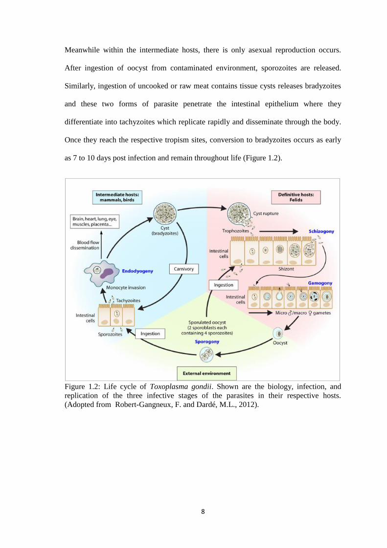
Meanwhile within the intermediate hosts, there is only asexual reproduction occurs.
After ingestion of oocyst from contaminated environment, sporozoites are released.
Similarly, ingestion of uncooked or raw meat contains tissue cysts releases bradyzoites
and these two forms of parasite penetrate the intestinal epithelium where they
differentiate into tachyzoites which replicate rapidly and disseminate through the body.
Once they reach the respective tropism sites, conversion to bradyzoites occurs as early
as 7 to 10 days post infection and remain throughout life (Figure 1.2).
Figure 1.2: Life cycle of Toxoplasma gondii. Shown are the biology, infection, and
replication of the three infective stages of the parasites in their respective hosts.
(Adopted from Robert-Gangneux, F. and Dardé, M.L., 2012).

9
Transmission
The horizontal transmissions to humans are cause by ingestion of tissue cysts in infected
meat or by ingestion of soil, water or food contaminated with sporulated cysts from
environment. Another way is by solid organ transplantation , where by a cyst-containing
organ from a donor to a non - immunised recipient in particular heart transplant patient
(Fernàndez-Sabé et al., 2011). Where else, vertical transmission occurs through
tachyzoites colonized at placental tissue to foetus by primary infection or reactivation
of latent infection.
Clinical features of Toxoplasma infection
In general, T. gondii infections are asymptomatic and self - limiting among healthy
immunocompetent individuals, in others cases, patients may experience fever or
cervical lymphadenopathy, sometimes associated with myalgia, asthenia, or other
nonspecific clinical signs. However among pregnant women, new born, infants and
immunocompromised patients the infection may cause severe clinical manifestation.
Manifestation during pregnancy and congenital toxoplasmosis
Classically, congenital infection occurs only if a pregnant women develops primary
acquired infection during pregnancy or 8 weeks before conceiving. Placenta plays a
major role as a barrier at the beginning of the gestation leading to less parasites
transmission but later it becomes more permeable allowing transmission of 30% in
second trimester and 60 to 70% third trimester (Dunn et al., 1999). Congenital
infection from a reactivation of chronic infection in an immunocompetent pregnant is a
rare event. This condition is postulated due to decreased cellular response during
pregnancy that interferes the parasitic control leading to increased risk of vertical
transmission (Garweg et al., 2005). No doubt this phenomenon might attribute to
10
reinfection with exposure to different genotypes or reactivation of chronic infection
(Elbez-Rubinstein et al., 2009).
Foetal infection during early gestation are at high risk of developing foetal anomaly
such as miscarriage, stillbirth, intrauterine growth retardation and birth defect. Major
squeal include mental retardation, seizures (80%), microcephalus and hydrocephalus
(28%), deafness, and psychomotor deficiency (Remington et al., 2001). Eye lesion is
more severe in early pregnancy where microopthalmia, cataract, increase intraocular
pressure, strabismus, optic neuritis, retinal necrosis, uveitis and retinochoroiditis can be
observed (Delair et al., 2011; Roberts et al., 2001). Clinical manifestation during second
trimester includes epilepsy, anaemia, thrombocytopenia induced petechial, hepatic
disorder, severe sepsis, pneumonitis or retinochoroiditis (Remington et al., 2001). By
contrast, late maternal infection mostly in third trimester results in subclinical
toxoplasmosis in new born, sometimes goes unnoticed , but later in life may develop
chorioretinitis (Montoya and Liesenfeld, 2004).
Role of placenta in T. gondii transmission and pathophysiology
Placenta is a key tissue in the mother-to-foetus relationship, apart from trophic role it
also provides the tolerant immune microenvironment necessary for gestation (Entrican,
2002). During primary infection, parasites cross intestinal barrier, invade monocytes in
contact with lamina propria, disseminates throughout the body including placenta.
Infection of placenta tissue leads to placentitis and subsequently infect the trophoblast
lining which interface with foetus compartment, proceeds to congenital infection. This
important process has two main consequences, firstly placental infection may adversely
affect this tenuous equilibrium between maternal and foetal compartments; and
secondly placenta is directly involved in parasite transmission to the foetus.

11
By immune response, interferon ᵧ (IFN- ᵧ) produced by natural killer (NK) cells or
CD8+ cytotoxic lymphocytes (CTL) directly controls both invasion of monocytes and
trophoblasts by T. gondii and replication of the parasite in infected cells. Massive IFN-ᵧ
release has immunopathological effects, of which apoptosis of decidual cells and spiral
artery dilation (Senegas et al., 2009). Some of these are essential immunomodulatory
mechanisms that compensate for the Th-1 inflammatory cytokines induced by
Toxoplasma, and could avoid foetal loss, particularly when infection occurs in early
pregnancy. Human trophoblast cells produce interleukin 10 (IL-10) and transforming
growth factor β1 (TGF-β1), which promote a Th-2 immune response to ensure
maternal–foetal tolerance but induce a significant increase in both T. gondii intracellular
replication and invasion (Barbosa et al., 2008). IFNᵧ secretion induces intercellular
adhesion molecule (ICAM)-1 up regulation on the trophoblast surface, enhancing
adhesion of infected monocytes to the trophoblast cell surface. Infected trophoblast cells
then lose the ability for apoptosis, which results in parasite persistence in placental
tissues (Pfaff and Candolfi, 2008), and this can be a reservoir for immediate or delayed
congenital infection. The strong expression of human leukocyte antigen-G (HLA-G) on
trophoblast cells can inhibit lysis by maternal NK cells and can mediate suppression of
the allocytotoxic T cell response against the foetus. HLA-G expression also drives
mononuclear phagocytes into suppressive pathways (Hunt et al., 2005) (Figure 1.3).

12
Figure 1.3:Tentative scheme of the pathophysiological hypotheses controlling
trophoblast–cell infection and transplacental transfer of Toxoplasma gondii.(Adopted
from Robert-Gangneux et al., 2011)
Toxoplasmosis in immunocompromised patients
Typically occurs as a reactivation of a chronic disease leading to life threatening
condition. The immunocompromised patients consists of HIV – infected patient,
tuberculosis patient, on prolonged immune suppressive therapies such as Systemic
Lupus Erythematous (SLE), haematological malignancy, non-communicable disease
such as diabetes mellitus and even old aged individuals. The major factor promoting to
the reactivation of infection is profoundly impaired cellular immunity. In HIV –
infected patients, the incidence is closely related to CD4 T cell counts, especially when
count falls under100 cell/ul. Toxoplasmosis encephalitis (TE) most predominant
manifestation with various symptoms, ranging from headache, lethargy, incoordination,
or ataxia to hemiparesis, loss of memory, dementia, seizure usually associated with
fever (Luft and Remington, 1992).

13
Ocular toxoplasmosis
T. gondii is the most common pathogen of retinochroiditis leading to reccurent
posterior uveitis worldwide (Butler et al., 2013). Common complaints are eye redness,
blurring of vision and ocular pain. Chorioretinal lesion may develope from congenital or
postnatal acquired infection and occur during the acute or latent stage of infection.
Generally it is difficult to determine the infection was congenital or acquired with
recurrences of chorioretinitis. Patients whom present with acute toxoplasmosis between
the fourth and sixth decade of life, often have unilateral eye involvement, usually spare
the macula with no scars. Whereas those acquired infection postnatally, often
subclinical, may result in partial or complete loss of vision (Delair et al., 2008). By
contrast in congenital infection, the chorioretinitis is bilateral and they might face severe
complications such as optic nerve atrophy, glaucoma, cataract and retinal detachment.
Among immunocompromised patients, ocular toxoplasmosis occur with atypical and
severe necrotizing form of retinochoroiditis (Antoniazzi et al., 2008).
Diagnostic methods
There are several diagnostic approaches including detection of parasitic agent and
Toxoplasma antibodies. Parasitic agents can be detected through histological
identification, isolation from tissue culture and molecular technique by polymerase
chain reaction (PCR). Whereas the serodiagnostic test are mainly to detect different
classes of antibodies or antigen.
Serology diagnosis
Understanding the kinetic of antibody response is the basis of serological test.
Interpretation of the results depends on patient’s immune background and disease
setting followed by clinical signs. Many serology tests are to measure different types of
antibody, including IgG, IgM, IgA, and IgE, which show unique increase or decrease
14
during the course of infection. The established serological methods available include
Sabin – Feldman Dye test (SFDT), Indirect fluorescent Assay (IFA) agglutination tests,
Enzyme-linked immunosorbent assays (ELISA) and Avidity of Toxoplasma IgG. In
avidity of Toxoplasma IgG, a high avidity ratio exclude a recent infection preceding 4
months, test is performed during first trimester of pregnancy. Whereby, if index is low
or intermediate, the interpretation is ambivalent. This cannot exclude an infection
acquired in the preceding 4 months, or prove that it is recent, unless the index is
extremely low. Also should be kept in mind that treatment delays IgG avidity
maturation (Meroni et al., 2009).
Kinetic of antibody responses
T. gondii-specific IgM antibodies rises from day 5 to weeks following acute infection
(Paquet et al., 2013) peaks at 2 months and disappear within 6-9 months, more rapidly
than IgG. A positive IgM result indicates an early, acute or a recent Toxoplasma
infection. In recent infection there will be presence of persistent IgM (Meek et al.,
2001; Montoya and Liesenfeld, 2004). IgM antibodies can persist over years without
any clinical significance and thus hamper the interpretation of test results especially in
case of pregnancy screening (Remington et al., 2001; Wong and Remington, 1994) T.
gondii-specific IgG antibody are detectable within 1 or 2 weeks after infection, peaks
within 12 weeks to 6 months and persist for decades (Paquet et al., 2013). (Figure 1.4).
A positive result indicates a previous exposure (latent infection) and provide immunity
to reinfection unless from a different genotype and virulence genes. It may also indicate
recent or reactivation of latent infection. In case of recent or reactivation of infection
usually a significant rise or 4 fold titer in IgG antibody level can be expected in serial
samples obtained in 2 – 3 weeks apart (Montoya and Liesenfeld, 2004). Passive
immunity is detection of IgG antibody in newborn or infant, is from mother and may
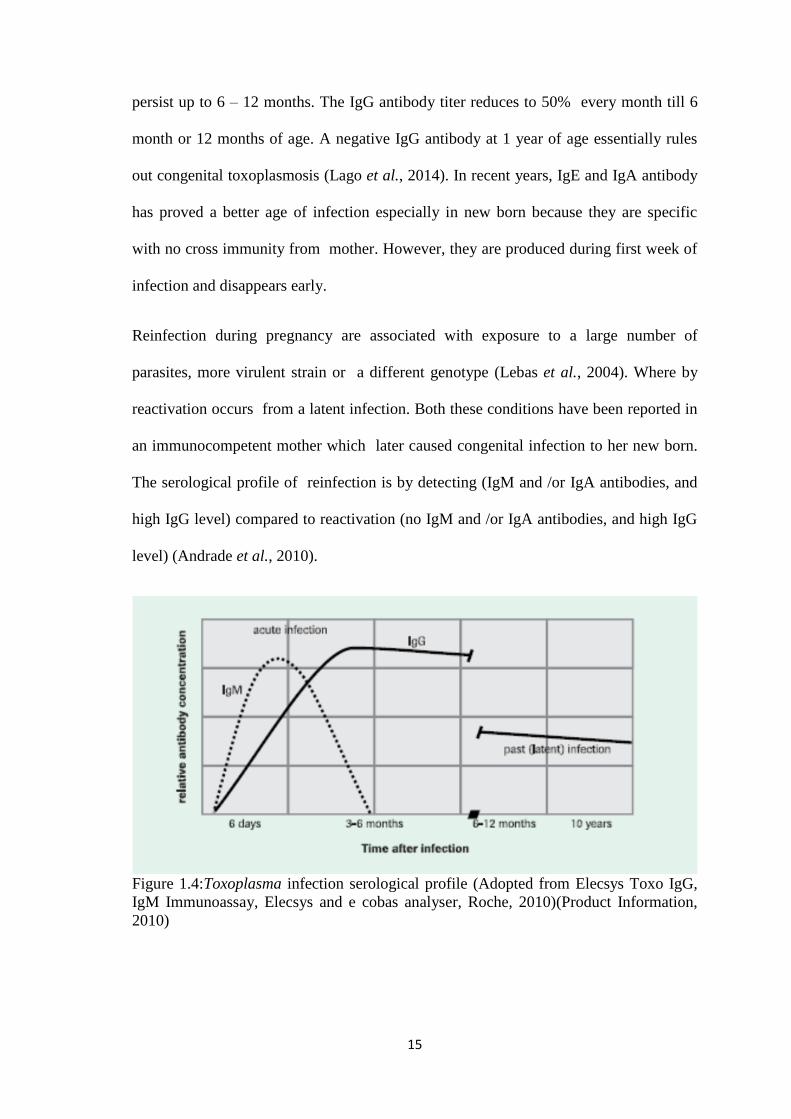
15
persist up to 6 – 12 months. The IgG antibody titer reduces to 50% every month till 6
month or 12 months of age. A negative IgG antibody at 1 year of age essentially rules
out congenital toxoplasmosis (Lago et al., 2014). In recent years, IgE and IgA antibody
has proved a better age of infection especially in new born because they are specific
with no cross immunity from mother. However, they are produced during first week of
infection and disappears early.
Reinfection during pregnancy are associated with exposure to a large number of
parasites, more virulent strain or a different genotype (Lebas et al., 2004). Where by
reactivation occurs from a latent infection. Both these conditions have been reported in
an immunocompetent mother which later caused congenital infection to her new born.
The serological profile of reinfection is by detecting (IgM and /or IgA antibodies, and
high IgG level) compared to reactivation (no IgM and /or IgA antibodies, and high IgG
level) (Andrade et al., 2010).
Figure 1.4:Toxoplasma infection serological profile (Adopted from Elecsys Toxo IgG,
IgM Immunoassay, Elecsys and e cobas analyser, Roche, 2010)(Product Information,
2010)
16
1.3.6.3. Sabin – Feldman dye test (SFDT)
Sabin – Feldman dye test (SFDT) is the first assay developed and still considered as the
gold standard with high sensitivity and specificity. This test is applied based on
incubation of live tachyzoites on the slide with patients serum and complement. If the
serum contains specific antibodies against T. gondii, the coated tachyzoite will be lysed
by the complement system and fail to stain with dye methylene blue. Finally the
stained and unstained tachyzoites are counted and used as end-point titer. The
limitation for this test is application of live tachyzoites which is considered as biohazard
and fails to determine the stage of infection (Udonsom et al., 2010).
Indirect fluorescent assay (IFA)
This method is applied based on killed tachyzoites fixed on a glass slide to react with
the antibody in the serum. The detection of this reaction is by fluorescence – labelled
anti human IgG and IgM antibodies and reviewed under a fluorescence microscope.
This test is simple, safe and inexpensive but it is operator dependent. There are also
possibilities of false positive results in cases of patients with rheumatoid factors or
antinuclear antibodies (Rorman et al., 2006).
Agglutination tests
The available tests are direct agglutination test (DAT), indirect hemagglutination test
(IHAT) and latex agglutination test (LAT). Development of these tests are to identify
antibodies against T. gondii. DAT starting with coating of microtiter plates containing
formalinized Toxoplasma tachyzoites will agglutinate if antibodies are present in the
diluted patients serum. In IHAT, sensitised red blood cells (RBCs) are used that shows
agglutination if serum are positive with anti – T. gondii antibodies (Liu et al., 2015). In
LAT, tachyzoites are fixed to latex beads and visible flocculation reveals positive serum
containing specific IgG antibody. The agglutination test are generally simple and

17
inexpensive but there a tendencies for false positive results to occur especially in
immunocompromised individual.
Enzyme – linked immunosorbent assay (ELISA)
The principle of this assay is a microtiter plate is coated with antigens, and diluted sera
are applied. The anti – Toxoplasma antibodies will bind to the antigen and detected by
secondary antibody. Unbound reagents are washed and substrate is added colour
reaction occurs which correlates with the quantity of the antibody. The determination of
results depends on optic density of the serum. This test is highly sensitive, measures
quantitative and semiquantitative antibody, detects IgG, IgM, IgA and IgE antibodies
and applied as a large scale of samples in short duration (Sudan et al., 2013). In current
settings, the latest generation of ELISA, electrochemiluminescence immunoassay
“ECLIA”is applied in most laboratories as a screening tool for Toxoplasma infection.
The initial principle is similar to ELISA but the end result is measured with a cell that
uses electrodes and magnet to gather the bound immunocomplexes. Finally application
of a defined voltage induces the electrochemiluminescent reaction and the resulting
light emission is measured directly by the photomultiplier.
A study conducted by the new Roche Elecsys Toxo IgG and IgM immunoassay was
compared with Sabin–Feldman dye test and immunosorbent agglutination assay-IgM as
a reference test. Single serum samples were analysed from 927 pregnant women,
including 100 negative, 706 chronic, and 121 acute infections. The combination of both
Elecsys IgG and Ig M assays demonstrated high sensitivity and specificity of 97.1% and
100.0%, respectively, and a positive and negative predictive value of 100.0% and
81.3%, respectively (Prusa et al., 2010).

18
Parasite isolation
Isolation of T. gondii from blood, body fluids and tissue always indicate acute
infection. Isolation is by inoculation of the parasites from patients sample in the tissue
cultures or laboratory animals. In mice, the test is performed by injecting the clinical
samples either intraperitoneally or subcutaneously. The mice will be subjected for
serological test after 3 to 6 weeks to detect the presence of anti T.gondii antibodies.
Tissues from positive antibodies will be tested with PCR or microscopic examination
to look for cysts for final confirmation (Hill and Dubey, 2002).
Histologic diagnosis
Demonstration of tachyzoites in tissue or smears of body fluids establish the diagnosis
of acute infection. Conventional staining is difficult to demonstrate tachyzoites,
therefore the immunoperoxidase technique with antisera is applied successfully to detect
the parasite in toxoplasmosis encephalitis cases of HIV patients (Montoya, 2002).
Molecular diagnosis
PCR amplification for detection of T. gondii DNA in body fluids and tissues are used to
diagnose congenital, ocular, cerebral and disseminated toxoplasmosis. The most popular
used gene is the 35 – fold repetitive gene B, one of the first gene used in the PCR
detection of T.gondii (Sarkari et al., 2014). PCR has evolved in the diagnosis of
intrauterine Toxoplasma infection by early detection in placenta tissue or amniotic
fluids and helped to guide for prompt treatment and avoid unnecessary invasive
procedures (Montoya, 2002). Amniotic fluids from amniocentesis is the cornerstone
for diagnosis of congenital toxoplasmosis. If maternal primary infection is suspected
and abnormal ultrasound findings were diagnosed (intracranial calcification,
microcephaly, hydrocephalus, ascites, hepatosplenomegaly or severe intrauterine
growth retardation), PCR should be offered to identify Toxoplasma infection in the

19
amniotic fluid. This procedure is not performed at less than 18 weeks gestation because
of high rate of false positive results, nevertheless should be done not less than 4 weeks
after time of suspected maternal infection (Paquet et al., 2013). Performance of
amniocentesis after 18 weeks has sensitivity of 91%, specificity 96 % (Romand et al.,
2001).
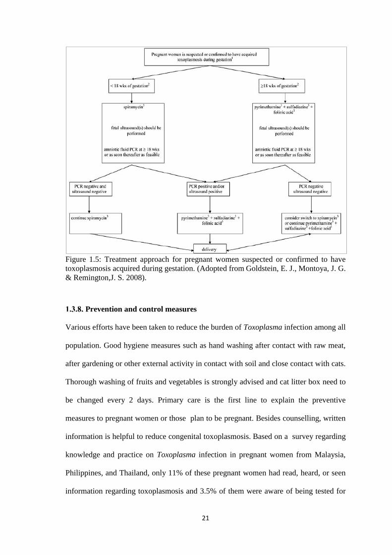
Treatment
Treatment in immunocompetent pregnant women with previous infection with T. gondii
should not be necessary. Women who are immunosuppressed or HIV-positive should be
offered screening because of the risk of reactivation and toxoplasmosis encephalitis. A
non-pregnant woman, diagnosed with an acute toxoplasmosis should be counselled to
wait 6 months before attempting to become pregnant. Each case should be considered
separately in consultation with an expert. The existing indication for treatment in
pregnant women is to achieve two goals. Firstly, if maternal infection occurred but
foetus is not infected, aimed at preventing vertical transmission, before foetal infection.
Spiramycin, a macrolide, does not readily cross the placenta, therefore is not reliable
for treatment of foetal infection. As a foetal prophylaxis, is used to prevent spread of
organisms across the placenta (Goldstein et al., 2008). Secondly if foetal infection has
been confirmed or is highly suspected by amniotic fluid PCR or foetal ultrasound,
pyrimethamine and sulfadiazine is indicated. Pyrimethamine is a folic acid antagonist
acts synergistically with sulfonamides. This drug not be used in the first trimester,
potentially teratogenic and a reversible dose related depression of the bone marrow and
therefore must be combined with folinic acid. The combination of pyrimethamine and
sulfadiazine results in a significant decrease in disease severity. Neonates with
congenital toxoplasmosis, even asymptomatic at birth should be treated early to reduce
long term squeal (Paquet et al., 2013) (Figure 1.5).

20
In patients with AIDS whom developed reactivation of cerebral toxoplasmosis, CD4
cell count of < 100 cells/mm3 has a strong association as a prognostic marker in
progression of the disease, especially with other opportunistic infection. Toxoplasmosis
encephalitis is a preventable disease when adequate chemoprophylaxis of trimethoprim-
sulfamethoxazole or dapsone-pyrimethamine plus folinic acid is administered (Passos et
al., 2000).
The diagnosis for retinochoroiditis depends on an accurate opthalmological examination
which shows typical of white focal lesion associated with a vitreous inflammatory
reaction and Toxoplasma seropositivity. This indicates for treatment and further
confirmed with therapeutic response (Robert-Gangneux and Dardé, 2012). Intravitreous
clindamycin with dexamethasone seems to be as effective as systemic treatments, where
as other preferred oral antibiotic would be trimethoprim-sulfamethoxazole,
azithromycin or clindamycin which prevents recurrence of the disease (Harrell and
Carvounis, 2014).
21
Figure 1.5: Treatment approach for pregnant women suspected or confirmed to have
toxoplasmosis acquired during gestation. (Adopted from Goldstein, E. J., Montoya, J. G.
& Remington,J. S. 2008).
Prevention and control measures
Various efforts have been taken to reduce the burden of Toxoplasma infection among all
population. Good hygiene measures such as hand washing after contact with raw meat,
after gardening or other external activity in contact with soil and close contact with cats.
Thorough washing of fruits and vegetables is strongly advised and cat litter box need to
be changed every 2 days. Primary care is the first line to explain the preventive
measures to pregnant women or those plan to be pregnant. Besides counselling, written
information is helpful to reduce congenital toxoplasmosis. Based on a survey regarding
knowledge and practice on Toxoplasma infection in pregnant women from Malaysia,
Philippines, and Thailand, only 11% of these pregnant women had read, heard, or seen
information regarding toxoplasmosis and 3.5% of them were aware of being tested for

22
the infection (Andiappan et al., 2014). Screening for pregnant women and neonate is
implemented in many European countries. Hence the decisions on implementation of
screening in a country are considered based on prevalence data, disease burden,
technical resources and diagnostic costs.
23
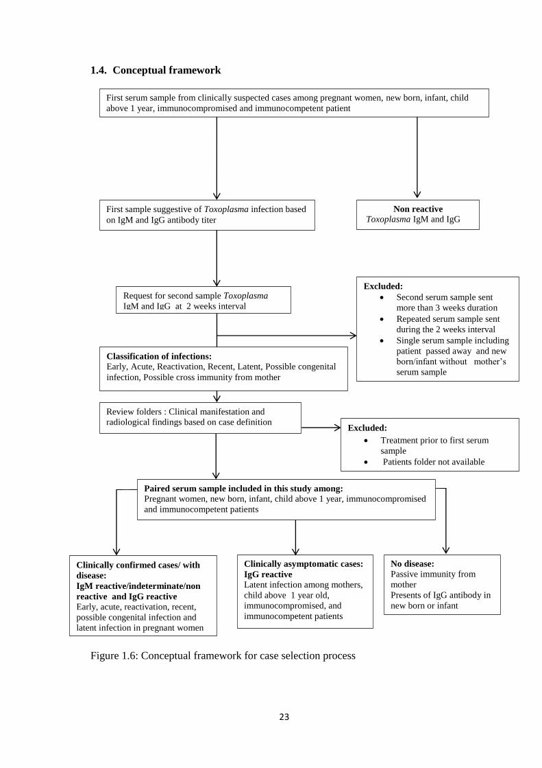
1.4. Conceptual framework
Figure 1.6: Conceptual framework for case selection process
First serum sample from clinically suspected cases among pregnant women, new born, infant, child
above 1 year, immunocompromised and immunocompetent patient
First sample suggestive of Toxoplasma infection based
on IgM and IgG antibody titer
Non reactive Toxoplasma IgM and IgG
Request for second sample Toxoplasma
IgM and IgG at 2 weeks interval
Review folders : Clinical manifestation and
radiological findings based on case definition
Clinically confirmed cases/ with
disease:
IgM reactive/indeterminate/non
reactive and IgG reactive
Early, acute, reactivation, recent,
possible congenital infection and
latent infection in pregnant women
Clinically asymptomatic cases:
IgG reactive
Latent infection among mothers,
child above 1 year old,
immunocompromised, and
immunocompetent patients
No disease: Passive immunity from
mother
Presents of IgG antibody in
new born or infant
Excluded:
Treatment prior to first serum
sample
Patients folder not available
Excluded:
Second serum sample sent
more than 3 weeks duration
Repeated serum sample sent
during the 2 weeks interval
Single serum sample including
patient passed away and new
born/infant without mother’s
serum sample
Paired serum sample included in this study among:
Pregnant women, new born, infant, child above 1 year, immunocompromised
and immunocompetent patients
Classification of infections:
Early, Acute, Reactivation, Recent, Latent, Possible congenital
infection, Possible cross immunity from mother

24
1.5. OBJECTIVES
General objectives
To describe the clinical manifestation and determine the seroprevalance classification of
infection among the selected populations, validity of the test and percentage of patient
on treatment after first and paired serum sample.
Specific objectives
1. To describe the clinical manifestation of Toxoplasma infection among pregnant
women, new born, infant, child above 1 year old, immunocompromised and immuno
- competent patients.
2. To determine seroprevalence of early, acute, reactivation, recent, latent, passive
immunity from mother and possible congenital infection with paired serum sample
using Elecsys IgM / IgG assay among pregnant women , new born, infant, child
above 1 year old, immunocompromised and immunocompetent patients.
3. To determine the validity of paired serum sample to diagnose Toxoplasma infection
by calculating the sensitivity, specificity, PPV, NPV and ROC curve analyses in
clinically confirmed cases ( by gold standard).
4. To describe the percentage of patient on treatment after the first and paired serum
sample.
Research hypothesis
The classification of Toxoplasma infection into early , acute, reactivation, recent , latent,
passive immunity from mother and possible congenital infection can be established by
using paired serum sample of Toxoplasma IgM and IgG antibody titer in pregnant
women , new born, infant, child above 1 year old, immunocompromised and
immunocompetent patients.