using tertiary sulci to map the “cognitive globe” of
TRANSCRIPT

Miller et al. | Situating tertiary sulci in the prefrontal cortex 1
Using tertiary sulci to map the “cognitive globe”
of prefrontal cortex
Jacob A. Miller1, Mark D’Esposito1,2, Kevin S. Weiner1,2
1Helen Wills Neuroscience Institute, 2Department of Psychology, University of California-Berkeley
Corresponding author: Jacob A. Miller [email protected] 210 Barker Hall University of California-Berkeley, Berkeley, CA, 94720
Keywords: prefrontal cortex, functional neuroanatomy, morphology, gradients, brain mapping, higher-order cognition
Miller et al. | Situating tertiary sulci in the prefrontal cortex 2
ABSTRACT Stuss (1984) considered the human prefrontal cortex (PFC) as a “cognitive globe” on which functions of the frontal lobe could be mapped. Here, we discuss classic and recent findings regarding the evolution, development, function, and cognitive role of shallow indentations, or tertiary sulci, in PFC with the goal of using tertiary sulci to map the “cognitive globe” of PFC. First, we discuss lateral PFC (LPFC) tertiary sulci in classical anatomy and modern neuroimaging, as well as their development, with a focus on those within the middle frontal gyrus (MFG). Second, we discuss tertiary sulci in comparative neuroanatomy, focusing on primates. Third, we summarize recent findings showing the utility of tertiary sulci for understanding structural-functional relationships with functional network insights in ventromedial and LPFC. Fourth, we revisit and update unresolved theoretical perspectives considered by Vogt and Vogt (1919) and Sanides (1964) that tertiary sulci serve as landmarks for cortical gradients. Together, the consideration of these classic and recent findings indicate that tertiary sulci are situated in a unique position within the complexity of the “cognitive globe” of PFC: they are the smallest and shallowest of sulci in PFC, yet can offer insights that bridge spatial scales (microns to networks), modalities (functional connectivity to behavior), and species. As such, the map of tertiary sulci within each individual participant serves as a coordinate system specific to that individual on which functions may be further mapped. We conclude with new theoretical and methodological questions that if answered in future research, will likely lead to mechanistic insight regarding the structure and function of human LPFC.

Miller et al. | Situating tertiary sulci in the prefrontal cortex 3
INTRODUCTION
“The anatomy of the frontal lobe and its connections suggests a possible cognitive globe on
which our knowledge of frontal lobe functions may be mapped” (Stuss, 1984).
Understanding how anatomical structures of the brain support functional networks
underlying human-specific aspects of cognition is a major goal in cognitive neuroscience. Of the
many anatomical structures to study, prefrontal cortex (PFC), which is disproportionately
expanded in the human brain, is particularly important given its central role in cognitive control
and goal-directed behavior (Donahue, Glasser, Preuss, Rilling, & Van Essen, 2018; E. K. Miller
& Cohen, 2001; Stuss, 2011; Stuss & Alexander, 2000; Stuss & Knight, 2012). Throughout his
career, Don Stuss used maps of lesion damage to begin parcellating the PFC into the component
pieces of a “cognitive globe” as he described in the above quote (Stuss, 1984; Stuss, 2011; Stuss
& Alexander, 2000; Stuss & Knight, 2012). In the decades since, functional maps and parcellations
of the PFC have been developed across many cognitive domains using a variety of neuroimaging
techniques. For example, modern neuroimaging research shows widespread support for a
hierarchical functional gradient organized along the rostral-caudal axis of lateral PFC (LPFC;
(Badre & D'Esposito, 2009; Demirtas et al., 2019; Koechlin, Ody, & Kouneiher, 2003; Koechlin
& Summerfield, 2007; Nee & D'Esposito, 2016; Reid et al., 2016). Additionally, several different
anatomical, functional, and multimodal approaches have further parcellated PFC into dozens of
different areas (Eickhoff, Yeo, & Genon, 2018; Glasser et al., 2016; Goulas, Uylings, & Stiers,
2012; Kong et al., 2018; Sallet et al., 2013). Despite this progress, we still lack fundamental
neuroanatomical details of the functional maps and parcellations within PFC largely for two main
reasons. First, invasive methods commonly used to uncover the neuroanatomical details of cortical

Miller et al. | Situating tertiary sulci in the prefrontal cortex 4
networks in animal models cannot be used in humans. This is not a new issue and over three
decades ago was summarized as the “backwardness of human neuroanatomy” (Crick & Jones,
1993). Second, human and non-human hominoid brains contain neuroanatomical structures that
other primate brains lack. For instance, the expanded association cortices in hominoid brains
contain shallow indentations known as tertiary sulci that are missing in the more lissencephalic
brains of other species widely studied in cognitive neuroscience such as mice, marmosets, and
macaques (Armstrong, Schleicher, Heyder, Maria, & Zilles, 1995; Petrides, 2019; Sanides, 1962;
Welker, 1990).
Here, we propose that identifying and studying tertiary sulci contribute a forwardness to
human neuroanatomy as this approach offers an opportunity to examine neuroanatomical-
functional relationships in humans that cannot be conducted in other animal models – even non-
human hominoids. Specifically, we cannot map non-human hominoid brains in the way that we
can map the human brain due to ethical reasons. As such, examining the relationship between
tertiary sulci and different types of functional and neuroanatomical maps across spatial scales is a
unique opportunity specific to the human brain. In this paper, we propose that tertiary sulci in the
human cerebral cortex serve as a coordinate map for linking neuroanatomical and functional
properties of the human brain across spatial scales and modalities. Consideration of tertiary sulci
as anatomical features allows the “cognitive globe” of PFC function to be mapped within
individuals without relying on an average cortical surface or template across individuals within
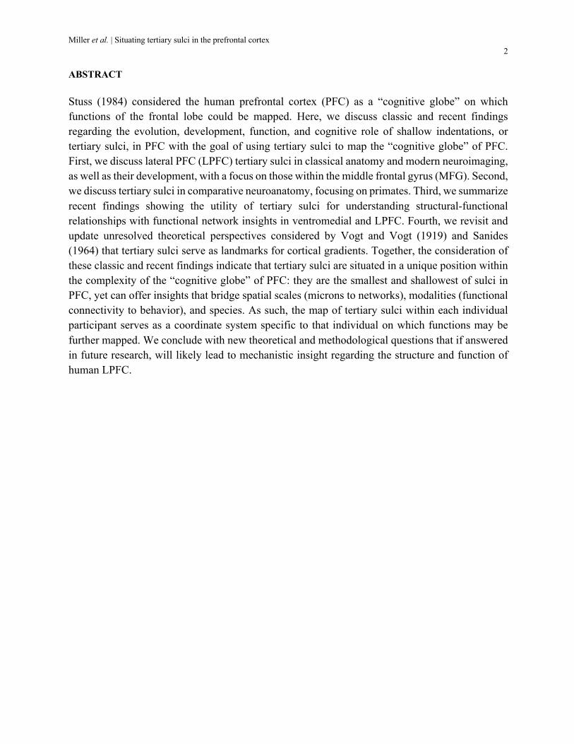
which tertiary sulci disappear (Fig. 1). The map of tertiary sulci within each individual participant
serves as a coordinate system specific to that individual on which functions may be further mapped.
Miller et al. | Situating tertiary sulci in the prefrontal cortex 5
Figure 1. Tertiary sulci in LPFC disappear on averaged cortical surface reconstructions from anatomical MRIs. Top, three individually labeled left hemispheres with tertiary sulci in the middle frontal gyrus (MFG) outlined in white. The superior and inferior frontal sulci (sfs, ifs) are labeled for reference above and below the MFG, respectively. Bottom, average cortical surfaces show much smaller tertiary sulci compared to individual subjects. As more subjects are included in the averaged cortical surface, tertiary sulci within the MFG disappear almost entirely, which is inconsistent with their prominence in individual hemispheres.
This review is divided into four main sections. First, we discuss LPFC tertiary sulci in
classical anatomy and modern neuroimaging with a focus on those within the middle frontal gyrus
(MFG). Second, we discuss tertiary sulci in development and comparative neuroanatomy, focusing
on primates. Third, we summarize recent findings showing the utility of tertiary sulci for
understanding structural-functional relationships in LPFC and likely in association cortices more
broadly. Fourth, we revisit unresolved theoretical perspectives that tertiary sulci can serve as
landmarks for cortical gradients. We conclude with new theoretical and methodological questions
that if answered in future research, will likely lead to mechanistic insight regarding the structure
and function of LPFC.

Miller et al. | Situating tertiary sulci in the prefrontal cortex 6

Miller et al. | Situating tertiary sulci in the prefrontal cortex 7
Figure 2. Characteristics of tertiary sulci in PFC and their historical identification. (a) Principle morphological features distinguishing tertiary sulci from larger primary sulci. Nissl stain downloaded from Allen atlas (http://human.brain-map.org/), developmental images adapted from Welker (1990). (b) We use the tertiary sulci in the posterior middle frontal gyrus (MFG) as an example to outline the main types of ambiguity across post-mortem studies leading to the neglect of tertiary sulci in LPFC. Top left, sulci in the MFG were depicted in diagrams and in individual samples but left unlabeled in official schematics as in Eberstaller (1890). Bottom left, a sulcus in the anterior MFG, the frontomarginal sulcus, was improperly used as the label for tertiary sulci in the more posterior MFG, as in Connolly (1950). Top middle, Cunningham (1892) used the same label of the frontomarginal sulcus for all tertiary sulci in the posterior MFG. Bottom middle, tertiary sulci were identified and even assigned numbers, but not given any distinguishing labels in brain sections or schematics (Connolly, 1950). Top right, separate labels were applied to different tertiary sulci within the posterior MFG. Retzius (1896) acknowledged a posterior transverse component (Retzius’ fmt in Fig. 1) that was often distinct from the rest of the middle frontal sulcus. Ariens-Kappers (1928, 1929, 1936) also acknowledged three components of the middle frontal sulcus (Appendix) largely consistent with the most modern and thorough schematic of sulcal morphology from Petrides, which identifies the posterior middle frontal
sulcus (pmfs) into three purported structures, an anterior, intermediate, and posterior component (Petrides, 2019). Bottom right, sulci within the MFG have been referred to using different labels (top, middle) such as the medifrontal
sulcus, frontomarginal sulcus, intermediate frontal sulcus, middle frontal sulcus, and posterior middle frontal sulcus, among others (see Appendix).
I: LPFC tertiary sulci in classic anatomy and modern neuroimaging: Clarity vs. Ambiguity It is widely accepted that LPFC has expanded with evolution (Amiez, Kostopoulos,
Champod, & Petrides, 2006; Amiez & Petrides, 2009; Croxson et al., 2005; Neubert, Mars, Sallet,
& Rushworth, 2015; Petrides, 2005; Petrides & Pandya, 1999; Petrides, Tomaiuolo, Yeterian, &
Pandya, 2012; Sallet et al., 2013) and is “phylogenetically novel” (Stuss & Benson, 1984). This
evolutionary expansion included the addition of anatomical structures within LPFC - such as
tertiary sulci (Armstrong et al., 1995; Welker, 1990) - that are lacking in brains that are
phylogenetically older (Barrett et al., 2020; Catani, 2019). Tertiary sulci are shallow indentations
in the cerebral cortex that emerge latest in development (Fig. 2a). Ongoing research attempts to
better understand the function of these evolved structures that are expanded, and even unique, to
the human LPFC. Specifically, large, freely available multi-modal datasets have provided the
ability to finally assess typical variations in tertiary sulcal morphology and functional organization
within individuals that was not possible in classical studies of post-mortem brains. Indeed, tertiary
Miller et al. | Situating tertiary sulci in the prefrontal cortex 8
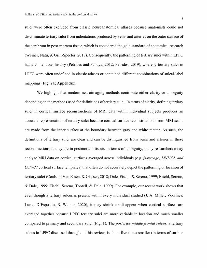
sulci were often excluded from classic neuroanatomical atlases because anatomists could not
discriminate tertiary sulci from indentations produced by veins and arteries on the outer surface of
the cerebrum in post-mortem tissue, which is considered the gold standard of anatomical research
(Weiner, Natu, & Grill-Spector, 2018). Consequently, the patterning of tertiary sulci within LPFC
has a contentious history (Petrides and Pandya, 2012; Petrides, 2019), whereby tertiary sulci in
LPFC were often undefined in classic atlases or contained different combinations of sulcal-label
mappings (Fig. 2a; Appendix).
We highlight that modern neuroimaging methods contribute either clarity or ambiguity
depending on the methods used for definitions of tertiary sulci. In terms of clarity, defining tertiary
sulci in cortical surface reconstructions of MRI data within individual subjects produces an
accurate representation of tertiary sulci because cortical surface reconstructions from MRI scans
are made from the inner surface at the boundary between gray and white matter. As such, the
definitions of tertiary sulci are clear and can be distinguished from veins and arteries in these
reconstructions as they are in postmortem tissue. In terms of ambiguity, many researchers today
analyze MRI data on cortical surfaces averaged across individuals (e.g. fsaverage, MNI152, and
Colin27 cortical surface templates) that often do not accurately depict the patterning or location of
tertiary sulci (Coalson, Van Essen, & Glasser, 2018; Dale, Fischl, & Sereno, 1999; Fischl, Sereno,
& Dale, 1999; Fischl, Sereno, Tootell, & Dale, 1999). For example, our recent work shows that
even though a tertiary sulcus is present within every individual studied (J. A. Miller, Voorhies,
Lurie, D’Esposito, & Weiner, 2020), it may shrink or disappear when cortical surfaces are
averaged together because LPFC tertiary sulci are more variable in location and much smaller
compared to primary and secondary sulci (Fig. 1). The posterior middle frontal sulcus, a tertiary
sulcus in LPFC discussed throughout this review, is about five times smaller (in terms of surface
Miller et al. | Situating tertiary sulci in the prefrontal cortex 9
area) and half as deep as the primary central sulcus. Taken together, tertiary sulci have an unclear
history for methodological reasons, which can be corrected with methods that instead preserve
neuroanatomical structures and take individual anatomical organization into consideration.
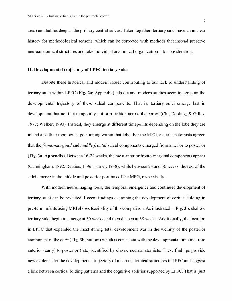
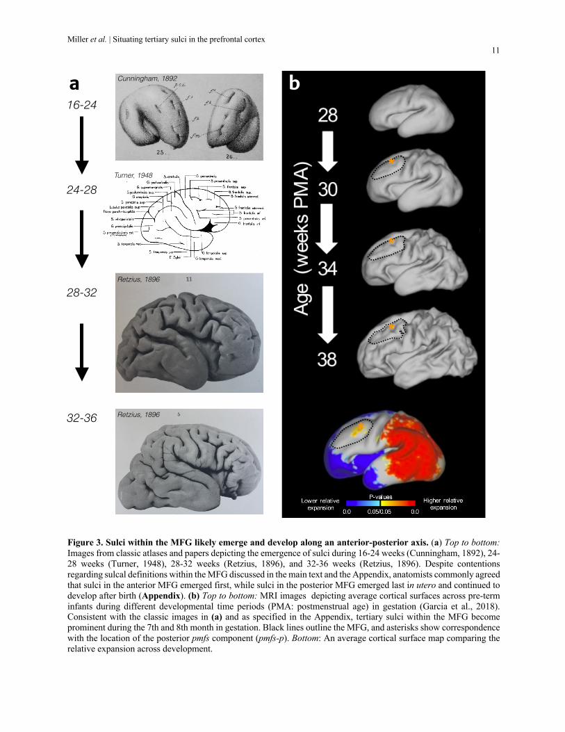
II: Developmental trajectory of LPFC tertiary sulci Despite these historical and modern issues contributing to our lack of understanding of
tertiary sulci within LPFC (Fig. 2a; Appendix), classic and modern studies seem to agree on the
developmental trajectory of these sulcal components. That is, tertiary sulci emerge last in
development, but not in a temporally uniform fashion across the cortex (Chi, Dooling, & Gilles,
1977; Welker, 1990). Instead, they emerge at different timepoints depending on the lobe they are
in and also their topological positioning within that lobe. For the MFG, classic anatomists agreed
that the fronto-marginal and middle frontal sulcal components emerged from anterior to posterior
(Fig. 3a; Appendix). Between 16-24 weeks, the most anterior fronto-marginal components appear
(Cunningham, 1892; Retzius, 1896; Turner, 1948), while between 24 and 36 weeks, the rest of the
sulci emerge in the middle and posterior portions of the MFG, respectively.
With modern neuroimaging tools, the temporal emergence and continued development of
tertiary sulci can be revisited. Recent findings examining the development of cortical folding in
pre-term infants using MRI shows feasibility of this comparison. As illustrated in Fig. 3b, shallow
tertiary sulci begin to emerge at 30 weeks and then deepen at 38 weeks. Additionally, the location
in LPFC that expanded the most during fetal development was in the vicinity of the posterior
component of the pmfs (Fig. 3b, bottom) which is consistent with the developmental timeline from
anterior (early) to posterior (late) identified by classic neuroanatomists. These findings provide
new evidence for the developmental trajectory of macroanatomical structures in LPFC and suggest
a link between cortical folding patterns and the cognitive abilities supported by LPFC. That is, just

Miller et al. | Situating tertiary sulci in the prefrontal cortex 10
as prolonged myelination is a hallmark of association cortex development (D. J. Miller et al.,
2012), tertiary sulcal development in LPFC may be an important marker for a complex array of
genetic and environmental factors related to variations in human cognitive abilities.
Miller et al. | Situating tertiary sulci in the prefrontal cortex 11
Figure 3. Sulci within the MFG likely emerge and develop along an anterior-posterior axis. (a) Top to bottom: Images from classic atlases and papers depicting the emergence of sulci during 16-24 weeks (Cunningham, 1892), 24-28 weeks (Turner, 1948), 28-32 weeks (Retzius, 1896), and 32-36 weeks (Retzius, 1896). Despite contentions regarding sulcal definitions within the MFG discussed in the main text and the Appendix, anatomists commonly agreed that sulci in the anterior MFG emerged first, while sulci in the posterior MFG emerged last in utero and continued to develop after birth (Appendix). (b) Top to bottom: MRI images depicting average cortical surfaces across pre-term infants during different developmental time periods (PMA: postmenstrual age) in gestation (Garcia et al., 2018). Consistent with the classic images in (a) and as specified in the Appendix, tertiary sulci within the MFG become prominent during the 7th and 8th month in gestation. Black lines outline the MFG, and asterisks show correspondence with the location of the posterior pmfs component (pmfs-p). Bottom: An average cortical surface map comparing the relative expansion across development.
16-24
24-28
28-32
32-36
Retzius, 1896
Retzius, 1896
Turner, 1948
Cunningham, 1892a b
*
*
*
*

Miller et al. | Situating tertiary sulci in the prefrontal cortex 12
III. Tertiary sulci in comparative neuroanatomy and primate evolution Among primates, the level of cortical folding (or gyrification index) tracks with
evolutionary complexity, suggesting that sulcal structures are at the bare minimum markers of
primate evolution (Zilles, Palomero-Gallagher, & Amunts, 2013). Classic and recent research
findings indicate that some primate species adisplay tertiary sulci that are likely homologous to
human tertiary sulci in these cortical expanses. For example, Amiez and colleagues compared the
sulcal morphology of vmPFC using anatomical MRI from human, chimpanzee, baboon, and
macaque brains. Across the four analyzed primate species, the majority of tertiary sulci in the
vmPFC were only identifiable in hominoids (humans and chimpanzees) and a subset were human-
specific (Fig. 4a). For example, the dorsomedial polar sulcus (dmps) was identified as a hominoid-
specific marker present in most human brains and ~50% of chimpanzee brains, while the
ventromedial polar sulcus (vmps) was present in most individual human brains (64%), but entirely
absent in chimpanzees and other primate brains. Interestingly, while the presence of major
cytoarchitectonic areas is largely conserved across species in vmPFC, the most expanded areas
(BA 9 and 10) were the locations for the identified tertiary sulci (Amiez et al., 2019).
In LPFC, consistent with Amiez and colleagues (2019), it is likely that some, but not all
chimpanzees have all three pmfs components as in humans. Two different chimpanzee brains with
and without tertiary sulci in the MFG are shown in Fig. 4b, in contrast to an example human brain,
in which tertiary sulci are always present within the MFG. Future studies performing
morphological comparisons of tertiary sulci between chimpanzees and humans will further
quantify these observations. This homology may be important since the pmfs appears to delineate
borders or transitional zones for cytoarchitectonic areas in both humans and chimpanzees. For
example, the seminal papers of Rajkowska & Goldman-Rakic analyzed the cytoarchitecture of the

Miller et al. | Situating tertiary sulci in the prefrontal cortex 13
LPFC within nine post-mortem human brains. In the presented coronal sections of
cytoarchitectonic areas 9 and 46 (Fig. 4c), a common trend appears that the posterior middle
frontal sulcus (or MFS, as it was labeled) delineates a transitional zone between areas 9 and 46
(Rajkowska & Goldman-Rakic, 1995a, 1995b). The characteristics of areas 9 and 46 are different
enough (area 9 with a more prominent layer 3, area 46 with more prominent deeper layers) that
Petrides coined area 9/46d to describe this transitional zone (Petrides, 2005; Petrides & Pandya,
1999). We posit that this transitional zone is tightly coupled to the pmfs and may point to a
mechanistic association between granular cortex and tertiary sulci in primates, with a specific
expansion of middle cortical layers. Support for this hypothesis is evident from re-examining
cytoarchitectonic studies of chimpanzees showing that the pmfs also appears to mark
cytoarchitectonic transitions between granular regions FD and FDΔ in chimpanzee brains differing
in the extent of granular cell proliferation in layer 3 (Bailey & Bonin, 1951; Bailey, Bonin, &
McCulloch, 1950). These observations suggest that tertiary sulci could serve as an organizing
framework to study PFC evolution and to compare with the evolution of cytoarchitecture and
connectivity patterns across species. They further lead to potential mechanistic links relating
tertiary sulci to the expansion of granular cortex and white matter across primate evolution
(Donahue et al., 2018; Garcia-Cabezas, Zikopoulos, & Barbas, 2019; Van Essen et al., 2019).
As tertiary sulci are identified as important landmarks in functional areas that are most
expanded in humans, comparing tertiary sulci among primate species also leads to questions about
whether sulci would serve as trait-level behavioral markers within and across species. Is variability
in sulcal morphology in macaques, baboons, and chimpanzees associated with behavioral traits?
Do animals with more “human-like” sulcal morphology (e.g. a chimpanzee with a dmps or pmfs)
exhibit cognitive control or other behaviors closer to human-level performance? As more cognitive

Miller et al. | Situating tertiary sulci in the prefrontal cortex 14
paradigms from the human literature are adapted for non-human primates (Badre, Frank, & Moore,
2015), these anatomical-behavioral relationships can begin to shed light on PFC structural-
functional relationships across evolutionary timescales.
Figure 4. Tertiary sulci across primate PFC. (a) The sulcal morphology of the medial PFC and anterior cingulate cortex shown in example sagittal MRI sections of human and chimpanzee brains. The vmps (top) is a human-specific tertiary sulcus, while the dmps (bottom) is found in humans and over half of chimpanzee hemispheres examined. Figure adapted from Amiez et al. (2019). (b) Top, cortical surface (left hemisphere) from the NIH macaque brain template (https://afni.nimh.nih.gov/NMT), where only the principal sulcus can be observed with the LPFC. Middle, two chimpanzee cortical surfaces showing variability in the presence or absence of a pmfs (outlined in white) within the MFG. The inset shows the pmfs (then labeled as “fm”) as the boundary between areas FD and FDΔ. Adapted from Bailey, Bonin, & McCulloch (1950). Bottom, example human cortical hemisphere with the prominent pmfs outlined in white. (c) Schematic of a post-mortem coronal section with the pmfs (labeled then as the middle frontal sulcus, MFS) indicating a transitional area between cytoarchitectonic areas 9 and 46. Inlets show Nissl stains of cytoarchitectural organization in areas 9, 46, and transitional area 9/46d. Figures adapted from Rajkowska & Goldman-Rakic, 1995, and Petrides & Pandya, 1999.

Miller et al. | Situating tertiary sulci in the prefrontal cortex 15
IV: Tertiary sulci as markers of structural-functional relationships in PFC: Network insight from neuroanatomical precision
One of Stuss’ research goals was to understand brain networks from improved
neuroanatomical precision in lesion-behavioral studies. Many of Stuss’ studies examined the
relationship between neuroanatomical damage and behavioral performance in order to determine
brain-behavioral relationships (Stuss, 2011; Stuss & Alexander, 2000; Stuss & Levine, 2002). For
these studies, Stuss and colleagues generated methods (Stuss et al., 2005; Stuss & Levine, 2002)
to examine lesion location relative to cytoarchitectonic parcellations of cortex beyond Brodmann
- specifically from Petrides and Pandya (1994). Such an approach not only provided
cytoarchitectonic links to the lesion location, but also functional properties of that
cytoarchitectonic region. As Stuss writes:
“The location of the lesions provides the clues to dissociating processes.” Stuss, 2011, pg. 760
Often, if a researcher strives for improved anatomical detail regarding either the cortical
location of a lesion or a functional region (or both), it is assumed that the researcher favors a
modular versus distributed theory of how information is represented in cortex. Or, that improved
anatomical insight in general is just a type of “modern phrenology,” without relation to
fundamental properties of neuronal functioning. On the contrary, Stuss was very clear about how
his approach was a preliminary attempt to use improved anatomical precision in lesion research to
gain insight into functional brain networks. Stuss wrote:
“Our research on the effect of focal frontal lobe lesions on separable cognitive and noncognitive
processes revealed distinct roles for different regions of the frontal lobes. Careful reading of the
results leads to the conclusion that this is not a modern phrenology but a preliminary effort in the
use of lesion research to understand integrated neural networks. Converging evidence from
Miller et al. | Situating tertiary sulci in the prefrontal cortex 16
multiple methodologies compellingly argues for the regulatory role of the frontal lobes in networks
involving posterior regions.” Stuss et al., 2002 pgs. 400-401
We echo Stuss’ clarity and emphasize that zooming in to tertiary sulci in PFC forms a foundation
for understanding how these largely overlooked neuroanatomical structures contribute to typical
brain function and cognition. Using modern multi-modal neuroimaging datasets, two recent
parallel lines of work show that meticulously labeling tertiary sulci within individuals uncovers
new structural-functional relationships within PFC at the network level. In a recent study, the
sulcal morphology of the ventral medial PFC (vmPFC) was carefully divided into four distinct
patterns across individuals and the superior rostral sulcus (ros-s), a tertiary sulcus, was found to
co-localize with a hub of the default mode network (Lopez-Persem, Verhagen, Amiez, Petrides, &
Sallet, 2019). Further, the presence or absence of nearby tertiary sulci shifted the functional
organization within vmPFC. For example, the presence or absence of the tertiary inferior rostral
sulcus (ros-i) shifted the location of the vmPFC peak of the default mode network superiorly (Fig.
5a). Moreover, individual differences showed higher connectivity strength of the vmPFC with the
rest of the default-mode network in individuals with a ros-i (64.04%) compared to those without
a ros-i. These results indicate a relationship between sulcal variability and functional variability
within vmPFC as well as other cortical locations. For instance, the presence of the ros-i also
resulted in a functional cluster in PCC that was not present in individuals lacking a ros-i.
A second line of modern neuroimaging evidence comes from our recent work
characterizing the pmfs in LPFC. We applied a recently proposed labeling scheme of tertiary sulci
in LPFC from post-mortem brains (Petrides, 2019) to test whether these sulci could be defined in
the LPFC of individual subjects in-vivo. Within each of 72 individual hemispheres, we identified
the pmfs as a prominent tertiary sulcal structure that consists of three distinct components along an

Miller et al. | Situating tertiary sulci in the prefrontal cortex 17
anterior-posterior axis. While the pmfs was present within each individual, its location was the
most variable across hemispheres of all sulci examined in LPFC. This variability likely contributed
to the historical ambiguity of its identification, as well as contributes to its disappearance in
commonly used average cortical surfaces (Miller et al., 2020). To determine if pmfs components
have distinct functional network organization, we analyzed multi-modal data from the Human
Connectome Project (HCP) after manual labeling of all sulci in lateral PFC. The three pmfs
components were dissociable based on myelin content, resting-state functional connectivity
profiles, and task activations across a meta-analysis of 83 cognitive tasks (Fig. 5b). Together, these
results show thateach pmfs component has a different network fingerprint (Fig. 5b).
The combination of these results with additional recent findings show the functional
relevance of using tertiary sulci to study functional organization and properties (Amiez et al., 2006;
Amiez et al., 2013; Amiez & Petrides, 2009; Lorenz et al., 2017; Weiner et al., 2014; Weiner et
al., 2018). Because tertiary sulci are prominent within individuals, they can serve as a scaffolding
to link microanatomical and functional features across all of the cerebral cortex and allow for
communication and replication between research groups. These findings also lead to new questions
regarding the emergence of tertiary sulci and the development of anatomical and functional cortical
connectivity patterns across individuals. For example, does the emergence of the ros-i in
development precede the detection of a default mode network? Promising new lines of infant fMRI
scanning may be able to reveal how the developmental trajectory for tertiary sulci in gestation
relates to the layout and functioning of whole-brain functional networks (Ellis & Turk-Browne,
2018). Long-range (Schilling et al., 2018; Van Essen et al., 2014; Welker, 1990) and short-range
(Reveley et al., 2015) connectivity differ between gyri and sulci, and early-life anatomical and

Miller et al. | Situating tertiary sulci in the prefrontal cortex 18
functional data open a new array of research tools and questions for structural-functional
relationships in PFC.
Figure 5. Network insights from identifying tertiary sulci. (a) The superior and inferior rostral sulci (ros-s, ros-i) serve as landmarks for default-mode network connectivity strength and organization. Top left, sulcal organization in vmPFC showing the inferior rostral tertiary sulcus (ros-i). Bottom Left, the location of peak connectivity to the default-mode network within the vmPFC is consistently highest at the anterior tip of the ros-s. Right, individuals with versus without a ros-i show a shift in the peak location of default-mode network connectivity in the vmPFC. Figures adapted from Lopez-Persem et al. (2018). (b) Left, the three components of the posterior middle frontal sulcus (pmfs) in the MFG outlined in white on five individual cortical surfaces (left hemispheres). Right, the three pmfs components exhibit different patterns of network connectivity along an anterior-posterior gradient, adapted from Miller et al., 2020.

Miller et al. | Situating tertiary sulci in the prefrontal cortex 19
V: Tertiary sulci as landmarks in association cortices: unresolved theoretical perspectives and future directions
The potential importance of tertiary sulci as landmarks in human association cortices was
championed by Vogt & Vogt (1919) and further refined by Sanides (1964). Sanides proposed
“gradations” or “streams” which matched the directions of neocortical development during brain
evolution (Henssen et al., 2016; Sanides, 1964). Consistent with observations by the Vogts,
Sanides noted that tertiary sulci often served as boundaries for cortical gradations in the
myeloarchitecture of PFC sections (for a detailed description of Sanides’ methods, see Henssen et
al., 2016).
More recent work in the functional neuroimaging and neurology literature seem to also
support this correspondence. For example, examining the relationship between the pmfs
components and the recent whole-brain multimodal parcellation from Glasser and colleagues
suggests that 1) the pmfs-p and pmfs-i likely identify boundaries between area 8Av superiorly from
8C inferiorly, 2) the pmfs-i likely identifies a boundary between a posterior cluster containing areas
8Av and 8c and a more anterior cluster containing areas 46 and p9/46v, and 3) the pmfs-a likely
identifies the boundary between areas 46 and p9/46v (Glasser et al., 2016). As this is a parcellation
derived from averaging brain data across individuals, advancement in single-subject level
parcellations that can be applied to new datasets will be needed to directly explore these hypotheses
that tertiary sulci correspond to transitions between multimodal areas.
Additionally, identifying the location of the pmfs components in previously published
images from Stuss’ work shows that lesions superior to these components likely result in response
time deficits (Fig. 6, left). Functional and behavioral links are not limited to the location of pmfs
components, but also other nearby tertiary sulci in LPFC. For instance, the intermediate frontal

Miller et al. | Situating tertiary sulci in the prefrontal cortex 20
sulcus (imfs), which is anterior to the pmfs, seems to identify a functional activations for the
monitoring (or manipulation) of working memory content (Petrides, 2005); Fig. 6, right). Finally,
a functional region preferentially activated during higher-order, temporal control appears to co-
localize with the three pmfs components, while the imfs seems to co-localize with a functional
region preferentially activated during contextual control (Nee & D'Esposito, 2016). The
combination of these interpretations of previously published data demonstrates the utility of
tertiary sulci to situate functional maps across findings across different research groups and to
ground functional maps to evolutionary new anatomical features of cortex. In sum, increasing
evidence from classic and recent studies suggests that some tertiary sulci serve as landmarks that
bridge spatial scales (microns to networks), modalities (functional connectivity to behavior), and
species.
We emphasize that examining the relationship between tertiary sulci and other types of
data presently requires accurate, validated, and manually defined sulci in individual subjects. For
example, in our work, we manually defined ~800 sulci in 72 hemispheres. While 72 is a large
sample size compared to other labor-intensive anatomical studies in which 20 hemispheres was
considered sufficient to encapsulate individual differences (Amunts & Zilles, 2015), over 2400
hemispheres are available just from the HCP dataset alone (Glasser et al., 2013). Defining tertiary
sulci in only the LPFC of every HCP participant would require ~26,400 manual definitions, while
defining all tertiary sulci in the entire HCP dataset would require over a quarter of a million
(~256,800) manual definitions. Consequently, manual identification of tertiary sulci will continue
to limit sample sizes until new automated methods are generated. Future work using deep learning
algorithms and expanded training data may help to identify tertiary structures in novel brains
without manual labeling or intervention (Hao et al., 2020). Uncovering structural-functional

Miller et al. | Situating tertiary sulci in the prefrontal cortex 21
relationships in PFC will be advanced by leveraging anatomical expertise to develop
computational approaches to neuroanatomy. This work is especially important as individual-level
data has been increasingly recognized as important for basic and clinical human research, where
it is critical to understand within-subject native anatomy, function, and behavior (Fisher, Medaglia,
& Jeronimus, 2018; Gordon et al., 2017; Seitzman et al., 2019).
Figure 6. Tertiary sulci are useful for building an understanding of PFC across scales of organization. Tertiary sulci (pmfs, imfs) are relevant boundaries across studies of PFC organization, including multi-modal parcellations (Top left), lesions leading to response timing deficits (Bottom left), and functional neuroimaging of working memory and cognitive control (Right). Figures adapted from listed studies.

Miller et al. | Situating tertiary sulci in the prefrontal cortex 22
Box 1. Open questions for future investigation regarding tertiary sulci and the anatomical and functional organization of the PFC and other association cortices. Concluding remarks
Motivated by Stuss’ provocative concept of considering PFC as a “cognitive globe,” we
have discussed classic and recent findings regarding the development, evolution, function, and
cognitive role of PFC tertiary sulci with a particular focus on vmPFC and LPFC. Tertiary sulci are
situated in a unique position within the complexity of this cognitive globe as they are the smallest
and shallowest of sulci in PFC, yet can offer insights that bridge spatial scales (microns to
networks), modalities (functional connectivity to behavior), and species. The unique position of
tertiary sulci generates innumerable open questions, some that we have included in the main text
and five of which we highlight in Box 1. Future research will continue to shed light on the unique
position of tertiary sulci not only in PFC, but also in other association cortices.

Miller et al. | Situating tertiary sulci in the prefrontal cortex 23
References
Amiez, C., Kostopoulos, P., Champod, A. S., & Petrides, M. (2006). Local morphology predicts functional organization of the dorsal premotor region in the human brain. J Neurosci, 26(10), 2724-2731.
Amiez, C., Neveu, R., Warrot, D., Petrides, M., Knoblauch, K., & Procyk, E. (2013). The location of feedback-related activity in the midcingulate cortex is predicted by local morphology. J Neurosci, 33(5), 2217-2228.
Amiez, C., & Petrides, M. (2009). Anatomical organization of the eye fields in the human and non-human primate frontal cortex. Prog Neurobiol, 89(2), 220-230.
Amiez, C., Sallet, J., Hopkins, W. D., Meguerditchian, A., Hadj-Bouziane, F., Ben Hamed, S., et al. (2019). Sulcal organization in the medial frontal cortex provides insights into primate brain evolution. Nat Commun, 10(1), 3437.
Amunts, K., & Zilles, K. (2015). Architectonic Mapping of the Human Brain beyond Brodmann. Neuron, 88(6), 1086-1107.
Armstrong, E., Schleicher, A., Heyder, O., Maria, C., & Zilles, K. (1995). The Ontogeny of Human Gyrification. Cereb Cortex, 5(1).
Badre, D., & D'Esposito, M. (2009). Is the rostro-caudal axis of the frontal lobe hierarchical? Nat Rev Neurosci, 10(9), 659-669.
Badre, D., Frank, M. J., & Moore, C. I. (2015). Interactionist Neuroscience. Neuron, 88(5), 855-860. Bailey, P., & Bonin, G. V. (1951). The Isocortex of Man (Vol. 1). Urbana: University of Illinois Press. Bailey, P., Bonin, G. V., & McCulloch, W. S. (1950). The isocortex of the chimpanzee. Urbana, IL
University of Illinois Press. Barrett, R. L. C., Dawson, M., Dyrby, T. B., Krug, K., Ptito, M., D'Arceuil, H., et al. (2020). Differences
in Frontal Network Anatomy Across Primate Species. J Neurosci, 40(10), 2094-2107. Catani, M. (2019). The anatomy of the human frontal lobe. Handb Clin Neurol, 163, 95-122. Chi, J. G., Dooling, E. C., & Gilles, F. H. (1977). Gyral development of the human brain. Ann Neurol, 1(1),
86-93. Coalson, T. S., Van Essen, D. C., & Glasser, M. F. (2018). The impact of traditional neuroimaging methods
on the spatial localization of cortical areas. Proc Natl Acad Sci U S A, 115(27), E6356-E6365. Crick, F., & Jones, E. (1993). Backwardness of human neuroanatomy. Nature, 361, 109-110. Croxson, P. L., Johansen-Berg, H., Behrens, T. E., Robson, M. D., Pinsk, M. A., Gross, C. G., et al. (2005).
Quantitative investigation of connections of the prefrontal cortex in the human and macaque using probabilistic diffusion tractography. J Neurosci, 25(39), 8854-8866.
Cunningham, D. J. (1892). Contribution to the surface anatomy of the cerebral hemispheres. Dublin: Academy House.
Dale, A. M., Fischl, B., & Sereno, M. I. (1999). Cortical surface-based analysis. I. Segmentation and surface reconstruction. Neuroimage, 9(2), 179-194.
Demirtas, M., Burt, J. B., Helmer, M., Ji, J. L., Adkinson, B. D., Glasser, M. F., et al. (2019). Hierarchical Heterogeneity across Human Cortex Shapes Large-Scale Neural Dynamics. Neuron, 101(6), 1181-1194 e1113.
Donahue, C. J., Glasser, M. F., Preuss, T. M., Rilling, J. K., & Van Essen, D. C. (2018). Quantitative assessment of prefrontal cortex in humans relative to nonhuman primates. Proc Natl Acad Sci U S A.
Eickhoff, S. B., Yeo, B. T. T., & Genon, S. (2018). Imaging-based parcellations of the human brain. Nat Rev Neurosci, 19(11), 672-686.
Ellis, C. T., & Turk-Browne, N. B. (2018). Infant fMRI: A Model System for Cognitive Neuroscience. Trends Cogn Sci, 22(5), 375-387.
Fischl, B., Sereno, M. I., & Dale, A. M. (1999). Cortical surface-based analysis. II: Inflation, flattening, and a surface-based coordinate system. Neuroimage, 9(2), 195-207.
Fischl, B., Sereno, M. I., Tootell, R. B. H., & Dale, A. M. (1999). High-Resolution Intersubject Averaging and a Coordinate System for the Cortical Surface. Human Brain Mapping, 8, 272-284.

Miller et al. | Situating tertiary sulci in the prefrontal cortex 24
Fisher, A. J., Medaglia, J. D., & Jeronimus, B. F. (2018). Lack of group-to-individual generalizability is a threat to human subjects research. Proc Natl Acad Sci U S A, 115(27), E6106-E6115.
Garcia, K. E., Robinson, E. C., Alexopoulos, D., Dierker, D. L., Glasser, M. F., Coalson, T. S., et al. (2018). Dynamic patterns of cortical expansion during folding of the preterm human brain. Proc Natl Acad Sci U S A, 115(12), 3156-3161.
Garcia-Cabezas, M. A., Zikopoulos, B., & Barbas, H. (2019). The Structural Model: a theory linking connections, plasticity, pathology, development and evolution of the cerebral cortex. Brain Struct Funct.
Glasser, M. F., Coalson, T. S., Robinson, E. C., Hacker, C. D., Harwell, J., Yacoub, E., et al. (2016). A multi-modal parcellation of human cerebral cortex. Nature, 536(7615), 171-178.
Glasser, M. F., Sotiropoulos, S. N., Wilson, J. A., Coalson, T. S., Fischl, B., Andersson, J. L., et al. (2013). The minimal preprocessing pipelines for the Human Connectome Project. Neuroimage, 80, 105-124.
Gordon, E. M., Laumann, T. O., Gilmore, A. W., Newbold, D. J., Greene, D. J., Berg, J. J., et al. (2017). Precision Functional Mapping of Individual Human Brains. Neuron.
Goulas, A., Uylings, H. B., & Stiers, P. (2012). Unravelling the intrinsic functional organization of the human lateral frontal cortex: a parcellation scheme based on resting state fMRI. J Neurosci, 32(30), 10238-10252.
Hao, L., Bao, S., Tang, Y., Gao, R., Parvathaneni, P., Miller, J. A., et al. (2020). Automatic Labeling of Cortical Sulci using Convolutional Neural Networks in a Developmental Cohort. Paper presented at the IEEE International Symposium on Biomedical Imaging (ISBI)
Henssen, A., Zilles, K., Palomero-Gallagher, N., Schleicher, A., Mohlberg, H., Gerboga, F., et al. (2016). Cytoarchitecture and probability maps of the human medial orbitofrontal cortex. Cortex, 75, 87-112.
Koechlin, E., Ody, C., & Kouneiher, F. (2003). The Architecture of Cognitive Control in the Human Prefrontal Cortex. Science, 302.
Koechlin, E., & Summerfield, C. (2007). An information theoretical approach to prefrontal executive function. Trends Cogn Sci, 11(6), 229-235.
Kong, R., Li, J., Orban, C., Sabuncu, M. R., Liu, H., Schaefer, A., et al. (2018). Spatial Topography of Individual-Specific Cortical Networks Predicts Human Cognition, Personality, and Emotion. Cereb Cortex.
Lopez-Persem, A., Verhagen, L., Amiez, C., Petrides, M., & Sallet, J. (2019). The Human Ventromedial Prefrontal Cortex: Sulcal Morphology and Its Influence on Functional Organization. J Neurosci, 39(19), 3627-3639.
Lorenz, S., Weiner, K. S., Caspers, J., Mohlberg, H., Schleicher, A., Bludau, S., et al. (2017). Two New Cytoarchitectonic Areas on the Human Mid-Fusiform Gyrus. Cereb Cortex, 27(1), 373-385.
Miller, D. J., Duka, T., Stimpson, C. D., Schapiro, S. J., Baze, W. B., McArthur, M. J., et al. (2012). Prolonged myelination in human neocortical evolution. Proc Natl Acad Sci U S A, 109(41), 16480-16485.
Miller, E. K., & Cohen, J. D. (2001). AN INTEGRATIVE THEORY OF PREFRONTAL CORTEX FUNCTION. Annu Rev Neurosci, 24, 167-202.
Miller, J. A., Voorhies, W., Lurie, D. J., D’Esposito, M., & Weiner, K. S. (2020). A new sulcal landmark for anatomical and functional gradients in human lateral prefrontal cortex bioRxiv
Nee, D. E., & D'Esposito, M. (2016). The hierarchical organization of the lateral prefrontal cortex. eLife, 5. Neubert, F. X., Mars, R. B., Sallet, J., & Rushworth, M. F. (2015). Connectivity reveals relationship of
brain areas for reward-guided learning and decision making in human and monkey frontal cortex. Proc Natl Acad Sci U S A, 112(20), E2695-2704.
Petrides, M. (2005). Lateral prefrontal cortex: architectonic and functional organization. Philos Trans R Soc Lond B Biol Sci, 360(1456), 781-795.
Petrides, M. (2019). Atlas of the Morphology of the Human Cerebral Cortex on the Average MNI Brain (1 ed.). London, UK: Elsevier.

Miller et al. | Situating tertiary sulci in the prefrontal cortex 25
Petrides, M., & Pandya, D. N. (1999). Dorsolateral prefrontal cortex: comparative cytoarchitectonic analysis in the human and the macaque brain and corticocortical connection patterns. Eur J Neurosci, 11, 1101-1036.
Petrides, M., Tomaiuolo, F., Yeterian, E. H., & Pandya, D. N. (2012). The prefrontal cortex: comparative architectonic organization in the human and the macaque monkey brains. Cortex, 48(1), 46-57.
Rajkowska, G., & Goldman-Rakic, P. S. (1995a). Cytoarchitectonic definition of prefrontal areas in the normal human cortex: I. Remapping of areas 9 and 46 using quantitative criteria. Cereb Cortex, 5(4), 307-322.
Rajkowska, G., & Goldman-Rakic, P. S. (1995b). Cytoarchitectonic Definition of Prefrontal areas in the Normal Human Cortex: II. Variability in Locations of Areas 9 and 46 and Relationship to the Talairach Coordinate System. Cereb Cortex, 5.
Reid, A. T., Bzdok, D., Langner, R., Fox, P. T., Laird, A. R., Amunts, K., et al. (2016). Multimodal connectivity mapping of the human left anterior and posterior lateral prefrontal cortex. Brain Struct Funct, 221(5), 2589-2605.
Retzius, G. (1896). Das Menschenhirn. Stockholm, Sweden: Norstedt and Soener. Reveley, C., Seth, A. K., Pierpaoli, C., Silva, A. C., Yu, D., Saunders, R. C., et al. (2015). Superficial white
matter fiber systems impede detection of long-range cortical connections in diffusion MR tractography. Proc Natl Acad Sci U S A, 112(21), E2820-2828.
Sallet, J., Mars, R. B., Noonan, M. P., Neubert, F. X., Jbabdi, S., O'Reilly, J. X., et al. (2013). The organization of dorsal frontal cortex in humans and macaques. J Neurosci, 33(30), 12255-12274.
Sanides, F. (1962). Die Architektonik Des Menschlichen Stirnhirns Zugleich Eine Darstellung Der Prin. In M. Muller, H. Spatz & P. Vogel (Eds.), Monographien aus dem Gesamtgebiete der Neurologie und Psychiatrie (Vol. 98, pp. 176-190). Berlin: Springer Berlin Heidelberg.
Sanides, F. (1964). STRUCTURE AND FUNCTION OF THE HUMAN FRONTAL LOBE. Neuropsychologia, 2, 209-219.
Schilling, K., Gao, Y., Janve, V., Stepniewska, I., Landman, B. A., & Anderson, A. W. (2018). Confirmation of a gyral bias in diffusion MRI fiber tractography. Hum Brain Mapp, 39(3), 1449-1466.
Seitzman, B. A., Gratton, C., Laumann, T. O., Gordon, E. M., Adeyemo, B., Dworetsky, A., et al. (2019). Trait-like variants in human functional brain networks. Proc Natl Acad Sci U S A, 116(45), 22851-22861.
Stuss, D. T. (2011). Functions of the frontal lobes: relation to executive functions. J Int Neuropsychol Soc, 17(5), 759-765.
Stuss, D. T., & Alexander, M. P. (2000). Executive functions and the frontal lobes: a conceptual view. Psychol Res, 63, 289-298.
Stuss, D. T., Alexander, M. P., Shallice, T., Picton, T. W., Binns, M. A., Macdonald, R., et al. (2005). Multiple frontal systems controlling response speed. Neuropsychologia, 43(3), 396-417.
Stuss, D. T., & Benson, D. F. (1984). Neuropsychological Studies of the Frontal Lobes. Psychological Bulletin, 95(1), 3-28.
Stuss, D. T., & Knight, R. T. (2012). Principles of Frontal Lobe Function (2 ed.). Oxford: Oxford University Press.
Stuss, D. T., & Levine, B. (2002). Adult clinical neuropsychology: lessons from studies of the frontal lobes. Annu Rev Psychol, 53, 401-433.
Turner, O. A. (1948). Growth and development of the cerebral cortical pattern in man. Arch Neurol Psychiatry, 59(1).
Van Essen, D. C., Donahue, C. J., Coalson, T. S., Kennedy, H., Hayashi, T., & Glasser, M. F. (2019). Cerebral cortical folding, parcellation, and connectivity in humans, nonhuman primates, and mice. Proc Natl Acad Sci U S A.
Van Essen, D. C., Jbabdi, S., Sotiropoulos, S. N., Chen, C., Dikranian, K., Coalson, T. S., et al. (2014). Mapping Connections in Humans and Non-Human Primates. In Diffusion MRI (pp. 337-358).

Miller et al. | Situating tertiary sulci in the prefrontal cortex 26
Weiner, K. S., Golarai, G., Caspers, J., Chuapoco, M. R., Mohlberg, H., Zilles, K., et al. (2014). The mid-fusiform sulcus: a landmark identifying both cytoarchitectonic and functional divisions of human ventral temporal cortex. Neuroimage, 84, 453-465.
Weiner, K. S., Natu, V. S., & Grill-Spector, K. (2018). On object selectivity and the anatomy of the human fusiform gyrus. Neuroimage, 173, 604-609.
Welker, W. (1990). Why does cerebral cortex fissure and fold? A review of determinants of gyri and sulci. In A. Peters & E. G. Jones (Eds.), (pp. 3-136). New York: Plenum Press.
Zilles, K., Palomero-Gallagher, N., & Amunts, K. (2013). Development of cortical folding during evolution and ontogeny. Trends Neurosci, 36(5), 275-284.