uterine contraction patterns in patients with severe
TRANSCRIPT

908 SA MEDICAL JOURNAL 31 May 1980
IUD occurred within 1 week of the test are taken intoaccount, however, the incidence is reduced to 0,4%. Thisfigure is further reduced to 0,3 % when cases in whichthe fetus wa alive on admission are also excluded almost the same incidence (0,4 %) was found by Evertsonet al."" When it is taken into account that disasters suchas abruptio placentae, amniotic fluid infection or haemorrhage due to placenta praevia cannot be predicted, thereal proportion of false-positive test results is very mall.Other clinical conditions in which an aCT should beinterpreted with caution are diabetes, fetal haemolyticdisease, and pre-eclampsia suddenly increasing in severity.
I wish to thank Dr C. de W. Vivier, Superintendent ofTygerberg Hospital, for permission to publish. This study wassupported by the South African Medical Research Council.
REFERE 'CES
I. Huddleston. J. F. and Freeman, R. K. ill Bolognese, R. 1. andSchwartz, R. H. eds (1977): Perinatal Medicine. Baltimore: Williams
& Wilkins.
2. B>skett, T. F. and Sandy, E. A. (1971): Brit. J. Obstel. Gynaee.,84, 39.
3. Idem (1975): Amer. J .. Obs'et. Gynec., 123, 106.4. Van Coeverden de Groot, H. A. (1979): S. Afr. med. J., SS, 823.5. aeye, R. L., Tafari, N. and Marboe, C. C. (1979): Acta obstel.
gynec. scand., 58, 37.6. Freeman, R. K. (1975): Amer. J. Obstet. Gynec., 121, 481.7. Odendaa1, H. J. and Smdenbergh, H. A. (1977): S. Afr. med. J.,
52, 473.8. Manseau, P., Vaquier, J., Chavinie, J. et al. (1972): J. Gynec.
Obstet. BioI. Repr., 1, 343.9. Modanlou, H. D., Freeman, R. K., Ortiz, O. er al. (1977): Obstet.
and Gynec., 49, 1977.10. Kubli, F., Ruttgers, H., HaIler, U. et al. (1972): Z. Geburtsh.
Perinatalol., 176, 1972.11. Seski, J. and Compton, A. A. (1976): Amer. J. Obstel. Gynee., 125,
276.12. Lee, C. G., Di Loreto, P. C. and Logrand, B. (1976): Obstet. and
Gynec., 48, 19.13. MeCranie, W. M. and Nieby1, J. (1971): Ibid., 49, 241. .14. Farah,ni, G., Vasudeva, K., Petrie, R. et al. (1976): Ibid., 47, 159.15. Egley, C. C. and Suzuki, K. (1977): Ibid., 50, 54.16. Gal, D., Neuhoff, S., Lilling, M. 1. et al. (1979): Amer. J. Obstet.
Gynec., 133, Ill.17. Lorenz, R. P. and Pagano, J. S. (1978): Ibid., 130, 232.18. Salemo, N. 1. and Kay. T. R. (1978): Ibid., 130, 849.19. KlaphoIz, H. (1975): J. reprod. Med., 15,169.20. Evertson, L. R., Gauthier, R. J. and Colle., J. V. (1978): Obstet.
and Gynec., 51. 671.
Uterine Contraction Patterns in Patients with SevereAbruptio Plac'entae
H. J. ODENDAAL
SUMMARYUterine contractions were monitored in 54 patients inwhom severe abruptio placentae had caused intra-uterinedeath of the fetus. The frequency at the beginning ofmonitoring ranged from 2 to 14 contractions in 10 minutes,with a mean of 8,4. Only 4 patients had 4 or fewer contractions in the first 10 minutes of monitoring. No changein contraction frequency was observed during the courseof la'bour. A high frequency of contractions before orduring labour is highly suggestive of severe abruptio
placentae.
S. Afr. med. l., 57, 908 (l980).
Department of Obstetrics an;} Gynaecology, University ofStellenbosch and Tygerberg Hospital, Parowvallei, CP
H. J. ODE TDAAL, M.B. rn.B., F.C.O.G. (S.A.), M.R.C.O.G.,M.MED. (0. & G.) (Present address: Department of Obstetricsand Gynaecology, University of the Orange Free State,Bloemfontein)
D3le received: 31 October 1979.
An increase in the frequency of uterine contractions occursin patients with abruptio placentae. 1 Although frequentcontractions are rarely present during normal labour (H. J.Vandeput - unpublished data), it is still uncertain howoften they occur in patients with severe abruptio placentae, .since uterine atonia has also been described in this condition.' This study was therefore undertaken to investigateuterine activity in patients with severe abruptio placentae.
PATIENTS AND METHODSPatients in whom the diagnosis of c..bruptio placentaecould be made on definite clinical grounds and in whomno fetal heart beat could be heard with a Doppler apparatus were selected for the study. After admission to theobstetric special care unit, the patient's clinical conditionwas assessed and the necessary resuscitation was performed. A vaginal examination was done to assess the condition of the cervix and to rupture the membranes. Anopen-tipped plastic catheter was introduced into the uterinecavity for direct recording of intra-uterine pressures. Whenthe cervix was not sufficiently dilated for insertion of the
31 Mei 1980 SA MEDIESE TYDSKRIF 909
TABLE I. CONTRACTION FREQUENCY OF THE UTERUSDURING LABOUR
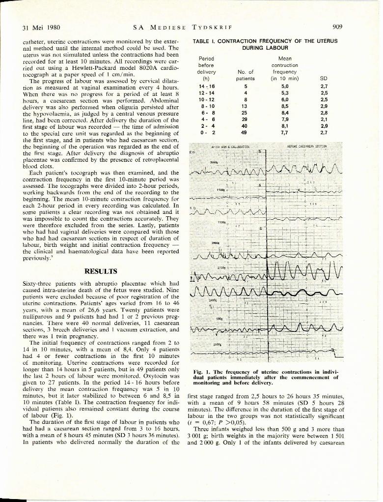
Fig. 1. The frequency of uterine contractions in individual patients immediately after the commencement ofmonitoring and before delivery.
first stage ranged from 2,5 hours to 26 hours 35 minutes,with a mean of 9 hours 58 minutes (SD 5 hours 28minutes). The difference in the duration of the first stage oflabour in the two groups was not statistically significant(t = 0,67; P >0,05).
Three infants weighed less than 500 g and 3 more than3001 g; birth weights in the majority were between 1 501and 2000 g. Only I of the infants delivered by caesarean
SO2,72,52,52,92,82,12,92,7
8EFORE CA£.S!\R£AN SECTION
Meancontractionfrequency
(in 10 min)
5,05,36,08,58,47,98,17,7
No. ofpatients
548
13
25294049
AFTER Aa.", & CALt8RATION
Periodbeforedelivery
(h)
14 -_1612 -1410 - 128 - 106 - 84 - 62 - 40- 2
RESULTS
Sixty-three patients with abruptio placentae which hadcaused intra-uterine death of the fetus were studied. Ninepatients were excluded because of poor registration of theuterine contractions. Patients' ages varied from 16 to 46years, with a mean of 26,6 years. Twenty patients werenulliparous and 9 patients had had I or 2 previous pregnancies. There were 40 normal deliveries, 11 caesareansections, 3 breech deliveries and 1 vacuum extraction, andthere was 1 twin pregnancy.
The initial frequepcy of contractions ranged from 2 to14 in 10 minutes, with a mean of 8,4. Only 4 patientshad 4 or fewer contractions in the first 10 minutesof monitoring. Uterine contractions were recorded forlonger than 14 hours in 5 patients, but in 49 patients onlythe last 2 hours of labour were monitored. Oxytocin wasgiven to 27 patients. In the period 14 - 16 hours beforedelivery the mean contraction frequency was 5 in 10minutes, but it later stabilized to between 6 and 8,5 in10 minutes (Table I). The contraction frequency for individual patients also remained constant during the courseof labour (Fig. 1).
The duration of the first stage of labour in patients whohad had a caesarean section ranged from 3 to 16 hours,with a mean of 8 hours 45 minutes (SD 3 hours 36 minutes).In patients who delivered normally the duration of the
catheter, uterine contractions were monitored by the external method until the internal method could be used. Theuterus was not stimulated unless the contractions had beenrecorded for at least 10 minutes. All recordings were carried out using a Hewlett-Packard model 8020A cardiotocograph at a paper speed of 1 cm/min.
The progress of labour was assessed by cervical dilatation as measured at vaginal examination every 4 hours.When there was no progress for a period of at least 8hours, a caesarean section was performed. Abdominaldelivery was also performed when oliguria persisted afterthe hypovolaemia, as judged by a central venous pressureline, had been corrected. After delivery the duration of thefirst stage of labour was recorded - the time of admissionto the special care unit was regarded as the beginning ofthe first stage, and in patients who had caesarean section,the beginning of the operation was regarded as the end ofthe first stage. After delivery the diagnosis of abruptioplacentae was confirmed by the presence of retroplacentalblood clots.
Each patient's tocograph was then examined, and thecontraction frequency in the first ID-minute period wasassessed. The tocographs were divided into 2-hour periods,working backwards from the end of the recording to thebeginning. The mean ID-minute contraction frequency foreach 2-hour period in every recording was calculated. Insome patients a clear recording was not obtained and itwas impossible to count the contractions accurately. Theywere therefore excluded from the series. Lastly, patientswho had had vaginal deliveries were compared with thosewho had had caesarean sections in respect of duration oflabour, birth weight and initial contraction frequency the clinical and haematological data have been reportedpreviously.3

910 SA MEDICAL JOURNAL 31 May 1980
section weighed more than 2500 g and and only 5 weighedmore than 2 000 g. The mean birth we'ight for infantsdelivered by caesarean section was 1 714 g (SD 806),which is slightly less than the 1 824 g (SD 728) of infantsdelivered vaginally. The difference in birth weight is notsignificant (t = 1,90; P <0,05).
DISCUSSION
For intra-uterine death to occur, at least two-thirds of theplacenta has to be separated from the uterine wall: Allthe patients included in the series could be regarded ascases of severe or grade III abruptio placentae, becauseintra-uterine death of the fetus had occurred in all.
The frequency of uterine contractions in severe abruptioplacentae is unique - in normal labour the uterus rarelycontracts more than 3 or 4 times during a lO-minuteperiod. During this study the initial contraction frequencywas less than 5 in only 4 patients, while the mean was8,4. These contraction frequencies were measured immediately after the commencement of monitoring, and theincrease could therefore not have been caused by oxytocin,which was not administered during the first 10 minutes ofmonitoring. The contraction frequency did not changeduring labour, although contractions were slightly less frequent long before the commencement of labour; the smallnumber of patients studied in this period could have ledto this observation.
There seems to be a correlation between contractionfrequency and the outcome of labour, the initial frequencybeing significantly higher in patients in whom caesareansection was later performed for lack of progress.
The aetiology of this almost diagnostic contraction pattern is unknown, but several factors may contribute to it.Abruption of the placenta could cause a sudden decreasein the progesterone-blocking effect on the uterus and therefore result in a high-frequency or low-amplitude contraction pattern. A decrease in progesterone levels was seenwhen placental function was destroyed by intra-amnioticinjection of hypertonic saline! A sudden release of prostaglandins due to decidual damage could also play a role,since it has been demonstrated that stripping of the membranes can increase prostaglandin release and cause achange in the contraction pattern of the uterus." eobo etal.' described the increased frequency of uterine contractions in 1965 and at that time attributed it to a 'severefunctional disturbance on the uterine fibers'. Eskes et al."recorded intra-uterine pressures in a few cases of abruptio
placentae, describing a high frequency of contractions aswell as a higher tonus, and postulated that this incompleterelaxation of the uterus could have been due to a sympathetic influence. This group of workers also describedelevated intra-uterine pressures and a high frequency ofcontractions in patients with polyhydramnios" Duringdrainage of amniotic fluid the pressure during contractionsas well as the frequency of contractions decreased. Distension of the uterus, as is the case in severe abruptioplacentae, could have the same effect as polyhydramnios-,However, in·-this series no correlation was found betweenthe size 'of the retroplacental clot and the contractionfrequency.
In this series it is unlikely that cephalopelvic ·disproportion necessitated the caesarean sections, because theseinfant's birth weights did not differ from those of infantsdelivered vaginally and in any case the birth weights werelow (only 1 infant weighed more than 2 500 g).
Although the cause for the increased contraction frequency in abruptio placentae remains debatable, it servesas an important diagnostic tool, since contractions caneasily be recorded by external methods. If contractions arefound to be frequent before or during labour, abruptioplacentae should seriously be considered in the differentialdiagnosis.
The contraction pattern is probably also a cause of thehigh incidence of intra-uterine deaths in patients withabruptio placentae, since the frequent contractions preventadequate blood flow to the intervillous space. Patients withabruptio placentae should therefore be delivered as soonas possible, unless the fetal heart rate can be monitoredadequately and facilities are available for 'immediatecaesarean section in the case of fetal distress.
I wish to thank the registrars of the Department of Obstetricsand Gynaecology, Tygerberg Hospital, for their help with thisstudy, which was supported by the South African MedicalResearch Council.
REFERENCES
I. Odendaal, H. 1. (1976): S. Afr. med. J., 50, 2129.2. Scher, G. (1977): Amer. J. Obstet. Gynec., 129, 164.3. Odendaal, H. J., Brink, S. and Steytler, J. G. (1978): S. Afr. med. J.,
54, 476. .4. Page, E. W., King, E. B. and Merrill, J. A. (1954): Obste!. and Gynec.,
3, 358.5. Csapo, A. 1., Herczeg, J., Pulkkinen, M. er al. (1976): Amer. J.
Obstet. Gynec., 124, I.6. Mitchell, M. D., Flint, A. P. F., Bibby, J. er al. (1977): Brit. med. J., ,
2, 1183.7. Coba, E., Quintero, C. A., Strada, G. ei al. (1965): Amer. J. Obstet.
Gynec., 93, 1151.8. Eskes, T., Seelen, J. and Stolte, L. (1967): Ned. T. Verlosk., 67, 74.9. Idem (1967): Ibid., 67, 251.