utility of pt3 substaging in lymph node-negative
TRANSCRIPT

Vol.:(0123456789)1 3
World Journal of Urology (2021) 39:4021–4027 https://doi.org/10.1007/s00345-021-03697-3
TOPIC PAPER
Utility of pT3 substaging in lymph node‑negative urothelial carcinoma of the bladder: do pathologic parameters add to prognostic sub‑stratification?
Moritz Maas1 · Johannes Mischinger2 · Eva Compérat3 · Marcus Scharpf4 · Falko Fend4 · Tilman Todenhöfer5 · Arnulf Stenzl1 · Georgios Gakis6 · Steffen Rausch1
Received: 1 November 2020 / Accepted: 8 April 2021 / Published online: 21 April 2021 © The Author(s) 2021
AbstractPurpose The value of bladder cancer (BC) substaging into macroscopic (pT3b) and microscopic (pT3a) perivesical fat extension in lymph node (Ln)-negative patients is controversially discussed and limited evidence for prognostic relevance of additional histopathological factors in pT3 BC exists. We evaluated the prognostic value of pT3 substaging and established pathological and clinical parameters with focus on tumor invasive front (TIF) and tumor size.Methods Specimens of 52 patients treated with radical cystectomy (RC) for pT3 a/b muscle-invasive BC were reviewed and re-evaluated by a pathologist specialized in uropathology. Clinical variables and standard histopathologic characteristics were assessed including TIF and tumor size. Their value as prognosticators for overall survival (OS) and recurrence-free survival (RFS) was evaluated.Results Mean age of patients was 67.55 years. Tumors were staged pT3a in 28 patients (53.8%) and pT3b in 24 (46.8%). Median OS was 34.51 months. Median tumor size was 3.2 cm, median TIF was 11.0 mm. Differences in OS between pT3a and pT3b were not significant (p = 0.45). Carcinoma in situ (CIS) and lymphovascular invasion (LVI) were significantly associated with pT3b tumors. Univariate analysis could not identify pathological prognosticators like TIF or tumor size for OS and RFS (p for all > 0.05).Conclusion No significant differences in OS or RFS were observed comparing Ln-negative pT3 BC following radical cys-tectomy. Additional pathologic variables like TIF could not be identified as prognosticator. Relevance of pT3 BC substaging needs reevaluation in larger prospective cohorts.
Keywords Muscle-invasive bladder cancer · Pathological staging · Patient outcome · Perivesical extension · Tumor invasion front
Introduction
Bladder cancer (BC) is one of the ten most common malig-nancies worldwide and the second most common uro-onco-logical entity [1]. Urothelial carcinoma represents the largest proportion; about 30% of the diseases are muscle-invasive at the time of their diagnosis or will become muscle-invasive in the course of their progress [2].
In patients with muscle-invasive urothelial bladder cancer (MIBC), radical cystectomy with bilateral lymphadenectomy is the treatment of choice for patients with non-metastatic disease [2]. The removed surgical specimen is pathologi-cally examined and classified according to the American Joint Committee on Caner (AJCC) TNM staging system. This classification helps the treating physician to evaluate
* Georgios Gakis [email protected]
1 Department of Urology, University Hospital, Tübingen, Germany
2 Department of Urology, University Hospital, Graz, Austria3 Department of Pathology, University Hospital, Wien, Austria4 Department of Pathology, University Hospital, Tübingen,
Germany5 Clinical Trials Unit, Studienpraxis Urologie, Nürtingen,
Germany6 Department of Urology, University Hospital Würzburg,
Oberdürrbacher Straße 6, 97080 Würzburg, Germany
4022 World Journal of Urology (2021) 39:4021–4027
1 3
the disease, decide on necessary treatment options (e.g. need for adjuvant therapies) and to estimate the prognosis.
One important aspect of this classification is the patho-logic tumor stage (pT). According to the currently applied classification, tumors extending the bladder without invad-ing surrounding organs are separated into pT3a and pT3b. In this context, pT3a tumors show a microscopic and pT3b a macroscopic invasion of the perivesical fat. The prognostic significance of this distinction remains subject of contro-versial debates: In comparison to pT2 tumors, pT3 disease including both, pT3a and pT3b is associated with impaired prognosis [3]. However, this prognostic relevance has not consistently been reported for the comparison of pT3a and pT3b tumors, which led to discussion about the clinical importance of the pT3 sub-classification [4].
Among other clinicopathological variables with puta-tive impact on the prognostic relevance are tumor size or the histopathological tumor invasion front (TIF) which was earlier discussed as a discriminative variable in pT3 bladder cancer [5].
Therefore, the aim of our study was to evaluate the prog-nostic value of pT3 substaging in dependence of additional histopathological variables including TIF and tumor size in patients with Ln-negative MICB.
Patients and methods
From our institutional bladder cancer database, medical records of patients with diagnosis of pT3a/b MIBC that underwent radical cystectomy from 2004 to 2013 were iden-tified. Patients with other than urothelial cancer, lymph-node metastases, or unknown nodal status were excluded from the analysis.
Files and original pathology reports were reviewed for clinical and pathologic standard variables tumor category (pT) and grade (G), lymphovascular invasion (LVI), con-comitant carcinoma in situ (CIS), microvascular invasion (V1), surgical margin status (R0/1). Staging and grading was performed according to the AJCC TNM system (2015 version), and the World Health Organization staging system, respectively. All pathology specimens were re-evaluated by one uropathology specialist (E.C.) and additional pathologic parameters tumor size and tumor invasion front, defined as the maximum width of the tumor invasion front in the perivesical fat, were evaluated for the final analysis. Follow-up of patients after surgery was performed in an outpatient setting according to the respective contemporary guideline (European Association of Urology Guidelines; S3-Guideline of the German Society of Urology) recommendations. All included patients could be followed up for tumor recurrence and death.
Chi-square tests were performed to assess the association of individual clincopathological risk-factors. For categori-cal analyses, tumor size and TIF were grouped according to their median values. Kaplan–Meier analyses were per-formed to evaluate overall survival (OS) and recurrence-specific survival (RFS). Differences between subgroups were evaluated using Log-rank test. Statistical significance was regarded as p < 0.05. For statistic analysis, commercial software (MedCalc®; Version 12.7.3.0) was used. Written informed consent was obtained by all participants. The study was approved by the Ethics Committee of the University of Tübingen (Approval Number: 417/2010 A).
Results
We identified 52 node-negative patients with pT3a/b urothe-lial cancer that underwent radical cystectomy and lymphad-enectomy. Of the patients, 21.2% were female and 78.8% were male. One (1.9%) of the patients received neo-adju-vant chemotherapy prior to radical cystectomy, two patients (3.8%) received adjuvant platinum-based chemotherapy. The median age at surgery was 67.55 years (range 46.43–88.99). Median follow-up was 33.74 months (mean: 51.8; range 1–184.5). Of all individuals with stage pT3, 53.8% were staged pT3a and 46.2% pT3b. Median tumor size was 3.2 cm (95% CI 2.6–3.5% cm) and median TIF was 11.0 mm (95% CI 7.0–15.53 mm). Detailed patient and tumor characteris-tics are summarized in Table 1.
Overall, 21 patients (40.4%) had cancer recurrence, 33 (63.5%) patients died during follow-up. Table 2 provides survival data for the analyzed patient cohort.
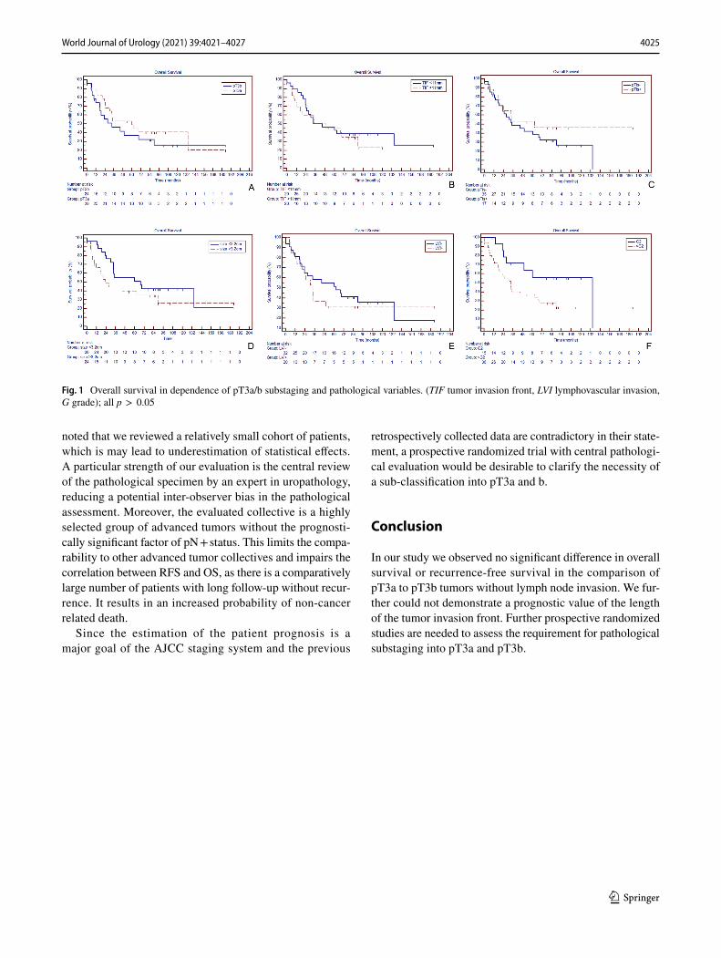
Median overall survival (OS) was 34.51 months (24.762–89.458) for the entire cohort and 60.07 months (30.443–134.483) for pT3a patients, while the detected OS for pT3b patients accounted for 33.62 (17.800–89.458) months. The observed differences in OS were however not statistically significant. In addition no correlation between pT3 substage and RFS was identified (data not shown). Association of pT3a/b substage to the individual pathologic factors revealed CIS and LVI to be significantly associated with pT3b (Table 3).
Overall, univariate analysis could not identify additional prognosticators of OS in the pT3a/b subgroup as shown in Fig. 1.
Discussion
The current AJCC TNM classification distinguishes blad-der tumors into pT3a (microscopic perivesical tissue inva-sion) and pT3b (macroscopic perivesical tissue invasion) sub-stages. Taking anatomical and biological aspects into

4023World Journal of Urology (2021) 39:4021–4027
1 3
account, this subdivision is evident and one would expect a worse prognosis for tumors with macroscopic invasion of the surrounding perivesical tissue: The lymphatic vessel topography shows larger lymphatic vessels more distant from the bladder’s serosa, resulting in a higher probability of lymphonodal spread in pT3b tumors [6, 7]. Correspond-ing observations were made by Neuzillet et al. and Tilki et al. showing more frequent LN invasion in pT3b tumors than in pT3a tumors [8, 9]. Furthermore, a macroscopic invasion is usually associated with a higher tumor bur-den, which leads to an increased probability of phenotypic mutations for metastatic spread and a higher probability of circulating tumor cells, capable of forming metastases [9, 10].
Despite these theoretical considerations, the expected prognostic difference between pT3a and pT3b tumors has not been consistently observed in the literature and thus leads to a discrepancy between the existing staging classifi-cation and its prognostic implication. Particularly in patients with pT3 pN0 disease no clear conclusion regarding the prognostic significance of the sub-classification is evident:
A large multicenter study by Tilki et al. shows significant differences in cancer-specific survival (CSS) and recurrence-free survival (RFS) for 456 patients with pT3, pN0 bladder cancer [8]. These observations are supported by Sonpavde et al.: Their multicentric analysis of 578 patients shows the prognostic potential of the pT3a/b sub-classification for RFS and postulates a prognostic risk model for patients with pT3
Table 1 Patient characteristics (n = 52)
Variable N %
Gender Male 41 78.8 Female 11 21.2
Age (years) at surgery (Radical Cystectomy) Median (range) 67.55 (46.43–88.99)
Pathological stage pT3a 28 53.8 pT3b 24 46.8
Pathological grade G2 15 29.4
> G2 36 70.6 N/A 1 –
Carcinoma in situ (CIS) No 35 67.3 Yes 17 32.7
Lymphovascular invasion (LVI) No 32 61.5 Yes 20 38.5
Necrosis No 44 84.6 Yes 8 15.4
Vascular invasion (V1) V0 45 88.2 V1 6 11.8 N/A 1 –
Surgical margin status R0 50 96.2 R1 2 3.8 R2 0 –
Tumor size (cm) Median 3.2 cm (95% CI 2.6–3.5) Arithmetic mean 3.35 (95% CI 2.8–3.85)
Tumor invasion front (TIF) in mm Median 11 (95% CI 7–15) Arithmetic mean 12.7 (95% CI 10–15.5)

4024 World Journal of Urology (2021) 39:4021–4027
1 3
pN0 based on T3 substage at radical cystectomy, lymphovas-cular invasion and margin status [11]. Important limitations of the aforementioned studies are caused by their multicenter nature. Here, the pathology was not centrally reviewed and they cover long periods of time, possibly including different surgical and pathological approaches.
Conversely, a number of smaller, mainly monocentric studies in the mentioned subgroup pT3 pN0 revealed no prognostic impact of the pT3 sub-classification: Boudreaux et al. could not find any significant difference concerning OS, CSS and RFS between lymph node-negative pT3a and pT3b tumors (p = 0.79; p = 0.21 and p = 0.53, respectively) [12]. Similar observations were made by Dincel et al., Kim
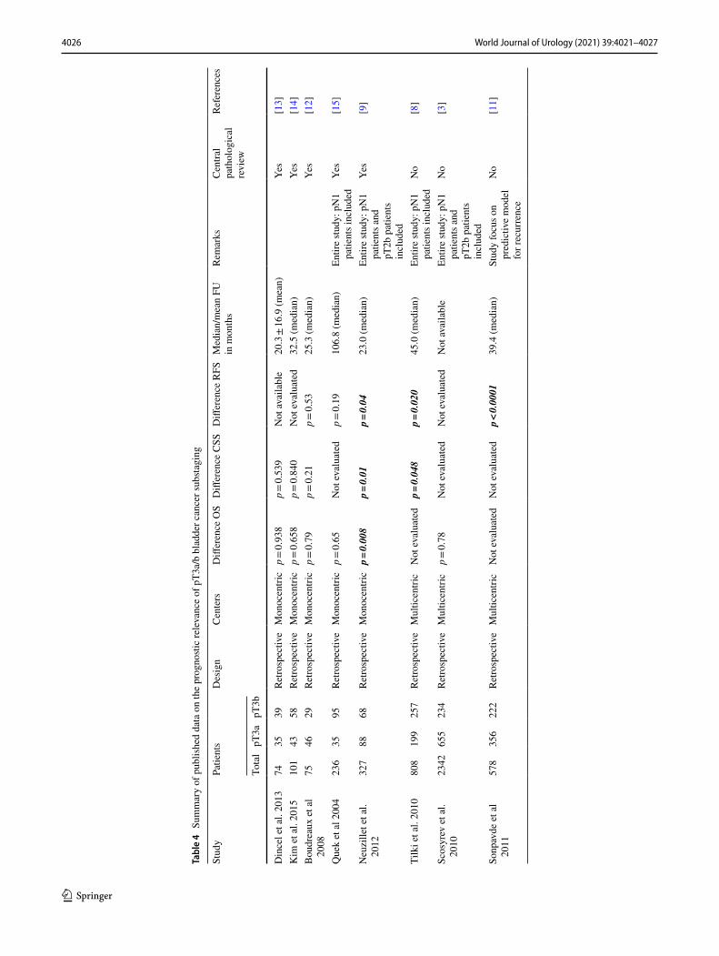
et al. and Quek et al. [13–15]. Scosyrev et al. retrospectively evaluated the SEER database (Surveillance, Epidemiology, and End results) and found impaired survival rates of pT3a BC as compared to pT2b disease with the same nodal status. Furthermore, they revealed an increased risk of nodal metas-tases with increased substaging and a decreased survival in pT3b patients compared to pT3a patients for node-positive tumors. However, in the node-negative subgroup, pT3a/b substage did not significantly impact OS [3]. Table 4 sum-marizes the available evidence on the prognostic impact of substaging in lymph node-negative pT3 patients.
The results of our study are consistent with the reported studies above: We could not prove any negative prognostic influence of pT3b substaging compared to the pT3a patients. Moreover, given the assumption that pT3 disease may be pathologically classified and sub-stratified by alternative pathological variables, we sought to evaluate tumor size and TIF. As for the other standard pathological factors, these parameters were not shown to be prognosticators in the pre-sent analysis.
In a comparative approach, Zarei et al. evaluated the prog-nostic value of the depth of tumor invasion into perivesical fat in pT3 tumors and demonstrated a significant improved CSS in patients with less than 4.5 mm invasion (p = 0.02) [5]. A combination of both parameters, width and depth of tumor invasion, could provide new insights in this context.
Our study has several limitations which have to be addressed: First, the inherent limitation of any retrospec-tive study should be mentioned. Additionally, it has to be
Table 2 Tabular summary of survival data for the analyzed cohort
Patients with recurrence n = 21
Location of recurrence Osseous n = 4 Pulmonary n = 2 Local recurrence n = 5 Hepatic n = 2 Lymphonodal recurrence n = 4 Peritoneal carcinomatosis n = 2 Renal recurrence n = 1 Soft tissue n = 1
Death n = 33
Cause of death Death after recurrence n = 17 postoperative complications n = 2 non-cancer related deaths n = 14
Table 3 Association of pathological variables with pT3 substaging
LVI lymphovascular invasion, CIS carcinoma in situ, TIF tumor inva-sion front, V1 vascular invasionp-values in bold indicate statistical significance
Variable P
Grade > 2 0.7508LVI 0.0329Necrosis 0.1634CIS 0.0472Positive surgical margin 0.5405TIF > 11 mm 0.2912Tumor size > 3.2 cm 0.1624V1 0.7782
4025World Journal of Urology (2021) 39:4021–4027
1 3
noted that we reviewed a relatively small cohort of patients, which is may lead to underestimation of statistical effects. A particular strength of our evaluation is the central review of the pathological specimen by an expert in uropathology, reducing a potential inter-observer bias in the pathological assessment. Moreover, the evaluated collective is a highly selected group of advanced tumors without the prognosti-cally significant factor of pN + status. This limits the compa-rability to other advanced tumor collectives and impairs the correlation between RFS and OS, as there is a comparatively large number of patients with long follow-up without recur-rence. It results in an increased probability of non-cancer related death.
Since the estimation of the patient prognosis is a major goal of the AJCC staging system and the previous
retrospectively collected data are contradictory in their state-ment, a prospective randomized trial with central pathologi-cal evaluation would be desirable to clarify the necessity of a sub-classification into pT3a and b.
Conclusion
In our study we observed no significant difference in overall survival or recurrence-free survival in the comparison of pT3a to pT3b tumors without lymph node invasion. We fur-ther could not demonstrate a prognostic value of the length of the tumor invasion front. Further prospective randomized studies are needed to assess the requirement for pathological substaging into pT3a and pT3b.
Fig. 1 Overall survival in dependence of pT3a/b substaging and pathological variables. (TIF tumor invasion front, LVI lymphovascular invasion, G grade); all p > 0.05
4026 World Journal of Urology (2021) 39:4021–4027
1 3
Tabl
e 4
Sum
mar
y of
pub
lishe
d da
ta o
n th
e pr
ogno
stic
rele
vanc
e of
pT3
a/b
blad
der c
ance
r sub
stag
ing
Stud
yPa
tient
sD
esig
nC
ente
rsD
iffer
ence
OS
Diff
eren
ce C
SSD
iffer
ence
RFS
Med
ian/
mea
n FU
in
mon
ths
Rem
arks
Cen
tral
path
olog
ical
re
view
Refe
renc
es
Tota
lpT
3apT
3b
Din
cel e
t al.
2013
7435
39Re
trosp
ectiv
eM
onoc
entri
cp =
0.93
8p =
0.53
9N
ot av
aila
ble
20.3
± 16
.9 (m
ean)
Yes
[13]
Kim
et a
l. 20
1510
143
58Re
trosp
ectiv
eM
onoc
entri
cp =
0.65
8p =
0.84
0N
ot e
valu
ated
32.5
(med
ian)
Yes
[14]
Bou
drea
ux e
t al
2008
7546
29Re
trosp
ectiv
eM
onoc
entri
cp =
0.79
p = 0.
21p =
0.53
25.3
(med
ian)
Yes
[12]
Que
k et
al 2
004
236
3595
Retro
spec
tive
Mon
ocen
tric
p = 0.
65N
ot e
valu
ated
p = 0.
1910
6.8
(med
ian)
Entir
e stu
dy: p
N1
patie
nts i
nclu
ded
Yes
[15]
Neu
zille
t et a
l. 20
1232
788
68Re
trosp
ectiv
eM
onoc
entri
cp =
0.00
8p =
0.01
p = 0.
0423
.0 (m
edia
n)En
tire
study
: pN
1 pa
tient
s and
pT
2b p
atie
nts
incl
uded
Yes
[9]
Tilk
i et a
l. 20
1080
819
925
7Re
trosp
ectiv
eM
ultic
entri
cN
ot e
valu
ated
p = 0.
048
p = 0.
020
45.0
(med
ian)
Entir
e stu
dy: p
N1
patie
nts i
nclu
ded
No
[8]
Scos
yrev
et a
l. 20
1023
4265
523
4Re
trosp
ectiv
eM
ultic
entri
cp =
0.78
Not
eva
luat
edN
ot e
valu
ated
Not
ava
ilabl
eEn
tire
study
: pN
1 pa
tient
s and
pT
2b p
atie
nts
incl
uded
No
[3]
Sonp
avde
et a
l 20
1157
835
622
2Re
trosp
ectiv
eM
ultic
entri
cN
ot e
valu
ated
Not
eva
luat
edp <
0.00
0139
.4 (m
edia
n)St
udy
focu
s on
pred
ictiv
e m
odel
fo
r rec
urre
nce
No
[11]

4027World Journal of Urology (2021) 39:4021–4027
1 3
Author contributions MM: manuscript writing, editing, data collection and management; JM: data collection and management, data analysis; EC: project development, pathological examination; MS: data collec-tion or management; FF: protocol development; TT: protocol devel-opment; AS: protocol development; GG: project and protocol devel-opment, data collection and management; SR: manuscript writing, editing, data management and analysis, project development.
Funding Open Access funding enabled and organized by Projekt DEAL. This research did not receive any specific grant from funding agencies in the public, commercial or not-for-profits sectors.
Data availability Data and material available.
Declarations
Conflict of interest/competing interests The authors have no relevant conflict of interest in the context of the work presented.
Ethics approval The study was approved by the Ethics Committee of the University of Tübingen (APPROVAL number: 417/2010 A).
Consent to participate Consent to participate was given by the patients or their legal representatives.
Open Access This article is licensed under a Creative Commons Attri-bution 4.0 International License, which permits use, sharing, adapta-tion, distribution and reproduction in any medium or format, as long as you give appropriate credit to the original author(s) and the source, provide a link to the Creative Commons licence, and indicate if changes were made. The images or other third party material in this article are included in the article’s Creative Commons licence, unless indicated otherwise in a credit line to the material. If material is not included in the article’s Creative Commons licence and your intended use is not permitted by statutory regulation or exceeds the permitted use, you will need to obtain permission directly from the copyright holder. To view a copy of this licence, visit http:// creat iveco mmons. org/ licen ses/ by/4. 0/.
References
1. Siegel RL, Miller KD, Jemal A (2019) Cancer statistics. CA A Cancer J Clin 69(1):7–34. https:// doi. org/ 10. 3322/ caac. 21551
2. Witjes JA, Bruins HM, Cathomas R, Compérat EM, Cowan NC, Gakis G, Hernández V, Linares Espinós E, Lorch A, Neuzillet Y, Rouanne M, Thalmann GN, Veskimäe E, Ribal MJ, van der Hei-jden AG (2020) European Association of Urology Guidelines on Muscle-invasive and Metastatic Bladder Cancer: summary of the 2020 guidelines. EurUrol. https:// doi. org/ 10. 1016/j. eururo. 2020. 03. 055
3. Scosyrev E, Yao J, Messing E (2010) Microscopic invasion of perivesical fat by urothelial carcinoma: implications for prognosis and pathology practice. Urology 76(4):908–913. https:// doi. org/ 10. 1016/j. urolo gy. 2010. 02. 073 (Discussion 914)
4. Paner GP, Montironi R, Amin MB (2017) Challenges in patho-logic staging of bladder cancer: proposals for fresh approaches of assessing pathologic stage in light of recent studies and observa-tions pertaining to bladder histoanatomic variances. AdvAnat-Pathol 24(3):113–127. https:// doi. org/ 10. 1097/ pap. 00000 00000 000152
5. Zarei S, Frank I, Boorjian SA, Thompson RH, Kim S, Weight C, Tarrell R, Thapa P, Cheville JC (2012) Prognostic significance of measured depth of invasion of urothelial carcinoma of the bladder compared to the 2010 American Joint Committee on Cancer pT2 and pT3 classifications. J Urol 188(5):1706–1711. https:// doi. org/ 10. 1016/j. juro. 2012. 07. 035
6. Matsumoto K, Soh S, Satoh T, Iwamura M, Ishikawa Y, Ishii T, Baba S (2008) Distribution of lymphatic vessel network in nor-mal urinary bladder. Urology 72(3):706–710. https:// doi. org/ 10. 1016/j. urolo gy. 2007. 12. 061
7. Marchetti C, Poggi P, Farina A, Gritti A, Scelsi L, Scelsi R (1996) Structure of the initial lymphatics of the human urinary bladder with invasive urothelial tumors. Lymphology 29(3):118–125
8. Tilki D, Svatek RS, Karakiewicz PI, Novara G, Seitz M, Son-pavde G, Gupta A, Kassouf W, Fradet Y, Ficarra V, Skinner E, Lotan Y, Sagalowsky AI, Stief CG, Reich O, Shariat SF (2010) pT3 Substaging is a prognostic indicator for lymph node nega-tive urothelial carcinoma of the bladder. J Urol 184(2):470–474. https:// doi. org/ 10. 1016/j. juro. 2010. 04. 007
9. Neuzillet Y, Lebret T, Molinie V, Yonneau L, Herve JM, Theo-dore C, Botto H (2012) Perivesical fat invasion in bladder cancer: implications for prognosis comparing pT2b, pT3a and pT3b stages and consequences for adjuvant chemotherapy indications. BJU Int 110(11):1736–1741. https:// doi. org/ 10. 1111/j. 1464- 410X. 2012. 11104.x
10. Msaouel P, Koutsilieris M (2011) Diagnostic value of circulating tumor cell detection in bladder and urothelial cancer: systematic review and meta-analysis. BMC Cancer 11:336. https:// doi. org/ 10. 1186/ 1471- 2407- 11- 336
11. Sonpavde G, Khan MM, Svatek RS, Lee R, Novara G, Tilki D, Lerner SP, Amiel GE, Skinner E, Karakiewicz PI, Bastian PJ, Kassouf W, Fritsche HM, Izawa JI, Scherr DS, Ficarra V, Dinney CP, Lotan Y, Fradet Y, Shariat SF (2011) Prognostic risk strati-fication of pathological stage T3N0 bladder cancer after radical cystectomy. J Urol 185(4):1216–1221. https:// doi. org/ 10. 1016/j. juro. 2010. 11. 082
12. Boudreaux KJ Jr, Chang SS, Lowrance WT, Rumohr JA, Barocas DA, Cookson MS, Smith JA Jr, Clark PE (2009) Comparison of American Joint Committee on Cancer pathologic stage T3a versus T3b urothelial carcinoma: analysis of patient outcomes. Cancer 115(4):770–775. https:// doi. org/ 10. 1002/ cncr. 24110
13. Dincel C, Kara C, Balci U, Ozer K, Ozbir S, Sefik E, Gorgel SN, Girgin C (2013) Comparison of microscopic (pT3a) and gross extravesical extension (pT3b) in pathological staging of bladder cancer: analysis of patient outcomes. Int Urol Nephrol 45(2):387–393. https:// doi. org/ 10. 1007/ s11255- 013- 0381-7
14. Kim HS, Piao S, Moon KC, Jeong CW, Kwak C, Kim HH, Ku JH (2015) Adjuvant chemotherapy correlates with improved sur-vival after radical cystectomy in patients with pT3b (macroscopic perivesical tissue invasion) bladder cancer. J Cancer 6(8):750–758. https:// doi. org/ 10. 7150/ jca. 12259
15. Quek ML, Stein JP, Clark PE, Daneshmand S, Miranda G, Cai J, Groshen S, Cote RJ, Lieskovsky G, Quinn DI, Skinner DG (2004) Microscopic and gross extravesical extension in pathological stag-ing of bladder cancer. J Urol 171(2):640–645. https:// doi. org/ 10. 1097/ 01. ju. 00001 08664. 39035. 51
Publisher’s Note Springer Nature remains neutral with regard to jurisdictional claims in published maps and institutional affiliations.