utility of viper (virtual imaging for pathology education
TRANSCRIPT
Utility of VIPER (Virtual Imaging for Pathology Education & Research) in Continuing Medical Education and Slide Surveys
T Barr, K Nicol, D Billiter, K Wohlever, P Baker, & V Prasad Biopathology Center
The Research Institute at Nationwide Children’s Hospital
Department of Pathology & Laboratory Medicine College of Medicine, The Ohio State University
Nationwide Children’s Hospital Columbus, Ohio
For 15 years Virtual Congresses with virtual libraries of “static images” have been available on the Internet. With the evolution of technology we can now transmit high-resolution whole-slide images of glass slides for consultation, diagnosis and research.1,2 Despite these advances, a majority of current slide survey programs are still using glass slides for continuing medical education or proficiency tests and these are fraught with challenges. For example, it is very labor intensive to obtain, cut, stain and ship glass slides to multiple locations. Other challenges include the potential for discrepancy among slides due to loss of tissue in deeper sections and difficulty annotating glass slides. Use of digital pathology in teaching venues addresses these issues with slides by reducing the quantity of tissue and slides required, permitting multiple annotations and allowing all participating pathologists to review the same image.
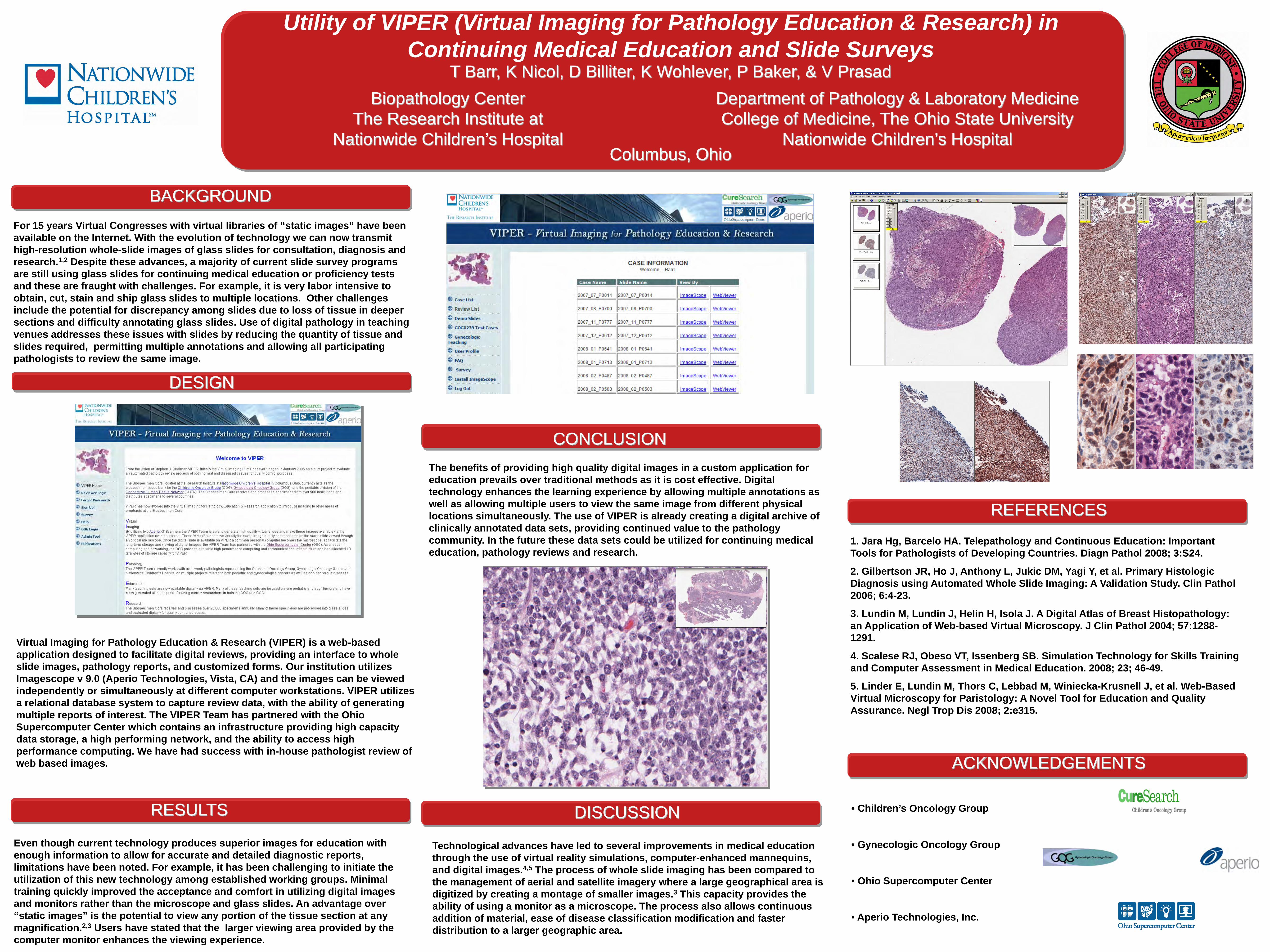
Virtual Imaging for Pathology Education & Research (VIPER) is a web-based application designed to facilitate digital reviews, providing an interface to whole slide images, pathology reports, and customized forms. Our institution utilizes Imagescope v 9.0 (Aperio Technologies, Vista, CA) and the images can be viewed independently or simultaneously at different computer workstations. VIPER utilizes a relational database system to capture review data, with the ability of generating multiple reports of interest. The VIPER Team has partnered with the Ohio Supercomputer Center which contains an infrastructure providing high capacity data storage, a high performing network, and the ability to access high performance computing. We have had success with in-house pathologist review of web based images.
Even though current technology produces superior images for education with enough information to allow for accurate and detailed diagnostic reports, limitations have been noted. For example, it has been challenging to initiate the utilization of this new technology among established working groups. Minimal training quickly improved the acceptance and comfort in utilizing digital images and monitors rather than the microscope and glass slides. An advantage over “static images” is the potential to view any portion of the tissue section at any magnification.2,3 Users have stated that the larger viewing area provided by the computer monitor enhances the viewing experience.
The benefits of providing high quality digital images in a custom application for education prevails over traditional methods as it is cost effective. Digital technology enhances the learning experience by allowing multiple annotations as well as allowing multiple users to view the same image from different physical locations simultaneously. The use of VIPER is already creating a digital archive of clinically annotated data sets, providing continued value to the pathology community. In the future these data sets could be utilized for continuing medical education, pathology reviews and research.
Technological advances have led to several improvements in medical education through the use of virtual reality simulations, computer-enhanced mannequins, and digital images.4,5 The process of whole slide imaging has been compared to the management of aerial and satellite imagery where a large geographical area is digitized by creating a montage of smaller images.3 This capacity provides the ability of using a monitor as a microscope. The process also allows continuous addition of material, ease of disease classification modification and faster distribution to a larger geographic area.
1. Jara Hg, Barcelo HA. Telepathology and Continuous Education: Important Tools for Pathologists of Developing Countries. Diagn Pathol 2008; 3:S24.
2. Gilbertson JR, Ho J, Anthony L, Jukic DM, Yagi Y, et al. Primary Histologic Diagnosis using Automated Whole Slide Imaging: A Validation Study. Clin Pathol 2006; 6:4-23.
3. Lundin M, Lundin J, Helin H, Isola J. A Digital Atlas of Breast Histopathology: an Application of Web-based Virtual Microscopy. J Clin Pathol 2004; 57:1288-1291.
4. Scalese RJ, Obeso VT, Issenberg SB. Simulation Technology for Skills Training and Computer Assessment in Medical Education. 2008; 23; 46-49.
5. Linder E, Lundin M, Thors C, Lebbad M, Winiecka-Krusnell J, et al. Web-Based Virtual Microscopy for Paristology: A Novel Tool for Education and Quality Assurance. Negl Trop Dis 2008; 2:e315.
• Children’s Oncology Group
• Gynecologic Oncology Group
• Ohio Supercomputer Center
• Aperio Technologies, Inc.
BACKGROUND
DESIGN
CONCLUSION
DISCUSSION
REFERENCES
ACKNOWLEDGEMENTS
RESULTS