uv mutagenesis in escherichia coli frequency of the ... · uv mutagenesis in escherichia coli k-12:...
TRANSCRIPT

Journal of Experimental Microbiology and Immunology (JEMI) Vol. 1:32-46 Copyright December 2001, M&I UBC
UV mutagenesis in Escherichia coli K-12: Cell survival and mutation frequency of the chromosomal genes lacZ, rpoB, ompF, and ampA
KEVIN LIN AND ALICE WANG
Department of Microbiology and Immunology, UBC
UV-induced mutation frequency and cell survivability was examined in Escherichia coli K-12 strains. The effects of UV (254nm) induced damage on deoxyribonucleic acid (DNA) was investigated using two protocols termed Direct-Plate Irradiation (DPI) and Liquid-Plate Irradiation (LPI). A biological system needed to be designed to facilitate future experiments involving UV and gene mutation frequencies. The conditions for the DPI protocol involved plating E. coli onto nutrient agar plates and exposing them to 0, 5, 10, and 15 seconds of UV irradiation at 60cm away from the plates. The LPI protocol involved exposing cells suspended in M9-minimal salts with 0.2% MgSO4 to 0, 10, 20, 30, 40 and 60 seconds. Both negative screening and positive screening procedures were applied to determine the mutational frequency of lacZ, rpoB, ompF, and ampC. Several strains and plasmids were also tested in these selections to determine the best biological system for carrying out the experiment. These tests showed that the best strain was B23. This strain produced a maximum mutational frequency of 8.5 x 10⁻⁶ for rpoB (rifampicin resistance) and 1.5 x 10⁻⁵ for ompF (chloramphenicol resistance). The results are discussed in terms of the SOS response or SOS system because this system is responsible for mutagenesis. Our results provide a framework for future assays involving mutagenesis.
UV radiation has become a global concern due to the fact that it is a major environmental mutagen and carcinogen, leading to conditions such as skin cancer [8]. Coupled with the fact that ozone layer (the atmospheric layer that filters out solar UV radiation from the sun) is continuously depleted by pollution, there is pressure to better understand the mechanics in how mutations arise so that possible remedies in the future can be devised. UV radiation may be a hazard to the human population but it is also an environmental stress for other organisms such as bacteria. Such environmental stress caused by UV may in some way induce different evolutionary changes on bacteria that would have otherwise not been selected for. This area thus provides avenues of physiological, ecological, and genetic investigation because mutations play a key role in biological processes such as evolution, carcinogenesis, aging, and generation of somatic genetic diversity [8]. In this series of experiments, the K-12 strain of Escherichia coli were used because of its ease of cultivation, genetic characterization and because it is the most often used organism in UV mutagenesis experiments. The genetic markers chosen for mutational frequency analysis were rpoB (also known as rif) located at about 90 centisomes in the K-12 chromosome, ampA located at about 94 centisomes, and ompF located at about 21 centisomes [6]. Mutations at these sites confer resistance to rifampicin (an antibiotic that strongly inhibits RNA polymerase), ampicillin, and chloramphenicol respectively [9]. The lacZ locus, located at about 8 centisomes [6], was also selected as a genetic marker; lacZ− mutants are not able to use lactose. When deoxyribonucleic acid (DNA) is exposed to UV light (254nm), the most frequent DNA damage, or lesions, results at dimers of any two adjacent pyrimidine bases (T, thymine; C cytosine) causing T-T, C-T, and C-C dimers, but T-T dimers are the most common cyclobutane pyrimidine dimers [3, 10, 13, 18, 19]. Another type of DNA damage is the 6-4 pyrimidine-pyrimidone photoproducts. These occur at a lesser frequency than cyclobutane pyrimidine dimers but are less mutagenic because they’re more efficiently repaired than cyclobutane pyrimidine dimers [3]. The remaining types of DNA lesions include pyrimidine hydrates, purine photoproducts, strand brakes, and DNA cross-links. These occur at a much lower frequency however [3]. Thus, UV radiation induces mutations such as frameshifts, base substitutions, deletions, recombination and other types of genetic rearrangements [13]. To cope with DNA damage, Escherichia coli has two main categories of repair, one is the “error-free” system and is independent of the SOS regulon or response (see below), while the other is the “error-prone” repair system and is part of the SOS regulon [15, 20]. There are three strategies employed by E. coli that are considered relatively error-free, they are photoreactivation, excision repair and post-replication repair [21].
32

Journal of Experimental Microbiology and Immunology (JEMI) Vol. 1:32-46 Copyright December 2001, M&I UBC
However, not all DNA lesions can be repaired because it some situations, the cell may have sustained significant damage; the damage was poorly repaired; or the DNA damage occurred during the S phase of cell growth [17]. Under such stressful conditions, the cell must make a last, desperate effort to repair itself. Or else UV-induced lesions will block DNA polymerases from replicating its genetic material, ultimately leading to cell death. This push to repair itself is commonly referred to as the SOS response or SOS system [see reviews 1, 4, 7, 8, 16 for more detailed information]. Basically, the SOS system avoids replication blocks caused by UV induced lesions by activating a series of pathway that causes DNA polymerases to be highly tolerant of these bulky groups of photodimers. In order to achieve this tolerance to replicate pass these lesions, the proofreading ability and specificity of polymerases in faithfully replicating the template strand is greatly reduced. Consequently, the wrong nucleotides are added during the elongation process of DNA replication and thus mutations are introduced to the genome. As a result, the SOS system is ultimately responsible for mutagenesis in a majority of the cases. When this project was initially designed, we planned on using the DH5α strain of Escherichia coli transformed with the plasmid pUC19 as our biological system to look at gene mutation frequency on lacZ located on pUC19 (see [12] for in-depth discussion of the lac system) because this system was used in one of the undergraduate biology courses at the University of British Columbia. Furthermore, their experiment involved naked pUC19 plasmid irradiation and subsequent transformation of irradiated pUC19 back into DH5α host which was useful for our purposes. We set out to repeat this experiment without realizing some of the constraints involved with the strains DH5α and INVα. Therefore, we expected to observe some kind of lac mutation frequency. The second part of the project evolved out of difficulties in our first set of experiments to include gene mutation frequency on the chromosome. With the second half of the project we expected the frequencies to show results similar to Miller’s experiment [11]. The results from this project are discussed based on the SOS response and further avenues of investigation are provided.
MATERIALS AND METHODS Chemicals and bacteria growth nutrients used in this project were standard grade reagents. Most equipment are standard scientific equipment. Bacterial Strains. Several strains of the K-12 line of Escherichia coli were used in this project (Table 1) The stock strains, provided by Dr. William Ramey (UBC Microbiology), were kept at -70°C in nutrient LB broth supplemented with 25% glycerol. LB broth agar plates were streaked with these freezer stocks to grow a working set of individual colonies. Individual colonies were picked for use of growing the overnights. Overnight cultures for the direct-plate irradiation were grown up in flasks containing 20ml of LB broth at 37°C in an orbit-shaking water bath (New Brunswick Scientific, model G76). Whereas overnight cultures for the liquid-plate irradiation were grown up in 16x125mm test tubes containing 5ml of LB broth placed on a rotary test tube rack (Departmental custom built) in the 37°C incubator room. All overnight cultures were grown to saturation. Strains of bacteria employed in this experiment. Direct-Plate Irradiation. For the series of direct-plate kill experiments, sub-cultures were grown by inoculating (1:50 dilution) LB broth (20ml final volume) with the overnights and grown to about 1 x 10⁸ cells per ml, approximately 0.7 to 0.8 OD660nm. These sub-cultures were grown in flasks placed in a 37°C orbit-shaking water bath at power level 3.5. When the sub-culture reached the desired optical density, a series of dilutions of the sub-culture was prepared; these dilutions are 10-1, 10-2, 10-3, 10-4, and 10-5. These dilutions were then used to make final plates for the ultraviolet irradiation by spread-plating 0.1 ml of the diluted sub-cultures onto various nutrient plates. LB agar plates were used unless otherwise noted. Therefore, the final plated dilutions are 10-2, 10-3, 10-4, 10-5, and 10-6. For most experiments, the dilutions of 10-2, 10-3, and 10-4 were used for the treatment set while 10-5 and 10-6 dilutions were the control. After plating, the plates were kept in a black box (plastic cooler box) before and after irradiation unless otherwise noted. All UV irradiations were done in a custom-built UV chamber with a glass front. The UV lamp (Philips UVC lamp, 15W) could be adjusted vertically to a desired height of up to 1.2 metres. The majority of the experiments were done in the dark to avoid photoreactivation. The only time a light source was present was during the transfer of the plates into the UV chamber. Before each irradiation, the UV lamp was warmed up for at least 30 minutes. For each UV exposure time point one plate from each treatment set (10-2, 10-3, and 10-4 dilutions) were randomly placed in the centre of the chamber and irradiated for the set time; this was done by using a digital timer and by manually adjusting the power switch of the UV lamp. The lids of the treatment plates were removed before placing the plates into the chamber to avoid shielding by the lids. After irradiation, the lids were replaced and the plates were immediately placed into the black box. All the plates were grown in the 37°C incubator for 24 to 36 hours before scoring the number of colonies. Kill curve variables. Escherichia coli strains DH5α(pUC19) and INVα(pUC19) were used in the initial tests to determine the appropriate parameters for UV irradiation involving the direct-plate irradiation protocol. Heights tested were 30 cm and 60 cm from floor of the chamber to the lamp. Various time sets were also tested, the first set involved exposures of 0, 10, 30 and 60 seconds. The second set involved exposures of 0, 5, 10 and 15 seconds. Lastly, the final set contained exposures of 0, 2, 4 and 6 seconds. Photoreactivation after UV mutagensis. The effects of natural light on UV irradiated cells were briefly investigated. This was done by carrying out the standard direct irradiation protocol with a new time set. This new time set had the same exposure length as that of the longest exposure to UV; the only difference is that the one of these sets were placed in natural light for approximately two hours before placing them into the incubator. Response of strains and plasmids to UV irradiation. Responses to UV irradiation in the different strains were compared by applying the direct-plate irradiation protocol. The effect of plasmids on UV response was looked at by comparing host strains with different plasmids, different hosts with the same plasmid, and hosts with or without the same plasmid. Negative screening of lac and Amp mutants. The direct-plate irradiation protocol was carried out with the α-complemented strains with the plasmid pUC19. After a day of incubation, UV survivors where randomly picked and transfer to a fresh LB agar plate to make a master stamp plate of 100 colonies per plate. The master plate was allowed to grow for 24 hours at 37°C before it was replicate-plated to negative-screen plates such as MacConkey agar and LB agar supplemented with 100ug/ml of ampicillin. These negative-screen plates were incubated for 24 to 48
33

Journal of Experimental Microbiology and Immunology (JEMI) Vol. 1:32-46 Copyright December 2001, M&I UBC
hours in a 37°C incubator and then the number of negative mutants was counted. To quickly ensure that the mutants were not contaminants, they were Gram stained to check their microscopic properties. Further tests on the mutants were carried out by performing plasmid extraction using commercial mini-prep kits (Wizard), and then transforming α-complemented hosts to see whether the mutation was chromosomal related by screening the transformed hosts with the MacConkey and ampicillin supplemented LB agar plate
Table I. Strains of Escherichia coli used in the experiments
Strain Plasmid Description B23
-none-
K-12 wildtype
DH5α (host) -none- deoR, endA1, gyrA96, hsdR17(rk− mk
+) recA1, relA1, supE44, thi-1, ∆(lacZYA-argFV169) φ80lacZ ∆M15, F−
DH5α pUC19 same as DH5α, Amp+, Lac+
INVα (host) -none- F'{lacI:Tn10(tetr)}, mcrA, ∆(mrr-hsdRMS-mcr), φ80lac, ∆M15, ∆lacX74, recA1, araD139, ∆(ara-leu)7697, galU, galK, rpsL, endA1, nupG
INVα pUC19 same as INVα, Amp+, Lac+
HB101 (host) -none- F- hsdS20(r−B m−B), recA13, ara-14, proA2, lacY1, galK2, rpsL20(Smr), xyl-5,
mtl-1, supE44, λ-, leu HB101 p309.1 same as HB101 host except that a HIND III F fragment of φ29 is cloned into
the HIND III site of pBR322 HB101 p328.5 same as HB101 host except that a HIND III C fragment of φ29 is cloned into
the HIND III site of pBR322 SF8 (host) -none- hsdR, hsdM, recB, recC, lop, leu, thr, thi, Amps
SF8 p309.1 same as SF8 host except that a HIND III F fragment of φ29 is cloned into the HIND III site of pBR322
SF8 p328.5 same as SF8 host except that a HIND III C fragment of φ29 is cloned into the HIND III site of pBR322
Negative screening with S-Gal differential medium. The direct-plate irradiation protocol was also carried out with S-Gal agar (Sigma) plate. Instead of plating the dilution sets to LB agar, the cells were directly plated onto S-Gal agar plates and irradiated. This allows a direct screen for negative mutants because all negative mutants will appear white on this agar while the normal population will grow up to be black in colour; this forgoes the intermediate step of replica-plating. The direct-plate protocol was slightly modified for this test by changing the dilution scheme to 10-1, 10-2, and 10-3 final plated dilutions for the test sets; controls were still 10-6 final dilution. Liquid-Plate Irradiation. This method was adapted from Miller (21). In the protocol E. coli B23, DH5α(host), and INVα(host) were used. The height of the UV lamp from the bottom of the chamber was 60 cm and the times of exposure are typically much longer than the ones involved with the direct-plate irradiation method. The time set which seemed to give the best result included exposure times of 0, 10, 20, 30, and 40 seconds to UV. Sub-cultures were prepared by inoculating Luria broth with the overnight culture to make a final volume of 30ml (1:30 dilution). The sub-culture was then allowed to grow up to approximately 1.0 to 1.2 OD660nm in a 37°C orbit-shaking water bath. When the culture has reached the desired optical density, the cells were centrifuged at 8500 RPM for 15 minutes. The pellet was resuspended in the same volume of Minimal M9 salts supplemented with MgSO4 to give a final concentration of 0.2%. The resuspended solution of cells was then allowed to cool on ice for about 15 minutes. At this point, 5 ml of the cell resuspension was pipetted into shallow-bottomed glass petri dishes. These dishes were then placed into a black box lined with cold packs. For each exposure time point, the glass petri dish was centred under the UV lamp on a piece of cardboard; the lids were removed. During the exposure to UV, the cell suspension was gently swirled around to allow for even irradiation by shuffling the piece of cardboard on which the petri dish sat on; a shaking platform could be substituted but the distance between the lamp and the platform will need to be adjusted accordingly. Exposure time was monitored manually by controlling the power to the UV lamp and by a digital timer. The lids were then replaced on the dish and transferred back into the black box. After all the plates were irradiated, the viable plate count for the number of survivors were completed by plating final dilutions of 10-6 for 0 seconds, 10-3 for 10 seconds, 10-2 for 20 and 30 seconds and 10-1 dilution for 40 seconds. X-gal (5-bromo-4-chloro-3-indolyl-β-D-galactopyranoside) LB agar plates were used for the viable plate count. X-gal plates were made by adding 50mg (50ug/ml Luria agar) of X-gal (GBT) dissolved in 1ml of dimethylforamide, and 50mg (50ug/ml Luria agar) of IPTG (isopropyl-thio-galactosidaseactoside) (GBT) to 1 litre of Luria agar. Numbers of viable cells were counted after 24 to 36 hours of incubation at 37°C. Positive screening for rifampicin resistance, chloramphenicol resistance, and ampicillin resistance. Outgrowths of the irradiated cells were also prepared for positive screening by inoculating 5ml of LB broth in 16x125mm test tubes with 0.1ml of UV exposed cells. These were placed in the rotary test tube rack and grown overnight at 37°C. The outgrowths were plated the next day by spread-plating 0.1ml of overnight cultures on each positive-screen plate. The plates used were rifampicin LB agar (100ug/ml), chloramphenicol LB agar (5ug/ml), and ampicillin LB agar (100ug/ml). To determine the total number of cells in the overnight cultures, 0.1ml of 10-5 dilution was plated onto Luria agar plates. These were incubated for 24 to 36 hours in the 37°C incubator and number of colonies was counted after sufficient growth.
34

Journal of Experimental Microbiology and Immunology (JEMI) Vol. 1:32-46 Copyright December 2001, M&I UBC
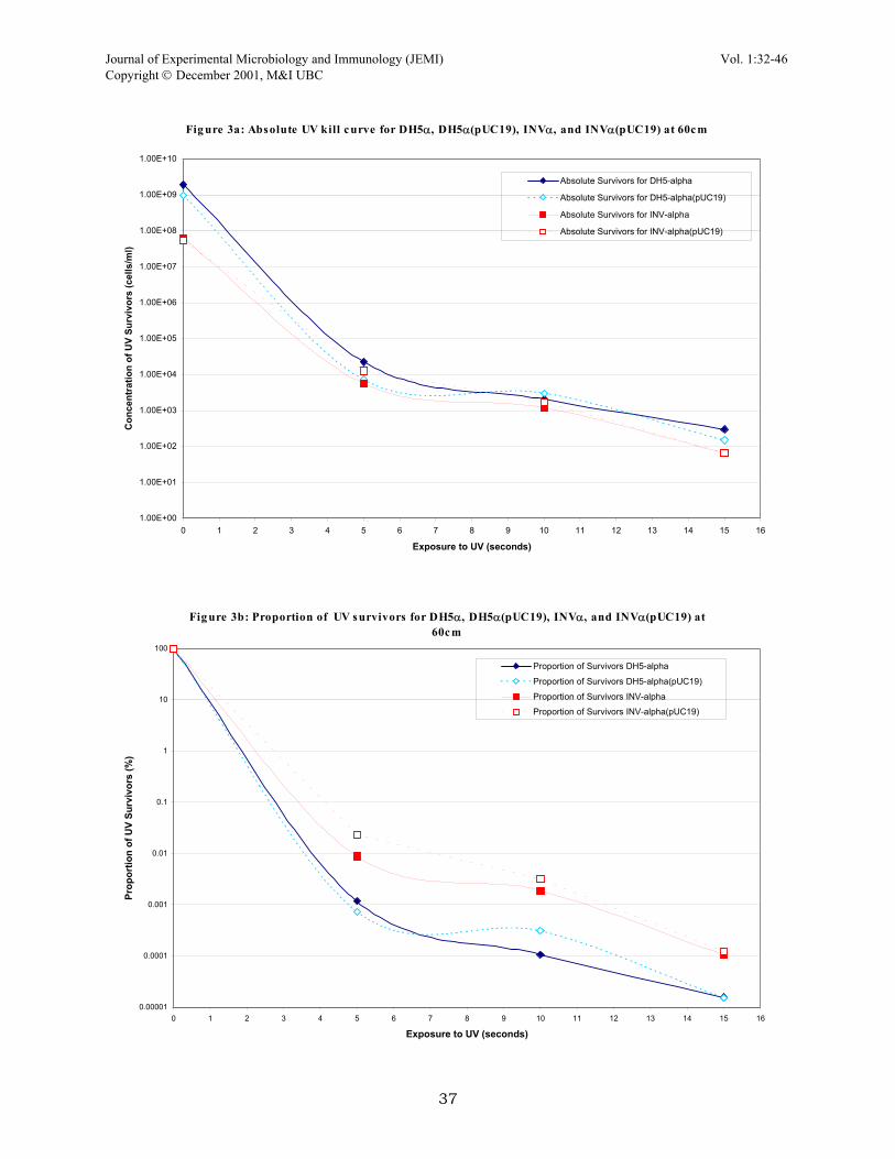
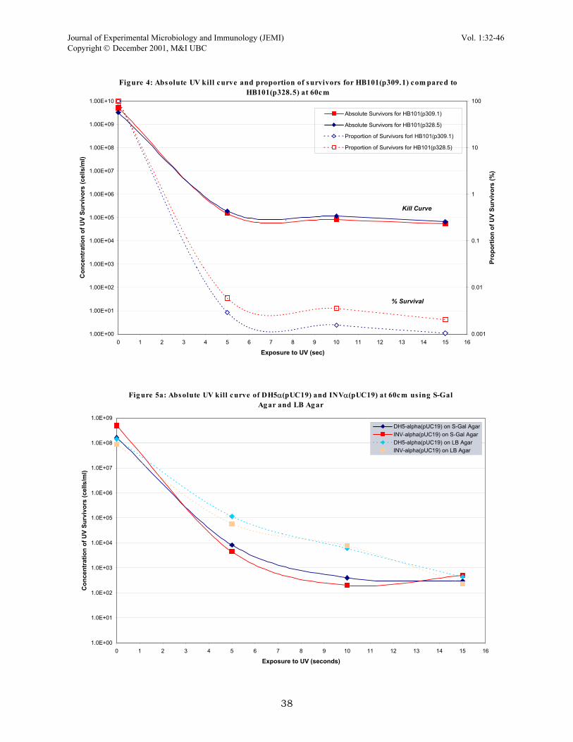
RESULTS Kill curve variables. Since a UV monitor or lux monitor was not available, the actual amount of radiation exposed to the cells in the direct plate irradiation was unknown. Therefore various variables involving time of exposure to UV and distance between the UV lamp and plates had to be worked out to determine a useful kill curve (Fig. 1). Two main schemes were tested for DPI, the first set included irradiation times of 0, 10, 30 and 60 seconds at 30cm from the plate. This kill curve actually appears to be an ideal exponential decay curve except that we were not satisfied with one main area. The curve only contained 4 points and the actual shape of the curve was determined in large by the time point at 10 seconds of exposure. Looking back at this set, it may have prove beneficial to repeat this set of conditions with several more time points. The second set of conditions involved exposure times of 0, 5, 10, and 15 seconds at 60cm away from the plate. This provided a better curve because it was more reliable due to several points falling closely together, giving more confidence to the curve. Furthermore, this set gave a 105-fold killing compared to 104-fold killing in the first set which is much closer to the ideal kill of 106 (Dr. Ramey, personal communication). In the end, the second set was chosen as the kill parameters for a majority of the project. Photoreactivation after UV irradiation. Photoreactivation was briefly looked at in this experiment by exposing UV irradiated plates to natural sunlight. The strains DH5α(pUC19) and INVα(pUC19) were usually irradiated for 10 seconds and then placed at the windowsill for an hour to two hours. The results obtained from these quick tests were varied, during some runs there was about 100-fold reactivation by sunlight while on other runs, the photoreactivation was only 5-fold. But in general, all plates that were left in natural light showed greater viability that those plates which were never exposed to any natural light. Response of strains and plasmids to UV irradiation. This battery of tests was linked to finding the best UV mutagenesis setup. Several strains were irradiated to see how they respond to the effects of UV; different combinations of plasmids and host cells were also examined. Of all the strains tested, the wild types SF8 and B23 displayed the most tolerance of UV light; plates with SF8 cells continuously grew as confluent lawns even when irradiated for lengthy periods of time. The α-complemented strains (DH5α and INVα) and HB101 were more sensitive to the effects of UV light. The two α-complemented strains behaved very similar in response to UV irradiation (Fig. 2). The presence of pUC19 plasmid in both α-complemented strains was tested (Fig. 3a, 3b), as were p309.1 and p328.5 in HB101 host cells (Fig. 4). The plasmid pUC19 did not confer any changes in cell response to UV light as demonstrated by similar survival percentages. The other two plasmids were tested as well because of conflicting data acquired during the earlier parts of this project; SF8 responded different to UV light depending on whether or not it carried p309.1 or p328.5. Therefore, these two were tested in HB101 and again in host SF8 (data not shown) and the results suggest that same host cells with either plasmid will behave similarly Negative screening of lac and Amp mutants. Much of the project was centred on obtaining a rough estimate of mutational frequency in genes. But this part of the project proved to be the least successful, at least for gene mutational frequency on the pUC19 plasmid. Many UV irradiated runs were completed and only one set returned mutants, white forming colonies on MacConkey plates (indicating a lac− mutation, loss of lac function). The ratio of white mutants to normal, deep violet forming colonies was 1:10, since the total number of colonies on the replicate plate was 100, this implies a 10% mutation frequency of the lac gene in UV survivors. This number is suspiciously high (we were expecting a frequency of about 0.1%) and since this run was only produced once while many other runs failed to turn up mutants, this set can be disregarded as an anomaly until further tests show otherwise. Negative screening with S-Gal differential media. The DPI protocol was applied with a new type of differential media called S-Gal (which screens for lac expression). Direct-plate irradiation of DH5α(pUC19) and INVα(pUC19) were performed on this type of media and compared to regular LB agar (Fig. 5a, 5b). From looking at this, not much difference in the percentage of survivors except that the killing appears to be steeper on the S-Gal media was observed. However, a few sets done with DPI and S-Gal agar provided mutants (white colonies vs. normal colonies which are black) at a frequency of about 0.1%; approximately for every 1000 survivor, black-forming colony, there was a white colony or a sectored colony. Even though the observed frequency is similar o the one expected, the confidence in this observed frequency is debatable because the results were not consistently reproducible. Thus, there’s reason to believe that these white colonies were cells that originally lacked the pUC19 plasmid or lost it during the course of the procedure. In short, more tests needed to be performed to determine whether or not this frequency is real.
35

Journal of Experimental Microbiology and Immunology (JEMI) Vol. 1:32-46 Copyright December 2001, M&I UBC
36
Fig ure 1: Absolute UV kill curve and proportion of s urvivors of DH5α(pUC19) at various he ig hts and duration of UV e xpos ure s
1.00E+00
1.00E+01
1.00E+02
1.00E+03
1.00E+04
1.00E+05
1.00E+06
1.00E+07
1.00E+08
1.00E+09
0 5 10 15 20 25 30 35 40 45 50 55 60 65
Exposure to UV (seconds)
Con
cent
ratio
n of
UV
Surv
ivor
s (c
ells
/ml)
0.0001
0.001
0.01
0.1
1
10
100
Prop
ortio
n of
UV
Surv
ivor
s (%
)
Absolute Survivors - DH5-alpha @ 60cmAbsolute Survivors - DH5-alpha @ 30cm 02.21.01Proportion of Survivors DH5-alpha @ 60cmProportion of Survivors DH5-alpha @ 30 cm 02.21.01
Fi g u re 2 : Ab s o l u te UV k il l c u rv e a n d p ro p o rtio n o f s u rv iv o rs fo r D H5α (p UC 1 9 ) a n d IN Vα (p UC 1 9 ) a t 6 0c m
1.00 E+00
1.00 E+01
1.00 E+02
1.00 E+03
1.00 E+04
1.00 E+05
1.00 E+06
1.00 E+07
1.00 E+08
1.00 E+09
0 1 2 3 4 5 6 7 8 9 10 11 1 2 13 14 15 16
Ex posu re to U V (sec onds)
Con
cent
rati
on o
f UV
Sur
vivo
rs (c
ells
/ml)
0 .000 1
0 .001
0 .01
0 .1
1
1 0
1 00
Pro
por
tion
of U
V S
urvi
vors
(%)
Absolute Survivors - DH5-alphaAbsolute Survivors - INV-alphaProportion of Survivors - DH5-alpha
Proportion of Survivors - INV-alpha
Kill Cu rve
% Surviva l
s
Journal of Experimental Microbiology and Immunology (JEMI) Vol. 1:32-46 Copyright December 2001, M&I UBC
37
Fig ure 3a: Absolute UV kill curve for DH5α, DH5α(pUC19), INVα, and INVα(pUC19) at 60cm
1.00E+00
1.00E+01
1.00E+02
1.00E+03
1.00E+04
1.00E+05
1.00E+06
1.00E+07
1.00E+08
1.00E+09
1.00E+10
0 1 2 3 4 5 6 7 8 9 10 11 12 13 14 15 16
Exposure to UV (seconds)
Con
cent
ratio
n of
UV
Surv
ivor
s (c
ells
/ml)
Absolute Survivors for DH5-alpha
Absolute Survivors for DH5-alpha(pUC19)
Absolute Survivors for INV-alpha
Absolute Survivors for INV-alpha(pUC19)
Fig ure 3b: Proportion of UV s urvivors for DH5α, DH5α(pUC19), INVα, and INVα(pUC19) at 60cm
0.00001
0.0001
0.001
0.01
0.1
1
10
100
0 1 2 3 4 5 6 7 8 9 10 11 12 13 14 15 16
Exposure to UV (seconds)
Prop
ortio
n of
UV
Surv
ivor
s (%
)
Proportion of Survivors DH5-alpha
Proportion of Survivors DH5-alpha(pUC19)
Proportion of Survivors INV-alpha
Proportion of Survivors INV-alpha(pUC19)
Journal of Experimental Microbiology and Immunology (JEMI) Vol. 1:32-46 Copyright December 2001, M&I UBC
38
Figure 4: Absolute UV kill curve and proportion of s urvivors for HB101(p309.1) com pared to HB101(p328.5) at 60cm
1.00E+00
1.00E+01
1.00E+02
1.00E+03
1.00E+04
1.00E+05
1.00E+06
1.00E+07
1.00E+08
1.00E+09
1.00E+10
0 1 2 3 4 5 6 7 8 9 10 11 12 13 14 15 16
Exposure to UV (sec)
Con
cent
ratio
n of
UV
Surv
ivor
s (c
ells
/ml)
0.001
0.01
0.1
1
10
100
Prop
ortio
n of
UV
Surv
ivor
s (%
)
Absolute Survivors for HB101(p309.1)
Absolute Survivors for HB101(p328.5)
Proportion of Survivors forl HB101(p309.1)
Proportion of Survivors for HB101(p328.5)
.Kill Curve
% Survival
Fig ure 5a: Absolute UV kill curve of DH5α(pUC19) and INVα(pUC19) at 60cm us ing S-Gal Ag ar and LB Ag ar
1.0E+00
1.0E+01
1.0E+02
1.0E+03
1.0E+04
1.0E+05
1.0E+06
1.0E+07
1.0E+08
1.0E+09
0 1 2 3 4 5 6 7 8 9 10 11 12 13 14 15 16
Exposure to UV (seconds)
Con
cent
ratio
n of
UV
Surv
ivor
s (c
ells
/ml)
DH5-alpha(pUC19) on S-Gal AgarINV-alpha(pUC19) on S-Gal AgarDH5-alpha(pUC19) on LB AgarINV-alpha(pUC19) on LB Agar

Journal of Experimental Microbiology and Immunology (JEMI) Vol. 1:32-46 Copyright December 2001, M&I UBC
Fi g u re 5 b : P ro p o rti o n o f UV s u rv i v o rs fo r D H5α (p UC 1 9) a n d IN Vα (p UC 1 9) a t 6 0 c m o n S-G a l Ag a r a n d LB A g a r
0.000 01
0.00 01
0.0 01
0.01
0 .1
1
10
1 00
0 1 2 3 4 5 6 7 8 9 10 11 12 13 14 15 16
Exp osure to UV (s econ ds)
Pro
port
ion
of U
V S
urvi
vors
(%)
DH5-alpha(pUC19) on S-Gal AgarINV-alpha(pUC19) on S-Gal AgarDH5-alpha(pUC19) on LB AgarINV-alpha(pUC19) on LB Agar
Standard kill curve using LIQUID-PLATE IRRADIATION (LPI)DH5α(host), INVα(host) and B23. The standard conditions for the LPI protocol consisted of irradiating three strains of cells: DH5a, INVα, and B23. The most useful exposure times were 0, 10, 20, 30, and 40 seconds for the α-complemented strains and 0, 10, 20, 30, 40 and 60 seconds for B23. All of these runs were carried out with the UV lamp 60cm away from the plates. Several runs were completed for all three strains; therefore the data from all the sets are complied and averaged as a percent survival plot (Fig. 6a, 6b, and 6c). The survival percentages of all three strains were also compared to each other and to a DPI run on LB agar (Fig. 7). From this figure, we can see several ideas that were suggested earlier in the DPI runs. Firstly, of all three strains, B23 shows the most resistance to UV effects as demonstrated by its requirement for longer exposure times to produce substantial killing (106). Whereas with the α-complemented strains, it took about 25% less exposure time to produce the same amount of killing. Secondly, the LPI protocol agrees with data from DPI protocol regarding to the similarity in response to UV light in the α-complemented strands. The best-fit lines for percent-survival of DH5α and INVα are nearly identical. This is similar to the set of data acquired in DPI regarding strain differences (Fig. 2). Lastly, this plot reveals a substantial difference in cell response (percentage survival) of DH5α irradiated by the DPI protocol compared to DH5α irradiated with the LPI protocol. The kill curve is much sharper for DH5α irradiated by the DPI protocol and it requires about one-third the time (approximately 15 seconds) for substantial killing compared to 45 seconds with DH5α irradiated with the LPI protocol. Negative screening of lac with X-Gal differential media. Viable plant counts of irradiated cells were plated immediately after irradiation to screen for possible lac mutation on the B23 chromosome and to serve as an indicator of the number of survivors. Of the four runs completed with LPI protocol involving B23, the screening process failed to pick up any white-forming colonies. This agrees with earlier DPI runs with MacConkey and S-Gal LB agar differential media. Positive screening for rifampicin resistance, chloramphenicol resistance and ampicillin resistance. Irradiated cells by LPI protocol were out-grown overnight and plated to various positive screening media. Of all three strains, only B23 showed a change in mutation frequency at the rpoB, and ompF loci (Fig. 8). The two α-complemented strains only displayed background levels of mutations at these sites; therefore, no observable mutation frequency was present. In the B23 strain, these mutation frequencies increased substantially during the periods of early UV exposures and reached a maximum at about 20 seconds or 30 seconds of irradiation. There was variation between
39
Journal of Experimental Microbiology and Immunology (JEMI) Vol. 1:32-46 Copyright December 2001, M&I UBC
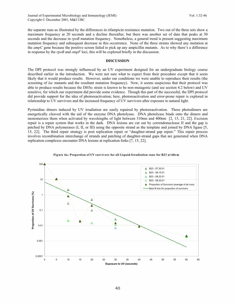
the separate runs as illustrated by the differences in rifampicin resistance mutation. Two out of the three sets show a maximum frequency at 20 seconds and a decline thereafter, but there was another set of data that peaks at 30 seconds and the decrease in rpoB mutation frequency. Nonetheless, a general trend is present suggesting maximum mutation frequency and subsequent decrease in this occurrence. None of the three strains showed any mutation at the ampC gene because the positive screen failed to pick up any ampicillin mutants. As to why there’s a difference in response by the rpoB and ompF loci, this will be explored briefly in the discussion.
DISCUSSION The DPI protocol was strongly influenced by an UV experiment designed for an undergraduate biology course described earlier in the introduction. We were not sure what to expect from their procedure except that it seem likely that it would produce results. However, under our conditions we were unable to reproduce their results (the screening of lac mutants and the resultant mutation frequency). Now, it seems suspicious that their protocol was able to produce results because the DH5α strain is known to be non-mutagenic (and see section 4.2 below) and UV sensitive, for which our experiment did provide some evidence. Though this part of the successful, the DPI protocol did provide support for the idea of photoreactivation; here, photoreactivation and error-prone repair is explored in relationship to UV survivors and the increased frequency of UV survivors after exposure to natural light. Pyrimidine dimers induced by UV irradiation are easily repaired by photoreactivation. These photodimers are energetically cleaved with the aid of the enzyme DNA photolyase. DNA photolyase binds onto the dimers and monomerizes them when activated by wavelengths of light between 310nm and 400nm [2, 15, 21, 22]. Excision repair is a repair system that works in the dark. DNA lesions are cut out by correndonuclease II and the gap is patched by DNA polymerases (I, II, or III) using the opposite strand as the template and joined by DNA ligase [5, 15, 22]. The third repair strategy is post replication repair or “daughter-strand gap repair.” This repair process involves recombination interchange of strands and patching of daughter-strand gaps that are generated when DNA replication complexes encounter DNA lesions at replication forks [7, 15, 22].
Fig ure 6a: Proportion of UV survivors for all Liquid-Irradiation runs for B23 at 60cm
0.0001
0.001
0.01
0.1
1
10
100
0 5 10 15 20 25 30 35 40 45 50 55 60 65
Exposure to UV (seconds)
Prop
ortio
n of
UV
Surv
ivor
s (%
)
B23 - 07.30.01
B23 - 08.15.01
B23 - 08.22.01
B23 - 08.30.01
Proportion of Survivors (average of all runs)
Best fit line for proportion of survivors
40

Journal of Experimental Microbiology and Immunology (JEMI) Vol. 1:32-46 Copyright December 2001, M&I UBC
41
Fig ure 6c : Proportion of UV s urvivors for all Liquid-Irradiation runs for INVα at 60cm
0.00001
0.0001
0.001
0.01
0.1
1
10
100
0 5 10 15 20 25 30 35 40 45 50
Exposure to UV (seconds)
Prop
ortio
n of
UV
Surv
ivor
s (%
)
INV-alpha -07.30.01INV-alpha -08.15.01INV-alpha - 08.22.01INV-alpha - 08.30.01Proportion of Survivors (average of all runs)Best fit line for proportion of survivors
Figure 6b: Proportion of UV survivors for all Liquid-Irradiation runs forDH5
α at60
0.0001
0.001
0.01
0.1
1
10
100
0 5 10 15 20 25 30 35 40 45 50
Exposure to UV (seconds)
ProportionofUVSurvivors(%)
DH5-alpha - 07.30.01
DH5-alpha - 08.15.01
DH5-alpha - 08.22.01
DH5-alpha - 08.30.01
Proportion of Survivors (average of all runs)
Best fit line for proportion of survivors

Journal of Experimental Microbiology and Immunology (JEMI) Vol. 1:32-46 Copyright December 2001, M&I UBC
42
Fig ure 7: Com parison of diffe rent proportions of UV survivors of E. coli afte r Liquid Irradiation at 60cm
0.00001
0.0001
0.001
0.01
0.1
1
10
100
0 5 10 15 20 25 30 35 40 45 50 55 60 65
Exposure to UV (seconds)
Perc
ent S
urvi
val
B23DH5-alphaINV-alphaDH5-alpha (Agar Plate)Fitted line for B23Fitted line for DH5-alphaFitted line for INV-alphaFitted line for DH5-alpha (Irradiated using Direct-Plate method)
Fig ure 8: Fre que ncy of E. coli m utants re s is tant to rifam pic in and chloram phe nic ol afte r UV irradiation (LPI) at 60cm
-1.0E-05
-5.0E-06
0.0E+00
5.0E-06
1.0E-05
1.5E-05
2.0E-05
0 5 10 15 20 25 30 35 40 45 50 55 60 65
Exposure to UV (seconds)
Freq
uenc
y of
Mut
ants
B23 - Rifampcin Mutants 08.30.01B23 - Rifampcin Mutants 08.22.01B23 - Rifampcin Mutants 07.30.01HB101 - Rifampcin Mutants 08.22.01DH5-alpha - Rifampcin Mutants 08.22.01INV-alpha - Rifampcin Mutants 08.22.01B23 - Chloramphenicol Mutants 07.30.01B23 - Chloramphenicol Mutants 08.30.01
Journal of Experimental Microbiology and Immunology (JEMI) Vol. 1:32-46 Copyright December 2001, M&I UBC
According to the these repair strategies, this is probably how these UV survivors were able to endure the stress caused by genetic damage as a result of UV light. Since the DPI protocol strictly required steps which limited any exposure of light to the freshly irradiated cells, it seems highly plausible that the survivors that arise on the LB plates or MacConkey plates are cells that have used excision repair, and postreplication repair mechanisms to remove these dimers. The ones that were not able to survive may be the ones that have suffered considerate damage such as multiple strand brakes, numerous DNA cross-linking, and cyclobutane pyrimidine dimers. The role of cyclobutane pyrimidine dimers in UV survival is demonstrated by the increase in number of UV survivors when exposed to light (photoreactivation). Cells that have undergone the DPI protocol of irradiation and a period of exposure to natural light shows that the number of surviving cells increase on a order of several or countless magnitudes. Due to the reason that the amount of light available was not controlled because of weather conditions and so forth, the amount of photoreactivation for each run would vary considerably depending on the amount of light. Thus, we see this wide range of UV survivors depending on the photoreactive ability in Escherichia coli. After looking into the SOS system, it seems strange why would a UV mutagenic system involving a recA− strain (i.e. DH5α and INVα) appear feasible because it was shown that mutagenesis is linked to the function of this gene. The SOS system is described briefly below and linked to idea and to some of the observations in this project. Basically, non-repaired or improperly repaired DNA lesions will cause the replicative assembly (DNA polymerases) to momentarily halt, leaving the strand being replicated single-stranded. This acts as a signal for the induction of the SOS response; single strand binding proteins and the RecA protein (coded for by the recA gene) will bind the single-stranded DNA in the presence of nucleoside triphosphates to form a nucleoprotein filament, becoming RecA* (activated form of RecA). This complex will then in turn aid in the mediate the cleavage of the LexA protein, a regulatory molecule that represses the expression of SOS genes (e.g., recA, umuD, umuC, polB, recN, sulA, uvrA, uvrB, and uvrD) by binding their operators and preventing transcription. Aside from activating LexA protein, the RecA protein also actives UmuD protein by mediating its cleavage to yield the active form of UmuD’. UmuD’ and UmuC proteins are important to SOS response because they interact with the replicative assembly along with RecA to allow for translesion synthesis. Translesion synthesis is a term used to describe the cell’s ability to replicate DNA pass miscoding and noncoding lesions arising from DNA damage. The exact mechanism of translesion synthesis is still unclear but it is now believed that the UmuC family of DNA polymerases (polymerase IV, V in E. coli) is involved in translesion synthesis. It is believed that UmuC, UmuD’ and RecA helps these highly tolerant polymerases to replicate pass the lesions by relaxing proofreading ability of these polymerases, increasing the polymerases’ processivity. Furthermore, these polymerases have loose-and-flexible active sites which facilitate the passage of DNA lesions more readily [see reviews 1, 4, 7, 8, 16, 17, 18, for more detailed information about SOS response and DNA repair]. It is this action by these highly tolerant DNA polymerases that incorrect nucleotides are inserted in the replicated strand, opposite the template strand with the lesions. As a result, mutations are introduced into subsequent generations of DNA. Thus, mutagenesis arises through the error-prone replication through lesion sites by polymerases mediated by the SOS system. Without the SOS system, mutagenesis would not be possible. The SOS response model can account for quite a few observations within this project. Strain B23 can be referred to as a benchmark for all the other strains since it is one that closely relates to the wild type strain. Since B23 is wild type it can be assured that it contains regular levels of SOS regulating molecules such as LexA and RecA when not induced, and the appropriate levels of RecA*, UmuC and UmuD when induced. The strain SF8 is highly resistant to UV mutagenesis indicating that its error-free pathway is more adapted to deal with UV damage than B23. Also, the SOS system in SF8 may somehow be repressed so that mutagenesis does not occur in SF8 as observed. Therefore, there are several gene targets in the SOS regulon of SF8 that could be damaged. For example, the genes lexA maybe constitutively expressed at high levels so the SOS genes are always repressed, resulting in the absence of error-prone repair. Or proteins such as the UmuC and UmuD may be dysfunctional, also preventing the error-prone machinery to introduce mutations during translesion synthesis. Shutting down the SOS response is not enough to confer UV resistance to SF8 since they show a very high rate of survival. So SF8 may have a highly tolerant polymerase that does not need activation like the UmuC family of polymerases. Or, the DNA of SF8 is naturally more resistant to UV damage because of special DNA folding rearrangement and DNA packaging proteins that may shield the chromosome from damage. The α-complemented strains (DH5α and INVα) on the other hand are UV-sensitive. The link that connects the SOS system and these cells is the lack or engineered damage to the recA gene. Thus, these strains of bacteria are deficient in the RecA protein, the key regulatory and active protein part of the SOS system. Experiments in the past (see above mentioned reviews) have described recA− strains as being non-UV mutable, UV sensitive and deficient in some repair pathways. This can be reasoned by understanding how RecA protein is required for the down-regulation of the LexA protein. If a cell is unable to produce RecA proteins, this cell would never be able to induce
Journal of Experimental Microbiology and Immunology (JEMI) Vol. 1:32-46 Copyright December 2001, M&I UBC
it’s SOS system and as result, has a higher chance of cell death when DNA polymerases fall off or lock up at UV-induced lesions. This accounts for how and why recA− strains are UV sensitive, in conjunction with the lost of some other DNA pathways due to the lost of recA. This brings up the second characteristic of being recA−. Strains deficient in the RecA protein are non-mutable because the system which introduces the mutations, SOS, is permanently turned off because DNA polymerases needs UmuC, UmuD and RecA interactions in order for it to be lenient and tolerant of UV lesions. UV non-mutability was observed in this project. The α-complemented strains and HB101 are all recA− and these three strains displayed zero mutation frequency at the sites selected as markers. Just as predicted by and described in the literature. The lack of DH5α(pUC19) and INVα(pUC19) lac mutants with the DPI protocol can also be attributed to the lost of recA function because translesion synthesis was not able to introduce errors at either lac site on the chromosome or plasmid. Beside from the SOS regulatory system, there are other variables that can influence mutation frequencies as witnessed in the differences between rifampicin resistance when compared to chloramphenicol resistance in B23 UV irradiated mutants. For example, the amount of UV-induced lesions a strand of DNA receives is dependent on its packaging and accessibility by UV. If a certain segment is packed deep within the genome than the chances of it developing UV lesions is certainly less due to DNA packaging proteins such as histones and the shielding effect. Furthermore, DNA supercoiling and DNA scaffolding also provides a means to keep DNA away from the damaging sources of UV. Therefore, mutations are dependent on the amount of lesions formed from UV damage. This could account for the differences in mutational frequency observed in ompF (chloramphenicol resistance) and rpoB (rifampicin resistance). Out of the 2 genes, ompF was mutated the most frequently, and according to these lines of thought it can be inferred that ompF is less packaged and more exposed to the UV light. Another possible variable that may determine the mutation frequency is the length of the gene in question. Longer genes are more susceptible to mutation because their genetic code is longer and thus more inclined to UV lesions. But this may not necessarily be the case all the time. Larger genes tend to code for bigger and complex proteins. These complex proteins and their structure are highly specific so that a mutation may lead to a devastating lost of function depending on where the mutation is located and the number of mutations imparted by UV mutagenesis. The rpoB gene is about 1342 base pairs long while the ompF gene is about 362 base pairs long [6]. In this case, the smaller gene is the one with the higher mutational frequency. Explained earlier, this can be due to the fact that functionality of a smaller protein is not as dependent on its structure as a more complex protein. Or another possible explanation is that the DNA sequence of the ompF gene contains more pyrimidines and thus, more susceptible to cylcobutane pyrimidine dimers. A larger pool of thymine dimers mean that a larger number of dimers will escape repair or be repaired poorly leading to mutations by the SOS system. In Fig. 8 there’s a trend showing mutational frequency of the ompF and rpoB loci as a function of time. The mutational frequency increases steady up until the 20 or 30 seconds of UV exposure mark and then it starts to decrease. It seems reasonable that the frequency of mutation increases for a period of time because the number of lesions increases as a function of irradiation. Therefore, more and more of the population are developing mutations at a certain site because of the increased number of lesions that are escaping repair accumulates; and accumulation leads to a better chance of getting a mutation at that certain locus (ie. ompF). After a certain point, the frequency begins to drop because the accumulation of lesions becomes too deadly and cells with mutations at these sites begin to die. The lack of observable mutants with DH5α(pUC19) and INVα(pUC19) can also be explained from a statistical point of view. The plasmid pUC19 is a high-copy plasmid, meaning it exists as many copies within the cell. Let’s assume that the chance of inducing a mutation at the lac gene in this plasmid is 1 in a 1000. And in order for the cell to become a lac mutant, each plasmid must be hit in the lac gene. Since each and every hit is independent, the chance of creating a lac mutant by mutating the plasmids is: (1/1000)p = chance of getting a lac mutant due to plasmid mutation (1) Where p is the copy number for a particular plasmid. Thus, if there were three pUC19 plasmids inside a cell, the probability that all three plasmids will be hit in the same locus is 1/109. Thus, someone looking through 109 UV survivors may only find one mutant in 109 cells. With α-complementation, the functional lacZ product, β-galactosidase, is a dimer. One subunit is coded on the plasmid while the other is coded on the chromosome. So theoretically, it is possible to cause a lac− mutant by mutating the gene on the chromosome. But this is not as simple as it seems because the chromosome is much larger and is subject to higher levels of organization which may provide protection from UV (see above). Thus, the chance of inducing a mutation at either gene loci seems like a daunting task.
44

Journal of Experimental Microbiology and Immunology (JEMI) Vol. 1:32-46 Copyright December 2001, M&I UBC
One of the main goals behind this project was to perfect an assay system for mutation frequency in genes located on both plasmids and the chromosome. If this proved successful, we hoped to compare both in vivo and in vitro effects of UV radiation on gene mutation frequency and to genetically analyse hotspots. However, too much confidence was placed in another design and not enough scrutiny was applied in questioning that protocol when this project was designed. As a result, several problems had to be overcome before this project provided any meaningful insights. Our finding suggests that the α-complemented strains DH5α and INVα are poor biological systems for testing UV mutagenesis because of its recA− phenotype. In accordance with current knowledge in UV mutagenesis and SOS response, the RecA protein is essential to the process of mutation. On the other hand, the B23 strain can be a satisfactory system for analysing certain chromosomal gene mutation frequencies such as rifampicin and chloramphenicol resistance. Several strain sensitivities to UV light were also investigated and the resulting data fits the present stream of thought. The plasmid looked at in this project, pUC19, p309.1 and p328.5 did not confer any special responses to a range of hosts such as DH5α, INVα, HB101, and SF8. One key area we would have liked to investigate was the application of our LPI protocol with a strain of E. coli that was α-complemented but not of the recA− phenotype. This strain is U163 (thi−, lac−, α-complementation) and provides several advantages over the α-complemented strains in this project and the B23 strain. Basically, the U163 combines the UV mutagenic properties of B23 along with the α-complement properties of the DH5α and INVα strains. U163 could potentially provide insight into both chromosomal and plasmid gene mutation frequency. In the early 1960’s, Witkin and others noted that post-treatment of irradiated cells with caffeine and other repair inhibitors such as acriflavine (or other basic dyes) helped to increase the yield of UV-induced mutations dramatically [21]. It is shown that caffeine and these inhibitors were able to increase the number of unexcised pyrimidine dimers from 2 to 20, potentially increase the probability of mutation in any gene ten times. Therefore, this addition may be a useful alternative to skew the ratios of mutants so the screening process becomes easier. Another method of increasing a cell’s susceptibility to mutagenesis involves a plasmid called pKM101 (derived from R46, related to R205, TP110 and ColI) [15]. pKM101 when introduced to E. coli exhibits an increased probability of developing base-substitutions and frameshift mutations in response to a wide range of DNA damaging agents while only increasing the spontaneous mutation frequency slightly [15]. An alternative method for studying mutation frequency involves a heat-sensitive plasmid called pMut which contains the gene mutD. This plasmid when inserted into a host increases the mutation frequency by 20- to 4000- fold. The advantage of this system is that when a desired mutation is found, the “mutagenic” plasmid can be removed by growing it at high temperatures [14]. This system allows for mutation frequency analysis in both directions if applied to the proper system.
REFERENCES 1. Bradley, T.S., and G.C. Walker. 1998. Mutagenesis and more: umuDC and the Escherichia coli SOS response. Genetics. 148:1599-1610. 2. Brash, D.E., W.A. Franklin, G.B. Sancar, A. Sanear, and W.A. Haseltine. 1985. Escherichia coli DNA photolyase reverse cyclobutane
pyrimidine dimers but not pyrimidine-pyrimidone (6-4) photoproducts. Journal of Biological Chemistry. 260(21):11438-11441. 3. Chandrasekhar, D., and B.V. Houten. 2000. In vivo formation and repair of cyclobutane pyrimidine dimers and 6-4 photoproducts measured
at the gene and nucleotide level in Escherichia coli. Mutation Research. 450:19-40. 4. Friedberg, E.C., G.C. Walker, and W. Siede. 1995. DNA repair and mutagenesis. American Society for Microbiology Press. Washingtong
DC. 5. Hanawalt, P.C., P.K. Cooper, A.K. Ganesan, and C.A. Smith. 1979. DNA repair in bacteria and mammalian cells. Annual Review of
Biochemistry. 48:783-836. 6. Karp, P.D., M. Riley, M. Saier, I.T. Paulsen, S.M. Parley, and A. Pellegrini-Toole. 2000. The EcoCyc and MetaCyc databases. Nucleic
Acid Research. 28(1):56-59 7. Kuzminov, A. 1999. Recombinational repair of DNA damage in Escherichia coli and Bacteriophage λ. Microbiology and Molecular
Biology Reviews. 63(4):751-813. 8. Livneh, Z., O. Cohen-Fix, R. Skaliter, and T. Elizur. 1993. Replication of damaged DNA and the molecular mechanism of ultraviolet light
mutagenesis. Critical Reviews in Biochemistry and Molecular Biology. 28(6):465-513. 9. Miller, J.H. 1972. Experiment 32: Isolation of valine-resistant and antibiotic-resistant mutants. Pages 221-229 in J.H. Miller, Experiments in
Molecular Genetics. Cold Spring Harbour Laboratory Press, New York.
45

Journal of Experimental Microbiology and Immunology (JEMI) Vol. 1:32-46 Copyright December 2001, M&I UBC
46
10. Miller, J.H. 1985. Mutagenic specificity of ultraviolet light. Journal of Molecular Biology. 182:45-68. 11. Miller, J.H. 1992. Experiment 15: Mutagenesis with UV. Pages 150-156 in J.H. Miller, A Short Course in Bacterial Genetics. Cold Spring
Harbour Laboratory Press, New York. 12. Müller-Hill, B. 1996. The lac operon. Walter de Gruyter & Co., Berlin, Germany. 13. Schaaper, R.M., R.L. Dunn, and B.W. Glickman. 1987. Mechanisms of ultraviolet-induced mutation: mutational spectra in Escherichia coli
lacI gene for wild-type excision-repair-deficient strain. Journal of Molecular Biology. 195:187-202. 14. Selifonova, O., F. Valle, and V. Schellenberger. 2001. Rapid evolution of novel traits in microorganisms. Applied and environmental
Microbiology. 67(8):3645-3649. 15. Walter, G.C. 1984. Mutagenesis and inducible responses to deoxyribonucleic acid damage in Escherichia coli. Microbiological Reviews.
48(1):60-93. 16. Walker, G.C. 1995. SOS-regulated proteins in translesion DNA synthesis and mutagenesis. Trends in Biochemical Sciences. 20(10):416-
420. 17. Wang, Z. 2001. Translesion synthesis by the UmuC family of DNA polymerases. Mutation Research. 486:59-70. 18. Wehner, J., and G. Horneck. 1995. Effects of vacuum UV and UVC radiation on dry Escherichia coli plasmid pUC19: I. Inactivation of
lacZ− mutation induction and strand breaks. Journal of Photochemistry and Photobiology. 28:77-85. 19. Wehner, J., and G. Horneck. 1995. Effects of vacuum UV and UVC radiation on dry Escherichia coli plasmid pUC19: II. Mutational
specificity at the lacZ gene. Journal of Photochemistry and Photobiology. 30:171-177. 20. Witkin, E.M. 1967. Mutation-proof and mutation-prone modes of survival in derivatives of Esherichia coli B differing in sensitivity to
ultraviolet light. Brookhaven Symp. Biol. 20:17-55. 21. Witkin, E.M. 1969. Ultraviolet-induced mutation and DNA repair. Annual Review of Genetics. 23:487-514. 22. Witkin, E.M. 1976. Ultraviolet mutagenesis and enducible DNA repair in Esherichia coli. Bateriological Reviews. 40(4):869-907.