uva-dare (digital academic repository) infectious diseases ... · pinpoints par-1 as an important...
TRANSCRIPT
UvA-DARE is a service provided by the library of the University of Amsterdam (http://dare.uva.nl)
UvA-DARE (Digital Academic Repository)
Infectious diseases and fibrotic disorders: Potential novel targets
Duitman, J.
Link to publication
Citation for published version (APA):Duitman, J. (2014). Infectious diseases and fibrotic disorders: Potential novel targets.
General rightsIt is not permitted to download or to forward/distribute the text or part of it without the consent of the author(s) and/or copyright holder(s),other than for strictly personal, individual use, unless the work is under an open content license (like Creative Commons).
Disclaimer/Complaints regulationsIf you believe that digital publication of certain material infringes any of your rights or (privacy) interests, please let the Library know, statingyour reasons. In case of a legitimate complaint, the Library will make the material inaccessible and/or remove it from the website. Please Askthe Library: https://uba.uva.nl/en/contact, or a letter to: Library of the University of Amsterdam, Secretariat, Singel 425, 1012 WP Amsterdam,The Netherlands. You will be contacted as soon as possible.
Download date: 28 May 2020
10Protease activated receptor-1 (PAR-1) deficiency
ameliorates skin fibrosis in a mouse model of bleomycin-induced scleroderma.
JanWillem Duitman1, Roberta R. Ruela-de-Sousa1, Kun Shi1, Onno J. de Boer2, Keren S. Borensztajn3, Sandrine Florquin2, Maikel P. Peppelenbosch4, C. Arnold Spek1.
1Center for Experimental and Molecular Medicine (CEMM), 2Department of Pathology; Academic Medical Center, University of Amsterdam, Amsterdam, The Netherlands.
3Unité INSERM 700, Physiopathologie et Epidémiologie de l’Insuffisance Respiratoire, Faculté de Médecine Xavier Bichat, Paris, France, 4Department of Gastroenterology
and Hepatology, Erasmus Medical Center, Rotterdam, The Netherlands.
Submitted
PS JanWillem Duitman_2014-01v2.indd 141 28-03-14 10:37

142
CHAPTER 10
ABSTRACTObjective. Accumulating evidence shows that Protease Activated Receptor-1 (PAR-1) plays
an important role in the development of fibrosis, including systemic sclerosis (SSc) induced
lung fibrosis. However, whether PAR-1 also plays a role in the development of scleroderma
induced by SSc remains elusive. The aim of this study was to determine the role of PAR-1 in
the development of skin fibrosis.
Methods. In order to explore possible mechanisms in which PAR-1 could play a role, hu-
man dermal fibroblasts and keratinocytes were stimulated with specific PAR-1 agonists
or antagonists. To investigate the role of PAR-1 in skin fibrosis, we subjected wildtype and
PAR-1 deficient mice to a model of bleomycin-induced skin fibrosis.
Results. PAR-1 activation leads to increased proliferation and extra cellular matrix (ECM)
production, but not migration of human dermal fibroblasts (HDF) in vitro. Moreover, trans-
forming growth factor (TGF)-β production was increased in keratinocytes upon PAR-1 acti-
vation, but not in HDF. The loss of PAR-1 in vivo significantly attenuated bleomycin-induced
scleroderma. The bleomycin-induced increase in dermal thickness and ECM production
was significantly reduced in PAR-1 deficient mice compared to wildtype mice. Moreover,
TGF-β expression and the number of proliferating fibroblasts were reduced in PAR-1 defi-
cient mice although the difference did not reach statistical significance.
Conclusion. This study demonstrates that PAR-1 contributes to the development of skin
fibrosis in a mouse model of SSc. We suggest that PAR-1 potentiates the fibrotic response
by inducing fibroblast proliferation and ECM production.
INTRODUCTIONScleroderma (also referred to as systemic sclerosis (SSc)) is an rheumatic autoimmune dis-
ease of unknown etiology characterized by excessive extracellular matrix (ECM) production
in (amongst others) skin and lung.1 The activation, proliferation and migration of resident
fibroblasts at the site of trauma induces deposition of ECM proteins like fibronectin and
collagen.2 Inhibiting fibroblast activation may thus provide therapeutic strategies for SSc
due to its direct anti-fibrotic effect.2
Protease activated receptor (PAR)-1 is a G-coupled receptor belonging to the protease ac-
tivated receptor family that consists of 4 members (PAR-1-4). PARs show a unique mecha-
nism of activation, being proteolytically cleaved by serine proteases.3 Removal of the
PS JanWillem Duitman_2014-01v2.indd 142 28-03-14 10:37

143
PAR-1 IN SKIN FIBROSIS
10
N-terminal extracellular domain releases a tethered ligand that binds to the body of the re-
ceptor to induce transmembrane signaling.4 Interestingly, PAR-1 induces proliferation, mi-
gration and ECM production of fibroblasts and PAR-1 deficiency (either genetic or pharma-
cologic) limits experimental fibrosis in lung5;6 and liver.7 In the skin, PAR-1 is expressed on
keratinocytes, endothelial cells and fibroblasts and the density of PAR-1 positive fibroblasts
is increased in the skin of SSc patients compared to that of healthy controls.8 Moreover,
PAR-1 is expressed in both the epidermis and dermis of normal and hypertrophic scars and
in keloid lesions.9 The functional consequence of PAR-1 expression in scleroderma remains
elusive however although accelerated wound healing in mice after topical PAR-1 activation
pinpoints PAR-1 as an important receptor in the skin.10
Here we show that PAR-1 drives pro-fibrotic responses of human dermal fibroblasts (HDF)
and keratinocytes in vitro. Furthermore, we show that PAR-1 plays a pivotal role in the
development of skin fibrosis in a murine model of bleomycin-induced skin fibrosis.
METHODSAnimals. Heterozygous PAR-1 KO mice on a C57Bl/6 background were purchased from
The Jackson Laboratory (Bar Harbor, ME, USA).11 Animals were intercrossed to obtain
homozygous PAR-1 KO mice as described before.12 Wildtype C57BL/6 mice were purchased
from Charles River (Maastricht, the Netherlands). All experiments were approved by the
Institutional Animal Care and Use Committee of the University of Amsterdam. All mice
were maintained according to institutional guidelines. Animal procedures were carried
out in compliance with the Institutional Standards for Humane Care and Use of Laboratory
Animals. The Animal Care and Use Committee of the Academic Medical Center approved
all experiments.
Induction of Scleroderma. Eight to ten-weeks old mice (n=8 per group) received daily
intradermal injections of bleomycin (100 μl containing 10 μg bleomycin sulphate in PBS) or
saline into their shaved backs for 10 consecutive days.
Histological Analysis of skin. After sacrifice, skin sections were fixed in formalin, embed-
ded in paraffin and 4-μm-thick sections were subsequently deparaffinized, rehydrated and
washed in deionized water. Slides were stained with H&E and Masson’s trichrome accord-
ing to routine procedures. Dermal thickness was measured on H&E stained slides using
pictures taken at 10× magnification. The average of three measurements per section was
used for each skin section.
PS JanWillem Duitman_2014-01v2.indd 143 28-03-14 10:37
144
CHAPTER 10
In vivo proliferation. Proliferative cells were detected using a rabbit anti-Ki67 (#RM-9106;
Lab Vision) antibody essentially as described before.13 In short, after deparaffinization and
endogenous peroxidase inhibition, sections were boiled in citrate buffer (pH 6.0) for 10
minutes, blocked with Normal Goat serum for 30 minutes and incubated over-night with
the primary antibody (1:500) at 4°C. Subsequently, slides were incubated with Brightvision
PolyHRP-anti-rabbit IgG (DPVR-110HRP; Immunologic) for 30 minutes at room temperature
and stained using DAB (BS04-999; Immunologic). Proliferating cells of the epidermis, hair
follicles and sebaceous glands were excluded in the analysis.
Detection of fibronectin. Fibronectin stainings were performed using a fibronectin anti-
body (1:50; sc-6953, Santa Cruz Biotechnology). In short, sections were boiled in citrate
buffer (pH 6.0) for 20 minutes, blocked with 5% normal rabbit serum in PBS for 30 minutes
and incubated over-night with the primary antibody at 4°C. Subsequently, slides were in-
cubated with a rabbit-anti-goat-HRP antibody (1:100; P0160, Dako) for 30 minutes at room
temperature and stained using DAB (BS04-999; Immunologic).
Cell culture. Human keratinocytes (HaCaT cells; a gift from Dr Versteeg; passages 50-55)
and human dermal fibroblasts (HDF; ATCC; passages 2-6) were maintained in DMEM
supplemented with 10% FCS. Unless stated otherwise, cells were serum-starved for 4
hours and subsequently stimulated as described. Cells were lysed in Laemmli lysis buffer,
incubated for 5 minutes at 95°C and stored at -20°C for further analysis. Medium of the
cells was stored at -20°C for TGF-β detection.
Lenti-viral knockdown of PAR-1. For lenti-viral PAR-1 silencing, PAR-1 and control shRNA
in the pLKO.1-puro backbone were purchased from Sigma (MISSION® shRNA library). We
selected clones TRCN0000003689 (CCGGCCCGGTCATTTCTTCTCAGGACTCGAGTCCTGAGA-
AGAAATGACCGGGTTTTT), TRCN0000003691 (CCGGCCTACTACTTCTCAGCCTTCTCTCGAGA-
GAAGGCTGAGAAGTAGTAGGTTTTT) and SHC004 (CCGGCGTGATCTTCACCGACAAGATCTC-
GAGATCTTGTCGGTGAAGATCACGTTTTT; control; shTGFP). Lenti-viral production and cell
transduction was performed using standard protocols14 and shRNA transduced HDF were
selected in the presence of 2 µg/ml puromycin for 72 hours.
Cell viability assays. HDFs, seeded in 96-well plates at a concentration of 5000 cells/well,
were stimulated with thrombin (1 U/mL), PAR-1 agonist peptide (H-SFLLRN-NH2; PAR-1-AP;
PS JanWillem Duitman_2014-01v2.indd 144 28-03-14 10:37

145
PAR-1 IN SKIN FIBROSIS
10
100 μM) or human recombinant TGF-β (5 ng/ml) after which cell viability was determined
using a 3-(4,5-dimethylthiazol-2-yl)-2,5-diphenyltetrazolium (MTT) assay according to rou-
tine procedures. When indicated, cells were pre-incubated for 30 minutes with P1pal-12
(palmitate-RCLSSSAVANRS-NH2; 5 μM).
Western Blot. Samples were subjected to SDS-PAGE (10% gel), after which proteins were
transferred onto Immobilon-P membranes (Millipore) as described before.15 Next, mem-
branes were blocked for 1h at room temperature in either Odyssey Blocking buffer (OBB;
LI-COR Biosciences)/PBS (for p44/42) or 5% BSA in TBS + 0.1% Tween-20 (TBS-T; for fibro-
nectin, collagen type I and α-tubulin). The membranes were incubated overnight at 4 ºC
with antibodies against fibronectin (1:1000 in TBS-T; sc-6953, Santa Cruz Biotechnology),
α-tubulin (1:1000 in TBS-T; sc-23948, Santa Cruz Biotechnology), collagen type I (1:800 in
TBS-T; #1310-01, Southern Biotech) or phospho-p44/42 (1:1000 in OBB/PBS-T; #9106, Cell
Signaling). After washing, the blots were incubated with secondary antibodies (HRP-conju-
gated for fibronectin, collagen type I and α-tubulin (1:1000 in TBS-T) or IRDye 700-GAR for
phospho-p44/42 (1:5000 in OBB/PBS-T with 0.04% SDS). Finally, membranes were im-
aged using Lumi-Light (12015200001; Roche) on an ImageQuant™ LAS 4000 biomolecular
imager (GE Healthcare) or on a LI-COR Odyssey IR Imager.
TGF-β detection. Active-TGF-β levels were measured by ELISA (R&D Systems) according to
the manufacturer’s recommendations. For in vitro experiments, total TGF-β levels were de-
termined. Before start of the assay, samples were incubated with 1 N HCl in order to cleave
all pro-TGF-β and subsequently with 1.2N NaOH/0.5M HEPES to neutralize the reaction.
Statistical Analysis. For the in vivo experiment, differences between groups were analyzed
by t-test or Mann-Whitney U-test for nonparametric values. For the in vitro experiments,
1-way-ANOVA analysis or Kruksal-Wallis test (for nonparametric values) was performed,
followed by Bonferroni’s or Dunns multiple comparison tests respectively. Analyses were
performed using GraphPad Prism version 4.0 (GraphPad Software, San Diego, CA).
RESULTSPAR-1 induces fibrotic responses in keratinocytes and dermal fibroblasts. To assess
whether PAR-1 drives fibrotic responses in vitro, HDF were cultured with or without throm-
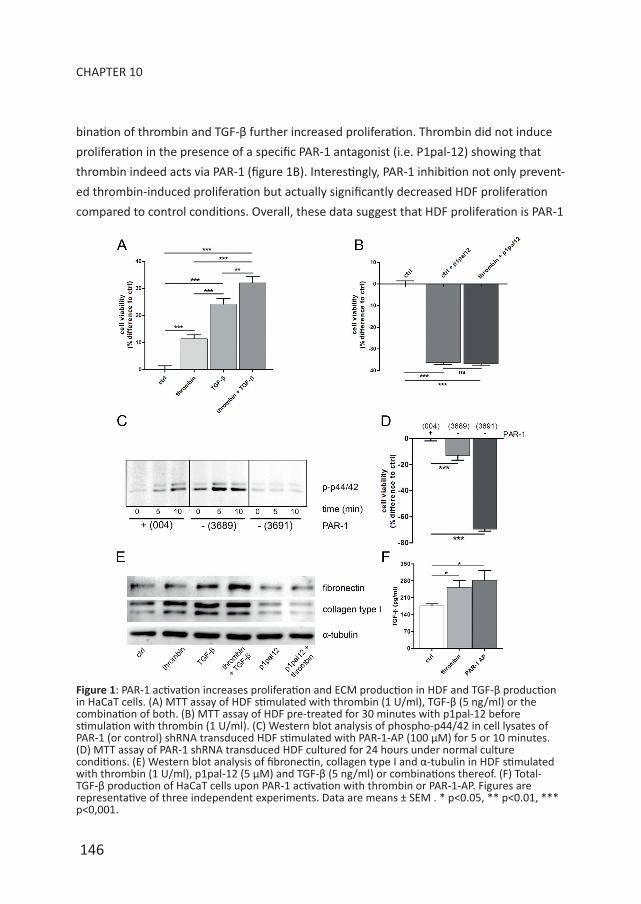
bin and/or TGF-β (positive control). As shown in figure 1A, thrombin-dependent PAR-1
activation and TGF-β stimulation both increased proliferation of HDF, whereas the com-
PS JanWillem Duitman_2014-01v2.indd 145 28-03-14 10:37
146
CHAPTER 10
bination of thrombin and TGF-β further increased proliferation. Thrombin did not induce
proliferation in the presence of a specific PAR-1 antagonist (i.e. P1pal-12) showing that
thrombin indeed acts via PAR-1 (figure 1B). Interestingly, PAR-1 inhibition not only prevent-
ed thrombin-induced proliferation but actually significantly decreased HDF proliferation
compared to control conditions. Overall, these data suggest that HDF proliferation is PAR-1
Figure 1: PAR-1 activation increases proliferation and ECM production in HDF and TGF-β production in HaCaT cells. (A) MTT assay of HDF stimulated with thrombin (1 U/ml), TGF-β (5 ng/ml) or the combination of both. (B) MTT assay of HDF pre-treated for 30 minutes with p1pal-12 before stimulation with thrombin (1 U/ml). (C) Western blot analysis of phospho-p44/42 in cell lysates of PAR-1 (or control) shRNA transduced HDF stimulated with PAR-1-AP (100 μM) for 5 or 10 minutes. (D) MTT assay of PAR-1 shRNA transduced HDF cultured for 24 hours under normal culture conditions. (E) Western blot analysis of fibronectin, collagen type I and α-tubulin in HDF stimulated with thrombin (1 U/ml), p1pal-12 (5 μM) and TGF-β (5 ng/ml) or combinations thereof. (F) Total-TGF-β production of HaCaT cells upon PAR-1 activation with thrombin or PAR-1-AP. Figures are representative of three independent experiments. Data are means ± SEM . * p<0.05, ** p<0.01, *** p<0,001.
PS JanWillem Duitman_2014-01v2.indd 146 28-03-14 10:37

147
PAR-1 IN SKIN FIBROSIS
10
dependent.
Although P1pal-12 is frequently used as a specific PAR-1 inhibitor5 and no toxicity has
been described, we next aimed to refute this latter suggestion. To this end, we lenti-virally
transduced HDF with two different PAR-1 shRNA constructs (designated 3689 and 3691)
and a control construct (004). As shown in figure 1C, PAR-1 activation by PAR-1-AP rap-
idly induces phosphorylation of the well-known PAR-1 target p44/42. Importantly, PAR-1
signaling was effectively reduced in HDF transduced with shRNA 3691 as evident from the
absence of p44/42 phosphorylation upon stimulation with PAR-1-AP. shRNA 3689 was less
effective in reducing PAR-1 signaling as evident from residual p44/42 phosphorylation after
PAR-1 activation.
As expected, effective inhibition of PAR-1 signaling (i.e. shRNA 3691) led to similar effects
on proliferation as compared to P1pal-12 treatment, whereas non-effective PAR-1 inhibi-
tion (i.e. shRNA 3689) only showed a 15% reduction in proliferation as compared to control
HDF (Figure 1D). Taken together, these data show PAR-1 dependent proliferation of HDF. As
opposed to proliferation, PAR-1 did not influence HDF migration (data not shown).
Next we assessed whether PAR-1 would modify ECM production by HDF. Indeed, throm-
bin stimulation of HDF slightly increased production of collagen type I, but it did not
modify fibronectin levels (Figure 1E). PAR-1 inhibition by P1pal-12 pre-treatment abol-
ished thrombin-induced collagen production but also diminished baseline collagen levels
showing that PAR-1 induces ECM synthesis. As expected, TGF-β stimulation also induced
collagen and fibronectin production. Interestingly, the combination of thrombin and TGF-β
further induced fibronectin levels without significantly affecting collagen levels.
PAR-1 deficiency reduces TGF-β levels during pulmonary fibrosis6 and next to direct
pro-fibrotic effects of PAR-1 on HDF, PAR-1 may also drive skin fibrosis by inducing TGF-β
production. As shown in Figure 1F, both thrombin and PAR-1-AP stimulation of HaCaT cells
indeed induced TGF-β production. TGF-β levels produced by HDF were low and did not
change upon PAR-1 activation (data not shown). Overall these data suggest an important
role for PAR-1 during dermal fibrosis by modifying HDF proliferation and ECM production
and potentially by modifying TGF-β production by keratinocytes.
Amelioration of bleomycin-induced scleroderma by loss of PAR-1. To determine whether
PAR-1 is critical for skin fibrosis, we subjected PAR-1 deficient and wildtype mice to a
bleomycin-induced scleroderma model. As shown in Figure 2A-C, dermal thickness was
significantly reduced in PAR-1 deficient mice as compared to wildtype mice. In line, the
accumulation of ECM was also reduced in PAR-1 deficient mice as compared to wildtype
PS JanWillem Duitman_2014-01v2.indd 147 28-03-14 10:37

148
CHAPTER 10
mice as evident from reduced collagen content in Masson’s trichrome stained skin sections
(Figure 2D). Together these data show that PAR-1 deficient mice are protected against
bleomycin-induced skin fibrosis.
Modification of profibrotic processes in PAR-1 deficient mice. In our in vitro experiments
we showed that PAR-1 drives fibroblast proliferation and consequently we next assessed
the number of proliferating cells (excluding cells of the epidermis, hair follicles and
sebaceous glands) in the dermis of wildtype and PAR-1 deficient mice during bleomycin-
induced skin fibrosis. As shown in figure 3A-B, the number of proliferating cells as analyzed
Figure 2: PAR-1 contributes to skin fibrosis in a model of bleomycin-induced SSc. (A-B) HE staining of skin sections of wildtype (A) and PAR-1 deficient (B) mice upon bleomycin treatment (10x magnification). Dermal thickness is indicated with a black line. (C) Quantification of dermal thickness upon bleomycin treatment in wildtype and PAR-1 deficient mice (n=7-8). (D) Representative pictures of Masson’s trichrome staining of skin sections from wildtype and PAR-1 deficient mice upon bleomycin treatment (10x magnification). Data are means ± SEM. * p<0.05.
PS JanWillem Duitman_2014-01v2.indd 148 28-03-14 10:37
149
PAR-1 IN SKIN FIBROSIS
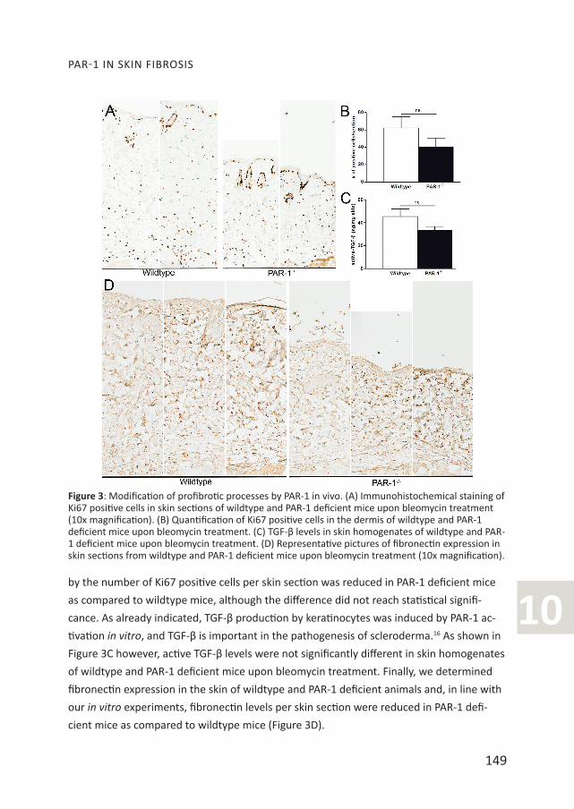
10by the number of Ki67 positive cells per skin section was reduced in PAR-1 deficient mice
as compared to wildtype mice, although the difference did not reach statistical signifi-
cance. As already indicated, TGF-β production by keratinocytes was induced by PAR-1 ac-
tivation in vitro, and TGF-β is important in the pathogenesis of scleroderma.16 As shown in
Figure 3C however, active TGF-β levels were not significantly different in skin homogenates
of wildtype and PAR-1 deficient mice upon bleomycin treatment. Finally, we determined
fibronectin expression in the skin of wildtype and PAR-1 deficient animals and, in line with
our in vitro experiments, fibronectin levels per skin section were reduced in PAR-1 defi-
cient mice as compared to wildtype mice (Figure 3D).
Figure 3: Modification of profibrotic processes by PAR-1 in vivo. (A) Immunohistochemical staining of Ki67 positive cells in skin sections of wildtype and PAR-1 deficient mice upon bleomycin treatment (10x magnification). (B) Quantification of Ki67 positive cells in the dermis of wildtype and PAR-1 deficient mice upon bleomycin treatment. (C) TGF-β levels in skin homogenates of wildtype and PAR-1 deficient mice upon bleomycin treatment. (D) Representative pictures of fibronectin expression in skin sections from wildtype and PAR-1 deficient mice upon bleomycin treatment (10x magnification).
PS JanWillem Duitman_2014-01v2.indd 149 28-03-14 10:37
150
CHAPTER 10
DISCUSSIONPAR-1 may play an important role during the initiation and progression of both lung and
liver fibrosis.5-7 Here, we determined whether PAR-1 would also drive skin fibrosis in the
setting of Ssc. Indeed, PAR-1 activation leads to increased proliferation and ECM produc-
tion in HDF, whereas it also potentiates TGF-β production of HaCaT. In line with these in vi-
tro data, we also show reductions in dermal thickening and collagen/fibronectin deposition
in PAR-1 deficient mice during experimental skin fibrosis. Overall, our data thus pinpoint
PAR-1 as a novel mediator of skin fibrosis.
Our in vitro data suggest that PAR-1 modifies scleroderma by acting as a pleiotropic medi-
ator affecting multiple profibrotic responses (i.e. proliferation, TGF-β production and ECM
deposition). However, during experimental scleroderma, PAR-1 deficiency only showed
significant effects on ECM deposition whereas the differences in fibroblast proliferation
did not reach significance. It may thus be that PAR-1 mainly affects ECM deposition during
bleomycin-induced scleroderma although the observed variability in fibroblast prolifera-
tion in vivo may explain the lack of significance. TGF-β production was also not significantly
reduced in PAR-1 deficient mice which may be due to the fact that PAR-1 only contributes
to TGF-β production in keratinocytes and not in dermal fibroblasts. As PAR-1 modifies
inflammation during pulmonary fibrosis6, it may be tempting to speculate that PAR-1-de-
pendent inflammation may potentiate skin fibrosis. Importantly however, inflammatory
cell influx (neutrophils and macrophages) and cytokine production were similar in wildtype
and PAR-1 deficient mice (data not shown).
An interesting finding of our manuscript is that PAR-1 inhibition (both pharmacologically
and genetically) reduces both proliferation and ECM production of HDF. These data strong-
ly suggest that HDF secrete an endogenous PAR-1 agonist that drives proliferation and
ECM production under normal culture conditions in an autocrine manner. Expression levels
of the endogenous PAR-1 ligand seem to be rate limiting for these pro-fibrotic processes as
exogenous PAR-1 activation still further increases both proliferation and ECM production.
Although several PAR-1 agonist (like MMP1317 and kallikreins18 have been described to be
produced in skin fibroblasts, the exact nature of the endogenous PAR-1 agonist remains
elusive and is currently under investigation.
We used a bleomycin-induced model of scleroderma that largely mimics human sclero-
derma.19 Indeed, dermal thickening is accompanied by deposition of dense packed col-
lagen fibrils that are extensively cross-linked in both murine and human scleroderma. The
fact that we show that PAR-1 deficiency limits skin fibrosis in this model, together with the
fact that PAR-1 is upregulated in the skin during human Ssc8 and that PAR-1 activation on
PS JanWillem Duitman_2014-01v2.indd 150 28-03-14 10:37

151
PAR-1 IN SKIN FIBROSIS
10
human cell types potentiates key profibrotic processes, suggests that PAR-1 may have clini-
cal relevance for human scleroderma. One should realize however that PAR-1 also medi-
ates platelet activation and targeting PAR-1 could lead to bleeding complications. It may
consequently be better to target the, still elusive, endogenous PAR-1 agonist.
In conclusion, we identify PAR-1 as a novel potential mediator of skin fibrosis which may
open novel therapeutic treatment strategies for limiting scleroderma (or other cutaneous
fibrotic disorders) for which no effective treatment strategy is available at this time.
Acknowledgements: This study was supported by grant 09.102 from the Dutch Burns
foundation to J.D. The authors like to thank Marieke ten Brink and Joost Daalhuisen for
their technical assistance during the animal experiments.
REFERENCES 1. Gabrielli, A., S. Svegliati, G. Moroncini, and D. Amico. 2012. New insights into the role of oxida-
tive stress in scleroderma fibrosis. Open Rheumatol J 6:87-95. 2. Gilbane AJ, Denton CP, and Holmes AM. 2013. Scleroderma pathogenesis: a pivotal role for fibro-
blasts as effector cells. Arthritis Res Ther 15:215-223. 3. Vu, T. K. H., D. T. Hung, V. I. Wheaton, and S. R. Coughlin. 1991. Molecular-Cloning of A Functional
Thrombin Receptor Reveals A Novel Proteolytic Mechanism of Receptor Activation. Cell 64:1057-1068.
4. Ramachandran, R. and M. Hollenberg. 2008. Proteinases and signalling: pathophysiological and therapeutic implications via PARs and more. Br J Pharmacol 153:S263-S282.
5. Lin, C., J. Duitman, J. Daalhuisen, M. ten Brink, J. von der Thüsen, T. van der Poll, K. Borensztajn, and C. A. Spek. 2013. Targeting protease activated receptor-1 with P1pal-12 limits bleomycin-induced pulmonary fibrosis. Thorax 69:152-160.
6. Howell, D. C. J., R. H. Johns, J. A. Lasky, B. Shan, C. J. Scotton, G. J. Laurent, and R. C. Chambers. 2005. Absence of proteinase-activated receptor-1 signaling affords protection from bleomycin-induced lung inflammation and fibrosis. Am J Pathol 166:1353-1365.
7. Rullier, A., J. Gillibert-Duplantier, P. Costet, G. Cubel, V. Haurie, C. Petibois, D. Taras, N. Dugot-Senant, G. Deleris, P. Bioulac-Sage, et al. 2008. Protease-activated receptor 1 knockout reduces experimentally induced liver fibrosis. Am J Physiol Gastrointest Liver Physiol 294:G226-G235.
8. Cevikbas, F., S. Seeliger, M. Fastrich, H. Hinte, D. Metze, C. Kempkes, B. Homey, and M. Steinhoff. 2011. Role of protease-activated receptors in human skin fibrosis and scleroderma. Exp Dermatol 20:69-71.
9. Materazzi, S., S. Pellerito, C. Di Serio, M. Paglierani, A. Naldini, C. Ardinghi, F. Carraro, P. Geppetti, G. Grino, M. Santucci, et al. 2007. Analysis of protease-activated receptor-1 and-2 in human scar formation. J Pathol 212:440-449.
10. Strukova, S. M., T. N. Dugina, I. V. Chistov, M. Lange, E. A. Markvicheva, S. Kuptsova, V. P. Zubov, and E. Glusa. 2001. Immobilized thrombin receptor agonist peptide accelerates wound healing in mice. Clin Appl Thromb Hemost 7:325-329.
11. Connolly, A. J., H. Ishihara, M. L. Kahn, R. V. Farese, and S. R. Coughlin. 1996. Role of the throm-bin receptor In development and evidence for a second receptor. Nature 381:516-519.
12. Schouten, M., C. van’t Veer, J. J. Roelofs, M. Levi, and T. van der Poll. 2012. Protease-activated receptor-1 impairs host defense in murine pneumococcal pneumonia: a controlled laboratory
PS JanWillem Duitman_2014-01v2.indd 151 28-03-14 10:37
152
CHAPTER 10
study. Crit Care 16:R238.13. Bijlsma, M. F., P. J. Leenders, B. J. Janssen, M. P. Peppelenbosch, H. ten Cate, and C. A. Spek.
2008. Endogenous hedgehog expression contributes to myocardial ischemia-reperfusion-induced injury. Exp Biol Med (Maywood) 233:989-996.
14. Anastasov, N., M. Klier, I. Koch, D. Angermeier, H. Hofler, F. Fend, and L. Quintanilla-Martinez. 2009. Efficient shRNA delivery into B and T lymphoma cells using lentiviral vector-mediated transfer. J Hematop 2:9-19.
15. Duitman, J., M. Schouten, A. P. Groot, J. B. Daalhuisen, S. Florquin, T. van der Poll, and C. Spek. 2012. CCAAT/enhancer-binding protein delta facilitates bacterial dissemination during pneumo-coccal pneumonia in a platelet-activating factor receptor-dependent manner. Proc Natl Acad Sci U S A 109:9113-9118.
16. Yamamoto, T., S. Takagawa, I. Katayama, and E. Nishioka. 1999. Anti-sclerotic effect of trans-forming growth factor-beta antibody in a mouse model of bleomycin-induced scleroderma. Clin Immunol 92:6-13.
17. Ravanti, L., M. Toriseva, R. Penttinen, T. Chrombleholme, M. Foschi, J. H. Han, and V. M. Kahari. 2001. Expression of human collagenase-3 (MMP-13) by fetal skin fibroblasts is induced by trans-forming growth factor-beta via p38 mitogen-activated protein kinase. FASEB J 15:1098-1100.
18. Komatsu, N., M. Takata, N. Otsuki, T. Toyama, R. Ohka, K. Takehara, and K. Saijoh. 2003. Expres-sion and localization of tissue kallikrein mRNAs in human epidermis and appendages. J Invest Dermatol 121:542-549.
19. Vorstenbosch, J., H. Al-Ajmi, S. Winocour, A. Trzeciak, L. Lessard, and A. Philip. 2013. CD109 overexpression ameliorates skin fibrosis in a mouse model of bleomycin-induced scleroderma. Arthritis Rheum. 65:1378-1383.
PS JanWillem Duitman_2014-01v2.indd 152 28-03-14 10:37