uva-dare (digital academic repository) susceptibility … · changes in ion channel function 11 and...
TRANSCRIPT

UvA-DARE is a service provided by the library of the University of Amsterdam (http://dare.uva.nl)
UvA-DARE (Digital Academic Repository)
Susceptibility to ischemic ventricular fibrillation
de Jong, J.S.S.G.
Link to publication
Citation for published version (APA):de Jong, J. S. S. G. (2013). Susceptibility to ischemic ventricular fibrillation
General rightsIt is not permitted to download or to forward/distribute the text or part of it without the consent of the author(s) and/or copyright holder(s),other than for strictly personal, individual use, unless the work is under an open content license (like Creative Commons).
Disclaimer/Complaints regulationsIf you believe that digital publication of certain material infringes any of your rights or (privacy) interests, please let the Library know, statingyour reasons. In case of a legitimate complaint, the Library will make the material inaccessible and/or remove it from the website. Please Askthe Library: http://uba.uva.nl/en/contact, or a letter to: Library of the University of Amsterdam, Secretariat, Singel 425, 1012 WP Amsterdam,The Netherlands. You will be contacted as soon as possible.
Download date: 30 Aug 2018
Platelets and cardiac arrhythmia.
Chapter 4.1 J.S.S.G. de Jong1, L.R. C. Dekker2
1 Department of Cardiology, Academic Medical Center, Amsterdam, Netherlands2 Department of Cardiology, Catharina Hospital, Eindhoven, Netherlands
Published in: Frontiers in Physiology 2010;1:166doi: 10.3389/fphys.2010.00166
4.1Abstract
78
Sudden cardiac death remains one of the most prevalent modes of death in
industrialized countries, and myocardial ischemia due to thrombotic coronary
occlusion is its primary cause. The role of platelets in the occurrence of SCD extends
beyond coronary flow impairment by clot formation. Here we review the substances
released by platelets during clot formation and their arrhythmic properties. Platelet
products are released from three types of platelet granules: dense core granules,
alpha-granules, and platelet lysosomes. The physiologic properties of dense granule
products are of special interest as a potential source of arrhythmic substances. They
are released readily upon activation and contain high concentrations of serotonin,
histamine, purines, pyrimidines, and ions such as calcium and magnesium. Potential
arrhythmic mechanisms of these substances, e.g. serotonin and high energy
phosphates, include induction of coronary constriction, calcium overloading, and
induction of delayed after-depolarizations. Alpha-granules produce thromboxanes
and other arachidonic acid products with many potential arrhythmic effects mediated
by interference with cardiac sodium, calcium and potassium channels. Alpha-
granules also contain hundreds of proteins that could potentially serve as ligands to
receptors on cardiomyocytes. Lysosomal products probably do not have an important
arrhythmic effect. Platelet products and ischemia can induce coronary permeability,
thereby enhancing interaction with surrounding cardiomyocytes. Antiplatelet therapy
is known to improve survival after myocardial infarction. Although an important part
of this effect results from prevention of coronary clot formation, there is evidence to
suggest that antiplatelet therapy also induces anti-arrhythmic effects during ischemia
by preventing the release of platelet activation products.
79
Platelets and cardiac arrhythmia
1. Cardiac arrhythmias Sudden cardiac death (SCD) remains one of the most prevalent modes of death in
industrialized countries, claiming almost a million deaths annually in Western Europe
and the United States1,2. Ventricular fibrillation (VF) in the setting of coronary artery
disease is the most common underlying arrhythmia2 as acute coronary thrombosis is
observed in 74–79% of SCD victims at autopsy3,4.
Arrest of blood flow to the myocytes results in a complex ischemic reaction that
includes an increase in outward potassium current5, a triphasic increase of extracellular
potassium6, a decrease of action potential (AP) duration7, and depolarization of resting
membrane potential. Decrease in pH results in activation of the Na+-H+ exchange
pathway extruding H+ from the cell. This leads to Ca2+ loading as a consequence
of reverse-mode action of the Na-Ca exchanger (NXC)8,9. Ca2+ loading of ischemic
cardiomyocytes has several pro-arrhythmic effects: abnormal sarcoplasmic reticulum
Ca2+ cycling and promotion of AP alternans, delayed after-depolarizations (DADs),
and decreased gap-junction conductance ultimately leading to uncoupling of cell-
to-cell connections10. Furthermore, reactive oxygen species are released, resulting in
changes in ion channel function11 and mitochondrial dysfunction8. These events create
a hostile environment that renders the myocardium prone to arrhythmias12.
VF during ischemia results from re-entrant excitation. Onset of re-entry may not only
result from injury current13,14, but may also be evoked by triggered beats originating
from early and delayed after-potentials (EADs and DADs)15.
Platelets play an important role in the occurrence of SCD. Traditionally, the role
of platelets in SCD was believed to be limited to their ability to halt coronary flow
by clot formation. However, there is an increasing body of evidence suggesting that
the process of clot formation has arrhythmic properties beyond the arrest of distal
perfusion. Animal experiments have shown that platelet activation increases the
susceptibility of ischemic myocardium to VF. Coronel et al. showed that intracoronary
thrombi have profibrillatory effects16. In their pig model, the left anterior descending
artery (LAD) was ligated, and blood with thrombin-induced platelet activation or
heparinized blood injected distally. Notwithstanding a comparable area of ischemia,
thrombi injection resulted in VF in 4/7 pigs vs. 2/19 control pigs. Similarly, in a dog
model of regional myocardial ischemia, 51% of control animals developed VF after
coronary artery embolization. However, following pre-treatment with carbenicillin, a
strong inhibitor of platelet aggregation, or estradiol cypionate, which induces severe
thrombocytopenia, the incidence of VF was reduced to 9% and zero, respectively17.
Platelet activation results in the release of platelet products, the so-called secretome,
which includes organic substances (e.g., adenosine-5’-triphosphate [ATP], serotonin,
Chapter 4.1
80
and histamine) and more than 2000 proteins18. Many of these substances can alter
electrophysiological properties of the heart in various animal species, supporting the
notion that activated platelets exert pro-arrhythmic effects (Figure 1)19,20,21. Conversely,
platelet antagonists counteract ischemia-induced arrhythmias in animal studies22,23.
2. Platelet adhesion and activationPlatelets are small anuclear cells (2-3 µm, 7.5–1.5 fL), which are derived from
defragmentation of megakaryocytes. Platelets have a life-span of 8–12 days, and the
blood of healthy individuals has a platelet concentration of 150–400 x 109/L .24 Coronary
plaque rupture results in exposure of subendothelium-containing collagen, collagen-
bound-von-Willebrand factor (vWf), fibronectin and laminin. Platelet membranes
expose an array of adherence proteins waiting to adhere with these exposed molecules.
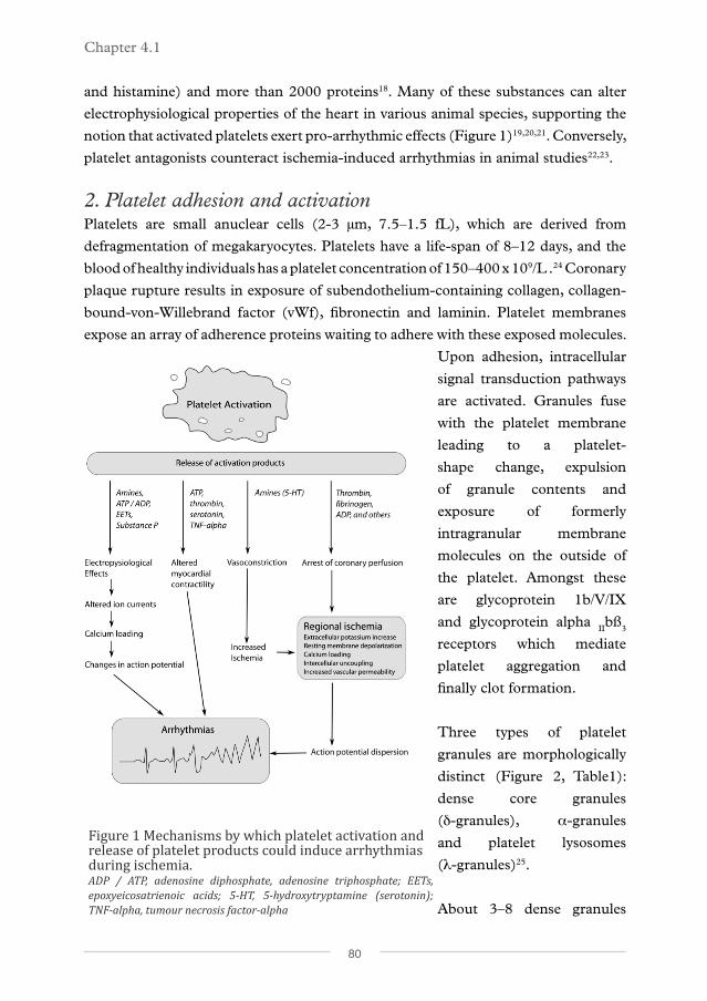
Upon adhesion, intracellular
signal transduction pathways
are activated. Granules fuse
with the platelet membrane
leading to a platelet-
shape change, expulsion
of granule contents and
exposure of formerly
intragranular membrane
molecules on the outside of
the platelet. Amongst these
are glycoprotein 1b/V/IX
and glycoprotein alpha IIbß
3
receptors which mediate
platelet aggregation and
finally clot formation.
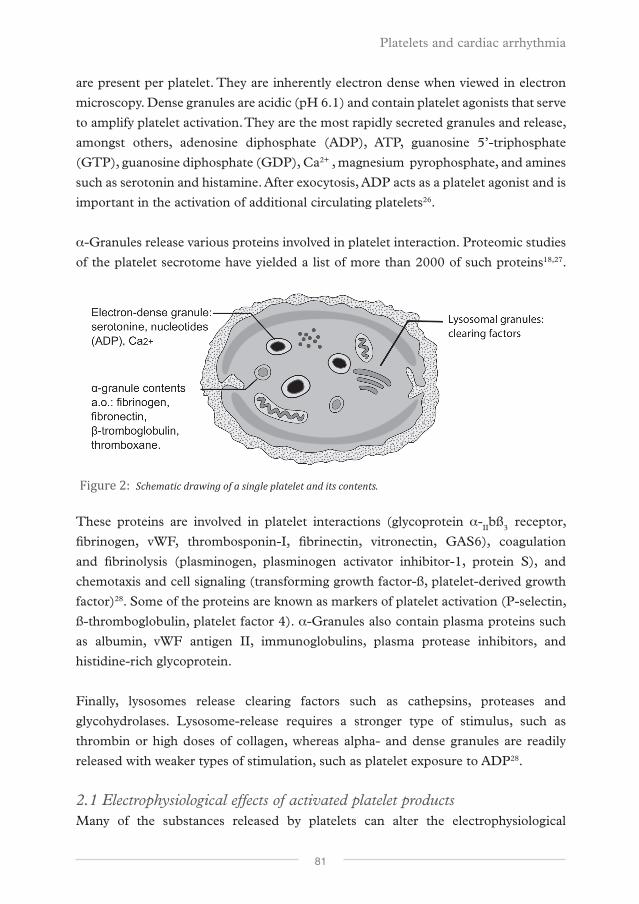
Three types of platelet
granules are morphologically
distinct (Figure 2, Table1):
dense core granules
(d-granules), α-granules
and platelet lysosomes
(λ-granules)25.
About 3–8 dense granules
Figure 1 Mechanisms by which platelet activation and release of platelet products could induce arrhythmias during ischemia.ADP / ATP, adenosine diphosphate, adenosine triphosphate; EETs, epoxyeicosatrienoic acids; 5-HT, 5-hydroxytryptamine (serotonin); TNF-alpha, tumour necrosis factor-alpha
Platelets and cardiac arrhythmia
81
are present per platelet. They are inherently electron dense when viewed in electron
microscopy. Dense granules are acidic (pH 6.1) and contain platelet agonists that serve
to amplify platelet activation. They are the most rapidly secreted granules and release,
amongst others, adenosine diphosphate (ADP), ATP, guanosine 5’-triphosphate
(GTP), guanosine diphosphate (GDP), Ca2+ , magnesium pyrophosphate, and amines
such as serotonin and histamine. After exocytosis, ADP acts as a platelet agonist and is
important in the activation of additional circulating platelets26.
α-Granules release various proteins involved in platelet interaction. Proteomic studies
of the platelet secrotome have yielded a list of more than 2000 of such proteins18,27.
These proteins are involved in platelet interactions (glycoprotein α-IIbß
3 receptor,
fibrinogen, vWF, thrombosponin-I, fibrinectin, vitronectin, GAS6), coagulation
and fibrinolysis (plasminogen, plasminogen activator inhibitor-1, protein S), and
chemotaxis and cell signaling (transforming growth factor-ß, platelet-derived growth
factor)28. Some of the proteins are known as markers of platelet activation (P-selectin,
ß-thromboglobulin, platelet factor 4). α-Granules also contain plasma proteins such
as albumin, vWF antigen II, immunoglobulins, plasma protease inhibitors, and
histidine-rich glycoprotein.
Finally, lysosomes release clearing factors such as cathepsins, proteases and
glycohydrolases. Lysosome-release requires a stronger type of stimulus, such as
thrombin or high doses of collagen, whereas alpha- and dense granules are readily
released with weaker types of stimulation, such as platelet exposure to ADP28.
2.1 Electrophysiological effects of activated platelet productsMany of the substances released by platelets can alter the electrophysiological
Figure 2: Schematic drawing of a single platelet and its contents.
Chapter 4.1
82
properties of the heart in various animal species29.
2.1.1 AminesSerotonin or 5-hydrocytryptamine (5-HT) is not synthesized in platelets but is
actively taken up from the plasma and accumulated in dense granules where it is
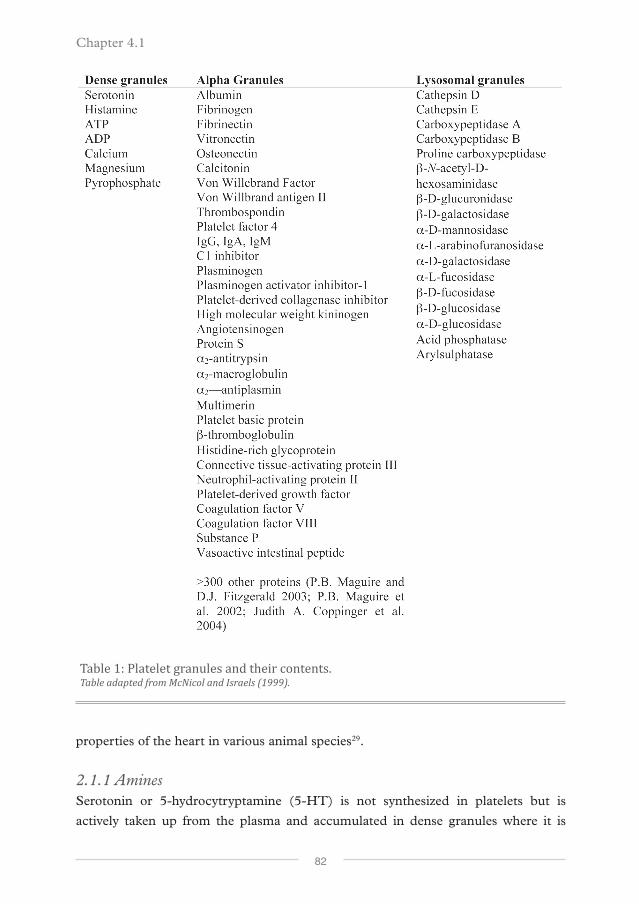
Table 1: Platelet granules and their contents.Table adapted from McNicol and Israels (1999).
Platelets and cardiac arrhythmia
83
likely complexed with ATP and potentially with calcium30. The serotonin released
by exocytosis is relatively stable and functions as a weak platelet agonist on 5HT2
receptors30, although it is probably less important in this respect than ADP. The positive
feedback effect of serotonin may be of secondary importance to its vasoconstrictive
action, which reduces flow at the site of injury and thereby limits blood loss31 Dense
granular concentration of serotonin was calculated to be 65 mM32, whereas (platelet-
rich) plasma serotonin concentration was 842 ± 58 nmol/L in healthy individuals33.
Serotonin can be used to assess the platelet release reaction: it locally increased 18
to 27-fold from normal levels in tissue surrounding a coronary thrombus in a dog
model34.
Human atrial and ventricular cardiomyocytes express 5-HT4 receptor35,36, and
exposure to serotonin results in tachycardia, increased atrial contractility, and atrial
arrhythmias37. 5-HT causes an up to 6-fold increase in L-type Ca2+ channel current
through 5-HT4 receptors, in atrial myocytes from patients in sinus rhythm38. Likewise,
serotonin has positive inotropic and lusitropic effects on ventricular cardiomyocytes 39. Arrhythmic effects have also been observed and are probably mainly related to
Ca2+-overload mediated by an increase in L-type Ca2+ current40. Antidepressants from
the selective serotonin reuptake inhibitor class (SSRI) have QT prolonging effects in
patients with SSRI intoxications. The major clinical interaction with cardiovascular
disease is likely its platelet inhibiting effect, which may reduce incidence of MI41.
Like, serotonin, histamine is another amine present in dense granules. Resting human
platelets contain 25 ng histamine / 108 platelets, which can increase towards 47 ng
/ 108 platelets upon platelet activation42. The major arrhythmic effects of histamine
consist of H1-receptor-mediated slowing of AV nodal conduction and H2-receptor
mediated increase in sinus rate and ventricular automaticity43. Histamine also has a
positive inotropic effect on ventricular cardiomyocytes44.
In addition to serotonin, histamine can also cause vasoconstriction as further discussed
below.
2.1.2 Nucleotide derivativesExtracellular purines (adenosines, ADP, and ATP) and pyrimidines (uridine
diphosphate [UDP] and uridine triphosphate [UTP]) can initiate a wide range
of intracellular signaling cascades through purinergic receptors45. Platelet-dense
granules contain a high concentration of ATP (436mM) and ADP (653 mM)32, which
can accelerate platelet activation binding to ADP receptors upon release. Normal

Chapter 4.1
84
extracellular ATP concentration is about 2 µM 46 and may increase to 100 μM under
pathological conditions47. Under normal conditions, ATP is rapidly converted to
adenosine29.
In rat and mouse ventricular cardiomyocytes, exposure to ATP induces a cytosolic
Ca2+ rise and has positive inotropic effects 48-50. By activation of purinergic receptors,
ATP induces sustained increases in diastolic Ca2+ and triggers multiple Ca2+ waves,
leading to DADs in current clamped mouse heart51.
2.1.3 IonsThe dense granular concentration of calcium was calculated to be 2.2 M.32 The
total amount of calcium in human platelets is 10.1 μmol/1011 platelets52. In humans,
arrhythmias related to high calcium and magnesium levels are rare53. The normal plasma
concentration of Ca2+ ranges from 1.03 to 1.23 mmol/L 54. The Ca2+ concentration
in dense granules is very high, but the amount of released Ca2+ relative to the plasma
concentration is small. The platelets in 1 mL blood (about 250 x 109) contain about
25.3 nmol of Ca2+, and upon release would increase the local Ca2+ concentration by
about 2% (presuming a volume of dispersion of 1 mL). This raw estimate does not
take into account the high concentration of platelets in an actual thrombus and the
washout of Ca2+ during the non-occlusive phase of thrombus formation.
The platelet content of Mg2+, the second most abundant ion, is much smaller that the
Ca2+ content. Platelets contain about 0.4 μmol/1011 platelets of releasable magnesium
(Meyers, Holmsen, and Seachord 1982). Normal plasma concentrations of Mg2+
range between 0.6 and 0.7 mmol/L. A large influence of platelet Mg2+ release during
platelet aggregation is therefore less likely than it is for Ca2+.
2.1.4 Thromboxane, arachidonic acid and other alpha-granule contentsDuring platelet activation, arachidonic acid is liberated by phospholipase-A2 from
membrane phospholipids and is converted enzymatically to epoxyeicosatrienoic
acids (EETs) via the cyclo-oxygenase and lipoxygenase pathways. EETs have several
effects on cardiac tissues55,56. EETs inhibit cardiac Na+ channels57, significantly
increase intracellular Ca2+ concentrations in isolated guinea pig cardiomyocytes58,
and significantly modulate the activities of cardiac Ca2+ channels59 and KATP
channels 57. Furthermore EETs can uncouple neonatal rat cardiac myocytes by reducing gap-
junction conductance60.
Thromboxane is an arachidonic-acid derivative that is prothrombotic and released by
platelets. Thromboxane is produced by cyclooxygenase from prostanoids, a process
Platelets and cardiac arrhythmia
85
that is inhibited by aspirin. Thromboxane is chemically unstable in blood, but its
stable analogue, U41669, has been shown to increase automaticity and to increase
both resting and peak intracellular Ca2+ concentrations and induce irregular Ca2+
transients in isolated neonatal rat ventricular myocytes61. This is supported by the fact
that these effects can be reversed in rabbit cardiomyocytes by blockade of U41669.62
Chien et al. examined the effects of platelet products of rabbit platelets on cytosolic
calcium in chick embryonic heart cells. They identified the arrhythmic platelet
product(s) as small trypsin-sensitive peptide(s).63 Examples of such small peptides,
found in α-granules, are substance P, calcitonin, vasoactive intestinal peptide, and
angiotensinogen. Of the latter three no direct cardiac effects have been described.
Substance P has only recently been found to be present in relatively high concentrations
in platelets.64 It is primarily known as a neurotransmitter of pain sensation, but also has
cardiovascular effects which are primarily mediated through vasodilatation. However,
direct cardiac effects have been described: induction of bradycardia and hypotension
in denervated and anesthesized rats.65
Besides these, α-granules contain a long list of substances (Table 1) which can be
subdivided in proteoglycans, adhesive glycoproteins, hemostatic factors, cellular
mitogens, protease inhibitors and miscellaneous other molecules25. Platelet proteomic
approaches have tried to assess all proteins present in platelets27,66. Proteomics of
the products of activated platelets have revealed more than 300 proteins18,67 which
are mostly secreted in α-granules. Most of these proteins are present in only small
quantities. Large electrophysiologic effects would therefore likely be mediated through
membrane channels on cardiomyocytes.
2.1.5 Lysosomal productsPlatelet lysosomes contain ‘clearing factors’ that break down the platelet clot: acid
proteases and glycohydrolases. These lysosomal products have no known effect
on cardiomyocytes. Breakdown products of platelet clots could potentially be
arrhythmogenic, however a review on the incidence of early ventricular arrhythmias
after thrombolytic therapy did not show evidence for increased early arrhythmias68.
2.2 VasoconstrictionSeveral dense granule products have been shown to have effects on coronary smooth
muscle cells that mediate coronary constriction. ATP, UTP and ADP can mediate
prostacyclin and nitric-oxide release by interaction with the P2Y receptors on
endothelial cells69. Serotonin injected in coronary arteries of angina patients leads to

Chapter 4.1
86
intense coronary constriction70, probably mediated by vascular 5-HT1B and 5-HT2A
receptors37. Like serotonin, histamine is another strong vasoconstrictor. In patients
with variant angina, histamine could induce coronary spasm in 47% of subjects in
one study71. Thrombus-released vasoconstrictive substances could reduce coronary
perfusion and thereby increase local ischemia and reduce blood supply by collateral
vessels. Ischemia due to coronary spasm in the absence of severe atherosclerosis has
been described as a cause of ventricular fibrillation72-77.
2.3 Endothelial permeabilityIncreased endothelial permeability during myocardial infarction can facilitate leakage
of platelet products from the coronary lumen to epicardial myocytes. Increased
endothelial permeability could be induced by platelet products or by ischemia. Indeed
in a rat model of cerebral ischemia, thrombin injection induced vascular disruption
and increased permeability78. In another study, platelet activating factor (PAF) greatly
increased coronary permeability by the inhibition of endogeneous NO synthesis79.
And, in a hamster model, PAF increased permeability of the microvasculature of the
cheek pouch80. Also, serotonin has been shown to increase vascular permeability in
different animal models81.
Ischemia also induces coronary permeability as animal studies have shown. In a dog
model, 15 mins of coronary occlusion could increase protein leakage by 50%.82 And,
in a rat model, 20 mins of severe ischemia increased transcapillary albumin flux by
100% 83,84. Both the direct ischemic effects and the effects induced by platelet products
promote interaction of platelet products and the myocardial tissue surrounding the
ischemic coronary.
2.4 Effects on myocardial contractilityBoth increases and decreases in myocardial contractility have been found in
experiments with substances released by platelets. ATP has strong positive inotropic
effects in rats. ATP stimulates a large increase in cytosolic Ca2+ transients85,49. ATP also
increases the L-type Ca2+ current in rats, and both mechanisms can induce positive
inotropic effects86. Further upstream in rats and mice, activation of P2 purinergic
receptors exerts a positive inotropic effect on cardiomyocytes and intact hearts by
increasing intracellular ATP levels50. Thrombin promotes Ca2+ entry and release in
cardiomyocytes63. Serotonin has positive inotropic effects on the human atria through
the HT4 receptor, but such a receptor is absent in human ventricular cardiomyocytes 40.

Platelets and cardiac arrhythmia
87
Negative inotropic effects have been described for tumour necrosis factor-α in rats 88,89. It is likely that these effects leverage towards a positive inotropic effect of platelet
products, as was confirmed by a study that added aggregating platelets to a bath of cat
papillary muscles resulting in positive inotropic effects90. Also, injection of low-dosed
platelet-activating factor into coronary arteries resulted in strong positive inotropic
effects in isolated rabbit hearts91.
2.5 Interaction of effects of platelet products and ischemic effectsArrhythmias are very common during cardiac ischemia and are the result of a
multifactorial process. Ischemia is the most important factor in arrhythmia occurrence.
This is evidenced by the fact that the absence of active platelets does not completely
eliminate occurrence of ventricular fibrillation in coronary ligation studies described
above12,17,63. Platelet products strongly increase the risk of arrhythmias in the setting
of myocardial ischemia. It is tempting to speculate which platelet products have
the strongest role in arrhythmia occurrence. From the clinical setting we know that
ischemic VF often occurs within the first minutes after onset of chest pain, and it’s risk
declines during the first hours. An arrhythmic mediator should therefore be released
readily upon platelet activation and exert it’s effect within seconds as also has been
observed in the animal models.
Amines from dense granules are readily released in high concentrations and alter
cardiomyocyte electrophysiology directly, which makes them likely candidates. On
top of that serotonin induced vasoconstriction could undermine collateral perfusion
of the ischemic area. As a result of the findings of Chien et al. small trypsin-sensitive
peptide(s) released by platelets, such as substance P, deserve further investigation.
Increased endothelial permeability induced by ischemia promotes interaction
between these substances and cardiomyocytes. An unstable thrombus with fragment
embolization could lead to increased local heterogeneity.
3. Platelet antagonists and arrhythmia preventionAntiplatelet therapy is known to improve survival after myocardial infarction92, while
patients with increased platelet aggregation have a worse prognosis post myocardial
infarction93. However, it is difficult to separate the beneficial effect of preventing
thrombotic coronary occlusion from the potential anti-arrhythmic effects achieved
during ischemia by preventing formation of platelet activation products. However,
some evidence does exist for such an effect. Platelet antagonists effectively induced
ventricular arrhythmias in models of ischemia by coronary ligation in rats22 and
dogs23,94. In a dog model of regional ischemia, the threshold of epinephrine-induced
ventricular fibrillation was reduced after aspirin pre-treatmen95. In a rat model of
Chapter 4.1
88
coronary occlusion, sarpogrelate, a 5-hydroxy tryptamine 2A receptor antagonist,
but not cilostazol, a phosphodiesterase-III inhibitor, was able to prevent ventricular
arrhythmias during ischemia96. Sarpogrelate has multiple fields of action, including
inhibition of serotonin-induced coronary spasm. However, administering aspirin
after platelet activation had occurred did not block anti-arrhythmic effects of platelet
products in isolated rabbit hearts91.
4. Summary and conclusionThe role of platelets in the occurrence of SCD extends beyond coronary flow
impairment by clot formation. During clot formation platelets release a plethora of
substances, many with potent arrhythmic properties. Platelet products are released
from three types of platelet granules: dense core granules, alpha-granules and platelet
lysosomes. The physiologic properties of dense granule products are of special
interest as a potential source of arrhythmic substances. They are released readily
upon activation and contain high concentrations of serotonin, histamine, purines
(adenosines, ADP, and ATP), pyrimidines (UDP and UTP), and ions such as Ca2+
and Mg2+. The mode of action of these substances ranges from induction of coronary
constriction (serotonin), Ca2+ overloading (serotonin), and the induction of DADs
(ATP).
α-Granules contain thromboxanes and other arachidonic acid products with
many potential arrhythmic effects mediated by interference with cardiac K+ and
Ca2+ channels. α-Granules also contain a large number of proteins in much lower
concentrations that could potentially serve as a ligand to receptors on cardiomyocytes.
Substance P is of particular interest. Lysosomal products are clearing factors that
result in breakdown products of clots; but clinical studies do not suggest an important
role of lysosomal products in arrhythmias, as evidenced by the absence of arrhythmia-
induction during thrombolysis.
Antiplatelet therapy is known to improve survival after myocardial infarction. Although
an important part of this effect results from the prevention of coronary clot formation,
there is evidence to suggest that antiplatelet therapy also has anti-arrhythmic effects
during ischemia by preventing the release of platelet activation products.
DisclosuresNone
Platelets and cardiac arrhythmia
89
Author affiliationsJ.S.S.G. de Jong1, L.R.C. Dekker2
1 Department of Cardiology, Academic Medical Center, Amsterdam, The Netherlands.
2 Department of Cardiology, Catharina Hospital, Eindhoven, The Netherlands.
References1. Chugh SS, Reinier K, Teodorescu C, Evanado A, Kehr E, Al Samara M, et al. Epidemiology of
sudden cardiac death: clinical and research implications. Prog Cardiovasc Dis. 2008 Dec;51(3):213-228.
2. Zipes DP, Camm AJ, Borggrefe M, Buxton AE, Chaitman B, Fromer M, et al. ACC/AHA/ESC
2006 guidelines for management of patients with ventricular arrhythmias and the prevention of sudden
cardiac death: a report of the American College of Cardiology/American Heart Association Task Force and
the European Society of Cardiology Committee for Practice Guidelines (Writing Committee to Develop
Guidelines for Management of Patients With Ventricular Arrhythmias and the Prevention of Sudden
Cardiac Death). J Am Coll Cardiol. 2006 Sep 5;48(5):e247-346.
3. Davies M, Thomas A. Thrombosis and acute coronary-artery lesions in sudden cardiac ischemic
death. N Engl J Med. 1984 Mei 3;310(18):1137-1140.
4. Frink RJ, Rooney PA, Trowbridge JO, Rose JP. Coronary thrombosis and platelet/fibrin
microemboli in death associated with acute myocardial infarction. Br Heart J. 1988 Feb;59(2):196-200.
5. Wilde AA, Escande D, Schumacher CA, Thuringer D, Mestre M, Fiolet JW, et al. Potassium
accumulation in the globally ischemic mammalian heart. A role for the ATP-sensitive potassium channel.
Circ Res. 1990 Oct;67(4):835-43.
6. Coronel R, Fiolet JW, Wilms-Schopman FJ, Schaapherder AF, Johnson TA, Gettes LS, et
al. Distribution of extracellular potassium and its relation to electrophysiologic changes during acute
myocardial ischemia in the isolated perfused porcine heart. Circulation. 1988 May;77(5):1125-1138.
7. Saito T, Sato T, Miki T, Seino S, Nakaya H. Role of ATP-sensitive K+ channels in
electrophysiological alterations during myocardial ischemia: a study using Kir6.2-null mice. Am J Physiol
Heart Circ Physiol. 2005 Jan;288(1):H352-357.
8. Akar JG, Akar FG. Regulation of ion channels and arrhythmias in the ischemic heart. J
Electrocardiol. 2007 Dec;40(6 Suppl):S37-41.
9. Dekker LR, Fiolet JW, VanBavel E, Coronel R, Opthof T, Spaan JA, et al. Intracellular Ca2+,
intercellular electrical coupling, and mechanical activity in ischemic rabbit papillary muscle. Effects of
preconditioning and metabolic blockade. Circ. Res. 1996 Aug;79(2):237-246.
10. Kléber AG, Riegger CB, Janse MJ. Electrical uncoupling and increase of extracellular resistance
after induction of ischemia in isolated, arterially perfused rabbit papillary muscle. Circ Res. 1987
Aug;61(2):271-279.
11. Carmeliet E. Cardiac ionic currents and acute ischemia: from channels to arrhythmias. Physiol.
Rev. 1999 Jul;79(3):917-1017.
Chapter 4.1
90
12. Coronel R, Wilms-Schopman FJ, Opthof T, van Capelle FJ, Janse MJ. Injury current and
gradients of diastolic stimulation threshold, TQ potential, and extracellular potassium concentration
during acute regional ischemia in the isolated perfused pig heart. Circ Res. 1991 May;68(5):1241-1249.
13. Lown B, Wolf M. Approaches to sudden death from coronary heart disease. Circulation. 1971
Jul;44(1):130-142.
14. Verkerk AO, Veldkamp MW, Bouman LN, van Ginneken AC. Calcium-activated Cl(-) current
contributes to delayed afterdepolarizations in single Purkinje and ventricular myocytes. Circulation. 2000
Jun 6;101(22):2639-2644.
15. Ter Keurs HEDJ, Boyden PA. Calcium and arrhythmogenesis. Physiol Rev. 2007 Apr;87(2):457-
506.
16. Coronel R, Wilms-Schopman FJ, Janse MJ. Profibrillatory effects of intracoronary thrombus in
acute regional ischemia of the in situ porcine heart. Circulation. 1997 Dec 2;96(11):3985-3991.
17. Johnson G, Heckel R, Leis L, Franciosa J. Effect of inhibition of platelet function with carbenicillin
or aspirin on experimental canine sudden death. J Lab Clin Med. 1981 Nov;98(5):660-672.
18. Coppinger JA, Cagney G, Toomey S, Kislinger T, Belton O, McRedmond JP, et al. Characterization
of the proteins released from activated platelets leads to localization of novel platelet proteins in human
atherosclerotic lesions. Blood. 2004 Maart 15;103(6):2096-2104.
19. Hoffman BF, Guo SD, Feinmark SJ. Arrhythmias caused by platelet activating factor. J
Cardiovasc Electrophysiol. 1996 Feb;7(2):120-133.
20. Hoffman BF, Feinmark SJ, Guo SD. Electrophysiologic effects of interactions between activated
canine neutrophils and cardiac myocytes. J Cardiovasc Electrophysiol. 1997 Jun;8(6):679-687.
21. Flores N, Botchway A, Stavrou B, Sheridan D. Cardiac electrophysiological effects of platelet-
derived substances. Exp.Physiol. 1999 Maart;84(2):253-274.
22. Ahn Y, Cho J, Park W, Kim N, Kim J, Kim S, et al. The effects of antiplatelet agents in the
prevention of ventricular tachyarrhythmias during acute myocardial ischemia in rats. Jpn Heart J. 1999
Jan;40(1):79-86.
23. Wainwright C, Parratt J, Bigaud M. The effects of PAF antagonists on arrhythmias and platelets
during acute myocardial ischaemia and reperfusion. Eur Heart J. 1989 March;10(3):235-243.
24. Greer JP. Wintrobe’s Clinical Hematology. Lippincott Williams & Wilkins; 2008.
25. Rendu F, Brohard-Bohn B. The platelet release reaction: granules’ constituents, secretion and
functions. Platelets. 2001 Aug;12(5):261-273.
26. McNicol A, Israels SJ. Platelet dense granules: structure, function and implications for
haemostasis. Thromb. Res. 1999 Jul 1;95(1):1-18.
27. McRedmond J, Park S, Reilly D, Coppinger J, Maguire P, Shields D, et al. Integration of
Proteomics and Genomics in Platelets: a profile of platelet proteins and platelet-specific genes. Mol Cell
Proteomics. 2004 Feb 1;3(2):133-144.
28. Gresele P, Page CP, Vermylen J. Platelets in thrombotic and non-thrombotic disorders.
Cambridge University Press; 2002.
Platelets and cardiac arrhythmia
91
29. Flores NA, Botchway AN, Stavrou BM, Sheridan DJ. Cardiac electrophysiological effects of
platelet-derived substances. Exp. Physiol. 1999 Mar;84(2):253-274.
30. De Clerck F, Xhonneux B, Leysen J, Janssen PA. Evidence for functional 5-HT2 receptor sites
on human blood platelets. Biochem. Pharmacol. 1984 Sep 1;33(17):2807-2811.
31. De Clerck F. Blood platelets in human essential hypertension. Agents Actions. 1986 Aug;18(5-
6):563-580.
32. Holmsen H, Weiss HJ. Secretable storage pools in platelets. Annu. Rev. Med. 1979;30:119-134.
33. Vikenes K, Farstad M, Nordrehaug JE. Serotonin is associated with coronary artery disease and
cardiac events. Circulation. 1999;100(5):483-489.
34. Ashton J, Benedict C, Fitzgerald C, Raheja S, Taylor A, Campbell W, et al. Serotonin as a mediator
of cyclic flow variations in stenosed canine coronary arteries. Circulation. 1986 Maart 1;73(3):572-578.
35. Kaumann AJ, Sanders L, Brown AM, Murray KJ, Brown MJ. A 5-hydroxytryptamine receptor in
human atrium. Br. J. Pharmacol. 1990 Aug;100(4):879-885.
36. Bach T, Syversveen T, Kvingedal AM, Krobert KA, Brattelid T, Kaumann AJ, et al. 5HT4(a)
and 5-HT4(b) receptors have nearly identical pharmacology and are both expressed in human atrium and
ventricle. Naunyn Schmiedebergs Arch. Pharmacol. 2001 Feb;363(2):146-160.
37. Kaumann AJ, Levy FO. 5-hydroxytryptamine receptors in the human cardiovascular system.
Pharmacol. Ther. 2006 Sep;111(3):674-706.
38. Ouadid H, Seguin J, Dumuis A, Bockaert J, Nargeot J. Serotonin increases calcium current in
human atrial myocytes via the newly described 5-hydroxytryptamine4 receptors. Mol. Pharmacol. 1992
Feb;41(2):346-351.
39. Brattelid T, Qvigstad E, Lynham JA, Molenaar P, Aass H, Geiran O, et al. Functional serotonin
5-HT4 receptors in porcine and human ventricular myocardium with increased 5-HT4 mRNA in heart
failure. Naunyn Schmiedebergs Arch. Pharmacol. 2004 Sep;370(3):157-166.
40. Kaumann AJ. Do human atrial 5-HT4 receptors mediate arrhythmias? Trends in Pharmacological
Sciences. 1994 Dec;15(12):451-455.
41. Sauer WH, Berlin JA, Kimmel SE. Effect of antidepressants and their relative affinity for the
serotonin transporter on the risk of myocardial infarction. Circulation. 2003 Jul 8;108(1):32-36.
42. Jancinová V, Nosál R. Increased histamine content in Ca2+-ionophore A23187-activated human
blood platelets. Platelets. 1998;9(3-4):203-206.
43. Wolff AA, Levi R. Histamine and cardiac arrhythmias. Circ. Res. 1986 Jan;58(1):1-16.
44. Ginsburg R, Bristow MR, Stinson EB, Harrison DC. Histamine receptors in the human heart.
Life Sciences. 1980 Jun 30;26(26):2245-2249.
45. Ralevic V, Burnstock G. Receptors for Purines and Pyrimidines. Pharmacological Reviews.
1998;50(3):413-492.
46. Born G, Kratzer M. Source and concentration of extracellular adenosine triphosphate during
haemostasis in rats, rabbits and man. J Physiol. 1984;354:419-429.
47. Coade S, Pearson J. Metabolism of adenine nucleotides in human blood. Circ.Res.
Chapter 4.1
92
1989;65(3):531-537.
48. Pucéat M, Clément O, Scamps F, Vassort G. Extracellular ATP-induced acidification leads to
cytosolic calcium transient rise in single rat cardiac myocytes. Biochem. J. 1991 Feb 15;274 ( Pt 1):55-62.
49. Danziger RS, Raffaeli S, Moreno-Sanchez R, Sakai M, Capogrossi MC, Spurgeon HA, et al.
Extracellular ATP has a potent effect to enhance cytosolic calcium and contractility in single ventricular
myocytes. Cell Calcium. 1988 Aug;9(4):193-199.
50. Mei Q, Liang BT. P2 purinergic receptor activation enhances cardiac contractility in isolated rat
and mouse hearts. Am. J. Physiol. Heart Circ. Physiol. 2001 Jul;281(1):H334-341.
51. Gurung IS, Kalin A, Grace AA, Huang CL. Activation of purinergic receptors by ATP induces
ventricular tachycardia by membrane depolarization and modifications of Ca2+ homeostasis. J. Mol. Cell.
Cardiol. 2009 Nov;47(5):622-633.
52. Meyers KM, Holmsen H, Seachord CL. Comparative study of platelet dense granule constituents.
Am. J. Physiol. 1982 Sep;243(3):R454-461.
53. Diercks DB, Shumaik GM, Harrigan RA, Brady WJ, Chan TC. Electrocardiographic
manifestations: electrolyte abnormalities. J Emerg Med. 2004 Aug;27(2):153-160.
54. Larsson L, Ohman S. Serum ionized calcium and corrected total calcium in borderline
hyperparathyroidism. Clin. Chem. 1978 Nov;24(11):1962-1965.
55. Flores N, Sheridan D. Electrophysiological and arrhythmogenic effects of platelet activating
factor during normal perfusion, myocardial ischaemia and reperfusion in the guinea-pig. Br.J Pharmacol.
1990 Nov;101(3):734-738.
56. Xiao Y. Cyclic AMP-dependent modulation of cardiac L-type Ca2+ and transient outward K+
channel activities by epoxyeicosatrienoic acids. Prostaglandins Other Lipid Mediat. 2007 Jan;82(1-4):11-
18.
57. Lee HC, Lu T, Weintraub NL, VanRollins M, Spector AA, Shibata EF. Effects of
epoxyeicosatrienoic acids on the cardiac sodium channels in isolated rat ventricular myocytes. J. Physiol.
(Lond.). 1999 Aug 15;519 Pt 1:153-168.
58. Moffat MP, Ward CA, Bend JR, Mock T, Farhangkhoee P, Karmazyn M. Effects of
epoxyeicosatrienoic acids on isolated hearts and ventricular myocytes. Am. J. Physiol. 1993 Apr;264(4 Pt
2):H1154-1160.
59. Chen J, Capdevila JH, Zeldin DC, Rosenberg RL. Inhibition of cardiac L-type calcium channels
by epoxyeicosatrienoic acids. Mol. Pharmacol. 1999 Feb;55(2):288-295.
60. Massey KD, Minnich BN, Burt JM. Arachidonic acid and lipoxygenase metabolites uncouple
neonatal rat cardiac myocyte pairs. Am. J. Physiol. 1992 Aug;263(2 Pt 1):C494-501.
61. Hoffmann P, Heinroth-Hoffmann I, Toraason M. Alterations by a thromboxane A2 analog
(U46619) of calcium dynamics in isolated rat cardiomyocytes. J Pharmacol Exp.Ther. 1993 Jan;264(1):336-
344.
62. Wacker MJ, Kosloski LM, Gilbert WJR, Touchberry CD, Moore DS, Kelly JK, et al. Inhibition of
thromboxane A2-induced arrhythmias and intracellular calcium changes in cardiac myocytes by blockade
Platelets and cardiac arrhythmia
93
of the inositol trisphosphate pathway. J. Pharmacol. Exp. Ther. 2009 Dec;331(3):917-924.
63. Chien, W.W., R. Mohabir, D. Newman, L.L. Leung, and W.T. Clusin. (1990). Effect of platelet release
products on cytosolic calcium in cardiac myocytes. Biochem.Biophys.Res.Commun. 170, no. 3: 1121-1127.
64. Jones, Sarah, Katherine L. Tucker, Tanya Sage, William J. Kaiser, Natasha E. Barrett, Philip J. Lowry,
Andreas Zimmer, Stephen P. Hunt, Michael Emerson, and Jonathan M. Gibbins. (2008). Peripheral
tachykinins and the neurokinin receptor NK1 are required for platelet thrombus formation. Blood 111, no.
2: 605-612. doi:10.1182/blood-2007-07-103424.
65. Tompkins, J D, D B Hoover, and J C Hancock. (1999). Substance P evokes bradycardia by stimulation
of postganglionic cholinergic neurons. Peptides 20, no. 5: 623-628.
66. Maguire P, Fitzgerald D. Platelet proteomics. Journal of Thrombosis and Haemostasis.
2003;1(7):1593-1601.
67. Maguire P, Wynne K, Harney D, O’Donoghue N, Stephens G, Fitzgerald D. Identification of the
phosphotyrosine proteome from thrombin activated platelets. Proteomics. 2002 Jun;2(6):642-648.
68. Solomon SD, Ridker PM, Antman EM. Ventricular arrhythmias in trials of thrombolytic therapy
for acute myocardial infarction. A meta-analysis. Circulation. 1993 Dec;88(6):2575-2581.
69. da Silva CG, Specht A, Wegiel B, Ferran C, Kaczmarek E. Mechanism of purinergic activation of
endothelial nitric oxide synthase in endothelial cells. Circulation. 2009 Feb 17;119(6):871-879.
70. McFadden EP, Clarke JG, Davies GJ, Kaski JC, Haider AW, Maseri A. Effect of intracoronary
serotonin on coronary vessels in patients with stable angina and patients with variant angina. N. Engl. J.
Med. 1991 Mar 7;324(10):648-654.
71. Kaski JC, Crea F, Meran D, Rodriguez L, Araujo L, Chierchia S, et al. Local coronary
supersensitivity to diverse vasoconstrictive stimuli in patients with variant angina. Circulation. 1986
Dec;74(6):1255-1265.
72. Sanna T, Lanza GA, Niccoli G, La Torre G, Cosentino N, Crea F. Coronary artery vasospasm
causing ventricular fibrillation - an external loop recording. Resuscitation. 2009 Apr;80(4):393-394.
73. Sansone F, Trichiolo S, Ceresa F, Attisani M, Berardo A, Rinaldi M. Recurrent ventricular
fibrillation due to coronary artery spasm immediately after ascending aorta replacement. J Cardiovasc Med
(Hagerstown). 2009 Oct;10(10):810-812.
74. Hendriks ML, Allaart CP, Bronzwaer JGF, Res JJC, de Cock CC. Recurrent ventricular
fibrillation caused by coronary artery spasm leading to implantable cardioverter defibrillator implantation.
Europace. 2008 Dec;10(12):1456-1457.
75. Al-Sayegh A, Shukkur AM, Akbar M. Automatic implantable cardioverter defibrillator for
the treatment of ventricular fibrillation following coronary artery spasm: a case report. Angiology. 2007
Mar;58(1):122-125.
76. Hung M, Cheng C, Yang N, Hung M, Cherng W. Coronary vasospasm-induced acute coronary
syndrome complicated by life-threatening cardiac arrhythmias in patients without hemodynamically
significant coronary artery disease. Int. J. Cardiol. 2007 Apr 12;117(1):37-44.
77. Seniuk W, Mularek-Kubzdela T, Grygier M, Grajek S, Cieβliβski A. Cardiac arrest related to
Chapter 4.1
94
coronary spasm in patients with variant angina: a three-case study. J. Intern. Med. 2002 Oct;252(4):368-
376.
78. Chen B, Cheng Q, Yang K, Lyden PD. Thrombin mediates severe neurovascular injury during
ischemia. Stroke. 2010 Oct;41(10):2348-2352.
79. Filep JG, Földes-Filep E, Sirois P. Nitric oxide modulates vascular permeability in the rat
coronary circulation. Br J Pharmacol. 1993 Feb;108(2):323-326.
80. Ramírez MM, Quardt SM, Kim D, Oshiro H, Minnicozzi M, Durán WN. Platelet Activating
Factor Modulates Microvascular Permeability through Nitric Oxide Synthesis. Microvascular Research.
1995 Sep;50(2):223-234.
81. Clerck F, Nueten JM, Reneman RS. Platelet-vessel wall interactions: Implication of
5-hydroxytryptamine. A review. Agents and Actions. 1984 12;15(5-6):612-626.
82. Dauber I, VanBenthuysen K, McMurtry I, Wheeler G, Lesnefsky E, Horwitz L, et al. Functional
coronary microvascular injury evident as increased permeability due to brief ischemia and reperfusion. Circ
Res. 1990 Apr 1;66(4):986-998.
83. Sunnergren KP, Rovetto MJ. Microvascular permeability characteristics of the isolated perfused
ischemic rat heart. Journal of Molecular and Cellular Cardiology. 1980 Oct;12(10):1011-1031.
84. Sunnergren KP, Rovetto MJ. Myocyte and endothelial injury with ischemia reperfusion in
isolated rat hearts. Am J Physiol Heart Circ Physiol. 1987 Jun 1;252(6):H1211-1217.
85. Christie A, Sharma VK, Sheu SS. Mechanism of extracellular ATP-induced increase of cytosolic
Ca2+ concentration in isolated rat ventricular myocytes. J. Physiol. (Lond.). 1992 Jan;445:369-388.
86. Scamps F, Vassort G. Mechanism of extracellular ATP-induced depolarization in rat isolated
ventricular cardiomyocytes. Pflugers Arch. 1990 Nov;417(3):309-316.
87. Chien W, Mohabir R, Newman D, Leung L, Clusin W. Effect of platelet release products on
cytosolic calcium in cardiac myocytes. Biochem.Biophys.Res.Commun. 1990;170(3):1121-1127.
88. Tabrizchi R. The influence of tumour necrosis factor-alpha on the cardiovascular system of
anaesthetized rats. Naunyn Schmiedebergs Arch. Pharmacol. 2001 Mar;363(3):307-321.
89. Edmunds NJ, Lal H, Woodward B. Effects of tumour necrosis factor-alpha on left ventricular
function in the rat isolated perfused heart: possible mechanisms for a decline in cardiac function. Br. J.
Pharmacol. 1999 Jan;126(1):189-196.
90. Shah AM, Meulemans AL, Brutsaert DL. Myocardial inotropic responses to aggregating platelets
and modulation by the endocardium. Circulation. 1989 Jun;79(6):1315-1323.
91. Alloatti G, Montrucchio G, Camussi G. Prostacyclin inhibits the platelet-dependent effects of
platelet-activating factor in the rabbit isolated heart. J. Cardiovasc. Pharmacol. 1990 May;15(5):745-751.
92. Lau J, Antman EM, Jimenez-Silva J, Kupelnick B, Mosteller F, Chalmers TC. Cumulative meta-
analysis of therapeutic trials for myocardial infarction. N. Engl. J. Med. 1992 Jul 23;327(4):248-254.
93. Trip MD, Cats VM, van Capelle FJ, Vreeken J. Platelet hyperreactivity and prognosis in survivors
of myocardial infarction. N. Engl. J. Med. 1990 May 31;322(22):1549-1554.
94. Moschos C, Haider B, De La Cruz C, Lyons M, Regan T. Antiarrhythmic effects of aspirin
95
during nonthrombotic coronary occlusion. Circulation. 1978 Apr 1;57(4):681-684.
95. Shehadeh AA, Arena J, Moschos CB, Regan TJ. Nonplatelet effects of aspirin during acute
coronary occlusion: electrophysiologic and cation alterations in ischemic myocardium. J. Cardiovasc.
Pharmacol. Ther. 2000 Apr;5(2):113-120.
96. Barta J, Sanganalmath SK, Kumamoto H, Takeda N, Edes I, Dhalla NS. Antiplatelet agents
sarpogrelate and cilostazol affect experimentally-induced ventricular arrhythmias and mortality. Cardiovasc.
Toxicol. 2008;8(3):127-135.