validation of ion physics in geant4 against...
TRANSCRIPT

Validation of Ion Physics in Geant4 against carbon
Takashi SasakiKEK

Outline• Introduction
– Geant4 validation • Protons
• Tsukasa Aso’s talk• Carbons
– Bragg Peak• Does distribution in water• Satoru Kameoka’s talk at IEEE-NSS• To be submitted to PMB
– Cross section measurement • Emulsion • Toshiyuki Toshito’s talk at IEEE-NSS• To be submitted to NIM
• Voxel graphics – gMocren http://geant4.kek.jp/gMocren

Verification and Validation
• Verification – The aim is obtaining a right program code for
a process• To see the code is working as intended • Code review by users is good aspect of open
source
• Validation– To test the given model is right in what
precision at what parameter ranges

Validation of Geant4
• For validation purposes, we need to pay an extra attention to compare with measurement and simulation – Normalization should be done very carefully
• In many cases, normalization is done to obtain good agreements between measurement and simulation

Normalization
• Why? 1. Total number of incident particles are not
known– We cannot avoid this
2. Knowledge on geometry is not complete e.g.missing material or wrong dimension– Use “good experimental data”– Do the experiment by ourselves if necessary
3. Beam profiles are not well known – Again data from “good experiments” is mandate
Simulation of heavy ion therapy system using Geant4
Satoru Kameoka (KEK, JST-CREST)Takashi SASAKI※1,※2, Koichi MURAKAMI※1,※2, Tsukasa ASO※2※3,
Akinori KIMURA※2※4, Masataka KOMORI※5, Tatsuaki KANAI※5, Nobuyuki KANEMATSU※5, Yuka KOBAYASHI※5, Syunsuke YONAI※5,
Yousuke KUSANO※6,Takeo NAKAJIMA※6, Osamu TAKAHASHI※6, Mutsumi TASHIRO※7, Yoshihisa IHARA※8, Hajime KOIKEGAMI※8
High Energy Accelerator Research Organization (KEK)※1, CREST JST※2, Toyama National College of Maritime Technology※3,
Ashikaga Institute of Technology※4, National Institute of Radiological Sciences※5, Accelerator Engineering Corporation※6, Gunma University※7
Ishikawa-harima Heavy Industries※8

• Background / Motivation– Rising attention to Heavy ion therapy – Three-dimensional Monte-Carlo simulation
- the most reliable and accurate mean for treatment planning in complex geometry
– Yet to be understood fragmentation reaction of heavy ion beam significantly modifies dose distribution from that expected from ionization of primary particle.
• Objective of this work– Develop a practical simulator of a heavy ion beamline
of HIMAC based on Geant4, implementing geometries of beamline instruments
– Validate the capability of ion reaction models of Geant4 to predict expected dose distribution in a clinical target by comparison with irradiation experiments of carbon beam on a water target.
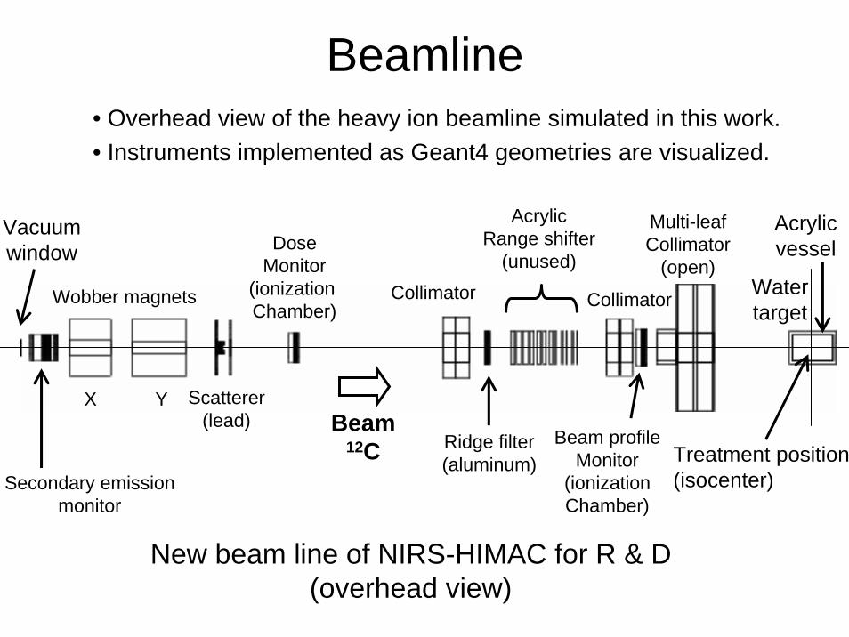
Beamline
Treatment position(isocenter)
Vacuum window
Watertarget
Acrylicvessel
New beam line of NIRS-HIMAC for R & D(overhead view)
Secondary emissionmonitor
Wobber magnets
X Y Scatterer(lead)
DoseMonitor
(ionization Chamber)
Collimator
Ridge filter(aluminum)
AcrylicRange shifter
(unused)
Multi-leafCollimator
(open)
Collimator
Beam profileMonitor
(ionizationChamber)
Beam12C
• Overhead view of the heavy ion beamline simulated in this work.• Instruments implemented as Geant4 geometries are visualized.

Water target
400 mm
300 mm
2 mm
0.1 mm
Watertarget
2 mm
Beam (12C)
Dose-measured region
• Experiment– Dose delivered to the target was measured with an ionization chamber with 2
mm-spaced channels in horizontal direction.– Depth-dose distribution was obtained by moving the dosimeter along the beam
direction. • Simulation
– Dose-measured region is divided into voxels and energy deposits in each of them were accumulated.

Physical processes in Geant4- Determine the secondaries produced in the interaction
and calculates the momenta of the particles• Ions – Electromagnetic interactions
• Ionization• Multiple scattering
– Inelastic hadronic reaction• Shen’s formula for inclusive reaction cross section• G4BinaryLightIonReaction or JQMD
– Radioactive Decay• 5He, 5Li, 8Be etc.
• Secondaries– Electron / positron
• Ionization, multiple scattering, bremsstrahlung, annihilation– Gamma ray
• Photo-electric effect, compton scattering, pair production– Proton / neutron
• Pre-compound model (-150 MeV)• Binary cascade model (-3 GeV)

Ion reaction models
• G4BinaryLightIonReaction(1) Binary Cascade model
• Cascade of binary collisions between individual nucleons composing projectile and target nuclei
(2) Pre-compound model• Formation of fragment nuclei based on level densities of possible final states
and matrix elements deduced from nucleon-nucleon scattering data
• JQMD ( developed by Niita et al., and interfaced to Geant4 by T. Koi)(1) QMD (Quantum Molecular Dynamics)
• Gaussian wave function for each nucleon• Time evolution described by classical Hamiltonian
(2) SDM (Statistical Decay Model)• Calculation of emission probabilities of nucleon and light nuclei based on
Fermi gas model• Determination of fragment species based on final positional distribution of the
nucleons
- Treat the process by two stages: (1) initial dynamical phase and (2) later statistical phase

MultileafCollimatorRidge Filter
Scatterer
Collimator
WobblerMagnets
Collimator
Event display
Dose monitor

Depth-dose distribution(12C 290 MeV/n)
Depth in water (mm) Depth in water (mm)
Rel
ativ
e do
se
Rel
ativ
e do
se
Simulated dose is normalized to agree with the experimental data of pristine Bragg peak at the surface of the water target, and the same normalization factor is applied to SOBP.
w/ Ridge filterwo/ Ridge filterPristine Bragg peak Spread-out Bragg peak
Normalization factordetermined here

Depth-dose distribution(12C 400 MeV/n)
w/ Ridge Filterwo/ Ridge FilterPristine Bragg peak Spread-out Bragg peak
Depth in water (mm) Depth in water (mm)
Rel
ativ
e do
se
Rel
ativ
e do
se
Normalization factordetermined here

Simulation of penumbra measurement (1)Beam
Background
Half-closedMLC
Penumbra widening is well reproduced
by simulation.

Simulation of penumbra measurement (2)Beam Beam
MLC MLC

Summary
• In simulation of an irradiation experiment of 290 MeV/n and 400 MeV/n carbon beam on water target, both of two inelastic ion reaction models, G4BinaryLightIonReaction and JQMD, reproduce pristine (mono-energetic) Bragg peak pretty well.
• As for reproduction of spread-out Bragg peak, there is still room for improvement especially for higher beam energy.
• Lateral beam profile is well reproduced.


Typical processing time in the water target simulation
• Computing environment : Compiled by gcc 3.3.3 and executed on
Linux 2.6.5 on AMD Opteron 2.4 GHz• G4BinaryLightIonReaction:
: 5.8 min / k events• JQMD
: 7.0 min / k events
~ 100 hours / CPU for 10% statistics & 2 x 2 mm resolution

Heavy ion therapy at HIMAC
• NIRS – National Institute of Radiological Science, Chiba, Japan
• HIMAC – First heavy ion accelerator facility dedicated to heavy ion therapy in the world
• Over 2,000 cases have been treated on trial basis.
• Broad beam method using wobbler-scatterersystem was developed.

General introduction of Geant4
• C++ library for the simulation of the passage of particle through matter
• Designed with object-oriented software technology
• Abundant physics models covering wide energy range down to a few eV
• Powerful capability to describe complex geometry

Broad beam method Patient body
Wobbler magnets
YXRidgeFilter
Scatterer
RangeShifter
Collimator
Compensator(Bolus)
Target volume(tumor)
Bragg peak
Beam
RidgeFilter
Spread-outBragg peak
Depth
dose
By = Ay sin(ωt)
Bx = Ax sin(ωt+π/2)

Physical (dis)advantage of heavy ion beam
• Dose-localizing capability (Bragg peak)• High biological effect (cell-killing capability)• Beam fragmentation significantly modifies dose
distribution from that expected from ionization of beam particle
Site of cancer © NIRSDepth of penetration
Rel
ativ
e do
se (%
)
proton
Heavy ion
X-ray
γ-ray neutron
tail
Bragg peak

Contribution of different Z nuclei to dose
G4BinaryLightIonReaction JQMD
Study on Nuclear Fragmentation by High Speed Emulsion Read-Out System
Toshiyuki Toshito(JST-CREST, KEK)
On behalf of the NIRS-HIMAC P152 collaboration
Nov.1 2006NSS2006 San Diego, California

Purpose:To collect data of heavy ion interactions with H, C, N,O, Ca, P, etc in the energy region of ion therapy with the emulsion chamber technology.Also to collect data of heavy ion interactions which are important in the radiation shielding of space laboratories.
Organization:12 institutes from HEP, medical and space domains
Nagoya Univ. Toho Univ.Aichi Univ. of EducaitonKobe Univ.High Energy Accelerator Research Organization (KEK)Ritsumeikan Univ.Naruto Univ. of EducationSLACNational Institute of Radiological Science (NIRS) Gunma Univ., Faculty of MedicineJapan Aerospace Exploration Agency (JAXA) Univ. of Tokyo
Experiment started in 2003
Medical
Space
Emulsion
NIRS-HIMAC (Chiba in Japan) P152 experiment
Geant4

Heavy ion therapy Mostly carbon is used.
Carbon has a tail dose after the Bragg peak.
Ref. http://www.nirs.go.jp/tiryo/himac/himac2.htm
protoncarbon
X-ray
γ-rayneutron
Rel
ativ
e D
ose
(%)
50
100
5.0 10.0 15.00.0Depth - Human Body (cm) 15
Validation of physics models is necessary.Reaction data of fundamental process are required.
The tail dose can be calculated by using semi-empirical modelsor Monte-Carlo simulation, for example Geant4.


C-Water data taking on Dec.2004


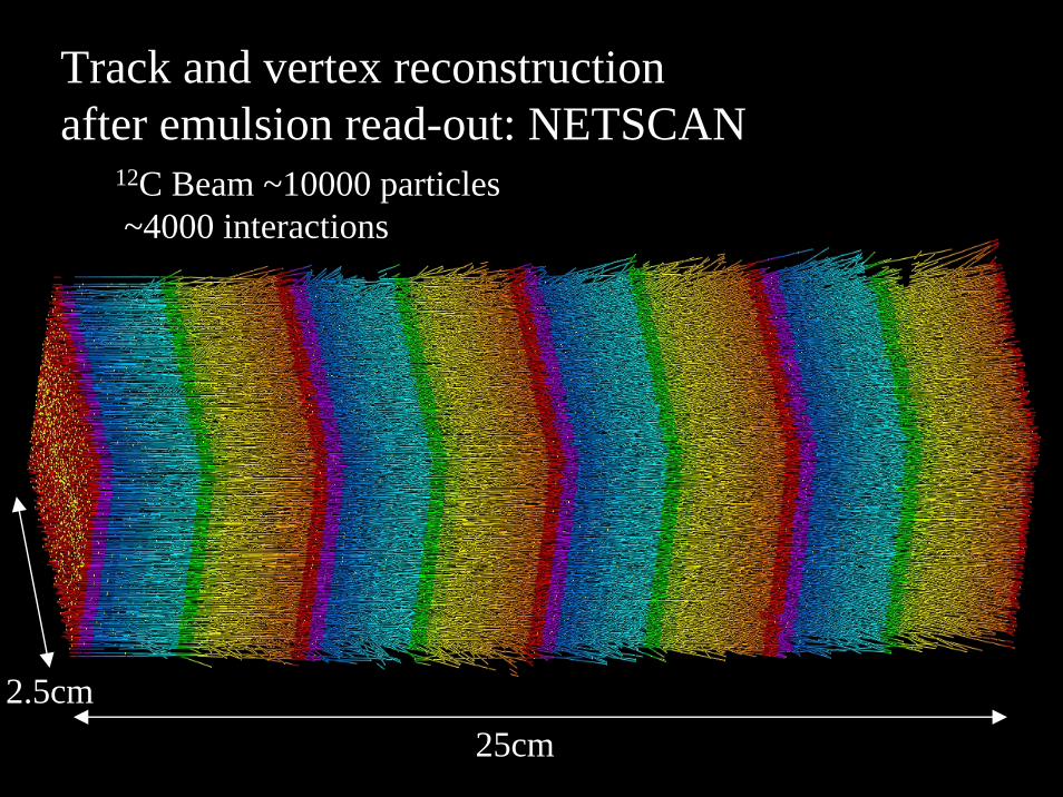
25cm2.5cm
12C Beam ~10000 particles~4000 interactions
Track and vertex reconstructionafter emulsion read-out: NETSCAN

Pulse heights
Film A: sensitive for M.I.P.
Film
B: d
esen
sitiz
ed
Film D: desensitized
Z=1 2≥ZZ=6
Z=5Z=4
Z=3
Z=2

Detection of charge-changing
carbon beryllium
Puls
e he
ight
s in
dese
nsiti
zed
film
Module number
example
Film BFilm D

A.N.Golovchenko et al.,PRC 66 014609(2002) I.Schall et al.,
NIM B 117 221(1996)
Our results Sihver’s modelPRC47,1225(1993)
C-Water(H2O) total charge-changing reaction cross sections

Study on Nuclear Fragmentation by High Speed Emulsion Read-Out System
Toshiyuki Toshito(JST-CREST, KEK)
On behalf of the NIRS-HIMAC P152 collaboration
Nov.1 2006NSS2006 San Diego, California

Purpose:To collect data of heavy ion interactions with H, C, N,O, Ca, P, etc in the energy region of ion therapy with the emulsion chamber technology.Also to collect data of heavy ion interactions which are important in the radiation shielding of space laboratories.
Organization:12 institutes from HEP, medical and space domains
Nagoya Univ. Toho Univ.Aichi Univ. of EducaitonKobe Univ.High Energy Accelerator Research Organization (KEK)Ritsumeikan Univ.Naruto Univ. of EducationSLACNational Institute of Radiological Science (NIRS) Gunma Univ., Faculty of MedicineJapan Aerospace Exploration Agency (JAXA) Univ. of Tokyo
Experiment started in 2003
Medical
Space
Emulsion
NIRS-HIMAC (Chiba in Japan) P152 experiment
Geant4

Heavy ion therapy Mostly carbon is used.
Carbon has a tail dose after the Bragg peak.
Ref. http://www.nirs.go.jp/tiryo/himac/himac2.htm
protoncarbon
X-ray
γ-rayneutron
Rel
ativ
e D
ose
(%)
50
100
5.0 10.0 15.00.0Depth - Human Body (cm) 15
Validation of physics models is necessary.Reaction data of fundamental process are required.
The tail dose can be calculated by using semi-empirical modelsor Monte-Carlo simulation, for example Geant4.


C-Water data taking on Dec.2004


25cm2.5cm
12C Beam ~10000 particles~4000 interactions
Track and vertex reconstructionafter emulsion read-out: NETSCAN

Pulse heights
Film A: sensitive for M.I.P.
Film
B: d
esen
sitiz
ed
Film D: desensitized
Z=1 2≥ZZ=6
Z=5Z=4
Z=3
Z=2

Detection of charge-changing
carbon beryllium
Puls
e he
ight
s in
dese
nsiti
zed
film
Module number
example
Film BFilm D


A.N.Golovchenko et al.,PRC 66 014609(2002) I.Schall et al.,
NIM B 117 221(1996)
Our results Sihver’s modelPRC47,1225(1993)
C-Water(H2O) total charge-changing reaction cross sections

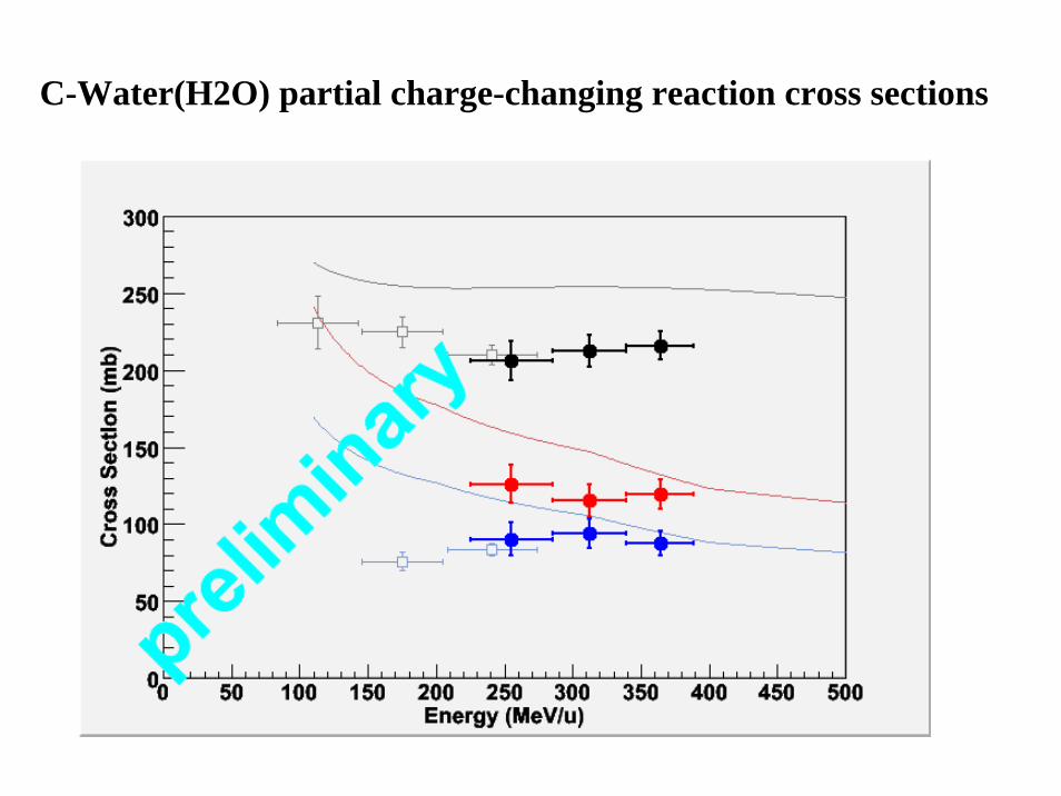
C-Water(H2O) partial charge-changing reaction cross sections
Our results
A.N.Golovchenko et al.,PRC 66 014609(2002)


H
Li
He
C 400MeV/u220MeV/u
25cm
C Li+He+Hexample

HeHe
HeC
150MeV/u400MeV/u
25cm
C 3He
example
SummaryPrecise data of fragment reactions are required in the field of
heavy ion therapy and radiology.
New emulsion technology developed for large scale neutrino experiment is applied to the investigation of fragment reactions.
Charge-changing cross sections of Carbon in Water were obtained in the energy region 200-400MeV/u.
Data taking in more wide-ranging energy using various target materials and validation of physics models (for example Geant4) with event-by-event basis will be performed in near future.


Charge identification(3≦Z ≦ 6)by refreshing method
35 grains/100μm ~8 grains/100μm
30℃,R.H.98%,3days
δray~5tracks/44μm
Z=1
Z=2
Z=6
Refreshing is used as a method to reduce sensitivity
38℃40℃

Two dedicated exposure to study Low energy interaction
Sep.05 230MeV/u 12C
Dec.05 100MeV/u 12C
For lower energy region <200MeV/u : more important
Thin targetFor charge identification, emulsion films with much more reducedsensitivity is required because of higher dE/dx in low energy.
More refresh: longer time, higher temperature
New emulsion processing so called gold development is under study!

290MeV/u 9B 290MeV/u 9B
Normal development Gold development Low sensitivity by ~1/30& Fine grain

Development
• Normal development(Chemical development)Ag+ is provided from AgBr crystal.
• Gold development(Physical development)Au is provided from developer
RedOx
e-Latent image
Ag+
AgBr
Silver filament
Latent image
Au
AgBr
hemisphere
Prof. K.Kuge (Chiba Univ.)
gold thiocyanate complex solution

Bending in magnetic field
180MeV/u 12C
18.3cm
3.0cmx
zThis technique will be usefulfor mass identification.p,d,t 3He,4He …

More to study in P152
More statistics to study interaction in more detailPure Ca,P targetTarget fragment

Treatment for cancer by heavy ion

Treatment for cancer

Heavy ion therapy FacilityHIMAC at NIRS (National Institute of Radiological Sciences)
Chiba/Japan– Operation since 1994– About 1,800 patients treated– Treatment beam: 12C
Experiment Areas
Linac800 KeV/u
Linac6 MeV/u
Synchrotron800 MeV/u
Treatment Rooms
Ion Source
~65 m

In Japan
The Energy Research Center Wakasa Bay (Tsuruga: 200 MeV)
Hyogo Ion Beam Medical Center
(Nishi-Harima: 320 MeV/u)
Shizuoka Cancer Center(Mishima: 230 MeV)
NIRS(Chiba: 90 MeV,
400MeV/u)
NCC East Hospital (Kashiwa: 235 MeV)
U. of Tsukuba PMRC (Tsukuba: 250 MeV)
Ion beamProton beam
#Proton beam facilities: 5 #Ion beam facilities: 2

Hadrontherapy in the worldA radiation therapy technique for tumor
treatment using hadron beams.Facilities in operation in the world
Proton beam: 23Ion beam: 3
Patients treated: > 46,000
Proton beamIon beam
GSI

Target Be(2mm)
Degrader(Aluminum)TOF(start)
ΔE(silicon)TOF(stop)
Bending magnet
Secondary beam line at HIMAC to produce He,Li,Be,B beamhaving almost same velocity.
ECC

3He
7Li
11B
9Be
12C
ReferenceNot refreshed
30℃38℃45℃ Volume pulse height
Secondary beam from Z=2 to 6 produced by 290MeV/u 12C
Apr.May.2004
Be
LiB
Available for charge identification up to Z=6
β~0.65
average
Accepted for publication in NIM A

Charge identification by normal OPERA film
・・・・・・
Z=1 Z=2
Z=6secondary beam
16layer
pixel0.3μm× 0.3μm
Ekine=430~100MeV/u
44μm
Detected track
Volume pulse height
Z≧3
Volume pulse height∝dE/dx(β) near MIPs Saturated for multiple charge

C-Water data taking in P152 Jan.04

R&D for Charge identificationin P152
Apr.04 and May.04

・Water(H2O) and Lucite(C5H8O2) hybrid target・Emulsion film with reduced sensitivity for charge identificationto avoid saturation of pulse height
・Reduced sensitivity by 1/5 and 1/10 are combined
Emulsion with normal sensitivity
Reduced sensitivity by 1/5
Water Water Water
2mmReduced sensitivity by 1/10
Lucite(1mm)
Dec.04 exp. 400MeV/u 12C
1/10
sens
itivi
ty
1/5 sensitivity
BC
Be
LiHe
Pulse height
65layers
Emulsion chamber to study C-Water and C-Lucite interactions

Velocity distribution of Hydrogen fragments
Volume pulse height⇔dE/dx(LET)⇔velocity
Ebeam:430~395MeV/u Ebeam:395~360MeV/u
Ebeam:325~285MeV/uEbeam:360~325MeV/u
Ebeam:285~245MeV/u Ebeam:245~195MeV/u
β
Calibrated by proton beam@ KEK PS
protonπ+
Volume pulse height

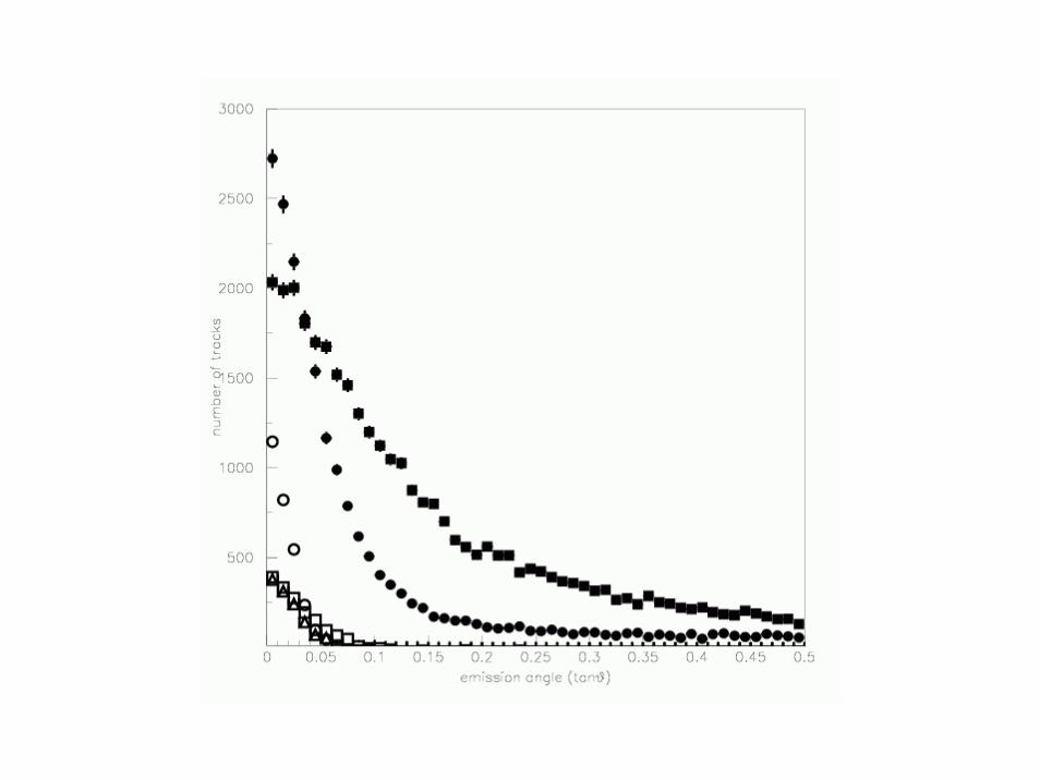
B
Be LiLi
He
H
Emission angle with respect to the beam(tanθ)
Emission angle of secondly particles classified by charge


Apr.03, Jun.03, Sep.03, Apr.04, May.04
R&D for emulsion readout, chamber design and charge identification
Jan.04 and Dec.04
physics result of C-Water and C-Lucite interaction
in >150MeV/u
Cross section : consistent with other experiments
Comparison between our data and theoretical model
Validity test of QMD build Geant4 is set going.
Summary and Current status of P152

C-Water(H2O) partial charge-changing reaction cross sections

C-Water(H2O) partial charge-changing reaction cross sections
Our results
A.N.Golovchenko et al.,PRC 66 014609(2002)


H
Li
He
C 400MeV/u220MeV/u
25cm
C Li+He+Hexample

HeHe
HeC
150MeV/u400MeV/u
25cm
C 3He
example

SummaryPrecise data of fragment reactions are required in the field of
heavy ion therapy and radiology.
New emulsion technology developed for large scale neutrino experiment is applied to the investigation of fragment reactions.
Charge-changing cross sections of Carbon in Water were obtained in the energy region 200-400MeV/u.
Data taking in more wide-ranging energy using various target materials and validation of physics models (for example Geant4) with event-by-event basis will be performed in near future.


Charge identification(3≦Z ≦ 6)by refreshing method
35 grains/100μm ~8 grains/100μm
30℃,R.H.98%,3days
δray~5tracks/44μm
Z=1
Z=2
Z=6
Refreshing is used as a method to reduce sensitivity
38℃40℃

Two dedicated exposure to study Low energy interaction
Sep.05 230MeV/u 12C
Dec.05 100MeV/u 12C
For lower energy region <200MeV/u : more important
Thin targetFor charge identification, emulsion films with much more reducedsensitivity is required because of higher dE/dx in low energy.
More refresh: longer time, higher temperature
New emulsion processing so called gold development is under study!

290MeV/u 9B 290MeV/u 9B
Normal development Gold development Low sensitivity by ~1/30& Fine grain

Development
• Normal development(Chemical development)Ag+ is provided from AgBr crystal.
• Gold development(Physical development)Au is provided from developer
RedOx
e-Latent image
Ag+
AgBr
Silver filament
Latent image
Au
AgBr
hemisphere
Prof. K.Kuge (Chiba Univ.)
gold thiocyanate complex solution

Bending in magnetic field
180MeV/u 12C
18.3cm
3.0cmx
zThis technique will be usefulfor mass identification.p,d,t 3He,4He …
More to study in P152
More statistics to study interaction in more detailPure Ca,P targetTarget fragment

Treatment for cancer by heavy ion

Treatment for cancer

Heavy ion therapy FacilityHIMAC at NIRS (National Institute of Radiological Sciences)
Chiba/Japan– Operation since 1994– About 1,800 patients treated– Treatment beam: 12C
Experiment Areas
Linac800 KeV/u
Linac6 MeV/u
Synchrotron800 MeV/u
Treatment Rooms
Ion Source
~65 m

In Japan
The Energy Research Center Wakasa Bay (Tsuruga: 200 MeV)
Hyogo Ion Beam Medical Center
(Nishi-Harima: 320 MeV/u)
Shizuoka Cancer Center(Mishima: 230 MeV)
NIRS(Chiba: 90 MeV,
400MeV/u)
NCC East Hospital (Kashiwa: 235 MeV)
U. of Tsukuba PMRC (Tsukuba: 250 MeV)
Ion beamProton beam
#Proton beam facilities: 5 #Ion beam facilities: 2

Hadrontherapy in the worldA radiation therapy technique for tumor
treatment using hadron beams.Facilities in operation in the world
Proton beam: 23Ion beam: 3
Patients treated: > 46,000
Proton beamIon beam
GSI

Target Be(2mm)
Degrader(Aluminum)TOF(start)
ΔE(silicon)TOF(stop)
Bending magnet
Secondary beam line at HIMAC to produce He,Li,Be,B beamhaving almost same velocity.
ECC

3He
7Li
11B
9Be
12C
ReferenceNot refreshed
30℃38℃45℃ Volume pulse height
Secondary beam from Z=2 to 6 produced by 290MeV/u 12C
Apr.May.2004
Be
LiB
Available for charge identification up to Z=6
β~0.65
average
Accepted for publication in NIM A

Charge identification by normal OPERA film
・・・・・・
Z=1 Z=2
Z=6secondary beam
16layer
pixel0.3μm× 0.3μm
Ekine=430~100MeV/u
44μm
Detected track
Volume pulse height
Z≧3
Volume pulse height∝dE/dx(β) near MIPs Saturated for multiple charge

C-Water data taking in P152 Jan.04

R&D for Charge identificationin P152
Apr.04 and May.04

・Water(H2O) and Lucite(C5H8O2) hybrid target・Emulsion film with reduced sensitivity for charge identificationto avoid saturation of pulse height
・Reduced sensitivity by 1/5 and 1/10 are combined
Emulsion with normal sensitivity
Reduced sensitivity by 1/5
Water Water Water
2mmReduced sensitivity by 1/10
Lucite(1mm)
Dec.04 exp. 400MeV/u 12C
1/10
sens
itivi
ty
1/5 sensitivity
BC
Be
LiHe
Pulse height
65layers
Emulsion chamber to study C-Water and C-Lucite interactions

Velocity distribution of Hydrogen fragments
Volume pulse height⇔dE/dx(LET)⇔velocity
Ebeam:430~395MeV/u Ebeam:395~360MeV/u
Ebeam:325~285MeV/uEbeam:360~325MeV/u
Ebeam:285~245MeV/u Ebeam:245~195MeV/u
β
Calibrated by proton beam@ KEK PS
protonπ+
Volume pulse height

B
Be LiLi
He
H
Emission angle with respect to the beam(tanθ)
Emission angle of secondly particles classified by charge


Apr.03, Jun.03, Sep.03, Apr.04, May.04
R&D for emulsion readout, chamber design and charge identification
Jan.04 and Dec.04
physics result of C-Water and C-Lucite interaction
in >150MeV/u
Cross section : consistent with other experiments
Comparison between our data and theoretical model
Validity test of QMD build Geant4 is set going.
Summary and Current status of P152

C-Water(H2O) partial charge-changing reaction cross sections

gMocren
• Voxel graphics+dose viewer for Geant4– DICOM is only supported currently– Any other voxel data can be adopted easily
• Free to download– http://geant4.kek.jp/gMocren– Binary only distributed
• Hardware rendering– Under development