vap, vat & co - hôpitaux universitaires henri...
TRANSCRIPT

Christian Brun-Buisson, Medical ICU, Creteil - FR
VAP, VAT & CO.
ENCORE ET TOUJOURS LE DIAGNOSTIC DES LRTI
?
JAVA 2017

Agenda
La problématique
Le diagnostic d’infection respiratoire basse et la
place de la VAT et autres VAC
L’intégration clinico-microbiologique pour une
prise en charge adaptée
C. Brun-Buisson JAVA 2017

The European Sepsis Study:
Sources of infection
% s
ite
s
C. Alberti et al, ICM 2002
0
10
20
30
40
50
60
70
LRTI IAS UTI PBSI SST CNS Others Unknown
CA
HA
ICU-Acq
LRTI is, by far, the first cause of sepsis and of antibiotic prescription in the ICU

Quelles leçons de l’étude ALARM ? Routine management of 1st episode of suspected VAP, 20
ICUs, 398 pts
20 ICUs; 398 ICU patients meeting predefined criteria for suspected VAP
Baseline CPIS: 8.4 ± 2.3 (1-14)
APACHE II score: 22.8 ± 8.3 (2-52)
Duration of MV prior to VAP diagnosis: 7.3 (0-44) days
162 (40.7%) pts receiving AbRx prior to or during VAP treatment for non-VAP indications, including:
quinolone (14.6%), ureidopenicillin/monobactam (11.1%), cefepime (9.3%) or carbapenem (5.8%)
Diagnosis:
ACCP criteria (V. Baselski & al, Chest 1992)
Cultures: TA (58.3%), BAL (33.7%), or both (1.8%);
6.3% of patients had neither TA or BAL performed
Major pathogens identified in 197 patients (49.5%)
MRSA 15%, PA 14%, Enterobacteriaceae 10%
No PPMO in 37%; no growth in 6%
M. Kollef & al, CHEST 2006; 129:1210–18.
C. Brun-Buisson JAVA 2017

Quelles leçons de l’étude ALARM ? Routine management of 1st episode of suspected VAP, 20
ICUs, 398 pts
Initial coverage of GNB
(larger spectrum drug, % pts)
*
* incl. van
Range 0-25 0-73 0-70 2-38 0-65 %
Mean duration of therapy 11.8 ± 5.9 (0-51) days
Change in regimen - No pathogen identified 13%
- Pathogen identified 57%
Definitive therapy - Unchanged /same spectrum 289 (72.6%)
- Escalation 15.3% pts,
- Deescalation 22.1% pts
Deescalation - major pathogen isolated 15.6%
- No major pathogen identified 6.5%
- No pathogen identified 13%
M. Kollef & al, Chest 2006; 129:1210–
18
No. Drugs prescribed . 1 : 28% . 2 : 48% . 3+ : 23% w. vancomycin: 52% pts
C. Brun-Buisson JAVA 2017
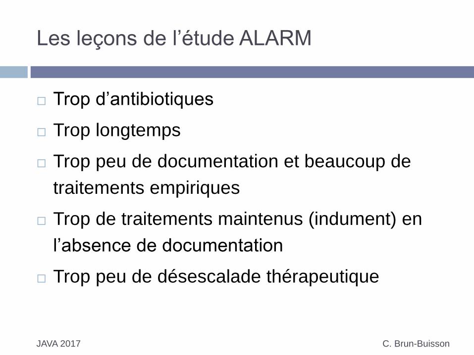
Les leçons de l’étude ALARM
Trop d’antibiotiques
Trop longtemps
Trop peu de documentation et beaucoup de
traitements empiriques
Trop de traitements maintenus (indument) en
l’absence de documentation
Trop peu de désescalade thérapeutique
C. Brun-Buisson JAVA 2017

C. Brun-Buisson JAVA 2017
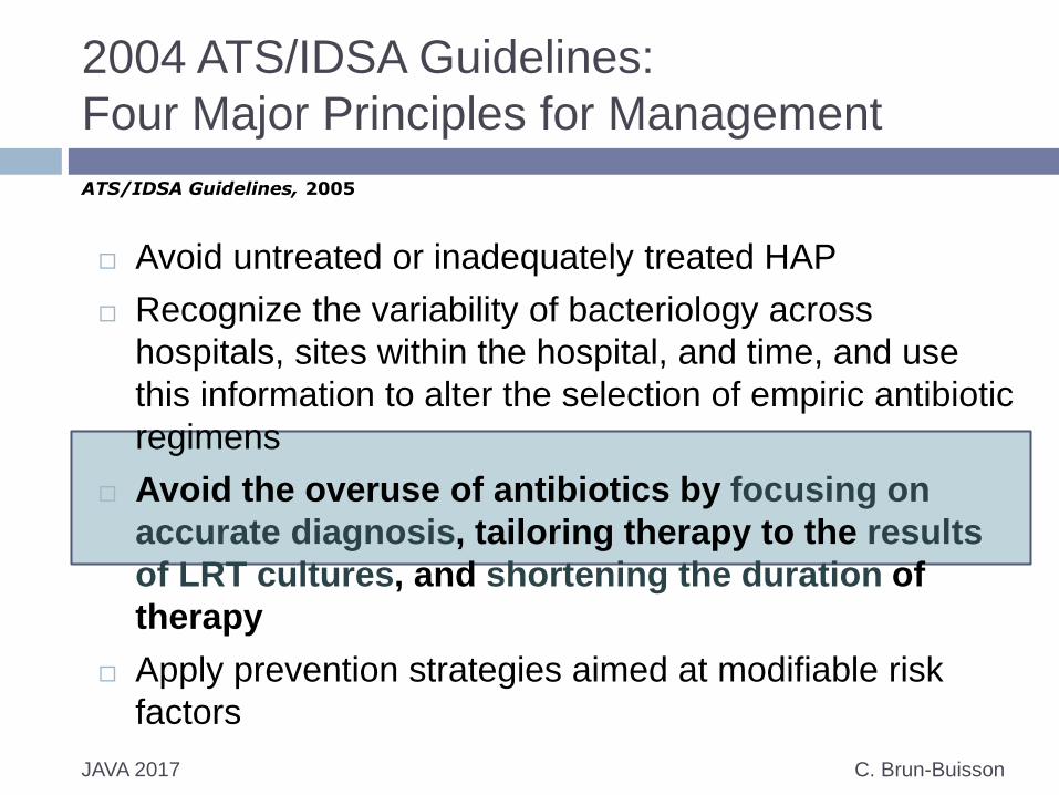
Avoid untreated or inadequately treated HAP
Recognize the variability of bacteriology across
hospitals, sites within the hospital, and time, and use
this information to alter the selection of empiric antibiotic
regimens
Avoid the overuse of antibiotics by focusing on
accurate diagnosis, tailoring therapy to the results
of LRT cultures, and shortening the duration of
therapy
Apply prevention strategies aimed at modifiable risk
factors
2004 ATS/IDSA Guidelines:
Four Major Principles for Management
ATS/IDSA Guidelines, 2005
C. Brun-Buisson JAVA 2017

LRT samples culture: Principles of
Interpretation for Diagnosing VAP
The incidence of colonization in hospitalized patients
and even more in patients requiring endotracheal
intubation, is high
A positive EA culture cannot differentiate colonization
from infection
Antibiotic therapy of simple colonization is strongly
discouraged
A sterile culture of the lower respiratory tract (in the
absence of recent change in therapy) is strong
evidence that a pneumonia is not present
ATS/IDSA Guidelines, 2005
C. Brun-Buisson JAVA 2017

Clinical Infectious Diseases Advance Access published July 14, 2016
C. Brun-Buisson JAVA 2017
Eur Respir J 2017; 50: 1700582

1. Know what you treat: the HAP/VAP &
co diagnostic issues
Cultures of respiratory secretions should be obtained from (virtually) all patients with suspected VAP
Noninvasive sampling with semi-quantitative cultures to diagnose VAP, rather than invasive sampling with quantitative cultures and rather than noninvasive sampling with quantitative cultures (weak recommendation, low-quality
evidence) *
We suggest obtaining distal quantitative samples (prior to any antibiotic treatment) in order to reduce antibiotic exposure in stable patients with suspected VAP and to improve the accuracy of the results (weak recommendation, low
quality of evidence) * For patients with suspected VAP whose invasive quantitative culture results are
below the diagnostic threshold for VAP, we suggest that antibiotics be withheld
rather than continued
ATS-IDSA Guidelines 2016
ERS-ESICM-ESCMID Guidelines 2016

2. Identify RF for MDRB
Specific exposures
Local epidemiology
Classification schemes
C. Brun-Buisson JAVA 2017

Identify risk factors for MDRB: The new
classification scheme for HAP/VAP
Community-onset Hospital-onset
HAP
(no MV)
VAP
(MV)
Classical
CAP
CAP
with RF
for
MDRB
HCAP VAC
RF for
MDRB RF for
MDRB
C. Brun-Buisson JAVA 2017

Risk Factors for Health Care-Associated
Infections and for Infection with Drug-
Resistant Bacteria Risk factors for health care-associated infections (ATS-IDSA GL
2004)
Hospitalization for ≥ 2 days in preceeding 90 days
Residence in a nursing home or long-term care facility
Home infusion therapy, including antimicrobial agents
Long-term dialysis within 30 days
Home wound care
Family member with multidrug-resistant pathogen
Risk factors for infection with drug-resistant bacteria
Antimicrobial therapy in preceeding 90 days
Current hospitalization for ≥ 5 days
High frequency of antibiotic resistance in the community or in the specific
hospital environment
Immunosuppression
AY. Peleg & DC. Hooper: Hospital-Acquired Infections Due to Gram-Negative Bacteria. N Engl J Med 2010; 362: 1804-13
C. Brun-Buisson JAVA 2017
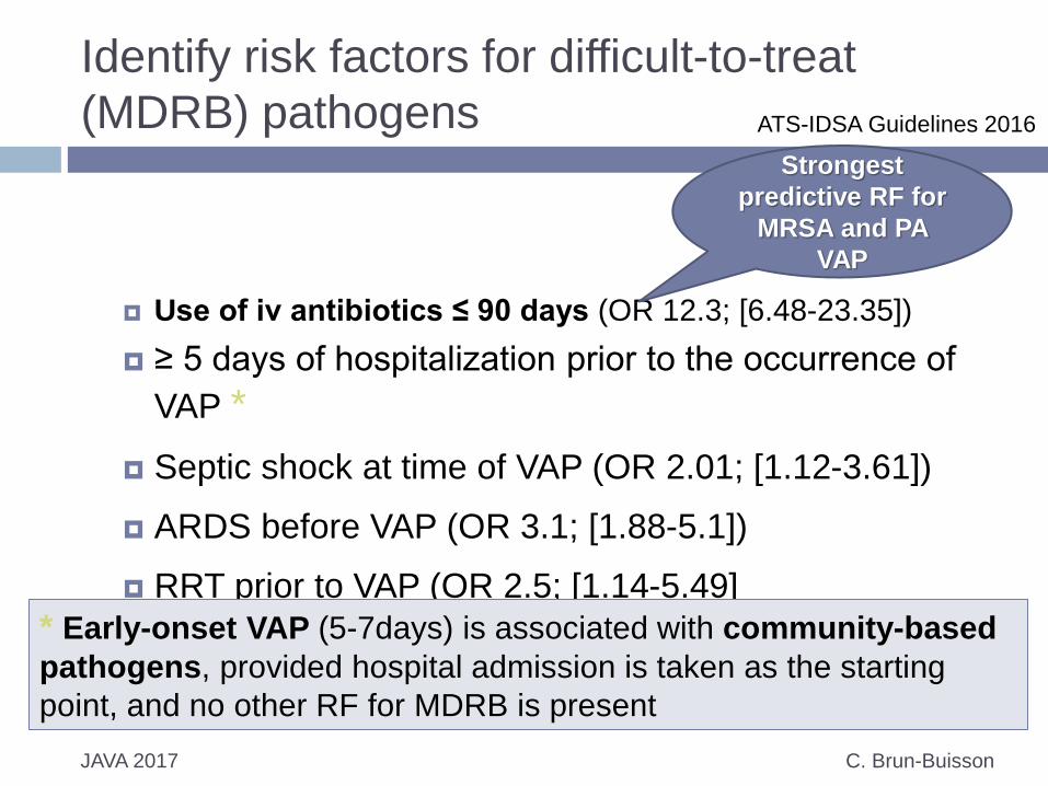
Identify risk factors for difficult-to-treat
(MDRB) pathogens
Use of iv antibiotics ≤ 90 days (OR 12.3; [6.48-23.35])
≥ 5 days of hospitalization prior to the occurrence of
VAP *
Septic shock at time of VAP (OR 2.01; [1.12-3.61])
ARDS before VAP (OR 3.1; [1.88-5.1])
RRT prior to VAP (OR 2.5; [1.14-5.49]
* Early-onset VAP (5-7days) is associated with community-based
pathogens, provided hospital admission is taken as the starting
point, and no other RF for MDRB is present
Strongest
predictive RF for
MRSA and PA
VAP
C. Brun-Buisson JAVA 2017
ATS-IDSA Guidelines 2016

VAP, VAC, iVAC, VAT & co.
C. Brun-Buisson JAVA 2017

Does this patient have VAP?
The clinical suspicion of VAP: all three of
(1) new or progressive persistent radiographic infiltrates;
(2) clinical observations suggesting infection, e.g. the new onset of fever, purulent sputum, leukocytosis, increased minute ventilation, arterial oxygenation decline and/or the need for increased vasopressor infusion to maintain blood pressure;
(3) “positive” microbiological culture results for a potentially pathogenic microorganism isolated from endotracheal aspirates (ETAs), bronchoalveolar lavage fluid, pleural fluid and/or blood
New or just
persistent ?
J.Chastre & CE Luyt. Intens Care Med 2016; 42:1159
How much
is
« positive »
?
C. Brun-Buisson JAVA 2017

Does this patient have VAP?
Criteria VAP VAT VAT (CDC) iVAC
New/progressiv
e persitent
infiltrate
(+) (-) (-) NA
Clinical
features of
infection
Two of:
New fever
Purulent
secretions
Leukocytosis
Decreased
oxygenation
Vasopressor need
Fever
Purulent
secretions
leukocytosis
Fever
Cough
Wheezing
Purulent
secretions
Leukocytosis
Worsening
oxygenation (2 d)
+
Fever or
Leukocytosis
+
New ab initiated
Microbiology Blood/pleural fluid
Positive
quantitative culture
(BAL, ETA)
Positive
quantitative
culture (BAL,
ETA)
Positive
culture (no
threshold)
Positive
quantitative
(probable) or
non-quantitative
(possible VAP)
J.Chastre & CE Luyt. Intens Care Med 2016; 42:1159
C. Brun-Buisson JAVA 2017

C. Brun-Buisson JAVA 2017
Outcomerea database (1996-2012); 3,028 pts
Epidemiologie VAE et relation avec VAP
2,331 patients (77%) ≥ 1 VAC; 869 patients (29%) had one iVAC
episode
Confirmation VAP quantitative cultures
Correlation avec VAP
VAC : 0.67 (p < 0.0001)
IVAC : 0.82 (p < 0.0001),
Se/Sp for VAP
VAC 0.92 / 0.28
iVAC : 0.67 / 0.75
Crit Care Med. 2015; 43: 1798-806.

Definitions
C. Brun-Buisson JAVA 2017
L.Bouadma & al, Crit Care Med, 2015

Outcomes
C. Brun-Buisson JAVA 2017
No VAC (n=697) VAC (n=2331) iVAC (n=869)
VAP - 339 (14.5) 240 (27.6)
Tracheobronchit
is
- 23 (1) 12 (1.4)
Nosocomial inf - 637 (27.3) 381 (43.8)
SAPS II 47 [35 – 59] 44 [34 – 55] 44 [33 – 55]
Days alive wo. ab 24 [2 – 26] 17 [4 – 23] 10 [4 – 23]
ICU LOS 9 [7 – 13] 18 [11 – 29] 22 (14 – 34]
30d ICU mortality 217 (31.1) 514 (22.1) 222 (25.6)
Hosp mortality 278 (39.9) 856 (36.7) 386 (44.4)
Multiple causes or the lack of identified cause were frequent.
IVAC episodes were strongly correlated to VAP; only 27.6% IVAC episodes were
related to VAP and less than half to a nosocomial infection.
VAC and IVAC associated with poor outcome and correlated with an increase in ab
use.
VAEs are common and associated with high morbidity, and the
VAE rate seems to be a good indicator for quality-
improvement purpose.
L.Bouadma & al, Crit Care Med, 2015
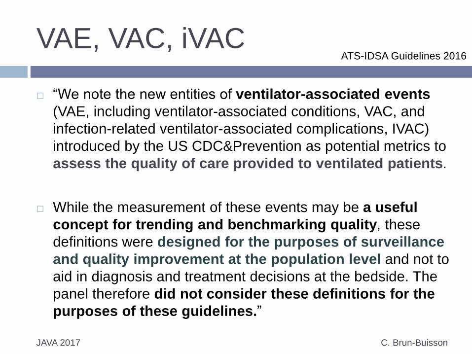
VAE, VAC, iVAC
C. Brun-Buisson JAVA 2017
“We note the new entities of ventilator-associated events
(VAE, including ventilator-associated conditions, VAC, and
infection-related ventilator-associated complications, IVAC)
introduced by the US CDC&Prevention as potential metrics to
assess the quality of care provided to ventilated patients.
While the measurement of these events may be a useful
concept for trending and benchmarking quality, these
definitions were designed for the purposes of surveillance
and quality improvement at the population level and not to
aid in diagnosis and treatment decisions at the bedside. The
panel therefore did not consider these definitions for the
purposes of these guidelines.”
ATS-IDSA Guidelines 2016

Treatment of Ventilator-associated
Tracheobronchitis (VAT) ?
C. Brun-Buisson JAVA 2017
VAT : fever with no other recognizable cause,
with new or increased sputum production,
positive ETA culture (>106 CFU/mL) yielding a
new bacteria, and no radiographic evidence of
nosocomial pneumonia (Nseir & al , Crit Care 2005)
In patients with VAT, we suggest not providing
antibiotic therapy (weak recommendation, low-quality
evidence).
ATS-IDSA Guidelines 2016

C. Brun-Buisson JAVA 2017
12 ICUs; June 2005-June 2007
Excluded : severe ID, tracheostomy on admission; prior VAP;
SAPS II >65
Weekly Q-EA and on inclusion
VAT : Fever >38°C, purulent secretions, Q-EA ≥106 , and ‘’no
radiographic signs of new pneumonia”
Randomisation avec ou sans traitement (ouvert)
VAP: new or progressive radiographic infiltrate + 2 of : fever
>38.5°C, leukocytosis (>10 000), purulent secretions and +ve
Q-EA
Analysis ITT and mITT (excl. pts : DNR, subsequent active ab
Rx)
Critical Care 2008, 12:R62

Antibiotics for VAT: a RCT
C. Brun-Buisson JAVA 2017
Eligible
N=65
Randomised
N=58
Antibiotic Rx
N=22
No atb
N=36
mITT
N=18
Subsequent active atb,
n=6
DNR order, n=4
DNR order, n=4
mITT
N=26
Nseir & al, Crit Care 2008
Étude arrétée ‘prématurément’

Antibiotics for VAT: a RCT
C. Brun-Buisson JAVA 2017
ATB (+), N=22 ATB (–) , N=36
Age 62 67
SAPS II 47 47
Medical 19 (86) 30 (83)
COPD 9 (40) 17 (47)
CAP 6 (27) 10 (27)
Cardiac failure 3 (13) 1 (2)
AE-COPD 3 (13) 14 (38)
Neurologic failure 2 (9) 5 (13)
Duration MV, d 17 13
Infection admission 18 (81) 25 (69)
Nseir & al, Crit Care 2008

Events & Outcome
C. Brun-Buisson JAVA 2017
ATB + ATB -
Tracheostomy 5 (22) 5 (13)
Septic shock 1 (4) 7 (19)
ICU-aquired inf
Non-VAP 7 (31) 18 (50)
VAP within 8 d 0 15 (41)
ABT Rx 22 (100) 21 (58)
Duration MV 29 ± 17 26 ± 15
VAP (28d) 3 (13) 17 (47)
ICU death 4 (18) 17 (47)
Nseir & al, Crit Care 2008

114 ICUs, 1 yr
2960 eligible patients; 689 (23%) with VA-LRTI
320 pts VAT*, all treated
39 progress to VAP 19/250 ttmt approprié vs
20/70 ttmt inapproprié
Deaths
VAP 146/369 (40%)
VAT 93/320 (29%)
None 673/2271 (30%)
C. Brun-Buisson JAVA 2017
Lancet Respir Med 2015; 3: 859–68
*fever, or leukocytosis and purulent
secretions
• Positive Q culture LRT secretions (ETA,
BAL, PSB)

Etudes fibroscopiques chez les BPCO
C. Brun-Buisson JAVA 2017
Auteur Patients Prelèvemen
ts
%
positifs
Fagon 50 pts
VM
PSB 50%
Monso 29 pts
EABC
PSB
52%
Soler 50 pts
VM
PSB
LBA
EA
46%
Pela 40 pts
ambulatoire
s
PSB 52%
Zalachai
n
88 pts
stables
PSB 41%
MO
• H.
influenzae/parainflu.
• S. pneumoniae
• M. catarrhalis
• P. aeruginosa
• Strep viridans
• (autres BGN , CGP)

1. Know what you treat: the HAP/VAP,
VAT, iVAC diagnostic issues
In patients with VAT (ie, fever, purulent sputum,
positive culture, no new infiltrate), we suggest
NOT providing antibiotic therapy *
ATS-IDSA Guidelines 2016
* In the presence of new respiratory signs of infection, such as an
increased amount of purulent sputum and a high-quality sample with
positive Gram stain, in conjunction with new systemic signs of
infection plus worsening oxygenation and/or increasing ventilator
settings, [and absence of other infectious focus], antibiotic treatment
may be considered even in the absence of new or progressive
persistent infiltrates C. Brun-Buisson JAVA 2017
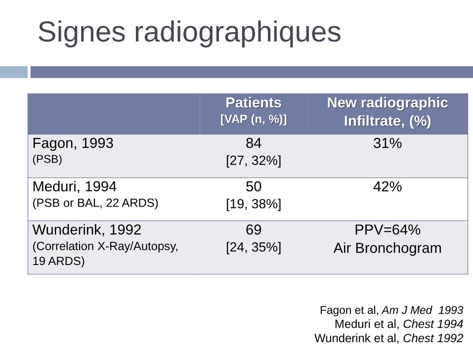
Signes radiographiques
Fagon et al, Am J Med 1993
Meduri et al, Chest 1994
Wunderink et al, Chest 1992
Patients [VAP (n, %)]
New radiographic
Infiltrate, (%)
Fagon, 1993 (PSB)
84
[27, 32%]
31%
Meduri, 1994 (PSB or BAL, 22 ARDS)
50
[19, 38%]
42%
Wunderink, 1992 (Correlation X-Ray/Autopsy,
19 ARDS)
69
[24, 35%]
PPV=64%
Air Bronchogram

Infection et SDRA: Diagnostic clinique et
données autopsiques Andrews et al, Chest 1981
Histology VAP +
(n=14)
VAP -
(n=10)
All pts
(n=24)
Fever 14 (100) 8 (80) 22 (92)
Leukocytosis 14 (100) 8 (80) 22 (92)
Asymmetry on CXR 8 (57) 3 (30) 11 (46)
Pathogens
(Tracheal Aspirate)
12 (86) 7 (70) 19 (79)
Clinical Dg accurate 9 (64%) 8 (80) 17 (71%)
Clinical Dg inaccurate 5 (36%) 2 (20%) 7 (29%)
C. Brun-Buisson JAVA 2017

Early CT-Scan for Community-Acquired Pneumonia at the Emergency Department (ESCAPED)
Zoom ---
Imagerie TDM thorax
Claessens YE. Am J Respir Crit Care Med 2015; 192:974-82
→ Discordance diagnostique entre Probabilité pré-test et
Probabilité post-test diagnostic = 58,6%
Comité d’adjudication (J28) diagnostic = 31,3%
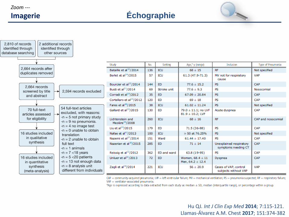
Hu QJ. Int J Clin Exp Med 2014; 7:115-121. Llamas-Álvarez A.M. Chest 2017; 151:374-382
Zoom ---
Imagerie Échographie

3. Initiate antibiotic therapy promptly
Initiation of therapy is based on clinical
suspicion (and severity of presentation)
(Serum PCT level is not an option to help decide
whether or not to start therapy)
But:
Therapy may be witheld in patients in whom
invasive quantitative sampling shows growth
below the threshold
C. Brun-Buisson JAVA 2017

Choosing the right moment to start antibiotic
treatment
C. Brun-Buisson JAVA 2017
71 patients with VAP; 43 (61 %) with”gradual” VAP
Nonquantitative EA and serum inflammatory biomarkers every 48–72 h.
Clinical VAP defined as 2+ of: T° > 38 °C, WBC >12,000/mm3 or
<4000/mm3, purulent respiratory secretions, and w. a new or
progressive pulmonary infiltrate on the CXR
VAP confirmed if Q-EA ≥ 105, BAL ≥104, or mini-BAL ≥103 cfu/ml
‘Gradual VAP’ : presence in the 96h pre-VAP of purulent respiratory
secretions, plus one or both of the following: temperature > 38 °C and a
WBC >12,000/mm3, and wo. new or progressive pulmonary infiltrate on
CXR
P.Ramirez & al. Critical Care (2016) 20:169
At time of VAP Acute onset (n=28) Gradual (n=43)
MV days 4.5 [3 – 9] 8 [6.5 – 11.5]
SOFA 10 [8 – 12] 7 [5 – 11]
mCPIS 7 [6 – 8] 7 [6 – 8]
No difference in outcome (28d mortality 61%)

Conclusions
C. Brun-Buisson JAVA 2017
La bonne question: quand débuter le traitement ?
Il y a des pneumonies précoces et tardives
Il y a des trachéobronchites tardives, qui peuvent occasionnellement bénéficier d’un traitement
Le diagnostic d’infiltrat « nouveau et/ou progressif » est hasardeux -> échographie ?
A remplacer par altération des échanges gazeux ?
La décision thérapeutique est clinique, et peut s’appuyer sur la microbiologie (ex. direct)
Il faut se tenir à une approche diagnostique microbiologique standardisée
Pour pouvoir poursuivre, modifier, arrêter le traitement empirique selon un protocole suivi.