varicella and isolated acute peripheral facial nerve palsy
TRANSCRIPT
473
Varicella and isolated acute peripheral facial nerve palsy: A systematic review on natural history, prognosis and treatment*1Lip Yuen Teng, *2Wai Quen Lee, 1Aina Mariana Binti Abdul Manaf*LY Teng and WQ Lee contributed equally to this work and are joint first author
1Department of Paediatrics, Hospital Port Dickson; 2Department of Paediatrics, Hospital Tuanku Ja’afar Seremban, Malaysia.
Abstract
Background & Objective: Varicella is a common infection during childhood and generally self-limiting. However, it can rarely cause neurological complications. Isolated acute peripheral facial palsy (APFP) is extremely rare during primary varicella infection with estimated incidence of <0.01%. There have also been conflicting opinions on its natural history, prognosis and management worldwide. We aimed to establish the natural history, prognosis and treatment for varicella-related isolated APFP in immunocompetent individuals, without co-morbids. Methods: Systematic review was performed with systematic literature search in Google Scholar and PubMed. Data was analysed with statistical analysis software.Results: Thirty cases were included. The complete remission rate of APFP was 66.67% for non-treatment group and 72.22% for treatment group (p=0.643). Early and late treatment group had a similar complete remission rate of 88.89% and 80% respectively (p=1.000). However, early treatment group (within 3 days of onset) had achieved complete remission 3 weeks earlier than the late treatment group (p=0.091). Antiviral group tends to have better outcome than steroid monotherapy group, although statistically insignificant (p=0.055). Conclusions: This condition generally has good prognosis even without treatment. However, early treatment and antiviral therapy may at least accelerate remission and reduce morbidities although these cannot alter the final outcome. Clinicians may consider antiviral therapy if patients present within 3 days of onset. These findings need to be applied with caution, considering the limitations of our review.
Keywords: Varicella, chickenpox, peripheral facial nerve palsy, facial paralysis
Neurology Asia 2020; 25(4) : 473 – 484
Address correspondence to: Lip Yuen Teng; Department of Paediatrics, Hospital Port Dickson, KM11, Jalan Pantai, 71050 Port Dickson, Negeri Sembilan, Malaysia. Email: [email protected]
Date of Submission: 26 April 2020; Date of Acceptance: 13 May 2020
vaccine introduction, hospitalization and mortality rates declined by >70% and 88% respectively.4
Neurological complications from varicella are estimated to be 0.01-0.03% with cerebellar ataxia (1 in 4000 cases in unvaccinated children) and encephalitis (1 in 33,000 cases) being the commonest.5 Other rarer neurological complications include transverse myelitis, optic neuritis, Reye syndrome, peripheral motor neuropathy, Guillain-Barre syndrome and facial nerve palsy.5-11 Facial nerve palsy is not an uncommon condition in the paediatric population. There have been various causes attributable to the nerve palsy, i.e. congenital, infectious, neoplastic, traumatic or idiopathic.12-14 Causative infections include herpes simplex virus (HSV), mumps, Coxsackie
INTRODUCTION
Varicella-zoster Virus (VZV) is a DNA virus under the family of Herpesviridae and also called human herpesvirus 3.1 There are two clinically distinct forms of disease caused by VZV infection, i.e. varicella (chickenpox) and herpes zoster (shingles) infections.2 Varicella is the primary infection of VZV, resulting in lifelong latent infection of sensory ganglia while reactivation of the latent infection results in herpes zoster.3 Varicella is highly contagious yet generally benign and self-limiting. During the pre-vaccine era, there were approximately 11,000 admissions for varicella (2-3 in 1000 healthy children) and 103 deaths (1 in 60,000 cases) annually in the United States.4 After

Neurology Asia December 2020
474
virus, influenza, Borrelia burgdorferi and VZV. Almost half of the acute peripheral facial palsy (APFP) cases are due to Bell’s palsy which is the appellation used to describe an APFP of unknown cause, with an incidence rate of 20-30 per 100,000 annually.12,15 Its pathogenesis remained unknown. In 2008, Hato et al. postulated that Bell’s palsy may be associated with reactivation of HSV in 31-79% of cases.15 Other studies showed its association with VZV reactivation up to 37% of cases.18-21 VZV reactivation has been demonstrated using polymerase chain reaction or serological assays in patients with zoster sine herpete and also Ramsay Hunt Syndrome (also called herpes zoster oticus).16-20 Ramsay Hunt syndrome is characterized by a triad of ipsilateral APFP, otalgia and the presence of painful vesicular eruption in the external ear whereas zoster sine herpete is characterized by APFP in the absence of typical zoster skin lesions.16-20
The Copenhagen Facial Nerve study had evaluated the natural history of 2,570 cases of peripheral nerve palsy of various aetiologies over a 25-year period, including 349 children younger than 15 years old.22 The majority of cases included were Bell’s palsy (66.2%, n=1701), herpes zoster or Ramsay Hunt Syndrome (4.5%, n=116) and trauma (3.7%, n=95).22 6.6% cases reported in neonatal period were due to either congenital paresis or birth trauma (n=169).22 70% of patients with Bell’s palsy (n=1189) had complete paralysis and 30% (n=512) incomplete paralysis on initial assessment.22 Without treatment, 85% regained function within 3 weeks while the remaining 15% of patients within 3-5 months.22 58% achieved complete remission within 2 months. 71% had final complete remission without any sequelae, 12% had mild dysfunction (House-Brackmann Grade II), 13% moderate dysfunction (Grade III), while only 4% had moderately severe or severe dysfunction (Grade IV-V) and none remained completely paralysed.22 Children with Bell’s palsy had favourable outcome with 90% achieving complete remission while herpes zoster patients had poor outcome with only 21% regaining full function.22 Although this study had collected a huge amount of data on peripheral facial nerve palsy of different aetiologies, as most of the studies, Peitersen et al. 2002 described mainly on Bell’s palsy and, to a smaller extent, Ramsay Hunt syndrome.22 In fact, none of the 2,570 cases reported were due to primary varicella infection. Again, it showed the rarity of this condition. In our center, we treated a 2-year-old, previously well girl who was admitted for varicella infection
on day 4 of illness. She had classical presentation with fever, malaise and typical vesicles in different stages with some being crusted on the trunk and left pinna but none in the external auditory canal or tympanic membrane. On day 5 of illness, she developed isolated left APFP (House-Brackmann Grade II). She was otherwise well with no other neurological deficits. She was given oral acyclovir 800mg 5 times a day for 7 days. She improved significantly after 5 days of treatment and achieved complete remission after 7 days of treatment. She remained well with no sequelae during serial clinic reviews up to 1 year after this APFP episode. Due to the rarity of this condition, we were uncertain about the treatment effectiveness and prognosis. The natural history, management and prognosis of this clinical entity are far from well-established. They are mainly based on anecdotal experience. Hence, we performed a systematic review on acute peripheral facial nerve palsy during active varicella infection. We aimed to establish the natural history, prognosis and treatment for varicella-related isolated APFP in immunocompetent individuals, without co-morbids.
METHODS
We have performed a systematic review using Preferred Reporting Items for Systematic Reviews and Meta-analysis of Individual Participant Data (PRISMA-IPD) guideline.
Search strategy
We conducted a PubMed and Google Scholar search with a subject headings of “varicella”, “chickenpox”, “chicken pox”, “facial palsy”, “facial nerve palsy” and “facial paralysis”. We reviewed publications from year 1960 to August 2019. The articles were screened and reviewed independently by two authors. We identified 7,860 records from Google Scholar and 122 from PubMed search with additional 32 records from Tanaka et al. (2001)23 and Muñoz-Sellart et al. (2010)24 (Figure 1).
Inclusion and exclusion criteria
The inclusion criteria were case reports or case series of acute peripheral facial palsy associated with acute phase of varicella. The exclusion criteria were cases with other concurrent neurological complications such as cerebellar ataxia, cerebellitis, meningitis, encephalitis, transvers myelitis, optic neuritis and vasculopathy; cases with other concurrent cranial nerve involvement;

475
cases with familial cause or recurrent course; Ramsay Hunt syndrome, zoster sine herpete and Guillain-Barre syndrome. We also excluded cases with immunodeficiency, immunocompromised individuals and those with other pre-existing chronic medical illness. Varicella or chickenpox was defined based on clinical diagnosis and/or serological testing while peripheral facial palsy was defined with clinical manifestation of lower motor neuron lesion of the facial nerve.
Data collection
After removing duplicates, screening and eligibility assessment, data were manually extracted from
the text for analysis. We recorded demographics; APFP laterality, severity, onset, timing of first sign of improvement, and remission; treatment; and study duration. Data were insufficient to compare APFP severity, treatment dosages and first sign of improvement among the groups. Early treatment was defined when the treatment was initiated within 3 days from the onset of facial palsy while it was considered as late treatment if it was initiated after 3 days. We defined complete remission as complete recovery of facial nerve function without any residual sequelae while those with residual sequelae were regarded as partial remission.
IDEN
TIFICA
TION
PubMed(n=122)
GoogleScholar
(n=7,860)
AdditionalrecordsidentifiedfromTanakaetal(2001)andMuñoz-Sellartetal(2010)
(n=32)
SCRE
ENING
Recordsidentifiedthroughdatabasesearching
(n=8,014)
Duplicates(n=103) Recordsafterduplicateswereremoved
(n=7,911)
Recordsscreened
(n=7,911) Recordsexcluded
(n=7,864)
ELIGIBILITY
Articlesexcluded(n=18)
- Duetoexclusioncriteria(n=13)
- Nofulltextandabstractavailable(n=4)
- Nofulltextavailableandtherequireddataalsonotavailablefromtheabstract(n=1)
Articlesassessedforeligibilitythrough
fulltextandabstract(n=48)
INCLUDED
Studiesincluded
inanalysis(n=30)
Figure1showedthestudysearchandselectionusingPRISMA-IPDguideline.
Figure 1: Showed the study search and selection using PRISMA-IPD guideline

Neurology Asia December 2020
476
Statistical analysis
All the statistical analyses and calculations were conducted using IBM SPSS Statistics Version 22 (SPSS Inc., Chicago Illinois). Depending on the sample size distribution, the differences between two categorical groups were analysed using Pearson Chi-square for large sample size while Fisher’s Exact Test for small sample size. With our small sample size (n=30), the differences between two scale variables were analysed using non-normally distributed method or non-parametric test, Mann Whitney U test. Non-normally distributed scale variables were presented using median and interquartile range. A significant analysis was considered as 95% confidence level or 5% level of statistical significance.
RESULTS
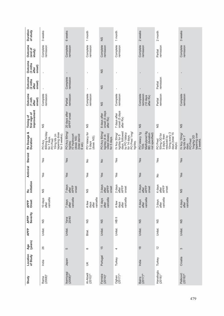
We have reviewed published articles as far back as the year 1961 and identified 30 which met our criteria (Table 1). Among these, 21 were paediatrics (70%) with a median age of 5 years (range: 0.3-17 years) while 9 were adults (30%) with a median age of 26 years (range: 18-35 years). 76.7% of the cases had unilateral facial palsy, 16.6% bilateral palsy and 2 cases were not specified by the authors. Similar to the description by other authors from previous literature, we found in our review that APFP had a median onset of 7 days after chickenpox eruption, ranging from 3 days pre-eruption and 17 days post-eruption, with a median onset of 6 days post-eruption for paediatrics and 10 days post-eruption for adults. Eight cases (26.7%) were not started on any treatment while 22 cases (73.3%) were started on treatment. The demographics and clinical characteristics of the 30 cases were summarized in Table 2.
Natural history
Without treatment, the patients had a median remission onset of 2.36 weeks and complete remission at 4.50 weeks (median) from APFP onset. 42.86% achieved complete remission and 52.14% remained partial remission at 1 month after APFP onset. 62.50% achieved complete remission and 37.50% remained partial remission at the end of the studies with a median study duration of 1.25 months (range: 1.00-8.00 months). From our review and postulation, those with APFP caused by chickenpox had 42.86% complete remission rate at 1 month after APFP onset; peaked
with 66.67% at 2 months and remained 66.67% at 3 months after APFP. This finding is similar to the natural history for Bell’s palsy described by Peitersen et al. 2002 (58% achieved complete remission within 2 months).22 Its prognosis is also much better than herpes zoster-associated APFP which had 21 % complete remission, 75% partial remission with varying severities of sequalae and 4% persistent complete loss of facial nerve function.
Prognosis with treatment
In the treatment (either antiviral, steroid or in combination) group, 77.27% achieved complete remission and only 22.72% had partial remission at the end of the study with a median study duration of 1 month (range: 0.50-24.00) while non-treatment group had 62.50% complete remission and 37.50% partial remission rate (p=0.643). The odd of partial remission at the end of study for those receiving treatment is 51% less likely than those without treatment (odd ratio=0.49; 95% CI=0.086-2.805) (p=0.423). The patients in the treatment group had a complete remission rate of 61.11% at 1 month, 72.22% at 2 months and 82.35% at 3 months after APFP onset while the non-treatment group had 42.86% complete remission rate at 1 month; peaked with 66.67% at 2 months and remained 66.67% at 3 months from APFP onset. With treatment, the patients had a median remission onset of 2.86 weeks and complete remission at 3.16 weeks (range: 1.00-11.43) from APFP onset. Those who received treatment achieved complete remission by 1.36 weeks (median) earlier than those without treatment (p=0.085). However, these were not statistically significant.
Comparison between early treatment and late treatment
Albeck et al. 1989 and Yilmaz et al. 2005 suggested that patients who received therapy early (≤3 days after APFP onset) responded fully while those who received treatment late (>3 days after onset) only had partial remission.7,25 From our systematic review, those receiving therapy early (≤3 days after onset) had 85.71% complete remission while those receiving therapy late (>3 days after onset) had only 25% complete remission and 75% remained partial remission at 1 month after APFP onset (p=0.088). However, both groups had similar outcome at the end of studies (median study duration: 1.25 months) with 88.89%

477
Tabl
e 1:
Sho
wed
the
sum
mar
y of
art
icle
s on
var
icel
la-a
ssoc
iate
d ac
ute
peri
pher
al fa
cial
ner
ve p
alsy
(Yea
r 19
61 to
201
9)
Stud
yLo
catio
n of
Stu
dyA
ge
(yea
rs)
APF
PA
PFP
Seve
rity
APF
P O
nset
Rx
initi
atio
nA
ntiv
iral
Ster
oid
Rx
Dos
age
& D
urat
ion
Tim
ing
of
first
sig
n of
im
prov
emen
t
Out
com
e (1
mth
af
ter
onse
t)
Out
com
e (2
mth
s af
ter
onse
t)
Out
com
e (3
mth
s af
ter
onse
t)
Out
com
e (e
nd o
f st
udy)
Dur
atio
n of
stu
dy
Rav
in
(196
1)30
US
7U
nila
t.N
S14
day
s af
ter
varic
ella
NS
No
Yes
Pre
d do
se &
du
ratio
n no
t sp
ecifi
ed
1 m
th a
fter
AP
FP o
nset
Par
tial
rem
issi
onP
artia
l re
mis
sion
Par
tial
rem
issi
onP
artia
l re
mis
sion
(m
inim
al
resi
dual
pa
raly
sis)
2 ye
ars
Cha
rach
on31
(1
971)
*Fr
ance
1N
SN
S1
day
befo
re
varic
ella
-N
oN
o-
NS
NS
NS
NS
Par
tial
rem
issi
onN
S
Man
ning
(1
972)
32U
S3
Uni
lat.
NS
NS
-N
oN
o-
NS
Com
plet
e re
mis
sion
--
Com
plet
e re
mis
sion
4 w
eeks
Sho
ji
(197
5)33
Japa
n17
Uni
lat.
NS
17 d
ays
afte
r va
ricel
la
-N
oN
o-
NS
Com
plet
e re
mis
sion
Com
plet
e re
mis
sion
-C
ompl
ete
rem
issi
on2
mon
ths
Ogi
no
(198
0)34
Japa
n5
Uni
lat.
NS
3 da
ys
befo
re
varic
ella
-N
oN
o-
NS
Par
tial
rem
issi
onC
ompl
ete
rem
issi
on-
Com
plet
e re
mis
sion
5 w
eeks
Mut
o35
(198
2)*
Japa
n11
Uni
lat.
NS
7 da
ys
afte
r va
ricel
la
NS
No
Yes
NS
NS
Com
plet
e re
mis
sion
--
Com
plet
e re
mis
sion
3 w
eeks
Mur
thy
(198
4) 3
6In
dia
22U
nila
t.N
S15
day
s af
ter
varic
ella
-N
oN
o-
1 m
th a
fter
AP
FP o
nset
Par
tial
rem
issi
on-
-P
artia
l re
mis
sion
1 m
onth
Yam
amot
o37
(198
7)*
Japa
n27
Bila
t.N
SR
ight
A
PFP
: 10
day
s af
ter
varic
ella
-N
oN
o-
NS
Par
tial
rem
issi
onP
artia
l re
mis
sion
Par
tial
rem
issi
onC
ompl
ete
rem
issi
on8
mon
ths
NS
Left
AP
FP:
11 d
ays
afte
r va
ricel
la
-N
oN
o-
NS
Par
tial
rem
issi
onP
artia
l re
mis
sion
Par
tial
rem
issi
onC
ompl
ete
rem
issi
on
Pun
tous
38
(198
9)**
Fran
ce30
Uni
lat.
NS
14 d
ays
afte
r va
ricel
la
NS
Yes
Yes
IV A
cy
10m
g/kg
.P
O P
red
1mg/
kg/d
ay.
(dur
atio
n no
t sp
ecifi
ed)
NS
Com
plet
e re
mis
sion
--
Com
plet
e re
mis
sion
4 w
eeks
Gan
joo
39
(198
9)**
In
dia
35B
ilat.
NS
1 da
y af
ter
varic
ella
NS
No
Yes
PO
Pre
d-ni
solo
ne
60m
g/da
y
NS
Par
tial
rem
issi
onP
artia
l re
mis
sion
Par
tial
rem
issi
onP
artia
l re
mis
sion
24
wee
ks
Um
emur
a (1
991)
40Ja
pan
1.3
Uni
lat.
NS
4 da
ys
afre
r va
ricel
la
9 da
ys
afte
r A
PFP
on
set
Yes
No
IV A
cy 5
0mg
tds
for 7
da
ys
39 d
ays
afte
r A
PFP
ons
etP
artia
l re
mis
sion
Par
tial
rem
issi
onC
ompl
ete
rem
issi
onC
ompl
ete
rem
issi
on2.
6 m
onth
s

Neurology Asia December 2020
478
Stud
yLo
catio
n of
Stu
dyA
ge
(yea
rs)
APF
PA
PFP
Seve
rity
APF
P O
nset
Rx
initi
a-tio
nA
ntiv
iral
Ster
oid
Rx
Dos
age
&
Dur
atio
nTi
min
g of
fir
st s
ign
of
impr
ovem
ent
Out
com
e (1
mth
af
ter
onse
t)
Out
com
e (2
mth
s af
ter
onse
t)
Out
com
e (3
mth
s af
ter
onse
t)
Out
com
e (e
nd o
f st
udy)
Dur
atio
n of
stu
dy
Bor
det 41
(1
992)
**Fr
ance
24U
nila
t.N
S10
day
s af
ter
varic
ella
NS
Yes
Yes
Acy
1.5
g/da
y fo
r 6 d
ays,
th
en 6
00m
g/da
y.IV
Met
hylp
red
1g/d
ay fo
r 3
days
, the
n P
O P
red
1mg/
kg/d
ay fo
r 2
mon
ths
NS
Par
tial
rem
issi
onP
artia
l re
mis
sion
Par
tial
rem
issi
onP
artia
l re
mis
sion
16 w
eeks
Wat
anab
e(1
994)
26Ja
pan
6U
nila
t.N
S8
days
af
ter
varic
ella
-N
oN
o-
NS
Com
plet
e re
mis
sion
--
Com
plet
e re
mis
sion
1 m
onth
Van
der
Flie
r (1
999)
27
Hol
land
5S
eq.
Bila
t. N
SR
ight
A
PFP
: 7
days
be
fore
va
ricel
la
-N
oN
o-
NS
Com
plet
e re
mis
sion
Com
plet
e re
mis
sion
Com
plet
e re
mis
sion
Com
plet
e re
mis
sion
3 m
onth
s
NS
Left
AP
FP:
3 da
ys
afte
r va
ricel
la
-N
oN
o-
NS
Par
tial
rem
issi
onP
artia
l re
mis
sion
Par
tial
rem
issi
onP
artia
l re
mis
sion
Iked
a 42
(1
999)
*Ja
pan
7N
SN
S11
day
s af
ter
varic
ella
NS
No
Yes
NS
NS
NS
NS
NS
Com
plet
e re
mis
sion
NS
Tana
ka
(200
1)23
Japa
n0.
3(4
mth
)U
nila
t.N
S5
days
af
ter
varic
ella
1 da
y af
-te
r AP
FP
onse
t
Yes
No
IV A
cy 4
0mg
tds
for 3
day
s30
day
s af
ter
AP
FP o
nset
Par
tial
rem
issi
onC
ompl
ete
rem
issi
on-
Com
plet
e re
mis
sion
6 w
eeks
Ded
a (2
002)
43Tu
rkey
2U
nila
t.N
S3
days
af
ter
varie
lla
2 da
ys a
f-te
r AP
FP
onse
t
Yes
No
PO
Acy
20m
g/kg
/day
(4 d
i-vi
ded
dose
s)
for 5
day
s.
3 w
eeks
afte
r A
PFP
NS
NS
NS
Par
tial
rem
issi
on3
wee
ks
Ode
mis
(2
004)
19Tu
rkey
4U
nila
t.H
B II
(on-
set)
then
be
cam
e H
B II
I 10
day
s af
ter
onse
t
7 da
ys
afte
r va
ricel
la
10 d
ays
afte
r A
PFP
on
set
Yes
Yes
PO
Acy
40m
g/kg
/day
(4 d
i-vi
ded
dose
s).
PO
Pre
d 2m
g/kg
/day
for 1
4 da
ys.
18 d
ays
afte
r A
PFP
ons
et
(8 d
ays
afte
r R
x)
Com
plet
e re
mis
sion
23
day
s af
ter o
nset
(1
4 da
ys
afte
r Rx)
--
Com
plet
e re
mis
sion
1 m
onth
Yilm
az
(200
5)7
Turk
ey7.
5U
nila
t.N
S15
day
s af
ter
varic
ella
15 d
ays
afte
r A
PFP
on
set
Yes
No
PO
Acy
20m
g/kg
/day
(4 d
i-vi
ded
dose
s)
for 5
day
s.
NS
NS
NS
NS
Com
plet
e re
mis
sion
NS
Giri
ja
(200
7)5
Indi
a31
Uni
lat.
NS
9 da
ys
afte
r va
ricel
la
NS
Yes
No
PO
Acy
dos
e &
dur
atio
n no
t sp
ecifi
ed
NS
Com
plet
e re
mis
sion
--
Com
plet
e re
mis
sion
1 m
onth
479
Stud
yLo
catio
n of
Stu
dyA
ge
(yea
rs)
APF
PA
PFP
Seve
rity
APF
P O
nset
Rx
initi
a-tio
nA
ntiv
iral
Ster
oid
Rx
Dos
age
&
Dur
atio
nTi
min
g of
fir
st s
ign
of
impr
ovem
ent
Out
com
e (1
mth
af
ter
onse
t)
Out
com
e (2
mth
s af
ter
onse
t)
Out
com
e (3
mth
s af
ter
onse
t)
Out
com
e (e
nd o
f st
udy)
Dur
atio
n of
stu
dy
Bor
det 41
(1
992)
**Fr
ance
24U
nila
t.N
S10
day
s af
ter
varic
ella
NS
Yes
Yes
Acy
1.5
g/da
y fo
r 6 d
ays,
th
en 6
00m
g/da
y.IV
Met
hylp
red
1g/d
ay fo
r 3
days
, the
n P
O P
red
1mg/
kg/d
ay fo
r 2
mon
ths
NS
Par
tial
rem
issi
onP
artia
l re
mis
sion
Par
tial
rem
issi
onP
artia
l re
mis
sion
16 w
eeks
Wat
anab
e(1
994)
26Ja
pan
6U
nila
t.N
S8
days
af
ter
varic
ella
-N
oN
o-
NS
Com
plet
e re
mis
sion
--
Com
plet
e re
mis
sion
1 m
onth
Van
der
Flie
r (1
999)
27
Hol
land
5S
eq.
Bila
t. N
SR
ight
A
PFP
: 7
days
be
fore
va
ricel
la
-N
oN
o-
NS
Com
plet
e re
mis
sion
Com
plet
e re
mis
sion
Com
plet
e re
mis
sion
Com
plet
e re
mis
sion
3 m
onth
s
NS
Left
AP
FP:
3 da
ys
afte
r va
ricel
la
-N
oN
o-
NS
Par
tial
rem
issi
onP
artia
l re
mis
sion
Par
tial
rem
issi
onP
artia
l re
mis
sion
Iked
a 42
(1
999)
*Ja
pan
7N
SN
S11
day
s af
ter
varic
ella
NS
No
Yes
NS
NS
NS
NS
NS
Com
plet
e re
mis
sion
NS
Tana
ka
(200
1)23
Japa
n0.
3(4
mth
)U
nila
t.N
S5
days
af
ter
varic
ella
1 da
y af
-te
r AP
FP
onse
t
Yes
No
IV A
cy 4
0mg
tds
for 3
day
s30
day
s af
ter
AP
FP o
nset
Par
tial
rem
issi
onC
ompl
ete
rem
issi
on-
Com
plet
e re
mis
sion
6 w
eeks
Ded
a (2
002)
43Tu
rkey
2U
nila
t.N
S3
days
af
ter
varie
lla
2 da
ys a
f-te
r AP
FP
onse
t
Yes
No
PO
Acy
20m
g/kg
/day
(4 d
i-vi
ded
dose
s)
for 5
day
s.
3 w
eeks
afte
r A
PFP
NS
NS
NS
Par
tial
rem
issi
on3
wee
ks
Ode
mis
(2
004)
19Tu
rkey
4U
nila
t.H
B II
(on-
set)
then
be
cam
e H
B II
I 10
day
s af
ter
onse
t
7 da
ys
afte
r va
ricel
la
10 d
ays
afte
r A
PFP
on
set
Yes
Yes
PO
Acy
40m
g/kg
/day
(4 d
i-vi
ded
dose
s).
PO
Pre
d 2m
g/kg
/day
for 1
4 da
ys.
18 d
ays
afte
r A
PFP
ons
et
(8 d
ays
afte
r R
x)
Com
plet
e re
mis
sion
23
day
s af
ter o
nset
(1
4 da
ys
afte
r Rx)
--
Com
plet
e re
mis
sion
1 m
onth
Yilm
az
(200
5)7
Turk
ey7.
5U
nila
t.N
S15
day
s af
ter
varic
ella
15 d
ays
afte
r A
PFP
on
set
Yes
No
PO
Acy
20m
g/kg
/day
(4 d
i-vi
ded
dose
s)
for 5
day
s.
NS
NS
NS
NS
Com
plet
e re
mis
sion
NS
Giri
ja
(200
7)5
Indi
a31
Uni
lat.
NS
9 da
ys
afte
r va
ricel
la
NS
Yes
No
PO
Acy
dos
e &
dur
atio
n no
t sp
ecifi
ed
NS
Com
plet
e re
mis
sion
--
Com
plet
e re
mis
sion
1 m
onth
Stud
yLo
catio
n of
Stu
dyA
ge
(yea
rs)
APF
PA
PFP
Seve
rity
APF
P O
nset
Rx
initi
atio
nA
ntiv
iral
Ster
oid
Rx
Dos
age
&
Dur
atio
nTi
min
g of
fir
st s
ign
of
impr
ovem
ent
Out
com
e (1
mth
af
ter
onse
t)
Out
com
e (2
mth
s af
ter
onse
t)
Out
com
e (3
mth
s af
ter
onse
t)
Out
com
e (e
nd o
f st
udy)
Dur
atio
n of
stu
dy
Ram
a (2
008)
8In
dia
26U
nila
t.N
S10
day
s af
ter
varic
ella
NS
Yes
Yes
PO
Acy
80
0mg
5x/d
ay
for 7
day
s.P
O P
red
10m
g td
s (in
ta
perin
g do
se
for 3
wee
ks).
NS
Com
plet
e re
mis
sion
--
Com
plet
e re
mis
sion
3 w
eeks
Ikem
iyag
i (2
008)
29Ja
pan
5U
nila
t.Ya
na
20/4
07
days
af
ter
varic
ella
7 da
ys
afte
r A
PFP
on
set
Yes
Yes
PO
Acy
80m
g/kg
/day
.IV
Hyd
roco
rt (d
ose:
NS
)(to
tal s
tero
id
6 w
k).
20 d
ays
afte
r A
PFP
ons
etP
artia
l re
mis
sion
Com
plet
e re
mis
sion
-C
ompl
ete
rem
issi
on6
wee
ks
Al-A
badi
(2
010)
44U
K8
Bila
t.N
SA
few
da
ys
afte
r va
ricel
la
NS
Yes
No
PO
Val
acy
for
5 da
ys(d
ose:
NS
).
NS
Com
plet
e re
mis
sion
--
Com
plet
e re
mis
sion
1 m
onth
Ferr
eira
(2
014)
9P
ortu
gal
15U
nila
t.N
S11
day
s af
ter
varic
ella
3 da
ys
afte
r A
PFP
on
set
Yes
No
PO
Acy
80m
g/kg
/day
(4 d
i-vi
ded
dose
s)
for 5
day
s.
8 da
ys a
fter
onse
t (5
days
af
ter R
x)
NS
NS
NS
Com
plet
e re
mis
sion
NS
Ogh
an
(201
7)10
Turk
ey4
Uni
lat.
HB
IIA
few
da
ys
afte
r va
ricel
la
2 da
ys
afte
r A
PFP
on
set
Yes
Yes
IV A
cy 3
0mg/
kg/d
ay fo
r 5
days
, fol
low
ed
by P
O V
alac
y fo
r 14
days
.P
O P
red
1mg/
kg/d
ay.
7 da
ys a
fter
onse
t (5
days
af
ter R
x)
Com
plet
e re
mis
sion
--
Com
plet
e re
mis
sion
1 m
onth
Bai
ns
(201
7)45
Indi
a18
Uni
lat.
NS
4 da
ys
afte
r va
ricel
la
3 da
ys
afte
r A
PFP
on
set
Yes
Yes
PO
Val
acy
1g
TDS
.M
ethy
lpre
d 8g
B
D. (
dura
tion
not s
peci
fied)
NS
Com
plet
e re
mis
sion
(10
days
af
ter R
x)
--
Com
plet
e re
mis
sion
2 w
eeks
Han
alio
glu
(201
8)11
Turk
ey12
Uni
lat.
NS
3 da
ys
afte
r va
ricel
la
4 da
ys
afte
r A
PFP
on
set
No
Yes
PO
Pre
d 50
mg
OD
for
3 da
ys, t
hen
redu
ced
by
10m
g ev
ery
3 da
ys (t
otal
15
days
).
NS
Par
tial
rem
issi
onP
artia
l re
mis
sion
-P
artia
l re
mis
sion
2 m
onth
Pet
kovi
ć
(201
8)46
Cro
atia
3U
nila
t.N
S4
days
af
ter
varic
ella
NS
Yes
Yes
IV A
cy 1
0mg/
kg td
s fo
r 7
days
.P
O P
red
1mg/
kg O
D
tape
ring
over
2
wee
ks.
NS
Com
plet
e re
mis
sion
-
-C
ompl
ete
rem
issi
on3
wee
ks

Neurology Asia December 2020
480
complete remission from early treatment group and 80% from late treatment group (p=1.000). Although statistically insignificant, early treatment group tends to have earlier complete remission (median 3.0 weeks earlier) than late treatment group (p=0.091). The early treatment group achieved complete remission at median 2.00 weeks (range: 1.00-6.00 weeks) from APFP onset while the late treatment group at median 6.00 weeks (range: 3.29-11.43 weeks).
Comparison between treatment options
Eight cases were (36.4%) started on antiviral monotherapy, 5 cases (22.7%) on steroid monotherapy and 9 (40.9%) on combination therapy (antiviral+steroid). Those who received antiviral monotherapy had a median interval of 5 days between varicella and APFP onset; those with antiviral-steroid combination therapy 7 days; and those with steroid monotherapy had the longest median interval of 9 days. Treatment were initiated at a median of 2.5 days (range: 1-15 days) after the APFP onset with a median study duration of 1.13 months (range: 0.5-24 months) stating the patient outcome. We analysed the outcomes at the end of studies among the use of antiviral, systemic glucocorticoid and its combination:
(a) Antiviral therapy vs steroid monotherapy
The antiviral group (antiviral monotherapy and antiviral-steroid combination therapy) tends to have better outcome than steroid monotherapy at the end of studies with p value barely <0.05. The antiviral group had 88.24% complete remission while the steroid monotherapy group had only 40% complete remission. (p=0.055). Among the steroid monotherapy group (n=5), only 1 case developed APFP early during chickenpox infection (within first week) but the timing of initiation of steroid therapy was not specified. The remaining cases either developed APFP later during chickenpox infection (after 1 week of onset) or received steroid 1 week after varicella onset.
(b) Antiviral monotherapy vs steroid monotherapy
The antiviral monotherapy group had 87.5% complete remission while the steroid monotherapy group had only 40% complete remission. However, it was not statistically significant. (p=0.217).
Stud
yLo
catio
n of
Stu
dyA
ge
(yea
rs)
APF
PA
PFP
Seve
rity
APF
P O
nset
Rx
initi
atio
nA
ntiv
iral
Ster
oid
Rx
Dos
age
& D
urat
ion
Tim
ing
of
first
sig
n of
im
prov
emen
t
Out
com
e (1
mth
af
ter
onse
t)
Out
com
e (2
mth
s af
ter
onse
t)
Out
com
e (3
mth
s af
ter
onse
t)
Out
com
e (e
nd o
f st
udy)
Dur
atio
n of
stu
dy
Cha
tterje
(2
019)
47In
dia
19B
ilat.
NS
5 da
ys
afte
r va
ricel
la
2 da
ys a
f-te
r AP
FP
onse
t
Yes
Yes
PO
Acy
40
00m
g ev
ery
day
for 1
4 da
ys.
PO
Pre
d 60
mg
OD
for
14 d
ays.
NS
Com
plet
e re
mis
sion
--
Com
plet
e re
mis
sion
2 w
eeks
Teng
(201
9)In
dex
case
Unp
ublis
hed
Mal
aysi
a2
Uni
lat.
HB
II4
days
af
ter
varic
ella
1 da
y af
-te
r AP
FP
onse
t
Yes
No
PO
Acy
80
0mg
5x/d
ay
for 7
day
s.
5 da
ys a
fter
onse
tC
ompl
ete
rem
issi
on
(7 d
ays
afte
r Rx)
Com
plet
e re
mis
-si
on
Com
plet
e re
mis
sion
Com
plet
e re
mis
sion
2 ye
ars
Abb
revi
atio
nsA
PFP
, Acu
te p
erip
hera
l fac
ial n
erve
pal
sy; R
x, T
reat
men
t; M
th(s
), M
onth
(s);
Wk(
s), W
eek(
s); U
nila
t., U
nila
tera
l; B
ilat.,
Bila
tera
l; S
eq. B
ilat.,
Seq
uent
ial B
ilate
ral;
NS
, Not
spe
cifie
d or
unk
now
n;
IV, I
ntra
veno
us; P
O, o
ral;
Acy
, Acy
clov
ir; V
alac
y, V
alac
yclo
vir;
Pre
d, P
redn
isol
one;
Hyd
roco
rt, H
ydro
corti
sone
; Met
hylp
red,
Met
hylp
redn
isol
one;
HB
II, H
ouse
-Bra
ckm
ann
Cla
ssifi
catio
n gr
ade
II; H
B II
I, H
ouse
-Bra
ckm
ann
Cla
ssifi
catio
n gr
ade
III; Y
ana
20/4
0, Y
anag
ihar
a Fa
cial
Ner
ve G
radi
ng s
yste
m s
core
20/
40“ –
“: n
ot g
iven
or n
ot a
pplic
able
*The
dat
a w
as e
xtra
cted
from
Tan
aka
et a
l 200
1.23
**Th
e da
ta w
as e
xtra
cted
from
Muñ
oz-S
ella
rt et
al 2
010.
481
(c) Antiviral-steroid combination therapy vs steroid monotherapy
Those receiving antiviral-steroid combination therapy had 88.89% complete remission while steroid monotherapy had 40% complete remission at the end of studies with a p value of 0.095.
(d) Antiviral monotherapy vs antiviral-steroid combination therapy
There was no statistically significant difference in complete remission at the end of studies between these 2 groups with the former having 87.5% complete remission and the latter 88.89% (p=1.000). Those receiving antiviral monotherapy started to recover at 3.00 weeks from APFP onset and achieved complete remission at 4.00 weeks while those receiving antiviral-steroid combination therapy had recovery at 2.57 weeks (p=0.456) and achieved complete remission at 3.15 weeks (p=0.825).
DISCUSSION
Watanabe et al. described that facial nerve palsy can appear 5 days before and 16 days after chickenpox eruption.11,26 Watanabe et al. also hypothesized haematogenous spread of infection for those with pre-eruptive onset of APFP and
neurogenous route for those with post-eruptive onset.26 As described by previous studies, we postulated in our patient that the VZV from the pinna vesicles may have invaded the facial nerve sensory branches and then to adjacent motor branches, causing nerve injury by either direct viral invasion or immunologically mediated inflammatory responses.9,11 The hypothesized pathogenesis has been quite consistent throughout the years, whereas the natural history, prognosis and treatment for this clinical entity has been so far contradicting. In 1999, van der Flier et al. described that 50% of cases recovered completely within 1 month and their recovery was independent of the therapy prescribed.27 On the other hand, due to limited literature available, some extrapolated its prognosis and management from large scale studies of Bell’s palsy, stating high spontaneous recovery rate. However, some other authors extrapolated from studies of Herpes zoster-related APFP that this index clinical entity had poorer prognosis and early treatment should be given.19,28 Just as described by Peitersen22,
the prognosis of individual cases of facial nerve palsy, with or without treatment, varies with aetiologies.12 We should be cautious as the APFP in our context is associated with primary VZV infection, i.e. varicella. From our review, we found that acute
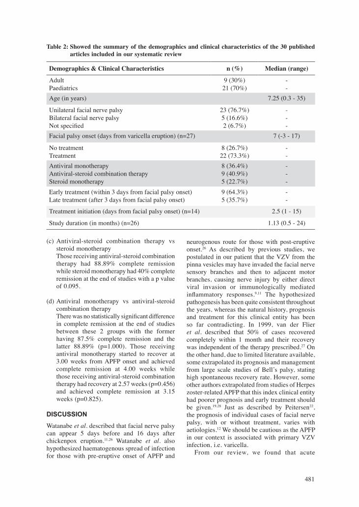
Table 2: Showed the summary of the demographics and clinical characteristics of the 30 published articles included in our systematic review
Demographics & Clinical Characteristics n (%) Median (range)
Adult Paediatrics
9 (30%)21 (70%)
--
Age (in years) 7.25 (0.3 - 35)
Unilateral facial nerve palsyBilateral facial nerve palsyNot specified
23 (76.7%)5 (16.6%)2 (6.7%)
---
Facial palsy onset (days from varicella eruption) (n=27) 7 (-3 - 17)
No treatmentTreatment
8 (26.7%)22 (73.3%)
--
Antiviral monotherapyAntiviral-steroid combination therapySteroid monotherapy
8 (36.4%)9 (40.9%)5 (22.7%)
---
Early treatment (within 3 days from facial palsy onset)Late treatment (after 3 days from facial palsy onset)
9 (64.3%)5 (35.7%)
--
Treatment initiation (days from facial palsy onset) (n=14) 2.5 (1 - 15)
Study duration (in months) (n=26) 1.13 (0.5 - 24)

Neurology Asia December 2020
482
peripheral facial palsy occurring at the time of primary chickenpox infection generally had good prognosis with 66.67% complete remission 2 months after its onset even without treatment. There was no statistically significant difference in the complete remission rate between treatment and non-treatment groups (p=0.643). Similarly, there was no statistically significant difference in the rate of complete remission between the early and late treatment group. However, early treatment group tends to achieve complete remission earlier (median 3.0 weeks earlier) than late treatment group, although statistically insignificant (p=0.091). Early treatment could potentially accelerate recovery and reduce morbidities even if it could not alter the final outcome. Among the treatment options, we found that the antiviral group (antiviral monotherapy and antiviral-steroid combination therapy) tends to have better outcome than steroid monotherapy group at the end of studies with p value barely <0.05. We also made assumptions that the statistical significance of the superiority with early treatment over late treatment and the use of either antiviral monotherapy or antiviral-steroid combination therapy over steroid monotherapy was likely limited to a certain extent by the small available sample size of our review. As stated above, our systematic review has some important limitations. Firstly, due to its rarity, the sample size is too small. Secondly, quite a number of previous reports had only a brief summary of the case, overlooking certain important information, including facial palsy severity. Among the 30 reports from our review, there were only 4 articles (13%) objectively stating the severity of APFP using well-recognised classification systems, i.e. House-Brackmann classification10,19 and Yanagihara Facial Nerve Grading System.29 We think the palsy severity is particularly important as it is potentially one of the main outcome predictors. Other potential factors influencing the prognosis were not adequately documented including treatment dosage, treatment duration and the timing of the first sign of recovery. Thirdly, in most reports, the patients were only studied for a short duration with a median of 1.13 months. This is particularly true for those with partial remission who should be studied for a longer period to determine the rate and extent of recovery at different points of time. Considering the limitations of our review, we should be cautious when interpreting the findings. Future case reports should include all
of the following information to facilitate review articles of better quality: 1. Age of the patient; 2. Facial palsy laterality (unilateral/bilateral); 3. Facial palsy severity classification (e.g. House-Brackmann Classification); 4. Facial palsy onset in relation to varicella infection; 5. Interval between treatment initiation and facial palsy onset; 6. Treatment dosage and duration; 7. Timing of first sign of improvement; 8. Longer period of study (e.g. up to 1 year after onset) especially for those with incomplete remission; 9. Serial extent of recovery documented at each review. More frequent review for the first 3-4 months after facial palsy (e.g. 1 month, 2 month, 3 month after onset, etc); 10. Objectively specify the severity of residual facial nerve dysfunction if no complete remission at the end of study (using tools e.g. House-Brackmann Classification). In conclusion, our review showed that APFP caused by varicella had similar prognosis as Bell’s palsy but significantly better prognosis than herpes zoster-associated APFP. It generally has good prognosis even without treatment. However, clinicians may consider using antiviral with or without steroid therapy during early course of the disease (within 3 days of onset) as early treatment may accelerate remission. Nevertheless, our findings need to be interpreted or applied with caution considering the limitations of our systematic review. Important and standardized information are required in future case reports to facilitate systematic review of better quality for guideline development.
ACKNOWLEDGEMENTS
We would like to thank the Director of Hospital Port Dickson and Director General, Ministry of Health, Malaysia for the permission to publish this paper.
DISCLOSURE
Financial support: None
Conflicts of interest: None
REFERENCES 1. Poumerol G, Wilder-Smith A. International travel and
health. World Health Organisation (2012). Retrieved from https://www.who.int/ith.
2. Albrecht MA. Clinical features of varicella-zoster virus infection: chickenpox. Post TW, ed: UpToDate. Waltham, MA: UpToDate Inc. https://www.uptodate.com (Accessed on August 9, 2019.)
3. LaRussa PS, Marin M. Varicella-zoster virus

483
infections. In: Kliegman RM, Stanton BF, St. Geme JW, Schor NF, Behrman RE, eds: Nelson textbook of pediatrics. 19th ed. Philadelphia, PA: Elsevier Saunders; 2011:1104-10.
4. Hamborsky J, Kroger A, Wolfe S, eds: Epidemiology and prevention of vaccine-preventable diseases. 13th ed. Washington D.C.: Centers for disease control and prevention, Public health foundation; 2015:353-76.
5. Girija AS, Rafeeque M, Abdurehman KP. Neurological complications of chickenpox. Ann Indian Acad Neurol 2007;10:240-6.
6. Rack AL, Grote V, Streng A, et al. Neurologic varicella complications before routine immunization in Germany. Pediatr Neurol 2010;42(1):40-8.
7. Yilmaz C, Çaksen H. Severe neurological complications of chickenpox: report of four cases. Eur J Gen Med 2005;2(4):177-9.
8. Rama Rao G, Amareswar A, Kishan Kumar Y, Rani R. Isolated facial palsy in varicella. Indian J Dermatol Venereol Leprol 2008;74(3):261-2.
9. Ferreira H, Dias A, Lopes A. Acute peripheral facial palsy after chickenpox: a rare association. Case Rep Pediatr 2014;2014:754390.
10. Oghan F, Topuz MF, Erdogan O. Facial paralysıs during varicella zoster infectıon in a child. Heighpubs Otolaryngol Rhinol 2017; 1:16-9.
11. Hanalioğlu D, Özsürekci Y, Büyükçam A, Gültekingil-Keser A, Tekşam Ö, Ceyhan M. Acute peripheral facial paralysis following varicella infection: An uncommon complication. Turk J Pediatr 2018;60(1):99-101.
12. Geller TJ. Facial nerve palsy in children. Post TW, ed: UpToDate. Waltham, MA: UpToDate Inc. https://www.uptodate.com (Accessed on May 28, 2019.)
13. Shargorodsky J, Lin HW, Gopen Q. Facial nerve palsy in the pediatric population. Clin Pediatr (Phila) 2010;49(5):411-7.
14. Karalok ZS, Taskin BD, Ozturk Z, Gurkas E, Koc TB, Guven A. Childhood peripheral facial palsy. Childs Nerv Syst 2018 May;34(5):911-7.
15. Hato N, Murakami S, Gyo K. Steroid and antiviral treatment for Bell’s palsy. Lancet 2008;371(9627):1818-20.
16. Sarnat HB. Bell palsy. In: Kliegman RM, Stanton BF, St. Geme JW, Schor NF, Behrman RE, eds: Nelson textbook of pediatrics. 19th ed. Philadelphia, PA: Elsevier Saunders; 2011:2146-7.
17. Furuta Y, Fukuda S, Suzuki S, Takasu T, Inuyama Y, Nagashima K. Detection of varicella-zoster virus DNA in patients with acute peripheral facial palsy by the polymerase chain reaction, and its use for early diagnosis of zoster sine herpete. J Med Virol 1997;52(3):316-9.
18. Furuta Y, Ohtani F, Kawabata H, Fukuda S, Bergström T. High prevalence of varicella-zoster virus reactivation in herpes simplex virus-seronegative patients with acute peripheral facial palsy. Clin Infect Dis 2000;30(3):529-33.
19. Ödemis E, Türkay S, Tunca A, Karadag A. Acute peripheral facial palsy during chickenpox in a child. J Pediatric Neurol 2004;2(4):245-6.
20. Furuta Y, Ohtani F, Aizawa H, Fukuda S, Kawabata H, Bergström T. Varicella-zoster virus reactivation is
an important cause of acute peripheral facial paralysis in children. Pediatr Infect Dis J 2005;24(2):97-101.
21. Santos MA, Caiaffa Filho HH, Vianna MF, Almeida AG, Lazarini PR. Varicella zoster virus in Bell’s palsy: a prospective study. Braz J Otorhinolaryngol 2010;76(3):370-3.
22. Peitersen E. Bell’s palsy: the spontaneous course of 2,500 peripheral facial nerve palsies of different etiologies. Acta Otolaryngol Suppl 2002;(549):4-30.
23. Tanaka T, Iwai K, Sudo M. A Case Report of Facial Nerve Palsy in an Infant Associated with Chickenpox. Practica Oto-Rhino-Laryngologica 2001; 94(5):421-25.
24. Muñoz-Sellart M, García-Vidal C, Martínez-Yelamos S, et al. Peripheral facial palsy after varicella. Report of two cases and review of the literature. Enferm Infecc Microbiol Clin 2010;28(8):504-8.
25. Albeck H, Ninn- Pedersen K. Acyclovir in the treatment of facial palsy due to zoster virus. Ugeskr Laeger 1989; 151: 90-2.
26. Watanabe Y, Ikeda M, Kukimoto N, Kuga M, Tomita H. A case report of facial nerve palsy associated with chickenpox. J Laryngol Otol 1994;108(8):676-8.
27. van der Flier M, van Koppenhagen C, Disch FJ, Mauser HW, Bistervels JH, van Diemen-Steenvoorde JA. Bilateral sequential facial palsy during chickenpox. Eur J Pediatr 1999;158(10):807-8.
28. Grose C, Bonthius D, Afifi AK. Chickenpox and the geniculate ganglion: facial nerve palsy, Ramsay Hunt syndrome and acyclovir treatment. Pediatr Infect Dis J 2002;21(7):615-7.
29. Ikemiyagi Y, Yamamoto M, Yoshida T, Nomura T, Takazawa R, Shigeta F. A case report of facial nerve palsy in childhood associated with chickenpox. Practica Oto-Rhino-Laryngologica 2008; 101(11):841-4.
30. Ravin LC. Facial paralysis as a complication of chickenpox. Am J Ophthalmol 1961;52:723-4.
31. Charachon R, Micoud M, Junien-Lavillauroy C, Serero C. Facial paralysis and varicelliform eruption. J Fr Otorhinolaryngol Audiophonol Chir Maxillofac 1971;20(10):1159-60. (French)
32. Manning JJ, Adour KK. Facial paralysis in children. Pediatrics 1972;49(1):102-9.
33. Shoji H, Hirose K, Uono M, Sugita R, Motodo R. Peripheral facial palsy following varicella. Rinsho Shinkeigaku 1975;15(9):587-91. (Japanese)
34. Ogino S, Tamaki H, Furukawa Y. Two cases of peripheral facial paralysis associated with varicella. Practica Oto-Rhino-Laryngologica 1980;73(2):358-62.
35. Muto J, Takeda E, Takahashi S. Facial nerve palsy as a neurological complication of chickenpox: case report and literature review. Otolaryngology (Tokyo) 1982; 54(1):71-4. (Japanese)
36. Murthy VK, Sawhney IM, Prabhakar S, Chopra JS. Isolated facial palsy in chickenpox. J Neurol Neurosurg Psychiatry 1984;47(7):754.
37. Yamamoto K, Noda M, Ito H, et al. Bilateral peripheral facial nerve palsy associated with chickenpox. Neurology 1987;27: 510-2. (Japanese)
38. Puntous M, Imbert Y, Pellegrin JL, Ducos P, Dupont E. Peripheral facial paralysis following varicella. Presse Med 1989;18(34):1707. (French)
Neurology Asia December 2020
484
39. Ganjoo RK, Roy AK, Kumar A. Bilateral facial palsy following chicken pox. J Assoc Physicians India 1989;37(12):798.
40. Umemura H, Ozaki M, Kitamura K. Peripheral facial paralysis in an infant with chicken-pox. Pract Otol (Kyoto) 1991;84(5):627-31.
41. Bordet R, Destée A. Facial paralysis and chicken-pox. Rev Neurol (Paris) 1992;148(1):62-3. (French)
42. Ikeda M, Watanabe Y, Tsuji K. Facial nerve palsy caused by chickenpox. Journal of Otolaryngology, Head and Neck Surgery -- JOHNS 1999;15: 1283-6. (Japanese)
43. Deda G, Çaksen H, İçağasioğlu D, et al. A case of chickenpox associated with facial nerve palsy. Pediatr Dermatol 2002;19(1): 95-6.
44. Al-Abadi E, Milford DV, Smith M. A patient with bilateral facial palsy associated with hypertension and chickenpox: learning points. BMJ Case Rep 2010;2010:bcr0620103083.
45. P Bains. Peripheral facial palsy as a complication of varicella. J Pakistan Assoc Dermatol 2017;27(4): 410-3.
46. Petković D, Vuković B, Cviljević S. A case report of facial nerve palsy associated with varicella infection. Abstract Book 2018. Mind and Brain – 58th International Neuropsychiatric Congress, Pula, Congress. 2018:72-73.
47. Chatterjee N, Chatterjee C. Isolated bilateral facial palsy due to chicken pox – an unique presentation. Int J Dermatol Clin Res 2019;5(1): 001-002.