varun nejm
TRANSCRIPT

T h e n e w e ngl a nd j o u r na l o f m e dic i n e
n engl j med 375;21 nejm.org November 24, 2016 2067
Review Article
Von Willebrand’s disease is an inherited bleeding disorder characterized by defective platelet adhesion and aggregation. The disorder was first described in 1926 by Erik von Willebrand, who recognized that it
differed from hemophilia and named it “hereditary pseudohemophilia.”1 The fac-tor in plasma that corrects the disease was not identified until many years later and was called von Willebrand factor. It binds to collagen at sites of vascular in-jury, mediates platelet adhesion and aggregation, and serves as a carrier protein for coagulation factor VIII (Fig. 1).3 Von Willebrand factor also has other functions and may be involved in such processes as inflammation and angiogenesis.4
Von Willebrand’s disease is the most common inherited bleeding disorder and has an autosomal inheritance pattern. The disease is characterized mainly by mucosa-associated bleeding and bleeding after surgery and trauma. The diagnosis is based on a personal or family history of bleeding and laboratory evidence of abnormalities in von Willebrand factor, factor VIII, or both. Affected patients have reduced levels of functional von Willebrand factor, and various types of von Wille-brand’s disease can be distinguished on the basis of phenotypic characteristics. The goal of treatment is to increase von Willebrand factor levels by administering desmopressin or by the infusion of exogenous von Willebrand factor–containing concentrates. In this review, we discuss the latest findings regarding the patho-physiology, diagnosis, and treatment of von Willebrand’s disease.
Epidemiol o gy a nd Cl a ssific ation
On the basis of population studies, the prevalence of von Willebrand’s disease is 0.6 to 1.3%.5,6 Although the autosomal inheritance pattern would suggest an equal distribution of male patients and female patients, the disease is diagnosed in more females because of female-specific hemostatic challenges. Not all persons with low von Willebrand factor levels have clinically relevant bleeding symptoms. On the basis of referrals to specialized centers, the estimated prevalence of von Wille-brand’s disease is approximately 1 case per 10,000 persons.7 The prevalence is strongly dependent on the diagnostic cutoff level for von Willebrand factor. The normal range is generally between 50 and 150 IU per deciliter. In several sets of guidelines, the diagnosis is based on a cutoff level for von Willebrand factor of 30 IU per deciliter, whereas the principles of care for von Willebrand’s disease specify a cutoff level of 40 IU per deciliter.5,8-10 In daily practice, clinicians do not always follow these guidelines, especially since the official classification of the International Society on Thrombosis and Haemostasis (ISTH) does not specify cutoff values.11
Von Willebrand’s disease is subdivided into types 1, 2, and 3.11 Type 1, which accounts for 70 to 80% of cases, is characterized by a quantitative deficiency of von Willebrand factor. Type 2, accounting for approximately 20% of cases, is
From the Department of Hematology, Erasmus University Medical Center, Rotter-dam (F.W.G.L.), and the Department of Thrombosis and Hemostasis, Leiden Uni-versity Medical Center, Leiden (J.C.J.E.) — both in the Netherlands. Address re-print requests to Dr. Leebeek at the De-partment of Hematology, Erasmus Uni-versity Medical Center, Rm. Na-820, P.O. Box 2040, 3000 CA Rotterdam, the Nether-lands, or at f . leebeek@ erasmusmc . nl.
N Engl J Med 2016;375:2067-80.DOI: 10.1056/NEJMra1601561Copyright © 2016 Massachusetts Medical Society.
Dan L. Longo, M.D., Editor
Von Willebrand’s DiseaseFrank W.G. Leebeek, M.D., Ph.D., and Jeroen C.J. Eikenboom, M.D., Ph.D.
The New England Journal of Medicine Downloaded from nejm.org on November 23, 2016. For personal use only. No other uses without permission.
Copyright © 2016 Massachusetts Medical Society. All rights reserved.

n engl j med 375;21 nejm.org November 24, 20162068
T h e n e w e ngl a nd j o u r na l o f m e dic i n e
caused by dysfunctional von Willebrand factor, resulting in a normal or reduced von Willebrand factor antigen concentration but a large reduc-tion in von Willebrand factor function. Type 2 is further subdivided on the basis of specific phe-notypic characteristics (Fig. 2). Type 3 von Will-ebrand’s disease is rare (accounting for <5% of
cases), is the most severe form, and is caused by the absence of circulating von Willebrand factor.
In rare cases, von Willebrand’s disease is ac-quired, rather than inherited, by means of vari-ous mechanisms that result in rapid degradation or clearance of von Willebrand factor. The ac-quired syndrome is not discussed here because
Cleaved Disulfidebond
VWF dimer
Noncovalentbond
VWFmultimer
SP D1D2 D'D3 D4 CKC1–C6A1A2A3
PropeptideSignalpeptide
FVIII GPIbα αIIb
β3
Collagen
CollagenVWF monomer
E N D O T H E L I A LC E L L
N U C L E U S
E N D O P L A S M I CR E T I C U L U M
G O L G IA P P A R A T U S
Fusion with cell membrane
A
B
Weibel–Paladebody
Helixes of VWFmultimers
Helixes uncoil
GPIbα
Collagenreceptor
Coiled,inactive VWF
Subendothelial collagen
Active αIIb
β3
Inactiveα
IIbβ
3
Uncoiled, active VWF
I N A C T I V A T E DP L A T E L E T
E N D O T H E L I A L C E L L
F L O W O F B L O O D
A C T I V A T E DP L A T E L E T
ADAMTS13 proteolyzes long VWF strands into
shorter strands
ADAMTS13VWFstrand
The New England Journal of Medicine Downloaded from nejm.org on November 23, 2016. For personal use only. No other uses without permission.
Copyright © 2016 Massachusetts Medical Society. All rights reserved.
n engl j med 375;21 nejm.org November 24, 2016 2069
Von Willebr and’s Disease
it requires diagnostic strategies and treatment that differ from those used for the inherited disease.12
Clinic a l Pr esen tation
The symptoms of von Willebrand’s disease vary among patients, depending on the level of re-sidual von Willebrand factor activity, the disease
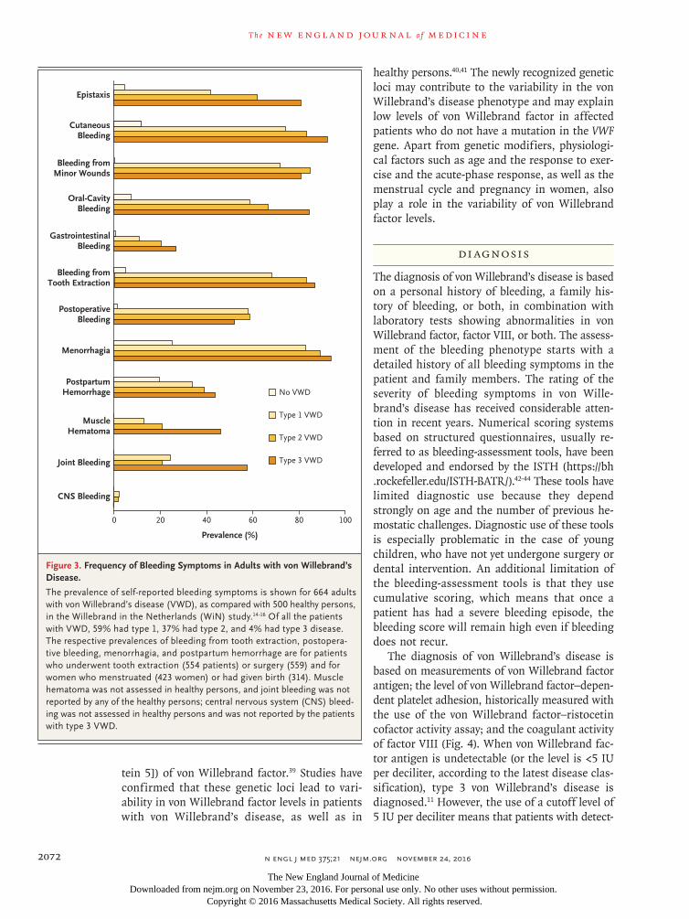
subtype, and to some extent, age and sex. In children with von Willebrand’s disease, the most frequent presenting symptoms are bruising and epistaxis.13 In adults, the most common symp-toms are hematomas, menorrhagia, and bleed-ing from minor wounds. The majority of pa-tients (60 to 80%) have bleeding after surgery or dental extractions.14 Figure 3 shows the preva-lence of self-reported bleeding symptoms derived from the large Willebrand in the Netherlands (WiN) study14; similar prevalences have been reported in other studies.5,17
A well-known, serious, and possibly life-threatening bleeding complication is gastroin-testinal bleeding from angiodysplasia.18 It is most common in elderly patients with type 2 or 3 von Willebrand’s disease.14,18 Intraarticular (joint) bleeding is a frequent complication in patients with hemophilia but has not been reported as a major problem in patients with von Willebrand’s disease, although it may be a presenting symp-tom in those with type 2N (type 2 subtype Nor-mandy) or type 3 disease.19 It is now known that joint bleeding occurs in a considerable number of severely affected patients, potentially leading to arthropathy and reduced joint function.19 The risk of joint bleeding is strongly dependent on the level of residual factor VIII, as well as the severity of von Willebrand’s disease, and patients with type 3 disease who have very low factor VIII levels are at greatest risk.14,19,20 A lower health-related quality of life for patients with von Wille-brand’s disease than for the general population is strongly associated with the bleeding phenotype.21
The majority of women with von Willebrand’s disease have menorrhagia, which also impairs the quality of life (Fig. 3).22 Conversely, von Wille-brand’s disease is diagnosed in 5 to 20% of women who present with menorrhagia. The link to menorrhagia accounts for the more frequent identification of von Willebrand’s disease in women than in men.23,24 In the WiN study, more than 80% of women with von Willebrand’s dis-ease reported excessive blood loss during the menstrual cycle, and more than 20% underwent hysterectomy, a proportion that is twice as high as in the general population.25 Other reproductive tract symptoms also occur, including bleeding of an ovarian cyst, which should be considered in women with von Willebrand’s disease who present with a sudden onset of severe abdominal pain.26
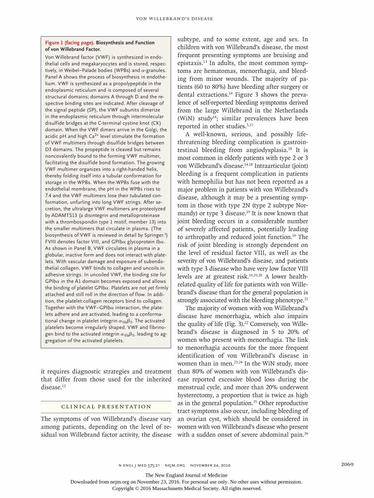
Figure 1 (facing page). Biosynthesis and Function of von Willebrand Factor.
Von Willebrand factor (VWF) is synthesized in endo-thelial cells and megakaryocytes and is stored, respec-tively, in Weibel–Palade bodies (WPBs) and α-granules. Panel A shows the process of biosynthesis in endothe-lium. VWF is synthesized as a propolypeptide in the endoplasmic reticulum and is composed of several structural domains; domains A through D and the re-spective binding sites are indicated. After cleavage of the signal peptide (SP), the VWF subunits dimerize in the endoplasmic reticulum through intermolecular disulfide bridges at the C-terminal cystine knot (CK) domain. When the VWF dimers arrive in the Golgi, the acidic pH and high Ca2+ level stimulate the formation of VWF multimers through disulfide bridges between D3 domains. The propeptide is cleaved but remains noncovalently bound to the forming VWF multimer, facilitating the disulfide bond formation. The growing VWF multimer organizes into a right-handed helix, thereby folding itself into a tubular conformation for storage in the WPBs. When the WPBs fuse with the endothelial membrane, the pH in the WPBs rises to 7.4 and the VWF multimers lose their tubulated con-formation, unfurling into long VWF strings. After se-cretion, the ultralarge VWF multimers are proteolyzed by ADAMTS13 (a disintegrin and metalloproteinase with a thrombospondin type 1 motif, member 13) into the smaller multimers that circulate in plasma. (The biosynthesis of VWF is reviewed in detail by Springer.2) FVIII denotes factor VIII, and GPIbα glycoprotein Ibα. As shown in Panel B, VWF circulates in plasma in a globular, inactive form and does not interact with plate-lets. With vascular damage and exposure of subendo-thelial collagen, VWF binds to collagen and uncoils in adhesive strings. In uncoiled VWF, the binding site for GPIbα in the A1 domain becomes exposed and allows the binding of platelet GPIbα. Platelets are not yet firmly attached and still roll in the direction of flow. In addi-tion, the platelet collagen receptors bind to collagen. Together with the VWF–GPIbα interaction, the plate-lets adhere and are activated, leading to a conforma-tional change in platelet integrin αIIbβ3. The activated platelets become irregularly shaped. VWF and fibrino-gen bind to the activated integrin αIIbβ3, leading to ag-gregation of the activated platelets.
The New England Journal of Medicine Downloaded from nejm.org on November 23, 2016. For personal use only. No other uses without permission.
Copyright © 2016 Massachusetts Medical Society. All rights reserved.

n engl j med 375;21 nejm.org November 24, 20162070
T h e n e w e ngl a nd j o u r na l o f m e dic i n e
Primary and secondary postpartum bleeding also occurs frequently.25,27,28
As a result of the physiologic rise in von Wil-lebrand factor levels throughout life, patients with type 1 von Willebrand’s disease may have levels within the normal range when they be-come older.29 It is still not known whether this rise results in fewer bleeding episodes. A recent
study showed that among patients with type 1 disease, bleeding symptoms occurred as fre-quently in patients who were older than 65 years of age as in those who were 18 to 65 years of age, which suggests that the age-dependent rise in von Willebrand factor levels does not lead to a mitigation of bleeding symptoms.30 Even though von Willebrand factor antigen levels also
Healthy Persons, Normal VWF
Disease Mechanisms
Deficiency of Functionally Normal VWF
Decreased Platelet Adhesion Due to Deficiency of HMW VWF Multimers
Enhanced, Spontaneous GPIbα Binding
Decreased Platelet Adhesion or Collagen Binding with No Loss of HMW VWF Multimers
Decreased Factor VIII Binding
No protein synthesis Null alleles Type 3
Reduced synthesis of normal VWF
Defective multimerization
Defective dimerization
Enhanced proteolysis by ADAMTS13
Null alleles Type 1
Missense mutations in propeptide,D3, and A2 domains
Type 2A
Intracellular retention of VWF
Enhanced clearance of VWF
Missense mutations Type 1
Type 1Missense mutations
Type 2AMissense mutations in CK domain
Type 2A
Type 2B
Missense mutations in A2 domain
Missense mutations in A1 domain
Type 2MMissense mutations in A1 domain
Type 2NMissense mutations in D'D3 domain
Defects in VWF Types of VWD
Low VWF
MonomerDimer
Factor VIII
Mutant subunit
GPIbα
P L A T E L E T
Proteolytic fragments
Multimer
The New England Journal of Medicine Downloaded from nejm.org on November 23, 2016. For personal use only. No other uses without permission.
Copyright © 2016 Massachusetts Medical Society. All rights reserved.

n engl j med 375;21 nejm.org November 24, 2016 2071
Von Willebr and’s Disease
rise with age in patients with type 2 disease, von Willebrand factor activity remains low because of the functional defect in the protein. In these patients, an increase in bleeding symptoms is observed with increasing age.30 These observa-tions should be confirmed in larger prospective studies. An interesting finding in two recent studies is that the risks of cardiovascular disease and ischemic stroke are reduced among patients with von Willebrand’s disease.31,32
Pathoph ysiol o gic a l Fe at ur es
VWF Mutations
The inheritance pattern for most types of von Willebrand’s disease is autosomal dominant, but in some cases, the inheritance is recessive. In 80% of patients with type 3 von Willebrand’s disease, the genetic defects in the VWF gene are null alleles, explaining the complete absence of von Willebrand factor17,33 (mutation database, www . vwf . group . shef . ac . uk). Most patients with type 3 disease are homozygous or compound heterozy-
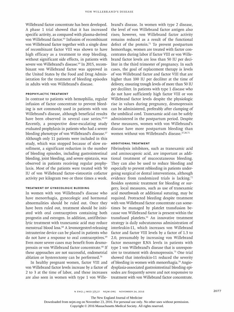
gous. Codominant rather than recessive inheri-tance is observed in up to 50% of patients with type 3 von Willebrand’s disease, with the hetero-zygous carriers having a mild type 1 disease phenotype.34 Although null alleles might be ex-pected in type 1 von Willebrand’s disease, since it is also characterized by a quantitative defi-ciency of von Willebrand factor, the majority of the mutations are in fact missense mutations. Type 1 disease often shows variable expression and incomplete penetrance. The missense muta-tions in type 1 disease may lead to impaired intracellular routing, storage, and secretion of von Willebrand factor or to faster clearance (Fig. 2).9,17,35,36 Missense mutations exert a domi-nant-negative effect on the normal allele as a result of the incorporation of mutant von Wille-brand factor subunits, together with normal sub-units, in von Willebrand factor multimers. This leads to a more severe phenotype than in the case of heterozygous null alleles (Fig. 2). In type 2 von Willebrand’s disease, the phenotype is deter-mined by specific functional defects or character-istics of the von Willebrand factor protein, and consequently, the mutations are usually dominant-negative missense mutations restricted to specific domains of von Willebrand factor. In contrast to type 1, the phenotype in type 2 disease is usually fully penetrant. Generally, the inheritance pattern of type 2 disease is autosomal dominant, with the exception of type 2N disease.33
Other Genetic Disease Modifiers and Physiological Factors
Even though many mutations causing type 1 von Willebrand’s disease have been identified, no mutations are found in approximately 30% of patients.9,35-37 Genetic modifiers outside the VWF gene and physiological factors probably play a role in reducing von Willebrand factor levels. One of the major genetic determinants of von Willebrand factor levels outside the VWF gene is the ABO blood group, with 25% lower von Wille-brand factor levels in persons with type O blood than in those with other types.38 Genomewide association studies have identified several other genetic loci that are associated with von Wille-brand factor levels in healthy persons.39 These studies have led to new insights into genetic loci that may be involved in clearance (e.g., CLEC4M [C-type lectin domain family 4, member M]) or exocytosis (e.g., STXBP5 [syntaxin-binding pro-
Figure 2 (facing page). Pathophysiological Mechanisms and Classification of von Willebrand’s Disease.
In healthy persons, VWF circulates as high-molecular-weight (HMW) multimers carrying factor VIII. Some persons have mildly reduced VWF levels, which may contribute to a bleeding phenotype but are not neces-sarily caused by defects in the VWF gene. Persons with low VWF levels and a bleeding tendency are classified as having low VWF, rather than von Willebrand’s dis-ease (VWD). There is a partial deficiency of functionally normal VWF in type 1 VWD and a complete deficiency in type 3 disease. This deficiency can result from a re-duction in protein synthesis, which is often caused by null alleles (large gene deletions, stop codons, frame-shift mutations, or splice-site mutations) but may also be due to mutations in the promotor regions. Homo-zygosity or compound heterozygosity for these defects results in type 3 VWD. Some heterozygous carriers have mild symptoms and receive a diagnosis of type 1 disease. However, most cases of type 1 VWD are caused by heterozygous missense mutations that exert a dom-inant-negative effect because the mutant subunits are incorporated into the multimer together with the nor-mal subunits, resulting in a disturbance of the entire multimer. Almost all cases of type 2 VWD are caused by missense mutations, which are usually restricted to specific functional domains. Inheritance of subtypes of type 2 disease is autosomal dominant, with the excep-tion of type 2N, which has a recessive pattern of inheri-tance. Patients may be either homozygous for two type 2N mutations or compound heterozygous for a type 1 defect and a type 2N defect.
The New England Journal of Medicine Downloaded from nejm.org on November 23, 2016. For personal use only. No other uses without permission.
Copyright © 2016 Massachusetts Medical Society. All rights reserved.
n engl j med 375;21 nejm.org November 24, 20162072
T h e n e w e ngl a nd j o u r na l o f m e dic i n e
tein 5]) of von Willebrand factor.39 Studies have confirmed that these genetic loci lead to vari-ability in von Willebrand factor levels in patients with von Willebrand’s disease, as well as in
healthy persons.40,41 The newly recognized genetic loci may contribute to the variability in the von Willebrand’s disease phenotype and may explain low levels of von Willebrand factor in affected patients who do not have a mutation in the VWF gene. Apart from genetic modifiers, physiologi-cal factors such as age and the response to exer-cise and the acute-phase response, as well as the menstrual cycle and pregnancy in women, also play a role in the variability of von Willebrand factor levels.
Di agnosis
The diagnosis of von Willebrand’s disease is based on a personal history of bleeding, a family his-tory of bleeding, or both, in combination with laboratory tests showing abnormalities in von Willebrand factor, factor VIII, or both. The assess-ment of the bleeding phenotype starts with a detailed history of all bleeding symptoms in the patient and family members. The rating of the severity of bleeding symptoms in von Wille-brand’s disease has received considerable atten-tion in recent years. Numerical scoring systems based on structured questionnaires, usually re-ferred to as bleeding-assessment tools, have been developed and endorsed by the ISTH (https:/ / bh . rockefeller . edu/ ISTH-BATR/ ).42-44 These tools have limited diagnostic use because they depend strongly on age and the number of previous he-mostatic challenges. Diagnostic use of these tools is especially problematic in the case of young children, who have not yet undergone surgery or dental intervention. An additional limitation of the bleeding-assessment tools is that they use cumulative scoring, which means that once a patient has had a severe bleeding episode, the bleeding score will remain high even if bleeding does not recur.
The diagnosis of von Willebrand’s disease is based on measurements of von Willebrand factor antigen; the level of von Willebrand factor–depen-dent platelet adhesion, historically measured with the use of the von Willebrand factor–ristocetin cofactor activity assay; and the coagulant activity of factor VIII (Fig. 4). When von Willebrand fac-tor antigen is undetectable (or the level is <5 IU per deciliter, according to the latest disease clas-sification), type 3 von Willebrand’s disease is diagnosed.11 However, the use of a cutoff level of 5 IU per deciliter means that patients with detect-
Figure 3. Frequency of Bleeding Symptoms in Adults with von Willebrand’s Disease.
The prevalence of self-reported bleeding symptoms is shown for 664 adults with von Willebrand’s disease (VWD), as compared with 500 healthy persons, in the Willebrand in the Netherlands (WiN) study.14-16 Of all the patients with VWD, 59% had type 1, 37% had type 2, and 4% had type 3 disease. The respective prevalences of bleeding from tooth extraction, postopera-tive bleeding, menorrhagia, and postpartum hemorrhage are for patients who underwent tooth extraction (554 patients) or surgery (559) and for women who menstruated (423 women) or had given birth (314). Muscle hematoma was not assessed in healthy persons, and joint bleeding was not reported by any of the healthy persons; central nervous system (CNS) bleed-ing was not assessed in healthy persons and was not reported by the patients with type 3 VWD.
Epistaxis
CutaneousBleeding
Bleeding fromMinor Wounds
Oral-CavityBleeding
GastrointestinalBleeding
Bleeding fromTooth Extraction
PostoperativeBleeding
Menorrhagia
PostpartumHemorrhage
MuscleHematoma
Joint Bleeding
CNS Bleeding
0 20 40 60 80 100
Prevalence (%)
No VWD
Type 1 VWD
Type 2 VWD
Type 3 VWD
The New England Journal of Medicine Downloaded from nejm.org on November 23, 2016. For personal use only. No other uses without permission.
Copyright © 2016 Massachusetts Medical Society. All rights reserved.

n engl j med 375;21 nejm.org November 24, 2016 2073
Von Willebr and’s Disease
able, although very low, von Willebrand factor antigen levels, who should actually be classified as having severe type 1 von Willebrand’s disease, will instead be classified as having type 3 dis-ease. Measurement of von Willebrand factor propeptide, a cleavage product formed during synthesis of von Willebrand factor (Fig. 1), helps discriminate between type 3 and type 1 disease.
In type 3 disease, both von Willebrand factor antigen and propeptide are absent or are present at very low levels, whereas in severe type 1 dis-ease, the antigen level is very low, but the pro-peptide level is minimally reduced or normal (Fig. 4).45
When von Willebrand factor antigen is mea-surable, the level is considered in relation to the
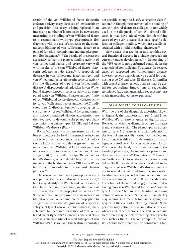
Figure 4. Diagnostic Algorithm for von Willebrand’s Disease.
The diagnosis of von Willebrand’s disease (VWD) begins with a relevant personal or family history of mucocutaneous bleeding. When VWD is suspected, the first level of testing comprises measurements of the VWF antigen (VWF:Ag) level, the platelet-binding activity of VWF (measured by means of a VWF–ristocetin cofactor activity [VWF:RCo] as-say), and factor VIII activity (FVIII:C). (The VWF:RCo assay may be replaced by newer assays that measure the bind-ing of VWF to a recombinant wild-type GPIb fragment with the use of ristocetin or the spontaneous binding of VWF to a gain-of-function recombinant mutant GPIb fragment.) When the results of all first-level tests are normal, VWD is ruled out; because of biologic variability, however, the tests should be repeated if values are at the low end of the normal range or if VWD is strongly suspected. If these first-level tests reveal definitive abnormalities, a diagnosis of VWD can be made; if the results are not conclusive, second-level tests are required. VWD can be subtyped on the basis of these second-level tests. A rare platelet defect due to a gain-of-function mutation in the GPIbα receptor, known as platelet-type VWD, has a phenotype similar to that of type 2B VWD; the two disorders can be distinguished with the use of genetic testing. RIPA denotes ristocetin-induced platelet aggregation, VWF:FVIIIB VWF–FVIII bind-ing activity, and VWFpp VWF propeptide. Persons with a bleeding tendency who have VWF levels between 30 and 50 IU per deciliter (the lower limit of the normal range) are classified as having “low VWF” or “possible type 1 dis-ease” but are not classified as having definitive VWD.
Bleeding Disorder Suspected
Second-Level Tests
VWF:Ag<5 IU/dl
Ratio of VWF:RCo
toVWF:Ag
≤0.6
Type 3 VWDSevere
Type 1 VWD
VWFpp<5 IU/dl VWFpp
≥5 IU/dl
Ratio of FVIII:C
toVWF:Ag
>0.6and
MultimersNormal
Type 1 VWD
Ratio of FVIII:Cto
VWF:Ag≤0.6
VWF:FVIIIBReduced
Type 2N VWD
MultimersLoss
of HMWmultimers
MultimersNormal
Type 2M VWD
RIPAEnhanced
RIPANormal
or reduced
Type 2B VWD
Type 2A VWD
MultimersLoss of HMW
multimers
VWF:RCo<30 IU/dl
VWF:RCo<30 IU/dl
and
Ratio of VWF:RCo
toVWF:Ag
>0.6
Low VWF
VWF:RCo30–50 IU/dl
Ratio of FVIII:Cto
VWF:Ag>0.6
and
and
and
First-LevelTests
The New England Journal of Medicine Downloaded from nejm.org on November 23, 2016. For personal use only. No other uses without permission.
Copyright © 2016 Massachusetts Medical Society. All rights reserved.
n engl j med 375;21 nejm.org November 24, 20162074
T h e n e w e ngl a nd j o u r na l o f m e dic i n e
results of the von Willebrand factor–ristocetin cofactor activity assay. Because of low sensitivity and precision, this assay is being replaced in an increasing number of laboratories by new assays measuring the binding of von Willebrand factor to a recombinant wild-type glycoprotein Ibα fragment with the use of ristocetin or the spon-taneous binding of von Willebrand factor to a gain-of-function recombinant mutant glycopro-tein Ibα fragment.46-48 The results of these assays accurately reflect the platelet-binding activity of von Willebrand factor and correlate very well with results of the von Willebrand factor–risto-cetin cofactor activity assay.48 A proportional decrease in von Willebrand factor antigen and von Willebrand factor–ristocetin cofactor activity fits the diagnosis of type 1 von Willebrand’s disease. A disproportional reduction in von Wille-brand factor–ristocetin cofactor activity as com-pared with von Willebrand factor antigen (ratio of von Willebrand factor–ristocetin cofactor activ-ity to von Willebrand factor antigen, ≤0.6) indi-cates type 2 disease. Further subtyping tests, such as assays of von Willebrand factor multimers and ristocetin-induced platelet aggregation, are then required to determine the phenotypic char-acteristics that define types 2A, 2B, and 2M von Willebrand’s disease (Fig. 4).
Factor VIII activity is also measured as a first-line test because the level is frequently reduced in any type of von Willebrand’s disease.20 A reduc-tion in factor VIII activity that is greater than the reduction in von Willebrand factor antigen (ratio of factor VIII activity to von Willebrand factor antigen, ≤0.6) may indicate type 2N von Wille-brand’s disease, which should be confirmed by measuring the binding of factor VIII to von Wille-brand factor in order to rule out mild hemo-philia A.49
The von Willebrand factor propeptide assay is not part of the official disease classification,11 but it may identify von Willebrand factor variants that have increased clearance, on the basis of an increased ratio of propeptide to antigen.45,50 Some authors have proposed that an increase in the ratio of von Willebrand factor propeptide to antigen warrants the designation of a specific subtype of type 1 von Willebrand’s disease, char-acterized by increased clearance of von Wille-brand factor (type 1C).50 However, enhanced clear-ance is a characteristic of several subtypes of von Willebrand’s disease, and this feature is probably
not specific enough to justify a separate classifi-cation.45 Although measurement of the binding of von Willebrand factor to collagen is not widely used in the diagnosis of von Willebrand’s dis-ease, it may have added value for identifying cases of type 2M disease that have specific de-fects in collagen binding, which are usually as-sociated with a mild bleeding phenotype.51
New assays that are faster and combine sev-eral functional aspects in a single approach are currently under development.48,52 Genotyping of the VWF gene is not performed routinely in the diagnosis of von Willebrand’s disease. In selected cases of suspected von Willebrand’s disease, however, genetic analysis may be useful for diag-nosing type 2N and type 2B disease. In families with type 3 disease, genetic analysis may be use-ful for counseling. Innovations in sequencing techniques (e.g., next-generation sequencing) have made genotyping easier to perform.9
Di agnos tic Con trov er sies
With the use of the diagnostic algorithm shown in Figure 4, the diagnosis of types 2 and 3 von Willebrand’s disease is quite straightforward. However, a definitive diagnosis of type 1 disease is more controversial. The phenotypic character-istic of type 1 disease is a partial reduction in the level of intrinsically normal von Willebrand factor, but it is difficult to determine an unam-biguous cutoff level for von Willebrand factor. The lower the level, the more consistent the bleeding phenotype, the inheritance pattern, and the identification of VWF mutations.35,37 Levels of von Willebrand factor–ristocetin cofactor activity below 30 IU per deciliter are considered to be diagnostic of von Willebrand’s disease. Accord-ing to several current guidelines, persons with a bleeding tendency who have von Willebrand fac-tor levels between 30 and 50 IU per deciliter (the lower limit of the normal range) are classified as having “low von Willebrand factor” or “possible type 1 disease” but are not classified as having definitive von Willebrand’s disease, although they may require treatment before undergoing sur-gery or in the event of a bleeding episode. Some persons may actually have mutations in VWF, whereas in other persons, the von Willebrand factor level may be determined by other genetic loci, such as the ABO blood group.37 A low von Willebrand factor level can be considered a bio-
The New England Journal of Medicine Downloaded from nejm.org on November 23, 2016. For personal use only. No other uses without permission.
Copyright © 2016 Massachusetts Medical Society. All rights reserved.

n engl j med 375;21 nejm.org November 24, 2016 2075
Von Willebr and’s Disease
marker for an increased risk of bleeding.53 It is also possible that other hemostatic defects, such as mild platelet-function disorders, contribute to the bleeding phenotype in these patients.54
A further diagnostic dilemma is posed by the physiologic increase in von Willebrand factor levels with age throughout life. This may have an effect on the diagnosis of von Willebrand’s dis-ease. In childhood, the physiologically lower levels may be erroneously interpreted as indica-tive of von Willebrand’s disease. Elderly patients who met the diagnostic criteria when they were younger may no longer have a von Willebrand factor level below the lower limit of the refer-ence range but may still appear to be at risk for bleeding.29,30
Tr e atmen t
Treatment of von Willebrand’s disease is based on normalizing von Willebrand factor and factor VIII levels in case of bleeding or before an inter-vention. This can be achieved by increasing the endogenous factor levels with the use of desmo-pressin or by infusing exogenous coagulation factors in the form of a high-purity von Wille-brand factor concentrate or a low-purity factor VIII–von Willebrand factor concentrate (Table 1).
Desmopressin
For most patients who have type 1 von Wille-brand’s disease or who are classified as having a low level of von Willebrand factor or possible type 1 von Willebrand’s disease, and for some
patients with type 2 von Willebrand’s disease, desmopressin can be given intravenously (0.3 μg per kilogram of body weight), intranasally (300 μg [150 μg per nostril] or, in a patient with a body weight of <50 kg, only one dose of 150 μg), or subcutaneously (0.3 μg per kilogram) to increase factor VIII and von Willebrand factor levels by two to four times. The use of a capped desmo-pressin dose of 15 or 20 μg (administered intra-venously or subcutaneously) has been suggested, but this approach requires further study before it can be recommended.55,56 The desmopressin dose can be repeated after 12 to 24 hours, de-pending on the individual response. Desmopres-sin is considered to be safe but may have mild side effects such as hypotension and flushing. More severe, rare side effects are cardiovascular complications and hyponatremia. The latter can be prevented by limiting fluid intake to 1500 ml for 24 hours after the administration of desmo-pressin.56,57 Various factors influence the response to desmopressin, including the genotype and phenotype of von Willebrand’s disease.58 There-fore, a test-dose infusion is recommended to establish the magnitude and duration of the re-sponse to desmopressin.
Factor Concentrate
In patients with type 3 von Willebrand’s disease and in most patients with type 2 disease, von Willebrand factor–containing concentrate is the treatment of choice. Plasma-derived concentrates of factor VIII and von Willebrand factor are com-mercially available in various ratios.59 Therefore,
Disease Type Treatment Alternative or Additional Treatment
Low VWF† Desmopressin, administered intravenously (0.3 μg per kilogram of body weight), intranasally (total dose, 300 μg [150 μg per nostril]; in patients with body weight <50 kg, only one dose of 150 μg), or subcutaneously (0.3 μg per kilogram)
Alternative or additional treatment: tranexamic acid (1 g, 3 or 4 times daily)
Type 1 Desmopressin, at same doses as above Additional treatment: tranexamic acid, at same dose as above
Type 2 Desmopressin, at same doses as above, or VWF–factor VIII or VWF concentrate‡
Additional treatment: tranexamic acid, at same dose as above
Type 3 VWF–factor VIII or VWF concentrate Additional treatment: tranexamic acid, at same dose as above
* VWF denotes von Willebrand factor.† Patients presenting with bleeding symptoms and VWF levels between 30 and 50 IU per deciliter (the lower limit of the
normal range) are classified as having low VWF but not von Willebrand’s disease.‡ Desmopressin is contraindicated in patients with type 2B disease.
Table 1. Treatment of von Willebrand’s Disease.*
The New England Journal of Medicine Downloaded from nejm.org on November 23, 2016. For personal use only. No other uses without permission.
Copyright © 2016 Massachusetts Medical Society. All rights reserved.

n engl j med 375;21 nejm.org November 24, 20162076
T h e n e w e ngl a nd j o u r na l o f m e dic i n e
dosing is concentrate-dependent. Recommenda-tions for the administration of von Willebrand factor concentrates are given in Table 2. High-purity von Willebrand factor concentrates with low amounts of factor VIII are also available.60 Infusion of pure von Willebrand factor concen-trate will normalize levels of von Willebrand fac-tor instantaneously, without an immediate in-crease in factor VIII levels. However, factor VIII levels will gradually increase through the bind-ing of endogenously synthesized factor VIII to the infused von Willebrand factor, which pre-vents the breakdown and increases the half-life of factor VIII. In the case of an acute bleeding episode or surgery, both of which require im-mediate normalization of von Willebrand factor and factor VIII levels, additional factor VIII con-centrate should be infused together with the high-purity von Willebrand factor concentrate.60
In a patient who has a bleeding episode or is undergoing surgery, the aim is to normalize the levels of von Willebrand factor–ristocetin cofac-tor activity and factor VIII activity with the use of desmopressin or von Willebrand factor con-centrate. During major surgery, both values should be higher than 100 IU per deciliter to ensure normal hemostasis. Trough levels should
be monitored regularly after the surgical proce-dure, with values maintained at a level above 50 IU per deciliter for 7 to 10 days in the case of major surgery and for 1 to 5 days in the case of minor surgery, depending on the specific intervention (Table 2).5,61 Whether factor VIII levels or von Willebrand factor levels should be used to mon-itor the response to treatment is still debated, and until that question is answered, it is reason-able to measure both. Especially during treat-ment with low-purity von Willebrand factor concentrates, factor VIII levels may rise to sup-raphysiologic levels, potentially predisposing patients to thrombotic complications, although such complications have been reported only sporadically.61,62
Patients with von Willebrand’s disease who are treated with coagulation factor concentrate should be treated in centers that have extensive experience in the management of bleeding dis-orders and have access to a local hemostasis laboratory. This also applies to the counseling and care of women with von Willebrand’s dis-ease during labor.5,10
Recombinant factor concentrates may reduce the risk of transferred viral infections and allergic reactions. In recent years, a recombinant von
Indication for VWF–Factor VIII or VWF Concentrate* Dose†
Target Levels for VWF–Ristocetin Cofactor Activity and Factor VIII Activity‡
Duration of Treatment
IU/kg IU/dl days
Bleeding
Mild to moderate 20–40 Peak, >50–80 on day 1; trough, >30 after day 1 1–3
Severe 50 Peak, >100 on day 1; trough, >50 after day 1 7–10
Intervention
Dental extraction 25 Peak, >50 on day 1 1
Minor surgery 30–60 Peak, >50–80 on day 1; trough, >30 after day 1 1–5
Major surgery 50–60 Peak, >100 on day 1; trough >50 after day 1 7–10
Delivery 40–50 Peak >100 on day 1; trough, >50 after day 1 3–4
* VWF–factor VIII or VWF concentrate is administered in patients with type 3 disease and in patients with type 1 or 2 dis-ease who do not have a response to desmopressin or in whom it is contraindicated.
† The dose of factor concentrate depends on the type of concentrate used. If VWF–factor VIII concentrate is used, the dose of factor concentrate also depends on the brand of concentrate. The dose is based on an anticipated in vivo recovery (2 IU per deciliter for every unit of factor VIII activity infused per kilogram of body weight and 1.5 IU per deciliter for every unit of VWF–ristocetin cofactor activity infused per kilogram) and the target levels of both VWF–ristocetin cofactor activity and factor VIII activity. If high-purity or recombinant VWF concentrate is administered, a single dose of factor VIII concentrate should also be administered in order to achieve the target level of factor VIII immediately.
‡ Factor VIII activity, and preferably also VWF–ristocetin cofactor activity, should be monitored regularly in all patients undergoing surgical procedures and all patients with severe bleeding episodes. If measurement of VWF–ristocetin co-factor activity is not immediately available at a local laboratory, dosing should be based on factor VIII activity levels.
Table 2. Indications for and Doses of Factor Concentrate in von Willebrand’s Disease.
The New England Journal of Medicine Downloaded from nejm.org on November 23, 2016. For personal use only. No other uses without permission.
Copyright © 2016 Massachusetts Medical Society. All rights reserved.
n engl j med 375;21 nejm.org November 24, 2016 2077
Von Willebr and’s Disease
Willebrand factor concentrate has been developed. A phase 1 trial showed that it has increased specific activity, as compared with plasma-derived von Willebrand factor.63 Infusion of recombinant von Willebrand factor together with a single dose of recombinant factor VIII was shown to have high efficacy as a treatment to stop bleeding, without significant side effects, in patients with severe von Willebrand’s disease.64 In 2015, recom-binant von Willebrand factor was approved in the United States by the Food and Drug Admin-istration for the treatment of bleeding episodes in adults with von Willebrand’s disease.
Prophylactic Treatment
In contrast to patients with hemophilia, regular infusion of factor concentrate to prevent bleed-ing is not commonly used in patients with von Willebrand’s disease, although beneficial results have been observed in several case series.65,66 Recently, a prospective dose-escalating study evaluated prophylaxis in patients who had a severe bleeding phenotype of von Willebrand’s disease.67 Although only 11 patients were included in this study, which was stopped because of slow en-rollment, a significant reduction in the number of bleeding episodes, including gastrointestinal bleeding, joint bleeding, and severe epistaxis, was observed in patients receiving regular prophy-laxis. Most of the patients were treated with 50 IU of von Willebrand factor–ristocetin cofactor activity per kilogram two or three times a week.
Treatment of Gynecologic Bleeding
In women with von Willebrand’s disease who have menorrhagia, gynecologic and hormonal abnormalities should be ruled out. Once they have been ruled out, treatment should be initi-ated with oral contraceptives containing both progestin and estrogen. In addition, antifibrino-lytic treatment with tranexamic acid may reduce menstrual blood loss.68 A levonorgestrel-releasing intrauterine device can be placed in patients who do not have a response to oral contraceptives.69 Even more severe cases may benefit from desmo-pressin or von Willebrand factor concentrate.68 If these approaches are not successful, endometrial ablation or hysterectomy can be performed.70
In healthy pregnant women, factor VIII and von Willebrand factor levels increase by a factor of 2 to 3 at the time of labor, and these increases are also seen in women with type 1 von Wille-
brand’s disease. In women with type 2 disease, the level of von Willebrand factor antigen also rises; however, von Willebrand factor activity remains reduced as a result of the functional defect of the protein.71 To prevent postpartum hemorrhage, women are treated with factor con-centrates during labor if factor VIII or von Wille-brand factor levels are less than 50 IU per deci-liter in the third trimester of pregnancy. In such cases, the goal of replacement therapy is levels of von Willebrand factor and factor VIII that are higher than 100 IU per deciliter at the time of delivery, ensuring trough levels of more than 50 IU per deciliter. In patients with type 1 disease who do not have sufficiently high factor VIII or von Willebrand factor levels despite the physiologic rise in values during pregnancy, desmopressin can be administered, preferably after clamping of the umbilical cord. Tranexamic acid can be safely administered in the postpartum period. Despite these measures, women with von Willebrand’s disease have more postpartum bleeding than women without von Willebrand’s disease.27,28,71
Additional Treatment
Fibrinolysis inhibitors, such as tranexamic acid and aminocaproic acid, are important as addi-tional treatment of mucocutaneous bleeding. They can also be used to reduce bleeding and especially to prevent rebleeding in patients under-going surgical or dental interventions, although evidence from randomized trials is lacking.72 Besides systemic treatment for bleeding or sur-gery, local measures, such as use of tranexamic acid mouthwash or additional suturing, may be required. Protracted bleeding despite treatment with von Willebrand factor concentrate can some-times be managed by platelet transfusion be-cause von Willebrand factor is present within the transfused platelets.61 An innovative treatment strategy is daily subcutaneous administration of interleukin-11, which increases von Willebrand factor and factor VIII levels by a factor of 1.3 to 2.0, presumably by increasing von Willebrand factor messenger RNA levels in patients with type 1 von Willebrand’s disease that is unrespon-sive to treatment with desmopressin.73 One trial showed that interleukin-11 reduced the severity of bleeding in women with menorrhagia.74 Angio-dysplasia-associated gastrointestinal bleeding epi-sodes are frequently severe and not responsive to treatment with von Willebrand factor concentrate.
The New England Journal of Medicine Downloaded from nejm.org on November 23, 2016. For personal use only. No other uses without permission.
Copyright © 2016 Massachusetts Medical Society. All rights reserved.

n engl j med 375;21 nejm.org November 24, 20162078
T h e n e w e ngl a nd j o u r na l o f m e dic i n e
Antiangiogenic drugs may be beneficial in such cases. Several case reports have described the successful use of atorvastatin and thalidomide.18,75 Interleukin-11, atorvastatin, and thalidomide have not yet been approved for use in von Willebrand’s disease.
Fu t ur e Per spec ti v es
In the past decade, new pathophysiological in-sights into von Willebrand’s disease and im-proved diagnostic approaches and treatment options have emerged. Diagnosis will be further improved by the introduction of more reproduc-ible and rapid assays of von Willebrand factor function. Next-generation sequencing will facili-tate the routine identification of mutations in VWF. More widespread use of prophylactic treat-ment for severely affected patients seems war-ranted, considering the reported beneficial effects.
Prospective studies are needed to address the question of whether persons who have low von Willebrand factor levels and a bleeding tendency but do not meet the diagnostic criteria for von Willebrand’s disease benefit from hemostatic treatment during surgical interventions. Similar studies are needed to determine whether treat-ment is warranted for elderly patients with von Willebrand’s disease in whom von Willebrand factor has risen to levels within the normal range.
Dr. Leebeek reports receiving consulting fees from UniQure and Baxalta (Shire) and grant support from CSL Behring and Baxter, all paid to his institution, and lecture fees and travel support from Roche; and Dr. Eikenboom, receiving lecture fees from Roche and grant support from CSL Behring, all paid to his institution. No other potential conflict of interest relevant to this article was reported.
Disclosure forms provided by the authors are available with the full text of this article at NEJM.org.
We thank Wichor M. Bramer, biomedical information spe-cialist at Erasmus University Medical Center, for assistance with the literature search (see the Supplementary Appendix, available with the full text of this article at NEJM.org).
References1. Von Willebrand E. Hereditar pseudo-hemofili. Fin Lakaresallsk Handl 1926; 68: 87-112.2. Springer TA. Von Willebrand factor,Jedi knight of the bloodstream. Blood 2014; 124: 1412-25.3. Zimmerman TS, Ratnoff OD, PowellAE. Immunologic differentiation of classic hemophilia (factor 8 deficiency) and von Willebrand’s disease, with observations on combined deficiencies of antihemo-philic factor and proaccelerin (factor V) and on an acquired circulating anticoagu-lant against antihemophilic factor. J Clin Invest 1971; 50: 244-54.4. Lenting PJ, Casari C, Christophe OD,Denis CV. von Willebrand factor: the old, the new and the unknown. J Thromb Hae-most 2012; 10: 2428-37.5. Nichols WL, Hultin MB, James AH,et al. Von Willebrand disease (VWD): evi-dence-based diagnosis and management guidelines, the National Heart, Lung, and Blood Institute (NHLBI) Expert Panel re-port (USA). Haemophilia 2008; 14: 171-232.6. Rodeghiero F, Castaman G, Dini E.Epidemiological investigation of the prev-alence of von Willebrand’s disease. Blood 1987; 69: 454-9.7. Sadler JE, Mannucci PM, Berntorp E,et al. Impact, diagnosis and treatment of von Willebrand disease. Thromb Haemost 2000; 84: 160-74.8. Laffan MA, Lester W, O’Donnell JS, etal. The diagnosis and management of von Willebrand disease: a United Kingdom Haemophilia Centre Doctors Organization guideline approved by the British Com-mittee for Standards in Haematology. Br J Haematol 2014; 167: 453-65.9. Batlle J, Pérez-Rodríguez A, Corrales I,
et al. Molecular and clinical profile of von Willebrand disease in Spain (PCM-EVW-ES): proposal for a new diagnostic paradigm. Thromb Haemost 2016; 115: 40-50.10. Castaman G, Goodeve A, Eikenboom J. Principles of care for the diagnosis and treatment of von Willebrand disease. Haematologica 2013; 98: 667-74.11. Sadler JE, Budde U, Eikenboom JC, et al.Update on the pathophysiology and classifi-cation of von Willebrand disease: a report of the Subcommittee on von Willebrand Factor. J Thromb Haemost 2006; 4: 2103-14.12. Cuker A, Connors JM, Katz JT, LevyBD, Loscalzo J. A bloody mystery. N Engl J Med 2009; 361: 1887-94.13. Sanders YV, Fijnvandraat K, Boender J,et al. Bleeding spectrum in children with moderate or severe von Willebrand dis-ease: relevance of pediatric-specific bleed-ing. Am J Hematol 2015; 90: 1142-8.14. de Wee EM, Sanders YV, Mauser-Bun-schoten EP, et al. Determinants of bleed-ing phenotype in adult patients with mod-erate or severe von Willebrand disease. Thromb Haemost 2012; 108: 683-92.15. Silwer J. Von Willebrand’s disease inSweden. Acta Paediatr Scand Suppl 1973; 238: 1-159.16. Sanders YV, de Wee EM, Meijer K, et al. Von Willebrand disease in the Nether-lands: the WiN study. Ned Tijdschr Geneeskd 2014; 158: A6518. (In Dutch.)17. Veyradier A, Boisseau P, Fressinaud E,et al. A laboratory phenotype/genotype correlation of 1167 French patients from 670 families with von Willebrand disease: a new epidemiologic picture. Medicine (Baltimore) 2016; 95(11): e3038.18. Makris M, Federici AB, Mannucci PM,et al. The natural history of occult or an-
giodysplastic gastrointestinal bleeding in von Willebrand disease. Haemophilia 2015; 21: 338-42.19. van Galen KPM, Mauser-BunschotenEP, Leebeek FWG. Hemophilic arthropa-thy in patients with von Willebrand dis-ease. Blood Rev 2012; 26: 261-6.20. Federici AB, Bucciarelli P, CastamanG, et al. The bleeding score predicts clini-cal outcomes and replacement therapy in adults with von Willebrand disease. Blood 2014; 123: 4037-44.21. de Wee EM, Mauser-Bunschoten EP,Van Der Bom JG, et al. Health-related quality of life among adult patients with moderate and severe von Willebrand dis-ease. J Thromb Haemost 2010; 8: 1492-9.22. Kadir RA, Edlund M, Von MackensenS. The impact of menstrual disorders on quality of life in women with inherited bleeding disorders. Haemophilia 2010; 16: 832-9.23. Kadir RA, Economides DL, Sabin CA,Owens D, Lee CA. Frequency of inherited bleeding disorders in women with menor-rhagia. Lancet 1998; 351: 485-9.24. Miller CH, Philipp CS, Stein SF, et al.The spectrum of haemostatic characteris-tics of women with unexplained menor-rhagia. Haemophilia 2011; 17(1): e223-9.25. De Wee EM, Knol HM, Mauser-Bun-schoten EP, et al. Gynaecological and ob-stetric bleeding in moderate and severe von Willebrand disease. Thromb Haemost 2011; 106: 885-92.26. James AH. More than menorrhagia:a review of the obstetric and gynaeco-logical manifestations of von Willebrand disease. Thromb Res 2007; 120: Suppl 1: S17-20.27. Stoof SCM, van Steenbergen HW,
The New England Journal of Medicine Downloaded from nejm.org on November 23, 2016. For personal use only. No other uses without permission.
Copyright © 2016 Massachusetts Medical Society. All rights reserved.

n engl j med 375;21 nejm.org November 24, 2016 2079
Von Willebr and’s Disease
Zwagemaker A, et al. Primary postpartum haemorrhage in women with von Wille-brand disease or carriership of haemo-philia despite specialised care: a retro-spective survey. Haemophilia 2015; 21: 505-12.28. Kouides PA. An update on the man-agement of bleeding disorders during pregnancy. Curr Opin Hematol 2015; 22: 397-405.29. Rydz N, Grabell J, Lillicrap D, James PD. Changes in von Willebrand factor level and von Willebrand activity with age in type 1 von Willebrand disease. Haemo-philia 2015; 21: 636-41.30. Sanders YV, Giezenaar MA, Laros-van Gorkom BAP, et al. Von Willebrand dis-ease and aging: an evolving phenotype. J Thromb Haemost 2014; 12: 1066-75.31. Sanders YV, Eikenboom J, de Wee EM, et al. Reduced prevalence of arterial thrombosis in von Willebrand disease. J Thromb Haemost 2013; 11: 845-54.32. Seaman CD, Yabes J, Comer DM, Ragni MV. Does deficiency of von Willebrand factor protect against cardiovascular dis-ease? Analysis of a national discharge register. J Thromb Haemost 2015; 13: 1999-2003.33. Hampshire DJ, Goodeve AC. The Inter-national Society on Thrombosis and Hae-matosis von Willebrand Disease Database: an update. Semin Thromb Hemost 2011; 37: 470-9.34. Bowman M, Tuttle A, Notley C, et al. The genetics of Canadian type 3 von Wil-lebrand disease: further evidence for co-dominant inheritance of mutant alleles. J Thromb Haemost 2013; 11: 512-20.35. Goodeve A, Eikenboom J, Castaman G, et al. Phenotype and genotype of a cohort of families historically diagnosed with type 1 von Willebrand disease in the Euro-pean study, Molecular and Clinical Mark-ers for the Diagnosis and Management of Type 1 von Willebrand Disease (MCMDM-1VWD). Blood 2007; 109: 112-21.36. James PD, Notley C, Hegadorn C, et al. The mutational spectrum of type 1 von Willebrand disease: results from a Cana-dian cohort study. Blood 2007; 109: 145-54.37. Flood VH, Christopherson PA, Gill JC, et al. Clinical and laboratory variability in a cohort of patients diagnosed with type 1 VWD in the United States. Blood 2016; 127: 2481-8.38. Jenkins PV, O’Donnell JS. ABO blood group determines plasma von Willebrand factor levels: a biologic function after all? Transfusion 2006; 46: 1836-44.39. Smith NL, Chen MH, Dehghan A, et al. Novel associations of multiple genetic loci with plasma levels of factor VII, factor VIII, and von Willebrand factor: the CHARGE (Cohorts for Heart and Aging Research in Genome Epidemiology) Consortium. Cir-culation 2010; 121: 1382-92.40. Rydz N, Swystun LL, Notley C, et al. The C-type lectin receptor CLEC4M binds, internalizes, and clears von Willebrand
factor and contributes to the variation in plasma von Willebrand factor levels. Blood 2013; 121: 5228-37.41. Sanders YV, van der Bom JG, Isaacs A, et al. CLEC4M and STXBP5 gene varia-tions contribute to von Willebrand factor level variation in von Willebrand disease. J Thromb Haemost 2015; 13: 956-66.42. Tosetto A, Rodeghiero F, Castaman G, et al. A quantitative analysis of bleeding symptoms in type 1 von Willebrand dis-ease: results from a multicenter European study (MCMDM-1 VWD). J Thromb Hae-most 2006; 4: 766-73.43. Rodeghiero F, Tosetto A, Abshire T, et al. ISTH/SSC bleeding assessment tool: a standardized questionnaire and a pro-posal for a new bleeding score for inher-ited bleeding disorders. J Thromb Hae-most 2010; 8: 2063-5.44. Elbatarny M, Mollah S, Grabell J, et al. Normal range of bleeding scores for the ISTH-BAT: adult and pediatric data from the Merging Project. Haemophilia 2014; 20: 831-5.45. Sanders YV, Groeneveld D, Meijer K, et al. Von Willebrand factor propeptide and the phenotypic classification of von Willebrand disease. Blood 2015; 125: 3006-13.46. Stufano F, Lawrie AS, La Marca S, Ber-benni C, Baronciani L, Peyvandi F. A two-centre comparative evaluation of new au-tomated assays for von Willebrand factor ristocetin cofactor activity and antigen. Haemophilia 2014; 20: 147-53.47. Flood VH, Gill JC, Morateck PA, et al. Gain-of-function GPIb ELISA assay for VWF activity in the Zimmerman Program for the Molecular and Clinical Biology of VWD. Blood 2011; 117(6): e67-74.48. Bodó I, Eikenboom J, Montgomery R, et al. Platelet-dependent von Willebrand factor activity — nomenclature and meth-odology: communication from the SSC of the ISTH. J Thromb Haemost 2015; 13: 1345-50.49. Gupta M, Lillicrap D, Stain AM, Fried-man KD, Carcao MD. Therapeutic conse-quences for misdiagnosis of type 2N von Willebrand disease. Pediatr Blood Cancer 2011; 57: 1081-3.50. Haberichter SL, Balistreri M, Christo-pherson P, et al. Assay of the von Wille-brand factor (VWF) propeptide to identify patients with type 1 von Willebrand dis-ease with decreased VWF survival. Blood 2006; 108: 3344-51.51. Favaloro EJ. Evaluation of commercial von Willebrand factor collagen binding assays to assist the discrimination of types 1 and 2 von Willebrand disease. Thromb Haemost 2010; 104: 1009-21.52. Roberts JC, Morateck PA, Christo-pherson PA, et al. Rapid discrimination of the phenotypic variants of von Willebrand disease. Blood 2016; 127: 2472-80.53. Sadler JE. Von Willebrand disease type 1: a diagnosis in search of a disease. Blood 2003; 101: 2089-93.
54. Gudmundsdottir BR, Marder VJ, Onun-darson PT. Risk of excessive bleeding associated with marginally low von Wille-brand factor and mild platelet dysfunc-tion. J Thromb Haemost 2007; 5: 274-81.55. Siew DA, Mangel J, Laudenbach L, Schembri S, Minuk L. Desmopressin re-sponsiveness at a capped dose of 15 μg in type 1 von Willebrand disease and mild hemophilia A. Blood Coagul Fibrinolysis 2014; 25: 820-3.56. Leissinger C, Carcao M, Gill JC, Jour-neycake J, Singleton T, Valentino L. Desmo-pressin (DDAVP) in the management of patients with congenital bleeding disor-ders. Haemophilia 2014; 20: 158-67.57. Stoof SCM, Cnossen MH, de Maat MPM, Leebeek FWG, Kruip MJHA. Side effects of desmopressin in patients with bleeding disorders. Haemophilia 2016; 22: 39-45.58. Castaman G, Lethagen S, Federici AB, et al. Response to desmopressin is influ-enced by the genotype and phenotype in type 1 von Willebrand disease (VWD): re-sults from the European Study MCMDM-1VWD. Blood 2008; 111: 3531-9.59. Lethagen S, Carlson M, Hillarp A. A comparative in vitro evaluation of six von Willebrand factor concentrates. Hae-mophilia 2004; 10: 243-9.60. Borel-Derlon A, Federici AB, Roussel-Robert V, et al. Treatment of severe von Willebrand disease with a high-purity von Willebrand factor concentrate (Wilfactin): a prospective study of 50 patients. J Thromb Haemost 2007; 5: 1115-24.61. Mannucci PM. Treatment of von Wille-brand’s disease. N Engl J Med 2004; 351: 683-94.62. Coppola A, Franchini M, Makris M, Santagostino E, Di Minno G, Mannucci PM. Thrombotic adverse events to coagu-lation factor concentrates for treatment of patients with haemophilia and von Wille-brand disease: a systematic review of pro-spective studies. Haemophilia 2012; 18(3): e173-87.63. Mannucci PM, Kempton C, Millar C, et al. Pharmacokinetics and safety of a novel recombinant human von Willebrand factor manufactured with a plasma-free method: a prospective clinical trial. Blood 2013; 122: 648-57.64. Gill JC, Castaman G, Windyga J, et al. Hemostatic efficacy, safety, and pharma-cokinetics of a recombinant von Wille-brand factor in severe von Willebrand disease. Blood 2015; 126: 2038-46.65. Berntorp E, Petrini P. Long-term pro-phylaxis in von Willebrand disease. Blood Coagul Fibrinolysis 2005; 16: Suppl 1: S23-6.66. Abshire T. The role of prophylaxis in the management of von Willebrand dis-ease: today and tomorrow. Thromb Res 2009; 124: Suppl 1: S15-9.67. Abshire T, Cox-Gill J, Kempton CL, et al. Prophylaxis escalation in severe von Willebrand disease: a prospective study from the von Willebrand Disease Prophy-
The New England Journal of Medicine Downloaded from nejm.org on November 23, 2016. For personal use only. No other uses without permission.
Copyright © 2016 Massachusetts Medical Society. All rights reserved.
n engl j med 375;21 nejm.org November 24, 20162080
Von Willebr and’s Disease
laxis Network. J Thromb Haemost 2015; 13: 1585-9.68. Kouides PA, Byams VR, Philipp CS,et al. Multisite management study of menorrhagia with abnormal laboratory haemostasis: a prospective crossover study of intranasal desmopressin and oral tranexamic acid. Br J Haematol 2009; 145: 212-20.69. Chi C, Huq FY, Kadir RA. Levonor-gestrel-releasing intrauterine system for the management of heavy menstrual bleeding in women with inherited bleed-ing disorders: long-term follow-up. Con-traception 2011; 83: 242-7.70. Ragni MV, Machin N, Malec LM, et al.Von Willebrand factor for menorrhagia:
a survey and literature review. Haemophilia 2016; 22: 397-402.71. James AH, Konkle BA, Kouides P, et al. Postpartum von Willebrand factor levels in women with and without von Wille-brand disease and implications for pro-phylaxis. Haemophilia 2015; 21: 81-7.72. van Galen KP, Engelen ET, Mauser-Bunschoten EP, van Es RJ, Schutgens RE. Antifibrinolytic therapy for preventing oral bleeding in patients with haemophilia or von Willebrand disease undergoing minor oral surgery or dental extractions. Cochrane Database Syst Rev 2015; 12: CD011385.73. Ragni MV, Novelli EM, Murshed A,Merricks EP, Kloos MT, Nichols TC. Phase II prospective open-label trial of recom-
binant interleukin-11 in desmopressin-unresponsive von Willebrand disease and mild or moderate haemophilia A. Thromb Haemost 2013; 109: 248-54.74. Ragni MV, Jankowitz RC, Jaworski K,Merricks EP, Kloos MT, Nichols TC. Phase II prospective open-label trial of recombi-nant interleukin-11 in women with mild von Willebrand disease and refractory men-orrhagia. Thromb Haemost 2011; 106: 641-5.75. Engelen ET, van Galen KP, SchutgensRE. Thalidomide for treatment of gastro-intestinal bleedings due to angiodysplasia: a case report in acquired von Willebrand syndrome and review of the literature. Haemophilia 2015; 21: 419-29.Copyright © 2016 Massachusetts Medical Society.
The New England Journal of Medicine Downloaded from nejm.org on November 23, 2016. For personal use only. No other uses without permission.
Copyright © 2016 Massachusetts Medical Society. All rights reserved.