vascular cli kanden 70 - shimadzu corporation
TRANSCRIPT
Vascular
Clinical Utility of RSM-DSA for Lower Extremity intervention
Dr. Kazuaki Kataoka Department of Cardiovascular Medicine, Kansai Denryoku Hospital
Kazuaki Kataoka
1. Introduction
Kansai Denryoku Hospital is located in the city of
Osaka. It comprises 23 medical departments with
400 beds. The original hospital was built in 1967,
but a new hospital is currently under construction.
Stage 1 is scheduled for completion in 2012 and
Stage 2 in 2014.
The hospital was originally established to look after
the health and welfare of employees at Kansai
Denryoku (Kansai Electric Power Co. Inc.) and
offered a lower level of medical care than other
hospitals in Osaka City. To enhance acute care
in the face of current medical circumstances,
the new hospital will incorporate a new
cardiovascular center that includes a new cardiac
surgery department. I was appointed director of
cardiovascular medicine in March 2010. Prior to
2009, the hospital handled less than 100
interventions a year, but in 2010 this grew to 209
coronary artery interventions and 50 peripheral
artery interventions per year. The greater number
of interventions and the large number of complex
lesions treated, such as chronic total occlusions,
stretched the I.I. angiography system we used at
the time to its limit. The angiography room at this
hospital is a single room containing a dual-plane
angiographic system used for neuro and abdominal
angiography as well as cardiac angiography, which
is somewhat unusual nowadays. All operations
have to be handled by a single system until the
new hospital is established, and the number of
manufacturers who could supply such a system
was limited to a few companies at the time.
After comparing various products, we decided
on the dual-plane angiography system with
direct-conversion flat panel detector (FPD) from
Shimadzu Corporation. We spent the month of
August 2010 installing the system, which
commenced operation in September.
2. System Overview
As described above, a dual-plane angiography
system is used at this hospital. A single room contains
a BRANSIST safire with 9" × 9" direct-conversion FPD
for cardiac angiography and angiography of the
lower extremities and a BRANSIST safire with
17" × 17" FPD for neuro and abdominal
angiography. Table 1 shows an overview of the
system. The 9" × 9" FPD is used for the coronary
artery and arteries in the lower extremities. The
system permits both coronary angiography and
lower-extremity arteriography by RSM-DSA with
the patient in the same posture.
Dual-plane system
Main examination region
Cardiac (PCI)
Lower extremities (PPI)
Abdominal angiography (TAE)
Neuroangiography
FPD view field
9 × 9 inches 17 × 17 inches
C-arm Floor-mounted 6-axis C-arm
Ceiling-mounted C-arm
FPD type Direct-conversion Direct-conversion
X-ray tube 3.0 MHU tube 3.0 MHU tube
Monitor 6-plane monitor 6-plane monitor (shared)
Image processing functions
SUREengine PRO
Realtime DSA
RSM-DSA
Dynamic reference
Saving video fluoroscopic images
SUREengine
Realtime DSA
RSM-DSA
Dynamic reference
Saving video fluoroscopic images
3D workstation
Table 1 System Overview
3. Clinical Experience Using RSM-DSA
Case 1
78-year-old male. The patient has a medical
history of stent placement in the right coronary
artery due to silent myocardial ischemia. The
patient was referred to this hospital because of
intermittent claudication in the left lower extremity
on walking approximately 500 m. Ankle-brachial
indices (ABI) of 1.01 at the right and 0.58 at the left
lead to suspicion of reduced blood flow in the left
lower extremity. Vascular ultrasonography confirmed
total occlusion approximately 20 cm below the
region of origin of the left superficial femoral artery.
The patient was therefore hospitalized for
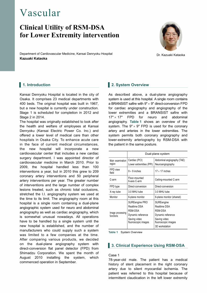
endovascular therapy. Initially, coronary angiography
confirmed restenosis in the right coronary artery
stent and severe stenosis of the left anterior
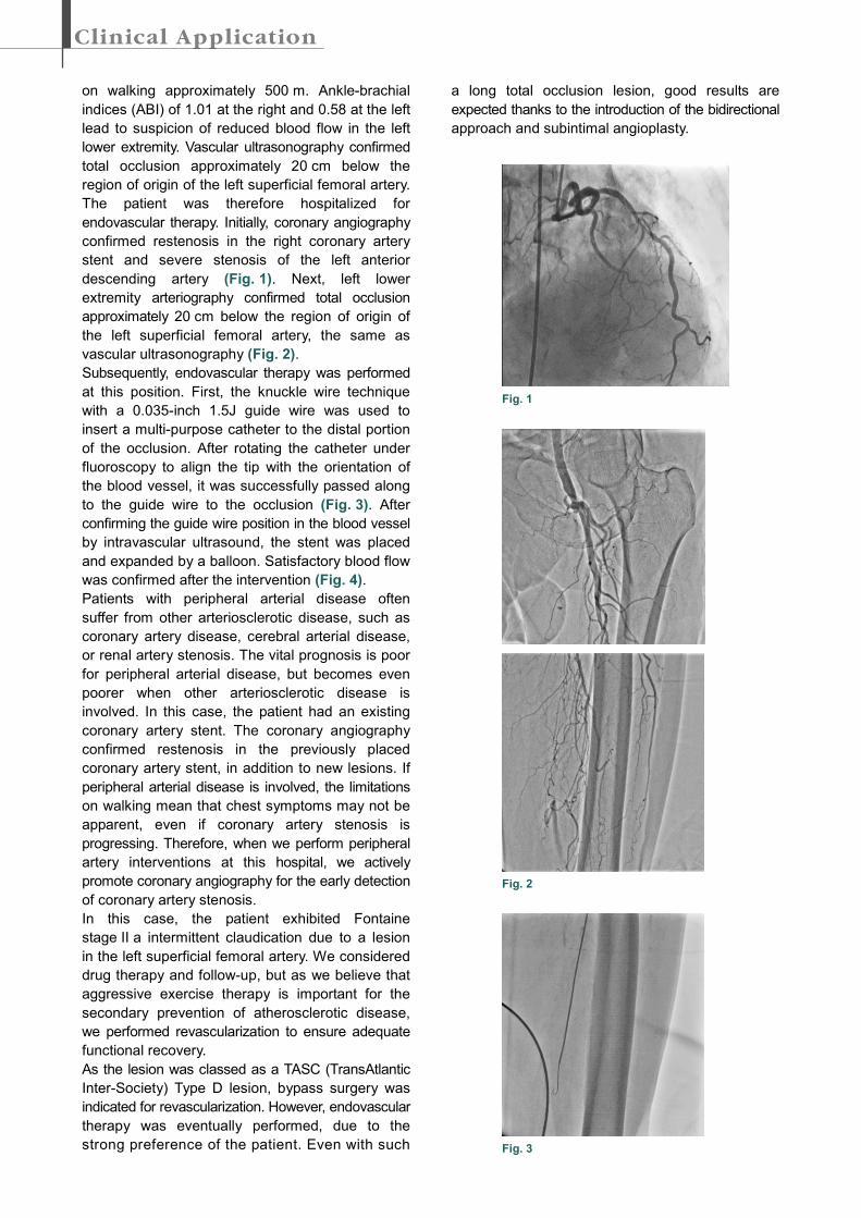
descending artery (Fig. 1). Next, left lower
extremity arteriography confirmed total occlusion
approximately 20 cm below the region of origin of
the left superficial femoral artery, the same as
vascular ultrasonography (Fig. 2).
Subsequently, endovascular therapy was performed
at this position. First, the knuckle wire technique
with a 0.035-inch 1.5J guide wire was used to
insert a multi-purpose catheter to the distal portion
of the occlusion. After rotating the catheter under
fluoroscopy to align the tip with the orientation of
the blood vessel, it was successfully passed along
to the guide wire to the occlusion (Fig. 3). After
confirming the guide wire position in the blood vessel
by intravascular ultrasound, the stent was placed
and expanded by a balloon. Satisfactory blood flow
was confirmed after the intervention (Fig. 4).
Patients with peripheral arterial disease often
suffer from other arteriosclerotic disease, such as
coronary artery disease, cerebral arterial disease,
or renal artery stenosis. The vital prognosis is poor
for peripheral arterial disease, but becomes even
poorer when other arteriosclerotic disease is
involved. In this case, the patient had an existing
coronary artery stent. The coronary angiography
confirmed restenosis in the previously placed
coronary artery stent, in addition to new lesions. If
peripheral arterial disease is involved, the limitations
on walking mean that chest symptoms may not be
apparent, even if coronary artery stenosis is
progressing. Therefore, when we perform peripheral
artery interventions at this hospital, we actively
promote coronary angiography for the early detection
of coronary artery stenosis.
In this case, the patient exhibited Fontaine
stage II a intermittent claudication due to a lesion
in the left superficial femoral artery. We considered
drug therapy and follow-up, but as we believe that
aggressive exercise therapy is important for the
secondary prevention of atherosclerotic disease,
we performed revascularization to ensure adequate
functional recovery.
As the lesion was classed as a TASC (TransAtlantic
Inter-Society) Type D lesion, bypass surgery was
indicated for revascularization. However, endovascular
therapy was eventually performed, due to the
strong preference of the patient. Even with such
a long total occlusion lesion, good results are
expected thanks to the introduction of the bidirectional
approach and subintimal angioplasty.
Fig. 1
Fig. 2
Fig. 3
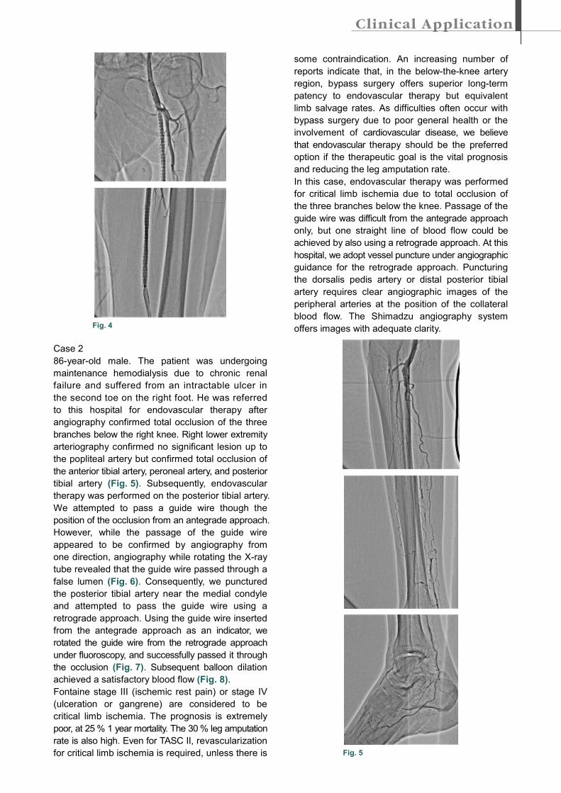
Fig. 4
Case 2
86-year-old male. The patient was undergoing
maintenance hemodialysis due to chronic renal
failure and suffered from an intractable ulcer in
the second toe on the right foot. He was referred
to this hospital for endovascular therapy after
angiography confirmed total occlusion of the three
branches below the right knee. Right lower extremity
arteriography confirmed no significant lesion up to
the popliteal artery but confirmed total occlusion of
the anterior tibial artery, peroneal artery, and posterior
tibial artery (Fig. 5). Subsequently, endovascular
therapy was performed on the posterior tibial artery.
We attempted to pass a guide wire though the
position of the occlusion from an antegrade approach.
However, while the passage of the guide wire
appeared to be confirmed by angiography from
one direction, angiography while rotating the X-ray
tube revealed that the guide wire passed through a
false lumen (Fig. 6). Consequently, we punctured
the posterior tibial artery near the medial condyle
and attempted to pass the guide wire using a
retrograde approach. Using the guide wire inserted
from the antegrade approach as an indicator, we
rotated the guide wire from the retrograde approach
under fluoroscopy, and successfully passed it through
the occlusion (Fig. 7). Subsequent balloon dilation
achieved a satisfactory blood flow (Fig. 8).
Fontaine stage III (ischemic rest pain) or stage IV
(ulceration or gangrene) are considered to be
critical limb ischemia. The prognosis is extremely
poor, at 25 % 1 year mortality. The 30 % leg amputation
rate is also high. Even for TASC II, revascularization
for critical limb ischemia is required, unless there is
some contraindication. An increasing number of
reports indicate that, in the below-the-knee artery
region, bypass surgery offers superior long-term
patency to endovascular therapy but equivalent
limb salvage rates. As difficulties often occur with
bypass surgery due to poor general health or the
involvement of cardiovascular disease, we believe
that endovascular therapy should be the preferred
option if the therapeutic goal is the vital prognosis
and reducing the leg amputation rate.
In this case, endovascular therapy was performed
for critical limb ischemia due to total occlusion of
the three branches below the knee. Passage of the
guide wire was difficult from the antegrade approach
only, but one straight line of blood flow could be
achieved by also using a retrograde approach. At this
hospital, we adopt vessel puncture under angiographic
guidance for the retrograde approach. Puncturing
the dorsalis pedis artery or distal posterior tibial
artery requires clear angiographic images of the
peripheral arteries at the position of the collateral
blood flow. The Shimadzu angiography system
offers images with adequate clarity.
Fig. 5
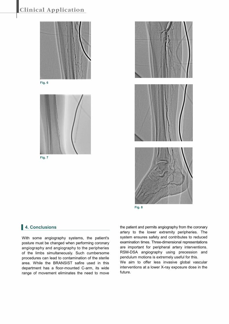
Fig. 6
Fig. 7
Fig. 8
4. Conclusions
With some angiography systems, the patient's
posture must be changed when performing coronary
angiography and angiography to the peripheries
of the limbs simultaneously. Such cumbersome
procedures can lead to contamination of the sterile
area. While the BRANSIST safire used in this
department has a floor-mounted C-arm, its wide
range of movement eliminates the need to move
the patient and permits angiography from the coronary
artery to the lower extremity peripheries. The
system ensures safety and contributes to reduced
examination times. Three-dimensional representations
are important for peripheral artery interventions.
RSM-DSA angiography using precession and
pendulum motions is extremely useful for this.
We aim to offer less invasive global vascular
interventions at a lower X-ray exposure dose in the
future.