vascular deformation mapping (vdm) of thoracic aortic ... · pdf filevascular deformation...
TRANSCRIPT

Vascular Deformation Mapping (VDM) ofThoracic Aortic Enlargement in AneurysmalDisease and DissectionNicholas S. Burris1, Benjamin A. Hoff1,2, Ella A. Kazerooni1, and Brian D. Ross1,2,3
1Department of Radiology; 2Center for Molecular Imaging; and 3Department of Biological Chemistry, University of Michigan, Ann Arbor, MI
Corresponding Author:Nicholas Burris, MDDepartment of Radiology, University of Michigan, 1500 E. MedicalCenter Drive, TC B1-132, SPC-5030, Ann Arbor, MI 48109-5030;E-mail: [email protected]
Key Words: aortic aneurysm, spatial Jacobian, aortic dissection, 3D printing, aneurysmalenlargement, vascular deformation mappingAbbreviations: Vascular deformation mapping (VDM), abdominal aortic aneurysm (AAA),thoracic aortic aneurysm (TAA), electrocardiogram (ECG), computed tomography angiography(CTA), Hounsfield unit (HU), computed tomography (CT), matrix metalloproteinases (MMPs)
Thoracic aortic aneurysm is a common and lethal disease that requires regular imaging surveillance to deter-mine timing of surgical repair and prevent major complications such as rupture. Current cross-sectional imag-ing surveillance techniques, largely based on computed tomography angiography, are focused on measure-ment of maximal aortic diameter, although this approach is limited to fixed anatomic positions and is proneto significant measurement error. Here we present preliminary results showing the feasibility of a novel tech-nique for assessing change in aortic dimensions, termed vascular deformation mapping (VDM). This tech-nique allows quantification of 3-dimensional changes in the aortic wall geometry through nonrigid coregistra-tion of computed tomography angiography images and spatial Jacobian analysis of aortic deformation.Through several illustrative cases we demonstrate that this method can be used to measure changes in theaortic wall geometry among patients with stable and enlarging thoracic aortic aneurysm and dissection.Furthermore, VDM results yield observations about the presence, distribution, and rate of aortic wall defor-mation that are not apparent by routine clinical evaluation. Finally, we show the feasibility of superposingpatient-specific VDM results on a 3-dimensional aortic model using color 3D printing and discuss future direc-tions and potential applications for the VDM technique.
INTRODUCTIONThe thoracic aorta is the largest blood vessel in the human bodyand is subject to most extreme hemodynamic forces. A healthyaorta is extremely durable, and it is able to absorb forces gen-erated by the heart owing to its thick walls and its elastic nature.Because of multiple factors (eg, hypertension, atherosclerosis,genetic aortic syndromes, infection), the structural integrity andelasticity of the aortic wall can deteriorate, leading to progres-sive dilation of the aortic lumen and formation of aortic aneu-rysm (1). Aortic dissection is a related form of aortic diseasecharacterized by tearing of the inner layers of the aortic wall (ie,intima and media), leading to the creation of a false lumen—orchannel—within the aortic wall itself, which is structurally com-promised and is subjected to high pressures. This results inaneurysm formation in �60% of patients with chronic aorticdissection of the descending thoracic aorta (Stanford type B) (2).The incidence of aortic aneurysm is increasing in the US popu-lation, and mildly dilated aortas are being incidentally detectedat higher rates owing to increased use of thoracic cross-sectionalimaging for nonaortic indications (eg, lung cancer screening)(3). Recent data suggest that the prevalence of thoracic aortic
dilation (�4 cm) is �3% of individuals older than 55 years ofage, which, on the basis of current US population estimates,means that �2.7 million people in the USA would be recom-mended to undergo regular imaging of the thoracic aorta on thebasis of the current American Heart Association guidelines forimaging surveillance (4-8).
Imaging surveillance has a central role in the managementof asymptomatic patients with aortic disease. The vast majorityof patients with an aortic aneurysm, �95%, are asymptomaticuntil the aneurysm ruptures, and only 40% of patients in whomthe aneurysm ruptures reach the hospital alive (6, 9). Althoughthe topic of aortic enlargement in abdominal aortic aneurysm(AAA) before and after endovascular repair has been the focus ofsignificant research effort, the natural history and mechanismsof thoracic aortic aneurysm (TAA) progression remain poorlyunderstood, and only a handful of studies have attempted tomeasure growth rates of the thoracic aorta (10-16). A majorlimitation in improving our understanding of TAAs is that thecurrent clinical imaging surveillance techniques rely primarilyon measurement of maximal aortic diameter. This parameter hasbeen most widely studied and is shown to correlate with future
RESEARCH ARTICLE
ABST
RA
CT
© 2017 The Authors. Published by Grapho Publications, LLC This is an open access article under the CC BY-NC-ND license (http://creativecommons.org/licenses/by-nc-nd/4.0/).ISSN 2379-1381 http://dx.doi.org/10.18383/j.tom.2017.00015
TOMOGRAPHY.ORG | VOLUME 3 NUMBER 3 | SEPTEMBER 2017 163
risk of aneurysm rupture (17). Although the simplicity of diam-eter measurements is appealing, this approach is subject to ahigh degree of measurement error, in the range of 2–5 mmdespite optimal measurement technique (18, 19). Error of thismagnitude makes confident determination of aortic enlarge-ment challenging considering that typical aortic growth ratesare slow (eg, 1 mm/y in the ascending aorta and 3 mm/y in thedescending aorta), and this issue is further compounded whenshorter follow-up intervals are analyzed (3 or 6 months) andwhen the aortic geometry is ovoid (17).
Although several sections of the aorta are vertically ori-ented and can be viewed in cross-section on axial images, mostof the aorta cannot be viewed in cross-section on standardimage planes, requiring image-processing software to effec-tively straighten the aorta and allow true orthogonal diametermeasurements to be made. The 2010 ACCF/AHA/AATS/ACR/ASA/SCA/SCAI/SIR/STS/SVM Guidelines for the Diagnosis andManagement of Patients With Thoracic Aortic Disease was thefirst set of guidelines to raise this issue and to recommendstandard measurement locations in addition to measurement ofmaximal aortic diameter (4). Even with orthogonal measure-ments, the aorta is often not perfectly round in the cross-section,but it is rather ovoid or irregular, particularly in the setting ofdisease, further compounding the issue of exactly which diam-eter measurements to record and use for follow-up. Measuringthe aortic diameter at predefined anatomic locations fails tocapture interval growth at nonmaximal locations, and this mea-surement does not detect the components of aortic enlargementin circumferential or longitudinal directions. Height-/weight-adjusted aortic area has been proposed as a better predictor offuture rupture than maximal diameter, and several studies haveinvestigated the use of volumetric measurements of TAA andAAA to improve the sensitivity for detecting aortic growth (10,20-24). However, similar to diameter measurements, measure-ment of aortic area and volumetric should be performed atpredetermined anatomic boundaries to ensure that measure-ments are comparable between studies, and small focal changesin aortic dimension may be camouflaged by a volumetricmeasurement approach. Although area or volume measuresmay be more sensitive to detect overall growth of an aorticsegment, information about localized change at a specificpoint along the aortic wall is not captured. Considering thatsurgical management recommendations are based on thresh-olds of size and growth rate, a diameter-based measurementtechnique may lead to treatment recommendations that areeither overly aggressive or conservative on the basis of themeasurement error alone. In addition, such size criteria usedfor surgical decision-making are based on historical mea-surement data and its inaccuracies, further emphasizing thesignificant ongoing need for accurate and reproducible aorticmeasurements.
A potential solution to these challenges exists in the field ofdiffeomorphic image registrations, particularly the applicationof spatial Jacobian matrices to quantify the deformation of theaortic wall by extracting nonrigid image transformations tomatch high-resolution thoracic electrocardiogram (ECG)-gatedcomputed tomography angiography (CTA) images acquired atdifferent time points, a technique that we term vascular defor-
mation mapping (VDM). Spatial Jacobian matrices in this con-text describe the relative local distortion at each point in theimage resulting from the automated image-based registration.Although nonrigid image warping coregistration techniqueshave been broadly used in diseases of the lungs and brain, to thebest of our knowledge, no prior studies have quantified spatialJacobian maps to assess interval aortic enlargement (25-27).Image intensity-based registration techniques have been re-ported to have submillimeter precision in many applications,which also translate to accuracy in the calculation of the result-ing spatial Jacobian with well-optimized workflows (28). Quan-titative assessment of registration accuracy is not straightfor-ward, with many potential sources of error and a large numberof degrees of freedom; however, the use of cost function penal-ties such as bending energy helps to constrain and smoothspatial Jacobian results while also maximizing anatomical fea-ture alignment. Jacobian maps may be directly calculated fromthe optimized nonrigid transform, and it could offer informationabout the local deformation of the aortic wall, including in thecircumferential and longitudinal directions, information that isnot currently assessed by other techniques.
A significant need exists for a more sensitive and accuratemethod of measuring change in thoracic aortic dimensions,considering that accurate detection of small-magnitude changeshas important implications for improving understanding of aor-tic aneurysm progression and better informing treatment deci-sions. The aim of this study was to demonstrate the feasibility ofusing the proposed VDM technique to measure interval changein aortic wall dimensions on routine clinical CTA studies ofpatients with TAA and dissection. In addition, we aim to com-pare the results of the VDM analysis with routine clinical CTAassessments in seeking to better understand the potential bene-fits and limitations of this novel technique.
METHODS AND MATERIALSStudy PopulationAll procedures were approved by the local institutional reviewboard with a waiver of informed consent obtained for thisretrospective study and were Health Insurance Portability andAccountability Act-compliant. Patients were identified througha review of local picture archiving and communication systemarchives to identify adult (�18 years) patients with dilation ofthe thoracic aorta undergoing imaging surveillance, with atleast 2 prior ECG-gated CTA examinations available for review.Patients were excluded if thoracic aortic enhancement was sub-optimal (�250 Hounsfield unit [HU]) or there was significantmotion/respiratory artifact affecting the clinical evaluation ofthoracic aortic segments. After reviewing 15 patients, severalwere excluded owing to obvious pulsation artifact affectingthe diseased aortic segment on CTA images (n � 5) or low-resolution baseline CTA studies (section thickness � 1.5 mm)that were acquired at outside hospitals and uploaded to ourpicture archiving and communication system (n � 4). Onepatient with type B aortic dissection was excluded fromanalysis owing to difficulties with accurate segmentation ofthe false lumen due to poor enhancement related to slow flowand partial thrombosis. The aortic pathologies of those pa-tients selected for analysis included ascending thoracic aortic
VDM of Thoracic Aortic Enlargement
164 TOMOGRAPHY.ORG | VOLUME 3 NUMBER 3 | SEPTEMBER 2017

dilation (n � 1), descending TAA (n � 1), and thoracic aorticdissection (n � 3).
Computed Tomography AngiographyCTA examinations were performed on 64-detector computedtomography (CT) scanners using helical acquisition mode(LightSpeed VCT or Discovery CT750HD, GE Healthcare, Wauke-sha, WI). Images were acquired through the entire thoracic aorta(lung apices to 2 cm below the celiac artery) during intravenousinjection of 95-mL iopamidol 370 mg I/mL (Isovue 370, BraccoDiagnostics, Inc., Princeton, NJ) at 4 mL/s, followed by a 100-mlsaline chaser at 4 mL/s. Retrospective ECG-gating was used,with axial reconstructions at 0.625-mm section thickness at75% of the R–R cycle with ECG-modulated tube current tech-nique (20% of maximum milliampere) and 40% adaptive sta-tistical iterative reconstruction for dose efficiency. Other scanparameters included the following: detector coverage, 40mm; Display Field of View, 25 cm; gantry rotation time, 0.4seconds; maximum tube current, 400–700 milliampere asdetermined by patient size; and tube voltage, 100–120 kVp.
Image SegmentationSegmentation of the aortic blood volume was accomplishedwith a user-defined threshold in contiguous regions followed bymanual adjustments, all performed using custom in-house al-gorithms developed in Matlab (The MathWorks, Inc, Natick,MA). In brief, a threshold was chosen on a case-by-case basis toseparate contrast-enhanced blood from the surrounding tissuesand organs. Manual separation was required at the aortic valveand arch vessel levels. The surface structure was determined onthe basis of the segmentation mask and then subject to curva-ture flow smoothing.
Image RegistrationImage registration was performed between sequential CTA stud-ies using a custom Matlab interface to the Elastix open sourcesoftware (Utrecht, Netherlands). Images were processed tempo-rarily for registration with the following after manually crop-ping around the region of the aorta:
(1) A 3D Wiener filter (3 � 3 � 3) was applied to limit theeffects of noise.
(2) Image values �0 HU were set to 0 HU to avoid includingthe lungs.
(3) The aortic blood segmentation mask was dilated by 6 mmto include the aortic wall.
Automated image registration included an affine optimizationfollowed by a multiresolution nonrigid b-spline warping opti-mization using mutual information (subsampled within the di-lated segmentation mask) with bending energy penalty (set to50). Three resolutions of b-spline grid spacing were used in thedescending order, as follows: 12, 6, and 3 mm. Total time forimage registration was �10 min on a standard high-end per-sonal computer.
Vascular Deformation MappingUsing the deformation fields generated from the final optimizednonrigid transformation, the spatial Jacobian tensor can bedefined as all first-order derivatives at each voxel location:
J � I���x
�x
��x
�y
��x
�z��y
�x
��y
�y
��y
�z��z
�x
��z
�y
��z
�z
� . (1)
The determinant of the 3D spatial Jacobian, |J|, or simply re-ferred to as the Jacobian map, was calculated from the finaloptimized image transform and normalized by the time differ-ence between imaging sessions (|J|/years) to indicate a defor-mation rate. Values of |J|, henceforth referred to as VDM values,were linearly interpolated to the vertex points of the aorticsegmentation surface for display. Image expansion is visualizedby greater values (red, |J| � 1), compression by |J| � 1 (blue),and no general deformation by |J| � 1 (green). Areas of expan-sion or compression were considered artificial if one of thefollowing criteria was present:
(1) visible motion artifact was present on source CTA images;(2) visible error was noted in image alignment after the image
warping coregistration step; or(3) regions of expansion/compression were adjacent to the
cut-planes of the 3D aortic segmentation (eg, at the levelof the aortic valve or proximal arch vessels), as these areasare susceptible to minor differences in geometry resultingfrom manual segmentation.
A simplified workflow of the VDM technique is displayed inFigure 1.
RESULTS AND DISCUSSIONEvaluation of Aortic AneurysmThe VDM analysis clearly depicted interval enlargement of thedescending aortic dimensions in our first representative case ofa 76-year-old female patient with a prior history of surgicalrepair of an ascending aortic aneurysm. The aortic arch anddescending aorta were not included in the initial surgical repairgiven the mild degree of preoperative dilation; however, thedistal arch and descending aorta were noted to progressivelyenlarge over 3 subsequent CTA examinations spanning a periodof 3.8 years (Figure 2). It is interesting to note that although theVDM shows enlargement of the proximal descending aorta ateach interval, the extent and rate of enlargement progress fromthe first interval to the last, consistent with the gradually accel-erating and outwardly expanding nature of aortic enlargementdescribed with aortic aneurysm (6). In addition, although theclinical radiologist’s assessment using maximal aortic diametersidentified enlargement at each interval, the growth rate ap-peared to be decelerating by diameter measurements, and thegrowth was reported to be limited to the distal arch, whereas theVDM clearly highlighted more extensive enlargement alongthe length of the aorta, involving the proximal and mid-de-scending aorta at the second and third intervals. In an attempt toquantify and validate the VDM results, aortic area measure-ments were performed at a single level in the distal descendingaorta with close attention paid to placing the measurementplane at precisely the same level and orientation in each study(Figure 2). The luminal area measurements revealed a small
VDM of Thoracic Aortic Enlargement
TOMOGRAPHY.ORG | VOLUME 3 NUMBER 3 | SEPTEMBER 2017 165

increase in area at the first interval (3.7 mm2), a larger increasein luminal area at the second interval (22.1 mm2), and the greatestincrease in luminal area at the third interval (100.2 mm2)consistent with the accelerating growth visualized on the VDMmap. For reference, an overall luminal increase of 100 mm2 isequal to a �1.1 mm increase in the diameter assuming that thelumen is circular.
In contrast to the above case, the VDM analysis was per-formed on a 66-year-old female patient undergoing imagingsurveillance of a mildly dilated ascending aorta (maximally4.1 cm at baseline), which revealed little deformation (Figure 3).This case was selected for analysis to serve as a negative control,as no enlargement was detected by clinical diameter assessment,and enlargement of the ascending aorta is both significantlyslower and less common than enlargement of the descendingaorta, particularly when the degree of dilation is mild (15). VDMdid not reveal any areas of rapid growth in the ascending ordescending aorta, and the majority of the 3D surface area of thethoracic aorta showed |J| values close to 1 (green), compatiblewith stable aortic dimensions. However, several small regions ofmoderate deformation were detected, one at the level of thesinotubular junction, another in the proximal arch in the regionof the origin of the innominate artery, and the last at themid-descending level. No motion artifact or image registrationerror was visually apparent, and, although the significance ofthese findings remains unclear, it is possible that they representareas of slow growth that are beneath the threshold of detectionby diameter measurements.
Evaluation of Aortic DissectionAortic dissection and aortic aneurysm are unique in their patho-physiology; however, the ultimate consequence of both pathol-ogies is the same—dilation of the aortic wall due to weakenedstructural integrity. In both aneurysm and dissection, clinicalsurveillance guidelines and surgical decision-making are basedon observation of the absolute aortic dimensions and the rate ofaortic enlargement. As such, the VDM technique can be used tomonitor progression (ie, enlargement) of patients with aorticdissection. In the first representative case, we present the resultsof a 56-year-old patient with a prior history of surgically re-paired dissection of the ascending aorta, with a residual dissec-tion flap involving the native aortic arch and descending aorta(Figure 4). VDM showed values close to 1 (green) throughout themajority of the aorta, compatible with stable dimensions of thetrue and false lumen during the 2-year time interval, in agree-ment with the clinical diameter assessment. There were severalsmall areas of apparent mild enlargement in the ascending aortaand the distal descending aorta, which are thought to be due toimprecisions in coregistration caused by slight differences in car-diac and respiratory phases between studies, resulting in minordifferences in aortic angulation (ie, “bending”), although no defi-nite misregistration was visually apparent.
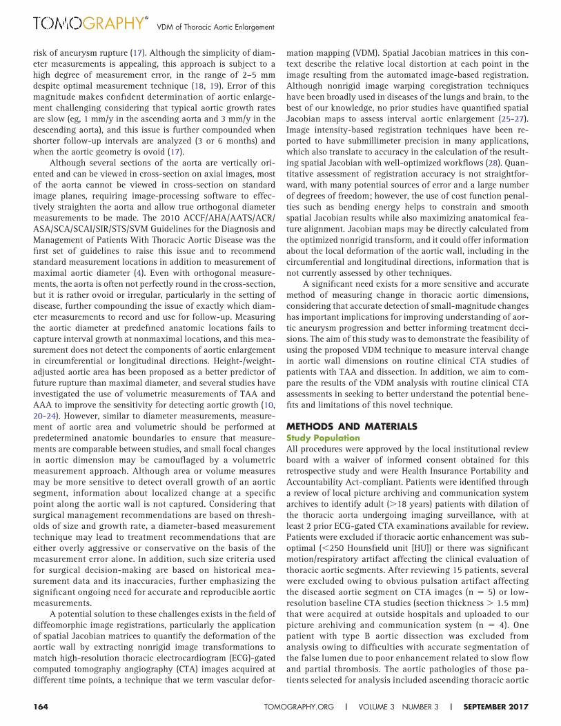
In contrast, Figure 5 illustrates a 52-year-old man with ahistory of type B aortic dissection who had 2 ECG-gated CTAstudies available for analysis, the first �1 year after the onset ofhis dissection and a follow-up study performed 6 months afterthe first. On the basis of the clinical report, there was suspicionfor �1–2 mm of interval enlargement of the distal aortic arch,
Figure 1. Vascular deformation mapping (VDM)workflow: computed tomography angiography(CTA) images undergo digital image processingand analysis that involves segmentation of thecontrast-enhancing aortic blood volume followedby nonrigid coregistration between baseline andfollow-up sessions. 3D maps of the determinant ofthe spatial Jacobian are then generated using thefinal optimized transform. These values are subse-quently mapped to the 3D surface of the aorticsegmentation, essentially allowing for visualiza-tion of the aortic wall deformation rate betweenimaging sessions. Expansion can be visualized asred (VDM � 1), compression as blue (VDM � 1),and no deformation as green (VDM � 0).
VDM of Thoracic Aortic Enlargement
166 TOMOGRAPHY.ORG | VOLUME 3 NUMBER 3 | SEPTEMBER 2017

but the conclusion of the clinical assessment was that there hadbeen no definite enlargement, as the observed change in diam-eter was within the range of measurement error. VDM of thispatient showed nearly diffuse enlargement of the false lumenthroughout the distal aortic arch and descending aorta, with acorresponding decrease in size of the true lumen, changes thatare frequently observed in chronic aortic dissection (29). It isimportant to note that although the absolute change in maximalaortic dimension was thought to be small (1–2 mm), the rate ofgrowth is noted to be significant owing to the short interval(6 months) between the 2 studies. This ability to detect growth
over short intervals is particularly useful in the setting of pa-tients with recent aortic dissection, as there is a proven clinicalbenefit to endovascular (TEVAR) repair in the subacute period (2weeks to 3 months after dissection) (30). Of note, there was avisually apparent motion artifact in the ascending aorta on CTAimages, leading to difficulty with image coregistration, which ismanifested on the VDM as a wavy aortic wall contour and areasof high and low Jacobian determinant (red and blue, respec-tively) on adjacent areas of the aortic wall.
Finally, Figure 6 illustrates a 76-year-old man with a his-tory of ascending aorta replacement, who developed a type Baortic dissection on the baseline study and had 4 surveillance
Figure 2. Progressive enlargement of an aortic aneurysm of the descending aorta in a 76-year-old patient with historyof prior surgical repair of the ascending aorta. Aortic enlargement is noted at all intervals, but increases in rate and ex-tent over time. Aortic lumen area (in square millimeter) measured at a single level (black line) in the distal descendingaorta was used to corroborate a focal region of enlargement in the distal descending aorta seen on the VDM map.VDM values for replaced ascending aorta are not displayed owing to artifact.
Figure 3. VDM analysis shows no areas of high-intensity wall expansion in a 66-year-old womanwith a mild dilated ascending aorta (4.1 cm maxi-mally). Although aortic dimensions were stable byclinical diameter assessment, low-intensity areasof potential aortic enlargement were noted at thesinotubular junction and in the region of the in-nominate artery, suggesting the possibility of lim-ited growth in these regions.
Figure 4. Overall stable aortic dimensions in a56-year-old man with a history of surgical repairof the ascending aorta for type A dissection withresidual dissection flap in the aortic arch and de-scending aorta. Small areas of apparent aorticexpansion at the aortic root and distal descendingaorta are likely because of mild coregistrationartifact (*).
VDM of Thoracic Aortic Enlargement
TOMOGRAPHY.ORG | VOLUME 3 NUMBER 3 | SEPTEMBER 2017 167
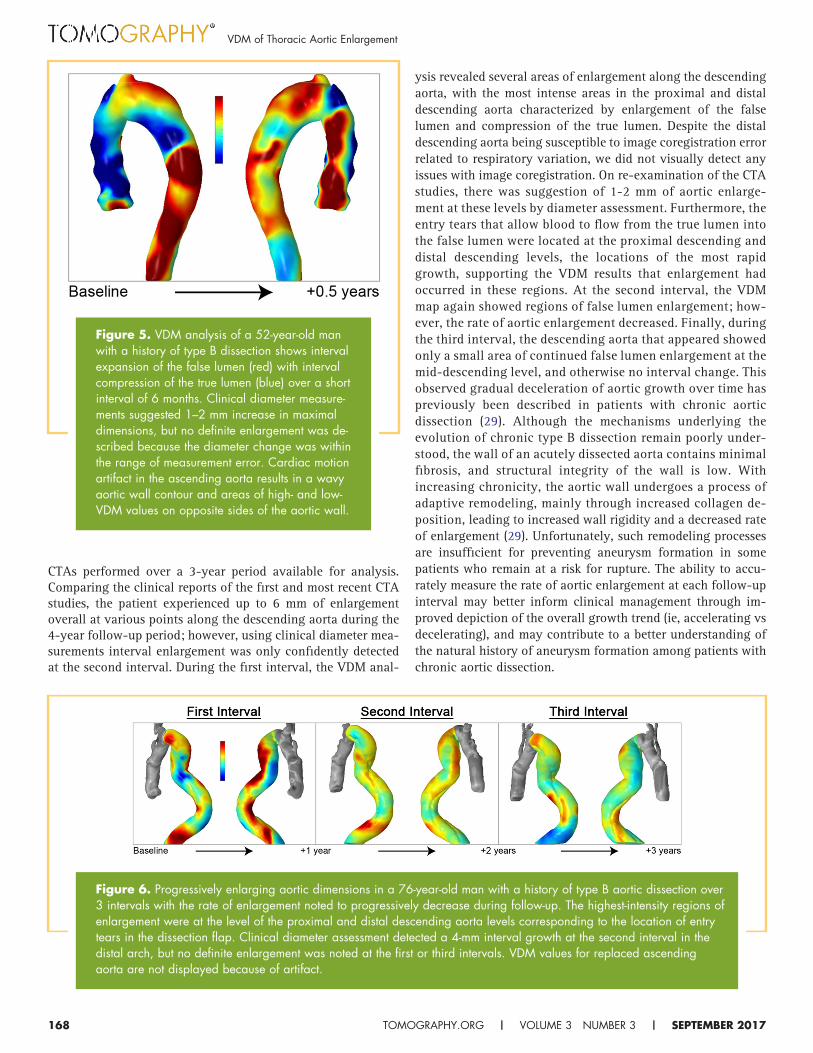
CTAs performed over a 3-year period available for analysis.Comparing the clinical reports of the first and most recent CTAstudies, the patient experienced up to 6 mm of enlargementoverall at various points along the descending aorta during the4-year follow-up period; however, using clinical diameter mea-surements interval enlargement was only confidently detectedat the second interval. During the first interval, the VDM anal-
ysis revealed several areas of enlargement along the descendingaorta, with the most intense areas in the proximal and distaldescending aorta characterized by enlargement of the falselumen and compression of the true lumen. Despite the distaldescending aorta being susceptible to image coregistration errorrelated to respiratory variation, we did not visually detect anyissues with image coregistration. On re-examination of the CTAstudies, there was suggestion of 1-2 mm of aortic enlarge-ment at these levels by diameter assessment. Furthermore, theentry tears that allow blood to flow from the true lumen intothe false lumen were located at the proximal descending anddistal descending levels, the locations of the most rapidgrowth, supporting the VDM results that enlargement hadoccurred in these regions. At the second interval, the VDMmap again showed regions of false lumen enlargement; how-ever, the rate of aortic enlargement decreased. Finally, duringthe third interval, the descending aorta that appeared showedonly a small area of continued false lumen enlargement at themid-descending level, and otherwise no interval change. Thisobserved gradual deceleration of aortic growth over time haspreviously been described in patients with chronic aorticdissection (29). Although the mechanisms underlying theevolution of chronic type B dissection remain poorly under-stood, the wall of an acutely dissected aorta contains minimalfibrosis, and structural integrity of the wall is low. Withincreasing chronicity, the aortic wall undergoes a process ofadaptive remodeling, mainly through increased collagen de-position, leading to increased wall rigidity and a decreased rateof enlargement (29). Unfortunately, such remodeling processesare insufficient for preventing aneurysm formation in somepatients who remain at a risk for rupture. The ability to accu-rately measure the rate of aortic enlargement at each follow-upinterval may better inform clinical management through im-proved depiction of the overall growth trend (ie, accelerating vsdecelerating), and may contribute to a better understanding ofthe natural history of aneurysm formation among patients withchronic aortic dissection.
Figure 5. VDM analysis of a 52-year-old manwith a history of type B dissection shows intervalexpansion of the false lumen (red) with intervalcompression of the true lumen (blue) over a shortinterval of 6 months. Clinical diameter measure-ments suggested 1–2 mm increase in maximaldimensions, but no definite enlargement was de-scribed because the diameter change was withinthe range of measurement error. Cardiac motionartifact in the ascending aorta results in a wavyaortic wall contour and areas of high- and low-VDM values on opposite sides of the aortic wall.
Figure 6. Progressively enlarging aortic dimensions in a 76-year-old man with a history of type B aortic dissection over3 intervals with the rate of enlargement noted to progressively decrease during follow-up. The highest-intensity regions ofenlargement were at the level of the proximal and distal descending aorta levels corresponding to the location of entrytears in the dissection flap. Clinical diameter assessment detected a 4-mm interval growth at the second interval in thedistal arch, but no definite enlargement was noted at the first or third intervals. VDM values for replaced ascendingaorta are not displayed because of artifact.
VDM of Thoracic Aortic Enlargement
168 TOMOGRAPHY.ORG | VOLUME 3 NUMBER 3 | SEPTEMBER 2017
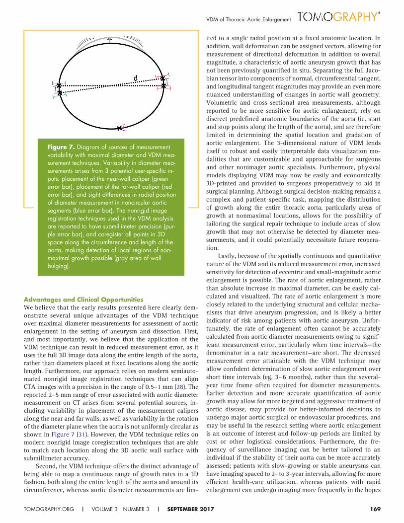
Advantages and Clinical OpportunitiesWe believe that the early results presented here clearly dem-onstrate several unique advantages of the VDM techniqueover maximal diameter measurements for assessment of aorticenlargement in the setting of aneurysm and dissection. First,and most importantly, we believe that the application of theVDM technique can result in reduced measurement error, as ituses the full 3D image data along the entire length of the aorta,rather than diameters placed at fixed locations along the aorticlength. Furthermore, our approach relies on modern semiauto-mated nonrigid image registration techniques that can alignCTA images with a precision in the range of 0.5–1 mm (28). Thereported 2–5 mm range of error associated with aortic diametermeasurement on CT arises from several potential sources, in-cluding variability in placement of the measurement calipersalong the near and far walls, as well as variability in the rotationof the diameter plane when the aorta is not uniformly circular asshown in Figure 7 (31). However, the VDM technique relies onmodern nonrigid image coregistration techniques that are ableto match each location along the 3D aortic wall surface withsubmillimeter accuracy.
Second, the VDM technique offers the distinct advantage ofbeing able to map a continuous range of growth rates in a 3Dfashion, both along the entire length of the aorta and around itscircumference, whereas aortic diameter measurements are lim-
ited to a single radial position at a fixed anatomic location. Inaddition, wall deformation can be assigned vectors, allowing formeasurement of directional deformation in addition to overallmagnitude, a characteristic of aortic aneurysm growth that hasnot been previously quantified in situ. Separating the full Jaco-bian tensor into components of normal, circumferential tangent,and longitudinal tangent magnitudes may provide an even morenuanced understanding of changes in aortic wall geometry.Volumetric and cross-sectional area measurements, althoughreported to be more sensitive for aortic enlargement, rely ondiscreet predefined anatomic boundaries of the aorta (ie, startand stop points along the length of the aorta), and are thereforelimited in determining the spatial location and gradation ofaortic enlargement. The 3-dimensional nature of VDM lendsitself to robust and easily interpretable data visualization mo-dalities that are customizable and approachable for surgeonsand other nonimager aortic specialists. Furthermore, physicalmodels displaying VDM may now be easily and economically3D-printed and provided to surgeons preoperatively to aid insurgical planning. Although surgical decision-making remains acomplex and patient-specific task, mapping the distributionof growth along the entire thoracic aorta, particularly areas ofgrowth at nonmaximal locations, allows for the possibility oftailoring the surgical repair technique to include areas of slowgrowth that may not otherwise be detected by diameter mea-surements, and it could potentially necessitate future reopera-tion.
Lastly, because of the spatially continuous and quantitativenature of the VDM and its reduced measurement error, increasedsensitivity for detection of eccentric and small-magnitude aorticenlargement is possible. The rate of aortic enlargement, ratherthan absolute increase in maximal diameter, can be easily cal-culated and visualized. The rate of aortic enlargement is moreclosely related to the underlying structural and cellular mecha-nisms that drive aneurysm progression, and is likely a betterindicator of risk among patients with aortic aneurysm. Unfor-tunately, the rate of enlargement often cannot be accuratelycalculated from aortic diameter measurements owing to signif-icant measurement error, particularly when time intervals—thedenominator in a rate measurement—are short. The decreasedmeasurement error attainable with the VDM technique mayallow confident determination of slow aortic enlargement overshort time intervals (eg, 3–6 months), rather than the several-year time frame often required for diameter measurements.Earlier detection and more accurate quantification of aorticgrowth may allow for more targeted and aggressive treatment ofaortic disease, may provide for better-informed decisions toundergo major aortic surgical or endovascular procedures, andmay be useful in the research setting where aortic enlargementis an outcome of interest and follow-up periods are limited bycost or other logistical considerations. Furthermore, the fre-quency of surveillance imaging can be better tailored to anindividual if the stability of their aorta can be more accuratelyassessed; patients with slow-growing or stable aneurysms canhave imaging spaced to 2- to 3-year intervals, allowing for moreefficient health-care utilization, whereas patients with rapidenlargement can undergo imaging more frequently in the hopes
Figure 7. Diagram of sources of measurementvariability with maximal diameter and VDM mea-surement techniques. Variability in diameter mea-surements arises from 3 potential user-specific in-puts: placement of the near-wall caliper (greenerror bar), placement of the far-wall caliper (rederror bar), and sight differences in radial positionof diameter measurement in noncircular aorticsegments (blue error bar). The nonrigid imageregistration techniques used in the VDM analysisare reported to have submillimeter precision (pur-ple error bar), and coregister all points in 3Dspace along the circumference and length of theaorta, making detection of local regions of non-maximal growth possible (gray area of wallbulging).
VDM of Thoracic Aortic Enlargement
TOMOGRAPHY.ORG | VOLUME 3 NUMBER 3 | SEPTEMBER 2017 169
of minimizing the incidence of potentially predictable and pre-ventable complications.
Technical ChallengesThe results presented here constitute the first steps in the appli-cation of a VDM technique for the evaluation of aortic disease,and while preliminary results show great promise, several chal-lenges remain. First, because this analysis has a high degree ofsensitivity to aortic wall deformation, errors can be introducedby factors resulting in differing spatial alignment of the 2compared aortic geometries. The 2 areas most susceptible tosuch error are at the aortic root (sinuses of Valsalva) and at thedistal descending aorta at the level of the diaphragm, with the 2main contributing factors being cardiac and respiratory motion.The effects of these factors on variation in aortic geometry havebeen previously described (32). The aortic root has the highestdegree of pulsatory motion of any thoracic aortic segment ow-ing to its close proximity to the heart, with the degree ofpulsation amplified during expiration. In addition, the entirethoracic aorta has a relatively uniform lateral and posteriordisplacement with expiration. However, although uniform dis-placement could be easily corrected for during image coregis-tration, the distal descending aorta remains relatively fixed inposition by the diaphragm, and this nonuniform motion intro-duces potential misalignment during image coregistration.
These observations stress the importance of acquiring theCTA images during the same phase of respiration (preferablyinspiration) and with ECG-gating (preferably in late diastole) tominimize errors attributable to the small phasic variations inaortic geometry. In addition to respiratory and pulsation arti-facts, “stair-step” artifact is occasionally encountered in ECG-gated CTAs, particularly when studies are performed on scan-ners with detector rows numbering 64 or less. The stair-stepartifact is problematic for the VDM technique, as it creates anabrupt shelf-like defect in the 3D aortic segmentation that limitsimage coregistration. Fortunately, modern CT scanners thathave been optimized for cardiovascular imaging can greatlyminimize the frequency and severity of stair-step artifacts ow-ing to the increased number of detector rows and decreasedgantry rotation time.
Second, the VDM method has significant technical demands,both in terms of user expertise and computing requirements. Manysoftware platforms are currently available to perform accuratesemiautomated aortic segmentation, which, if combined withour technique, may significantly decrease the time required foranalysis. In addition, although the required computing powermakes the VDM technique less practical for smaller centers thatlack dedicated high-performance computing capabilities, therecent rise of cloud-based medical image analysis software mayallow for analysis to be centralized, lessening technical demandsat the level of the end user. Development of this VDM techniqueis still in the early stages of development; however, routineclinical applicability of this approach will require streamliningof the analysis with improved semiautomated image segmenta-tion techniques and code optimization to minimize technicaldemands and user input.
Future Technical DirectionsFuture technical efforts will be focused on the following 5 keytopics:
(1) Further validating the accuracy and consistency of theVDM analysis is an important next step, as there is noclear noninvasive—or even invasive—tool to measurechanges in aortic dimension in situ. As such, VDM resultswill need to be compared with other more sensitive mea-surements of aortic enlargement such as CTA luminal areaand volumetric assessments. In addition, phantoms withprecisely controlled dimensions could be used to furthervalidate VDM results in a controlled experimental setting.
(2) Further developments in computer algorithms will beneeded to further automate and improve image segmen-tation, coregistration, and analysis steps. In particular,additional efforts need to be focused on accurate andreproducible false lumen segmentation in patients withaortic dissection, as the false lumen is prone to heteroge-neous enhancement related to either partial thrombosisand/or slow blood flow.
(3) Development of a user-friendly console for easy visual-ization and interaction with the VDM results will be animportant step in moving this technology into the realmof clinical practice.
(4) Evaluating the feasibility of quantifying changes in theaortic dimensions between different points in the cardiaccycle, rather than between different studies acquired atthe same point in the cardiac cycle. The aortic dimensionshave been shown to change significantly with pulsation,and measurement of these changes using VDM couldprovide important insights into the elasticity/rigidity ofthe aortic wall, a characteristic that has been associatedwith a large variety of cardiovascular diseases (33).
(5) Investigating the feasibility of the VDM technique toquantify enlargement of other pathologies that manifestas progressive vascular enlargement such as AAA, cere-bral aneurysm, pulmonary artery enlargement related topulmonary hypertension, and endoleak after endovascu-lar aortic repair.
Future Clinical DirectionsIn addition to ongoing technical developments, this technologyhas several unique potential clinical and research applicationsthat we plan to investigate. First, given the rapidly increasingavailability and decreasing costs of color 3D printing, we plan tostudy the clinical utility of superimposing VDM results on full-scale, color 3D-printed aortic models. To demonstrate feasibilityof this approach, we submitted the VDM results from Figure 2(Interval 2) for color 3D printing with the results shown in Figure 8.To the best of our knowledge, there are no prior publishedreports of superimposing imaging-based measurements of patho-physiology, such as the VDM results of aneurysm enlargement,on 3D-printed anatomic models. A large proportion of the lit-erature in the field of 3D printing has focused on the productionof high-fidelity anatomic models from medical imaging data,with some proposed applications including rapid prototypingfor medical device development, creation of individualizedmedical implants/prostheses, and operative planning and pa-
VDM of Thoracic Aortic Enlargement
170 TOMOGRAPHY.ORG | VOLUME 3 NUMBER 3 | SEPTEMBER 2017

tient-specific device sizing in the setting of complex anatomy(34, 35). However, despite such promising applications, the roleof 3D printing in clinical practice remains a topic of debate,largely owing to questions of cost versus benefit if similar obser-vations and measurements can be made by 3D medical imagingsoftware. As demonstrated here, the addition of VDM resultssuperimposed on the 3D model surface significantly increasesthe amount of information that the model contains and mayfurther justify the effort involved in creating patient-specific 3Dmodels. Although a computerized VDM provides a detailedoverview of the aneurysm enlargement, a 3D model that can behandled and closely studied by surgeons may allow for subtleanatomic and functional observations that are not as easilyappreciated on a digital image, and could facilitate patienteducation during clinic visits.
Second, the VDM technique may be extremely valuable as aresearch tool for studies of aneurysm pathophysiology owing toits high degree of sensitivity to changes in aortic dimensions.There has been increased interest in elucidating the cellularpathways involved in remodeling of the vascular wall leading tothe formation of aortic aneurysm (36, 37). Various cellularsignaling pathways along with host-immune interactions havebeen implicated in the pathogenesis of AAAs (38-41). Thesefactors are in part related to the complex underlying cellularprocesses responsible for the loss of extracellular matrix andwall remodeling. For example, chronic inflammation of theaorta wall has been implicated in the activation of matrix met-alloproteinases (MMPs) such as MMP-2 and MMP-9, which havebeen reported to play a role in aortic wall weakening andsubsequent AAA formation (42, 43). Furthermore, transforminggrowth factor � signaling alterations (44) have been widelyassociated with vascular smooth muscle disease with the genetic
basis now identified to involve 3 distinct pathomechanisms thatinclude perturbation of the transforming growth factor � sig-naling pathway, disruption of the vascular smooth muscle cellcontractile apparatus, and impairment of extracellular matrixsynthesis (36, 37, 45). Advances in our understanding of theunderlying pathogenetic alterations involved in the pathogen-esis of thoracic aortic disease are providing significant newopportunities for therapeutic interventions using novel pharma-ceutical approaches. The development of a validated imagingbiomarker would allow for longitudinal quantification of theeffects of drug interventions on modulation of disease progres-sion. This capability would provide unique opportunities to usethis imaging biomarker to facilitate development of therapeuticstrategies in both preclinical aneurysm models and for use inclinical translational trials undertaking novel therapeuticstrategies.
Finally, the potential impact of VDM results on clinicalpatient care will need to be thoroughly studied. Primary topicsof investigation will include investigation of the associationsbetween VDM measures and patient cardiovascular risk factors,the potential of VDM assessment to reclassify patient risk as-sessments, and the ability of VDM to predict patient outcomes.Although the VDM technique may indeed be more sensitive indetecting change in the aortic dimensions, an analysis of howdata from this novel method could change patient managementwill be needed to show value over the more easily obtainedaortic diameter measurements. Considering that the VDM anal-ysis can be performed respectively on routine clinical CTAscans, the VDM results can be compared with clinical reportsand a wide variety of patient demographic parameters andoutcomes such as surgical repair strategy, surgical complicationrate, reoperation rates, and occurrence of aorta-specific adverseevents during imaging surveillance. In addition, as the VDMtechnique allows for assessment of aortic enlargement atspecific spatial locations along the aortic wall, growth can becolocalized with pathological features of the aortic wall that arebelieved to promote aneurysm development such as atheroscle-rotic plaque (both calcified and lipid-rich), mural thrombus,intimal hyperplasia, or wall thickness. Identifying direct corre-lations between localized aortic wall pathology and regionalwall expansion by VDM analysis could greatly advance ourunderstanding of the underlying pathophysiology that leads toaortic aneurysm, and offer new strategies to predict aorticevents, risk-stratify patients, and monitor the effectiveness ofpharmacological therapy.
CONCLUSIONWe have demonstrated the feasibility of a spatial Jacobian-based technique to measure changes in the size of the aorticlumen between baseline and follow-up ECG-gated thoracic CTAexaminations in patients with mild aortic dilatation, aortic an-eurysm, and aortic dissection, and that this technique is capableof quantifying and visually displaying the degree of aorticenlargement in a 3-dimensional fashion. Furthermore, we haveshown that there are clear discrepancies between the VDMresults and clinical diameter assessments, with the VDM tech-nique appearing more sensitive for detection of changes inaortic dimensions owing to reduced measurement error, al-
Figure 8. Color 3D-printed model of the thoracicaorta produced from the VDM results of a patientwith progressively enlarging descending aorticaneurysm presented in Figure 2, interval 2. Thesuperimposition of pathophysiologic VDM data ona 3D-anatomic model represents a novel applica-tion of medical 3D printing and be valuable inoperative planning.
VDM of Thoracic Aortic Enlargement
TOMOGRAPHY.ORG | VOLUME 3 NUMBER 3 | SEPTEMBER 2017 171
though formal quantification of the degree of error reductionand the potential clinical impacts of a more sensitive analysis ofaortic dimension changes requires further investigation. TheVDM technique for measurement of change in aortic wall di-
mensions holds the promise of considerably improving the ac-curacy of aortic imaging surveillance, informing clinical deci-sion-making, furthering aortic research questions, and sheddinglight on the natural history of aortic disease.
ACKNOWLEDGMENTSThe authors would like to thank Drs. Himanshu Patel and Bo Yang for their guidancepertaining to the potential applications of vascular deformation mapping in the field ofaortic surgery. This study received support from BDR: National Institutes of HealthR35CA197701 and U01CA166104, and NSB: Radiologic Society of North AmericaResearch Fellow Grant (RF1502).
Disclosure: No disclosures to report.
Conflict of Interest: None reported.
REFERENCES1. Elefteriades JA. Thoracic aortic aneurysm: reading the enemy’s playbook. Curr
Probl Cardiol. 2008;33(5):203–277.2. Durham CA, Cambria RP, Wang LJ, Ergul EA, Aranson NJ, Patel VI, Conrad MF.
The natural history of medically managed acute type B aortic dissection. J VascSurg. 2015;61(5):11921198.
3. Morgan L, Choi H, Reid M, Khawaja A, Mazzone PJ. The frequency of incidentalfindings and subsequent evaluation in low-dose CT scans for lung cancer screen-ing. Ann Am Thorac Soc. 2017;14(9):1450–1456.
4. Hiratzka LF, Bakris GL, Beckman JA, Bersin RM, Carr VF, Casey DE Jr, Eagle KA,Hermann LK, Isselbacher EM, Kazerooni EA, Kouchoukos NT, Lytle BW, MilewiczDM, Reich DL, Sen S, Shinn JA, Svensson LG, Williams DM; American College ofCardiology Foundation/American Heart Association Task Force on PracticeGuidelines; American Association for Thoracic Surgery; American College ofRadiology; American Stroke Association; Society of Cardiovascular Anesthesiolo-gists; Society for Cardiovascular Angiography and Interventions; Society of Inter-ventional Radiology; Society of Thoracic Surgeons; Society for Vascular Medi-cine. 2010 ACCF/AHA/AATS/ACR/ASA/SCA/SCAI/SIR/STS/SVM guidelinesfor the diagnosis and management of patients with Thoracic Aortic Disease: areport of the American College of Cardiology Foundation/American Heart Asso-ciation Task Force on Practice Guidelines, American Association for Thoracic Sur-gery, American College of Radiology, American Stroke Association, Society ofCardiovascular Anesthesiologists, Society for Cardiovascular Angiography andInterventions, Society of Interventional Radiology, Society of Thoracic Surgeons,and Society for Vascular Medicine. Circulation. 2010;121(13):e266–e369.
5. Benedetti N, Hope MD. Prevalence and significance of incidentally noted dilationof the ascending aorta on routine chest computed tomography in older patients.J Comput Assist Tomogr. 2015;39(1):109–111.
6. Chau KH, Elefteriades JA. Natural history of thoracic aortic aneurysms: size mat-ters, plus moving beyond size. Prog Cardiovasc Dis. 2013;56(1):74–80.
7. Kälsch H, Lehmann N, Möhlenkamp S, Becker A, Moebus S, Schmermund A,Stang A, Mahabadi AA, Mann K, Jöckel KH, Erbel R, Eggebrecht H. Body-sur-face adjusted aortic reference diameters for improved identification of patientswith thoracic aortic aneurysms: results from the population-based Heinz NixdorfRecall study. Int J Cardiol. 2013;163(1):72–78.
8. State Health Facts Menlo Park (CA): The Kaiser Family Foundation. http://www.kff.org/other/state-indicator/distribution-by-age/? Updated March 2016.Accessed August 31, 2017.
9. Johansson G, Markstrom U, Swedenborg J. Ruptured thoracic aortic aneurysms:a study of incidence and mortality rates. J Vasc Surg. 1995;21(6):985–988.
10. Renapurkar RD, Setser RM, O’Donnell TP, Egger J, Lieber ML, Desai MY, StillmanAE, Schoenhagen P, Flamm SD. Aortic volume as an indicator of disease pro-gression in patients with untreated infrarenal abdominal aneurysm. Eur J Radiol.2012;81(2):E87–E93.
11. Juvonen T, Ergin MA, Galla JD, Lansman SL, Nguyen KH, McCullough JN, LevyD, de Asla RA, Bodian CA, Griepp RB. Prospective study of the natural history ofthoracic aortic aneurysms. Ann Thorac Surg. 1997;63(6):1533–1545.
12. Sueyoshi E, Sakamoto I, Hayashi K, Yamaguchi T, Imada T. Growth rate of aor-tic diameter in patients with type B aortic dissection during the chronic phase.Circulation. 2004;110(11 suppl 1):II256–261.
13. Coady MA, Rizzo JA, Hammond GL, Kopf GS, Elefteriades JA. Surgical interven-tion criteria for thoracic aortic aneurysms: a study of growth rates and complica-tions. Ann Thorac Surg. 1999;67(6):1922–1926.
14. Cheung K, Boodhwani M, Chan KL, Beauchesne L, Dick A, Coutinho T. Thoracicaortic aneurysm growth: role of sex and aneurysm etiology. J Am Heart Assoc.2017;6(2). pii: e003792.
15. Gagné-Loranger M1, Dumont É1, Voisine P1, Mohammadi S1, Dagenais F. Nat-ural history of 40-50 mm root/ascending aortic aneurysms in the current era ofdedicated thoracic aortic clinics. Eur J Cardiothorac Surg. 2016;50(3):562–566.
16. Hong H, Yang Y, Liu B, Cai W. Imaging of abdominal aortic aneurysm: the pres-ent and the future. Curr Vasc Pharmacol. 2010;8(6):808–819.
17. Elefteriades JA, Farkas EA. Thoracic aortic aneurysm clinically pertinent controver-sies and uncertainties. J Am Coll Cardiol. 2010;55(9):841–857.
18. Quint LE, Liu PS, Booher AM, Watcharotone K, Myles JD. Proximal thoracic aor-tic diameter measurements at CT: repeatability and reproducibility according tomeasurement method. Int J Cardiovasc Imaging. 2013;29(2):479–788.
19. Lu TLC, Rizzo E, Marques-Vidal PM, von Segesser LK, Dehmeshki J, Qanadli SD.Variability of ascending aorta diameter measurements as assessed with electro-cardiography-gated multidetector computerized tomography and computer as-sisted diagnosis software. Interact Cardiovasc Thorac Surg. 2010;10(2):217–221.
20. Trinh B, Dubin I, Rahman O, Botelho MPF, Naro N, Carr JC, Collins JD, BarkerAJ. Aortic volumetry at contrast-enhanced magnetic resonance angiography feasi-bility as a sensitive method for monitoring bicuspid aortic valve aortopathy. InvestRadiol. 2017;52(4):216–222.
21. Masri A, Kalahasti V, Svensson LG, Roselli EE, Johnston D, Hammer D, Schoen-hagen P, Griffin BP, Desai MY. Aortic cross-sectional area/height ratio and out-comes in patients with a trileaflet aortic valve and a dilated aorta. Circulation.2016;134(22):1724–1737.
22. Davies RR, Gallo A, Coady MA, Tellides G, Botta DM, Burke B, Coe MP, KopfGS, Elefteriades JA. Novel measurement of relative aortic size predicts rupture ofthoracic aortic aneurysms. Ann Thorac Surg. 2006;81(1):169–177.
23. Parr A, Jayaratne C, Buttner P, Golledge J. Comparison of volume and diametermeasurement in assessing small abdominal aortic aneurysm expansion examinedusing computed tomographic angiography. Eur J Radiol. 2011;79(1):42–47.
24. Stanley GA, Murphy EH, Knowles M, Ilves M, Jessen ME, Dimaio JM, ModrallJG, Arko FR 3rd. Volumetric analysis of type B aortic dissections treated with tho-racic endovascular aortic repair. J Vasc Surg. 2011;54(4):985–992.
25. Galbán CJ, Han MK, Boes JL, Chughtai KA, Meyer CR, Johnson TD, Galbán S,Rehemtulla A, Kazerooni EA, Martinez FJ, Ross BD. Computed tomography-basedbiomarker provides unique signature for diagnosis of COPD phenotypes and dis-ease progression. Nat Med. 2012;18(11):1711–1715.
26. Boes JL, Hoff BA, Hylton N, Pickles MD, Turnbull LW, Schott AF, Rehemtulla A,Chamberlain R, Lemasson B, Chenevert TL, Galbán CJ, Meyer CR, Ross BD. Im-age registration for quantitative parametric response mapping of cancer treat-ment response. Transl Oncol. 2014;7(1):101–110.
27. Keith L, Ross BD, Galban CJ, Luker GD, Galban S, Zhao B, Guo X, Chenevert TL,Hoff BA. Semiautomated workflow for clinically streamlined glioma parametricresponse mapping. Tomography. 2016;2(4):267–275.
28. Klein S, Staring M, Pluim JP. Evaluation of optimization methods for nonrigidmedical image registration using mutual information and B-splines. IEEE TransImage Process. 2007;16(12):2879–2890.
29. Peterss S, Mansour AM, Ross JA, Vaitkeviciute I, Charilaou P, Dumfarth J, FangH, Ziganshin BA, Rizzo JA, Adeniran AJ, Elefteriades JA. Changing pathology ofthe thoracic aorta from acute to chronic dissection literature review and insights.J Am Coll Cardiol. 2016;68(10):1054–1065.
30. Fanelli F, Cannavale A, O’Sullivan GJ, Gazzetti M, Cirelli C, Lucatelli P, SantoniM, Catalano C. Endovascular repair of acute and chronic aortic type B dissec-tions: main factors affecting aortic remodeling and clinical outcome. JACCCardiovasc Interv. 2016;9(2):183–191.
31. Kauffmann C, Tang A, Therasse E, Giroux MF, Elkouri S, Melanson P, Oliva VL,Soulez G. Measurements and detection of abdominal aortic aneurysm growth:Accuracy and reproducibility of a segmentation software. Eur J Radiol. 2012;81(8):1688–1694.
32. Suh GY, Beygui RE, Fleischmann D, Cheng CP. Aortic arch vessel geometriesand deformations in patients with thoracic aortic aneurysms and dissections.J Vasc Interv Radiol. 2014;25(12):1903–1911.
VDM of Thoracic Aortic Enlargement
172 TOMOGRAPHY.ORG | VOLUME 3 NUMBER 3 | SEPTEMBER 2017

33. Sethi S, Rivera O, Oliveros R, Chilton R. Aortic stiffness: pathophysiology, clinicalimplications, and approach to treatment. Integr Blood Press Control. 2014;7:29–34.
34. Foley TA, El Sabbagh A, Anavekar NS, Williamson EE, Matsumoto JM. 3D-print-ing: applications in cardiovascular imaging. Curr Radiol Rep. 2017;5(9):43.
35. Marro A, Bandukwala T, Mak W. Three-dimensional printing and medical imag-ing: a review of the methods and applications. Curr Probl Diagn Radiol. 2016;45(1):2–9.
36. Andelfinger G, Loeys B, Dietz H. A Decade of Discovery in the Genetic Under-standing of Thoracic Aortic Disease. Can J Cardiol. 2016;32(1):13–25.
37. Bowdin SC, Laberge AM, Verstraeten A, Loeys BL. Genetic testing in thoracicaortic disease–when, why, and how? Can J Cardiol. 2016;32(1):131–134.
38. Ghosh A, Lu G, Su G, McEvoy B, Sadiq O, DiMusto PD, Laser A, Futchko JS,Henke PK, Eliason JL, Upchurch GR Jr. Phosphorylation of AKT and abdominalaortic aneurysm formation. Am J Pathol. 2014;184(1):148–158.
39. Kim J, Procknow JD, Yanagisawa H, Wagenseil JE. Differences in genetic signal-ing, and not mechanical properties of the wall, are linked to ascending aorticaneurysms in fibulin-4 knockout mice. Am J Physiol Heart Circ Physiol. 2015;309(1):H103–H113.
40. Meng X, Yang J, Dong M, Zhang K, Tu E, Gao Q, Chen W, Zhang C, Zhang Y.Regulatory T cells in cardiovascular diseases. Nat Rev Cardiol. 2016;13(3):167–179.
41. Miner GH, Faries PL, Costa KD, Hanss BG, Marin ML. An update on the etiologyof abdominal aortic aneurysms: implications for future diagnostic testing. ExpertRev Cardiovasc Ther. 2015;13(10):1079–1090.
42. Rabkin SW. The Role Matrix metalloproteinases in the production of aortic aneu-rysm. Prog Mol Biol Transl Sci. 2017;147:239–265.
43. van der Pluijm I, van Vliet N, von der Thusen JH, Robertus JL, Ridwan Y, van Hei-jningen PM, van Thiel BS5, Vermeij M2, Hoeks SE6, Buijs-Offerman RM7, Verha-gen HJ8, Kanaar R9, Bertoli-Avella AM7, Essers J. Defective connective tissueremodeling in Smad3 mice leads to accelerated aneurysmal growth through dis-turbed downstream TGF-beta signaling. EBioMedicine. 2016;12:280–294.
44. Forte A, Galderisi U, Cipollaro M, De Feo M, Della Corte A. Epigenetic regula-tion of TGF-beta1 signalling in dilative aortopathy of the thoracic ascendingaorta. Clin Sci (Lond). 2016;130(16):1389–1405.
45. Brownstein AJ, Ziganshin BA, Kuivaniemi H, Body SC, Bale AE, Elefteriades JA.Genes associated with thoracic aortic aneurysm and dissection: an update andclinical implications. Aorta (Stamford). 2017;5(1):11–20.
VDM of Thoracic Aortic Enlargement
TOMOGRAPHY.ORG | VOLUME 3 NUMBER 3 | SEPTEMBER 2017 173