vascular insulin resistance through fat
TRANSCRIPT

Vascular Insulin Resistance Through Fat
Intracellular Signaling, Genetic Interferences and Hemodynamics

The research presented in this thesis is part of the research program of the Institute for Cardiovascular Research at the VU free university (ICAR-VU). The studies were performed at Laboratory of Physiology, VU medical center, Amsterdam, the Netherlands.
Financial support by the Dutch Heart Foundation is greatly acknowledged.
The publication of this thesis is further financially supported by: Dutch Diabetes foundation, Laboratory of Physiology, JE Juriaanse Foundation, Siemens, DSI, Astra Zeneca, Merck Sharp & Dohme, Bracco.
ISBN: 9789461080363
Printed by: Gildeprint BV, Enschede Cover: Inverted image of a fluorescent stained cross section of the arterial wall (made in 2006) Cover design: Marloes Bakker Lay out: Laura Kok
© 2010 W. Bakker All rights reserved. No part of this book may be reproduced or transmitted in any form or by any means, without prior
written permission of the author.

VRIJE UNIVERSITEIT
ACADEMISCH PROEFSCHRIFT
ter verkrijging van de graad Doctor aan de Vrije Universiteit Amsterdam,
op gezag van de rector magnificus prof.dr. L.M. Bouter
in het openbaar te verdedigen ten overstaan van de promotiecommissie
van de faculteit der Geneeskunde op donderdag 20 mei 2010 om 11.45 uur
in de aula van de universiteit, De Boelelaan 1105
door
Wineke Bakker geboren te Hoorn
Vascular Insulin Resistance Through Fat
Intracellular Signaling, Genetic Interferences and Hemodynamics

promotoren: prof.dr. V.W.M. van Hinsbergh prof.dr. C.D.A. Stehouwer
copromotoren: dr. P. Sipkema dr. E.C. Eringa

“Logic will get you from A to B. Imagination will take you everywhere.”
- Albert Einstein -

Overige leden promotiecommissie prof.dr. E.T. van Bavel prof.dr. M. Diamant prof.dr. A.C. Gittenberger-de Groot prof.dr. J.A. Maassen dr. D. Merkus prof.dr. J.C. Seidell dr. P.J. Voshol prof.dr. J.S. Yudkin

Table of Contents
CONTENTS
General Introduction and Outline of the Thesis
Endothelial Dysfunction and Diabetes: Roles of impaired insulin signaling, obesity and perivascular adipose tissue
Cell Tissue Research 2009; 335 (1): 165-189 Microcirculation 2007; 14 (4): 389-402
IRS1 Deficiency reduces Endothelin-1 Sensitivity in Muscle Arterioles and affect Muscle Vascularization
Submitted for publication
IRS2 Deficiency decreases Blood Pressure by Impairment of Insulin-mediated Endothelin-1 Activation in Muscle Arterioles
Submitted for publication
PKC-theta Activation induces Insulin-mediated Constriction of Muscle Arterioles Diabetes, 2008; 57 (3): 706-713
Selective PKC-theta Activation in Muscle- opposed to Adipose Tissue Arterioles during Obesity. Consequences for Tissue Perfusion
Submitted for publication
Muscle Perivascular Adipose Tissue interferes in Insulin-mediatedVasoreactivity by Activation of AMPK
Conclusions and General Discussion
Summary / Samenvatting
Acknowledgements / Dankwoord
List of Publications
Curriculum Vitae
I.
II.
III.
IV.
V.
VI.
VII.
VIII.
IX.
X.


General Introduction
I.1
General
Introduction and
Outline of the
Thesis

Chapter I
I.2

General Introduction
I.3
Textbox 1. Body Mass Index (BMI) BMI indicates weight in relation to height. Classification of overweight and obesity in adults: BMI (kg/m2) Definition1 <18.5 Underweight 18.5-24.9 Normal weight 25-29.9 Overweight 30-34.9 Moderate obese 35-39.9 Severe obese 40-44.9 Extreme obese ≥45 Very extreme obese
I. General Introduction Obesity, a worldwide problem Adipose tissue, or body fat, was previously thought to function as energy storage depot and as protection of organs. Nowadays adipose tissue is regarded to be involved in metabolic and inflammatory processes. Obesity is caused by an imbalance between energy intake and energy expenditure, resulting in a positive energy balance and associated weight gain. The prevalence of overweight and obesity continues to rise and has globally reached epidemic proportions.2 World-wide about 1.6 billion people are overweight (BMI > 25kg/m2) of which 400 million suffer from obesity (BMI>30kg/m2). In the Netherlands, the prevalence of overweight and obesity are 50% and 12% respectively among men and 63% and 15% respectively among women.3 Next to a sedentary lifestyle and physical inactivity, genetic components increase the risk for the development of obesity. About 40% of the total variation in body weight between individuals can be explained by genetic differences.4 Due to genetic predisposition, individuals react differently to changes in energy balance or dietary factors. The contribution of genetic factors to overweight and obesity increases as the severity of the metabolic disorder increases.4
A consequence of the increased incidence of obesity is a rise of metabolic and cardiovascular diseases. Overweight and obesity are major risk factors for cardiovascular disease and type 2 diabetes. Together with vascular dysfunction, insulin resistance, hypertension, dyslipidaemia, hyperglycemia, they are part of the metabolic syndrome (Fig1). Cardiovascular disease, mainly heart disease and stroke, is already the world’s number one cause of death and the incidence of type 2 diabetes will rapidly reach epidemic proportions.5 Both cardiovascular disease and type 2 diabetes are associated with and even partly caused by disturbed vascular function and insulin resistance. Vascular dysfunction and in particular endothelial dysfunction is regarded as an important and early factor in the pathogenesis of vascular complications in diabetes and hypertension,6,7 while insulin resistance is characterized by impaired insulin signaling,8,9 impaired capillary recruitment6 resulting in impaired glucose uptake in skeletal muscle.10,11
Figure 1. Overview of connected disorders studied in this thesis. The metabolic syndrome is a combination of disorders (partly consisting of: obesity, insulin resistance, vascular dysfunction, and hypertension) that are strongly associated with each other and with cardio-vascular disease and type 2 diabetes. Solid arrow depicts strongly related relations between 2 disorders, dashed lines depicts the role of vascular insulin resistance in type 2 diabetes and hypertension.
Obesity Vascular insulinresistance
Type 2 diabetes
Hypertension
Obesity Vascular insulinresistance
Type 2 diabetes
Hypertension
I

Chapter I
I.4
Textbox 2. Different definitions of Resistance
Resistance: the force that opposes (relative) motion. Vacular resistance: the resistance to flow that must be overcome in order for blood to circulate through the body. Insulin resistance: the insensitivy to insulin of glucose uptake in (mainly) skeletal muscle. Vascular insulin resistance: decreased ability to respond to insulin vasodilator effects, resulting increased resistance to blood flow and decreased glucose delivery to tissues.
Mircocirculation and Endothelial Function
Obesity and insulin resistance are associated with changes in microvascular function. The microcirculation consists of resistance arteries, capillaries and venules. In this thesis the function of resistance arteries, also called arterioles, is described and investigated. A
resistance artery has an intima consisting of endothelial cells and a media consisting of smooth muscle cells (Fig 2). The endothelial cells are in direct contact with blood and serum factors, like fatty acids, nutrients and hormones. The endothelial cells react to these factors
through signal transduction cascades that regulate the production of vasoactive substances. These vasoactive substances influence calcium homeostasis in the smooth muscle cells
and can either stimulate contraction, by
raising smooth muscle concentrations or sensitivity of calcium, or relaxation by lowering concentrations or sensitivity of
calcium.12 When contraction of vascular smooth muscle occurs, this results in a vasoconstriction and an increased
resistance in the vasculature. When vascular smooth muscle cells relax, the vascular diameter becomes larger, resulting
in vasodilation. Vasoconstriction and vasodilation induced by components in the blood are normally in equilibrium to maintain vascular tone in resistance arteries. The primary function of the
microcirculation is to regulate blood pressure, blood flow and the exchange of substances between blood and tissue. Small changes in diameter of resistance arteries can have large effects on blood flow and blood pressure. According to Poiseuille’s law, a small change of
10% in diameter of resistance arteries results in approximately 40% change in blood flow. Acute regulation of the vascular diameter is achieved by the production of
vasodilator and vasoconstrictor factors by the endothelial cells of resistance arteries. Two
important endothelial vasoregulatory factors discussed in this thesis are nitric oxide (NO) and endothelin-1 (ET-1). An important regulatory hormone is insulin, which is continuously present and adapts to the nutritional status, and which can induce the production of both NO
and ET-1 by the endothelial cells.

General Introduction
I.5
Textbox 3. Insulin
Insulin is a hormone produced by the islets of Langerhans of the pancreas. The name comes from the latin word insula for “island.” Insulin has extensive effects on metabolism and vasoregulation. In skeletal muscle insulin stimulates glucose uptake as main energy source. In the vascular system, insulin regulates vascular diameter in resistance arteries. Insulin resistance: see textbox 2
Vascular Insulin Signaling
Insulin circulates in the vascular system and when it binds to its receptor on endothelial cells of a resistance artery, it activates a complex signaling network. The two
most critical signaling branches downstream of the insulin receptor are mediated by the kinases phosphatidylinositol 3-kinase (PI3K)13 and extracellular
signal-regulated kinase 1/2 (ERK1/2).14 PI3K activates Akt and endothelial NO synthase (eNOS),15 leading to the production of NO by the endothelial cells13 and as
discussed stimulates vasodilation (Figure 2).16 ERK1/2 activation results in the production of ET-1, which stimulates vasoconstriction17 (Figure 2). The ability of
insulin to produce both factors that cause vasodilation and vasoconstriction and to keep these factors in balance makes this hormone an important vasoregulator.18
Important proteins in the regulation of insulin signaling are the insulin receptor
substrates (IRSs). IRS1 and IRS2 are the predominant isoforms in endothelial cells and mediate the vascular effects of insulin.19 The IRS proteins associate with the insulin receptor and act as a downstream regulator by binding signaling proteins that contain appropriate
binding domains.19,20 In this way, IRS proteins are at the head of the signaling cascade of insulin and can influence all functional effects of insulin.
Endothelial cells
Smooth muscle cells
Blood
Endothelial cells
Smooth muscle cells
Blood
Figure 2. Resistance artery and vascular insulin signaling. The resistance artery is composed of endothelial cell layer (yellow ovals) and a smooth muscle cell layer (pink). Insulin signaling occurs in the endothelial cells, with the insulin receptor in direct contact with blood. The vasoregulatory properties of insulin are mediated by a complex signaling network leading to the production of nitric oxide (NO) and endothelin-1 (ET-1). The production of NO by endothelial cells induces a decrease in calcium in smooth muscle cells resulting in vasodilation. The production of ET-1 by endothelial cells induces an increase in calcium in smooth muscle cells resulting in vasoconstriction.
Ca 2+
Constriction DilatationCa 2+
PI3K A kt
eNO S
NO
EC
“IR S”AktERK1/2
eNOS
SMC
Insulin
ET-1
Ca 2+
Constriction DilatationCa 2+
PI3K A kt
eNO S
NO
EC
“IR S”AktERK1/2
eNOS
SMC
Insulin
ET-1I

Chapter I
I.6
Two effects of vascular insulin signaling are muscle perfusion and possibly also blood
pressure regulation. Muscle perfusion contributes to the delivery of nutrients and insulin through the vasculature in muscle. In muscle, the delivery and uptake of glucose is the main determinant of energy generation. The amount of glucose that is delivered to the muscle
depends on the perfusion of capillaries. Capillaries consist largely of a monolayer of endothelial cells, and the capillary bed reacts to shear forces induced by flowing blood from the supplying resistance arteries. The resistance arteries studied in this thesis are involved
in muscle glucose uptake.21 When resistance arteries dilate, e.g. in response to exercise, more capillaries will be perfused (capillary recruitment). During recruitment of capillaries, the endothelial surface area is increased and diffusion of glucose from blood to tissue is
enhanced.22 Blood pressure is controlled by cardiac output, renal fluid resorption and vascular resistance. The resistance of arteries is determined by the vascular diameter. When the
vascular diameter of resistance arteries is decreased, the heart needs more energy to pump the blood into the circulation. When the circulating blood volume (determined by renal absorption) and cardiac output are constant, the differences in blood pressure are entirely
mediated by changes in vascular resistance. As insulin affects the production of vasodilator and vasoconstrictor factors, it possibly plays an important role in the regulation of vascular resistance.
Vascular Insulin Resistance
Insulin resistance is a condition in which cells become insensitive to the effects of insulin. This means that insulin can bind to its receptor but that intracellular insulin signaling is impaired. In vasoregulation, an impaired insulin signaling disturbs the production of NO or
ET-1 leading to a shift in the balance of vasodilator and vasoconstrictor effects on resistance arteries. As a consequence, vasoconstriction of resistance arteries often occurs, leading to disturbed regulation of muscle perfusion,23 glucose uptake11 and blood pressure.24 Vascular
insulin resistance contributes to general insulin resistance, by reducing the delivery of insulin and glucose towards muscle,25 resulting in decreased glucose uptake in the muscle.11,26
Vascular insulin resistance can contributes to Type 2 diabetes by impairment of
muscle perfusion. Disturbed vascular insulin signaling in type 2 diabetes is characterized by a decrease in insulin-mediated NO production and an increase in ET-1 production. This leads to insulin-mediated vasoconstriction in resistance arteries of muscle and probably
results in less perfused capillaries. A consequence is that the surface area for nutrient exchange and therefore glucose uptake by muscle is decreased. Initially, the pancreas compensates for insulin resistance by increasing insulin production. However, insulin
resistance leads to type 2 diabetes when pancreatic β-cells fail to compensate the increased amount of secreted insulin. As a result total blood glucose levels will increase. Vascular insulin resistance may contribute to Hypertension by the induction of vasoconstriction in

General Introduction
I.7
Textbox 4. Adipocyte Adipocytes are the primarily cells composing adipose tissue. The cells contain a single large fat droplet, which forces the nucleus to the cellmembrane. Adipocytes have an endocrine function by the production of adipokines.
Lipid dropletNucleus
cytoplasm
Secretion of adipokines
Lipid dropletNucleus
cytoplasm
Secretion of adipokines
resistance arteries.6,24 In hypertension, vascular dysfunction is characterized by a
predominance of insulin’s constrictor effects through a decreased NO production and an increased ET-1 production.25,27 This vasoconstriction will lead to increased resistance to flow in the arteries and increases blood pressure when the heart functions normally and the
blood volume is constant.21
Fat in relation to Vascular Insulin Resistance An important risk factor for the development of vascular insulin resistance is fat. Fat, in this
thesis comprises both adipose tissue and obesity in general, as well as the mutual interaction of the vasculature and surrounding tissue and adipose tissue derived factors, likes FFA.
In Obesity, the composition of blood plasma is changed, which also activates endothelium and impairs vascular function.
Adipose tissue secretes substances called adipokines. More than 50 different kinds of adipokines have been described, of which
most of them are inflammatory cytokines, hormones or FFA. In obesity, the increased fat mass secretes more FFA and cytokines,
which can negatively influence vascular function. For example, the increased levels of FFA are associated with impairment of
vasodilator responses of resistance arteries and with the development of insulin resistance and hypertension.
An important determinant of the risk of obesity to the development of metabolic disorders, is the location of fat. Especially adipose tissue around the waist (abdominal) and around the organs (visceral) are stronger risk factors than e.g. adipose tissue underneath
the skin (subcutaneous). Interestingly, adipose tissue pads have been identified around resistance arteries, so called perivascular adipose tissue (PVAT).28 The PVAT pads are increased in obesity.29 The local secretion of adipokines next to the vascular wall may have
adverse effects on vasoregulatory properties of arteries.30
I

Chapter I
I.8
Textbox 5. Adipose tissue
Adipose tissue (AT), also called body fat, is a loose connective tissue. In healthy people AT is mainly composed of adipocytes. In obese people, adipose tissue also contains inflammatory cells, like macrophages. The number of macrophages in adipose tissue is related to the severity to metabolic disorders (MD). Different kinds of (AT): Description: Severity to MD: Abdominal AT Depots of AT in region between thorax and pelvis, also called belly fat high Brown AT Abundant in newborns and hibernating animals low Ectopic AT Refers at AT depots outside normal location high Intramuscular AT Depot of AT between muscle bundles unknown Perivascular AT Depot of AT accumulated round arteries unknown Subcutaneous AT Depot of AT under the skin medium Paraosseal Depot of AT in the interface between muscle and bone unknown Visceral AT Depots of AT round organs, including epididymal, mesenteric, perirenal high White AT Used for energy storage, ~20-25% of body content (normal weight) normal
Adipose tissue is a highly vascularized tissue and the growth of adipose tissue requires
continuous remodeling of the vascular network. As adipose tissue mass increases during obesity, the perfusion is insufficient to maintain normal oxygen levels. In order to keep the fat mass growing, inflammation and the formation of new blood vessels are triggered.
Increased perfusion of adipose tissue by adipose tissue resistance arteries creates a larger surface area to diffuse nutrients. Changes in the regulation of nutrient metabolism in obesity promote nutrient storage in adipose tissue.

General Introduction
I.9
Outline of the Thesis This thesis focuses on the relation of vascular insulin resistance and fat in a mouse study. Vascular insulin resistance was studied by the determination of the effect of insulin to induce vasodilation and vasoconstriction in isolated resistance arteries in a pressure myograph.
Different aspects of fat were investigated. Fat in this study comprises both adipose tissue and obesity in general, as well as the mutual interaction of the vasculature and surrounding tissue and adipose tissue derived factors, likes FFA. First, an extensive introduction
concerning the role of endothelial dysfunction and diabetes induced by impaired insulin signaling, obesity and perivascular adipose tissue is given in Chapter II.
Polymorphisms and changes in phosphorylation of Insulin Receptor Substrates
(IRSs) proteins have been described to contribute to impaired insulin signaling. IRS proteins are important mediators of insulin signaling but their exact role in vascular function is not known. In Chapter III and IV the role of IRS in insulin-mediated vasoreactivity and their
physiologic effects on vascular function was investigated. Chapter III focuses on the role of IRS1 in insulin signaling and muscle vascularization. In mice with a genetic deletion of IRS1 we examined whether IRS1 deficiency impairs insulin responses in muscle resistance
arteries and whether this influences vascularization and growth of skeletal muscle. Chapter IV focuses on the role of IRS2 in insulin signaling and blood pressure regulation. Blood pressure is highly sensitive to stress. In order to overcome the effects of stress we studied
blood pressure with the use of radiotelemetry in mice. The effects of IRS2 deletion on blood pressure and whether possible changes could be related to changes in insulin-mediated vasoreactivity were examined to better understand the role of disturbed insulin signaling in
blood pressure regulation. In obesity, plasma levels of FFA are increased and are associated with impaired
insulin signaling, impaired capillary recruitment and impaired insulin-mediated glucose
uptake in muscle. The mechanisms of how FFAs affect insulin-mediated vasoreactivity were investigated in Chapter V and VI. In Chapter V, the hypothesis that FFA activates PKCθ in muscle resistance arteries and thereby impairs insulin-mediated vasoreactivity was tested.
The strong evidence of PKCθ activation in muscle resistance arteries by FFA was further investigated in resistance arteries of adipose tissue in Chapter VI. The study described in this chapter tested the hypothesis that PKCθ is specifically activated in resistance arteries of
muscle, as opposed to resistance arteries from adipose tissue and whether this selective activation of PKCθ affects blood flow towards skeletal muscle and adipose tissue.
The prelimary results of the effects of local secretion of adipokines on vascular
function are described in Chapter VII. This chapter examines the effects of perivascular adipose tissue isolated from lean and obese/diabetic mice on insulin sensitivity and insulin-mediated vasoreactivity of vessels of lean mice.
Finally, Chapter VIII provides a general discussion of the findings presented in this thesis and place these findings in perspective.
I

Chapter I
I.10
REFERENCE LIST 1. World health organization. Obesity: preventing and managing the global epidemic. Report of a WHO
consultation. World Health Organ Tech Rep Ser 2000;894:i-xii, 1-253. 2000. 2. Formiguera,X. & Cant£n,A. Obesity: epidemiology and clinical aspects. Best Practice & Research Clinical
Gastroenterology 18, 1125-1146 (2004). 3. Blokstra A,S.H.V.W. Changes in lifestyle factors and risk factors for chronic. diseases with ageing: The
Doetinchem Study 1987-2002 . 2006. 4. SW van den Berg,M.D.J.B. RIVM report 260401003, Genetic contribution to obesity: a literature review.
2007. 5. Campbell,R.K. Type 2 diabetes: where we are today: an overview of disease burden, current treatments,
and treatment strategies. 2315. J Am Pharm. Assoc. (2003. ) 49 Suppl 1, S3-S9 (2009). 6. Serne,E.H. et al. Impaired skin capillary recruitment in essential hypertension is caused by both functional
and structural capillary rarefaction. Hypertension JID - 7906255 38, 238-242 (2001). 7. Stehouwer,C.D., Lambert,J., Donker,A.J. & van Hinsbergh,V.W. Endothelial dysfunction and pathogenesis
of diabetic angiopathy. Cardiovasc. Res 34, 55-68 (1997). 8. Baron,A.D. et al. Insulin-mediated skeletal muscle vasodilation contributes to both insulin sensitivity and
responsiveness in lean humans. J Clin Invest 96, 786-792 (1995). 9. Laakso,M., Edelman,S.V., Brechtel,G. & Baron,A.D. Decreased effect of insulin to stimulate skeletal
muscle blood flow in obese man. A novel mechanism for insulin resistance. J Clin Invest 85, 1844-1852 (1990).
10. Baron,A.D. et al. Reduced postprandial skeletal muscle blood flow contributes to glucose intolerance in human obesity. J Clin Endocrinol Metab 70, 1525-1533 (1990).
11. Clerk,L.H. et al. The vasodilatory actions of insulin on resistance and terminal arterioles and their impact on muscle glucose uptake. Diabetes Metab Res Rev. 20, 3-12 (2004).
12. Somlyo,A.P. & Somlyo,A.V. Signal transduction and regulation in smooth muscle. Nature 372, 231-236 (1994).
13. Zeng,G. et al. Roles for insulin receptor, PI3-kinase, and Akt in insulin-signaling pathways related to production of nitric oxide in human vascular endothelial cells. Circulation 101, 1539-1545 (2000).
14. Eringa,E.C. et al. Vasoconstrictor effects of insulin in skeletal muscle arterioles are mediated by ERK1/2 activation in endothelium. Am J Physiol Heart Circ Physiol 00067 (2004).
15. Dimmeler,S. et al. Activation of nitric oxide synthase in endothelial cells by Akt- dependent phosphorylation. Nature 399, 601-605 (1999).
16. Scherrer,U., Randin,D., Vollenweider,P., Vollenweider,L. & Nicod,P. Nitric oxide release accounts for insulin's vascular effects in humans. J Clin Invest 94, 2511-2515 (1994).
17. Cardillo,C. et al. Insulin stimulates both endothelin and nitric oxide activity in the human forearm. Circulation 100, 820-825 (1999).
18. Vicent,D. et al. The role of endothelial insulin signaling in the regulation of vascular tone and insulin resistance. J Clin Invest 111, 1373-1380 (2003).
19. White,M.F. The IRS-signalling system: a network of docking proteins that mediate insulin action. Mol Cell Biochem 182, 3-11 (1998).
20. White,M.F. Insulin Signaling in Health and Disease. Science 302, 1710-1711 (2003). 21. Gudbjornsdottir,S., Elam,M., Sellgren,J. & Anderson,E.A. Insulin increases forearm vascular resistance in
obese, insulin-resistant hypertensives. J Hypertens. 14, 91-97 (1996). 22. Rattigan,S., Wallis,M.G., Youd,J.M. & Clark,M.G. Exercise training improves insulin-mediated capillary
recruitment in association with glucose uptake in rat hindlimb. Diabetes 50, 2659-2665 (2001). 23. Lteif,A., Vaishnava,P., Baron,A.D. & Mather,K.J. Endothelin limits insulin action in obese/insulin-resistant
humans. Diabetes 56, 728-734 (2007). 24. Levy,B.I., Ambrosio,G., Pries,A.R. & Struijker-Boudier,H.A.J. Microcirculation in Hypertension: A New
Target for Treatment? Circulation 104, 735-740 (2001). 25. Baron,A.D. & Brechtel,G. Insulin differentially regulates systemic and skeletal muscle vascular resistance.
Am J Physiol Endocrinol Metab 265, E61-E67 (1993). 26. Vincent,M.A., Barrett,E.J., Lindner,J.R., Clark,M.G. & Rattigan,S. Inhibiting NOS blocks microvascular
recruitment and blunts muscle glucose uptake in response to insulin. Am J Physiol Endocrinol Metab 285, E123-E129 (2003).
27. Cardillo,C., Campia,U., Iantorno,M. & Panza,J.A. Enhanced Vascular Activity of Endogenous Endothelin-1 in Obese Hypertensive Patients. Hypertension 43, 36-40 (2004).
28. Soltis,E.E. & Cassis,L.A. Influence of perivascular adipose tissue on rat aortic smooth muscle responsiveness. 2314. Clin Exp Hypertens A 13, 277-296 (1991).
29. Yudkin,J.S., Eringa,E. & Stehouwer,C.D. "Vasocrine" signalling from perivascular fat: a mechanism linking insulin resistance to vascular disease. The Lancet 365, 1817-1820 (2005).
30. Eringa,E.C. et al. Regulation of vascular function and insulin sensitivity by adipose tissue: focus on perivascular adipose tissue. Microcirculation. 14, 389-402 (2007).
Endothelial Dysfunction and Diabetes
II.1
EndothelialDysfunction
and
DiabetesRoles of impaired insulin signaling,
obesity and perivacular adipose tissue
EndothelialDysfunction
and
DiabetesRoles of impaired insulin signaling,
obesity and perivacular adipose tissue
Cell Tissue Research 2009; 335 (1): 165-189 Microcirculation 2007; 14 (4): 389-402

Chapter IIa
II.2

Endothelial Dysfunction and Diabetes
II.3
IIa. Endothelial Dysfunction and Diabetes: Roles of hyperglycemia, impaired insulin signaling and obesity Wineke Bakker, Etto C. Eringa, Pieter Sipkema, Victor W.M. van Hinsbergh
ABSTRACT Endothelial dysfunction comprises a number of functional alterations in the vascular endothelium that are associated with diabetes and cardiovascular disease, including changes in vasoregulation, enhanced generation of reactive oxygen intermediates, inflammatory activation and altered barrier function. Hyperglycemia is a characteristic feature of type 1 and type 2 diabetes and plays a pivotal role in diabetes-associated microvascular complications. Although hyperglycemia also contributes to the occurrence and progression of macrovascular disease, the major cause of death in type 2 diabetes, other factors such as dyslipidemia, hyperinsulinemia and adipose tissue-derived factors play a more dominant role. A mutual interaction between these factors and endothelial dysfunction occurs during the progression of the disease. Special attention is given to the possible involvement of endopalsmatic reticulum stress (ER stress) and role of obesity and adipose-derived adipokines as contributors to endothelial dysfunction in type 2 diabetes. The close interaction of adipocytes of perivascular adipose tissue with arteries and arterioles facilitates the exposure of their endothelial cells to adipokines, particularly if inflammation activates the adipose tissue, and thus affects vasoregulation and capillary recruitment in skeletal muscle. Thus, an initial dysfunction of endothelial cells underlies metabolic and vascular alterations that contribute to the development of type 2 diabetes.
II

Chapter IIa
II.4
1. Introduction
Diabetes mellitus is a common metabolic disease with a high and growing prevalence affecting 4% of the population and worldwide affecting 171 million people worldwide in 2000 and an expected 366 million in 2030.1 Type 1 diabetes is characterized by an absolute deficiency of insulin due to pancreatic insuffiency. In contrast, type 2 diabetes is characterized mainly by insulin resistance, a reduced response of glucose uptake rate during insulin exposure, and therefore represents a relative deficiency of insulin in spite of high plasma levels of insulin. By progressive dysfunction of the pancreatic -cells this eventually can also in type 2 diabetes lead to an absolute deficiency of insulin for tissue cells. Endothelial dysfunction comprises a number of functional alterations in the vascular endothelium, such as impaired vasodilation, angiogenesis and barrier function, inflammatory activation, and increased plasma levels of endothelial products, that are generally associated with cardiovascular disease. Endothelial dysfunction in type 1 diabetes is probably the consequence of the metabolic changes related to diabetes, in particular hyperglycemia. With age a number of microvascular complications develop in type 1 diabetes patients, in particular retinopathy, nephropathy and the diabetic foot. In contrast, the relation between endothelial dysfunction and diabetes is much more complex in type 2 diabetes and draws in particular a heavy burden on the patients by cardiovascular disease. In type 2 diabetes a common cause may underlie both endothelial dysfunction and the development of hyperglycemia, while other factors such as dyslipidemia additionally contribute to both. Endothelial dysfunction may thus play a primary role in the development of vascular complications of type 2 diabetes, that are aggravated by hyperglycemia, but are not primarily dependent on the development of hyperglycemia. In the present survey we shall discuss the nature of endothelial dysfunction in type 1 and 2 diabetes and how it relates to these condittions. After discussing the effects of hyperglycemia on endothelial functioning, we will discuss how in type 2 diabetes, obesity and fat-derived adipokines act locally on arteries and arterioles and can contribute to insulin resistance and reduced glucose uptake in muscle. Further insight into the interrelationship between endothelial/vascular (dys)functioning, type 1 and 2 diabetes and obesity may help to further improve treatment of these epidemically increasing metabolic disorders.
2. Normal Endothelial Functions
The endothelium of all blood vessels represents a diffuse organ of over 700 gram in the adult man.2 Although local differences exist in the endothelium of various types of conduit vessels, resistance vessels and tissue capillaries, a number of general functions are known that are crucial for proper functioning of the organism.3-5 In addition, the endothelium of many different organs have specialized functions as well.3 The endothelium can extend its repertoire of functions by adaptation to various stimuli, including mechanical stress, oxidative and metabolic stresses, inflammation, hypoxia and many other stresses.4,6

Endothelial Dysfunction and Diabetes
II.5
2.1 General functions By its location the endothelium acts as a blood container, but in addition to that it actively regulates the passage of nutrients, hormones and macromolecules into the surrounding tissue.7 It is covered by a glycocalyx that contributes to the selectivity of the endothelial barrier function.8 Furthermore, the endothelium ensures the fluidity of blood by its contribution to hemostasis. Indeed, living endothelial cells are needed to prevent and limit blood coagulation and the formation of a platelet thrombus, and to produce fibrinolysis regulators.9
The interaction between flowing blood and endothelium regards not only the interaction of blood constituents and cells with the endothelium, but also includes the sensing of mechanical forces in particular shear forces that are exerted by the flowing blood on the endothelium. This sensing enables the endothelial cell to respond by acute vasoregulation and by inducing chronic adaptation of the blood vessel. Acute vasoregulation is achieved by the production of vasodilator factors, such as nitric oxide (NO), endothelium-derived hyperpolarization factor (EDHF) and prostaglandins (PGI2/PGE2), of which the relative contribution varies between different types of vessels.10 The endothelium not only responds to vasoactive agents with usually vasodilation, but is also involved in the catabolism, metabolism and synthesis of various vasoactive agents, particular in the lung.11
Furthermore, in specific conditions the endothelium is also able to induce the potent vasoconstrictor endothelin-1. Insulin also acts as a regulator of vasoregulation, as it is able to induce nitric oxide and endothelin-1 release.12,13 Another important function of the endothelium lies in the regulation of a proper recruitment of leukocytes at sites of inflammation or an immune reaction. Again both acute responses and chronic adaptation can cause induction of leukocyte adhesion molecules and other gene products. Inflammatory activation of the endothelium can occur e.g. after exposure to bacterial lipopolysaccaride and inflammatory cytokines, of which the potent inducers IL-1 and tumor necrosis factor alpha (TNF ) have drawn the most attention. Inflammatory activation can also be induced by reactive oxygen intermediates (ROI), which can be generated by the inflammation process itself, as well as by disturbed metabolic conditions.14
Finally, the endothelium is the major vector in angiogenesis, the formation of new microvessels. This is not only important in development, growth and tissue repair, but also in capillary perfusion of muscle. Furthermore, in a number of diseases an improper angiogenesis response causes unwanted growth, risk for local haemorrhage by immature vessels, or insufficient blood supply.15 2.2 Endothelial function in glucose metabolism and insulin action Endothelial cells are metabolically very active cells with a high rate of protein synthesis. They can use both glucose and fatty acids as nutrients. Non-esterified fatty acids (NEFA) are liberated from triglyceride-rich lipoproteins by lipoprotein lipase that is bound to the endothelial glycocalyx or are taken up from the plasma. In endothelial cells, uptake of D-glucose occurs via the Glut-1 glucose transporter, which is not influenced by insulin, in
II

Chapter IIa
II.6
contrast to Glut-4 in muscle cells. Therefore, glucose uptake in the endothelial cells reflects the glucose level in the blood independently of insulin sensitivity. However, most of the glucose that reaches the endothelium should not be catabolized, but delivered to the underlying tissue cells.
As the endothelium forms a continuous sealing of the blood, it acts as the gateway for glucose and insulin delivery for tissue cells. Small molecules like glucose can pass through the interendothelial junctions, except for those in the endothelium of brain microvessels, which only allow transcellular receptor GLUT-1 mediated translocation.16 In principle, Glut-1 may also contribute to the exchange of D-glucose from the blood to the interstitium of other tissues, but its relative contribution is not systematically investigated and probably small. This contrasts to the exchange of proteins, like albumin, which pass endothelial cells transcellularly via shuttling of caveolar vesicles between the luminal to the abluminal side. These caveolae contain specific receptors facilitating the translocation.17,18 Only in conditions of enhanced demand, e.g. during inflammation, in caveolin-1 deficient animals which have no functional caveolae, or in hypoxia, the junctions widen and allow paracellular exchange of proteins. Insulin is a small protein (6,000 Da), but nevertheless there is ample evidence that insulin-receptor-mediated binding and exchange determines its exchange from plasma and interstitial fluid19,20 and thus its availability to the insulin-sensitive tissues, like muscle, adipose tissue and brain (Fig 1).
Figure 1. Delivery of insulin, D-glucose and acute insulin signaling in endothelial cells Uptake of D-glucose occurs via the glucose transporter Glut-1, which, in contrast to Glut-4 in muscle, is not affected by insulin signaling. Exchange of glucose from the plasma to the interstitial fluid proceeds mainly via intercellular gaps/junctions. In contrast, insulin is shuttled over the endothelium via caveolae after binding to its receptor. In addition, insulin receptor signaling affects vasoregulation by endothelial cells. It has a rapid effect on the release of endothelin-1 (ET-1) and nitric oxide (NO). Activation of the insulin receptor phosphorylates insulin receptor substrates (IRS), of which IRS-1 and IRS-2 have been demonstrated in endothelial cells. PI3 kinase complexes with the phosphorylated IRS-1, after which PKB/Akt and subsequently eNOS are activated by phosphorylations. The eNOS dimer generates NO. Activation of the insulin receptor also cause activation of MEK-1 and ERK1,2 and subsequently the activation and release of endothelin-1. In analogy with heart cells25 one may suggest that interaction of the activated IRS-2 with the adapter protein Shc causes phosphorylation of MEK and the subsequent activation steps.
eNOS eNOS
NO
IRS-1PI3K
Akt
Glut-1IRS-2
ERK1,2
Shc
MEK
Endothelin-1
Insulin
Insulin receptor
D-glucose
D-glucose
Glucose exchange to interstitium
Insulin
Insulin receptor
Insulin exchange to interstitium
Vesicular transfer
Vasoregulation
Passage via junctions

Endothelial Dysfunction and Diabetes
II.7
Only the liver escapes this control, as it has fenestrated endothelial cells. As a consequence, the endothelium may affect the relative exposure of insulin-sensitive tissue cells to insulin after a glucose challenge.21,22 The deliveries of glucose and insulin to a specific tissue depend on the size of the perfused capillary bed (capillary recruitment) and their passage rates over the endothelium. The perfused capillary bed is determined by the pre-existing capillaries and in particular by the vasoregulation of the proximal resistance vessels. Insulin affects this regulation and thus glucose and insulin delivery.
Insulin can dilate arteries and arterioles by a receptor-dependent stimulation of a pathway that involves IRS-1, PI3 kinase, Akt/PKB and eNOS and leads to generation of the potent vasodilator NO (Fig 1). In addition, insulin is also able to cause rapid induction of endothelin-1, which occurs via a pathway that involves activation of MEK, ERK1,2 and endothelin converting enzyme. Both effects occur via activation of the insulin receptor, which subsequently phosphorylates insulin receptor substrates (IRS), of which IRS-1 and IRS-2 have been demonstrated in endothelial cells.23,24 Deficiency of IRS-1 impairs NO induction by insulin.24 However, the roles of IRS-1 and IRS-2 in endothelial cells and the balance of their expressions in various metabolic conditions are not completely understood. In analogy with (diabetic) heart cells25 one may suggest that interaction of the activated IRS-2 with the adapter protein Shc causes phosphorylation of MEK and the subsequent activation steps. In addition to the acute regulation, there are also effects on gene expression. Mice with a vascular endothelial cell-specific insulin receptor deficiency show normal growth and glucose metabolism, but display a reduction in ET-1 and eNOS mRNAs.26
3. Endothelial Dysfunction and Diabetes 3.1 Endothelial dysfunction The functioning of the endothelium is flexible and adapts to various types of metabolic, mechanical and inflammatory stress.4,6 However when this functioning becomes inadequate, e.g. loss of NO generation, or exaggerated, e.g. improper inflammatory activation, one speaks of endothelial dysfunction. From a mechanistic point of view there are as many endothelial dysfunctions as endothelial functions exist. They include changes in barrier function and hemostasis, reduced vasodilator responses, improper inflammatory activation and angiogenesis (Table 1).
In the clinical context, endothelial dysfunction is regarded as an important and early factor in the pathogenesis of atherothrombosis14,27 and vascular complications of diabetes28 and is associated with a number of traditional risk factors including hypercholesterolemia, smoking, hypertension, diabetes mellitus and insulin resistance and, recently, obesity.29 In this context it is often thought to represent a collection of simultaneously occurring alterations in endothelial functioning that occur early in arterial disease and are causal to subsequent changes in structure and function of affected blood vessels. However, although
II

Chapter IIa
II.8
accumulation of reactive oxygen intermediates, loss of NO bioavailability and inflammatory activation of the endothelium play a role in most clinical conditions including diabetes (see below), the exact nature and degree of endothelial dysfunction can vary with the nature of the noxious stimulus and the type of vessel involved. 3.2 Endothelial dysfunction and vascular complications of diabetes Endothelial dysfunctions that are associated with the occurrence and severity of vascular complications in diabetes are summarized in Table 1. Some of them are mainly associated
with hyperglycemia and microangiopathy, while others are induced by more complex metabolic alterations in type 2 diabetes and particularly contribute to Macroangio-pathy. After a discussion of various aspects of endothelial dysfunction in diabetes in general, we shall discuss in the subsequent chapters how hyperglycemia, and insulin-resistance- and obesity-associated factors contribute to these aspects of type 1 and 2 diabetes.
3.3 Structural changes in endothelial extracellular matrix and barrier dysfunction The endothelial cell is polarized and has as extracellular matrix a glycocalyx at its luminal side and a basement membrane at its abluminal side. In diabetes the basement membrane is thickened and altered in composition, due to enhanced synthesis of matrix proteins by transforming growth factor beta (TGF- ) activity and possibly by inadequate counter regulation of matrix protein synthesis by the defective matrix itself.30,31 Chondroitin sulphate- and dermatan sulphate- proteoglycans are increased at the expensive of heparan sulphate proteoglycans, which are markedly reduced in diabetes.32 At the same time, the thickness of the glycocalyx, which contains high amounts of heparan sulphate proteoglycans is markedly reduced.33,34 Loss of the glycocalyx leads to a wide spectrum of vascular abnormalities, which include adhesion of mononunuclear cells and platelets to the endothelial surface, attenuated NO availability and reduced binding of prothrombin and lipoprotein lipase in addition to increased vascular permeability.35 The altered composition of the basement membrane and glycocalyx are thought to cause a moderately increased leakage of
Endothelia l dysfunction in Diabetes
Structural changes in endothelial barrie r and matrixIncreased ba sal membrane thicknessReduced glycocalyxAGE formation and improper matrix crosslinking
Microalbuminuria
Reduced vasodilator res ponse (hypertension)Reduced NO productionIncreased en dothelin-1 synthesis
Increased inflammatory ac tivationIncreased expression of cell adhesion molecules an d leukocyte adhe sionIncreased pro duction of and response to circulating med iators, incl CRP
Altered hemostasisElevated plasma leve ls of vWFRe duced TM, i ncreased PAI-1
Improper angiogenesisImproper vessel gro wth in diabetic retinopathyRe duced a ngiogenesis in wound healing and di abetic foo t
Table 1. Endothelial dysfunction associated with the occurrence and severity of vascular complications in diabetes.

Endothelial Dysfunction and Diabetes
II.9
macromolecules through the endothelium of many vessels in hyperglycemia and diabetes. This phenomenon might be the basis of the Steno Hypothesis,36 which proposed that microalbuminuria (see below) in diabetes reflects a systemic leakage of albumin and atherogenic lipoproteins over the endothelium, thus reflecting an enhanced risk for atherothrombosis and cardiovascular disease. Although this hypothesis may explain the more general localization of atherosclerosis in diabetes as compared to the more focused lesions in hypercholesterolemia,37 several studies were unable to demonstrate such association between the transcapillary leakage of albumin and microalbuminuria.38 This indicates that other additional factors determine the functioning of the endothelial barrier towards macromolecules in microalbuminuric patients.
In vitro and animal studies have indicated hyperglycemia as an etiological factor of endothelial barrier injury, with microvascular hyperpermeability and plasma leakage as consequence.39,40 Hyperglycemia can stimulate crosslinking and modification of matrix proteins by glyco-oxidation, and advanced glycation end products (AGEs) which are generated in this process, have been reported to alter the synthesis of matrix proteins in animal experiments.41 The role of hyperinsulinemia as a contributor to capillary leakage is still controversial.39 Several studies suggested that the exchange of insulin in muscle capillaries is retarded, which can be either due to reduced permeability or to reduced perfusion of the muscle capillary bed.22,42 3.4 Microalbuminuria Microalbuminuria, which is defined in humans as 30–300 mg urinary albumin excretion per 24h,43,44 is generally considered as an indicator of early kidney damage and atherosclerosis in diabetes.45,46 Its origin is still incompletely understood. In the Steno Hypothesis36 leakage of albumin into the urine is a reflection of widespread vascular damage and thus predicts cardiovascular disease. Indeed, epidemiological and prospective studies show that microalbuminuria is predictive for cardiovascular disease in particular in patients with diabetes and hypertension, but also in the general population, independently of other classical risk markers.46
Stehouwer et al.47 suggested that the close linkage between microalbuminuria and endothelial dysfunction in type 1 and 2 diabetes patients might explain the fact that microalbuminuria is a risk marker for atherosclerotic cardiovascular disease. The type of endothelial dysfunction appeared to be important in this aspect. In type 2 diabetes patients, these authors found that microalbuminuria, endothelial dysfunction as estimated from plasma von Willebrand factor levels, and low-grade inflammation, although tightly linked, were independently associated with risk for cardiovascular death.48 In constrast, in elderly individuals without and with diabetes, microalbuminuria was linearly associated with impaired endothelium-dependent, flow-mediated vasodilation, supporting the concept that impaired endothelial nitric oxide synthesis plays a role in the association of microalbuminuria with cardiovascular disease risk.49 Other investigators proposed that individual variation in
II

Chapter IIa
II.10
vascular function is simultaneously associated with a variable excretion of micro amounts of albumin and the susceptibility to develop cardiovascular disease subsequently.50
Both changes in the hydrostatic pressure and the permselectivity of the glomeruli are thought to contribute to microalbuminuria. One may anticipate that changes in the local availability of growth factors, such as vascular endothelial growth factor (VEGF) and TGF ,or unfavorable conditions, e.g. hyperglycemia, may affect the interaction between the podocyte foot ends and the glomerular endothelium and thus alter glomerular permselectivity, together with changes in the glomerular proteoglycans.51 In addition to increased glomerular leakage of albumin, decreased protein resorption in the renal tubuli will also contribute to the appearance of albumin in the urine.52
3.5 NO availability and dysfunctional vasoregulation A key feature of endothelial dysfunction is the inability of arteries and arterioles to dilate appropriately in response to stimuli. This limits the delivery of nutrients and hormones to the distal tissues. Two mechanisms play an important role. Dominant is a decreased bioavailability of the vasodilator nitric oxide (NO). In addition, increased synthesis of endothelin-1 by activated endothelial cells induces vasoconstriction. The bioavailability of NO is determined by a balance of NO production by endothelial NO synthase (eNOS, also called NOS-III) and reduction of active NO by quenching of NO by reactive oxygen intermediates (ROI), particularly the superoxide anion.53 The NO generation occurs in a tightly coupled sequence of reactions at the eNOS dimer, which is stabilized by BH4 and requires several cofactors.54 Uncoupling of eNOS causes the enzyme to produce superoxide rather than NO.55,56
Superoxide and other ROI inhibit NO bioavailability in several ways. First, superoxide reacts directly with NO and forms peroxynitrite. Peroxinitrite is a potential damaging agent and contributes by itself to eNOS uncoupling, thus aggravating reduced NO production. Second, ROI reduce the availability of tetrahydrobiopterin (BH4), a cofactor required for NO synthesis from eNOS. Loss of structural interaction of BH4 with eNOS results in eNOS uncoupling and production of superoxide instead of NO by eNOS.54,57
Infusion of BH4 partially counteracted the reduced acetylcholine-induced vasodilation in type 2 diabetes patients, supporting the concept that eNOS uncoupling and reduced NO bioavailability contribute to endothelial dysfunction in diabetes.58 A third mechanism by which ROI reduce NO availability is by inhibition of the enzyme dimethylarginine dimethylaminohydrolase (DDAH).59 As DDAH converts the endogenous eNOS inhibitor Asymmetric dimethylarginine (ADMA), inhibition of DDAH causes accumulation of ADMA and suppression of NO production.59 Elevated plasma ADMA levels are a risk marker for cardiovascular events and diabetic kidney disease in patients with type 1 and type 2 diabetes.60,61 Intensive treatement of type 2 diabetes patients reduced, amongst others, both ADMA levels and cardiovascular risk.62

Endothelial Dysfunction and Diabetes
II.11
Besides ROI, an increase in arginase is another mechanism of reduced NO availability. Arginase metabolizes L-arginine to urea and ornithine. As enhanced arginase activity can decrease tissue and cellular arginine levels, L-arginine availability to eNOS is reduced,63 which leads to a decreased NO production and increased superoxide generation.64 Recently, it has been reported that increased arginase activity in diabetes contributes to vascular endothelial dysfunction by reduced L-arginine availability to NO synthase.65 A possible mechanism involved is the activation of RhoA by high glucose levels, which increases arginase activity, which in turn initiates a feed-forward cycle of diminished NO levels and further oxidative stress.65 Insulin suppresses the expression of genes from urea synthesis pathway, including arginase. As insulin signaling is disturbed in diabetes, diabetes induced increase in arginase activity might explain the decreased L-arginine levels reported in plasma from diabetic animals and patients66,67 and in vascular tissue of diabetic rats.66
Insulin resistance and oxidative stress, such as induced by hyperglycemia, can both contribute to an increased production of the potent vasoconstrictor endothelin-1. As will be discussed below, the balance between NO en endothelin-1 dependent pathways plays a major role in the vasoregulation by insulin and the dysfunction of vasoregulation in diabetes and obesity.
Other vasodilating factors such as endothelium-derived hyperpolarisation factor (EDHF) may also be altered in diabetic animals.68 The contribution of EDHF is most pronounced in smaller vessels, which limits a possible role for EDHF in diabetic endothelial dysfunction to the smaller resistance arteries and arterioles. Within the limited number of studies available, variable effects of diabetes on EDHF production (reduction, compensatory increase, no contribution) have been reported depending on the type of vessel studied and experimental setting.68,69 3.6 Leukocyte adhesion and inflammation The generation of ROI also affects other functions of the endothelium. Either directly via ROI or via reduction of NO, the NF- B pathway is activated with subsequently the activation of numerous genes involved in inflammation4,6,70 In particular the cell adhesion molecules VCAM, ICAM-1 and E-selectin have drawn much attention. As they represent major receptors controlling the influx of monocytes and other inflammatory cells into the arterial wall, their expression is considered as a hallmark in the etiology of atherosclerosis.14 Their importance is further underlined by the observation that the proper arterial shear forces exerted by the flowing blood on the endothelium have anti-atherogenic properties by reducing inflammatory activation of and expression of these leukocyte adhesion molecules by the endothelium.14,71,72 Many studies in experimental animals have shown increased expression of leukocytes adhesion molecules and low-grade inflammation of the endothelium in diabetes and their effects on the development and aggravation of atherosclerotic lesions. In humans the moderate elevation of C-reactive protein (CRP) in atherosclerosis, insulin resistance and diabetes has been interpreted as being the
II

Chapter IIa
II.12
consequence of a systemic low-grade inflammatory of the arteries.73-76 Furthermore, an increase of soluble forms of VCAM-1 and ICAM-1 has been observed in diabetes patients and was associated with an increase risk of developing cardiovascular disease.77,78
It has been reported that advanced glycation end products (AGEs) can activate NF-B in endothelial cells via activation of the receptor RAGE. This has been found in studies in
vitro as well as in experimental animals.41,79 AGE/RAGE-mediated activation of NADPH oxidase was also reported.80 Further studies have shown that RAGE also has other ligands with a much higher affinity, such as S100 protein, which are also involved in inflammation too, and that other vascular cells, e.g. macrophages also contain RAGE.81 Hui et al.82
pointed out that radical generation by AGEs can be caused by the ability of AGEs to bind ROI-generating heavy metals. This may explain why considerable variation exists in reports on the effect of AGEs on endothelial cells.80,81,83,84
Hyperinsulinemia accelerates atherosclerosis by directly enhancing neutrophil transendothelial migration through increasing endothelial PECAM-1 expression via mitogen-activated protein kinase activation.85
3.7 Decreased thromboresistance Several proteins involved in hemostasis have been evaluated as potential risk indicators of cardiovascular disease in diabetes.86 An increase in soluble thrombomodulin may point to a decreased ability to activate the anticoagulant protein C pathway, while a decrease in tissue-type plasminogen activator and increase of its inhibitor PAI-1 may point to a reduced fibrinolysis.47,49 Of particular interest is von Willebrand factor (vWF), which is both involved in the adhesion of platelets to collagen and complexes with coagulation factor VIII. Increases in plasma vWF concentrations have consistently been associated with an increased risk of cardiovascular complications and death in diabetes patients.49,87 How vWF contributes to this risk is still uncertain.
3.8 Altered angiogenesis and tissue repair The regeneration function of endothelial cells represented by angiogenesis is dysfunctional in hyperglycemia and diabetes. Diabetes patients have poor wound healing, impaired collateral formation after vascular occlusion or myocardial infarction, and an increased risk of rejection of transplanted organs.88 Impaired vascularization probably also contributes to diabetic neuropathy. In contrast, an excessive neovasularization is observed in the eyes of patients with diabetic retinopathy.89 Although this may look surprising, one has to realize that the cause of this excessive neovascularization is improper vascularization of the retina itself. Indeed, narrower retinal arteriolar caliber, before the onset of neovascularisation predicts the development of diabetes, providing further evidence that microvascular changes may contribute to the pathogenesis of diabetes.90 Because of the reduced blood supply, an additional layer of unstable vessels is growing in the vitreous fluid over the retina, thereby

Endothelial Dysfunction and Diabetes
II.13
increasing the risk for vascular leakage and bleeding into the eye. In patients with proliferative diabetic retinopathy huge levels of VEGF have been found in the eye fluid, indicating an important contribution of this angiogenic factor.91 Furthermore, the level of VEGF was less in diabetes patients treated with angiotensin-converting enzyme (ACE) inhibitors suggesting that angiotensin 2 also contributes.92 Either diabetes itself or the hypoxia that results from endothelial and vascular injury may induce these factors. Hyperglycemia is a major determinant of vessel damage in diabetic retinopathy with ROI, accumulation of glycolysis intermediates and AGEs as potential mediators. Reduction of the accumulation of glycolysis intermediates and blockage of AGE formation has been shown to be effective in an animal setting.93,94 Anti-VEGF antibodies have been shown to be effective in counteracting neovascularization in adult macular degeneration in the eye and are evaluated in patients with diabetic retinopathy.95 Finally, hyperinsulinemia and overactivation of insulin and IGF-1 receptors in the retinal microcirculation have been shown, in rodents, to contribute to VEGF expression and retinopathy associated with diabetes.
Besides VEGF, the VEGF receptors can also be affected in diabetes. Chronic coronary heart disease in diabetic patients is accompanied by an increased VEGF myocardial expression and a decreased expression of its receptors along with a down-regulation of its signal transduction. The latter could be partially responsible for the reduced neoangiogenesis in diabetic patients with ischemic cardiomyopathy.96 Furthermore, the neurotrophin p75 receptor, which is upregulated in the ischemic hindlimbs of the diabetic mice induce endothelial apoptosis and has angiogenic properties.97
3. Hyperglycemia-related Endothelial Dysfunction in type 1 Diabetes
Hyperglycemia is a feature of both type 1 and type 2 diabetes. There is ample evidence that intensified regulation of blood glucose markedly reduces the development and progression of microvascular complications.98,99 The UKPDS and subsequent studies showed that the efficacy of tight glucose control was less pronounced for macrovascular complications, particularly atherosclerosis and its sequels, in type 2 diabetes patients,100 and very stringent control could even aggravate the disease.101,102 This indicates that determinants other than hyperglycemia play a dominant role in the development of macrovascular disease as well. Notwithstanding this, hyperglycemia is still considered not only pivotal in diabetes-associated microvascular complications, but also to contribute to worsening macrovascular complications.
II

Chapter IIa
II.14
4.1 Endothelial activation by hyperglycemia Major vascular defects in diabetes, in which hyperglycemia plays an important role, include increased arterial stiffness and reduced NO production in resistance arteries and arterioles, reduced glomerular function and microalbuminuria in the kidney, and inappropriate neovascularization in the eye.47 In nearly all of these cases it is thought that the hyperglycemic state affects endothelial functioning. A number of biochemical mechanisms has been observed, which Michael Brownlee unified in one mechanism.103,104 According to this mechanism (Fig 2), the production of reactive oxygen radicals generated in particular by mitochondrial uncoupling and the subsequent activation of PARP and inhibition of the glycolysis enzyme GAPDH caused accumulation of glycolysis pathway intermediates, which activated at least four biochemical pathways known to be altered in endothelial cells by hyperglycemia: PKC activation, generation of methylglyoxal and AGEs, activation of the hexosamine pathway, and reduction of the NADPH/NADP+ ratio by activation of the sorbitol pathway.
Figure 2. Role of hyperglycemia on endothelial activation. The production of reactive oxygen radicals generated in particular by mitochondrial uncoupling and the subsequent activation of PARP and inhibition of the glycolysis enzyme GAPDH caused accumulation of glycolysis pathway intermediates, which activated at least four biochemical pathways known to be altered in endothelial cells by hyperglycemia: PKC activation, generation of methylglyoxal and AGEs, activation of the hexosamine pathway, and reduction of the NADPH/NADP+ ratio by activation of the sorbitol pathway.
GlucoseGlucose-6-P
Fructose-6-P
Sorbitol pathway
Glyceraldehyde-3-P
Hexosamine pathway
Protein Kinase C activationMethylglyyoxal Glycation
GAPDH
Bezotiamine
Pentose phosphate shunt
Diphosphoglycerate
Pyruvate
GAPDH
NAD(P)H oxidase
Uncoupled eNOS
PARP
O2-

Endothelial Dysfunction and Diabetes
II.15
The accumulation of glycolysis-derived triose-phosphates can activate PKC by their conversion to DAG, a known activator of PKCs. In particular the isoforms PKC- 2, and PKC have received much attention, on the one hand because they increase the expression of genes that are enhanced in diabetes, such as plasminogen activator inhibitor-1 (PAI-1), endothelin-1, VEGF and TGF- , on the other hand because of the efficacy of PKC- 2 blockers in reducing diabetic microangiopathy in animals.105,106 However, the efficacy in man of these blockers is still unclear.
Furthermore, the accumulated glycolysis-derived triose-phosphates can be converted into methylglyoxal, which can modify proteins intracellularly, forming advanced glycation end products within the cell. Methylglyoxal modification of heat-shock protein Hsp27107 and mSin3A, which enhances angiopoietin-2 transcription,108 have been reported in endothelial cells. An increase in cellular methylglyoxal has also been found to arrest cell growth, to induce apoptosis109,110 and to stimulate endocytosis of macromolecules 111. Intracellular methylglyoxal is degraded by glyoxylase. Overexpression of glyoxylase I in endothelial cells results in a decrease of the intracellular hyperglycemia-induced methylglyoxal concentration accompanied by normalization of endocytosis.111 High glucose levels also cause the formation of extracellular AGEs. As discussed above such AGEs may induce inflammatory activation of endothelial cells.79
The ambient glucose concentration regulates the cellular concentration of uridine 5’diphosphate N-acetylglucosamine (UDP-GlcNAc), which is generated from the glycolysis intermediate fructose-6-phosphate by glutamine:fructose-6-phosphate aminotransferase in the hexosamine pathway.112,113 UDP-Glc is a precursor for proteoglycans and O-linked GlcNAc (O-GlcNAc) addition to nuclear and cytoplasmic proteins.112 Increased O-glycosylation of SP-1 causes increased activity of this transcription factor and a subsequent elevated gene transcription of PAI-1 and TGF- .114 Furthermore, an increased flux of glucose through the hexosamine pathway has been associated with insulin resistance associated with defects in Akt activation in 3T3 L1 adipocytes115 and insulin resistance in skeletal muscle.116,117
The sorbitol pathway is also stimulated by hyperglycemia and can contribute to hyperglycemic complications in animals, but its significance for endothelial dysfunction has been disputed as aldolase inhibitors have little effect in man and the sorbitol pathway is poorly active in endothelial cells.118
Brownlee’s unifying mechanism for the pathobiology of hyperglycemia-induced diabetic complications104 thus proposes that hyperglycemia induces generation of superoxide, which subsequently results –via activation of PARP - in the inhibition of GAPDH and accumulation of glycolysis intermediates. Subsequent animal studies demonstrated that application of benfotiamine, which lowered the levels of glycolysis intermediates by stimulation of the pentose phosphate shunt, had a beneficial effect on endothelial survival and microvascular in the eye of rodents.93 Additional evidence for the importance of this mechanism was provided by studies that interfered with superoxide production in diabetic mice, which corrected defective ischemia-induced neovascularization.119 In the original
II

Chapter IIa
II.16
model uncoupling of mitochondria is indicated as a major source of superoxide generation. However, it should be kept in mind that in addition to uncoupled mitochondria several other mechanisms could generate reactive oxygen intermediates (ROI). In particular the activation of NADPH oxidases has been indicated as an important contributor to ROI stress,120,121
while also uncoupling of eNOS contributes to the generation of superoxide.122
4.2 Pseudohypoxia To explain various changes in hyperglycemia-exposed endothelial cells, such as enhanced TGF- expression and collagen synthesis, Williamson et al.123 postulated that pseudohypoxia occurred in endothelial cells. In support of this concept, the transcription factor HIF-1 is increased when angiotensin II stimulates endothelial cells in the presence of high glucose concentrations.124 It is likely that superoxide and glycosylation do not affect HIF-1 itself, but enzymes that regulate its stability, in particular proline hydoxylases.125
Such mechanism may result in enhanced production of important factors in diabetes, such as VEGF and TGF- .
4.3 Hyperglycemic memory A common feature of all above-mentioned pathways is their reversible nature, once hyperglycemia is corrected. However, the progression of microvascular complications after euglycemia was established again in dogs led to the hypothesis that the mechanisms associated with hyperglycemia have an irreversible nature causing persistence of vascular damage, the so-called hyperglycemic memory.126,127 This phenomenon has been confirmed in man.118,128 Two mechanisms have been hypothesized to explain this phenomenon. First, the generation of irreversible advanced AGEs, the products of non-enzymatic glycation of proteins and nucleotides. In addition to their aforementioned effects, AGE-mediated cross-linking of collagens contributes to long-lasting arterial stiffness. As the visco-elastic artery dampens the pressure wave that is transferred to the periphery after every heart beat, arterial stiffness increases the force with which this pulse arrives in the microvessels of the extremities, with potential damaging effects in e.g. small resistance vessels in the legs. Second, enduring effects of oxidative stress induced by hyperglycemia have been proposed to induce enduring inflammatory activation.120,121,129 Recently Forbes et al.130 suggested that oxidative stress might also affect the methylation of specific proteins and thus contribute to hyperglycemic memory. Taken together, hyperglycemia causes activation of endothelial cells by various pathways resulting in endothelial dysfunction and vascular disease, in particular microangiopathy and arterial stiffness. Normalization of glucose levels is necessary to counteract these effects, but hyperglycemic memory causes a delay in the effectiveness of this treatment in reducing variable dysfunctions and complications.

Endothelial Dysfunction and Diabetes
II.17
5. Endothelial Dysfunction in type 2 Diabetes Type 2 diabetes can be characterized by insensitivity to insulin-mediated glucose uptake which in combination with impaired beta cell function increases circulating blood glucose. Blood vessels of patients with type 2 diabetes131 and diabetic mice132,133 show attenuated endothelium dependent vasodilatation, which is caused by attenuation of NO production and decreased NO sensitivity of the smooth muscle cells, enhanced breakdown of NO by reactive oxygen species, decreased Akt phosphorylation and enhanced vasoconstrictor tone.131 Although the precise origins of endothelial dysfunction in type 2 diabetes remain unclear, several studies have suggested that endothelial and vascular dysfunction initiates well before the occurrence of overt hyperglycemia. Impairment of endothelium-dependent vasodilation has been reported in first degree relatives of type 2 diabetes subjects134,135 and subjects with impaired glucose tolerance.134
5.1 Dyslipidemia In addition to hyperglycemia, dyslipidemia and chronic inflammatory activation of adipose tissue and the arterial wall are hallmarks of type-2 diabetes and its vascular complications.136 Triglyceride-rich lipoproteins are usually elevated and contribute to increased levels of non-esterified fatty acids (NEFA) in the circulation. Remnants of triglyceride-rich lipoproteins can affect endothelial cells directly via activation of the receptor LOX-1, by which they stimulated NAD(P)H oxidase-dependent superoxide formation and induced cytokine release and apoptosis in endothelial cells in vitro.137 NEFA also can activate endothelial cells (see below). Furthermore, triglyceride-rich lipoproteins and their remnants, as well as (oxidized) cholesterol-delivering low density lipoproteins (LDL), can activate endothelial cells indirectly as they contribute to lipid accumulation in macrophages and subsequently the production of inflammatory cytokines and oxidized products138-140 LDL oxidation can occur within the oxidative milieu of an inflamed vessel wall, after which oxidized products can damage or activate vascular cells and induce expression of leukocyte adhesion molecules on the endothelium.139,141 As type 2 diabetes patients have smaller LDL particles, their passage through the arterial endothelium will be increased, by which they can contribute more to cholesterol delivery into the arterial wall. This accumulation is aggravated by a reduction in cholesterol-removing HDL particles, which is generally observed in the plasma of type 2 diabetes patients. In addition to effects on the arterial wall, the altered circulating lipids in type 2 diabetes contribute to lipid loading and inflammatory activation of adipose tissue and production of adipokines with subsequent vascular effects, as further explained below.
II

Chapter IIa
II.18
5.2 Genetic predisposition to the development of type II diabetes In “the thrifty gene” theory of Neel142 a genetic selection for food storing is suggested which in today’s western lifestyle predisposes to the development of obesity and diabetes. Studies with ob/ob mice with obesity due to leptin deficiency, and db/db, mice with type 2 diabetes and obesity due to a defective leptin receptor, support this hypothesis. Heterozygeous animals, ob/+ and db/+, survived longer during fasting.143 Mutations in the leptin receptor in humans have been described to be associated with the development of obesity144 and to be expressed in the vasculature.145 Leptin-deficient ob/ob mice have impaired endothelial dysfunction, which is restored after leptin administration through a mechanism in which leptin enhances NO release from the endothelium.146 Db/db mice147 and fa/fa zucker rats,148
both with defective leptin receptors, also showed endothelial dysfunction. Furthermore, the fa/fa rats show a selective resistance to insulin signaling, in particular a selective resistance to activation of PI3 kinase, which normally is involved in NO production.149 These data point to comparable mechanisms involved in endothelial dysfunction in type 2 diabetes and obesity. Other interesting candidates in the genetic predisposition of endothelial function in type 2 diabetes are proteins from the insulin signaling pathway, e.g. insulin receptor substrates (IRS), eNOS but also newly discovered proteins present in the vascular endothelium like PKC and PPAR . Polymorphisms in IRS proteins are associated with insulin resistance150 and disrupted IRS phosphorylation in endothelial cells leading to decreased NO production.151 However the exact role of IRS in endothelial dysfunction in the microvasculature is not completely clear. PKC has recently been discovered in the vascular endothelium of mice and humans, and is involved in disturbed insulin-mediated vasoreactivity induced by fatty acids.152 PKC KO mice are protected from acute fatty acid-induced insulin resistance153 and an overactive PKC gene could be involved in endothelial dysfunction in type 2 diabetes. PPAR has mostly been known for regulating adipogenesis and lipid metabolism,154 but has also been described to be present and active in vascular endothelium.155 Interference with PPAR signaling produces endothelial dysfunction via a mechanism involving oxidative stress and causes vascular hypertrophy and inward remodelling.156 The observation of regulating insulin signaling by NEFA and PKCadditionally points to a relation between fatty acid-induced activation of PKC and endothelial dysfunction in obesity and diabetes.
5.3 Impaired insulin signaling and ER stress Insulin resistance and reduced insulin signaling are associated with endothelial dysfunction. The defective insulin signaling causes inadequate production of NO and endothelin-1 (ET-1). In the resting pre-prandial state, the vasodilator and vasoconstrictor effects of insulin are in balance and insulin adapts this balance to the demands of the the body to produce either more NO, causing vasodilatation, or more ET-1, causing vasoconstriction. However, in obese states the balance of vasodilatation and vasoconstriction is shifted towards

Unfolded proteins
PERK JNK IRS-1 Impaired insulin signaling
TNFαEndothelial dysfunction and insulin resistance
TNFα/peroxynitrite
Obesity ER stress
ATF6
PERK
IRE(ser307)
p g g
Reduced NO production
Inflammatory activationIKK NFκB
Endothelial Dysfunction and Diabetes
II.19
vasoconstriction. In obese rats, these signaling pathways are differently affected: insulin-mediated activation of the Akt and NO pathway is impaired, but insulin-mediated activation of ERK1/2 and ET-1 is intact.149 Accordingly, we have recently found that insulin induced ET-1 dependent vasoconstriction in skeletal muscle arterioles of obese rats.148 Experimental
studies in healthy rats demonstrate that ET-1 infusion in vivo severely blunted the increased capillary recruitment and limb blood flow caused by insulin.157 How insulin signaling becomes impaired in the vascular endothelium is still poorly understood. In type 2 diabetes, endoplasmic reticulum stress (ER stress) may be a plausible link between insulin resistance and endothelial dysfunction. In non-endothelial cells (liver, adipose tissue, pancreas) and intact mice it has been reported that a so-called unfolded protein stress or ER stress is involved in disturbed insulin signalling.158 Increases in protein synthesis, enhanced generation of reactive oxygen intermediates and other aspects of metabolic stress can cause ER stress, which evokes a series of reactions that result in changes in the translation of proteins, activation of JNK (Jun kinase) and IKK (I B kinase) and induction of new, often inflammation-related genes (see 159,160 for reviews on ER stress). This results simultaneously in a reduction of IRS-1-mediated insulin signaling and a state of low-degree inflammatory activation (Fig 3).158,161 not only pointed to the potential role of ER stress as a link between obesity, insulin resistance and diabetes, but also demonstrated in mice that reduction of ER stress by chemical chaperones could restore glucose homeostasis in type 2 diabetes. Endothelial cells have a high protein synthesis capacity, are maximally exposed to elevations of nutrients in the blood, contain all the key proteins involved in ER stress,162 and have been shown indeed to display ER stress after exposure to oxidized phospholipids163 or homocysteine.164 Therefore, it is highly likely that ER stress contributes to reduced NO availability and increased expression of leukocyte adhesion molecules that accompany endothelial dysfunction.
Figure 3. Simplified scheme of how ER stress can contribute to endothelial dysfunction in type 2 diabetes. In various cells the accumulation of unfolded proteins in the endoplasmic reticulum (ER stress) can activate the proteins (inositol-requiring 1 ) IRE, (double-stranded RNA-dependent protein kinase (PKR)-like ER kinase) PERK and (activating transcription factor 6) ATF6 in the ER membrane and cause various effects resulting in altered gene induction, protein translation and cell signaling (see 159,160 for reviews). Activated by ER stress, IRE starts to phosphorylate Jun kinase (JNK), which induces a disturbed insulin signaling in endothelial cells by inhibiting IRS1 by phosphorylation at ser307, and I B kinase (IKK), which leads to activation of NF B and inflammatory activation. This causes a reduced NO production by endothelial cells.
II

Chapter IIa
II.20
Moreover, Eringa et al.165 showed that JNK can be activated by TNF in the endothelium of skeletal muscle arterioles of rat and causes a reduced vasodilatory response to insulin. It is thought that JNK can phosphorylate serine-residues on IRS-1 and therewith reduce the ability of insulin to phosphorylate tyrosine residues on IRS-1, which becomes manifest as insulin resistance.166,167 As a consequence insulin-mediated AKT activation and NO production were impaired. As TNF can induce ER stress in other cells,168 TNF -induced activation of JNK in endothelial cells may also involve activation of the ER stress signaling pathway. Figure 4 shows a simplified scheme of how ER stress can affect endothelial cells, comparable to other cell types. The precise role of ER stress in endothelial dysfunction in type 2 diabetes is a challenging new field that needs to be further underpinned. The involvement of ER-stress may be complex. Uncoupling of eNOS and subsequent generation of peroxynitrite as well as exposure to inflammatory mediators, such as TNF ,can cause ER stress.168,169 However, ER stress may also facilitates eNOS impairment and TNF generation. Therefore, it is likely that a mutual interaction exists between factors that cause ER stress and factors that are generated by ER stress. This can cause a type of cell activation that is self-perpetuating and therefore is difficult to normalize. Particularly the growth and displacement of adipose tissue and adipokines derived thereof can markedly enhance endothelial activation and dysfunction.
5.4 Obesity and endothelial dysfunction Obesity is an independent risk factor for coronary170 and systemic29 endothelial dysfunction, which are detectable before the onset of diabetes.171 Impaired vasodilator responses at the level of the resistance vessels of the nutritive capillary beds develops progressively along with an increase in adiposity.172-174 The close association between measures of adiposity and microvascular function suggests communicative pathways between adipose tissue and the microvasculature. Obesity, in particular visceral obesity, is strongly associated with insulin resistance.175 Therefore, the presence of endothelial dysfunction in obese subjects or animal models might be related to the insulin-resistant state. However, a possible role ofobesity in inducing endothelial dysfunction before the development of and independent of insulin resistance has been suggested in a rat model of diet-induced obesity.176 We recently hypothesized that perivascular fat has an important effect on endothelial vasoregulation in diabetes and obesity. To better understand the effect of perivascular fat on endothelial function and vasoregulation, we shall briefly survey inflammation and adipokine production by adipose tissue.

TNFα
MCP-1
Leptin
Adiponectin
TNFα
TNFα
TNFα
TNFα
Weight increase Macrophage
NEFA
Other
NEFATNFα
Adiponectin
LeptinNEFA
TNFα
AdiponectinOther
Leptin
Other
TNFα
Weight reduction
Adipose tissue with macrophages
Increasing adipocyte size
TNFα
TNFα
TNFα
TNFα
Normal adipose tissue
Endothelial Dysfunction and Diabetes
II.21
5.5 Obesity and inflammation Changes in the regulation of nutrient metabolism in obesity, promote nutrient storage in adipose tissue.177 This change in nutrient metabolism rather than steady-state glucose and insulin concentration causes a pro-inflammatory state in adipose tissue. Adipose tissue exhibits distinct secretory profiles that depend on adipose tissue mass. The secretion of a number of bioactive molecules such as NEFAs, TNF , interleukin-6 (IL-6), angiotensinogen,178,179 and plasminogen activator inhibitor type 1,180,181 is significantly increased in adipose tissue from obese animals and humans. In contrast, the production of adiponectin, a hormone that increases fatty acid oxidation and inhibits hepatic glucose production, is diminished.182,183 Of note, weight loss as a result of lifestyle changes is associated with a reduction in the plasma levels of inflammatory markers such as IL-6, IL-18, and C-reactive protein, as well as an increase in the circulating concentration of adiponectin.184 These observations strongly support the notion that the production of adipose-derived signals are regulated at least partly by the adipose tissue mass. Inflammation in adipose tissue, as observed in obesity, is characterized by an increased size and number of fat cells and predicts the number of macrophages in adipose tissue.185-
187 In obesity, 50% of adipose tissue consists of macrophages and the size of fat cells is approximately 120 m, compared to 5-10% macrophages and a fat cell size of 70 m in adipose tissue of lean mice.187-189 Inflammation in adipose tissue is probably initiated by the secretion of low amounts of TNF , which stimulate pre-adipocytes to produce MCP-1 resulting in recruitment of macrophages190,191 and a changed excretion profile (Fig 4).
Figure 4. Inflammation of adipose tissue and altered secretion of adipokines. Inflammation in adipose tissue is probably initiated by the secretion low amounts of TNF , as a consequence of e.g. changes in membrane-cholesterol of in size increased adipocytes. TNF is able to regulate the secretion of other adipokines, by stimulating pre-adipocytes to produce MCP-1 and subsequently the recruitment of macrophages.275,276 Subsequently, TNF creates a hierarchy of cytokines within adipose tissue277,278 and will change the excretion profile of adipose tissue into a pro-inflammatory state. This is indicated as increased letter size of the indicated adipokines in the figure.
II

ECERK1/2 Akt
IRS
NEFA
PKCθ/ JNK
NEFA/ TNFαInsulin
PKCθ+ -
Ca2+
Constriction
DilatationCa2+
NO
SMC
ET-1
eNOS
Chapter IIa
II.22
5.6 Adipokines Adipose tissue secretes a variety of adipokines, such as fatty acids, TNF , leptin, adiponectin (and likely many more) that modulate vascular tone, nutritive blood flow and insulin sensitivity. These adipokines alter smooth muscle contractility both directly and indirectly, by interacting with endothelium-dependent vasodilation. Non-esterified fatty acids (NEFA) impair endothelium dependent vasodilation in conduit arteries192,193 as well as the microcirculation,194 insulin-mediated capillary recruitment and glucose uptake of humans and rats.194,195 Insulin-mediated nutritive blood flow is blunted by lipid infusion, and this effect correlates very well with inhibition of insulin-mediated muscle glucose uptake.195 While data from isolated rabbit femoral arteries has suggested that NEFA directly decrease eNOS activity,193 evidence from aortic endothelial cells has shown that NEFA also inhibit insulin-mediated activation of Akt and eNOS.196 Indeed, we have recently found in muscle resistance arteries that the fatty acid, palmitic acid, induce insulin-mediated vasoconstriction of muscle resistance arteries at concentrations observed in obesity, through the inhibition of insulin-mediated activation of Akt and eNOS. PKC plays an important role in this interaction.197 This study suggests a direct interaction between FFA and insulin in the muscle microcirculation, leading to vasoconstriction of muscle resistance arteries. We propose that NEFA shift the balance of insulin’s vasoactive effects towards insulin-mediated vasoconstriction in skeletal muscle arterioles (Fig 5), which causes a reduction in blood flow and capillary perfusion, which results in a decreased glucose delivery and uptake in muscle.
Figure 5. Effect on insulin signaling by TNF or NEFA. Normal insulin signaling is mediated by either insulin receptor substrate (IRS), Akt, eNOS and NO production leading to vasodilation or by ERK1/2 and ET-1 production leading to vasoconstriction. TNF and NEFA affect the insulin signaling pathway by the activation of JNK or PKC , leading to impaired Akt activation induced by TNF and NEFA, and increase in ERK1/2 activation by NEFA, which both leads to insulin-mediated vasoconstriction in muscle resistance arteries.

Endothelial Dysfunction and Diabetes
II.23
TNF release by adipose tissue is increased in obesity.198 Based on animal and in vitro data, TNF has been proposed to reduce endothelial NO production, to impair insulin-stimulated glucose uptake in skeletal muscle, impair capillary recruitment,199 impair insulin-mediated vasodilation in muscle resistance arteries200 and reduction of NO production in aortic endothelial cells.201,202 Several proteins have been shown to mediate the interaction between TNF and insulin, such as p38 mitogen-activated protein kinase,202 c-jun N-terminal kinase (JNK)165 and I B kinase (IKK).201 A new source of TNF that has recently been identified is perivascular adipose around coronary arteries.203 This implies that TNF is produced by tissue directly adjacent to the vascular wall and may mean that circulating levels of TNF , which remain very low in obesity,204 underestimate the biologically relevant concentrations of this cytokine. Leptin was the first endocrine product of adipose tissue to be identified205 and has recently been found to regulate vascular function through local206,207 and central208 mechanisms. Leptin is a vasodilator in coronary arteries209 and in human forearm resistance arteries210 through endothelium-dependent211 and endothelium-independent mechanisms.212 A direct interaction between leptin and insulin has been shown in aortic endothelium. In a synergistic mechanism, leptin and insulin were shown to enhance NO production by phosphorylation eNOS at Ser1177.213 In contrast to these vasodilator effects, leptin also increases sympathetic nerve activity214,215 and enhances ET-1 release from vascular endothelium.216 Paradoxically, circulating leptin levels are elevated in obesity, apparently contradicting the beneficial effects of leptin described above. To explain this, recent studies have demonstrated impairment of leptin’s metabolic effects and to leptin-induced nitric oxide production, i.e. “leptin resistance”, in obesity217,218 and human hypertension.219 Leptin resistance may be caused by inflammatory mechanisms, as CRP impairs intracellular leptin signalling.220,221 Therefore, resistance to vasodilator effects of leptin may contribute to vascular dysfunction in obesity.
Adiponectin has recently emerged as an adipose tissue-derived modulator of endothelial function. Adiponectin increases NO production in vascular endothelium by increasing phosphorylation of eNOS at Ser1177,222,223 inhibits endothelial cell activation222,224,225 and associates negatively with risk of a cardiovascular event.226 In obesity adiponectin levels are decreased,227 contributing to impaired vascular function.228 In the coronary circulation, adiponectin produced by epicardial adipose tissue regulates the cardiac flow reserve.229
II

Chapter IIa
II.24
5.7 Local versus systemic adipokine secretion Obesity related consequences, like insulin resistance and endothelial dysfunction, are dependent on the location of fat accumulation. For example, abdominal adipose tissue contains more monocytes and macrophages than the subcutaneous depots. Morphological studies revealed substantial differences between not-obese subcutaneous and intra-abdominal fat depots. Macrophages were found in direct contact with matured adipocytes in abdominal AT.230 Moreover, adipose tissue from obese mice show adipocytes with an increased size and clusters of small nucleated cells compared to not-obese mice. These small nucleated cells form giant cells by fusion of multiple macrophages, which is also observed in instances of chronic inflammation.231 Furthermore, waist to hip ratio, a measure of abdominal fat accumulation, is correlated with CVD and inflammatory factors, like TNF .232,233 In population studies, it has been proposed that an increased waist-to-hip ratiomay reflect a relative abundance of abdominal fat (increased waist circumference) and peripheral muscle atrophy (decreased hip circumference).234 The abundance of abdominal AT is associated with macrophage infiltration and cytokine production.235 The peripheral muscle atrophy is associated with a low glucose uptake in muscle and a decrease in insulin clearance.236
Local adipokine secretion causes a local high concentration of inflammatory products, which is often miscalculated due to inability to measure locally but predicts inflammation by systemic inflammation markers. However, increasing fat pads around insulin responsive organs like the vasculature, skeletal muscle and cardiac tissue cause local high concentrations of adipokines, which may have huge effects on insulin signaling. Perivascular adipose tissue (PAT) is present around all conduit arteries and in some microvascular beds, such as that of the mesentery.237 We observed that PAT round skeletal muscle arterioles is increased in a diabetic mouse model (Fig 6). Recent evidence has shown that the vascular adventitia and especially PAT regulate vascular tone238,239,endothelium-dependent vasodilatation,240 vessel wall thickness,241 angiogenesis242,243 and inflammation.244-246 Skeletal muscle is responsible for the majority of whole-body insulin-stimulated glucose disposal and therefore a relevant target tissue in obesity and type 2 diabetes. The accumulation of adipose tissue interspersed within skeletal muscle and triglycerides within muscle cells are related to insulin resistance, at least in sedentary subjects. Healthy muscle is characterized by the capacity to utilize either lipid or carbohydrate fuels and to switch effectively between these fuels depending on the stimulus and the energy demands. The inflexibility of skeletal muscle to switch between fat and glucose fuels appears to be an important aspect of insulin resistance of skeletal muscle in obesity and type 2 diabetes.247
Taken together, systemic measurements of circulating adipokines are likely a underestimation of the actual concentration and damaging effects of local produced adipokines.

Co
ntr
ol m
ice
A
V
F
Gracilis muscle
Gracilis muscle
Db
/Db
mic
e
Endothelial Dysfunction and Diabetes
II.25
5.8 Endothelial dysfunction, type 2 diabetes and hypertension Endothelial dysfunction and type 2 diabetesare accompanied by cardiovascular disorders, such as peripheral and coronary arterial disease due to atherosclerosis, hypertension and stroke. Endothelial injury reflected in endothelial dysfunction has been indicated as an early hallmark of the development of atherosclerosis27 as well as hypertension.248 It affects the development of type 2 diabetes, while the metabolic disturbances of type 2 diabetes in turn further aggravate the progression of these diseases. Similarly, obesity contributes to endothelial dysfunction and metabolic alterations that further accelerate the vascular diseases and occurrence of their clinical complications. Obese, hypertensive humans show an insulin-induced vasoconstriction249 as well as increased ET-1–dependent vasoconstrictor tone and decreased NO-dependent vasodilator
tone at the level of the resistance arteries.250 In hypertension, the structure and function of the microcirculation are altered.251 Blood pressure is inversely related to insulin sensitivity and capillary recruitment, a consequence of reduced endothelium-dependent vasodilatation at the precapillary level.174 Infusion of endothelin-1 in vivo blunted capillary recruitment and limb blood flow caused by insulin157 and resulted in an increased blood pressure and reduced muscle glucose uptake. In addition, insulin resistance in spontaneously
hypertensive rats is associated with endothelial dysfunction characterized by imbalance between NO and ET-1 production.252
Figure 6. Increased local perivascular adipose tissue in skeletal muscle arterioles of Db/Db mice. After dissecting the gracilis muscle, the vasculature of the corresponding muscle in the control and Db/Db mice (b and e, with scale bar of 1mm) becomes visible. At a higher magnification the artery and vene can be distinguished (c and f, with scale bar 0f 0.25 mm). The Db/Db mice (d-f) contain more and larger fat cells surrounding the gracilis artery compared to control mice (a-d). A:artery, V:vene, F: femoral artery.
II

Chapter IIa
II.26
Conclusion Endothelial dysfunction contributes to the generation of insulin resistance and subsequent vascular complications of type 2 diabetes. The close interaction of adipocytes with arteries and arterioles facilitates the exposure of endothelial cells to adipokines, particularly when inflammation activates the adipose tissue, and thus affects vasoregulation and capillary recruitment in skeletal muscle.
6. Does Endothelial Dysfunction precede Insulin Resistance in type II Diabetes and Obesity?
Dyslipidemia and circulating NEFA aggravate type 2 diabetes and its cardiovascular sequelae.136 Interestingly, several lines of evidence point to a causal role of endothelial function in insulin resistance, particularly in muscle. However, the discussion about the causal relation between type 2 diabetes and endothelial dysfunction is complicated on the one hand by the fact that various endothelial dysfunctions, namely reduced NO production and vasodilation, microalbuminuria, elevated plasma vWF, and low grade inflammatory activation accompanied by increased circulating VCAM and C-reactive protein, are independently associated with risk of cardiovascular complications in patients with diabetes and glucose intolerance.77 On the other hand, it is likely that endothelial dysfunction(s) and metabolic dysregulation in these patients act mutually on each other, so that the chicken and egg question will be difficult to solve. Notwithstanding, several arguments indicate that endothelial dysfunction as reflected by impaired vasoregulation indeed can contribute to reduced insulin sensitivity in skeletal muscle tissue.
Muscle insulin resistance is the main contributor to impaired insulin actions and impaired glucose uptake and thereby increased blood glucose levels. Skeletal muscle accounts for approximately 80% of the insulin-mediated glucose uptake (IMGU) after a meal253 and becomes resistant to insulin in type 2 diabetes and obesity.254 Interference with endothelial NO production results in an impaired insulin sensitivity in striated muscle in both mice and rats.255,256 Furthermore, in obesity, elevated plasma NEFA impair the ability of insulin to stimulate muscle glucose uptake.195 NEFA also have major effects on microvasculature. NEFA activate PKC in the endothelium of resistance arteries and thereby induce insulin-mediated vasoconstriction.197 Other studies showed that lipid infusion impairs capillary recruitment194 and glucose uptake in muscle.195 So both NO production and response to NEFA by endothelial cells can cause a defect of muscle glucose uptake by an impairment of the microcirculation leading to vasoconstriction. There are several other reports showing a blunted endothelial response before the onset of type 2 diabetes and also reports showing a reversal effect of endothelial function after restoring insulin sensitivity in e.g. obesity-associated insulin resistance. One report showed that vascular reactivity (responses to acetylcholine and sodium nitroprusside) in both microcirculation and

Type 2Diabetes
Endothelial Dysfunction
A B C D
1 2 3
HyperglycemiaGenetic factorsObesity
DiabetesDysfunction
Endothelial dysfunction
Reactive oxygen intermediates
Hyperglycemia
Impaired endothelium dependent
vasodilation
Decreased capillary
recruitment
Impaired glucose uptake HyperglycemiaA B C D
1 2 3
Endothelial Dysfunction and Diabetes
II.27
macrocirculation were not only reduced in diabetic subjects, but also in insulin resistant subjects and relatives of diabetic subjects without increased levels of insulin or glucose. These data support the concept that endothelial dysfunction predicts the development of insulin resistance and diabetes. In addition, elevated plasma levels of biomarkers reflecting endothelial dysfunction, like E-selectin, ICAM-1, and VCAM-1 were powerful independent
predictors of type 2 diabetes in initially healthy people.257 The mechanisms by which endothelial dysfunction and sub-clinical inflammation
may lead to the development of type 2 diabetes are not completely clear. It has been suggested that endothelial dysfunction may decrease insulin-mediated capillary recruitment and microvascular redistribution of skeletal muscle blood flow from non-nutritive to nutritive flow paths, limiting insulin availability to skeletal muscle as well as insulin delivery to the interstitium. Whatever the mechanism may be, the fact that an improvement in endothelial dysfunction can lead to a decrease in insulin resistance and perhaps to a reduction in the incidence of type 2 diabetes has already been appreciated in the secondary analyses of some trials using medications known to improve vascular function.258,259
Taken together, both metabolic and vascular effects contribute to the development of type 2 diabetes. Notwithstanding a mutual interaction of these aspects during the etiology of diabetes, initial dysfunction of endothelium function contributes to this process, even prior to the increased hepatic glucose production (Fig 7).
Figure 7. Endothelial dysfunction versus type 2 diabetes. Mutual inter-action of metabolic and vascular effects contribute to the development of type 2 diabetes, which can be initiated by either hyper-glycemia, genetic factors, obesity or other unidentified factors. One possible route through which endothelial dysfunction result in type 2 diabetes is showed in A-B-C-D, in which impaired endothelial vasodilation results in impaired capillary recruitment, impaired glu-cose uptake with increased levels of glucose as a result. A possible route in which type 2 diabetes result in endothelial dysfunction showed in 1-2-3 is through the formation of reactive oxygen intermediates produced from the glycation pathway.
II

Chapter IIa
II.28
7. Conclusion and Perspectives
From the foregoing discussion it will be clear that endothelial dysfunctions are associated and often causally related to the vascular complications of type 1 and/or 2 diabetes and insulin-resistance associated with obesity, in particular those related to improper generation of ROI, reduced availability of NO after stimulation by insulin and other vasoactive agents, and low-grade inflammatory activation of the endothelium, and altered composition of the extracellular matrix.
Hyperglycemia Hyperglycemia is an important causal factor in endothelial dysfunction and plays a dominant role in the complications of type 1 diabetes, in particular the development of nephropathy, retinopathy, neuropathy associated with the diabetic foot, as well as in the occurrence of arterial stiffness. Part of the effects of hyperglycemia is long lasting, which give affected tissues some kind of hyperglycemic memory. Early detection of hyperglycemia and control of plasma glucose level are important for reducing hyperglycemia-induced vascular complications.
While normalization of insulin secretion and improvement of the circulation glucose levels, by pharmacological treatment or pancreatic island transplantation, is the prime approach to reduce the complications of hyperglycemia, the inefficacy of current methods and the hyperglycemic memory urge to look for additional treatments. ROI overproduction from mitochondrial electron transport chains, serves as a causal link between elevated glucose and three major pathways responsible for hyperglycemic damage, namely activation of the hexosamine pathway, increased formation of AGE and activation of PKC isoforms. One possible way to reduce ROI is the detoxification of superoxide radicals by increasing superoxide dismutase (SOD). A recent study in mice with an overexpression of SOD in endothelial cells showed a suppression of the progression of diabetic retinopathy in vivo, by reducing expression of VEGF and fibronectin.260 Other ways of scavenging ROI or inhibiting ROI generating NADPH oxidase in vascular cells are presently evaluated. Another option to reduce hyperglycemia effects is reduction of gycolysis intermediates by bezotiamine, which appeared effective in the mouse eye, or by interfering with specific pathways in the endothelial cells. Inhibition of VEGF in the eye appears promising, while reduction of PKC activation and inhibitors targeted to AGE production reduce vascular complications in diabetic rodents, but have not been proven to be effective in man.

Endothelial Dysfunction and Diabetes
II.29
Impaired insulin signaling Although also important in type 2 diabetes, normalization of the plasma glucose concentration had only a relatively small effect on the reduction of cardiovascular disease (peripheral and coronary atherosclerosis, hypertension and stroke), suggesting that other factors have a larger impact. As outlined in chapters 5 and 6 improper functioning of the endothelium and blood supply to tissues, such as muscle, has a profound effect on insulin sensitivity of these tissues, and contributes to insulin resistance. In the subsequent chain of reactions that end with overt type 2 diabetes a mutual interaction between metabolic dysregulation and vascular dysfunction occurs, which is additionally fueled by genetic or acquired dyslipidemia. Normalization of metabolic control and endothelial/vascular function are therefore both required. Furthermore, we pointed to the importance of adipose tissue to vascular functioning. In particular, extension of perivascular fat directly adjacent to the arterioles and arteries and the enhanced production of adipokines by inflammation of this adipose tissue provide a new paracrine system with a high competence of inducing insulin resistance in the skeletal muscle. It will be of interest to know whether and how the extent and activity of this perivascular fat is influenced by diet and exercise, two important factors in influencing insulin sensitivity.
Various drugs are used to improve insulin sensitivity. In addition to drugs that aim at restoring pancreatic function and normalization of blood glucose, ACE inhibitors and PPAR- agonists, like thiazolidinediones (TZD), are regularly used to treat diabetes mellitus type II. ACE inhibitors reduce the conversion of angiotensin 1 into the potent angiotensin 2 and simultaneously reduce the breakdown of the vasoactive agent bradykinin. These inhibitors have an improving effect on nephropathy and cardiovascular complications in all diabetes patients. The main beneficial effects of TZD are the improvement of insulin sensitivity and glucose tolerance, the generation of small insulin sensitive adipocytes and a decrease in inflammation markers.261,262 TZDs lead to an improvement in vascular function through either a direct effect on the vascular wall or by reducing the release of adipokines by adipose tissue.263 Statins, which in addition to lowering plasma LDL cholesterol, also reduce ROI production in endothelial cells and improve endothelial function, are also shown to reduce cardiovascular disease in diabetes patients.264
Another way to improve insulin sensitivity is to inhibit the negative effects of adipokines on the induction of insulin-mediated vasoconstriction. In isolated arteries we showed that the insulin-mediated vasconstriction induced by either TNF or free fatty acids can be abolished by the inhibition of JNK or PKC activation respectively.165,197 Furthermore, mice models with a genetic deletion of JNK or PKC were protected from diet-induced insulin resistance. This makes JNK and PKC good targets to improve insulin sensitivity.
II

Chapter IIa
II.30
Obesity and endothelial dysfunction As obesity contributes to insulin resistance and endothelial dysfunction reduction of circulating NEFA and weight loss by diet and/or exercise, or surgery can be used to improve endothelial function in obesity. Overnight lowering of FFA in obese women improved capillary recruitment, independent from endothelial dependent vasodilation, whereas FFA elevation in lean subjects impaired both capillary recruitment and endothelial vasodilation.265
So only an acute reduction in FFA levels is not sufficient to improve endothelial function. In contrast, weight loss is associated with reduction of long-term total mortality of particularly deaths from diabetes and heart disease,266 an improvement of insulin sensitivity, decrease in insulin production267 and an improvement of flow mediated vasodilation.268 Most studies on the effect of weight loss by either a low caloric diet,269,270 diet and exercise,271 or diet and medication272 showed positive effects on endothelial dysfunction. Furthermore, Woo et al showed that improvement in arterial endothelial function was greater in the diet and exercise group compared with the diet alone group, due to an independent effect of exercise training on improved arterial function.273 That exercise has a positive effect on endothelial function has well been recognized and the additional improvement in endothelial function by diet and exercise is therefore not surprising. Even in hyperglycaemic obese mice, which have total blunted endothelial response from birth, exercise reverses vascular endothelial dysfunction.274
Taken together, the effect of adipokines from adipose tissue adjacent to the vessel wall is an important contributor to endothelial dysfunction and the understanding of intervention at the level of adipocytes and/or endothelial cells may improve the treatment of metabolic disorders.

Endothelial Dysfunction and Diabetes
II.31
REFERENCE LIST 1. Wild,S., Roglic,G., Green,A., Sicree,R. & King,H. . Diabetes Care 27, 1047-1053 (2004). 2. Wolinsky,H. A proposal linking clearance of circulating lipoproteins… Circ Res 47, 301-311 (1980). 3. Aird,W.C. Phenotypic heterogeneity of the endothelium: II. Circ Res 100, 174-190 (2007). 4. Pober,J.S. & Sessa,W.C. Evolving functions of endothelial cells… Nat Rev Immunol 7, 803-815 (2007). 5. Aird,W.C. Phenotypic heterogeneity of the endothelium: I… Circ Res 100, 158-173 (2007). 6. Cines,D.B. et al. Endothelial Cells in Physiology and in the Pathophysiology… Blood 91, 3527-3561 (1998). 7. Cersosimo,E. & DeFronzo,R.A. Insulin resistance and… Diabetes Metab Res Rev 22, 423-436 (2006). 8. van Haaren,P.M., VanBavel,E., Vink,H. & Spaan,J.A. AJP Heart Circ Physiol 285, H2848-H2856 (2003). 9. van Hinsbergh,V.W. The endothelium: vascular. Eur J Obstet. Gynecol. Reprod. Biol 95, 198-201 (2001). 10. Shimokawa,H. et al.1978. J Cardiovasc. Pharmacol 28, 703-711 (1996). 11. Shaul,P.W. Regulation of vasodilator synthesis during…. Early Hum. Dev. 54, 271-294 (1999). 12. Schroeder,C.A., Jr., Chen,Y.L. & Messina,E.J. Am J Physiol 276, 815-20 (1999). 13. Cardillo,C. et al. Insulin stimulates both endothelin and nitric oxide… Circulation 100, 820-825 (1999). 14. Gimbrone,M.A., Jr. Endothelial dysfunction, hemodynamic forces…. Thromb Haemost. 82, 722-726 (1999). 15. Carmeliet,P. Angiogenesis in life, disease and medicine. Nature 438, 932-936 (2005). 16. Pardridge,W.M., Boado,R.J. & Farrell,C.R. J Biol Chem. 265, 18035-18040 (1990). 17. Simionescu,M., Gafencu,A. & Antohe,F. Transcytosis…. Microsc. Res Tech. 57, 269-288 (2002). 18. Schnitzer,J.E., Oh,P., Pinney,E. & Allard,J.. J Cell Biol 127, 1217-1232 (1994). 19. Bottaro,D.P., Bonner-Weir,S. & King,G.L. Insulin receptor recycling... J Biol Chem. 264, 5916-5923 (1989). 20. Wiernsperger,N. Vascular defects in the aetiology of peripheral… Diabetes Metab Rev 10, 287-307 (1994). 21. Yang,Y.J., Hope,I.D., Ader,M. & Bergman,R.N. Impportnace of… Am J Physiol 266, E17-E25 (1994). 22. Sjostrand,M. et al. Delayed transcapillary transport of insulin to muscle... Diabetes 51, 2742-2748 (2002). 23. Kubota,T. et al. Lack of Insulin Receptor Substrate-2 Causes… Circulation 107, 3073-3080 (2003). 24. Montagnani,M., Ravichandran,L.V. Esposito,D.L. & Quon,M.J.Mol Endocrinol 16, 1931-1942 (2002). 25. Laviola,L. et al. Effects of Streptozocin Diabetes… Diabetes 50, 2709-2720 (2001). 26. Vicent,D. et al. The role of endothelial insulin signaling in... J Clin Invest 111, 1373-1380 (2003). 27. Ross,R. Atherosclerosis--an inflammatory disease. N Engl J Med 340, 115-126 (1999). 28. Stehouwer,C.D., Lambert,J., Donker,A.J. & van Hinsbergh,V.W.M. Cardiovasc. Res 34, 55-68 (1997). 29. Brook,R.D., Bard,R.L., Rubenfire,M et al. The American Journal of Cardiology 88, 1264-1269 (2001). 30. Chen,S. et al. The key role of the transforming growth factor-beta system… Ren Fail. 23, 471-481 (2001). 31. Wolf,G., Chen,S. & Ziyadeh,F.N. From the Periphery of the Glomerular… Diabetes 54, 1626-1634 (2005). 32. Heickendorff,L., Ledet,T. & Rasmussen,L.M. Glycosaminoglycans in the.. Diabetologia 37, 286-292 (1994). 33. Nieuwdorp,M. et al. Endothelial glycocalyx damage coincides with… Diabetes 55, 1127-1132 (2006). 34. Nieuwdorp,M. et al. Loss of endothelial glycocalyx during acute… Diabetes 55, 480-486 (2006). 35. van den Berg,B.M., Nieuwdorp,M., Stroes,E.S. & Vink,H.Pharmacol Rep. 58 Suppl, 75-80 (2006). 36. Deckert,T., Feldt-Rasmussen,B. Jensen,T. & Kofoed-Enevoldsen,A. Diabetologia 32, 219-226 (1989). 37. Jensen,J.S., Borch-Johnsen,K., Jensen,G. & Feldt-Rasmussen,B. Clin Sci (Lond) 88, 629-633 (1995). 38. Nosadini,R. et al. Altered transcapillary escape of albumin…. Diabetes 54, 228-233 (2005). 39. Yuan,S.Y. et al. Microvascular permeability in diabetes and insulin… Microcirculation. 14, 363-373 (2007). 40. Simionescu,M. Implications of Early Structural-Function..Arterioscler Thromb Vasc Biol 27, 266-274 (2007). 41. Naka,Y. et al. RAGE axis: Animal models and novel... Arterioscler Thromb Vasc Biol 24, 1342-1349 (2004). 42. Castillo,C., Bogardus,C., Bergman,R., Thuillez,P. & Lillioja,S. J Clin Invest 93, 10-16 (1994). 43. Mogensen,C.E. Microalbuminuria predicts clinical proteinuria and…. N Engl J Med 310, 356-360 (1984). 44. American Diabetes Association. Standards of Medical Care in Diabetes. Diabetes Care 28, S4-36 (2005). 45. Stehouwer,C.D. & Smulders,Y.M. Microalbuminuria and risk for…J Am Soc Nephrol 17, 2106-2111 (2006). 46. Weir,M.R. Microalbuminuria and cardiovascular disease. Clin J Am Soc Nephrol 2, 581-590 (2007). 47. Stehouwer,C.D., Lambert,J., Donker,A.J. & van Hinsbergh,V.W.M. Cardiovasc. Res 34, 55-68 (1997). 48. Stehouwer,C.D. et al. Increased urinary albumin excretion, endothelial… Diabetes 51, 1157-1165 (2002). 49. Stehouwer,C.D. et al. Microalbuminuria is associated with impaired…Kidney Int. Suppl S42-S44 (2004). 50. de Zeeuw,D., Parving,H.H. & Henning,R.H. Microalbuminuria…J Am Soc Nephrol 17, 2100-2105 (2006). 51. Wolf,G., Chen,S. & Ziyadeh,F.N. From the periphery of the glomerular…Diabetes 54, 1626-1634 (2005). 52. D'Amico,G. & Bazzi,C. Pathophysiology of proteinuria. Kidney Int. 63, 809-825 (2003). 53. Williams,I.L., Wheatcroft,S.B. & Kearney,M.T.Int. J Obes Relat Metab Disord. 26, 754-764 (2002). 54. Raman,C.S. et al. Crystal structure of constitutive endothelial nitric oxide… Cell 95, 939-950 (1998). 55. Schmidt,H.H. et al. No .NO from NO synthase. Proc Natl Acad Sci U. S. A 93, 14492-14497 (1996). 56. Wever,R.M., van Dam,T., van Rijn,H.J. et al Biochem Biophys. Res Commun. 237, 340-344 (1997). 57. Hodnett,B.L. & Hester,R.L. Regulation of muscle blood flow… Microcirculation. 14, 273-288 (2007). 58. Heitzer,T., Krohn,K., Albers,S. & Meinertz,T. Tetrahydrobiopterin…. Diabetologia 43, 1435-1438 (2000). 59. Lin,K.Y. et al. Impaired nitric oxide synthase pathway in diabetes…Circulation 106, 987-992 (2002). 60. Lajer,M. et al. Plasma concentration of asymmetric dimethylarginine…Diabetes Care 31, 747-752 (2008). 61. Krzyzanowska,K., Mittermayer,F., Wolzt,M. & Schernthaner,G. Diabetes Care 30, 1834-1839 (2007). 62. Yasuda,S. et al. Intensive treatment of risk factors in patients… Eur Heart J 27, 1159-1165 (2006). 63. Berkowitz,D.E. et al. Arginase Reciprocally Regulates Nitric Oxide…Circulation 108, 2000-2006 (2003). 64. Kaesemeyer,W.H., Ogonowski,A.A., Jin,L., Caldwell,R.B. et al. Br J Pharmacol 131, 1019-1023 (2000).
II

Chapter IIa
II.32
65. Romero,M.J. et al. Diabetes-induced Coronary Vascular Dysfunction… Circ Res 102, 95-102 (2008). 66. Pieper,G.M., Siebeneich,W., Moore-Hilton,G. & Roza,A.M. Diabetologia 40, 910-915 (1997). 67. Hagenfeldt,L., Dahlquist,G. & Persson,B. Plasma amino acids… Acta Paediatr. Scand. 78, 278-282 (1989). 68. De Vriese,A.S., Verbeuren,T.J., Van,d., V, Lameire,N.H. et al. Br J Pharmacol 130, 963-974 (2000). 69. Fitzgerald,S.M., Kemp-Harper,B.K.,et al. Am J Physiol Regul Integr Comp Physiol 293, R707-R713 (2007). 70. Tesfamariam,B. & DeFelice,A.F. Endothelial injury in the initiation... Vascul. Pharmacol 46, 229-237 (2007). 71. Dekker,R.J. et al. Endothelial KLF2 links local arterial shear stress…. Am J Pathol 167, 609-618 (2005). 72. SenBanerjee,S. et al. KLF2 Is a novel transcriptional regulator of… J Exp Med 199, 1305-1315 (2004). 73. Jager,A. et al. von Willebrand factor, CRP…. Arterioscler Thromb Vasc Biol 19, 3071-3078 (1999). 74. Ridker,P.M. Inflammatory biomarkers and risks of myocardial infarction… Nutr Rev 65, S253-S259 (2007). 75. Schalkwijk,C.G. et al. Plasma concentration of C-reactive protein…. Diabetologia 42, 351-357 (1999). 76. Yudkin,J.S., Stehouwer,C.D., Emeis,J.J. et al. Arterioscler Thromb Vasc Biol 19, 972-978 (1999). 77. Jager,A. et al. Increased levels of soluble vascular cell adhesion molecule…Diabetes 49, 485-491 (2000). 78. Becker,A. et al. Why is soluble intercellular adhesion molecule-1 related… Eur J Clin Invest 32, 1-8 (2002). 79. Yan,S.D. et al. Enhanced cellular oxidant stress by the interaction… J Biol Chem. 269, 9889-9897 (1994). 80. Wautier,M.P. et al. Activation of NADPH oxidase…Am J Physiol Endocrinol Metab 280, E685-E694 (2001). 81. Yan,S.F., Ramasamy,R. & Schmidt,A.M. Nat Clin Pract. Endocrinol Metab 4, 285-293 (2008). 82. Hui,Y.Y., McAmis,W.C., Baynes,J.W., Schaeffer,R.C., Jr. & Wolf,M.B. Diabetologia 44, 1310-1317 (2001). 83. Fa,M.L., Schalkwijk,C.G., Engelse,M. & van Hinsbergh,V.W.M. Thromb Haemost. 95, 320-328 (2006). 84. Lieuw-a-Fa,M.L., Schalkwijk,C.G., Engelse,M. & van Hinsbergh. Thromb Haemost. 95, 320-328 (2006). 85. Okouchi,M. et al. High insulin enhances neutrophil transendothelial… Diabetologia 45, 1449-1456 (2002). 86. Alessi,M.C. & Juhan-Vague,I. Metabolic syndrome, haemostasis… Thromb Haemost. 99, 995-1000 (2008). 87. Stehouwer,C.D., Stroes,E.S., Hackeng,W.H., Mulder,P.G. et al. Diabetes 40, 971-976 (1991). 88. Martin,A., Komada,M.R. & Sane,D.C. Abnormal angiogenesis in… Med Res Rev 23, 117-145 (2003). 89. Aiello,L.P. Angiogenic pathways in diabetic retinopathy. N Engl J Med 353, 839-841 (2005). 90. Nguyen,T.T. et al. Retinal Arteriolar Narrowing Predicts Incidence… Diabetes 57, 536-539 (2008). 91. Aiello,L.P. et al. Vascular endothelial growth factor in ocular fluid… N Engl J Med 331, 1480-1487 (1994). 92. Hogeboom,v.B., I et al. Angiotensin converting enzyme inhibiting… Diabetologia 45, 203-209 (2002). 93. Hammes,H.P. et al. Benfotiamine blocks three major pathways of… Nat Med 9, 294-299 (2003). 94. Tamarat,R. et al. Blockade of advanced glycation… Proc Natl Acad Sci U. S. A 100, 8555-8560 (2003). 95. Andreoli,C.M. & Miller,J.W. Anti-vascular endothelial growth… Curr. Opin. Ophthalmol. 18, 502-508 (2007). 96. Sasso,F.C. et al. Increased Vascular Endothelial Growth Factor… J Am Coll Cardiol 46, 827-834 (2005). 97. Caporali,A. et al. Neurotrophin p75 receptor (p75NTR) promotes…. Circ Res 103, e15-e26 (2008). 98. United Kingdom Prospective Diabetes Study (UKPDS). BMJ 310, 83-88 (1995). 99. The effect of intensive treatment of diabetes on the development… N Engl J Med 329, 977-986 (1993). 100. Stratton,I.M. et al. Association of glycaemia with macrovascular and…. BMJ 321, 405-412 (2000). 101. The Action to Control Cardiovascular Risk in Diabetes Study Group. N Engl J Med 358, 2545-2559 (2008). 102. The ADVANCE Collaborative Group. Intensive Blood Glucose… N Engl J Med 358, 2560-2572 (2008). 103. Brownlee,M. Biochemistry and molecular cell biology of diabetic… 1977. Nature 414, 813-820 (2001). 104. Brownlee,M. The Pathobiology of Diabetic Complications: A Unifying… Diabetes 54, 1615-1625 (2005). 105. Koya,D. & King,G.L. Protein kinase C activation and the development…. Diabetes 47, 859-866 (1998). 106. Noh,H. & King,G.L. The role of protein kinase C activation in diabetic… Kidney Int. Suppl S49-S53 (2007). 107. Schalkwijk,C.G. et al. Heat-shock protein 27 is a major…. FEBS Letters 580, 1565-1570 (2006). 108. Yao,D. et al. High Glucose Increases Angiopoietin-2…. J. Biol. Chem. 282, 31038-31045 (2007). 109. Okado,A. et al. Induction of apoptotic cell death…. Biochem Biophys. Res Commun. 225, 219-224 (1996). 110. Thornalley,P.J. The glyoxalase system: new developments towards function…Biochem J 269, 1-11 (1990). 111. Shinohara,M. et al. Overexpression of glyoxalase-I in bovine…. J Clin Invest 101, 1142-1147 (1998). 112. Hart,G.W., Housley,M.P. & Slawson,C. Cycling of…. Nature 446, 1017-1022 (2007). 113. Sayeski,P.P. & Kudlow,J.E. Glucose Metabolism to Glucosamine. J. Biol. Chem. 271, 15237-15243 (1996). 114. Du,X.L. et al. Proceedings of the National Academy of Sciences 97, 12222-12226 (2000). 115. Vosseller,K., Wells,L., et al. Proceedings of the National Academy of Sciences 99, 5313-5318 (2002). 116. Marshall,S., Bacote,V. & Traxinger,R.R. Discovery of a metabolic…. J. Biol. Chem. 266, 4706-4712 (1991). 117. Patti,M.E., Virkamaki,A., Landaker,E.J., Kahn,C.R. & Yki-Jarvinen,H. Diabetes 48, 1562-1571 (1999). 118. King,G.L. The role of hyperglycaemia and hyperinsulinaemia…. Ann Med 28, 427-432 (1996). 119. Ceradini,D.J. et al. Decreasing Intracellular Superoxide Corrects..J. Biol. Chem. 283, 10930-10938 (2008). 120. Dworakowski,R., Alom-Ruiz,S.P. & Shah,A.M. NADPH oxidase…Pharmacol Rep. 60, 21-28 (2008). 121. Munzel,T., Sinning,C., Post,F., Warnholtz,A. & Schulz,E. Ann Med 40, 180-196 (2008). 122. Wever,R.M.F. et al. Biochemical and Biophysical Research Communications 237, 340-344 (1997). 123. Williamson,J.R. et al. Hyperglycemic pseudohypoxia and diabetic… Diabetes 42, 801-813 (1993). 124. Williams,B., Baker,A.Q., Gallacher,B. & Lodwick,D. Angiotensin… Hypertension 25, 913-917 (1995). 125. Ratcliffe,P.J. Understanding hypoxia signalling in cells--a new therapeutic… Clin Med 6, 573-578 (2006). 126. Engerman,R.L. & Kern,T.S. Progression of incipient diabetic retinopathy… Diabetes 36, 808-812 (1987). 127. Roy,S., Sala,R., Cagliero,E. & Lorenzi,M. Overexpression…Proc Natl Acad Sci U. S. A 87, 404-408 (1990). 128. Fioretto,P., Steffes,M.W., Sutherland,D.E., Goetz,F.C. & Mauer,M. N Engl J Med 339, 69-75 (1998). 129. Rask-Madsen,C. & King,G.L. Mechanisms of Disease…Nat Clin Pract. Endocrinol Metab 3, 46-56 (2007). 130. Forbes,J.M., Coughlan,M.T. & Cooper,M.E. Oxidative stress as a major…Diabetes 57, 1446-1454 (2008). 131. Okon,E.B. et al. Compromised Arterial Function in Human…. Diabetes 54, 2415-2423 (2005).

Endothelial Dysfunction and Diabetes
II.33
132. Okon,E.B., Szado,T., Laher,I., McManus,B. & van Breemen,C. J Vasc Res 40, 520-530 (2003). 133. Bagi,Z., Koller,A. & Kaley,G. Superoxide-NO… Am J Physiol Heart Circ Physiol 285, H1404-H1410 (2003). 134. Caballero,A.E. et al. Microvascular and macrovascular reactivity is…. Diabetes 48, 1856-1862 (1999). 135. Goldfine,A.B. et al. Family History of Diabetes Is a Major…. J Am Coll Cardiol 47, 2456-2461 (2006). 136. Mazzone,T., Chait,A. & Plutzky,J. Cardiovascular disease risk in type 2… Lancet 371, 1800-1809 (2008). 137. Shin,H.K., Kim,Y.K., Kim,K.Y., Lee,J.H. & Hong,K.W. Circulation 109, 1022-1028 (2004). 138. Kume,N., Cybulsky,M.I. & Gimbrone,M.A., Jr. Lysophosphatidylcholine..J Clin Invest 90, 1138-1144 (1992). 139. Vora,D.K. et al. Induction of P-selectin by oxidized lipoproteins. Circ Res 80, 810-818 (1997). 140. Saraswathi,V. & Hasty,A.H. The role of lipolysis in mediating the… J Lipid Res 47, 1406-1415 (2006). 141. Cybulsky,M.I. & Gimbrone,M.A.,Jr. Endothelial expression of a mononuclear..Science 251, 788-791 (1991). 142. NEEL,J.V. Diabetes mellitus: a "thrifty" genotype rendered… Am J Hum. Genet. 14, 353-362 (1962). 143. Coleman,D.L. Obesity genes: beneficial effects in heterozygous mice. Science 203, 663-665 (1979). 144. Clement,K. et al. A mutation in the human leptin receptor gene causes obesity.Nature 392, 398-401 (1998). 145. Bouloumie,A., Drexler,H.C.A., Lafontan,M. & Busse,R. Leptin, the Product..Circ Res 83, 1059-1066 (1998). 146. Winters,B. et al. Reduction of obesity, as induced by leptin, reverses..J Appl Physiol 89, 2382-2390 (2000). 147. Pannirselvam,M., Verma,S., Anderson,T.J. & Triggle,C.R. Cellular… Br J Pharmacol 136, 255-263 (2002). 148. Eringa,E.C., Stehouwer,C.D.A., Roos,M.H. Am J Physiol Endocrinol Metab 293, E1134-E1139 (2007). 149. Jiang,Z.Y. et al. Characterization of selective resistance to insulin… J. Clin. Invest. 104, 447-457 (1999). 150. SESTI,G.I.O.R. et al. Defects of the insulin receptor substrate (IRS)… FASEB J. 15, 2099-2111 (2001). 151. Kim,F. et al. Activation of IKK… Journal of Molecular and Cellular Cardiology 39, 327-334 (2005). 152. Bakker,W. et al. Protein kinase C theta activation induces insulin-mediated… Diabetes 57, 706-713 (2008). 153. Kim,J.K. et al. PKC-{theta} knockout mice are protected from fat… J. Clin. Invest. 114, 823-827 (2004). 154. Kliewer,S.A., Xu,H.E., Lambert,M.H. & Willson,T.M. Recent Prog Horm Res 56, 239-265 (2001). 155. Marx,N., Bourcier,T., Sukhova,G.K., Libby,P. et al. Arterioscler Thromb Vasc Biol 19, 546-551 (1999). 156. Beyer,A.M. et al. Interference With PPAR{gamma} Signaling Causes…Hypertension 51, 867-871 (2008). 157. Ross,R.M., Kolka,C.M., Rattigan,S. & Clark,M.G. Acute blockade by…. Diabetologia 50, 443-451 (2007). 158. Ozcan,U. et al. Endoplasmic Reticulum Stress Links Obesity, Insulin Action…Science 306, 457-461 (2004). 159. Eizirik,D.L., Cardozo,A.K. & Cnop,M. The Role for Endoplasmic Reticulum…Endocr Rev 29, 42-61 (2008). 160. Zhang,K. & Kaufman,R.J. From endoplasmic-reticulum stress to the… Nature 454, 455-462 (2008). 161. Ozcan,U. et al. Chemical Chaperones Reduce ER Stress and Restore… Science 313, 1137-1140 (2006). 162. Luo,D. et al. AIP1 Is Critical in Transducing IRE1-mediated… J. Biol. Chem. 283, 11905-11912 (2008). 163. Gharavi,N.M. et al. High-Density Lipoprotein…. Arterioscler Thromb Vasc Biol 27, 1346-1353 (2007). 164. Austin,R.C., Lentz,S.R. & Werstuck,G.H. Cell Death. Differ. 11 Suppl 1, S56-S64 (2004). 165. Eringa,E.C.et al. Physiological Concentrations of Insulin..Arterioscler Thromb Vasc Biol 26, 274-280 (2006). 166. Hotamisligil,G.S. Role of Endoplasmic Reticulum Stress and c-Jun NH2… Diabetes 54, S73-S78 (2005). 167. Taniguchi,C.M., Emanuelli,B. & Kahn,C.R. Critical nodes… Nat Rev Mol Cell Biol 7, 85-96 (2006). 168. Xue,X. et al. Tumor Necrosis Factor {alpha} (TNFalpha) Induces...J. Biol. Chem. 280, 33917-33925 (2005). 169. Dickhout,J. et al. Peroxynitrite Causes Endoplasmic.Arterioscler Thromb Vasc Biol 25, 2623-2629 (2005). 170. Al Suwaidi,J., Higano,S.T., Holmes,D.R., Lennon,R. & Lerman,A. J Am Coll Cardiol 37, 1523-1528 (2001). 171. Galili,O. et al. Early experimental obesity is… Am J Physiol Heart Circ Physiol 292, H904-H911 (2007). 172. de Jongh,R.T. et al. Visceral and Truncal Subcutaneous… J Clin Endocrinol Metab 91, 5100-5106 (2006). 173. Perticone,F. et al. Obesity and body fat distribution induce endothelial… Diabetes 50, 159-165 (2001). 174. Serne,E.H. et al. Microvascular Function Relates to Insulin Sensitivity and…Circulation 99, 896-902 (1999). 175. Reaven,G.M. Pathophysiology of insulin resistance in human disease. Physiol. Rev. 75, 473-486 (1995). 176. Erdei,N. et al. High-fat diet-induced reduction…Am J Physiol Heart Circ Physiol 291, H2107-H2115 (2006). 177. Schoeller,D.A. & Buchholz,A.C. Journal of the American Dietetic Association 105, 24-28 (2005). 178. Harte,A.L. et al. Insulin increases angiotensinogen expression… Diabetes Obes Metab 5, 462-467 (2003). 179. Rahmouni,K., Mark,A.L., Haynes,W.G. Am J Physiol Endocrinol Metab 286, E891-E895 (2004). 180. Harte,A.L. et al. Rosiglitazone inhibits the insulin-mediated… Diabetes Obes Metab 5, 302-310 (2003). 181. Shimomura,I. et al. Enhanced expression of PAI-1 in visceral fat: possible…Nat. Med 2, 800-803 (1996). 182. Arita,Y. et al. Biochemical and Biophysical Research Communications 257, 79-83 (1999). 183. Combs,T.P. et al. Induction of Adipocyte Complement-Related… Endocrinology 143, 998-1007 (2002). 184. Esposito,K. et al. Effect of Weight Loss and Lifestyle Changes on Vascular…JAMA 289, 1799-1804 (2003). 185. Weisberg,S.P. et al. Obesity is associated with macrophage… J. Clin. Invest. 112, 1796-1808 (2003). 186. Xu,H. et al. Chronic inflammation in fat plays a crucial role in the…J. Clin. Invest. 112, 1821-1830 (2003). 187. Weisberg,S.P. et al. Obesity is associated with macrophage…J Clin Invest 112, 1796-1808 (2003). 188. Hauner,H. The new concept of adipose tissue function. Physiology & Behavior 83, 653-658 (2004). 189. Hauner,H. The new concept of adipose tissue function. Physiology & Behavior 83, 653-658 (2004). 190. Xu,H. et al. Chronic inflammation in fat plays a crucial role in the… J. Clin. Invest. 112, 1821-1830 (2003). 191. Wellen,K.E. & Hotamisligil,G.S. Obesity-induced inflammatory… J. Clin. Invest. 112, 1785-1788 (2003). 192. Lundman,P., Tornvall,P., Nilsson,L. & Pernow,J. A triglyceride-rich fat… Atherosclerosis 159, 35-41 (2001). 193. Davda,R.K. et al. Oleic Acid Inhibits Endothelial Nitric Oxide Synthase…Hypertension 26, 764-770 (1995). 194. de Jongh,R.T., Serne,E.H., IJzerman,R.G., de Vries,G. & Stehouwer,C.D.A.Diabetes 53,2873-2882 (2004). 195. Clerk,L.H., Rattigan,S. & Clark,M.G. Lipid Infusion Impairs Physiologic… Diabetes 51, 1138-1145 (2002). 196. Kim,F. et al. Free Fatty Acid Impairment of Nitric Oxide...Arterioscler Thromb Vasc Biol 25, 989-994 (2005). 197. Bakker,W. et al. PKC-theta activation induces insulin-mediated… Diabetes db07-0792 (2007). 198. Hotamisligil,G.S., Shargill,N.S. & Spiegelman,B.M. Adipose expression…Science 259, 87-91 (1993).
II

Chapter IIa
II.34
199. Youd,J.M., Rattigan,S. & Clark,M.G. Acute impairment of insulin-mediated..Diabetes 49, 1904-1909 (2000). 200. Eringa,E.C. et al. Physiological Concentrations…. Arterioscler Thromb Vasc Biol 26, 274-280 (2006). 201. Kim,F., Gallis,B. & Corson,M.A. TNF-{alpha}… Am J Physiol Cell Physiol 280, C1057-C1065 (2001). 202. Li,G., Barrett,E.J., Barrett,M.O., Cao,W. & Liu,Z. Endocrinology 148, 3356-3363 (2007). 203. Mazurek,T. et al. Human Epicardial Adipose Tissue Is a Source of… Circulation 108, 2460-2466 (2003). 204. Hotamisligil,G.S., Arner,P., Caro,J.F., Atkinson,R.L. & Spiegelman,B. J Clin Invest 95, 2409-2415 (1995). 205. Zhang,L., Newman,J.M.B., Richards,S.M., Rattigan,S. & Clark,M.G. Microvascular Research70,7-16 (2005). 206. Nakagawa,K. et al. Leptin causes vasodilation in humans. Hypertens. Res 25, 161-165 (2002). 207. Vecchione,C. et al. Cooperation Between Insulin and Leptin in the… Hypertension 42, 166-170 (2003). 208. Aizawa-Abe,M. et al. Pathophysiological role of leptin in obesity… J Clin Invest 105, 1243-1252 (2000). 209. Minokoshi,Y. et al. Leptin stimulates fatty-acid oxidation by activating AMP… Nature 415, 339-343 (2002). 210. Nakagawa,K. et al. Leptin causes vasodilation in humans. Hypertens. Res 25, 161-165 (2002). 211. Sierra-Honigmann,M.R. et al. Biological Action of Leptin as an… Science 281, 1683-1686 (1998). 212. Nakagawa,K. et al. Leptin causes vasodilation in humans. Hypertens. Res 25, 161-165 (2002). 213. Vecchione,C. et al. Cooperation Between Insulin and Leptin in the… Hypertension 42, 166-170 (2003). 214. Aizawa-Abe,M. et al. Pathophysiological role of leptin in obesity… J Clin Invest 105, 1243-1252 (2000). 215. Rahmouni,K., Morgan,D.A., Morgan,G.M., Mark,A.L. & Haynes,W.G. Diabetes 54, 2012-2018 (2005). 216. Quehenberger,P. et al. Leptin Induces Endothelin-1 in Endothelial Cells… Circ Res 90, 711-718 (2002). 217. Beltowski,J., Wojcicka,G. & Jamroz,A. Stimulatory effect of leptin on… Obes Res 11, 1571-1580 (2003). 218. Rahmouni,K., Morgan,D.A., Morgan,G.M., Mark,A.L. & Haynes,W.G. Diabetes 54, 2012-2018 (2005). 219. Tsuda,K. & Nishio,I. Leptin and nitric oxide production in normotensive… Obes Res 12, 1223-1237 (2004). 220. Correia,M.L. et al. The concept of selective leptin resistance: evidence… Diabetes 51, 439-442 (2002). 221. Chen,K. et al. Induction of leptin resistance through direct interaction of…. Nat Med 12, 425-432 (2006). 222. Chen,H., Montagnani,M., Funahashi,T., Shimomura,I. & Quon,M.J. J. Biol. Chem.278,45021-45026 (2003). 223. Hattori,Y., Suzuki,M., Hattori,S. & Kasai,K. Globular adiponectin… Diabetologia 46, 1543-1549 (2003). 224. Kobashi,C. et al. Adiponectin Inhibits Endothelial Synthesis of… Circ Res 97, 1245-1252 (2005). 225. Ouchi,N. et al. Novel Modulator for Endothelial Adhesion Molecules… Circulation 100, 2473-2476 (1999). 226. Hotta,K. et al. Plasma Concentrations of a Novel… Arterioscler Thromb Vasc Biol 20, 1595-1599 (2000). 227. Arita,Y. et al. Biochemical and Biophysical Research Communications 257, 79-83 (1999). 228. Pilz,S. et al. Early Atherosclerosis in Obese Juveniles Is… J Clin Endocrinol Metab 90, 4792-4796 (2005). 229. Date,H. et al. Adiponectin produced in coronary circulation regulates… Clin Cardiol 29, 211-214 (2006). 230. Bornstein,S.R. et al. Immunohistochemical and ultrastructural localization… Diabetes 49, 532-538 (2000). 231. Xu,H. et al. Chronic inflammation in fat plays a crucial role…. J. Clin. Invest. 112, 1821-1830 (2003). 232. Seidell,J.C., Perusse,L., Despres,J.P. & Bouchard,C. Am J Clin Nutr 74, 315-321 (2001). 233. Hotamisligil,G.S., Arner,P., Caro,J.F., Atkinson,R.L. & Spiegelman,B.M. J Clin Invest 95,2409-2415 (1995). 234. Seidell,J.C., Han,T.S., Feskens,E.J. & Lean,M.E. Narrow hips and… J Intern. Med 242, 401-406 (1997). 235. Bornstein,S.R. et al. Immunohistochemical and ultrastructural localization of...Diabetes 49, 532-538 (2000). 236. Seidell,J.C., Han,T.S., Feskens,E.J. & Lean,M.E. Narrow hips and… J Intern. Med 242, 401-406 (1997). 237. Verlohren,S. et al. Visceral periadventitial adipose tissue regulates… Hypertension 44, 271-276 (2004). 238. Gollasch,M. & Dubrovska,G. Paracrine role for periadventitial…Trends Pharmacol Sci 25, 647-653 (2004). 239. Soltis,E.E. & Cassis,L.A. Influence of perivascular… Clin Exp Hypertens. A 13, 277-296 (1991). 240. Rey,F.E., Li,X.C., Carretero,O.A., Garvin,J.L. & Pagano,P.J. Circulation 106, 2497-2502 (2002). 241. Meng,Q.H., Jamal,W., Hart,S.L. & McEwan,J.R. Hum. Gene Ther 17, 717-727 (2006). 242. Cai,W.J. et al. Remodeling of the adventitia during…Am J Physiol Heart Circ Physiol 284, H31-H40 (2003). 243. Rehman,J. et al. Secretion of angiogenic and antiapoptotic factor…Circulation 109, 1292-1298 (2004). 244. Moos,M.P. et al. The lamina adventitia is the major… Arterioscler Thromb Vasc Biol 25, 2386-2391 (2005). 245. Okamoto,E. et al. Perivascular inflammation after balloon angioplasty…Circulation 104, 2228-2235 (2001). 246. Mazurek,T. et al. Human epicardial adipose tissue is a source of… Circulation 108, 2460-2466 (2003). 247. Goodpaster,B.H. & Wolf,D. Skeletal muscle lipid accumulation in… Pediatric Diabetes 5, 219-226 (2004). 248. Serne,E.H. et al. Microvascular dysfunction: causative role in the… Essays Biochem 42, 163-176 (2006). 249. Gudbjornsdottir,S., Elam,M., Sellgren,J. & Anderson,E.A. Insulin increases.. J Hypertens. 14, 91-97 (1996). 250. Cardillo,C., Campia,U., Iantorno,M. & Panza,J.A. Enhanced Vascular…Hypertension 43, 36-40 (2004). 251. Levy,B.I., Ambrosio,G., Pries,A.R. & Struijker-Boudier,H.A.J. Circulation 104, 735-740 (2001). 252. Potenza,M.A. et al. Insulin resistance…. Am J Physiol Heart Circ Physiol 289, H813-H822 (2005). 253. DeFronzo,R.A. Lilly lecture 1987. The triumvirate: beta-cell, muscle, liver... Diabetes 37, 667-687 (1988). 254. DeFronzo,R.A., Bonadonna,R.C. & Ferrannini,E. Pathogenesis of…Diabetes Care 15, 318-368 (1992). 255. Vincent,M.A., Barrett,E.J., Lindner,J.R., Clark,M.G. & Rattigan,S. Am J Physiol E285, E123-E129 (2003). 256. Shankar,R.R., Wu,Y., Shen,H.Q., Zhu,J.S. & Baron,A.D. Diabetes 49, 684-687 (2000). 257. Meigs,J.B., Hu,F.B., Rifai,N. & Manson,J.E. Biomarkers of Endothelial… JAMA 291, 1978-1986 (2004). 258. Egashira,K. et al. Reduction in serum cholesterol with pravastatin… Circulation 89, 2519-2524 (1994). 259. Caballero,A.E. et al. The Differential Effects of Metformin… J Clin Endocrinol Metab 89, 3943-3948 (2004). 260. Goto,H. et al. Biochemical and Biophysical Research Communications 366, 814-820 (2008). 261. Cock,T.A., Houten,S.M. & Auwerx,J. Peroxisome proliferator-activated…EMBO Reports 5, 142-147 (2004). 262. Picard,F. & Auwerx,J. PPAR(gamma) and glucose homeostasis. Annu. Rev Nutr 22, 167-197 (2002). 263. Tonelli,J. et al. Mechanisms of Early Insulin-Sensitizing Effects of… Diabetes 53, 1621-1629 (2004). 264. Costa,J., Borges,M., David,C. & Vaz Carneiro,A. Efficacy of lipid lowering…BMJ 332, 1115-1124 (2006). 265. de Jongh,R.T., Serne,E.H., IJzerman,R.G., de Vries,G. & Stehouwer CDA. Diabetes 53, 2873-2882 (2004).

Endothelial Dysfunction and Diabetes
II.35
266. Adams,T.D. et al. Long-Term Mortality after Gastric Bypass Surgery. N Engl J Med 357, 753-761 (2007). 267. Schernthaner,G. & Morton,J.M. Bariatric Surgery in Patients…. Diabetes Care 31, S297-S302 (2008). 268. Williams,I.L. et al. Endothelial function and weight loss in obese humans. Obes Surg.15, 1055-1060 (2005). 269. Sasaki,S. et al. A low-calorie diet improves endothelium-dependent…Am J Hypertens. 15, 302-309 (2002). 270. Raitakari,M. et al. Weight Reduction With Very-Low… Arterioscler Thromb Vasc Biol 24,124-128 (2004). 271. Sciacqua,A. et al. Weight Loss in Combination With Physical Activity..Diabetes Care 26, 1673-1678 (2003). 272. Bergholm,R. et al. Lowering of LDL Cholesterol Rather Than… Diabetes Care 26, 1667-1672 (2003). 273. Woo,K.S. et al. Effects of Diet and Exercise on Obesity-Related…Circulation 109, 1981-1986 (2004). 274. Moien-Afshari,F. et al. Exercise restores endothelial function… Diabetologia 51, 1327-1337 (2008). 275. Xu,H. et al. Chronic inflammation in fat plays a crucial role in…. J. Clin. Invest. 112, 1821-1830 (2003). 276. Wellen,K.E. & Hotamisligil,G.S. Obesity-induced inflammatory… J. Clin. Invest. 112, 1785-1788 (2003). 277. Coppack,S.W. Pro-inflammatory cytokines and adipose tissue. Proc Nutr. Soc 60, 349-356 (2001). 278. Trayhurn,P. & Wood,I.S. Adipokines: inflammation and the pleiotropic… Br. J Nutr 92, 347-355 (2004).
II

Chapter IIa
II.36

Vascular Function and Insulin Sensitivity
II.37
IIb. Regulation of Vascular Function and Insulin Sensitivity by Adipose Tissue: Focus on Perivascular Adipose Tissue
Etto C. Eringa, PhD; Wineke Bakker, MSc; Yvo M. Smulders, MD, PhD; Erik H. Serné, MD, PhD; John S. Yudkin, MD, PhD; Coen D. A. Stehouwer, MD, PhD.
ABSTRACT Obesity is associated with insulin resistance, hypertension and cardiovascular disease, but the mechanisms underlying these associations are incompletely understood. Microvascular dysfunction may play an important role in the pathogenesis of both insulin resistance and
hypertension in obesity. Adipose tissue-derived substances (adipokines) and especially inflammatory products of adipose tissue control insulin sensitivity and vascular function. Recently, adipose tissue associated with the arterial tree, called perivascular adipose tissue
(PAT) has been shown to produce a variety of adipokines and to trigger vascular inflammation. This review summarizes the mechanisms linking adipose tissue to (micro)vascular function, inflammation and insulin resistance with a special focus on the role
of PAT in the regulation of vascular tone, endothelial function, inflammation and insulin sensitivity.
II

Chapter IIb
II.38
1. Introduction Obesity is an increasingly common condition1 that is associated with insulin resistance,2 i.e. impairment of insulin-mediated glucose uptake, and endothelial dysfunction.2 The mechanisms underlying these associations are incompletely understood. Recent studies
have suggested that endocrine products of adipose tissue regulate insulin-mediated glucose uptake and vascular function.4-7 These products, called adipokines (originally called adipocytokines or adipose tissue-derived cytokines), comprise a heterogeneous group of
substances including non-esterified fatty acids (NEFA),8 cytokines (i.e. TNFα5,73 and IL-64),
chemokines (i.e. IL-85 and MCP-111) and hormones (i.e. leptin12 and adiponectin13). Inflammation of adipose tissue, particularly abdominal adipose tissue, has been proposed to
contribute to obesity-related insulin resistance and vascular dysfunction in obesity.5,14 We and others have proposed that microvascular dysfunction plays an important role in the development of insulin resistance as well as hypertension,6 as microvascular dysfunction will
impair muscle blood flow and thereby muscle insulin sensitivity, but may also increase vascular resistance through vasoconstriction of resistance arteries.16
In this review, we will discuss the regulation of vascular function by adipose tissue
and its relevance to obesity-related vascular dysfunction and insulin resistance, with a special focus on perivascular adipose tissue. First, we will discuss the relationships between impairment of microvascular function, inflammation of adipose tissue and insulin resistance.
Second, we will review the products of adipose tissue that regulate vascular function and inflammation. Finally, we will discuss regulation of vascular function and inflammation by perivascular adipose tissue.
2. Regulation of Insulin Sensitivity and Inflammation by the Microcirculation
Two functions of the microcirculation are relate to insulin resistance and vascular dysfunction in obesity: regulation of tissue perfusion17,18 and regulation of local inflammation.19,20 Local blood flow is regulated through vasodilatation and/or
vasoconstriction of resistance arteries,7 whereas expression of adhesion molecules on vascular endothelium regulates local infiltration of inflammatory cells from the bloodstream.20,21
Muscle perfusion is an important regulator of insulin-mediated muscle glucose uptake, the main determinant of whole-body insulin sensitivity.8 Insulin itself has been shown to increase muscle perfusion by acting directly on microvascular endothelium,9 and, in
muscle, this effect contributes to insulin-mediated glucose uptake.10,11 In a series of elegant studies, the groups of Clark and Barrett have elucidated the mechanisms underlying the

II.39
relationship between muscle blood flow and muscle glucose uptake.18,26,27 They have shown that the distribution of blood flow between “nutritive” capillaries, i.e. capillaries that perfuse
sites of high glucose uptake, and “non-nutritive” capillaries, i.e. capillaries that perfuse sites of low glucose uptake, regulates muscle glucose uptake.12-16 Insulin redistributes muscle blood flow towards nutritive capillaries and thus increases muscle glucose uptake17. This
‘capillary recruitment’ is critically dependent on insulin-stimulated production of nitric oxide by the endothelial cells in the muscle microcirculation18 and is impaired by vasoconstrictor substances that constrict nutritive arterioles, such as serotonin.29,34
We have shown that insulin directly increases NO activity in muscle resistance arteries, and that this effect of insulin is mediated by phosphatidylinositol 3-kinase (PI3-kinase) and protein kinase B/Akt.19 In addition to this NO-dependent vasodilator effect,
insulin increases the activity of ET-1, a process mediated by extracellular signal-regulated kinase 1/2 (ERK1/2).24 This balance of NO- and ET-1-dependent effects has also been demonstrated in the microcirculation of the human forearm.35 Importantly, insulin becomes a
vasoconstrictor of muscle resistance arteries when insulin-stimulated NO production is blunted,20 mimicking the effect of serotonin.14,21 Thus, impairment of insulin-stimulated NO production in the muscle microcirculation results in insulin-mediated vasoconstriction,
impairment of nutritive blood flow and lowering of insulin-mediated glucose uptake in muscle.
The microcirculation of obese, insulin-resistant subjects is characterized by
reduced production of vasodilator substances such as nitric oxide22,23,24 and endothelium-derived hyperpolarizing factors, increased activity of vasoconstrictor substances such as endothelin-1 (ET-1)25 and structural narrowing of muscle resistance arteries.41 Insulin-
mediated capillary recruitment is blunted in obese, insulin-resistant rats26 and obese, insulin-resistant human subjects,42 but the causes of impaired insulin-mediated capillary recruitment in muscle of obese subjects have not been elucidated.
Key to understanding decreased nutritive blood flow in obesity may be the dual effects of insulin on vascular tone and its differential impairment in obesity. In insulin-resistant human subjects and in insulin-resistant rats, insulin-mediated activation of PI3-kinase, Akt and
eNOS is impaired, while the activation of ERK1/2 and ET-1 by insulin is normal.27-29 Moreover, insulin has been shown to constrict forearm resistance arteries in obese, hypertensive subjects,30 supporting a disturbed balance of NO and ET-1 in the muscle
microcirculation. The causes of this impairment of insulin signaling in endothelium of obese subjects have not been identified.
Obesity-related vascular dysfunction and insulin resistance may well be caused by
altered signaling from adipose tissue to blood vessels and to muscle, as adipose tissue produces a large number of substances (adipokines) that affect vascular function and insulin sensitivity.
Vascular Function and Insulin Sensitivity
II

II.40
3. Regulation of Vascular Function and Insulin Sensitivity by Adipokines
In the past years, several adipokines have been shown to alter vascular tone and vessel
wall inflammation. These effects may be achieved by direct interaction with vascular endothelium, or indirectly, by enhancing monocyte infiltration into the vessel wall. Adipokines that act directly on vascular endothelium include TNFα,31 IL-6,48 NEFA,8,49 leptin50 and
adiponectin,51 while MCP-1,11 IL-832 and resistin52,53 increase monocyte adhesion to vascular endothelium.
The first identified cytokine secreted by adipose tissue was TNFα.5,54 Obese
persons express markedly higher TNFα expression in adipose tissue compared to lean persons,5 and TNFα has been proposed to impair insulin-stimulated glucose uptake in skeletal muscle.33 TNFα associates with impaired capillary recruitment in man56 and directly
inhibits insulin-mediated capillary recruitment and insulin-mediated glucose uptake in rat muscle.34 We have shown that TNFα directly impairs insulin’s vasodilator effects in muscle resistance arteries of the rat through activation of the c-jun N-terminal kinase (JNK) and
inhibition of insulin signaling in microvascular endothelium.16 A new source of TNFα that has recently been identified is perivascular adipose around coronary arteries.57 This implies that TNFα is produced within one millimeter of the vascular endothelium and may mean that
circulating levels of TNFα, which remain very low in obesity,5 underestimate the biologically relevant concentrations of this cytokine.
Like TNFα, Non-Esterified Fatty Acids (NEFA) impair insulin-mediated capillary
recruitment and glucose uptake of humans and rats.8,49 Insulin-mediated nutritive blood flow is blunted by lipid infusion, and this effect correlates very well with inhibition of insulin-mediated muscle glucose uptake.49 Evidence from aortic endothelial cells has suggested
that these effects of NEFA are achieved by direct inhibition of insulin-mediated activation of Akt and eNOS.58 Indeed, we have recently found that the fatty acid Palmitic Acid (PA) induce insulin-mediated vasoconstriction of muscle resistance arteries at concentrations
observed in obesity, in which Protein Kinase C theta (PKCθ) plays an important role [unpublished data]. These studies suggest a direct interaction between FFA and insulin in the muscle microcirculation, leading to vasoconstriction of muscle resistance arteries. FFA
shift the balance of insulin’s vasoactive effects towards insulin-mediated vasoconstriction in skeletal muscle arterioles, which causes a reduction in blood flow and capillary perfusion, which results in a decreased glucose delivery and uptake in muscle.
IL-6 is the principal regulator of the production of C-reactive protein, an important marker of inflammation and established risk factor for cardiovascular disease.35 Although IL-6 levels are related to vascular dysfunction in man,36 direct evidence demonstrating that IL-6
impairs vascular function is scarce.60 Long term elevation of IL-6 in mice impairs endothelial function by increasing angiotensin-stimulated production of reactive oxygen species,60 and genetic deletion of IL-6 attenuates angiotensin-induced hypertension in mice.61 Other
Chapter IIb

II.41
studies, however, have suggested that IL-6 may be a marker rather than a cause of vascular dysfunction,62 and genetic deletion of IL-6 in mice does not affect insulin sensitivity.63
Resistin has received a lot of attention for its proposed role in obesity-related insulin resistance.37 While this role has not been convincingly demonstrated in man,38 some recent evidence has suggested that resistin modulates vascular function. Circulating resistin
levels are increased in obesity,39 resistin increases ET-1 release and expression of adhesion molecules in cultured endothelial cells40 and impairs endothelium-dependent vasodilatation to the endothelium-dependent vasodilator bradykinin in isolated arteries.67 In
contrast, circulating resistin levels are not independently associated with coronary heart disease68 and do not relate to vascular reactivity.41 While the former studies imply that endothelial cells carry a receptor for resistin, further research will be needed to clarify the
relationship between resistin and vascular function. Leptin was the first endocrine product of adipose tissue to be identified70 and has
recently been found to regulate vascular function through local42 and central43 mechanisms.
Leptin is a vasodilator in coronary arteries44 and in human forearm resistance arteries45 through endothelium-dependent75 and endothelium-independent mechanisms.46 Interestingly, a direct interaction between leptin and insulin has been shown in aortic
endothelium. In a synergistic mechanism, leptin and insulin were shown to enhance NO production by phosphorylating eNOS at Ser1177.72 In contrast to these vasodilator effects, leptin also increases sympathetic nerve activity47 and enhances ET-1 release from vascular
endothelium.48 Paradoxically, circulating leptin levels are elevated in obesity, apparently contradicting the beneficial effects of leptin described above. To explain this, recent studies have demonstrated impairment of leptin’s metabolic effects and to leptin-induced nitric oxide
production, i.e. “leptin resistance”, in obesity76,78 and human hypertension.79 Leptin resistance may be caused by inflammatory mechanisms, as CRP impairs intracellular leptin signaling.49 Therefore, resistance to vasodilator effects of leptin may contribute to vascular
dysfunction in obesity. Adiponectin has recently emerged as an adipose tissue-derived modulator of
endothelial function. Adiponectin increases NO production in vascular endothelium by
increasing Ser 1177 phosphorylation of eNOS,51,82 inhibits endothelial cell activation83-85 and associates negatively with risk of a cardiovascular event.86 In obesity adiponectin levels are decreased,13 contributing to impaired vascular function.87 In the coronary circulation,
adiponectin produced by epicardial adipose tissue regulates the cardiac flow reserve.88 In conclusion, adipose tissue secretes a variety of adipokines, such as fatty acids,
TNFα, IL-6, leptin, adiponectin that modulate vascular tone, nutritive blood flow and insulin
sensitivity. These adipokines alter smooth muscle contractility diretcly and indirectly, by interacting with endothelium-dependent vasodilatation.
Vascular Function and Insulin Sensitivity
II

II.42
4. Obesity, Inflammation of Adipose Tissue and Insulin Resistance
In obesity, adipose tissue initiates inflammation by local production and secretion of
cytokines and chemokines. As obesity increases, this inflammation becomes systemic.50 A central feature of inflammation in adipose tissue is accumulation of activated macrophages.51 Macrophages in adipose tissue are the main source of pro-inflammatory
cytokines in this tissue,52 and recent evidence suggests that the expression of proinflammatory cytokines by macrophages cause obesity-related insulin resistance.91,92 An increase in adipocyte size triggers the production of monocyte chemoattractant protein-1
(MCP-1), which attracts macrophages into adipose tissue.53,54 Immunohistochemical analysis of human subcutaneous adipose tissue showed that both BMI and adipocyte size were strong predictors of the percentage of CD68-expressing macrophages.55
Morphological studies reveal substantial differences in inflammation between subcutaneous and intra-abdominal fat depots. Abdominal adipose tissue contains more monocytes and macrophages than the subcutaneous depots. Moreover, the release of the
pro-inflammatory cytokines TNFα, is increased in abdominal adipose tissue, and is correlated with body mass index (BMI), and waist-to-hip ratio (WHR).11,93 It has been proposed that an increased WHR may reflect a relative abundance of abdominal fat
(increased waist circumference) and peripheral muscle atrophy (decreased hip circumference).94
The differences in inflammatory properties of different adipose tissue depots relates
to microvascular function in obesity. De Jongh et al. have shown that increased abdominal adipose tissue is associated with impairment of capillary recruitment, and that this relationship is partially explained by increased production of inflammatory cytokines.95
Consistent with this, the abundance of abdominal AT is associated with macrophage infiltration and cytokine production.11 This suggests that increased intra-abdominal adipose tissue is pro-inflammatory and associates with microvascular dysfunction and insulin
resistance
5. Location of Adipose Tissue as a Determinant of Insulin Resistance and Microvascular Function
Apart from inflammatory activity within adipose tissue, another important determinant of insulin sensitivity and vascular function in obesity is its localization. In obese and in hypertensive subjects, accumulation of visceral adipose tissue is associated with insulin
resistance,96 hypertension96 and impairment of endothelium-dependent vasodilatation.97 In recent years the hypothesis of ectopic fat has emerged as a possible cause of insulin
Chapter IIb

II.43
resistance. In man, accumulation of lipids inside myocytes relates to muscle insulin resistance,56 suggesting that increased exposure of muscles to lipids and/or altered lipid
metabolism in myocytes is involved in the development of insulin resistance. In spite of these associations, intramyocellular lipid is not likely to cause muscle insulin resistance directly, as lipids also accumulate in the highly insulin-sensitive muscles of endurance-
trained athletes.100 We101 and others102 have provided a possible solution to this paradox by showing that in obese, insulin-resistant rats, perivascular adipose tissue (PAT) forms around the arterioles
in muscle and relates well to insulin resistance. While these intramuscular adipose tissue depots are very small, they may be the most relevant to muscle insulin resistance.
6. Perivascular Adipose Tissue: Anatomy and Cellular Consistency
Perivascular adipose tissue is present around all conduit arteries and in some microvascular beds, such as that of the mesentery.103 It may have organ-specific names such as epicardial
adipose tissue57 (or “fat”), peri-aortic adipose tissue,104 mesenteric adipose tissue,105 vascular stroma of adipose tissue,106 and intramuscular adipose tissue.90,101 As all of these adipose tissue depots are situated around the local vasculature, they will therefore be
included in this review, although they may have different pathophysiological roles. PAT is essentially a differentiated form of the vascular adventitia.104,105 The
adventitia of blood vessels consists of fibroblasts,107,108 fibroblast-like stem cells,109-111
adipocytes,101 macrophages,112 and T-lymphocytes.112 In larger arteries such as the LAD and aorta, the vascular adventitia is connected to the endothelium and vessel lumen by the vasa vasorum.113 Interestingly, extension of the vasa vasorum precedes endothelial
dysfunction in coronary arteries of hypercholesterolemic pigs,114 indicating that increased communication between adventitia and endothelium precedes endothelial dysfunction.
A striking anatomical feature of the vascular adventitia is its plasticity. The resident
fibroblast-like stem cells in the adventitia can differentiate into adipocytes or macrophages,111 but also to smooth muscle cells migrating to the media.110,115 The cellular consistency of the adventitia is not only determined by differentiation of resident stem
cells,110 but also by infiltration of leukocytes116 and bone marrow-derived stem cells.117 After mechanical injury and hypoxic stress, immune cells and bone marrow-derived stem cells invade the adventitia.117 Given the potential of adventitial cells to differentiate to adipocytes,
the increase in PAT volume throughout the vasculature observed in obesity118 may well originate from the adventitia.
The physiological and (patho) physiological stimuli that regulate cellular
differentiation and infiltration of inflammatory cells in the adventitia are largely unknown. A model for studying the differentiation of the adventitia to perivascular adipose tissue is the
Vascular Function and Insulin Sensitivity
II

II.44
rat cremaster muscle, which contains very little PAT in normal rats. In obesity, PAT develops in the cremaster muscle at the most proximal branching point of the muscle arterial tree101
(around the proximal part of the first degree arteriole). Moreover, subjecting rats to four weeks of a high fat diet (as described by Ouwens et al.119) induces formation of PAT at this location (unpublished results). Interestingly, this diet does not result in hyperinsulinemia,119
which is known to induce differentiation of fibroblasts to adipocytes.120 This suggests that the formation of intramuscular PAT depends on a combination of circulating factors, possibly lipids, and local stimuli. It is tempting to speculate that turbulent flow, which occurs at
branching points in the vascular tree, is one of these local determinants of PAT formation. Studies on flow patterns in conduit arteries have shown that low shear stress and turbulent flow induce local accumulation of lipids and macrophages in the vessel wall.121 Therefore,
the adventitial response to hyperlipidemia may be determined by the local flow pattern through increased penetration of lipids into the vessel wall.122
7. Regulation of Vascular Function by PAT Traditionally, the vascular adventitia has been viewed as a tissue stabilizing the vessel wall.123 Aside from this function, recent evidence has shown that the vascular adventitia and especially PAT regulate vascular tone,124,125 endothelium-dependent vasodilatation,126
vessel wall thickness,127 angiogenesis128,129 and inflamma-tion.57,116,130 PAT and vascular tone Regulation of smooth muscle tone by perivascular adipose tissue was first demonstrated by Soltis and Cassis,125 further characterized by the groups of Gollasch105,131,132 and Fernandez-Alonso133 and has recently been confirmed in human conduit arteries.134 These
groups demonstrated the release of an adipose tissue-derived relaxing factor from PAT of the aorta104,131 and mesenteric arteries,105,132 which hyperpolarizes vascular smooth muscle131 and inhibits adrenergic and depolarization-induced vasoconstriction.125 In an
elegant series of experiments, Gollasch et al. showed that preincubation of culture medium with PAT resulted in a relaxing effect on arteries without PAT, demonstrating the production of an adventitia-derived relaxing factor (ADRF). Its release depends on extracellular calcium
and its effect is mediated by intracellular tyrosine kinases and voltage-dependent smooth muscle K+ channels but not NO, P450 or prostaglandins.131 This factor is therefore an adventitia-derived hyperpolarizing factor that has yet to be identified.
In obese Zucker rats the ADRF effect is not altered,104 but it may be in spontaneously hypertensive rats (SHR).132 Galvèz et al. have shown that the systemic vasodilator response to K+ channel blockade is attenuated in these rats and relates to the
amount of PAT around mesenteric arteries.132 Furthermore, K+ channel blockade constricted isolated mesenteric arteries of normotensive rats, but not SHR, only in the presence of their
Chapter IIb

II.45
PAT. However, inhibition of vasoconstriction by PAT of mesenteric arteries was not altered at physiological pressures, precluding definitive conclusions.132
In addition to the ADRF, known vasodilator products of adipose tissue may play a role. Epicardial adipose tissue, mesenteric adipose tissue and the stromal fraction of adipose tissue express leptin106,132,135 and adiponectin,88,135,136 but the role of these hormones in
effects of PAT on vascular tone has not been investigated. Aside from vasodilator products, PAT has also been shown to secrete
vasoconstrictor peptides. First, An et al. have shown that periaortic adipose tissue secretes
ET-1 upon stimulation by angiotensin II (AngII).107 Whether periaortic PAT releases ET-1 without Ang II stimulation is unclear, as this PAT decreases, rather than increases, vascular tone in isolated experimental setups.131 Second, PAT of conduit arteries synthesizes
angiotensinogen.137 Moreover, the presence of angiotensin-converting enzyme (ACE)138 has been demonstrated in periaortic PAT, suggesting a functional renin-angiotensin system.137,139 In support, ACE inhibitors have been shown to inhibit ACE in PAT with similar
potency as their inhibiting effect on circulating Ang II levels.139 These data suggest that PAT is capable of synthesizing the potent vasoconstrictor peptides Ang II and ET-1, but it remains to be determined if this is the case for PAT at other locations.
In addition to its direct effects on smooth muscle tone, PAT may influence vascular tone by impairment of endothelium-dependent vasodilatation.126,133 Angiotensin enhances the generation of reactive oxygen species by aortic and mesenteric PAT140 through activation of
p91 phox in mice,141 and ROS from PAT impair endothelium-dependent vasodilatation.126 PAT releases other adipokines, such as NEFA142-144 and TNFα,57,145 known inhibitors of endothelium-dependent vasodilatation.57
Regulation of inflammation by PAT Macrophage accumulation in adipose tissue and muscle is a crucial step in the development
of obesity-related insulin resistance91,92 and accumulating evidence suggests that PAT controls this process. PAT of mouse aortas,146 rat mesentery,147 vascular stroma of adipose tissue148 and human coronary arteries57 has been shown to produce the chemokines MCP-1
and IL-8, and supernatant from peri-aortic adipose tissue of mice fed a high-fat diet induces chemotaxis of monocytes and T-lymphocytes across the endothelial cell layer.146 Similar effects have been demonstrated in the vascular stroma of adipose tissue.106 These
chemotactic effects of PAT respond to its local environment, as attraction of leukocytes by PAT is induced by a lipid-rich diet.130 However, only 5% of cells found in PAT are macrophages, suggesting that adipocytes themselves produce chemokines and cytokines
production in the presence of infiltrated immune cells.146 Indeed, synergistic production of cytokines by adipocytes and macrophages has been shown in co-culture experiments.144
Vascular Function and Insulin Sensitivity
II

II.46
8. PAT in the Metabolic Syndrome: the Vasocrine Hypothesis
Obesity-related insulin resistance of muscle is associated with impairment of insulin-mediated nutritive blood flow42, reduction of endothelium-dependent vasodilatation and NO
synthesis,149-151 ectopic disposition of lipids58 and infiltration of macrophages.59 To unite all of these different elements of the pathophysiology of obesity-related insulin resistance, we have recently hypothesized that intramuscular PAT causes muscle insulin resistance.101 We
propose that ectopic lipid deposition in the vascular adventitia induces formation of intramuscular PAT around arterioles that regulate nutritive blood flow. This newly formed PAT secretes NEFA, cytokines and chemokines, which trigger the infiltration of monocytes
and differentiation to macrophages. Adipocytes and macrophages in PAT then secrete NEFA as well as cytokines, which impair endothelium-dependent vasodilatation and insulin-mediated nutritive muscle blood flow, resulting in a reduction of insulin-mediated glucose
uptake. A summary of these proposed mechanisms is provided in Figure 1.
The mechanism of impairment of capillary recruitment in muscle by PAT requires
downstream signaling from larger arterioles to precapillary arterioles, as intramuscular PAT associates with larger arterioles and capillary recruitment is mediated by vasodilatation of precapillary arterioles. We have termed this vessel-to-vessel-signaling “vasocrine”.101 We
hypothesize that this intramuscular vasocrine signaling reduces insulin-mediated muscle glucose uptake and effectively induces a redistribution of substrates to adipose tissue, which has been demonstrated in mice with a specific impairment in muscle insulin signaling.60
Insulin
PI3K
Akt
eNOS
ERK
Cytokines
Fatty acids
ET-1
NO
Nutritive blood flow Insulin resistance
Macrophage
Perivascular adipocyte
Insulin
PI3K
Akt
eNOSeNOS
ERK
Cytokines
Fatty acids
ET-1
NO
Nutritive blood flow Insulin resistance
Macrophage
Perivascular adipocytePerivascular adipocyte
Figure 1. Proposed mechanism involved in the regulation of endothelial function and insulin-mediated glucose uptake bij PVA.T EC: Endothelial Cell; VSMC: Vascular Smooth muscle cell; eNOS: endothelial Nitric Oxide Synthase; PI3K: Phospatidylinositol-3-kinase; ERK: Extracellular signal-Regulated protein Kinase; ET-1: Endothelin-1. Adapted form Yudkin et al.101
Chapter IIb

II.47
The evidence available from human and experimental obesity supports the vasocrine hypothesis. In muscle, PAT around resistance arteries may impair insulin-
mediated nutritive blood flow and muscle glucose uptake.101 Insulin-mediated capillary recruitment in muscle regulates insulin-mediated muscle glucose uptake and is impaired in muscle of obese Zucker rats,61 and we have observed accumulation of intramuscular PAT in
these rats.101 Consistent with a role for PAT in human insulin resistance, increased amounts of epicardial adipose tissue153 as well as cytokine expression in this tissue associate with insulin resistance in man.154 This hypothesis remains to be tested in future research.
Around resistance arteries such as those of the mesentery, PAT may be involved in the regulation of vascular resistance and blood pressure, but the evidence available is inconsistent. On the one hand, a decreased amount of PAT in the mesentery is related to
reduced blood pressure-elevating and vasoconstrictor effects of K+ channel blockade in hypertensive rats.132 On the other hand, PAT is not present around resistance arteries of other tissues, such as muscle, in normal rats.101 Furthermore, the amount of epicardial fat
positively associates with peripheral resistance in man.155 Whether and how PAT regulates vascular resistance needs to be clarified in subsequent studies. In the coronary circulation, increases in PAT associate with decreases in cardiac flow
reserve88 and local inflammation.57,135,156 The latter may be involved in accelerated atherosclerosis in coronary arteries, as has been previously shown for PAT around the aorta.130 Peri-aortic adipose tissue plays a critical role in vessel wall inflammation and
atherosclerosis in mice.92,115,130,157 PAT-induced oxidative stress and endothelial dysfunction, as described above, may also contribute to accelerated atherosclerosis in obesity.
Diverse as the proposed pathophysiological effects of PAT are, it can be questioned whether properties of PAT are specific for their respective locations or that gene expression in mesenteric adipose tissue is predictive of, for example, epicardial adipose tissue. While
the humoral factors to which PAT at different locations is exposed may be similar, local metabolism and mechanics may vary widely. Although expression of inflammatory genes has been demonstrated in intramuscular, mesenteric, peri-aortic and epicardial PAT, no
studies have compared gene expression in different PAT depots, preventing definitive conclusions on similarities and differences between depots of PAT. Obesity-related factors regulating formation of PAT and altered adipokine expression in it
have not been identified, but may involve increased levels of low-density lipoprotein (LDL). Adventitial fibroblasts act as scavengers of LDL,158 and a lipid-rich diet induces macrophage infiltration in PAT.130 As information on this topic is scarce, further studies are clearly
needed.
Vascular Function and Insulin Sensitivity
II

II.48
Perspectives Despite the recent rise of interest in physiological functions of PAT, the evidence available is
inconclusive. Furthermore, we do not know what factors mediate the formation, cellular composition and secretion pattern of PAT. To address these questions, it will be necessary to manipulate the amount of PAT, its cellular composition and its pattern of gene expression.
Complete removal of PAT may not always be the best way, as blood vessels are capable of new adventitial tissue.159 Transfection of the vascular adventitia has proved possible in recent years,160,161 opening possibilities to investigate PAT function at specific locations as
well as gene therapy at specific locations and directed at specific cell types present in PAT. Future research will elucidate the mechanisms involved in regulation of vascular function and insulin sensitivity by PAT.
Conclusions In the context of the worldwide increase of obesity and its cardiovascular and metabolic
complications, perivascular adipose tissue presents an exciting new field of research for obesity-related vascular disease and type 2 diabetes. Perivascular adipose tissue expresses a broad range of vasoactive substances that trigger local inflammation and modulate
vascular function, which relate to abdominal obesity and insulin resistance. Future research will further clarify the physiological functions and therapeutical potential of this tissue.
Table 1. Products of PAT involved in regulation of vascular function. NEFA, non-esterified fatty acids; ROS, reactive oxygen species; IL-6, interleukin-6; TNFα, tumour necrosis factor alpha; IL-1β, interleukin-1 beta; MCP-1, monocyte chemoattractant protein-1; IL-8, interleukin-8; ADRF, adventitia-derived relaxing factor; ADGF, adventitia-derived growth factor.
Periaortic Mesenteric Epicardial SVF
NEFA X X X
ROS X X
IL-6 X X
TNFα X X X
IL-1β X
MCP-1 X X X X
IL-8 X X X
ADRF X X
Endothelin X
Angiotensinogen X X
Leptin X X X
Adiponectin X X
Resistin X
ADGF X
References: 104, 105, 123, 129, 132, 135, 138, 57, 67, 88, 137, 90, 103, 128, 139, 133-134, 143 140, 142, 144 141, 150 145
Chapter IIb

II.49
REFERENCE LIST 1. Kopelman,P.G. Obesity as a medical problem. Nature 404, 635-643 (2000). 2. Kolterman,O.G. Insel J, Seakow M, Olefsky JM. Mechanimsms of… J Clin Invest 65, 1272-1284 (1980). 3. de Jongh,R.T. Serne EH, IJzerman RG, de Vries G, Stehouwer CDA Circulation 109, 2529-2535 (2004). 4. Abel,E.D. et al. Adipose-slective targeting of he GLUT4 gene impairs insulin.. Nature 409, 729-733 (2001). 5. Hotamisligil,G.S. Arner P, Caro JF, Atkinson RL, Spiegelman BM. J Clin Invest 95, 2409-2415 (1995). 6. Kahn,B.B. & Flier,J.S. Obesity and insulin resistance J. Clin. Invest 106, 473-481 (2000). 7. Uysal,K.T. , Wiesbrock SM. Protection from obesity-induced… Nature 389, 610-614 (1997). 8. de Jongh,R.T. Free fatty acid levels modulate microvascular…Diabetes 53, 2873-2882 (2004). 9. Kern,P.A.. Adipose tissue tumor….Am J Physiol Endocrinol Metab 280, E745-E751 (2001). 10. Bruun,J.M. Monocyte chemoattracktant protein 1… Eur J Endocrinol JID - 9423848 148, 535-542 (2003). 11. Bruun,J.M., Association between measures of insulin… J Clin Endocrinol Metab 90, 2282-2289 (2005). 12. Schutte,R.Leptin is independently associated with… J Hum. Hypertens 19 , 535-541 (2005). 13. Arita,Y. et al.. Biochemical and Biophysical Research Communications 257, 79-83 (1999). 14. Yudkin,J.S. C-Reactive Protein in healthy subjects…. ATVB 19, 972-978 (1999). 15. Serne,E.H. et al. Microvascular function related to insulin sensitivity…Circulation 99, 896-902 (1999). 16. Eringa,E.C. et al. Arteriosclerosis, Thrombosis, and Vascular Biology 26, 274-280 (2006). 17. Agapitov,A.V. Impaired skeletal muscle and skin….J Hypertens 20, 1401-1405 (2002). 18. Clark,M.G. et al. Blood flow and muscle…. Am J Physiol Endocrinol Metab 284,E241-E258 (2003). 19. Curat,C.A. et al. From blood monocytes to adipose… Diabetes 53, 1285-1292 (2004). 20. Cancello,R. BJOG: An International Journal of Obstetrics and Gynaecology 113, 1141-1147 (2006). 21. Herishanu,Y. Leukocytosis in obese individuals… Eur. J Haematol. 76, 516-520 (2006). 22. Bonadonna,R.C. Role of tissue-specific blood flow…. Circulation 98, 234-241 (1998). 23. Eringa,E.C. Physiological concentrations of insulin… Cardiovasc. Res 56, 464-471 (2002). 24. Eringa,E.C. et al. Vasoconstrictor effects… Am J Physiol Heart Circ Physiol 287, H2043-H2048 (2004). 25. Zeng,G. et al. Roles for insulin receptor. PI3-kinase and Akt…. Circulation 101, 1539-1545 (2000). 26. Clerk,L.H. et al. Obesity blunts insulin-mediated microvascular.. Diabetes 55, 1436-1442 (2006). 27. Coggins,M. Physiologic hyperinsulinemia enhances human skeletal.. Diabetes 50, 2682-2690 (2001). 28. Rattigan,S. Hemodynamic actions of insulin in rat skeletal muscle… Diabetes 46, 1381-1388 (1997). 29. Rattigan,S. Serotonin-mediated acute insulin resistance in the perfused… Life Sci 53, 1545-1555 (1993). 30. Wallis,M.G. et al. Insulin-mediated hemodynamic changes are impaired… Diabetes 51, 3492-3498 (2002). 31. Youd,J.M. Acute impairment of insulin-mediated capillary recruitment... Diabetes 49, 1904-1909 (2000). 32. Vincent,M.A. Micorvascular recruitment is an early insulin effect… Diabetes 53, 1418-1423 (2004). 33. Vincent,M.A. Inhibiting NOS blocks microvascular..Am J Physiol Endocrinol Metab 285, E123-E129 (2003). 34. Clark,M.G. et al. Vascular and endotcrine control…Am J Physiol Endocrinol Metab 268, E797-E812 (1995). 35. Cardillo,C. et al. Insulin stimulates both endothelin and nitric oxide… Circulation 100, 820-825 (1999). 36. Koch,L.G. In vivo effect of naftidrofuryl on… Eur. J Pharmacol. 254, 249-255 (1994). 37. Williams,I.L., Obesity, atherosclerosisand … Int. J Obes Relat Metab Disord. 26, 754-764 (2002). 38. Steinberg,H.O. Obesity/insulin resitance is associated with… J Clin Invest 97, 2601-2610 (1996). 39. Ziccardi,P. et al. Reduction of inflammatory cytokine concentrations… Circulation 105, 804-809 (2002). 40. Cardillo,C., Enhanced vascular activity of endogenous endothelin-1… Hypertension 01 (2003). 41. Frisbee,J.C. Remodeling of the skeletal muscle… Am J Physiol Heart Circ Physiol 285, H104-H111 (2003). 42. Clerk,L.H. et al. Obesity Blunts Insulin-Mediated Microvascular…. Diabetes 55, 1436-1442 (2006). 43. Cusi,K. et al. Insulin resistance differntially affects the PI3-kinase… J. Clin. Invest. 105, 311-320 (2000). 44. Ferri,C. et al. Circulating endothelin-1 levels increase during… Diabetes Care 18, 226-233 (1995). 45. Jiang,Z.Y. et al. Characterization of selective resistance to insulin… J Clin Invest 104, 447-457 (1999). 46. Gudbjornsdottir,S. Insulin increases forearm vascular resistance… J Hypertens. 14, 91-97 (1996). 47. Kim,F., Gallis,B. TNFalpha inhibits flow and insulin… Am J Physiol Cell Physiol 280, C1057-C1065 (2001). 48. Wassmann,S. et al. Interleukin-6 induces oxidative stress and endothelial.. Circ Res 94, 534-541 (2004). 49. Clerk,L.H. Lipid infusion impairs physiologic insulin-mediated capillary… Diabetes 51, 1138-1145 (2002). 50. Vecchione,C. et al. Cooperation between insulin and leptin in… Hypertension 42, 166-170 (2003). 51. Hattori,Y. Globular adiponectin upregulates nitric oxide production… Diabetologia 46, 1543-1549 (2003). 52. Reilly,M.P. et al. Resistin is an inflammatory marker of atherosclerosis… Circulation 111, 932-939 (2005). 53. Verma,S. et al. Resistin promotes endhothelial cell activation…. Circulation 108, 736-740 (2003). 54. Hotamisligil,G.S. Adipose expression of tumor necrosis factor aplha… Science 259, 87-91 (1993). 55. Rask-Madsen,C. et al. Tumor necrosis factor -aplha inhibits insulin's… Circulation 108, 1815-1821 (2003). 56. Ahima,R.S. Visceral and truncal subcutaneous adipose tissue are… Diabetes 55, S145-S154 (2006). 57. Mazurek,T. et al. Human epicaridal adipose tissue is source of… Circulation 108, 2460-2466 (2003). 58. Kim,F. et al Fatty acid impariment.. Arteriosclerosis, Thrombosis, and Vascular Biology 25, 989-994 (2005). 59. Esposito,K. et al. Effect of weight loss and lifestyle changes on vascular… JAMA 289, 1799-1804 (2003). 60. Baumgartner-Parzer,S.M. Exp Clin Endocrinol Diabetes 109 Suppl 2, S166-S179 (2001). 61. Fox,C.S. et al. Trends in the incidence of type 2 diabeted mellitus… Circulation 113, 2914-2918 (2006). 62. Feihl,F.Hypertension: a disease of the microcirculation? Hypertension 48, 1012-1017 (2006).
Vascular Function and Insulin Sensitivity
II

II.50
63. Chida,D. Combined interleukin-6 and interleukin-1 deficiency causes… Diabetes 55, 971-977 (2006). 64. Steppan,C.M. et al. The hormone resistin links obesity to diabetes. Nature 409, 307-312 (2001). 65. Lee,J.H. et al.Circulating resistin levels are not associated.. J Clin Endocrinol Metab 88, 4848-4856 (2003). 66. Degawa-Yamauchi,M. Serum resistin (FIZZ3) protein is… J Clin Endocrinol Metab 88, 5452-5455 (2003). 67. Dick,G.M. et al. Resistin impairs… Am J Physiol Heart Circ Physiol 291, H2997-H3002 (2006). 68. Burnett,M.S. Cross-sectional associations of resistin… J Clin Endocrinol Metab 91, 64-68 (2006). 69. Shetty,G.K. Circulating adiponectin and resistin levels… Diabetes Care 27, 2450-2457 (2004). 70. Zhang,Y. et al. Positional cloning of the mouse gene and its human… Nature 372, 425-432 (1994). 71. Nakagawa,K. et al. Leptin causes vasodilation in humans. Hypertens. Res 25, 161-165 (2002). 72. Vecchione,C. et al Cooperation between insulin and leptin modulates…. Hypertension 42, 166-170 (2003). 73. Aizawa-Abe,M. et al. Pathophysiological role of leptin in obesity… J Clin Invest 105, 1243-1252 (2000). 74. Minokoshi,Y. et al. Leptin stimulates fatty-acid oxidation by activating… Nature 415, 339-343 (2002). 75. Sierra-Honigmann,M.R. et al.Biological action of leptin as an… Science 281, 1683-1686 (1998). 76. Rahmouni,K. Role of selective leptin resistance in diet-induced… Diabetes 54, 2012-2018 (2005). 77. Quehenberger,P. et al. Leptin induces endothelin-1 in endothelial… Circ Res 90, 711-718 (2002). 78. Beltowski,J. Stimulatory effect of leptin on nitric oxide production… Obesity Res 11, 1571-1580 (2003). 79. Tsuda,K. Leptin and nitric oxide production in normotensive and…Obesity Res 12, 1223-1237 (2004). 80. Correia,M.L. et al. The concept of selective leptin resistance… Diabetes 51, 439-442 (2002). 81. Chen,K. et al. Induction of leptin resistance through direct interaction… Nat Med 12, 425-432 (2006). 82. Chen,H. Induction of leptin resistance through direct interaction… J. Biol. Chem. M307878200 (2003). 83. Ouchi,N.. Novel modulator for endothelial adhesion molecules… Circulation 100, 2473-2476 (1999). 84. Kobashi,C. et al. Adiponectin inhibits endothelial synthesis of IL-8. Circ Res 97, 1245-1252 (2005). 85. Engeli,S. et al. Association between adiponectin and mediators… Diabetes 52, 942-947 (2003). 86. Hotta,K. et al. Plasma concentrations of a novel… Arterioscler Thromb Vasc Biol 20, 1595-1599 (2000). 87. Pilz,S. et al. Early atherosclerosis in obese juveniles… J Clin Endocrinol Metab 90, 4792-4796 (2005). 88. Date,H. et al. Adiponectin produced in coronary circulation regulates… Clin Cardiol. 29, 211-214 (2006). 89. Xu,H. et al. Chronic inflammation in fat plays a crucial role in… J Clin Invest 112, 1821-1830 (2003). 90. Weisberg,S.P. et al. Obesity is associated with macropahge… J. Clin. Invest. 112, 1796-1808 (2003). 91. Arkan,M.C. et al. IKK-beta links inflammation and insulin resistance… Nat Med 11, 191-198 (2005). 92. Shoelson,S.E. Inflammation and insulin resistance. J Clin Invest 116, 1793-1801 (2006). 93. Hotamisligil,G.S. Tumor necrosis factor alpha: a key component… Diabetes 43, 1271-1278 (1994). 94. Seidell,J.C. Narrow hips and broad waist circumferences independentl…. J Intern Med 242, 401-406 (1997). 95. Ruderman,N.B. Interleukin-6 regulation of AMP-activated protein kinase. Diabetes 55, S48-S54 (2006). 96. Sironi,A.M. et al. Visceral fat in hypertension: influence on insulin…. Hypertension 44, 127-133 (2004). 97. Perticone,F. et al. Obesity and body fat distribution induce endothelial… Diabetes 50, 159-165 (2001). 98. de Jongh,R.T. Visceral and truncal subcutaneous adipose…J Clin Endocrinol Metab 91, 5100-5106 (2006). 99. Petersen,K.F. Impaired mitochondrial activity in the insulin-resistant..N Engl J Med 350, 664-671 (2004). 100. Greco,A.V. et al. Insulin resistance in morbid obesity: Reversal with… Diabetes 51, 144-151 (2002). 101. Yudkin,J.S. "Vasocrine"signaling from perivascular fat: a mechanism… The Lancet 365, 1817-1820 (2005). 102. van Loon,L.J. Increased intramuscular lipid storage in the insulin… Pflugers Arch 451, 606-616 (2006). 103. Herlinger,H. Fibrofatty proliferation of the mesentery in Crohn disease.Abdom. Imaging 23, 446-448 (1998). 104. Lohn,M. et al. Periadventitial fat releases a vascular relaxing factor. FASEB J 16, 1057-1063 (2002). 105. Verlohren,S. et al. Visceral periadventitial adipose tissue regulates… Hypertension 44, 271-276 (2004). 106. Curat,C.A. et al. From blood monocytes to adipose tissue-resisdent… Diabetes 53, 1285-1292 (2004). 107. An,S.J. Endothelin-1 expression in vascular… Am J Physiol Heart Circ Physiol 290, H700-H708 (2006). 108. Barandier,C. Mature adipocytes and… Am J Physiol Heart Circ Physiol 289, H1807-H1813 (2005). 109. Xu,Y. et al. Role of bone marrow… Arteriosclerosis, Thrombosis, and Vascular Biology 24, 477-482 (2004). 110. Torsney,E. Adventitial progenitor cells contribute to… Trends Cardiovasc Med 15, 64-68 (2005). 111. Sengenes,C.Preadipocytes in the human subcutaneous adipose… J Cell Physiol 205, 114-122 (2005). 112. Jongstra-Bilen,J. Low-grade chronic inflammation in regions of the… J Exp. Med. 203, 2073-2083 (2006). 113. Gossl,M., Impact of coronary vasa vasorum… Am J Physiol Heart Circ Physiol 285, H2019-H2026 (2003). 114. Herrmann,J. et al. Coronoary vasa vasorum neovascularization… Cardiovasc. Res 51, 762-766 (2001). 115. Hu,Y. et al.Abundant progenitor cells in the adventitia contribute… J Clin Invest 113, 1258-1265 (2004). 116. Okamoto,E.i. et al. Perivascular inflammation after balloon angioplasty...Circulation 104, 2228-2235 (2001). 117. Hayashida,K. et al. Bone marrow derived cells contribute to pulmonary… Chest 127, 1793-1798 (2005). 118. Iacobellis,G. et al. Echocardiographic epicardial adipose… J Clin Endocrinol Metab 88, 5163-5168 (2003). 119. Ouwens,D.M. et al. Cardiac dysfunction induced by high-fat diet is… Diabetologia 48, 1229-1237 (2005). 120. Russell,T.R. & Ho,R.Conversion of 3T3 fibroblasts… Proc. Natl. Acad. Sci. U. S. A 73, 4516-4520 (1976). 121. Cheng,C. et al. Atherosclerotic lesion size and vulnerability are… Circulation 113, 2744-2753 (2006). 122. Cheng,C. et al. Shear stress affects the intracellular distribution…. Blood 106, 3691-3698 (2005). 123. Chignier,E. Adventitial resection of small artery provokes… Surg. Gynecol. Obstet. 163, 327-334 (1986). 124. Gollasch,M. Paracrine role for periadventitial adipose tissue.. Trends Pharmacol Sci 25, 647-653 (2004). 125. Soltis,E.E. Influence of perivascular adipose tissue on rat… Clin Exp. Hypertens A 13, 277-296 (1991). 126. Rey,F.E. Perivascular superoxide anion contributes to impairment…Circulation 106, 2497-2502 (2002). 127. Meng,Q.H. Application to vascular adventitia of a nonviral… Hum. Gene Ther 17, 717-727 (2006). 128. Cai,W.J. et al. Remodeling of the adventitia during...Am J Physiol Heart Circ Physiol 284, H31-H40 (2003). 129. Rehman,J. et al. Secretion of angiogenic and antiapoptotic factors… Circulation 109, 1292-1298 (2004).
Chapter IIb

II.51
130. Moos,M.P. et al. The lamina adventitia is the major… Arterioscler Thromb Vasc Biol 25, 2386-2391 (2005). 131. Dubrovska,G. Mecanisms of ADRF release… Am J Physiol Heart Circ Physiol 286, H1107-H1113 (2004). 132. Galvez,B. et al. Perivascular…. Arteriosclerosis, Thrombosis, and Vascular Biology 26, 1297-1302 (2006). 133. Gonzalez,M.C. Effect of removal of adventitia…Am J Physiol Heart Circ Physiol 280, H2876-H2881 (2001). 134. Gao,Y.J. et al.Perivasular adipose tissue modulates… J Thorac. Cardiovasc Surg. 130, 1130-1136 (2005). 135. Baker,A.R. et al. Human epicardial adipose tissue expresses… Cardiovasc Diabetol. 5, 1 (2006). 136. Yamamoto,K. et al. Production of adiponectin, an anti-inflammatory protein… Gut 54, 789-796 (2005). 137. Rogerson,F.M. et al. Presence of angiotensin converting enzyme… J Hypertens 10, 615-620 (1992). 138. Campbell,D.J. Cellular localization of angiotensinogen… Endocrinology 121, 1616-1626 (1987). 139. Zhuo,J. Acute and chronic in vivo inhibition of angiotensin… J Cardiovasc Pharmacol 29, 297-310 (1997). 140. Gao,Y.J. et al. Perivascular adipose tissue promotes… Cardiovascular Research 71, 363-373 (2006). 141. Wang,H.D. et al. Superoxide anion from the adventitia of the rat… Circ Res 82, 810-818 (1998). 142. Marchington,J.M. Adipose tissue in the mammalian heart… Comp Biochem. Physiol B 94, 225-232 (1989). 143. Gandemer,G. Relative contribution of the main tissues and organs to body… Lipids 18, 223-228 (1983). 144. Suganami,T. A paracrine loop… Arteriosclerosis, Thrombosis, and Vascular Biology 25, 2062-2068 (2005). 145. Nara,M. Running exersize increases tumor necrosis factor-alpha secreting… Life Sci. 65, 237-244 (1999). 146. Henrichot,E. et al. Production of..Arteriosclerosis, Thrombosis, and Vascular Biology 25, 2594-2599 (2005). 147. Tsuchiya,K. et al. Angiotensin II induces…. Am J Physiol Endocrinol Metab 291, E771-E778 (2006). 148. Di Gregorio,G.B.. Expression of CD68 and MCP-1 genes in human… Diabetes 54, 2305-2313 (2005). 149. Frisbee,J.C. Reduced nitric oxide … Am J Physiol Regul Integr Comp Physiol 289, R307-R316 (2005). 150. Frisbee,J.C. Impaired NO-dependent… Am J Physiol Heart Circ Physiol 281 , H1304-H1311 (2001). 151. Kashyap,S.R. et al. Insulin resistance is associated with… J Clin Endocrinol Metab 90, 1100-1105 (2005). 152. Kim,J.K. et al. Redistribution of substrates to adipose tissue… J Clin Invest 105, 1791-1797 (2000). 153. Iacobellis,G. Epicardial adipose tissue and insulin… J Clin Endocrinol Metab 90, 6300-6302 (2005). 154. Kremen,J. Increased subcutaneous and epicaridal adipose..J Clin Endocrinol Metab 91, 4620-4627 (2006). 155. Kankaanpaa,M. et al. Myocardial triglyceride content and…J Clin Endocrinol Metab 91, 4689-4695 (2006). 156. Malavazos,A.E. et al. Influence of epicardial adiposInternational Journal of Cardiology 121, 132-134 (2007). 157. Liu,K.H.Mesenteric fat thickness is an independent determinant…. Diabetes Care 29, 379-384 (2006). 158. Patel,S. et al. Characteristics of coronary smooth muscle cells and… Circulation 101, 524-532 (2000). 159. Barker,S.G. et al. The adventita and atherogenesis: removal initiates..Atherosclerosis 105, 131-144 (1994). 160. Tsutsui,M. Adventitial expression o f recombinant… Arterioscler Thromb Vasc Biol 18, 1231-1241 (1998).
Vascular Function and Insulin Sensitivity
II

II.52
Chapter IIb

IRS1 Deficiency affects Muscle Vascularization
III.1
IRS1 deficiency reduces
Endothelin-1 sensitivity in
Muscle arterioles and affects muscle
Vascularization
Submitted to Diabetes

Chapter III
III.2

IRS1 Deficiency affects Muscle Vascularization
III.3
III. IRS1 Deficiency reduces Endothelin-1 Sensitivity in Muscle Arterioles and affects Muscle Vascularization
Wineke Bakker, Caro-Lynn A.F. Alta, Leonie Sluijs, Pieter Sipkema, Coen D.A. Stehouwer, Victor W.M. van Hinsbergh, Etto C. Eringa.
ABSTRACT
Objective: Insulin resistance is characterized by reduced expression and activity of insulin receptor substrates (IRSs) and is partly caused by impaired perfusion of skeletal muscle. This study examined whether IRS1 deficiency impairs insulin responses in muscle arterioles
and whether this influences vascularization and growth of skeletal muscle. Research design and methods: To determine the role of IRS1 in regulating microvascular
function and muscle growth, IRS1+/+ and IRS1-/- mice (aged 5 and 10 weeks) were studied. Subsequently, structural and functional properties of the muscle microcirculation were determined by myography of isolated arterioles and histology of gastrocnemius muscles.
Results: IRS1-/- mice showed a reduced body weight, muscle mass and myocyte size. The maximal diameter of arterioles and capillary ratio in skeletal muscle were structurally
reduced in IRS1-/- mice. Unexpectedly, insulin induced vasodilation in arterioles of IRS1-/- mice, despite a reduced insulin-mediated Akt activation. This vasodilation was caused by reduced sensitivity to ET-1 and a reduced ET-1-mediated vasoconstriction and was
associated with reduced media thickness, which limited contractile function. These functional changes were independent from vasodilation responses to acetylcholine and sodium nitroprusside, which were not altered in arterioles from IRS1-/- mice.
Conclusion: IRS1 deficiency leads to insulin-mediated vasodilation caused by reduced endothelin-1-dependent vasoconstriction in muscle arterioles, despite impaired insulin-
mediated Akt activation. These changes were accompanied by structural narrowing of arteries and reduced capillary ratio in skeletal muscle. Our data show that reduced IRS-1 expression may partly explain microvascular dysfunction associated with insulin resistance.
III

Chapter III
III.4
INTRODUCTION Deficiencies in Insulin Receptor Substrate 1 (IRS1) expression and function in target tissues of insulin, such as skeletal muscle, are associated with insulin resistance and type 2 diabetes in man.1 Insulin plays a key role in glucose uptake in skeletal muscle as well as in vascular delivery of glucose into skeletal muscle.2 In muscle this will lead to an increase of muscle size by induction of myogenesis3 and an increase in nutrient supply by vasodilation of small arteries and recruitment of capillaries.4,5 IRS1 can be activated by insulin and IGF1,6 but its exact role in microvascular function has not been investigated. Mice that are homozygous for targeted deletion of the IRS1 gene are growth retarded at birth and remain growth retarded into adulthood.7,8 Furthermore, these IRS1-deficient mice show impaired glucose tolerance and a decrease in insulin- or IGF1-mediated glucose uptake, but do not develop diabetes.7,8 The insulin resistance of IRS1-deficient mice occurs at the level of skeletal muscle, due to a decrease in the activation of PI3K/Akt and glucose transport.9 Glucose disposal is dependent on the ability of myocytes to take up glucose and on the delivery of glucose and insulin to muscle tissue, which is limited by the degree of perfusion of the skeletal muscle.5,10 Insulin regulates skeletal muscle perfusion chronically by inducing formation of new blood vessels and acutely by increasing perfusion of already existent capillaries.11,12 Capillary density in skeletal muscle is correlated with insulin-mediated glucose uptake.13 In muscle arterioles, insulin regulates vascular diameter and muscle perfusion by the induction of both vasodilation (mediated by PI3K/Akt/pathway leading to NO production4,14) and vasoconstriction (mediated by ERK1/2 pathway leading to ET-1 production).15,16 These two pathways are in balance or result in net vasodilation in healthy conditions and respond to physiological needs of insulin sensitive tissues, such as skeletal muscle. In insulin resistance, the balance of insulin-mediated vasoreactivity is shifted towards vasoconstriction in arterioles, due to reduced Akt and eNOS activity17-19 and normal to enhanced endothelin-1 activity.17,20 Furthermore, this functional change is characterized by inward remodeling of arterioles21 and reduced capillary-to-muscle ratio in skeletal muscle.22 IRS1 deficient mice are insulin resistant, but the role of IRS1 in the functioning of arterioles and insulin-mediated vasoreactivity is unclear. IRS1 activates the pathway of PI3K/Akt upon stimulation by insulin or IGF1 and influences both muscle growth3 and blood flow.11 Presently, the effects of IRS1 in the vasculature have been mainly described in the context of the NO pathway of endothelial cells in-vitro,23,24 but the exact role of IRS1 in microvascular function and the effect of IRS1 on muscle vascularization and growth is still poorly understood.
The present study aimed to assess whether IRS1 deficiency impairs microvascular responses to insulin in skeletal muscle, and whether it affects vascularization and growth of skeletal muscle. IRS1 deficient mice were used to characterize capillary-to-muscle ratio and density in skeletal muscle, as well as functional responses of arterioles to insulin, and to evaluate whether the observed changes were associated with differences in myocyte size and skeletal muscle mass.

IRS1 Deficiency affects Muscle Vascularization
III.5
RESEARCH DESIGN AND METHODS Animals: The investigation conformed to the Guide for the Care and Use of Laboratory Animals
published by the US National Institutes of Health (NIH publication No 85 to 23, revised 1996). The local
ethics committee for animal experiments approved the procedures. Male and female IRS1-/- mice and
their WT littermates of 5 and 10 weeks of age were obtained by heterozygous breeding (heterozygous
breeding couple was kindly provided by the laboratory of C.R. Kahn, Boston, USA) and genotypes were
determined with PCR. Mice were sacrificed with an overdose of isoflurane. Organs, including
gastrocnemius muscles, were isolated, weighed, frozen in liquid nitrogen and stored in –80ºC until
further analysis. Arteries (femoral artery and gracilis artery) were dissected from surrounding tissue and
studied in a pressure myograph.
Immunohistochemistry: Sections of 5 μm from the gastrocnemius muscle were either stained with
hematoxylin-eosin to determine myocyte size or with endothelial-selective glycocalyx stain Lectin
(Lycopersicon esculentum Lectin, FITC-conjugate, Sigma-Aldrich) to determine the number of
capillaries. Images of muscle samples were visualized with Leica DMRB light microscope using 20x
objective and myocyte size was determined with NIH-ImageJ software. Capillaries were visualized with
a ZEISS Axiovert 200 Marianas inverted digital imaging microscope workstation using Slidebook
software (Slidebook version 4.1; 3l Intelligent Imaging Innovations, Inc). The number of capillaries was
expressed as capillary ratio per muscle cell and number of capillaries per mm2.
Vasoreactivity experiments: Artery segments of the femoral and gracilis artery were dissected and
placed in a pressure myograph with corresponding in vivo pressure (100 and 80 mmHg, respectively) to
measure maximal diameters.25 To study changes in response to insulin in arterioles, segments of the
gracilis artery were preconstricted to ~50% of the maximal diameter with 25mM KCl, as described
before.26 To determine vasodilator and vasoconstrictor effect of the arteries, dose-response relations
with both acetylcholine or sodium nitroprusside and ET-1 were performed. Acute effects of insulin (Novo
Nordisk, Alphen a/d Rijn, the Netherlands), at four concentrations (0.02, 0.2, 2 and 20nM) on the
diameter of the gracilis artery were studied and each concentration was recorded for 30 minutes.25 The
role of ET-1 in insulin-mediated vasoreactivity was assessed by pre-treatment with a non-selective ET-1
receptor antagonist (PD142893, 10μM) for 30 minutes.
Chemicals: MOPS-buffer consisted of (in mM) 145 NaCl, 4.7 KCl, 2.5 CaCl2, 1.0 MgSO4, 1.2 NaH2PO4,
2.0 pyruvate, 0.02 EDTA, 3.0 MOPS (3-(N-morpholino) propanesulfonic acid), 5.5 glucose and 0.1%
bovine serum albumin (BSA), and was set at a pH of 7.4. BSA, and acetylcholine (ACh) were obtained
from Sigma (St. Louis, MO) and human insulin (Actrapid®) was obtained from Novo Nordisk (Alphen a/d
Rijn, the Netherlands).
III

Chapter III
III.6
Protein analysis: Protein analyses were performed by western blotting, as described before.26
Segments of gracilis arteries from the same mouse (3 mm in length) were exposed to solvent (controls)
or to insulin (2nM) for 15 minutes at 37ºC. Differences in phosphorylated protein of Akt at ser 473 were
adjusted for differences in the total Akt protein staining (antibodies obtained from Cell Signaling
Technology, Boston, MA).
Statistics: Values are expressed as mean±SEM. Statistical analyses for differences between IRS1+/+
and IRS1-/- mice were performed by student t-test. Differences in insulin responses were determined by
one-way ANOVA with Bonferroni post hoc test. Differences in probability values of p<0.05 were
considered statistically significant.
RESULTS General characteristics of IRS1-/- mice The body weight and the weight of several organs (heart, kidney and spleen) of 10-week-old
IRS1-/- mice were 20-40% less than those in control mice (Table 1). In contrast, the tibia length, an often used parameter of body growth, was reduced by only 12%. Similar to other organs, the muscle mass as well as the cross sectional area of single myocytes was
decreased by 36-38% in IRS1-/- mice.
Table 1: General characteristics of 10-week-old IRS1+/+ and IRS1-/- mice. Data are mean±SEM. Organs weights and diameter GA: IRS2+/+ n=14, IRS2-/- n=12. CSA, diameter FA, cap ratio and Cap/mm2: IRS2+/+ n=6, IRS2-/- n=6. CSA: Cross Sectional Area muscle myocytes, FA: Femoral Artery, GA: Gracilis Artery, Cap: Capillaries.
Characteristics IRS1+/+ IRS1-/- N p % change
BW (gr) 22.6±0.8 14.2±0.5 12 <0.0001 -38%
Tibia (mm) 16.2±0.2 14.5±0.2 12 <0.0001 -12%
Heart (mg) 13.8±0.7 9.6±0.9 12 <0.001 -30%
Liver (mg) 96.5±11.6 74.1±4.1 12 =0.14 -23%
Kidney (mg) 14.8±1.0 11.9±0.5 12 <0.01 -20%
Spleen (mg) 9.4±0.6 5.7±0.3 12 <0.0001 -39%
Muscle mass (mg) 14.2±0.9 8.0±0.4 12 <0.05 -36%
CSA (μm2) 2082±187 1304±88 6 <0.005 -38%
Diameter FA (μm) 313.3±19.7 281.3±14 6 <0.05 -15%
Diameter GA (μm) 130.1±2.4 97.3±3.4 12 <0.0001 -25%
Cap ratio (#/cell) 1.60±0.1 1.21±0.1 6 <0.01 -24%
Cap/mm2 746±79 949±98 6 P=0.14 +21%

IRS1 Deficiency affects Muscle Vascularization
III.7
Microvascular characteristics of IRS1-/- mice: reduced diameter of arterioles and reduced capillary-to-muscle cell ratio in skeletal muscle We further focused on the role of IRS1 in skeletal muscle and the muscle vasculature in 10-week-old IRS1+/+ and IRS1-/- mice. A conduit artery (femoral artery: FA) and muscle microvasculature, in particular the resistance gracilis artery (GA) and capillaries, were
investigated. The maximal diameter of conduit and arterioles were reduced in the IRS1-/- mice by 15% and 25%, respectively, as compared to their control counterparts. The number of capillaries expressed as number of capillaries per myocyte was reduced by 24%.
However, the capillary density (capillaries/mm2) in muscle was not significantly changed in IRS1-/- mice.
One may anticipate that the structural changes in skeletal muscle and vasculature
are influenced by the growth deficit. Therefore, muscle and vascular parameters were corrected for physical length and growth delay. Correction for physical length was achieved by dividing muscle and vascular parameters by tibia length. Table 2 compares these data for
10 weeks old IRS1+/+ and IRS1-/- mice, and shows that after tibia correction only the difference in the diameter of the conduit femoral artery was normalized. Muscle mass, myocyte size, diameter of arterioles and capillary-to-muscle cell ratio remained significantly
different after correction for tibia length (Table 2).
Table 2: Muscle and vascular characteristics corrected for tibia length in 10-week-old IRS1+/+ and IRS1-/- mice. Data are mean±SEM for at least 6 mice. CSA: Cross Sectional Area muscle myocytes, FA: Femoral Artery, GA: Gracilis Artery, Cap: Capillaries.
The growth of resistance artery diameter is impaired during growth between 5 and 10 weeks in IRS1-/- mice
As the body weight of 5-week-old IRS1+/+ mice (Table 3) is comparable to that of 10-week-old IRS1-/- mice (Table 1), we investigated IRS1+/+ and IRS1-/- mice of 5 and 10 weeks of age to determine whether a possible postnatal growth delay might explain our results. The
general characteristics of 5-week-old IRS1-/- mice (Table 3) showed similar muscle and vascular defects as the 10-week-old IRS1-/- mice (Table 1).
Correction tibia length IRS1+/+ IRS1-/- N p % change
Muscle mass / tibia (mg/mm) 0.87±0.05 0.55±0.02 9 <0.0001 -37%
CSA / tibia (μm2/mm) 125.9±10.4 90.3±6.1 5 <0.05 -28%
FA / tiba (μm/mm) 19.3±1.1 19.7±0.6 6 =0.77 2%
GA / tibia (μm/mm) 8.0±0.2 6.7±0.2 12 <0.0005 -16%
Cap ratio / tibia (#/cell/mm) 0.097±0.005 0.084±0.004 5 <0.05 -13%
III

Chapter III
III.8
Table 3: General characteristics of 5-week-old IRS1+/+ and IRS1-/- mice. Data are mean±SEM for at least 6 mice. CSA: Cross Sectional Area muscle myocytes, FA: Femoral Artery, GA: Gracilis Artery, Cap: Capillaries.
Comparison of the muscle characteristics of 5-week-old IRS1+/+ mice with 10-week-old IRS1-/- mice (dashed lines in Fig 1A-B) revealed that muscle weight (Fig 1A) and myocyte size (Fig 1B) of 10-week-old IRS1-/- mice are comparable to that of 5-week-old
IRS1+/+ mice. Furthermore, the increase in muscle mass (Fig 1A) and myocyte size (Fig 1B) is in the same range in IRS1+/+ and IRS1-/- mice during the growth period from 5 till 10 weeks of age. This suggests that although postnatal muscle growth still occurred, IRS1-/-
mice have a persistent delay in muscle growth. Comparison of the vascular characteristics of 5-week-old IRS1+/+ mice with 10-
week-old mice IRS1-/- mice (dashed lines in Fig 2A-B) revealed that capillary-to-muscle (Fig
2A) and capillary density (Fig 2B) of 10-week-old IRS1-/- mice are comparable to that of 5-week-old IRS1+/+ mice. Furthermore, the increase in capillary-to-muscle ratio (Fig 2A) is in the same range as that in IRS1+/+ mice, whereas the difference in capillary density (Fig 2B)
becomes relatively smaller during the growth period of 5-10 weeks in IRS1-/- mice. This suggests that although the number of capillaries still increases postnatally, IRS1-/- mice keep a persistent reduction in capillary-to-muscle ratio.
In contrast, the diameter of the resistance artery is smaller in 10-week-old IRS1-/- mice than in 5-week-old IRS1+/+ mice (dashed line in Fig 2C). Furthermore, the increase in diameter was lower in IRS1-/- mice during the growth period from 5 till 10 weeks of age (Fig
2C). Thus, the reduction in diameter of arterioles in IRS1-/- becomes more severe during the growth period of 5-10 weeks compared to IRS1+/+ mice.
Characteristics IRS1+/+ IRS1-/- N p % change
BW (gr) 16.0±1.9 9.8±0.3 6 <0.0001 -39%
Tibia (mm) 13.0±0.4 12.0±0.1 6 <0.001 -8%
Heart (mg) 9.8±0.6 6.4±0.2 6 <0.001 -35%
Liver (mg) 71.6±11.3 55.9±3.0 6 =0.28 -22%
Kidney (mg) 11.2±0.7 8.7±0.4 6 <0.05 -22%
Spleen (mg) 7.1±0.7 3.4±0.1 6 <0.001 -52%
Muscle mass (mg) 8.0±0.6 3.9±0.1 6 <0.0001 -51%
CSA (μm2) 1494±158 695±22 6 <0.005 -53%
FA (μm) 252.9±10.1 228.4±6.2 6 =0.29 -10%
GA (μm) 111.8±2.4 78.7±3.2 6 <0.0001 -30%
Cap ratio (# / cell) 1.32±0.1 0.85±0.1 6 <0.01 -36%
Cap/mm2 916±63 1474±34 6 <0.05 +37%

IRS1 Deficiency affects Muscle Vascularization
III.9
5 100.00
0.05
0.10
0.15
IRS1+/+IRS1-/-
(age, weeks)
mus
cle
mas
s (m
g)
5 100
500
1000
1500
2000
2500
IRS1+/+IRS1-/-
(age, weeks)
CS
A (
μμ μμm
2)
A B
C
100μm
Muscle mass Surface area
5 100.00
0.05
0.10
0.15
IRS1+/+IRS1-/-
(age, weeks)
mus
cle
mas
s (m
g)
5 100
500
1000
1500
2000
2500
IRS1+/+IRS1-/-
(age, weeks)
CS
A (
μμ μμm
2)
A B
C
100μm
Muscle mass Surface area
Figure 2. Vascular characteristics in 5 and 10 week old IRS1+/+ (○) and IRS1-/- mice (●). (A) Capillary-to-muscle ratio, (B) capillary density and (C) diameter of gracilis artery (GA) of 5 and 10 week old IRS1+/+ and IRS1-/- mice. Dashed line in A-C compares IRS1+/+ of 5 weeks, with IRS1-/- of 10 weeks of age. (D) Representative capillary staining with Lectin of cross sections of the gastrocnemius muscle of IRS1+/+ and IRS1-/- mice of 5 and 10 weeks of age. White arrows point to an example of one typical capillary counted for quantification. Scale bar:100μm.
Figure 1. Muscle characteristics in 5 and 10 week old IRS1+/+ (○) and IRS1-/- mice (●). (A) Skeletal muscle mass and (B) cross sectional area (CSA) of gastrocnemius muscle of 5 and 10 week old IRS1+/+ and IRS1-/- mice. Dashed line in A and B compares IRS1+/+ of 5 weeks, with IRS1-/- of 10 weeks of age. (C) Representative HE staining of cross sections of gastrocnemius muscle of IRS1+/+ and IRS1-/- mice of 5 and 10 weeks of age. Scale bar:100μm.
D
5 100.0
0.5
1.0
1.5
2.0
IRS1+/+IRS1-/-
(age, weeks)
capi
llary
/ m
uscl
e ce
ll
5 100
500
1000
1500
IRS1+/+IRS1-/-
(age, weeks)
capi
llary
/ m
m2
A B
100μm
Capillary ratio Capillary density
5 100
50
100
150
IRS1+/+IRS1-/-
(age, weeks)
dia
met
er (
μμ μμm
)
C Diameter resistance artery
D
5 100.0
0.5
1.0
1.5
2.0
IRS1+/+IRS1-/-
(age, weeks)
capi
llary
/ m
uscl
e ce
ll
5 100
500
1000
1500
IRS1+/+IRS1-/-
(age, weeks)
capi
llary
/ m
m2
A B
100μm
Capillary ratio Capillary density
5 100
50
100
150
IRS1+/+IRS1-/-
(age, weeks)
dia
met
er (
μμ μμm
)
C Diameter resistance artery
III

Chapter III
III.10
Insulin induces vasodilation in arterioles of IRS1-/- mice due to a reduced ET-1 mediated vasoconstriction Subsequently, functional parameters of the arterioles were determined with specific emphasis on their responsiveness to insulin. To that end, the responsiveness to insulin was evaluated in isolated gracilis arteries from IRS1+/+ and IRS1-/- mice of both 5 and 10 weeks
of age. Prior to the insulin experiments, general vasodilatory function was determined by
the responses to acetylcholine (ACh) and sodium nitroprusside (SNP). Arterial segments of
IRS1+/+ and IRS1-/- had similar responses both to ACh (Fig 3A) and SNP (Fig 3B). This indicates that both endothelium-dependent and -independent vasodilation was not affected by the deficiency of IRS1 in these arteries.
In IRS1+/+ mice, the addition of insulin alone induced in arterioles a vasoconstriction under physiological conditions, which is more pronounced in 10-week-old mice than in 5-week-old mice (Fig 4A). In IRS1-/- mice, the sole addition of insulin did not change the arterial
diameter in 5-week-old mice. However, in 10-week-old IRS1-/- mice, insulin induced vasodilation (Fig 4B). In contrast, insulin-mediated Akt phosphorylation was significantly increased in arteries of IRS1+/+ mice, but not significantly increased in arteries of IRS1-/-
mice (Fig 4C). Previous studies indicated that the vasoregulatory response of insulin in arterioles
is largely determined by a balance between Akt/NO-mediated vasodilation and ERK/ET-1-
mediated vasoconstriction.25,26 An increase in NO production in arteries from IRS1-/- mice as a explanation for the insulin-mediated vasodilation (Fig 4B) is not likely, because Montagnani et al noted that IRS1 is required for insulin-mediated NO production in
endothelial cells.23 Furthermore, protein analysis of insulin-mediated Akt phosphorylation showed a decrease in arteries from IRS1-/- compared to IRS1+/+ mice (Fig 4C), suggesting a decrease rather than increase in NO production.
-10 -9 -8 -7 -6 -5
0
25
50
75
IRS1-/-IRS1+/+
Log ACh
dia
met
er c
han
ge
(%)
A
-10 -9 -8 -7 -6 -5 -4
0
25
50
75IRS1+/+IRS1-/-
Log SNP
dia
met
er c
han
ge
(%)
B
-10 -9 -8 -7 -6 -5
0
25
50
75
IRS1-/-IRS1+/+
Log ACh
dia
met
er c
han
ge
(%)
A
-10 -9 -8 -7 -6 -5 -4
0
25
50
75IRS1+/+IRS1-/-
Log SNP
dia
met
er c
han
ge
(%)
B Figure 3. Vasodilator response in IRS1+/+ (○) and IRS1-/- mice (●) was determined by (A) dose response to endothelium-dependent vasodilator acethylcholine (ACh) and (B) dose response to endothelium-independent vasodilator sodium nitroprusside (SNP).

IRS1 Deficiency affects Muscle Vascularization
III.11
Therefore, we investigated whether the difference in insulin response of the IRS1-/- mice was caused by changes in the response to ET-1-mediated vasoconstriction. Fig 5A shows the different insulin responses in 5- and 10-week-old IRS1+/+ and IRS1-/- mice at a
concentration of 2nM. The insulin-mediated vasoconstriction in 10-week-old IRS1+/+ mice was inhibited with a non-selective ET-1 receptor antagonist (Fig 5B), whereas the non-selective ET-1 receptor antagonist had no effect on the insulin-mediated vasodilation in 10-
week-old IRS1-/- mice (Fig 5C). In addition, we studied the sensitivity of arterioles to ET-1 by the addition of
different concentrations of exogenous ET-1 to arterioles. The sensitivity of ET-1 was
reduced in IRS1-/- mice of 10 weeks of age compared to IRS1+/+ of 10 weeks. Fig 5D shows vasoconstriction at a concentration 0.1μM of ET-1. The reduced ET-1 constriction was not present in 5-weeks-old IRS1-/- mice, suggesting an adaptation of ET-1 sensitivity in
the arterial wall of developing IRS1-/- mice. Furthermore, this adaptation of ET-1 sensitivity in the 10 week-old IRS1-/- mice can explain the vasodilation to insulin as observed in the 10-week-old IRS1-/- mice (Fig 5A). To elucidate whether ET-1 sensitivity was caused by
structural changes in the vascular wall, media thickness in cross sections of the femoral artery were determined (Fig 5E). Arteries of 10-week-old IRS1-/- mice showed a reduced media thickness compared to IRS1+/+ mice (Fig 5F), suggesting a reduced degree of
smooth muscle contraction. In addition, 5-week-old IRS1+/+ mice showed a similar media thickness as 10-week-old IRS1-/- mice. (data not shown)
Taken together, IRS1 deficiency did not alter endothelium-dependent vasodilation
in arterioles. However, IRS1 deficiency altered insulin responses by a reduction in ET-1 sensitivity in the arterial wall, which resulted in an insulin-mediated vasodilation. This adaptive response to insulin might explain why muscle arterioles with a reduced diameter in
IRS1-/- mice can still supply the increased number of postnatal muscle capillaries to a similar extent as IRS1+/+ mice (Fig 2).
Figure 4. Responsiveness to insulin of arterioles of 5 and 10 week old IRS1+/+ (○,∆) and IRS1-/- mice (●,▲). Insulin-mediated vasoreactivity is depicted as percentile change from preconstricted diameter in 5 (∆,▲) and 10 (○,●) week old IRS1+/+ (A) and IRS1-/- mice (B). Response to insulin in 10 week old IRS1-/- mice is significantly different from IRS1-/- mice of 5 weeks and from IRS1+/+ mice of both 5 and 10 weeks of age, # p<0.01. (C) Insulin-mediated phosphorylation of Akt at ser473 was determined in control and insulin (2nM) stimulated conditions in femoral arteries of IRS1+/+ and IRS1-/- mice. *p<0.05 after insulin stimulation in IRS1+/+ mice.
0
-30
-20
-10
0
10
20
30
0.01 0.1 1 10
IRS1+/+ 5 weeks IRS1+/+ 10 weeks
Insulin (nM)
diam
eter
cha
nge
(%)
A
IRS1+/+ IRS1+/+ IRS1-/- IRS1-/-0
3
6
9ControlInsulin
*
pAkt
/ tA
kt (
A.U
.)
IRS1+/+ IRS1-/-
pAkttAkt
C
0
-30
-20
-10
0
10
20
30
0.01 0.1 1 10
IRS1-/- 5 weeksIRS1-/- 10 weeks
#
Insulin (nM)
diam
eter
cha
nge
(%)
B
0
-30
-20
-10
0
10
20
30
0.01 0.1 1 10
IRS1+/+ 5 weeks IRS1+/+ 10 weeks
Insulin (nM)
diam
eter
cha
nge
(%)
A
IRS1+/+ IRS1+/+ IRS1-/- IRS1-/-0
3
6
9ControlInsulin
*
pAkt
/ tA
kt (
A.U
.)
IRS1+/+ IRS1-/-
pAkttAkt
C
0
-30
-20
-10
0
10
20
30
0.01 0.1 1 10
IRS1-/- 5 weeksIRS1-/- 10 weeks
#
Insulin (nM)
diam
eter
cha
nge
(%)
B
III
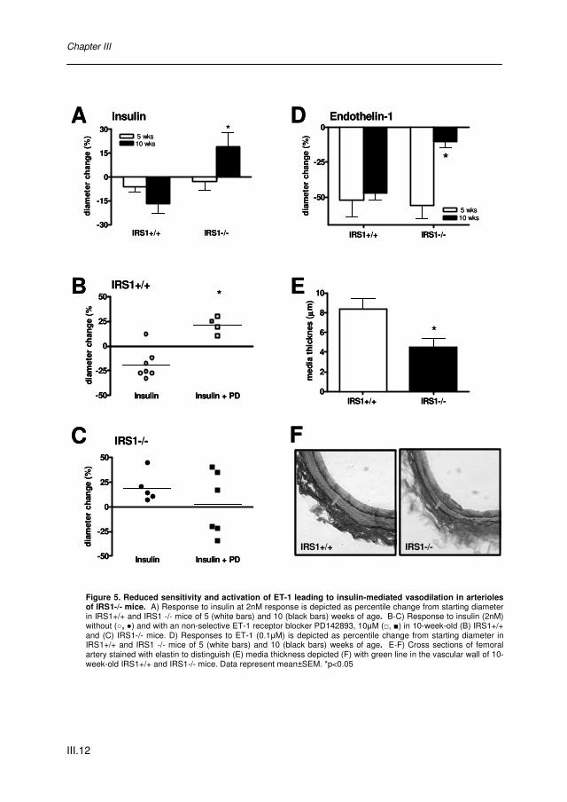
Chapter III
III.12
Figure 5. Reduced sensitivity and activation of ET-1 leading to insulin-mediated vasodilation in arterioles of IRS1-/- mice. A) Response to insulin at 2nM response is depicted as percentile change from starting diameter in IRS1+/+ and IRS1 -/- mice of 5 (white bars) and 10 (black bars) weeks of age. B-C) Response to insulin (2nM) without (○, ●) and with an non-selective ET-1 receptor blocker PD142893, 10μM (□, ■) in 10-week-old (B) IRS1+/+ and (C) IRS1-/- mice. D) Responses to ET-1 (0.1μM) is depicted as percentile change from starting diameter in IRS1+/+ and IRS1 -/- mice of 5 (white bars) and 10 (black bars) weeks of age. E-F) Cross sections of femoral artery stained with elastin to distinguish (E) media thickness depicted (F) with green line in the vascular wall of 10-week-old IRS1+/+ and IRS1-/- mice. Data represent mean±SEM. *p<0.05
IRS1+/+ IRS1-/-
-50
-25
0
5 wks10 wks
*
dia
met
er c
han
ge (
%)
IRS1+/+ IRS1-/--30
-15
0
15
30 5 wks10 wks
*
dia
met
er c
han
ge
(%)
A
C
D
Insulin Insulin + PD-50
-25
0
25
50
dia
met
er c
han
ge (
%)
Insulin Insulin + PD-50
-25
0
25
50 *
dia
met
er c
han
ge
(%
F
IRS1+/+
IRS1-/-
IRS1+/+ IRS1-/-0
2
4
6
8
10
*
med
ia t
hick
nes
(μμ μμ
m)
IRS1-/-IRS1+/+
EB
Insulin Endothelin-1

IRS1 Deficiency affects Muscle Vascularization
III.13
DISCUSSION This study demonstrates that IRS1 deficiency results in a reduced diameter of arterioles, but does not alter endothelium-dependent vasodilation. However, insulin responses in arterioles were changed in IRS1 deficient mice, resulting in insulin-mediated vasodilation caused by a
reduced ET-1 mediated vasoconstriction and ET-1 sensitivity. This functional change in arterioles was associated with structural narrowing of arteries and reduced capillary-to-muscle ratio in skeletal muscle.
The role of IRS1 in vascular insulin signaling This study showed for the first time that IRS1 is not only involved in the NO pathway,6,23 but
also in the ET-1 pathway of insulin. Unexpectedly, our study showed an insulin-mediated vasodilatation in arterioles of IRS1-/- mice, which was caused by a reduced sensitivity and ET-1 mediated vasoconstriction. The reduction in ET-1 sensitivity could be caused by the
reduced ability of the smooth muscle cells in the arterial wall to contract. In the femoral artery, media thickness was reduced in IRS1-/- mice. Although we were not able to investigate media thickness in arterioles, due to their small size, it is plausible to assume a
reduction of media thickness in arterioles, resulting in reduced capability to contract to vasoconstrictors, like ET-1. As ET-1 also has hypertrophic properties,27 the reduced media thickness could be a consequence of reduced ET-1 activation and the resulting reduced
contractile response in IRS1-/- mice. ET-1 activation is mediated by ERK1/2. One other study suggests the involvement
of IRS1 in ET-1 pathway and showed that IRS1 deletion reduces insulin-mediated ERK1/2
activation in cultured myocytes.28 Although we could not determine ERK1/2 activation in arterioles of IRS1-/- mice due to detection limitation, the inhibition of ET-1 with an ET-1 receptor blocker inhibited insulin-mediated vasoconstriction in IRS1+/+ mice, while it had no
effect in IRS1-/- mice. Taken together, our study shows the involvement of IRS1 in ERK1/2/ET-1 pathway of insulin, implying a more prominent role of IRS1 in insulin signaling than only its involvement in NO pathway.
In general, the mouse strain used in our study appears to have stronger reactivity to ET-1 than to NO. This became evident from the induction of insulin-mediated vasoconstriction in WT mice (Fig 4A). The vasoconstriction was abolished by the inhibition
of ET-1, but did not lead to NO-mediated vasodilation (Fig 5B), suggesting we could not detect NO responses to insulin in these isolated arterioles. It has to be taken into account that these findings cannot immediately be translated to the in vivo or human situation,
because an underestimated role of NO in our mouse model cannot be excluded. In this study, we determined insulin-mediated vasoreactivity in isolated arteries and showed no or a small role for NO. Akt activation, upstream of NO, was detectable, and was reduced in
IRS1-/- mice. However, in vivo the role of IRS1 in NO production is probably more pronounced, due to the presence of flow.
III

Chapter III
III.14
One logical explanation of the partly remaining insulin-mediated Akt phosphorylation and insulin-mediated vasodilatation in muscle arterioles of IRS1-/- mice is a compensated IRS2
expression. Although IRS2 is more expressed in arterial tissue than IRS1, IRS2 gene expression was not increased in arteries of IRS1-/- mice.29 However, it should be taken into account that a decrease in protein expression does not necessary mean a reduced
functionality of the protein. In agreement with this, insulin-mediated tyrosine-phosphorylation of IRS2 is enhanced in muscles of IRS1-/- mice.9,30 This suggests that IRS2 may partly compensate for defects of IRS1 in arteries,
which would explain the ability of arterioles with IRS1 deletions to activate Akt and vasodilate to insulin .
To our knowledge two other studies investigated vascular function in IRS1-/-
mice,29,31 but this is the first study that investigated IRS1 in the microcirculation. The present study shows that endothelium-dependent vasodilation was not altered in arterioles from 10-week-old IRS1-/- mice. In agreement to our study, Kubota et al showed no difference in
acetylcholine response in aortic rings of 20 week old IRS1-/- mice.29 In contrast, Abe et al showed in aortic rings from very old specify IRS1-/- mice an impaired endothelium-dependent vasodilation to acetylcholine.31 However, our study focused on the function of
arterioles and we used mice of 10 weeks of age, making it difficult to compare these data directly.
The role of IRS1 in regulation of muscle vascularization Despite an adaptive response to insulin in muscle arterioles and the ability of the muscles and capillaries to grow, IRS1-/- mice had a persistent decrease in muscle growth and
capillary-to-muscle ratio. IRS1 can activate Akt upon receptor stimulation by insulin or IGF1 and Akt regulates both the muscle growth3 and increased perfusion11 by inducing an enlarged capillary network.12 Akt activation in myocytes with an deletion for IRS1 is
decreased,28 suggesting an impaired muscle development and decreased capillary network in muscles of IRS1-/- mice. Pete et al showed that the gastrocnemius muscle mass was reduced in IRS1-/- mice due to an impaired IGF1-dependent growth of the skeletal muscle.
However, IGF1 overexpression could not overcome this body weight and muscle mass deficit that resulted from absolute IRS1 deficiency. This suggests that IGF1-stimulated muscle growth is impaired in IRS1-/- mice, and implies that an additional mechanism
preserves postnatal muscle growth, possible by an adaptation of vascular responses of insulin. In agreement with this, we showed that IRS1-/- mice have a different vascular response to insulin, as shown by comparing 5 and 10-week-old mice, which depends on ET-
1 activation and is associated with muscle growth. Furthermore, this study shows that Akt can be activated in arterioles from IRS1-/- mice, although probably to a lesser degree, which might contribute to both the observed insulin-mediated vasodilation, and ability of muscles
and capillaries to grow. Capillary-to-muscle ratio correlates with in-vivo insulin-mediated glucose uptake,32 but
glucose uptake also depends on the number of capillaries perfused.11 In IRS1-/- mice, the

IRS1 Deficiency affects Muscle Vascularization
III.15
capillary ratio was structurally reduced. However, the number of perfused versus non-perfused capillaries cannot be distinguished by the lectin staining used in this study. In
IRS1+/+ mice the capillary ratio is higher, but not all capillaries are necessarily perfused, while in the IRS1-/- mice all capillaries may be perfused, despite the lower capillary ratio. This may imply that the muscle perfusion of IRS1+/+ and IRS1-/- is not or less different than
expected on the basis of the capillary-to-muscle ratio. In favor of such compensatory mechanism we observed an insulin-mediated vasodilation in arterioles from IRS1-/- mice and not in IRS1+/+ mice.
Perspectives Structural and functional alterations of the muscle microcirculation have been proposed to
contribute to insulin resistance.33 These alterations comprise reduced insulin-mediated activation of Akt, reduced eNOS activity,17-19 normal to enhanced endothelin activity,17,20 inward remodeling of arterioles21 and a reduced capillary-to-muscle ratio in skeletal
muscle.22 The present study was undertaken to investigate whether decreased IRS1 expression contributes to these structural and functional changes.
Our data show that decreased IRS1 expression contributes to impaired Akt
activation, inward remodeling of arterioles and a reduction of capillary to myocyte ratio. However, reduced IRS1 expression does not explain the impaired endothelium-dependent vasodilatation or enhanced ET-1 activity observed in insulin resistance.17,20 Interestingly, this
study shows a novel additional role of IRS1 in the insulin-mediated vasoconstriction through ET-1. Its significance for impaired insulin signaling in insulin resistant subjects requires further clinical evaluation.
III

Chapter III
III.16
REFERENCE LIST 1. Sesti G. Federici M. Hribal,ML, Lauro D. Sbraccia P, Lauro R.: Defects of the insulin receptor
substrate (IRS) system in human metabolic disorders. FASEB J. 15:2099-2111, 2001. 2. Gazdag,AC, Dumke,CL, Kahn,CR, Cartee,GD: Calorie restriction increases insulin-stimulated
glucose transport in skeletal muscle from IRS-1 knockout mice. Diabetes 48:1930-1936, 1999.
3. Pete,G, Fuller,CR, Oldham,JM, Smith,DR, D'Ercole,AJ, Kahn,CR, Lund,PK: Postnatal Growth Responses to Insulin-Like Growth Factor I in Insulin Receptor Substrate-1-Deficient Mice. Endocrinology 140:5478-5487, 1999
4. Baron,AD: Hemodynamic actions of insulin. Am J Physiol Endocrinol Metab 267:E187-E202, 1994
5. Vincent,MA, Clerk,LH, Lindner,JR, Klibanov,AL, Clark,MG, Rattigan,S, Barrett,EJ: Microvascular Recruitment Is an Early Insulin Effect That Regulates Skeletal Muscle Glucose Uptake In Vivo. Diabetes 53:1418-1423, 2004
6. Alessi,DR, Andjelkovic,M, Caudwell,B, Cron,P, Morrice,N, Cohen,P, Hemmings,BA: Mechanism of activation of protein kinase B by insulin and IGF-1. EMBO J 15:6541-6551, 1996
7. Araki,E, Lipes,MA, Patti,ME, Bruning,JC, Haag,BLI, Johnson,RS, Kahn,CR: Alternative pathway of insulin signalling in mice with targeted disruption of the IRS-1 gene. Nature 372:186-190, 1994
8. Tamemoto,H, Kadowaki,T, Tobe,K, Yagi,T, Sakura,H, Hayakawa,T, Terauchi,Y, Ueki,K, Kaburagi,Y, Satoh,S, .: Insulin resistance and growth retardation in mice lacking insulin receptor substrate-1. Nature 372:182-186, 1994
9. Yamauchi,T, Tobe,K, Tamemoto,H, Ueki,K, Kaburagi,Y, Yamamoto-Honda,R, Takahashi,Y, Yoshizawa,F, Aizawa,S, Akanuma,Y, Sonenberg,N, Yazaki,Y, Kadowaki,T: Insulin signalling and insulin actions in the muscles and livers of insulin-resistant, insulin receptor substrate 1-deficient mice. Mol.Cell.Biol. 16:3074-3084, 1996
10. Baron,AD, Laakso,M, Brechtel,G, Edelman,SV: Reduced capacity and affinity of skeletal muscle for insulin-mediated glucose uptake in noninsulin-dependent diabetic subjects. Effects of insulin therapy 2299. J Clin Invest 87:1186-1194, 1991
11. Rattigan,S, Clark,MG, Barrett,EJ: Hemodynamic actions of insulin in rat skeletal muscle: evidence for capillary recruitment. Diabetes 46:1381-1388, 1997
12. Takahashi,A, Kureishi,Y, Yang,J, Luo,Z, Guo,K, Mukhopadhyay,D, Ivashchenko,Y, Branellec,D, Walsh,K: Myogenic Akt Signaling Regulates Blood Vessel Recruitment during Myofiber Growth. Mol.Cell.Biol. 22:4803-4814, 2002
13. Lillioja,S, Young,AA, Culter,CL, Ivy,JL, Abbott,WG, Zawadzki,JK, Yki-Jarvinen,H, Christin,L, Secomb,TW, Bogardus,C: Skeletal muscle capillary density and fiber type are possible determinants of in vivo insulin resistance in man. J Clin Invest 80:415-424, 1987
14. Dimmeler,S, Fleming,I, Fisslthaler,B, Hermann,C, Busse,R, Zeiher,AM: Activation of nitric oxide synthase in endothelial cells by Akt- dependent phosphorylation. Nature 399:601-605, 1999
15. Eringa,EC, Stehouwer,CDA, Nieuw Amerongen,GP, Ouwehand,L, Westerhof,N, Sipkema,P: Vasoconstrictor effects of insulin in skeletal muscle arterioles are mediated by ERK1/2 activation in endothelium. Am J Physiol Heart Circ Physiol 287:H2043-H2048, 2004
16. Potenza,MA, Marasciulo,FL, Chieppa,DM, Brigiani,GS, Formoso,G, Quon,MJ, Montagnani,M: Insulin resistance in spontaneously hypertensive rats is associated with endothelial dysfunction characterized by imbalance between NO and ET-1 production. Am J Physiol Heart Circ Physiol 289:H813-H822, 2005
17. Eringa,EC, Stehouwer,CDA, Roos,MH, Westerhof,N, Sipkema,P: Selective resistance to vasoactive effects of insulin in muscle arterioles of obese Zucker (fa/fa) rats. Am J Physiol Endocrinol Metab 293:E1134-E1139, 2007
18. Jiang,ZY, Lin,YW, Clemont,A, Feener,EP, Hein,KD, Igarashi,M, Yamauchi,T, White,MF, King,GL: Characterization of selective resistance to insulin signaling in the vasculature of obese Zucker (fa/fa) rats. J.Clin.Invest. 104:447-457, 1999
19. Okon,EB, Chung,AWY, Rauniyar,P, Padilla,E, Tejerina,T, McManus,BM, Luo,H, van Breemen,C: Compromised Arterial Function in Human Type 2 Diabetic Patients 2003. Diabetes 54:2415-2423, 2005

IRS1 Deficiency affects Muscle Vascularization
III.17
20. Cardillo,C, Campia,U, Bryant,MB, Panza,JA: Increased Activity of Endogenous Endothelin in Patients With Type II Diabetes Mellitus. Circulation 106:1783-1787, 2002
21. Frisbee,JC: Remodeling of the skeletal muscle microcirculation increases resistance to perfusion in obese Zucker rats. Am J Physiol Heart Circ Physiol 285:H104-H111, 2003
22. Gudbjornsdottir,S, Sjostrand,M, Strindberg,L, Lonnroth,P: Decreased muscle capillary permeability surface area in type 2 diabetic subjects. J Clin Endocrinol Metab 90:1078-1082, 2005
23. Montagnani,M, Ravichandran,LV, Chen,H, Esposito,DL, Quon,MJ: Insulin Receptor Substrate-1 and Phosphoinositide-Dependent Kinase-1 Are Required for Insulin-Stimulated Production of Nitric Oxide in Endothelial Cells. Mol Endocrinol 16:1931-1942, 2002
24. Federici,M, Pandolfi,A, De Filippis,EA, Pellegrini,G, Menghini,R, Lauro,D, Cardellini,M, Romano,M, Sesti,G, Lauro,R, Consoli,A: G972R IRS-1 Variant Impairs Insulin Regulation of Endothelial Nitric Oxide Synthase in Cultured Human Endothelial Cells. Circulation 109:399-405, 2004
25. Eringa,EC, Stehouwer,CDA, Merlijn,T, Westerhof,N, Sipkema,P: Physiological concentrations of insulin induce endothelin-mediated vasoconstriction during inhibition of NOS or PI3-kinase in skeletal muscle arterioles. Cardiovascular Research 56:464-471, 2002
26. Bakker,W, Sipkema,P, Stehouwer,CDA, Serne,EH, Smulders,YM, van Hinsbergh,VWM, Eringa,EC: PKC-theta activation induces insulin-mediated constriction of muscle arterioles. Diabetesdb07-0792, 2007
27. Schiffrin,EL, Lariviere,R, Li,JS, Sventek,P, Touyz,RM: Enhanced expression of endothelin-1 gene may cause blood pressure-independent vascular hypertrophy. J Cardiovasc Pharmacol 26 Suppl 3:S5-S8, 1995
28. Huang,C, Thirone,ACP, Huang,X, Klip,A: Differential Contribution of Insulin Receptor Substrates 1 Versus 2 to Insulin Signaling and Glucose Uptake in L6 Myotubes. J.Biol.Chem. 280:19426-19435, 2005
29. Kubota,T, Kubota,N, Moroi,M, Terauchi,Y, Kobayashi,T, Kamata,K, Suzuki,R, Tobe,K, Namiki,A, Aizawa,S, Nagai,R, Kadowaki,T, Yamaguchi,T: Lack of Insulin Receptor Substrate-2 Causes Progressive Neointima Formation in Response to Vessel Injury 1878. Circulation 107:3073-3080, 2003
30. Patti,ME, Sun,XJ, Bruening,JC, Araki,E, Lipes,MA, White,MF, Kahn,CR: 4PS/Insulin Receptor Substrate (IRS)-2 Is the Alternative Substrate of the Insulin Receptor in IRS-1-deficient Mice. J.Biol.Chem. 270:24670-24673, 1995
31. Abe,H, Yamada,N, Kamata,K, Kuwaki,T, Shimada,M, Osuga,J, Shionoiri,F, Yahagi,N, Kadowaki,T, Tamemoto,H, Ishibashi,S, Yazaki,Y, Makuuchi,M: Hypertension, hypertriglyceridemia, and impaired endothelium-dependent vascular relaxation in mice lacking insulin receptor substrate-1. J Clin Invest 101:1784-1788, 1998
32. Lillioja,S, Young,AA, Culter,CL, Ivy,JL, Abbott,WG, Zawadzki,JK, Yki-Jarvinen,H, Christin,L, Secomb,TW, Bogardus,C: Skeletal muscle capillary density and fiber type are possible determinants of in vivo insulin resistance in man. J Clin Invest 80:415-424, 1987
33. Barrett,EJ, Eggleston,EM, Inyard,AC, Wang,H, Li,G, Chai,W, Liu,Z: The vascular actions of insulin control its delivery to muscle and regulate the rate-limiting step in skeletal muscle insulin action. Diabetologia 52:752-764, 2009
III

Chapter III
III.18

IRS2 Deficiency decreases Blood Pressure
IV.1
IRS2deficiency decreases
Blood pressureby impairment of insulin-mediated
Endothelin-1activation in arterioles
IRS2deficiency decreases
Blood pressureby impairment of insulin-mediated
Endothelin-1activation in arterioles
Hypertension, in Revision

Chapter IV
IV.2

IRS2 Deficiency decreases Blood Pressure
IV.3
IV. IRS2 Deficiency decreases Blood Pressure by Impairment of Insulin-mediated Endothelin-1 activation in Muscle Arterioles
Wineke Bakker, Caro-Lynn A.F. Alta, Ronald Vlasblom, Pieter Sipkema, Coen D.A. Stehouwer, Victor W.M. van Hinsbergh, Etto C. Eringa.
ABSTRACT Objective: In type 2 diabetes, dysfunctional insulin receptor substrates (IRS) cause impaired insulin signaling, which is often associated with an increase in blood pressure. In
part, insulin regulates blood pressure by controlling vasodilation (mediated by NO) as well as vasoconstriction (mediated by endothelin-1). IRS2 is necessary for insulin signaling; however, the involvement of IRS2 in blood pressure regulation is not known. The aim of this
study is to investigate whether deletion of IRS2 alters blood pressure by changes in insulin-mediated vasoreactivity and cardiac output.
Research design and results: Radiotelemetry transmitters were implanted in IRS2-/- mice and their wildtype littermates for continuous blood pressure measurements without stress. The IRS2-/- mice showed a lower mean arterial pressure (MAP) than their wildtype
counterparts (96±6 vs. 111±2 mmHg respectively, p<0.0001), due to mainly a decrease in vascular resistance and partly by decrease in cardiac output. To further study vascular function, arterioles were isolated to study insulin-mediated vasoreactivity ex-vivo. Compared
to IRS2+/+, IRS2-/- mice showed an impaired insulin-mediated ERK1/2 and endothelin-1 activation, leading to insulin-mediated vasodilation. The sensitivity to endothelin-1 was not different between IRS2-/- and wildtype in arterioles.
Conclusion: IRS2-deficiency lowers blood pressure, which is caused by a decrease in cardiac output and a decrease in vascular resistance by a specific impairment of insulin’s
vasoconstrictor effects in arterioles. These data suggest that IRS2 plays a more important role in the ERK1/2/endothelin-1 pathway than in the Akt/NO pathway of insulin in arterioles.
IV

Chapter IV
IV.4
INTRODUCTION
Hypertension is associated with impaired insulin signaling, which is reflected by an prevalence of ~50% of insulin resistance in patients with hypertension.1 Impaired insulin signaling can be caused by environmental or genetic factors. Obesity is a frequent cause of
impaired insulin signaling, in which changes in the function of insulin receptor substrates (IRSs) play an important role. Genetic mutations have been found in two IRS proteins, IRS1 and IRS2, which have been reported to be associated with insulin resistance and/or type 2
diabetes.2,3 However, the exact role of IRS proteins in blood pressure regulation is unknown. Blood pressure is controlled by cardiac output and by arterioles.4,5 Insulin plays an
important regulatory role in these arteries.6 It determines vascular properties that contribute
to blood pressure regulation by stimulating both vasodilation (through Akt activation and NO production)7,8 and vasoconstriction (through ERK1/2 activation and endothelin-1 (ET-1) production)9,10 in arterioles. In hypertension, vascular dysfunction is characterized by a
predominance of insulin’s vasoconstrictor effects through decreased NO production and increased ET-1 production.11,12 This raises susceptibility to increased vascular peripheral resistance and hypertension.
IRS proteins are essential to insulin signaling, but their exact role in blood pressure regulation and vasoreactivity is unknown. The IRS isoforms IRS1 and IRS2 are involved in the regulation of insulin-stimulated glucose disposal. Both IRS1-/- and IRS2-/- mice show
endothelial dysfunction in aortic rings.13 How IRS proteins function in arterioles and thereby blood pressure regulation has not yet been studied. Whereas IRS1 has received more attention, and IRS1 and IRS2 are homologous proteins, yet IRS2 is more abundantly
expressed in the vascular endothelium.13 Furthermore, the metabolic phenotype of IRS2-/- mice is more severe. IRS2-/- mice are hyperinsulinemic and become hyperglycemic later in life,14 whereas IRS1-/- mice stay in a hyperinsulinemic, but normoglycemic state.15,16
In cultured endothelial cells, IRS1 is mainly described to function in insulin-mediated NO production17 through Akt dependent eNOS phosphorylation. IRS2 has been identified as an IRS1-independent pathway of insulin signaling.18-20 Furthermore, an
increased IRS2 expression under hyperinsulinemia is accompanied by increased Akt and eNOS expression and decreased ERK2 expression in rat aortas.21 Of note, activity of ERK1/2 in arterioles regulates their diameter and therefore susceptibility to hypertension.22
In addition, in a metabolic study, Kubota et al mentioned that systolic blood pressure is increased in IRS2-/- mice, as determined by tail cuff. In contrast, Bouzakri et al shows that IRS2 is mainly involved in the activation of ERK1/2 in muscle myoblasts.18 Thus, IRS2 might
be an interesting determinant of endothelial function, but its exact role in insulin signaling in arterioles and its role in blood pressure regulation is not clear.

IRS2 Deficiency decreases Blood Pressure
IV.5
The aim of this study was to investigate whether deletion of IRS2 will affect blood pressure and whether this is mediated by shifting the balance of insulin-mediated
vasoreactivity in arterioles. With the use of radiotelemetry, blood pressure was measured in IRS2-/- and IRS2+/+ mice. Resistance artery function and cardiac output were used as determinants of diastolic and systolic blood pressure.
RESEARCH DESIGN AND METHODS
Animals: The investigation conformed to the Guide for the Care and Use of Laboratory Animals
published by the US National Institutes of Health (NIH publication No 85 to 23, revised 1996). The local
ethics committee for animal experiments approved the procedures. Male and female IRS2-/- mice and
their WT littermates, on a hybrid background of the C57Bl/6 and SV129 mice strain, (Jackson
Laboratories, Maine, USA) were obtained by heterozygous breeding. PCR was used to distinguish
genotypes between IRS2+/+ and IRS2-/- mice. To check whether differences in genetic background
affect insulin responses, C57Bl6 (Harlan, Horst, the Netherlands) and Sv129 mice (Jackson
laboratories, Maine, USA) were used.
Blood glucose and insulin (non fasting): Blood glucose was measured from tail blood samples using
a glucose analyzer (HemoCue, Angelholm, Sweden). Plasma insulin was measured using ELISA
(EZRMI-13K; Linco Research, St Charles, MO, USA).
Blood pressure measurements: The radiotelemetry transmitters (TA 11 PA-C10, Data Science
International, St. Paul, MN) were implanted under 3.0% isoflurane anesthesia (in a mixture of O2 and
air) in 8-week-old mice. The pressure-sensing catheter-tip was placed through the left carotid artery into
the aortic arch and the transmitter was placed in a subcutaneous pocket along the right flank. After a
recovery period of 10 days, data of systolic (SBP), diastolic (DBP) and mean arterial pressure (MAP),
heart rate (HR) and activity were collected every 5 min for 10 sec during 72 hours. The data was
collected using the Dataquest ART software system, version 4.0 (Data Sciences International). This
allowed us to detect, collect, and analyze signals from several animals simultaneously. For statistical
analyses, we used mean values of 3 days for data analyses.
To determine peripheral vascular resistance, the time constant tau was used to describe the
individual diastolic pressure decay. The last third of the diastolic pressure decay was sampled (2 msec
interval) and the individual coordinate points (t,P) were plotted on a semilogarithmic scale. This results
in a nearly straight line with negative slope. The reciprocal of the slope is the time constant tau for that
diastolic decay and can be used as an index of peripheral vascular resistance when the arterial
compliance does not change.23,24 For each mouse, an average value was taken of at least 10 arterial
waveforms.
IV

Chapter IV
IV.6
Echocardiography: Non-invasive transthoracic echocardiograms were recorded in mice anesthetized
with 1.2% isoflurane in a mixture of N2O/O2 (1/2, vol/vol) by an Aloka echo-machine (ProSound SSD-
4000) using a 13-MHz linear array transducer. Aortic diameter and aortic outflow (using Doppler in
apical 5 chamber view) were measured and were used to determine stroke volume (SV: cross sectional
area of the aorta * Velocity time integral of the aorta). Cardiac output (CO) was calculated by multiplying
HR with SV. End-diastolic and end-systolic diameters (EDD and ESD, respectively) of the left ventricle
were measured perpendicularly to the interventricular septum at midseptal level. EDD and ESD were
used to calculate fractional shortening with the following formula: fractional shortening = [(EDD –
ESD)/EDD] x 100%.
Vasoreactivity: Mice were sacrificed with an overdose of isoflurane and arteries (aorta, femoral,
mesenteric and gracilis artery) were dissected, placed in a pressure myograph and pressurized to their
corresponding in vivo pressures (125, 100, 100 and 80 mmHg, respectively) to measure maximal
diameters25. Compliance is defined as the slope of the pressure-volume relation (C=dV/dP) and is
obtained by expanding a vessel by a known volume and measuring the change in pressure at steady-
state. The compliance (C) of the femoral artery was determined by the change in diameter (dD) divided
by the change in pressure (dP), and multiplied with (π*D/2). dP/dD was estimated by curve fitting of the
diameter-pressure relation. dP/dD and the femoral diameter (D) were then determined at in-vivo
pressure of the mouse (formula: C= (π*D*dD)/(2*dP)).
To study changes in response to insulin in arterioles, segments of the gracilis artery were
preconstricted to ~50% of the maximal diameter with 25 mM KCl in MOPS buffer, as described before26.
Endothelial integrity was determined by measuring vasodilator response to acetylcholine (ACh:
0.1μMol/l) before and after experiments. Acute effects of insulin (Human insulin (Actrapid®), Novo
Nordisk, Alphen a/d Rijn, the Netherlands) on the diameter of the gracilis artery were studied at four
concentrations of insulin (0.02, 0.2, 2 and 20nM) and diameter changes at each concentration were
recorded for 30 minutes25. The role of ET-1 in insulin-mediated vasoreactivity was assessed by pre-
treatment for 30 minutes with the non-selective ET-1 receptor antagonist (PD142893: 10μM, Kordia,
Leiden, the Netherlands) before the addition of insulin. Dose-relation responses with both acetylcholine
or sodium nitroprusside and ET-1 were performed to check endothelial or smooth muscle integrity of the
vessel wall.
Protein analysis: Protein analyses was performed by western blotting, as described before.26
Segments of gracilis arteries were exposed to solvent (controls) or to insulin (2nM) for 15 minutes at
37ºC. Differences in phosphorylated Akt at ser 473 were adjusted for differences in total Akt protein
staining. Antibodies were obtained from Cell Signaling Technology.
Statistics: Values are expressed as mean±SEM. Statistical analyses for differences in blood pressure
were performed by Student’s t-test and differences in protein activation by column t-test with
standardized mean. Differences in insulin responses were analyzed by one-way ANOVA and post hoc
analysis was performed with the Bonferroni method. Differences in Probability values of <0.05 were
considered statistically significant.

IRS2 Deficiency decreases Blood Pressure
IV.7
RESULTS
Table 1 shows the general characteristics of IRS2-/- mice and WT littermates. IRS2-/- mice had similar body weights and higher levels of insulin and glucose than the IRS2+/+ mice. The diameters of both gracilis artery (resistance artery) and femoral artery (conduit artery)
were reduced in IRS2-/- mice (Table 1). Blood pressure is decreased in IRS2-/- mice Blood pressure was measured continuously in conscious mice using radiotelemetry. Figure 1A shows an example of the circadian rhythm of blood pressure in IRS2+/+ and IRS2-/- mice. The continuously measured mean arterial pressure (MAP) over 48 hours shows a
decrease in IRS2-/- compared to IRS2+/+ mice (Fig 1B: 111±2 mm Hg vs. 96±6 mm Hg, p<0.01). The lower blood pressure in IRS2-/- mice was observed during the entire circadian cycle (Fig 1A). Both systolic and diastolic blood pressure are decreased by 10%, however
due to large variation in the systolic blood pressure data, only the decrease in diastolic blood pressure was significant (Table 2). The heart rate in conscious animals was not significantly different in IRS2-/- compared to IRS2+/+ mice (Table 2).
The cardiac output is decreased in IRS2-/- mice To further explore the decrease in blood pressure of the IRS2-/- mice, determinants for both
systolic and diastolic blood pressure were determined. Determinants for systolic blood pressure are the cardiac output and the compliance of the conduit arteries. In this study cardiac output was determined by echocardiography under isoflurane anesthesia in both
type of mice. Because isoflurane anesthesia influences blood pressure and heart rate, we measured both the arterial pressure by radiotelemetry and subsequently the cardiac output by echocardiography during one anesthetic period using a constant concentration of
isoflurane (Table 3). The cardiac output was lower in the IRS2-/- mice compared to IRS2+/+ mice (Table 3), which was mainly determined by a decrease in stroke volume (Table 3). The fractional shortening, as indicator of the systolic heart function, was determined by end
systolic and end diastolic lumen diameters and was not changed in IRS2-/- mice (Table 3). Changes in the compliance of the wall of conduit arteries were estimated from the
pressure-diameter-relationships in isolated femoral arteries at pressures ranging from 25 till
100 mmHg. The curve of the pressure-diameter-relation (Fig 2A) and the calculated compliance (Fig 2B) did not significantly differ between the femoral arteries from IRS2-/- mice and their IRS2+/+ counterparts. We assume that possible changes in compliance of
the femoral artery are representative for the total arterial compliance.
IV

Chapter IV
IV.8
General characteristics IRS2+/+ IRS2-/- P
Body weight (g) 23±1 24±1 0.84
Fasting Blood Glucose (mMol/l) 5.3±0.5 12.6±2.7 <0.05
Non-fasting Blood Glucose (mMol/l) 8.7±0.8 12.2±0.9 <0.01
Non-fasting Blood Insulin (μU/ml) 9±1 20±2 <0.01
Diameters arteries
Gracilis artery (μm) 127±5 111±6 <0.05
Femoral artery (μm) 298±8 244±10 <0.05
Mesenteric artery (μm) 201±14 197±10 0.51
Aorta (μm) 1342±5 1283±6 0.09
Characteristics IRS2+/+ IRS2-/- P
Heart weight (mg) 116±1 118±1 0.87
Mean arterial pressure (MAP: mmHg) 69.7±7.3 46.0±4.8 <0.05
Heart rate (HR: bpm) 444±16 428±45 0.71
Stroke volume (SV: ml) 0.048±0.004 0.032 ±0.004 <0.05
Cardiac output (HR*SV: ml/min) 21.5±2.2 13.3±2.1 <0.05
Total peripheral resistance (mmHg/ml/min) 3.6±0.7 4.1±1.1 0.69
End Diastolic Lumen Diameter (EDD: μm) 3.1±0.2 2.8±0.2 0.12
End Systolic Lumen Diameter (ESD: μm) 1.2 ±0.1 1.2±0.1 0.60
Fractional shortening (%) 60.9±2.8 57.7±3.2 0.45
Table 3. Cardiac
characteristics involving
blood pressure regulation
under 1.2% isoflurane
anesthetics Cardiac Output
(CO) was determined by
HR*SV. Peripheral Vascular
Resistance was determined by
MAP/CO. Fractional shortening
was determined by (EDD-
ESD/EDD)*100%.
Table 2. Blood pressure
characteristics of conscious
IRS2+/+ and IRS2-/- mice All
variables are an average of
collected data of 5 seconds
every 5 minutes for a period of
72 hours. IRS2+/+ n=15,
IRS2-/- n=8
Table 1. General
characteristics and artery
diameters of IRS2-/- mice.
Data represent mean±sem,
IRS2+/+ n=10, IRS2-/- n=7.
General characteristics IRS2+/+ IRS2-/- P
Mean arterial pressure (MAP) (mmHg) 111±2 96±6 <0.01
Systolic pressure (mmHg) 115±8 104±6 0.36
Diastolic pressure (mmHg) 101±2 90±4 <0.01
Pulse pressure (mmHg) 21±2 15±3 <0.05
Heart Rate (bpm) 589±11 553±19 0.08
Activity (moves / 5 seconds) 7 ±1 5 ±1 0.11
General characteristics IRS2+/+ IRS2-/- P
Mean arterial pressure (MAP) (mmHg) 111±2 96±6 <0.01
Systolic pressure (mmHg) 115±8 104±6 0.36
Diastolic pressure (mmHg) 101±2 90±4 <0.01
Pulse pressure (mmHg) 21±2 15±3 <0.05
Heart Rate (bpm) 589±11 553±19 0.08
Activity (moves / 5 seconds) 7 ±1 5 ±1 0.11

IRS2 Deficiency decreases Blood Pressure
IV.9
Figure 3. Decreased peripheral vascular resistance, determined by time constant tau of the diastolic pressure decay, in IRS2-/- mice. (A) Continuous recording of arterial waveform in IRS2+/+ (○) and IRS2-/- mice (●) . (B) Time constant tau of the diastolic pressure decay as an index for peripheral vascular resistance in IRS2+/+ (n=9) and IRS2-/- (n=8), * p<0.05.
Figure 1. Decreased blood pressure in IRS2-/- mice. (A) Representative day and night rhythm of a continuous blood pressure registration for 2 nights and 2 days in one representative IRS2+/+ (○) and one representative IRS2-/- mouse (●). (B) Mean arterial pressure (MAP), an average of diastolic and systolic pressure averaged over 48h was decreased in IRS2-/- (n=8) compared to IRS2+/+ (n=15), * p<0.01.
IRS2+/+ IRS2-/-0
25
50
75
100
125
*
mm
Hg
A B
40 50 60 700
25
50
75
100
125
150
IRS2-/-IRS2+/+
MA
P (
mm
Hg
)
night day night IRS2+/+ IRS2-/-0
25
50
75
100
125
*
mm
Hg
A B
40 50 60 700
25
50
75
100
125
150
IRS2-/-IRS2+/+
MA
P (
mm
Hg
)
night day night 40 50 60 700
25
50
75
100
125
150
IRS2-/-IRS2+/+
MA
P (
mm
Hg
)
night day night
Figure 2. Decreased cardiac output under 1.2% isoflurane anesthetics in IRS2-/- mice. (A) MAP, averaged over a continuous measurement of 15 minutes. (B) Cardiac output (CO) determined by multiplying stroke volume and heart rate, by echocardiography in IRS2+/+ (n=7) and IRS2-/- (n=5), * p<0.05.
A
0.000 0.025 0.050 0.075 0.10070
80
90
100
110
120
130
140
IRS2+/+IRS2-/-
time (msec)
Art
eria
l pre
ssur
e (m
m H
g)
dP
dt
IRS2+/+ IRS2-/-0.0
0.1
0.2
0.3
0.4
**
mse
c
BA
0.000 0.025 0.050 0.075 0.10070
80
90
100
110
120
130
140
IRS2+/+IRS2-/-
time (msec)
Art
eria
l pre
ssur
e (m
m H
g)
dP
dt
dP
dt
IRS2+/+ IRS2-/-0.0
0.1
0.2
0.3
0.4
**
mse
c
B
A
IRS2+/+ IRS2-/-0
200
400
600
com
pli
ance
(m
mH
g/ μμ μμ
m)
B
20 40 60 80 100
100
200
300
IRS2+/+IRS-/-
pressure mm Hg
dia
met
er (
μμ μμm
)
A
IRS2+/+ IRS2-/-0
200
400
600
com
pli
ance
(m
mH
g/ μμ μμ
m)
B
20 40 60 80 100
100
200
300
IRS2+/+IRS-/-
pressure mm Hg
dia
met
er (
μμ μμm
)
IV

Chapter IV
IV.10
Estimated peripheral vascular resistance is decreased in IRS2-/- mice Besides compliance, vascular resistance is the major determinant of diastolic blood
pressure, which can be determined in conscious mice by analyzing the diastolic pressure decay from the arterial waveforms obtained by the radiotelemetry recordings. Figure 3 shows a typical arterial waveform from IRS2+/+ and IRS2-/- mice (Fig 3A). One third of the diastolic pressure decay was used to determine the time constant tau (gray surface in Fig 3A), which was decreased in IRS2-/- mice compared to IRS2+/+ mice (Fig 3B). As compliance of the vessel wall of conduit arteries was similar between IRS2+/+ and IRS2-/-
mice, this implicates that the peripheral vascular resistance (PVR) in IRS2-/- mice is decreased, because the time constant is the product of the PVR and the compliance.
Insulin-mediated ET-1 and ERK1/2 activation is impaired in arterioles from IRS2-/- mice To study whether changes in vascular resistance in IRS2-/- mice could be explained by
changes in vascular function and protein expression, arterioles from IRS2+/+ and IRS2-/- mice were isolated.
Arterioles of IRS2-/- mice showed normal endothelial function, as reflected by
acetylcholine-induced vasodilation (Fig 4A), which is completely NO-dependent in these arteries.26 The sensitivity to respond to NO and ET-1 was not changed in IRS2-/- mice, as became evident from the dose response curves with the NO donor sodium nitroprusside and
exogenous ET-1 (Fig 4B and 4C). This shows that the arterioles from IRS2-/- mice were able to produce NO and to respond to NO and ET-1 in the absence of insulin.
In IRS2+/+ mice, insulin induced vasoconstriction, which was abolished after
preincubation with a non-selective ET-1 receptor antagonist (Fig 5A). This shows that the vasoconstriction in the arterioles of IRS2 +/+ mice was completely dependent on insulin-mediated ET-1 activity in the arterioles of IRS2+/+ mice. The vasoconstriction to insulin in
IRS2+/+ mice was different than in previous studies in C57Bl/6 mice, in which we found no change in diameter in response to insulin in arterioles.26 In contrast to that study, the IRS2 mice used in this study are inbred from two mice strains, C57Bl/6 and Sv129. We isolated
arteries from both strains and subjected them to insulin, to investigate whether insulin has different effects on arterioles from different strains of mice. Insulin induced a vasoconstriction in arteries from Sv129 mice, while it had no net effect on the diameter in
arteries from C57Bl/6 mice (Fig 6A). Endothelial function (response to acetylcholine-mediated vasodilation) was not different between both mouse strains (Fig 6B). This shows that arteries from different mouse strains display different responses to insulin and
underlines the importance of using littermates in the current study. In the IRS2-/- mice, insulin levels are higher than IRS2+/+ mice in-vivo (Table 1). However, insulin induced no change in vascular diameter at all concentrations and
preincubation with an ET-1 receptor antagonist had no effect (Fig 5B). As the responsiveness to ET-1 was equal (Fig 4C), this indicates that the ET-1 mediated
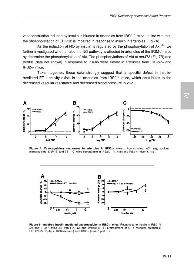
IRS2 Deficiency decreases Blood Pressure
IV.11
vasoconstriction induced by insulin is blunted in arterioles from IRS2-/- mice. In line with this, the phosphorylation of ERK1/2 is impaired in response to insulin in arterioles (Fig 7A).
As the induction of NO by insulin is regulated by the phosphorylation of Akt,27 we further investigated whether also the NO pathway is affected in arterioles of the IRS2-/- mice by determine the phosphorylation of Akt. The phosphorylations of Akt at ser473 (Fig 7B) and
thr308 (data not shown) in response to insulin were similar in arterioles from IRS2+/+ and IRS2-/- mice.
Taken together, these data strongly suggest that a specific defect in insulin-
mediated ET-1 activity exists in the arterioles from IRS2-/- mice, which contributes to the decreased vascular resistance and decreased blood pressure in-vivo.
Figure 5: Impaired insulin-mediated vasoreactivity in IRS2-/- mice. Responses to insulin in IRS2+/+ (A) and IRS2-/- mice (B) with ( ∆, ▲) and without (○, ●) pretreatment of ET-1 receptor antagonist, PD142893 (10μM) in IRS2+/+ (n=5) and IRS2-/- (n=4), * p<0.01).
Figure 4: Vasoregulatory responses in arterioles in IRS2-/- mice . Acetylcholine, ACh (A); sodium nitroprus-side, SNP (B) and ET-1 (C) were comparable in IRS2+/+ (○, n=5) and IRS2-/- mice (●, n=6).
-9 -8 -7 -6
0
25
50
75 IRS2+/+IRS2-/-
Log ACh
dia
met
er c
han
ge (
%)
A
-9 -8 -7 -6 -5
0
25
50
75IRS2+/+IRS2-/-
Log SNP
diam
eter
ch
ange
(%
)
B
-13 -12 -11 -10 -9
-75
-50
-25
0
IRS2-/-IRS2+/+
Log ET-1
dia
met
er c
han
ge (
%)
C
-9 -8 -7 -6
0
25
50
75 IRS2+/+IRS2-/-
Log ACh
dia
met
er c
han
ge (
%)
A
-9 -8 -7 -6 -5
0
25
50
75IRS2+/+IRS2-/-
Log SNP
diam
eter
ch
ange
(%
)
B
-13 -12 -11 -10 -9
-75
-50
-25
0
IRS2-/-IRS2+/+
Log ET-1
dia
met
er c
han
ge (
%)
C
0
-30
-20
-10
0
10
20
30
0.01 0.1 1 10
IRS2-/- + ET-1 inhibitionIRS2-/-
Insulin, nM
dia
met
er c
han
ge (
%)
0
-30
-20
-10
0
10
20
30
0.01 0.1 1 10
IRS2+/+
***
IRS2+/+ + ET-1 inhibition
Insulin, nM
diam
eter
cha
nge
(%
)
A B
0
-30
-20
-10
0
10
20
30
0.01 0.1 1 10
IRS2-/- + ET-1 inhibitionIRS2-/-
Insulin, nM
dia
met
er c
han
ge (
%)
0
-30
-20
-10
0
10
20
30
0.01 0.1 1 10
IRS2+/+
***
IRS2+/+ + ET-1 inhibition
Insulin, nM
diam
eter
cha
nge
(%
)
A B
IV

Chapter IV
IV.12
Figure 7: Decreased Phosphorylation ERK1/2, but not of Akt to insulin in arterioles of IRS2-/- mice. Insulin (2nM) stimulation (black bars) was compared to control situation (white bars) in the phosphorylation of ERK1/2 in (A) and in the phosphorylation of Akt at ser473 (B) in segments of the gracilis from IRS2+/+ (n=8) and IRS2-/- (n=9) mice, * p<0.05; # p<0.01.
Figure 6: Insulin-mediated vasoreactivity (A) and endothelial function (B) in arterioles of different mouse strains. Response to different concentrations of insulin in arterioles of C57Bl/6 and Sv129 mice (n=6, *P<0.001) (A). Response to different concentrations of acetylcholine (ACh) in arterioles of C57Bl/6 and Sv129 mice (n=4) (B).
B
0
1
2
3
4 ##Control
Insulin
IRS2+/+ IRS2-/-
pAkt
/ tA
kt (
A.U
.)
pAkt
tAktA
0.0
0.5
1.0
1.5
2.0*
IRS2+/+ IRS2-/-
ControlInsulin
pER
K/t
ER
K (
A.U
.)
pERK1/2
tERK1/2
B
0
1
2
3
4 ##Control
Insulin
IRS2+/+ IRS2-/-
pAkt
/ tA
kt (
A.U
.)
pAkt
tAktA
0.0
0.5
1.0
1.5
2.0*
IRS2+/+ IRS2-/-
ControlInsulin
pER
K/t
ER
K (
A.U
.)
pERK1/2
tERK1/2
-9 -8 -7 -6
0
20
40
60
80
100 C57Bl/6Sv129
Log ACh
dia
met
er c
han
ge
(%)
B
0
-30
-20
-10
0
10
20
30
0.01 0.1 1 10
C57Bl/6Sv129
**
Insulin, nM
dia
met
er c
han
ge (
%)
A
-9 -8 -7 -6
0
20
40
60
80
100 C57Bl/6Sv129
Log ACh
dia
met
er c
han
ge
(%)
B
0
-30
-20
-10
0
10
20
30
0.01 0.1 1 10
C57Bl/6Sv129
**
Insulin, nM
dia
met
er c
han
ge (
%)
A

IRS2 Deficiency decreases Blood Pressure
IV.13
DISCUSSION This study demonstrates for the first time that IRS2 deficiency lowers blood pressure, which is accompanied by a decrease in cardiac output and a decrease in vascular resistance by a
specific impairment of insulin’s vasoconstrictor effects in arterioles. Furthermore, this study shows that IRS2 is more involved in the ERK1/2-ET-1 pathway than in the Akt/NO pathway of insulin signalling in arterioles.
To our knowledge, one other study measured blood pressure in IRS2-/- mice, and demonstrated, with the tail-cuff method, that systolic blood pressure in IRS2-/- mice is increased.13 In contrast, the present study shows a decrease in mean arterial pressure in
IRS2-/- mice, mainly due to a decrease in diastolic blood pressure. Explanations for these different results may be the difference in the method to measure blood pressure and a difference in genetic background of the IRS2 mice. The tail cuff method measures the blood
pressure in restrained and stressed mice, which influences the blood pressure. The use of radiotelemetry in the present study overcomes the induction of stress. Furthermore, it determines an averaged central blood pressure for 72 hours in a normal environment,
instead of a determination of peripheral blood pressure for a short time period in a restrained condition.
Systolic blood pressure was not significantly decreased in IRS2-/- mice (Table 1).
However, a determinant of systolic blood pressure, cardiac output, was decreased in IRS2-/- mice. Cardiac output was determined under isoflurane anasthesia, which also has cardiodepressive28 and vasodilator effects29,30 and may influence cardiac output and blood
pressure.31 However, under these anesthesia conditions, blood pressure and heart rate decreased in both IRS2+/+ and IRS2-/- mice in the same extent (comparing table 2 and table 3). This suggests that the isoflurane anesthesia had no extra effect in IRS2-/- mice on
blood pressure regulation. The decrease in mean arterial pressure observed in IRS2-/- mice is partly
determined by a decrease in diastolic pressure (Fig 1 and Table 2), a decrease in vascular
resistance and an impaired insulin-mediated ET-1 activation (Fig 5). Diastolic blood pressure is largely regulated by adaptations in the vascular system. The diastolic pressure decay is caused by the drainage of blood from the large arteries through the peripheral arterioles and
can be used as a determinant for vascular resistance, if compliance does not change.23,24 The time constant tau of the diastolic pressure decay was decreased in IRS2-/- mice (Fig 3), which implicates a decrease in vascular resistance, because the compliance did not change
in the IRS2-/- mice. In these arterioles, insulin plays an important role in regulating vascular diameter, by inducing either vasoconstriction or vasodilation. This study points out that deletion of IRS2 impairs the capability of arterioles to activate the potent vasoconstrictor ET-
1 in response to insulin, resulting in a vasodilation. This impairment could be a reasonable explanation for the decreased vascular resistance and decreased diastolic blood pressure in IRS2-/- mice.
IV

Chapter IV
IV.14
IRS2 in insulin signaling has been described to participate in the activation of the ERK1/2 pathway in myoblasts,31,32 but also in the activation of the Akt pathway upon stimulation with
insulin in fibroblasts.33 These data, however, were obtained in-vitro and in non-vascular cell types. To our knowledge, this is the first study that investigated the functional role of IRS2 in insulin responses in the vascular wall. Our functional data showed that IRS2 is involved in
insulin-mediated vasoconstriction by activation of ET-1. Whereas IRS2 is structurally homologous to IRS1,19 it appears that these protein isoforms are functionally different. IRS1 is mainly described in the activation of Akt upon insulin stimulation,17 whereas this study
shows that IRS2 is not involved in activation of Akt in the vascular endothelium. IRS2 appears to be more involved in insulin induced MAPK-activation, insulin-mediated vasoconstriction and thereby negatively regulates blood pressure.
In the human microcirculation, insulin induces a net vasodilation, which is NO dependent.8 However, in this study, insulin’s vasodilator effects in arterioles were not functionally present in these types of mice. Insulin induces a vasoconstriction in arteries
from IRS2+/+ mice, and did not show a vasodilation by blocking insulin’s vasoconstrictor component. Insulin’s differential effects in different mice strain can explain this discrepancy between human and mice studies. It appears that arteries from certain mouse strains,
particular the Sv-strains, are less sensitive to insulin-mediated NO production in the vascular wall (See Fig 6A). This reduced NO production results in vasoconstriction in arterioles, which were crossbred on a Sv-hybrid background, like the IRS2 mice. However, by
constantly comparing the IRS2-/- with WT littermates these differences in strain effects do not influence our conclusion. Perspectives The relationship between insulin resistance and hypertension is poorly understood, as not all insulin-resistant subjects are hypertensive. Our finding that IRS2 deficiency decreases blood
pressure sheds new light on the relationship between insulin resistance and hypertension. The majority of studies on relationships between insulin signalling and hypertension have focused on IRS1, which mainly regulates NO activity in vascular endothelium.17 Our results
suggest that decreased IRS2 activity protects against hypertension. We propose that not a general decrease in expression of IRS1 and IRS2, but a predominance of IRS2 over IRS1 activity in vascular endothelium increases an individual’s risk of developing hypertension.

IRS2 Deficiency decreases Blood Pressure
IV.15
REFERENCE LIST
1. Lima,N.K.C., Abbasi,F., Lamendola,C. & Reaven,G.M. Prevalence of Insulin Resistance and
Related Risk Factors for Cardiovascular Disease in Patients With Essential Hypertension. Am J Hypertens 22, 106-111 (2008).
2. Esposito,D.L. et al. A Novel T608R Missense Mutation in Insulin Receptor Substrate-1 Identified in a Subject with Type 2 Diabetes Impairs Metabolic Insulin Signaling. J Clin Endocrinol Metab 88, 1468-1475 (2003).
3. Bodhini,D. et al. The G1057D polymorphism of IRS-2 gene and its relationship with obesity in conferring susceptibility to type 2 diabetes in Asian Indians. Int J Obes 31, 97-102 (2006).
4. Levy,B.I., Ambrosio,G., Pries,A.R. & Struijker-Boudier,H.A.J. Microcirculation in Hypertension: A New Target for Treatment? Circulation 104, 735-740 (2001).
5. Struijker Boudier,H.A., Cohuet,G.M., Baumann,M. & Safar,M.E. The heart, macrocirculation and microcirculation in hypertension: a unifying hypothesis. J Hypertens Suppl 21, S19-S23 (2003).
6. Baron,A.D. Cardiovascular actions of insulin in humans. Implications for insulin sensitivity and vascular tone. Baillieres Clin Endocrinol Metab 7, 961-987 (1993).
7. Baron,A.D. Hemodynamic actions of insulin. Am J Physiol Endocrinol Metab 267, E187-E202 (1994).
8. Cardillo,C. et al. Insulin Stimulates Both Endothelin and Nitric Oxide Activity in the Human Forearm. Circulation 100, 820-825 (1999).
9. Eringa,E.C. et al. Vasoconstrictor effects of insulin in skeletal muscle arterioles are mediated by ERK1/2 activation in endothelium. Am J Physiol Heart Circ Physiol 287, H2043-H2048 (2004).
10. Verma,S. et al. Endothelin Antagonism Uncovers Insulin-Mediated Vasorelaxation In Vitro and In Vivo. Hypertension 37, 328-333 (2001).
11. Lesniewski,L.A. et al. Decreased NO signaling leads to enhanced vasoconstrictor responsiveness in skeletal muscle arterioles of the ZDF rat prior to overt diabetes and hypertension. Am J Physiol Heart Circ Physiol 294, H1840-H1850 (2008).
12. Potenza,M.A. et al. Insulin resistance in spontaneously hypertensive rats is associated with endothelial dysfunction characterized by imbalance between NO and ET-1 production. Am J Physiol Heart Circ Physiol 289, H813-H822 (2005).
13. Kubota,T. et al. Lack of Insulin Receptor Substrate-2 Causes Progressive Neointima Formation in Response to Vessel Injury. Circulation 107, 3073-3080 (2003).
14. Withers,D.J. et al. Disruption of IRS-2 causes type 2 diabetes in mice. Nature 391, 900-904 (1998).
15. Araki,E. et al. Alternative pathway of insulin signalling in mice with targeted disruption of the IRS-1 gene. Nature 372, 186-190 (1994).
16. Tamemoto,H. et al. Insulin resistance and growth retardation in mice lacking insulin receptor substrate-1. Nature 372, 182-186 (1994).
17. Montagnani,M., Ravichandran,L.V., Chen,H., Esposito,D.L. & Quon,M.J. Insulin Receptor Substrate-1 and Phosphoinositide-Dependent Kinase-1 Are Required for Insulin-Stimulated Production of Nitric Oxide in Endothelial Cells. Mol Endocrinol 16, 1931-1942 (2002).
18. Bouzakri,K. et al. siRNA-based gene silencing reveals specialized roles of IRS-1/Akt2 and IRS-2/Akt1 in glucose and lipid metabolism in human skeletal muscle. Cell Metabolism 4, 89-96 (2006).
19. Patti,M.E. et al. 4PS/Insulin Receptor Substrate (IRS)-2 Is the Alternative Substrate of the Insulin Receptor in IRS-1-deficient Mice. J. Biol. Chem. 270, 24670-24673 (1995).
20. Yamauchi,T. et al. Insulin signalling and insulin actions in the muscles and livers of insulin-resistant, insulin receptor substrate 1-deficient mice. Mol. Cell. Biol. 16, 3074-3084 (1996).
21. Toba,H. et al. Hyperinsulinaemia increases the gene expression of endothelial nitric oxide synthase and the phosphatidylinositol 3-kinase/Akt pathway in rat aorta. Clin Exp Pharmacol Physiol 33, 440-447 (2006).
IV

Chapter IV
IV.16
22. Martens,F.M.A.C. et al. Vessel-Specific Stimulation of Protein Synthesis by Nitric Oxide Synthase Inhibition: Role of Extracellular Signal-Regulated Kinases 1/2. Hypertension 39, 16-21 (2002).
23. Walsh,J.T., Mehta,S.K., Topham,W.S. & Goldberg,A.D. An index of peripheral vascular resistance. J Hypertens 3, 631-637 (1985).
24. Bourgeois,M.J., Gilbert,B.K., Donald,D.E. & Wood,E.H. Characteristics of aortic diastolic pressure decay with application to the continuous monitoring of changes in peripheral vascular resistance. Circ Res 35, 56-66 (1974).
25. Eringa,E.C., Stehouwer,C.D.A., Merlijn,T., Westerhof,N. & Sipkema,P. Physiological concentrations of insulin induce endothelin-mediated vasoconstriction during inhibition of NOS or PI3-kinase in skeletal muscle arterioles. Cardiovascular Research 56, 464-471 (2002).
26. Bakker,W. et al. Protein kinase C theta activation induces insulin-mediated constriction of muscle resistance arteries. Diabetes 57, 706-713 (2008).
27. Dimmeler,S. et al. Activation of nitric oxide synthase in endothelial cells by Akt- dependent phosphorylation. Nature 399, 601-605 (1999).
28. Chu,D.K., Jordan,M.C., Kim,J.K., Couto,M.A. & Roos,K.P. Comparing isoflurane with tribromoethanol anesthesia for echocardiographic phenotyping of transgenic mice. J Am Assoc. Lab Anim Sci 45, 8-13 (2006).
29. Beaussier,M. et al. Role of nitric oxide and cyclooxygenase pathways in the coronary vascular effects of halothane, isoflurane and desflurane in red blood cell-perfused isolated rabbit hearts. Br. J. Anaesth. 88, 399-407 (2002).
30. Loeb,A.L., Godeny,I. & Longnecker,D.E. Anesthetics alter relative contributions of NO and EDHF in rat cremaster muscle microcirculation. Am J Physiol Heart Circ Physiol 273, H618-H627 (1997).
31. Bouzakri,K. et al. siRNA-based gene silencing reveals specialized roles of IRS-1/Akt2 and IRS-2/Akt1 in glucose and lipid metabolism in human skeletal muscle. Cell Metabolism 4, 89-96 (2006).
32. Huang,C., Thirone,A.C.P., Huang,X. & Klip,A. Differential Contribution of Insulin Receptor Substrates 1 Versus 2 to Insulin Signaling and Glucose Uptake in L6 Myotubes. J. Biol. Chem. 280, 19426-19435 (2005).
33. Bruning,J.C., Winnay,J., Cheatham,B. & Kahn,C.R. Differential signaling by insulin receptor substrate 1 (IRS-1) and IRS- 2 in IRS-1-deficient cells. Mol. Cell. Biol. 17, 1513-1521 (1997).

PKC-theta Activation induces Vasoconstriction
V.1
Diabetes, 2008; 57 (3): 706-713
PKCθ Activation induces
insulin-mediated
Vasoconstriction of muscle
Arterioles

Chapter V
V.2

PKC-theta Activation induces Vasoconstriction
V.3
V. PKC-theta Activation induces Insulin- mediated Constriction of Muscle Arterioles
Wineke Bakker, Pieter Sipkema, Coen D.A. Stehouwer, Erik H. Serne, Yvo M. Smulders, Victor W.M. van Hinsbergh, Etto C. Eringa.
ABSTRACT Objective:PKCθ-activation is associated with insulin resistance and obesity, but the
underlying mechanisms have not been fully elucidated. Impairment of insulin-mediated vasoreactivity in muscle contributes to insulin resistance, but it is unknown whether PKCθ is involved. In this study, we investigated whether PKCθ-activation impairs insulin-mediated
vasoreactivity and insulin signaling in muscle arterioles. Research Design and Methods: Vasoreactivity of isolated arterioles of mouse gracilis
muscles to insulin (0.02-20nM) was studied in a pressure myograph with or without PKCθ activation by palmitic acid (PA, 100μM). Results: In the absence of PKCθ activation, insulin did not alter arterial diameter, which was caused by a balance of NO-dependent vasodilator and endothelin-dependent vasoconstrictor effects. Using three-dimensional microscopy and Western blotting of muscle
arterioles, we found that PKCθ is abundantly expressed in endothelium of muscle arterioles of both mice and humans and is activated by pathophysiological levels of PA, as indicated by phosphorylation at Thr-538 in mouse arterioles. In the presence of PA, insulin induced
vasoconstriction (21±6% at 2nM insulin), which was abolished by pharmacological or genetic inactivation of PKCθ. Analysis of intracellular signaling in muscle arterioles showed that PKCθ activation reduced insulin-mediated Akt phosphorylation (Ser473) and increased
ERK1/2 phosphorylation. Inhibition of PKCθ restored insulin-mediated vasoreactivity and insulin-mediated activation of Akt and ERK1/2 in presence of PA. Conclusions: PKCθ activation induces insulin-mediated vasoconstriction by inhibition of Akt and stimulation of ERK1/2 in muscle arterioles. This provides a new mechanism linking PKCθ activation to insulin resistance.
V

Chapter V
V.4
INTRODUCTION
Obesity is associated with disturbed insulin signaling1 leading to muscle insulin resistance,
i.e. impaired insulin-mediated glucose uptake in muscle.2 Insulin resistance increases the risk for development of type 2 diabetes and hypertension.3 The impairment of vasoactive responses to insulin in the microcirculation4 has been described to contribute to insulin
resistance by reducing appropriate delivery of insulin to skeletal muscle myocytes. However, the exact mechanism behind impaired vascular insulin responses leading to insulin resistance remains to be elucidated.
Impairment of insulin-mediated vasoreactivity in the muscle microcirculation is characterized by an imbalance between insulin-mediated NO and endothelin-1 (ET-1) production. In the microcirculation, insulin regulates vasoactive responses by stimulating
both vasodilator and vasoconstrictor effects. Insulin has been shown to induce vasodilatation by activation of Akt, which enhances Ser1177 phosphorylation and activity of eNOS.5 This vasodilator effect regulates nutritive muscle blood flow and, consequently,
contributes to insulin-mediated glucose uptake in muscle.6 Vasoconstrictor effects of insulin are critically dependent on the activation of ERK1/2, which controls ET-1 release by the endothelium.7-9 Increased ET-1 activity, as observed in insulin resistant states, has been
shown to impair blood flow and glucose uptake.10 In microvessels of insulin-resistant animals, it has been observed that insulin-mediated Akt activation is selectively impaired, whereas ERK1/2 activation is not altered.11
PKCθ activation impairs insulin signaling and may be responsible for impaired capillary recruitment in muscle leading to a decrease in glucose uptake. PKCθ, one of the novel Ca2+-independent PKC isoforms,12 can be activated by lipid infusion,13,14 a high-fat
diet15 or direct stimulation with saturated fatty acids.16,17 PKCθ activation, induced by fatty acids, has been shown to impair insulin-mediated glucose uptake in skeletal muscle myocytes and in adipocytes by the inhibition of Akt in in-vitro studies.16-18 As outlined above,
however, impaired glucose uptake in muscle is also caused by impaired nutritive blood flow, which is critically dependent on activation of Akt.19 It has been reported that fatty acids directly impair insulin-mediated nutritive muscle blood flow and cause insulin resistance.20
Moreover, mice lacking PKCθ are protected from acute fatty acid-induced insulin resistance21 and PKCθ activity is increased in skeletal muscle from obese diabetic patients.22 However, whether PKCθ activation impairs insulin’s vasoactive effects and
thereby contributes to the development of insulin resistance is unknown. We hypothesize that PKCθ activation impairs insulin-mediated vasoreactivity in
skeletal muscle arterioles, by interfering in the insulin signaling pathway. To study the
vasoactive effects of insulin and PKCθ activation on arterioles, we used arterioles of gracilis muscles from wild type and PKCθ-knockout (PKCθ-KO) mice.

PKC-theta Activation induces Vasoconstriction
V.5
RESEARCH DESIGN AND METHODS Animals: The investigation conformed to the Guide for the Care and Use of Laboratory Animals
published by the US National Institutes of Health (NIH publication No 85 to 23, revised 1996). The local
ethics committee for animal experiments approved the procedures. Male C57Bl6 mice (Harlan; Zeist,
the Netherlands) and male PKCθ-KO mice (Jackson Laboratories, Maine, USA, stock: 005711)
weighing between 25 and 30g were sacrificed by CO2 inhalation and first order arterioles of the gracilis
muscle were isolated. PKCθ-KO mice were generated by inactivation of gene encoding PKCθ by
replacing the exon encoding the ATP-binding site of its kinase domain (amino acid residues 396-451)
with the neomycin resistance gene23. PCR was used to confirm the inactive PKCθ in these mice with
primers: 5'-TTGGTTCTCTTGAACTCTGC-3', 5'-ACTGCATCTGCGTGTTCGAA-3', 5'-
TAAGAGTAATCTTCCAGAGC-3'.
Patients: Human skeletal muscle biopsies were kindly provided by Dr. HW Niessen. Participants gave
informed consent for participation in the study. The study was undertaken with approval of the local
ethics committee and performed in accordance with the Declaration of Helsinki.
Chemicals: MOPS-buffer consisted of (in mM) 145 NaCl, 4.7 KCl, 2.5 CaCl2, 1.0 MgSO4, 1.2
NaH2PO4, 2.0 pyruvate, 0.02 EDTA, 3.0 MOPS (3-(N-morpholino) propanesulfonic acid), 5.5 glucose
and 0.1% bovine serum albumin, at pH 7.4. Palmitic acid (PA, C16:0, 10mM) was dissolved in 0.1M
NaOH, coupled to 10% bovine albumin serum (BSA) as described by Kim et al.24 and diluted to a final
concentration of 100 μM PA/0.1% BSA in MOPS-buffer (pH 7.4). Palmitic acid, BSA, L-NA, papaverine
and acetylcholine (ACh) were obtained from Sigma (St. Louis, MO). The PKCθ inhibitor (Biosource,
Carmarillo, USA), an isoform-specific pseudosubstrate (H2N-Leu-His-Gln-Arg-Arg-Gly-Ala-Ile-Lys-Gln-
Ala-Lys-Val-His-His-Val-Lys-Cys-NH2), inhibits the activity of PKCθ by binding to the substrate site in its
regulatory C1-domain.12,25-28
Vasoreactivity experiments: After dissection, the gracilis artery was placed in a pressure myograph
and studied at a pressure of 80 mmHg and a temperature of 37°C in MOPS-buffer as described.7 The
arteries were preconstricted to approximately 50% of the maximal diameter with 25 mM KCl. Endothelial
integrity was determined by measuring vasodilator response to acetylcholine (ACh: 0.1μM) before and
after experiments. Acute effects of insulin (Novo Nordisk, Alphen a/d Rijn, the Netherlands) on the
diameter of the gracilis artery were studied at four concentrations of insulin (0.02, 0.2, 2 and 20nM) and
diameter changes at each concentration were recorded for 30 minutes.7 The roles of NO and ET-1 in
insulin-mediated vasoreactivity were assessed by pre-treatment for 30 minutes with the non-selective
ET-1 receptor antagonist (PD142893: 10μM, Kordia, Leiden, the Netherlands) or an inhibitor of NO
synthase (N-Nitro-L-arginine (L-NA): 0.1mM) before addition of insulin. To study the interaction between
PKCθ and insulin in arterioles, artery segments were pre-treated with PA (100μM) for 30 minutes to
activate PKCθ and were thereafter subjected to insulin. To inhibit PKCθ activity, artery segments were
either pretreated with PKCθ-pseudosubstrate (PKCθi: 1μM, Biosource) before adding PA and insulin or
by the isolation of gracilis artery segments from PKCθ-KO mice.
V

Chapter V
V.6
Figure 2. Effects of PA on insulin responses in mouse arterioles. A: Effects of physiological concentrations of Palmitate (PA; 10-600 μM) on arterial diameter, in the absence (white circles) and presence of insulin (black circles) (I: 2nM). **P<0.01; # P <0.001, n=3. B: Vasoactive effects of insulin alone (black squares), with PA (black circles) (100μM), and with PA and PKCθ inhibition by a pseudosubstrate (black triangles) (PKCθi, 1μM). Responses are given as percent change from the baseline diameter. * P < 0.05; ** P <0.01; # P <0.001, insulin vs insulin + PA; insulin + PA vs insulin + PA + PKCθ inhibition, n=5.
Figure 1. Effects of insulin on the diameter of mouse arterioles. Vasoactive effects of insulin (I) alone (black squares) and during inhibition of NO (black diamonds) (LNA, 0.1mM), or ET-1 (black triangles) (PD142893, 10μM). Responses are given as percent change from the baseline diameter. * P<0.05, # P<0.001 vs. insulin, n=5.
General characteristics WT PKCθ-KO P
A. Passive Diameter (μm) 127±4 132±4 0.48
B. Basal Diameter (μm) 69±4 69±3 0.92
C. Active Response (%)* 55±3 49±2 0.29
D. Diameter after Acetylcholine (μm) 99±4 97±5 0.86
E. Diameter change after Acethylcholine (%)** 57±5 44±5 0.17
General characteristics WT PKCθ-KO P
A. Passive Diameter (μm) 127±4 132±4 0.48
B. Basal Diameter (μm) 69±4 69±3 0.92
C. Active Response (%)* 55±3 49±2 0.29
D. Diameter after Acetylcholine (μm) 99±4 97±5 0.86
E. Diameter change after Acethylcholine (%)** 57±5 44±5 0.17
Table 1. General characteristics of diameter from gracilis arteries of wild type and PKCθ-KO mice. Basal diameter was determined after preconstriction with KCl (25mM). NS: not significant, WT n=20, PKCθ-KO n=6, data expressed as mean ± SEM. * Basal arterial tone was calculated by ((A-B)/A)*100%, ** Diameter change after ACh was calculated by ((D-B)/A-B))*100%.
A BA B

PKC-theta Activation induces Vasoconstriction
V.7
Western blot experiments: Western blot analysis was performed as described.8 Segments of gracilis
arteries from the same mouse (3 mm in length, n=4) were exposed to solvent, insulin, insulin with PA or
insulin with PA and PKCθ pseudosubstrate for 15 minutes at 37ºC. The protein lysates of different
arterial segments were stained with a specific primary antibody against phosphorylated Akt (60 kDa),
phosphorylated ERK1/2 (44/42 kDa), phosphorylated PKCθ (79 kDa), total Akt and total ERK1/2
(Antibodies obtained from Cell signaling technology, Boston, MA) and were visualized with a
chemiluminescence kit (Amersham). Differences in phosphorylated protein, were adjusted for
differences in the corresponding total protein staining or Actin (50 kDa).
Immunohistochemistry: PKCθ was stained in endothelium and smooth muscle of arterial segments,
and in human arteries in skeletal muscle biopsies using a primary antibody against total-PKCθ (1:50,
New England Biosource) and a fluorescein isothiocyanate- (FITC-) labeled secondary antibody. DAPI
was used as nuclear counterstain.29 3D images of arterial segments were obtained using a ZEISS
Axiovert 200 Marianas inverted digital imaging microscope workstation using Slidebook software
(Slidebook version 4.1; 3 Intelligent Imaging Innovations, Inc).
Statistics: Steady-state responses are reported as mean changes in diameter from baseline (in
percent) ± SEM. The basal diameter was defined as the arteriolar diameter just before addition of the
first concentration insulin. Differences between means at each concentration and differences in
phosphorylation by western blot were assessed by one-way ANOVA with Bonferroni post hoc tests.
Data were expressed as relative to unstimulated controls (C), assigning a value of 1 to the control.
Differences were considered statistically significant if P < 0.05.
RESULTS General characteristics of arterioles were similar in the PKCθ-KO and the WT mice (Table1). Preconstriction with KCl (25mM) induced arterial tone, reducing the diameter of these
arteries to 55±2% and 49±2% in WT and PKC KO mice, respectively. All arterial segments dilated more than 25% in response to the endothelium dependent vasodilator ACh (0.1μM) at the start of the experiment. Palmitic Acid (PA) was used to activate PKCθ in this study.
Insulin exerts NO-dependent vasodilator and ET-1-dependent vasoconstrictor effects We first characterized, in wild type (WT) mice, the vasoactive effects of insulin in muscle
arterioles and the role of NO and ET-1 activity therein. Insulin alone had no acute effect on the diameter of these arteries due to a balance of vasoconstrictor and vasodilator effects
V

Chapter V
V.8
Figure 4
Nuclei PKC???? Merge
EC
SMC
Nuclei PKC???? Merge
EC
SMC
pPKC(thr538)
Actin
C I PA I+PA
Wild type
Mouse SMC
Mouse EC
Human skeletal muscle
PKCθ
CD31/ nuclei PKCθ Merge
Nuclei PKC???? MergeNuclei PKC???? MergePKCθ
PKCθ
CD31
Human skeletal muscle
Human skeletal muscle
A. C.B.
D. E. F.
G. H. I.
J.
K.
L.
Nuclei PKC???? Merge
EC
SMC
Nuclei PKC???? Merge
EC
SMC
pPKC(thr538)
Actin
C I PA I+PA
Wild type
Mouse SMC
Mouse EC
Human skeletal muscle
PKCθ
CD31/ nuclei PKCθ Merge
Nuclei PKC???? MergeNuclei PKC???? MergePKCθ
PKCθ
CD31
Human skeletal muscle
Human skeletal muscle
A. C.B.
D. E. F.
G. H. I.
J.
K.
L.
Figure 3. PKCθ localization in mouse and human arterioles and PKCθ activation by PA. A-F: Presence of PKCθ in the endothelial cell layer (A-C) and not in the smooth muscle cell layer (D-F) of mouse arterioles with immunohistochemistry at x63 magnification. EC endothelial cell layer, SMC=smooth muscle cell layer. G-K: Presence of PKCθ in small arteries in human quadriceps muscle of with co-staining of endothelial marker CD31 with fluorescence (G-I) and with bright light (J-K) at 40x magnification. L: PKCθ activation measured by the phosphorylation of PKCθ at Thr538 (n=4), * P<0.05. C= control, I=insulin (2nM), PA= Palmitic acid (100μM). Western blots shown are representative of four independent experiments.
C I PA I+PA0
1
2
3
4
5
*
*
*
pP
KC
/ a
ctin
(A
.U.)
pPKC
Actin
L
C I PA I+PA0
1
2
3
4
5
*
*
*
pP
KC
/ a
ctin
(A
.U.)
pPKC
Actin
L

PKC-theta Activation induces Vasoconstriction
V.9
(Fig 1). This balance became apparent after inhibition of either insulin’s vasodilator effects, by blocking the NO production (LNA), or insulin’s vasoconstrictor effects, by adding the ET-1
receptor blocker PD142893 (Fig 1). In the absence of insulin, the inhibitors of NO and ET-1 had no significant effect on the arterial diameter (-4.2±3.5% and 1.3±2.1%, respectively). Thus, the vasodilator effects of insulin are NO-dependent and the vasoconstrictor effects of
insulin are ET-1-dependent in muscle arterioles of WT mice. PA induces insulin-mediated vasoconstriction PA was used to study the effects of activation of PKCθ in arterioles of WT mice. PA induced a slight dose-dependent vasoconstriction in the absence of insulin and this vasoconstriction was enhanced in the presence of insulin (Fig 2A). Physiological levels of PA (10-50 μM)30
induced no vasoconstriction in the presence of insulin (2nM), whereas pathophysiological levels of PA (100-600μM)31 induced a dose-dependent vasoconstriction to insulin (2nM) (Fig2A).
Fig 2B shows that in the presence of PA, at a concentration that PA itself had no significant effect (100μM), insulin induced a dose dependent vasoconstriction (for example, vasoconstriction of 21%±6% at 2nM insulin). Inhibition of PKCθ with a pseudosubstrate
(1μM) abolished the insulin-mediated vasoconstriction in the presence of PA and restored the balance of insulin’s vasodilator and its vasoconstrictor effect. In the absence of PA, the PKCθ inhibitor itself had no significant effect on arterial diameter (8±6%). In contrast, 2nM
insulin induced vasoconstriction (-16±2%) during inhibition of PKCθ (data not shown). This suggests that basal activity of PKCθ is needed for normal insulin-mediated vasoreactivity in the absence of fatty acids.
PKCθ is present in endothelium of arterioles of both mice and humans and is activated by PA To further clarify the effects of PKCθ on insulin responses, we first investigated whether PKCθ is present and whether PKCθ can be activated in muscle arterioles of the mouse. The presence of PKCθ was demonstrated by staining PKCθ in mouse arterial segments. Figure
3A-C shows that PKCθ is abundantly expressed in the endothelial cells, but is almost absent in smooth muscle (Fig 3 D-F) of mouse gracilis arterioles. PKCθ is also present in small arteries of human skeletal muscle. Fig 3 G-K shows a co-staining of PKCθ with the
endothelium-specific marker CD31. The activation of PKCθ by PA in the arterial segments was demonstrated by measuring the phosphorylation of PKCθ at Thr538, which reflects catalytically active PKCθ.32,33 Figure 3L
shows that patho-physiological levels of PA (100μM), alone or in combination with physiological levels insulin (2nM) increased the phosphorylation of PKCθ at Thr538 in these arterioles.
Taken together, these data show that vascular PKCθ is present predominantly in the endothelium and is more than additionally activated by PA, insulin and the combination of both.
V

Chapter V
V.10
pAkt
C I I+PA I+PA+PKCi0
5
10
15
*
pA
kt /
tAkt
(A
.U.)
pAkt
tAkt
A
C I I+PA I +PA+PKCi0
2
4
6
**
pA
kt
/ tA
kt
(A.U
.)
tAkt
C
pAkt
C I I+PA I+PA+PKCi0
5
10
15
*
pA
kt /
tAkt
(A
.U.)
pAkt
tAkt
A
C I I+PA I +PA+PKCi0
2
4
6
**
pA
kt
/ tA
kt
(A.U
.)
tAkt
C
Figure 6. Effect of ET-1 inhibition on insulin-mediated vasoreactivity in WT and PKCθ-KO mice. Insulin-mediated vasoreactivity was studied with or without PA or ET-1 inhibition.Responses are given as percent change from the baseline diameter and bars in the graph correspond to table below. ** P<0.01. WT: n=5; PKCθ-KO: n=6 (I; I+PA) and n=2 (I+ET-1 inhibition). PA= Palmitic acid.
Figure 4. PKCθ-KO mice induce insulin-mediated vasoconstriction independent from PA in arterioles. Vasoactive effects of insulin alone (black squares) with PA (black circles) (100μM), and with PA and PKCθinhibition (black triangles) by a pseudosubstrate (PKCθi, 1μM) in PKCθ-KO mice. Responses are given as percent change (±SE) from the baseline diameter.
Figure 5. Effect of PKCθactivation on intracellular signaling of insulin. A: Insulin-mediated Akt phosphorylation at Ser-473 in WT mice, n=5. B: Effect of ERK1/2 phosphorylation at Thr202/Tyr204 in WT mice, n=5. C:Effect of Akt phosphorylation in PKCθ-KO mice, n=6. D: Effect of ERK1/2 phosphorylation in PKCθ-KO mice, n=5. C=control, I=insulin (2nM), PA=Palmitic acid (100μM), PKCθi=PKCθ pseudosubstrate (1μM). * P < 0.05; ** P <0.01. PKCθ-KO mice PKCθ-KO mice
Wild Type mice Wild Type mice
C I I+PA I+PA+PKCi0
1
2
p=0.10
pE
RK
/ t
ER
K (
A.U
.)
pERK1/2
tERK1/2
B
C I I+PA I +PA+PKCi0
1
2
3*** **
pE
RK
1/2
/ tA
kt (
A.U
.)
pERK1/2
tERK1/2
D
C I I+PA I+PA+PKCi0
1
2
p=0.10
pE
RK
/ t
ER
K (
A.U
.)
pERK1/2
tERK1/2
B
C I I+PA I +PA+PKCi0
1
2
3*** **
pE
RK
1/2
/ tA
kt (
A.U
.)
pERK1/2
tERK1/2
D

PKC-theta Activation induces Vasoconstriction
V.11
PKCθ-KO mice are protected from insulin-mediated vasoconstriction induced by PA To further explore whether PKCθ activation impairs insulin responses, we subsequently studied insulin-mediated vasoreactivity in gracilis arteries of mice, in which PKCθ was functionally inactivated (further indicated as PKCθ-KO mice). Arterioles of PKCθ-KO mice
had normal basal arterial tone and slightly attenuated vasodilator effects to ACh (0.1μM) (Table 1). Figure 4 shows that insulin induced a vasoconstriction, independent from PA, in PKCθ-KO mice. Furthermore, the addition of either PA (100μM) or the combination of PA
and the PKCθ pseudosubstrate (1μM) did not induce additional effects. PKCθ activation inhibits insulin-mediated Akt activation and enhances insulin-mediated ERK1/2 activation in muscle arterioles The vasodilator and vasoconstrictor effects of insulin in muscle arterioles require the activation of Akt and ERK1/2, respectively. To establish which of these signal transduction
pathways of insulin were affected by PKCθ activation, western blot analyses of Akt (Ser-473) and ERK1/2 (Thr202/Tyr204) phosphorylation were performed. Gracilis arterioles of WT and PKCθ-KO mice were exposed to insulin, insulin with PA, and insulin with PA with a
PKCθ inhibitor. In WT mice, the exposure of gracilis arterioles to PA reduced the insulin-mediated Akt activation and caused an increase in insulin-mediated ERK1/2 phosphorylation (fig 5A+B). PA alone had no significant effect on the phosphorylation of Akt and ERK1/2
(data not shown). Treatment with a PKCθ pseudosubstrate (1μM) restored the disrupted insulin-mediated activation of Akt and ERK1/2 in gracilis arteries of wild type mice in the presence of PA (fig 5A+B). In PKCθ-KO mice, insulin-mediated activation of Akt and ERK1/2
was not affected by either PA (100μM) or the combination of PA with a PKCθ pseudosubstrate (1μM) (Fig 5C+D). Taken together, these data show that activated PKCθ interferes in insulin signaling in
muscle arterioles by inhibiting Akt activation and enhancing of ERK1/2 activation, and shows that both vasodilator and vasoconstrictor effects of insulin are affected.
Insulin-mediated vasoconstriction during PKCθ activation is ET-1 dependent PKCθ activation shifts the balance of insulin-mediated vasoreactivity towards vasoconstriction in WT mice (Fig 2B), whereas in PKCθ-KO mice insulin also induces
vasoconstriction (Fig 4). To verify whether this vasoconstriction is indeed ET-1 dependent, arteries from WT and PKCθ-KO mice were pretreated with an ET-1 receptor antagonist (PD142893, 10μM). In the presence of PA, ET-1 receptor antagonist abolished the insulin-
mediated vasoconstriction induced by PA/PKCθ activation in WT mice (Fig 6). In PKCθ-KO mice, ET-1 receptor antagonist also abolished insulin-mediated vasoconstriction and even caused vasodilation (Fig 6), indicating that both vasodilator and vasoconstrictor effects of
insulin are present.
V
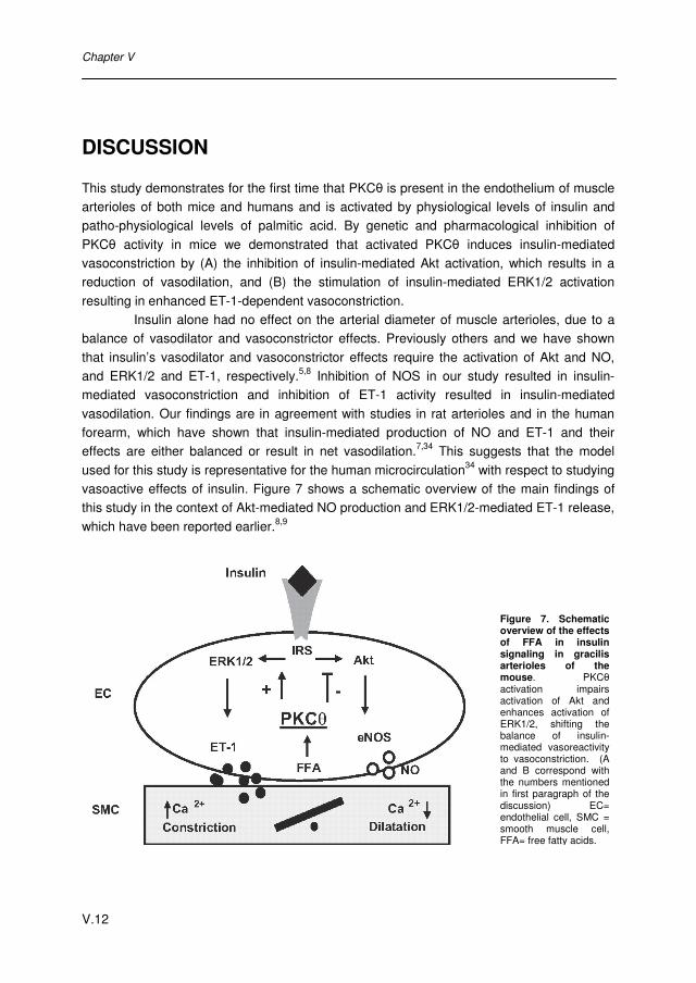
Chapter V
V.12
DISCUSSION
This study demonstrates for the first time that PKCθ is present in the endothelium of muscle arterioles of both mice and humans and is activated by physiological levels of insulin and patho-physiological levels of palmitic acid. By genetic and pharmacological inhibition of
PKCθ activity in mice we demonstrated that activated PKCθ induces insulin-mediated vasoconstriction by (A) the inhibition of insulin-mediated Akt activation, which results in a reduction of vasodilation, and (B) the stimulation of insulin-mediated ERK1/2 activation
resulting in enhanced ET-1-dependent vasoconstriction. Insulin alone had no effect on the arterial diameter of muscle arterioles, due to a
balance of vasodilator and vasoconstrictor effects. Previously others and we have shown
that insulin’s vasodilator and vasoconstrictor effects require the activation of Akt and NO, and ERK1/2 and ET-1, respectively.5,8 Inhibition of NOS in our study resulted in insulin-mediated vasoconstriction and inhibition of ET-1 activity resulted in insulin-mediated
vasodilation. Our findings are in agreement with studies in rat arterioles and in the human forearm, which have shown that insulin-mediated production of NO and ET-1 and their effects are either balanced or result in net vasodilation.7,34 This suggests that the model
used for this study is representative for the human microcirculation34 with respect to studying vasoactive effects of insulin. Figure 7 shows a schematic overview of the main findings of this study in the context of Akt-mediated NO production and ERK1/2-mediated ET-1 release,
which have been reported earlier.8,9
Figure 7. Schematic overview of the effects of FFA in insulin signaling in gracilis arterioles of the mouse. PKCθactivation impairs activation of Akt and enhances activation of ERK1/2, shifting the balance of insulin-mediated vasoreactivity to vasoconstriction. (A and B correspond with the numbers mentioned in first paragraph of the discussion) EC= endothelial cell, SMC = smooth muscle cell, FFA= free fatty acids.

PKC-theta Activation induces Vasoconstriction
V.13
In this study we used isolated first-order gracilis arterioles as a model for skeletal muscle
arterioles during preconstriction with KCl. All experiments in the present study were performed during preconstriction with KCl. Potassium influences the Ca2+ handling by depolarization of the cell-membrane of smooth muscle cells in the arterial wall, resulting in
vasoconstriction. This simulation of arterial tone is commonly used in other studies on arterial vasoregulation.35-37 It is unlikely that the induced state of tone will bear impact on the results of insulin-mediated vasoreactivity and PKCθ activation, because we found in that
insulin-mediated vasodilation is NO-dependent, insulin-mediated vasoconstriction is ET-1-dependent. Another difference between studies on insulin-mediated vasoreactivity in vivo and ex vivo is the presence of shear forces in vivo. As shear is a well-known stimulator of
endothelial NO production38 and NO inhibits insulin’s vasoconstrictor effects,7 this may explain the predominance of insulin’s vasodilator effects in vivo. In spite of this, vasoconstrictor effects of insulin have been demonstrated in a number of in vivo studies.34,39
An interesting novel observation of our study is that PKCθ is abundantly expressed in the endothelium of muscle arterioles of both mouse and man. PKCθ was until now mainly described in skeletal muscle samples,13-15 fibroblasts17 or cultured skeletal muscle
myocytes16 and T-cells.12 Moreover, PKCθ was encountered in cultured aortic endothelial cells,40 cultured human umbilical vein endothelial cells41,42 and small amounts have occasionally found in homogenates of tracheal43 and aortic44 smooth muscle cells of
rodents. However, the present study shows for the first time the presence and localization of PKCθ in situ in the vascular system of skeletal muscle of both mice and humans and particularly in the endothelial cell layer, in which the insulin-mediated production of NO and
endothelin-1 occurs. PKCθ activation interferes in insulin-mediated vasoreactivity by the inhibition of Akt
and stimulation of ERK, resulting in insulin-mediated vasoconstriction (Fig 2B). In the
present study, PKCθ activation by PA and insulin in muscle arterioles was determined by the phosphorylation of PKCθ at Thr-538. This phosphorylation-site is most important in PKCθ activation and mutations in this site inhibit the catalytical activity of PKCθ.13 We showed that
activated PKCθ inhibits insulin-mediated Akt activation, thereby reducing vasodilation. Simultaneously, it stimulates insulin-mediated ERK1/2, thereby enhancing vasoconstriction effects of insulin. In cultured fibroblasts, PKCθ activation is associated with the inhibition of
Akt activation17 and PKCθ was described as an upstream activator of the MAPK/ERK cascade.16,17 Furthermore, PKCθ can directly phosphorylate IRS1 at Ser-30717 and Ser-1101,18 and IRS1 in turn is described to be involved in both the activation of Akt18,45 as well
as the stimulation the MAPK/ERK pathway by insulin.46 It is possible that PKCθ can also influence IRS1 function in muscle arterioles directly, by specific phosphorylation and thereby influence insulin signaling. This results in the reduced Akt activation, which causes reduced
vasodilation, and increased ERK1/2 activation, resulting in enhanced vasoconstriction. Thus, PKCθ can be a key player in shifting insulin-mediated vasoreactivity towards vasoconstriction by modulating IRS1 phosphorylation.
V

Chapter V
V.14
Surprisingly, insulin induced vasoconstriction in WT mice during inhibition of PKCθ in the absence of PA and in PKCθ-KO mice (figure 4). As acetylcholine-mediated vasodilatation
was also slightly attenuated in PKCθ-KO mice (table 1), this effect may be caused by a positive effect of constitutional PKCθ activity on eNOS activity in absence of fatty acids. This may involve direct phosphorylation of eNOS by PKCθ at a serine residue that positively
regulates its activation, such as Ser 114, Ser 615 or Ser 633.47 This effect was absent in WT arterioles in the presence of PA (figure 2B), suggesting that (PA-induced) hyperactivity of PKCθ has predominantly unfavorable vasoconstrictive effects. More studies are required to
unravel this possible dual role of PKCθ. Our data support the hypothesis that PKCθ activation in muscle arterioles
contributes to fatty acid-induced insulin resistance. Clerk et al.20 described that fatty acid-
induced insulin resistance in muscle is partially caused by impairment of insulin-mediated nutritive muscle blood flow,20 which is dependent on activation of PI3-kinase/Akt in muscle arterioles.19 These authors suggest a possible role for PKCθ in the impairment of insulin
signaling in muscle arterioles.20 This suggestion is supported by the observation that mice lacking PKCθ are protected from acute fatty acid-induced insulin resistance.21 Indeed, this can be explained with our data, which show that fatty acids activate PKCθ in endothelium of
muscle arterioles, resulting in impaired insulin-mediated activation of Akt and a shift in insulin-mediated vasoreactivity to vasoconstriction. We propose that fatty acids, in addition to other metabolic effects, induce muscle insulin resistance by activation of PKCθ in
endothelium of muscle arterioles, which leads to reduction of insulin-mediated nutritive muscle blood flow. The findings in studies on muscle-specific PKCθ knockout mice are consistent with this hypothesis. Serra et al. have recently shown that specific expression of
dominant-negative PKCθ in skeletal muscle myocytes reduces, rather than enhances, insulin sensitivity.48 Therefore, the insulin-sensitizing effect of blocking PKCθ shown by Kim et al.21 cannot be explained by a direct effect on myocellular glucose uptake, but must be
caused by other mechanisms, such as improved nutritive blood flow. In summary, PKCθ activation by palmitic acid induces insulin-mediated
vasoconstriction in muscle arterioles, which can explain how fatty acids cause a decrease in
nutritive blood flow and impaired glucose uptake in muscle. This provides new mechanistic evidence of how PKCθ activation results in insulin resistance and suggest that PKCθ is promising novel target for improvement of vascular function in obesity.

PKC-theta Activation induces Vasoconstriction
V.15
REFERENCE LIST 1. Reaven,G.M. Banting lecture 1988. Role of insulin resistance in human disease. Diabetes 37,
1595-1607 (1988). 2. Kahn,B.B. & Flier,J.S. Obesity and insulin resistance. J. Clin. Invest 106, 473-481 (2000). 3. Frohlich,E.D., Messerli,F.H., Reisin,E. & Dunn,F.G. The problem of obesity and hypertension.
Hypertension 5, III71-III78 (1983). 4. Clark,M.G. et al. Blood flow and muscle metabolism: a focus on insulin action. Am J Physiol
Endocrinol Metab 284, E241-E258 (2003). 5. Dimmeler,S. et al. Activation of nitric oxide synthase in endothelial cells by Akt- dependent
phosphorylation. Nature 399, 601-605 (1999). 6. Vincent,M.A. et al. Microvascular Recruitment Is an Early Insulin Effect That Regulates
Skeletal Muscle Glucose Uptake In Vivo. Diabetes 53, 1418-1423 (2004). 7. Eringa,E.C., Stehouwer,C.D.A., Merlijn,T., Westerhof,N. & Sipkema,P. Physiological
concentrations of insulin induce endothelin-mediated vasoconstriction during inhibition of NOS or PI3-kinase in skeletal muscle arterioles. Cardiovascular Research 56, 464-471 (2002).
8. Eringa,E.C. et al. Vasoconstrictor effects of insulin in skeletal muscle arterioles are mediated by ERK1/2 activation in endothelium. Am J Physiol Heart Circ Physiol 287, H2043-H2048 (2004).
9. Potenza,M.A. et al. Insulin resistance in spontaneously hypertensive rats is associated with endothelial dysfunction characterized by imbalance between NO and ET-1 production. Am J Physiol Heart Circ Physiol 289, H813-H822 (2005).
10. Lteif,A., Vaishnava,P., Baron,A.D. & Mather,K.J. Endothelin Limits Insulin Action in Obese/Insulin-Resistant Humans. Diabetes 56, 728-734 (2007).
11. Jiang,Z.Y. et al. Characterization of selective resistance to insulin signaling in the vasculature of obese Zucker (fa/fa) rats. J. Clin. Invest. 104, 447-457 (1999).
12. Baier,G. et al. Molecular cloning and characterization of PKC theta, a novel member of the protein kinase C (PKC) gene family expressed predominantly in hematopoietic cells. J. Biol. Chem. 268, 4997-5004 (1993).
13. Yu,C. et al. Mechanism by Which Fatty Acids Inhibit Insulin Activation of Insulin Receptor Substrate-1 (IRS-1)-associated Phosphatidylinositol 3-Kinase Activity in Muscle. J. Biol. Chem. 277, 50230-50236 (2002).
14. Griffin,M.E. et al. Free fatty acid-induced insulin resistance is associated with activation of protein kinase C theta and alterations in the insulin signaling cascade. Diabetes 48, 1270-1274 (1999).
15. Donnelly,R., Reed,M.J., Azhar,S. & Reaven,G.M. Expression of the major isoenzyme of protein kinase-C in skeletal muscle, nPKC theta, varies with muscle type and in response to fructose- induced insulin resistance. Endocrinology 135, 2369-2374 (1994).
16. Jove,M. et al. Palmitate Induces Tumor Necrosis Factor-{alpha} Expression in C2C12 Skeletal Muscle Cells by a Mechanism Involving Protein Kinase C and Nuclear Factor-{kappa}B Activation. Endocrinology 147, 552-561 (2006).
17. Gao,Z. et al. Inhibition of Insulin Sensitivity by Free Fatty Acids Requires Activation of Multiple Serine Kinases in 3T3-L1 Adipocytes. Mol Endocrinol 18, 2024-2034 (2004).
18. Li,Y. et al. Protein Kinase C {theta} Inhibits Insulin Signaling by Phosphorylating IRS1 at Ser1101. J. Biol. Chem. 279, 45304-45307 (2004).
19. Bradley,E.A., Clark,M.G. & Rattigan,S. Acute effects of wortmannin on insulin's hemodynamic and metabolic actions in vivo. Am J Physiol Endocrinol Metab 292, E779-E787 (2007).
20. Clerk,L.H., Rattigan,S. & Clark,M.G. Lipid Infusion Impairs Physiologic Insulin-Mediated Capillary Recruitment and Muscle Glucose Uptake In Vivo. Diabetes 51, 1138-1145 (2002).
21. Kim,J.K. et al. PKC-{theta} knockout mice are protected from fat-induced insulin resistance. J. Clin. Invest. 114, 823-827 (2004).
V

Chapter V
V.16
22. Itani,S.I., Pories,W.J., MacDonald,K.G. & Dohm,G.L. Increased protein kinase C theta in skeletal muscle of diabetic patients. Metabolism 50, 553-557 (2001).
23. Sun,Z. et al. PKC-[thetas] is required for TCR-induced NF-[kappa]B activation in mature but not immature T lymphocytes. Nature 404, 402-407 (2000).
24. Kim,F. et al. Free Fatty Acid Impairment of Nitric Oxide Production in Endothelial Cells Is Mediated by IKK{beta}. Arterioscler Thromb Vasc Biol 25, 989-994 (2005).
25. Newton,A.C. Protein Kinase C: Structure, Function, and Regulation. J. Biol. Chem. 270, 28495-28498 (1995).
26. Hool,L.C. Protein kinase C isozyme selective peptides--a current view of what they tell us about location and function of isozymes in the heart. Curr. Pharm. Des 11, 549-559 (2005).
27. House,C. & Kemp,B.E. Protein kinase C contains a pseudosubstrate prototope in its regulatory domain. Science 238, 1726-1728 (1987).
28. Eichholtz,T., de Bont,D.B., de Widt,J., Liskamp,R.M. & Ploegh,H.L. A myristoylated pseudosubstrate peptide, a novel protein kinase C inhibitor. J. Biol. Chem. 268, 1982-1986 (1993).
29. Eringa,E.C. et al. Physiological Concentrations of Insulin Induce Endothelin-Dependent Vasoconstriction of Skeletal Muscle Arterioles in the Presence of Tumor Necrosis Factor-{alpha} Dependence on c-Jun N-Terminal Kinase. Arterioscler Thromb Vasc Biol 26, 274-280 (2006).
30. Steinberg,H.O. et al. Free fatty acid elevation impairs insulin-mediated vasodilation and nitric oxide production. Diabetes 49, 1231-1238 (2000).
31. Staehr,P., Hother-Nielsen,O., Levin,K., Holst,J.J. & Beck-Nielsen,H. Assessment of Hepatic Insulin Action in Obese Type 2 Diabetic Patients. Diabetes 50, 1363-1370 (2001).
32. Liu,Y., Graham,C., Li,A., Fisher,R.J. & Shaw,S. Phosphorylation of the protein kinase C-theta activation loop and hydrophobic motif regulates its kinase activity, but only activation loop phosphorylation is critical to in vivo nuclear-factor-kappaB induction. Biochem. J 361, 255-265 (2002).
33. Xu,Z.B. et al. Catalytic Domain Crystal Structure of Protein Kinase C-{theta} (PKC{theta}). J. Biol. Chem. 279, 50401-50409 (2004).
34. Cardillo,C. et al. Insulin Stimulates Both Endothelin and Nitric Oxide Activity in the Human Forearm. Circulation 100, 820-825 (1999).
35. Jones,R.D., Pugh,P.J., Hall,J., Channer,K.S. & Jones,T.H. Altered circulating hormone levels, endothelial function and vascular reactivity in the testicular feminised mouse. Eur J Endocrinol148, 111-120 (2003).
36. Okada,Y., Yanagisawa,T. & Taira,N. KCl-depolarization potentiates the Ca2+ sensitization by endothelin-1 in canine coronary artery. Jpn. J Pharmacol 60, 403-405 (1992).
37. Quaschning,T. et al. Endothelin B receptor-deficient mice develop endothelial dysfunction independently of salt loading. J Hypertens. 23, 979-985 (2005).
38. Cardillo,C., Kilcoyne,C.M., Cannon,R.O., III & Panza,J.A. Interactions Between Nitric Oxide and Endothelin in the Regulation of Vascular Tone of Human Resistance Vessels In Vivo. Hypertension 35, 1237-1241 (2000).
39. Gudbjornsdottir,S., Elam,M., Sellgren,J. & Anderson,E.A. Insulin increases forearm vascular resistance in obese, insulin-resistant hypertensives. J Hypertens. 14, 91-97 (1996).
40. Hempel,A. et al. High Glucose Concentrations Increase Endothelial Cell Permeability via Activation of Protein Kinase C{alpha}. Circ Res 81, 363-371 (1997).
41. Haller,H., Ziegler,W., Lindschau,C. & Luft,F.C. Endothelial Cell Tyrosine Kinase Receptor and G ProteinûCoupled Receptor Activation Involves Distinct Protein Kinase C Isoforms. Arterioscler Thromb Vasc Biol 16, 678-686 (1996).
42. Mattila,P., Majuri,M.L., Tiisala,S. & Renkonen,R. Expression of six protein kinase C isotypes in endothelial cells. Life Sci 55, 1253-1260 (1994).
43. Yang,C.M., Fen,L.W., Tsao,H.L. & Chiu,C.T. Inhibition of 5-hydroxytryptamine-induced phosphoinositide hydrolysis and Ca2+ mobilization in canine cultured tracheal smooth muscle cells by phorbol ester. Br J Pharmacol 121, 853-860 (1997).

PKC-theta Activation induces Vasoconstriction
V.17
44. Wang,Y.X. et al. Role of PKC in the novel synergistic action of urotensin II and angiotensin II and in urotensin II-induced vasoconstriction. Am J Physiol Heart Circ Physiol 292, H348-H359 (2007).
45. Aguirre,V. et al. Phosphorylation of Ser307 in Insulin Receptor Substrate-1 Blocks Interactions with the Insulin Receptor and Inhibits Insulin Action. J. Biol. Chem. 277, 1531-1537 (2002).
46. De Fea,K. & Roth,R.A. Modulation of Insulin Receptor Substrate-1 Tyrosine Phosphorylation and Function by Mitogen-activated Protein Kinase. J. Biol. Chem. 272, 31400-31406 (1997).
47. Mount,P.F., Kemp,B.E. & Power,D.A. Regulation of endothelial and myocardial NO synthesis by multi-site eNOS phosphorylation. Journal of Molecular and Cellular Cardiology 42, 271-279 (2007).
48. Serra,C. et al. Transgenic mice with dominant negative PKC-theta in skeletal muscle: a new model of insulin resistance and obesity. J Cell Physiol 196, 89-97 (2003).
V

Chapter V
V.18

Selective PKCθ Activation in Muscle Arterioles
VI.1
Submitted to Diabetes
Selective
PKCθactivation in
Muscle-opposed to
Adipose tissuearterioles during obesity
Selective
PKCθactivation in
Muscle-opposed to
Adipose tissuearterioles during obesity

Chapter VI
VI.2

Selective PKCθ Activation in Muscle Arterioles
VI.3
VI. Selective PKCθ Activation in Muscle Opposed to Adipose Tissue Arterioles. Consequences for tissue perfusion Wineke Bakker, Caro-Lynn A.F. Alta, Pieter Sipkema, Coen D.A. Stehouwer, Victor W.M.
van Hinsbergh, Etto C. Eringa.
ABSTRACT
Objective: Obesity is characterized by increased levels of fatty acids, decreased glucose
uptake in skeletal muscle and impairment of microvascular responses to insulin. Previously we have shown that PKCθ activation by fatty acids induces insulin-mediated vasoconstriction in muscle arterioles. The aim of this study was to investigate the
contribution of PKCθ activation in muscle and adipose tissue arterioles on insulin-mediated vasoreactivity and blood flow in skeletal muscle and adipose tissue during obesity. Research design and methods: Skeletal muscle and adipose tissue of lean and obese Db/Db mice were characterized for mass and structure, function and PKCθ-activation of their microcirculation.
Results: Skeletal muscle mass, myocyte size, diameter of muscle arterioles, and capillary ratio per myocyte were reduced in obese Db/Db mice. In contrast, adipose tissue mass,
adipocyte size, diameter of adipose tissue arterioles and capillary ratio per adipocyte were increased in obese Db/Db mice. We identified and isolated arterioles that branched from a common artery but nourished either muscle or adipose tissue. Palmitic acid (PA,100μmol/l)
activated PKCθ only in muscle arterioles but not in adipose tissue arterioles of lean mice. In muscle arterioles, this led to insulin-mediated vasoconstriction (30±6%, p<0.01) associated with a decreased Akt-activation. In adipose tissue arterioles, it resulted in a moderate
insulin-mediated vasodilation, due to increased Akt-activation. In obese Db/Db mice, PKCθ was activated in muscle arterioles and not in adipose tissue arterioles. In obese Db/Db mice, this led to insulin-mediated vasoconstriction of muscle arterioles and was associated with an
increased blood flow to adipose tissue compared to blood flow to skeletal muscle. Conclusion: Muscle and adipose tissue have functionally distinct vasculatures. PKCθ is
selectively activated by palmitic acid and in muscle arterioles from obese Db/Db mice, but not in adipose tissue arterioles. This was associated with insulin-mediated vasoconstriction in muscle and increased blood flow to adipose tissue, and may promote increased nutrient
storage in adipose tissue.
VI

Chapter VI
VI.4
INTRODUCTION Obesity is characterized by an increased fat mass, and is associated with microvascular
dysfunction and decreased insulin-mediated glucose uptake in skeletal muscle.1 The obesity-dependent impairment of microvascular responses to insulin in obesity is thought to contribute to insulin resistance by reducing perfusion and appropriate delivery of insulin and
glucose to skeletal muscle.2 However, it is unknown whether and how disturbed muscle perfusion and adipose tissue expansion are related in obesity.
In skeletal muscle, the vasodilation of arterioles by insulin and the recruitment of
capillaries increases muscle glucose uptake, by increasing microvascular volume.3 Arteriolar diameter is regulated by insulin’s vasodilator (mediated through Akt/NO pathway) and vasoconstrictor effects (mediated by ERK1/2/ET-1 pathway).4 These two pathways are in
balance or result in net vasodilation in healthy conditions, and the balance is shifted towards vasoconstriction in obesity.5,6 Both obesity and lipid infusion are characterized by reduced muscle perfusion, inhibition of insulin-mediated muscle glucose uptake7 and impaired
insulin-mediated capillary recruitment.7,8 Furthermore, in a previous study we have shown that free fatty acids (FFA), activates PKCθ in muscle arterioles, which results in insulin-mediated vasoconstriction.9 This suggests PKCθ activation as a new mechanism in the
reduction of muscle perfusion.9 In obesity, increased adipose tissue mass is characterized by adipocyte
hyperplasia, adipocyte hypertrophy, and a proinflammatory state.10,11 The growth of adipose
tissue is dependent on neovascularization and remodeling of existing capillaries.12 Postprandial adipose tissue blood flow is positively correlated with insulin sensitivity13 and is attenuated in obesity.14 However, regulation of adipose tissue arteriolar function in the
context of obesity has not been studied yet, and it is unclear whether blood flow to adipose tissue is linked to vascular responses in muscle.
We hypothesized that muscle and adipose tissue have functionally distinct
vasculatures; that PKCθ is selectively activated in muscle arterioles; and that this is associated with an increased blood flow to adipose tissue. To investigate PKCθ activation and vasoactive responses, muscle and adipose tissue arterioles from lean mice were
subjected to the free fatty acid palmitic acid (PA) and insulin. In addition, arterioles were isolated from obese Db/Db mice and responses to insulin were compared to those of lean mice. Different vascular responses of muscle and adipose tissue arterioles were observed,
which revealed structural and functional changes in capillary density, arteriolar diameter and blood flow of muscle and adipose tissue.

Selective PKCθ Activation in Muscle Arterioles
VI.5
RESEARCH DESIGN AND METHODS Mice: The investigation conformed to the Guide for the Care and Use of Laboratory Animals published
by the US National Institutes of Health (NIH publication No 85 to 23, revised 1996). The local ethics
committee for animal experiments approved the procedures. Male C57Bl6 and Obese Db/Db mice
(Harlan; Zeist, the Netherlands) of 10 weeks were sacrificed by overdose of isoflurane inhalation. Obese
Db/Db mice have a mutation in their leptin receptor and are characterized by increased body weight,
hyperglycemia, hyperinsulimia and hyperlipidemia.15 Body weight (40.6±0.8 vs. 24.8±0.4 g), fasting
blood glucose (10.3±0.6 vs. 5.4±0.2 mM) and non-fasting blood glucose (25.4±1.5 vs. 12.4±0.1 mM)
were higher in obese Db/Db mice than in lean mice (10 vs14 animals; P<0.0001 for all parameters) with
a similar genetic background (C57Bl/6). Gastrocnemius muscle, subcutaneous adipose tissue and
corresponding arterioles were isolated to analyze structural and functional characteristics.
Immunochemistry: Muscle samples were snap frozen at -80°C and adipose tissue samples were
embedded in paraffin. Tissue sections of 5 μm were either stained with heamatoxyline-eosine to
determine cell size or with the endothelium-selective glycocalyx stain lectin (Lycopersicon esculentum
Lectin, FITC-conjugate, Sigma-Aldrich) to determine the number of capillaries.
Blood flow: Blood flow was determined with FluoSpheres polystyrene microspheres (15μm blue green
fluorescent 430/465, Molecular probes, Invitrogen).16 Microspheres (2500 beads/gram body weight)
were injected into the left ventricle, while a reference blood sample with constant flow (100μl/min) was
collected. Blood flow of gastrocnemius muscle and subcutaneous adipose tissue of the hindlimb were
calculated using a reference blood sample and is expressed as a mean muscle or adipose tissue blood
flow. Fluorescent microspheres were extracted from muscle and adipose tissue according to Raab et
al16 and fluorescence intensity was measured using a luminescence spectrophotometer.
Vasoreactivity: After dissection, muscle and adipose arterioles were cannulated in a pressure
myograph and studied at physiologic pressure and temperature in MOPS-buffer, as described.9 The
arterioles were preconstricted to approximately 50% of the maximal diameter with 25mM KCl.
Endothelial integrity was determined by measuring vasodilator response to acetylcholine (ACh: 0.1μM)
before and after experiments. Acute effects of insulin (Novo Nordisk, Alphen a/d Rijn, the Netherlands),
at four concentrations (0.02, 0.2, 2 and 20nM) on arteriolar diameter were studied and each
concentration was recorded for 30 minutes.9 To study the interaction between PKCθ and insulin in
arterioles, arteriolar segments were pre-treated with PA (100 μM) for 30 minutes to activate PKCθ and
were thereafter subjected to insulin.
Western blot: Western blot analysis was performed as described.9 Segments of gracilis or adipose
tissue arterioles from the same mouse (3mm in length) were exposed to solvent, insulin (2nM) or insulin
with PA (100μM) for 15 minutes at 37ºC. The protein lysates of different arteriolar segments were
stained with specific primary antibodies against phosphorylated Akt (60 kDa), phosphorylated PKCθ (79
kDa), total Akt and total PKCθ (Antibodies obtained from Cell signaling technology, Boston, MA) and
VI
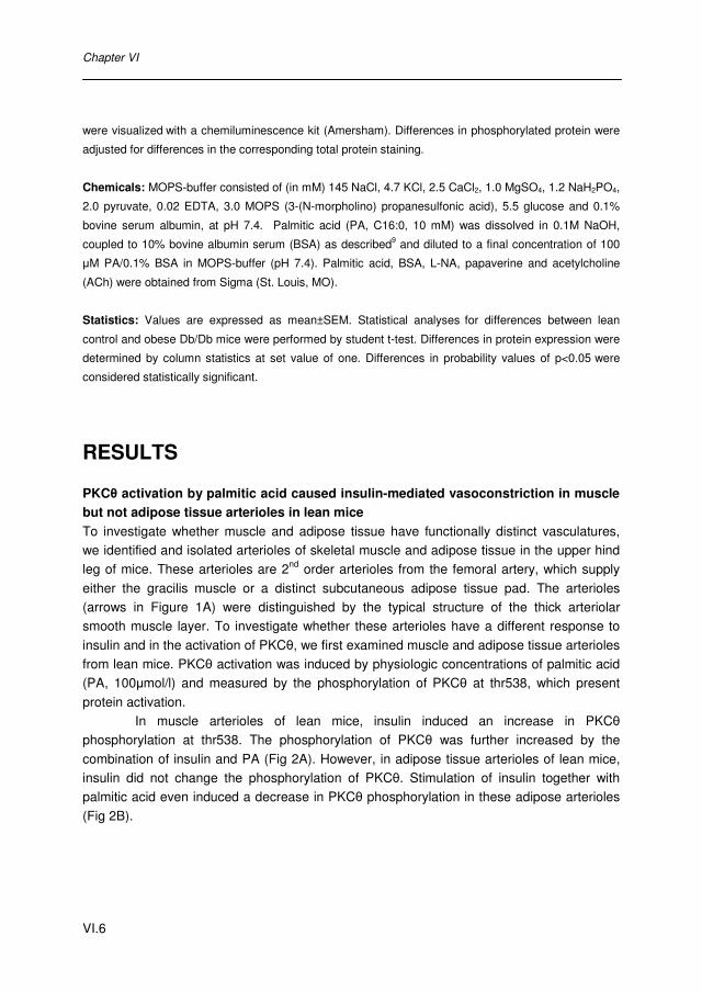
Chapter VI
VI.6
were visualized with a chemiluminescence kit (Amersham). Differences in phosphorylated protein were
adjusted for differences in the corresponding total protein staining.
Chemicals: MOPS-buffer consisted of (in mM) 145 NaCl, 4.7 KCl, 2.5 CaCl2, 1.0 MgSO4, 1.2 NaH2PO4,
2.0 pyruvate, 0.02 EDTA, 3.0 MOPS (3-(N-morpholino) propanesulfonic acid), 5.5 glucose and 0.1%
bovine serum albumin, at pH 7.4. Palmitic acid (PA, C16:0, 10 mM) was dissolved in 0.1M NaOH,
coupled to 10% bovine albumin serum (BSA) as described9 and diluted to a final concentration of 100
μM PA/0.1% BSA in MOPS-buffer (pH 7.4). Palmitic acid, BSA, L-NA, papaverine and acetylcholine
(ACh) were obtained from Sigma (St. Louis, MO).
Statistics: Values are expressed as mean±SEM. Statistical analyses for differences between lean
control and obese Db/Db mice were performed by student t-test. Differences in protein expression were
determined by column statistics at set value of one. Differences in probability values of p<0.05 were
considered statistically significant.
RESULTS PKCθ activation by palmitic acid caused insulin-mediated vasoconstriction in muscle but not adipose tissue arterioles in lean mice To investigate whether muscle and adipose tissue have functionally distinct vasculatures, we identified and isolated arterioles of skeletal muscle and adipose tissue in the upper hind leg of mice. These arterioles are 2nd order arterioles from the femoral artery, which supply
either the gracilis muscle or a distinct subcutaneous adipose tissue pad. The arterioles (arrows in Figure 1A) were distinguished by the typical structure of the thick arteriolar smooth muscle layer. To investigate whether these arterioles have a different response to
insulin and in the activation of PKCθ, we first examined muscle and adipose tissue arterioles from lean mice. PKCθ activation was induced by physiologic concentrations of palmitic acid (PA, 100μmol/l) and measured by the phosphorylation of PKCθ at thr538, which present
protein activation. In muscle arterioles of lean mice, insulin induced an increase in PKCθ
phosphorylation at thr538. The phosphorylation of PKCθ was further increased by the
combination of insulin and PA (Fig 2A). However, in adipose tissue arterioles of lean mice, insulin did not change the phosphorylation of PKCθ. Stimulation of insulin together with palmitic acid even induced a decrease in PKCθ phosphorylation in these adipose arterioles
(Fig 2B).
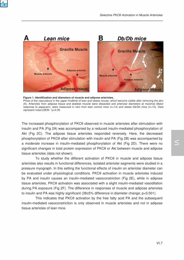
Selective PKCθ Activation in Muscle Arterioles
VI.7
The increased phosphorylation of PKCθ observed in muscle arterioles after stimulation with insulin and PA (Fig 2A) was accompanied by a reduced insulin-mediated phosphorylation of
Akt (Fig 2C). The adipose tissue arterioles responded reversely. Here, the decreased phosphorylation of PKCθ after stimulation with insulin and PA (Fig 2B) was accompanied by a moderate increase in insulin-mediated phosphorylation of Akt (Fig 2D). There were no
significant changes in total protein expression of PKCθ or Akt between muscle and adipose tissue arterioles (data not shown).
To study whether the different activation of PKCθ in muscle and adipose tissue
arterioles also results in functional differences, isolated arteriolar segments were studied in a pressure myograph. In this setting the functional effects of insulin on arteriolar diameter can be evaluated under physiological conditions. PKCθ activation in muscle arterioles induced
by PA and insulin causes an insulin-mediated vasoconstriction (Fig 2E), while in adipose tissue arterioles, PKCθ activation was associated with a slight insulin-mediated vasodilation during PA exposure (Fig 2F). The difference in responses of muscle and adipose arterioles
to insulin and PA was highly significant (38±5% difference in diameter change; p<0.001). This indicates that PKCθ activation by the free fatty acid PA and the subsequent
insulin-mediated vasoconstriction is only observed in muscle arterioles and not in adipose
tissue arterioles of lean mice.
Figure 1. Identification and diameters of muscle and adipose arterioles. Photo of the vasculature in the upper hindlimb of lean and obese mouse, which become visible after removing the skin (A). Arterioles from adipose tissue and skeletal muscle were dissected and arteriolar diameters at maximal dilator response to papavarin, were measured in vitro from lean control mice (n=14) and obese Db/Db mice (n=10). Data represent mean±SEM. *p<0.05.
Lean mice Db/Db mice
Adipose artery
Gracilis Muscle
Adipose tissue
Muscle artery
Adipose arteriole
Gracilis Muscle
Adipose tissue
Muscle arteriole
A
Adipose arteriole
Muscle arteriole
Adipose tissue
Gracilis Muscle
BLean mice Db/Db mice
Adipose artery
Gracilis Muscle
Adipose tissue
Muscle artery
Adipose arteriole
Gracilis Muscle
Adipose tissue
Muscle arteriole
A
Adipose arteriole
Muscle arteriole
Adipose tissue
Gracilis Muscle
B
VI

Chapter VI
VI.8
Figure 2. PKCθ activation by palmitic acid induces opposite functional effects in muscle and adipose tissue arterioles from lean mice. Phosphorylation of PKCθ at thr538 was used to determine PKCθ activation in muscle, n=9 (A) and adipose tissue arterioles, n=7 (B); Phosphorylation of Akt at ser473 was used to determine Akt activation in muscle (n=9) ( C) and adipose tissue arterioles, n=7 (D) from lean mice in the following conditions: control (c) insulin (I) and insulin (I: 2nM) with palmitic acid (PA:100μM). Functional response to PKCθ activation, induced by insulin (I: 2nM) and insulin with palmitc acid (PA: 100μM) on % diameter change, in muscle (E) and adipose tissue arterioles (F). Data depicted as percental change from preconstricted diameter in lean control mice (n=5). Data represent mean±SEM. *p<0.05, #p<0.01.
GA CC GA CI GA C I+PA0
1
2
3
4
5
*
#
pPK
C /
tPK
C (A
.U.)
AA CC AA CI AA C I+PA0
1
2
3
4
5
*pP
KC
/ tP
KC
(A.U
.)
AA C AA I AA I+PA0
2
4
6p=0.15
**
pA
kt /
tAK
t (A
.U.)
C I I+PA0
2
4
6 **
pA
kt /
tAkt
(A.U
.)
I I + PA
-40
-30
-20
-10
0
10#
diam
eter
cha
nge
to in
sulin
(%)
I I + PA-40
-30
-20
-10
0
10
dia
met
er c
han
ge t
o in
sulin
(%
)
Muscle arterioles Adipose arterioles
A BpPKCθ
tPKCθ
pPKCθ
tPKCθ
C D
C I I+PA
I I+PA I I+PA
pAkt
tAkt
pAkt
tAkt
C I I+PA C I I+PA
E F
C I I+PAGA CC GA CI GA C I+PA
0
1
2
3
4
5
*
#
pPK
C /
tPK
C (A
.U.)
AA CC AA CI AA C I+PA0
1
2
3
4
5
*pP
KC
/ tP
KC
(A.U
.)
AA C AA I AA I+PA0
2
4
6p=0.15
**
pA
kt /
tAK
t (A
.U.)
C I I+PA0
2
4
6 **
pA
kt /
tAkt
(A.U
.)
I I + PA
-40
-30
-20
-10
0
10#
diam
eter
cha
nge
to in
sulin
(%)
I I + PA-40
-30
-20
-10
0
10
dia
met
er c
han
ge t
o in
sulin
(%
)
Muscle arterioles Adipose arterioles
A BpPKCθ
tPKCθ
pPKCθ
tPKCθ
C D
C I I+PA
I I+PA I I+PA
pAkt
tAkt
pAkt
tAkt
C I I+PA C I I+PA
E F
C I I+PA

Selective PKCθ Activation in Muscle Arterioles
VI.9
Figure 3. PKCθ activation is increased and Akt activation is not changed in obese muscle arterioles and decreased in obese adipose tissue arterioles. Phosphorylation of PKCθ at thr538 was used to determine PKCθ activation in muscle, n=9 (A) and adipose tissue arterioles, n=8 (B); Phosphorylation of Akt at ser473 was used to determine Akt activation in muscle (n=9) ( C) and adipose arterioles, n=7 (D) from lean control and obese Db/Db mice in the following conditions: control (c) and insulin (I: 2nM ). Data represent mean±SEM. *p<0.05 compared c with I from lean and obese, $ p<0.05 compared I lean with I obese. E-F: Functional response to PKCθ activation, induced by insulin, on % diameter change, depicted as percental change from preconstricted diameter in muscle (E) and adipose arterioles (F) from lean control (n=6) and obese Db/Db mice (n=5). Data represent mean±SEM. *p<0.05
AA CC AA CI AA DC AA DI0
1
2
3
4
5
*pP
KC
/ tP
KC
(A
.U.)
AA C AA I AA DB C AA DB I0
2
4
6
8
#*
pA
kt /
tAkt
(A
.U.)
Bl6 C Bl6 I db/db C db/db I0
2
4
6
8*
*
pAkt
/ t
Akt
(A
.U.)
GA CC GA CI GA DC GA DI0
1
2
3
4
5
*
* $
pPK
C /
tPK
C (
A.U
.)
C I C ILean Db/Db
pPKCθtPKCθA B
pAkttAkt
C D
pPKCθtPKCθ
C I C ILean Db/Db
C I C ILean Db/Db
pAkttAkt
C I C ILean Db/Db
E F
-40
-30
-20
-10
0
10
*
dia
met
er c
han
ge
to in
sulin
(%
)
-40
-30
-20
-10
0
10
dia
met
er c
han
ge
to in
sulin
(%
)
Muscle arterioles Adipose arterioles
Lean Db/Db Lean Db/Db
AA CC AA CI AA DC AA DI0
1
2
3
4
5
*pP
KC
/ tP
KC
(A
.U.)
AA C AA I AA DB C AA DB I0
2
4
6
8
#*
pA
kt /
tAkt
(A
.U.)
Bl6 C Bl6 I db/db C db/db I0
2
4
6
8*
*
pAkt
/ t
Akt
(A
.U.)
GA CC GA CI GA DC GA DI0
1
2
3
4
5
*
* $
pPK
C /
tPK
C (
A.U
.)
C I C ILean Db/Db
C I C ILean Db/Db
pPKCθtPKCθA B
pAkttAkt
C D
pPKCθtPKCθ
C I C ILean Db/Db
C I C ILean Db/Db
C I C ILean Db/Db
C I C ILean Db/Db
pAkttAkt
C I C ILean Db/Db
C I C ILean Db/Db
E F
-40
-30
-20
-10
0
10
*
dia
met
er c
han
ge
to in
sulin
(%
)
-40
-30
-20
-10
0
10
dia
met
er c
han
ge
to in
sulin
(%
)
Muscle arterioles Adipose arterioles
Lean Db/Db Lean Db/Db
VI

Chapter VI
VI.10
PKCθ activation is increased in muscle and decreased in adipose tissue arterioles from Db/Db mice As Db/Db mice have elevated levels of circulating free fatty acids, we subsequently investigated whether PKCθ activation by endogenous factors also bears impact on the regulation of muscle and adipose tissue perfusion by insulin. To that end, we compared
muscle and adipose tissue arterioles of lean and obese diabetic Db/Db mice in control and insulin-stimulated conditions.
In muscle arterioles from both lean and Db/Db mice, insulin induced an increase in
PKCθ phosphorylation at thr538 (Fig 3A). Interestingly, the increase in PKCθ phosphorylation was approximately 3-fold higher in muscle arterioles isolated from Db/Db mice than in those of lean mice. In the adipose tissue arterioles from lean mice, insulin
induced no change in PKCθ phosphorylation and even slightly reduced it in Db/Db mice (Fig 3B).
The different activation of PKCθ in muscle arterioles did not result in significantly
different activations of Akt (Fig 3C and 3D), although the Db/Db arterioles tended to display less insulin-mediated Akt activation (p=0.40). Adipose tissue arterioles from lean and Db/Db showed a similar degree of insulin-mediated Akt activation (Fig 3D). The insulin-induced
activation of Akt was considerably less than in muscle arterioles (comparing Fig 3C and 3D). Functional studies in isolated arteriolar segments revealed that PKCθ activation in
muscle arterioles isolated from Db/Db mice resulted in insulin-mediated vasoconstriction,
which was significantly different from muscle arterioles of lean mice (Fig 3E). In contrast, insulin-mediated vasoreactivity was similar in adipose tissue arterioles of lean and Db/Db mice, and their diameters did not change in response to insulin (Fig 3F).
This indicates that insulin-induced PKCθ activation in muscle arterioles isolated from Db/Db mice (Fig 3A) is comparable to the PKCθ activation induced by PA in lean mice (Fig 2A). As a consequence, the difference in insulin-induced PKCθ activation between in
muscle and adipose tissue arterioles is blunted in Db/Db mice. Consequences of PKCθ activation for tissue vascularization in lean and Db/Db mice To investigate whether the different activation of PKCθ in muscle and adipose tissue arterioles influence tissue vascularization, we determined the maximal diameters of the arterioles, capillary density and blood flow of skeletal muscle and adipose tissue in lean and
Db/Db mice. The maximal diameter was measured of both arterioles towards gracilis muscle and adipose tissue from both lean and Db/Db mice. The diameter of muscle arterioles was
less in lean mice (145±2μm) than in obese mice (130±4μm), while the diameter of adipose tissue arterioles was larger in lean (177±4μm) than in obese mice (189±5μm) (Table 1).
The number of capillaries was less in skeletal muscle of Db/Db mice than in their
lean control counterparts, as determined by both ratio of number of capillaries per myocyte and number of capillaries per mm2 muscle cross-section (Fig 4A). In contrast, adipose tissue was expanded in obese Db/Db mice, accompanied by fat cell enlargement (Fig 4B). While

Selective PKCθ Activation in Muscle Arterioles
VI.11
the increase in adipocyte size was accompanied by an increase in capillary ratio per adipocyte, the number of capillaries per mm2 adipose tissue cross-section decreased
(Table 1). The reduced vascularization of skeletal muscle of Db/Db mice was accompanied by a 40% reduction of skeletal muscle mass and a smaller size of muscle myocytes (Table 1). The
increased vasculairzation of adipose tissue of Db/Db mice was accompanies by a 14-fold increase in adipose tissue mass and enlarged adipocyte size (Table 1). This was despite a 2-fold increase in body mass (40.6±0.8g as compared to 24.8±0.4g in lean mice).
Taken these structural data together, increase in tissue mass was positively correlated with the ratio of number of capillaries per cell and maximal diameter of arterioles.
Db/DbLean
Lec
tin
HE
B Lean Db/Db
Lec
tin
HE
A Db/DbLean
Lec
tin
HE
B Lean Db/Db
Lec
tin
HE
A
Figure 4. Cross sections of skeletal muscle (A) and adipose tissue (B) of lean and Db/Db mice. Cross sectional area (CSA) of muscle myocytes and adipocytes were determined by HE staining. The ratio of number of capillaries per myocyte/adipocyte and the capillary density per mm2 were determined by Lectin (Lycopersicon esculentum Lectin, FITC-conjugate) staining in lean and Db/Db mice. Scalebars represent 100μm.
VI

Chapter VI
VI.12
Blood flow to adipose tissue is increased in obese mice In a final experiment, we evaluate whether PKCθ activation in muscle arterioles is
associated with changes in blood flow to muscle and adipose tissue in obese mice. Blood flow in muscle and adipose tissue was measured using fluorescent 15μm microspheres, which are retained in small blood vessels. Blood flow expressed as ratio of muscle and
adipose tissue was predominated towards muscle in lean mice in total tissue blood flow (Fig 5A) and in tissue blood flow per gram (Fig 5B). In contrast, Blood flow in obese mice expressed as ratio of muscle and adipose tissue was predominated towards adipose tissue
in total tissue blood flow (Fig 5A) and in tissue blood flow per gram (Fig 5B).
Table 1: Mice and tissue characteristics of skeletal muscle and adipose tissue
<0.0011.50±0.070.93±0.05Capillary ratio(#/cell)
<0.05130±4145±2Maximal arteriolar diameter (μm)
<0.000115.8±0.07 1.1±0.01 Mass (g)
Adipose tissue
=0.16796±56989±115Capillary density (#/ mm2)
Mice / Tissue characteristics Lean Db/Db P
Body weight (g) 24.8±0.4 40.6±0.8 <0.0001
Skeletal muscle
Mass (g) 1.7±0.08 1.1±0.05 <0.001
CSA myocytes (μm2) 1366±77 1142±56 <0.05
Capillary ratio (#/cell) 1.56±0.14 1.16±0.06 <0.05
CSA adipocytes (μm2) 1185±101 6187±104 <0.005
Maximal arteriolar diameter (μm) 177±4 189±5 <0.05
Capillary density (#/ mm2) 485±10 245±39 <0.001
Figure 5. Blood flow ratio of muscle and adipose tissue in lean and Db/Db mice. Blood flow depicted as flow in ml/min (A) and flow in ml/min/mg (B) in ratio of BF in skeletal muscle divided by BF in adipose tissue. P<0.01
Lean Db/Db0
1
2
3
*BF
rati
o (m
l/min
)
A
Lean Db/Db0
1
2
3
4
5
p=0.07
p=0.06
BF
rati
o (m
l/min
/mg)
B
Lean Db/Db0
1
2
3
*BF
rati
o (m
l/min
)
A
Lean Db/Db0
1
2
3
4
5
p=0.07
p=0.06
BF
rati
o (m
l/min
/mg)
B

Selective PKCθ Activation in Muscle Arterioles
VI.13
DISCUSSION
The novel finding of this study is that muscle and adipose tissue have functionally distinct
vasculatures, in which PKCθ is activated differently by fatty acids and in obesity. In a previous study, we have shown that the free fatty acid PA enhances PKCθ activation in muscle arterioles, which subsequently results in insulin-mediated vasoconstriction.9 The
present study extends these data by showing that in adipose tissue arterioles of lean mice, PA reduced insulin-mediated PKCθ activation. Furthermore, in the absence of PA (a) insulin-mediated PKCθ activation is higher in muscle arterioles of obese Db/Db mice than in
those of lean mice, and (b) adipose tissue arterioles do not display this PKCθ activation. As the maximal diameter of muscle arterioles of obese Db/Db mice is decreased by 12% as compared to lean mice, while that of adipose tissue arterioles is increased by 10%. These
effects cause a decrease in the ratio of blood flow between muscle and adipose tissue as calculated on total and per gram tissue basis. Increased PKCθ activation in muscle arterioles Our data suggest that PKCθ activation in muscle arterioles may contribute to a reduced muscle glucose uptake in obesity. PKCθ is activated in muscle arterioles by fatty acids and
in obesity, leading to insulin-mediated vasoconstriction. Constriction of muscle arterioles is associated with reduced blood volume.3 Rattigan et al showed that a reduced blood volume is related to glucose uptake in muscle.3 In addition, other studies showed a reduced insulin-
mediated microvascular recruitment and impaired muscle glucose uptake both in an obese animal model and induced by FFA.17,18 Although blood volume was not directly measured in this study, the reduction in arteriolar diameter and decreased capillary density in skeletal
muscle do suggest a decreased microvascular volume in muscle of the obese Db/Db mice. Only the activation of PKCθ by FFA reduced the activation of Akt resulting in a
prominent insulin-mediated vasoconstriction in muscle arterioles, whereas this was not
observed in muscle arterioles in obese Db/Db mice. Similarly, another study described that arterioles of obese Zucker rats showed no difference in insulin-mediated Akt activation compared to lean controls.5 Because FFA levels are increased in obesity,8,19 it is probable
that the in vivo vascular response to insulin, including Akt activation, will be affected. The activation of endogenous FFA will facilitate the insulin-mediated activation of PKCθ in obese Db/Db mice.
Reduced PKCθ activation in adipose tissue arterioles and increased blood flow in adipose tissue of Db/Db mice Our data suggest that the lack of PKCθ activation in adipose tissue arterioles permits an increased blood flow and thereby an increased supply of nutrients to adipose tissue in obesity. Another study showed that postprandial adipose tissue blood flow (ATBF) is
attenuated in obesity.14 However, the attenuated response of ATBF only accounts for postprandial conditions and does not imply long-term nutrient uptake. As nutrient uptake in
VI

Chapter VI
VI.14
skeletal muscle is impaired in obesity,20 one can expect that nutrients persists in the circulation and eventually will be stored in adipose tissue. A possible mechanism of how
nutrients are stimulated for adipose tissue storage is the increased blood flow induced by an increased arteriolar diameter of adipose tissue arterioles. In the arteriole towards adipose tissue, PKCθ activation was reduced resulting in an increased Akt activation and moderate
vasodilation to insulin, which could stimulate an increase in ATBF. Unlike the increase in capillary ratio, the capillary density in this study and other
studies21,22 was decreased in adipose tissue of obese mice. In fast growing tissue, the
number of cells that can be supported by a blood vessel varies, which in turn influences the capillary density.12,23 As a consequence of the expansion of adipose tissue, the surface of vascular-tissue exchange over the adipose tissue as a whole increased, but the relative
perfusion per gram of tissue is decreased. The metabolic consequences of the latter in terms of local hypoxia and the subsequent activation of the enlarged adipocytes and accompanying macrophages are unknown.
Effect of selective PKCθ activation in muscle arterioles on blood flow to adipose tissue and nutrient uptake in obesity Our data suggest that a selective PKCθ activation in muscle arterioles increases ATBF in obesity. Other studies showed that muscle-specific insulin receptor deficiency promotes the redistribution of substrates to adipose tissue24 and that the restriction of glucose uptake by
skeletal muscle favors the uptake of triglycerides in connective tissue fat cells.25 The present study showed a connection between muscle and adipose tissue vascularization with a joint arteriole which branches either towards skeletal muscle or to a subcutaneous fat pad. PKCθ
activation induced a selective insulin-mediated vasoconstriction in muscle arterioles and was associated with predominant blood flow towards adipose tissue opposed to muscle. This will probably lead to a decreased nutrient supply towards muscle, as muscle arterioles are
described as nutritive arteries stimulating glucose uptake in skeletal muscle.26 This suggests that PKCθ can act as a switch in blood flow from muscle towards adipose tissue, supporting the storage of nutrients in adipose tissue in overnutritional conditions.
As PKCθ is differently activated in muscle- and adipose tissue arterioles, it provides a potential target for improvement of vascular function, glucose delivery towards muscle and inhibition of adipose tissue expansion in obesity.

Selective PKCθ Activation in Muscle Arterioles
VI.15
REFERENCE LIST 1. Clerk LH, Vincent MA, Jahn LA, Liu Z, Lindner JR, Barrett EJ. Obesity blunts insulin-mediated
microvascular recruitment in human forearm muscle. Diabetes. 2006;55:1436-1442. 2. Barrett EJ, Eggleston EM, Inyard AC, Wang H, Li G, Chai W, Liu Z. The vascular actions of
insulin control its delivery to muscle and regulate the rate-limiting step in skeletal muscle insulin action. Diabetologia. 2009;52:752-764.
3. Rattigan S, Clark MG, Barrett EJ. Hemodynamic actions of insulin in rat skeletal muscle: evidence for capillary recruitment. Diabetes. 1997;46:1381-1388.
4. Cardillo C, Nambi SS, Kilcoyne CM, Choucair WK, Katz A, Quon MJ, Panza JA. Insulin Stimulates Both Endothelin and Nitric Oxide Activity in the Human Forearm. Circulation. 1999;100:820-825.
5. Eringa EC, Stehouwer CDA, Roos MH, Westerhof N, Sipkema P. Selective resistance to vasoactive effects of insulin in muscle resistance arteries of obese Zucker (fa/fa) rats. Am J Physiol Endocrinol Metab. 2007;293:E1134-E1139.
6. Gudbjornsdottir S, Elam M, Sellgren J, Anderson EA. Insulin increases forearm vascular resistance in obese, insulin-resistant hypertensives. J Hypertens. 1996;14:91-97.
7. Clerk LH, Rattigan S, Clark MG. Lipid Infusion Impairs Physiologic Insulin-Mediated Capillary Recruitment and Muscle Glucose Uptake In Vivo. Diabetes. 2002;51:1138-1145.
8. de Jongh RT, Serne EH, IJzerman RG, de Vries G, Stehouwer CDA. Free Fatty Acid Levels Modulate Microvascular Function: Relevance for Obesity-Associated Insulin Resistance, Hypertension, and Microangiopathy. Diabetes. 2004;53:2873-2882.
9. Bakker W, Sipkema P, Stehouwer CD, Serne EH, Smulders YM, van Hinsbergh VW, Eringa EC. Protein kinase C theta activation induces insulin-mediated constriction of muscle resistance arteries. Diabetes. 2008;57:706-713.
10. Weisberg SP, McCann D, Desai M, Rosenbaum M, Leibel RL, Ferrante AW, Jr. Obesity is associated with macrophage accumulation in adipose tissue. J Clin Invest. 2003;112:1796-1808.
11. Xu H, Barnes GT, Yang Q, Tan G, Yang D, Chou CJ, Sole J, Nichols A, Ross JS, Tartaglia LA, Chen H. Chronic inflammation in fat plays a crucial role in the development of obesity-related insulin resistance1. J Clin Invest. 2003;112:1821-1830.
12. Lijnen HR. Angiogenesis and obesity. Cardiovasc Res. 2008;78:286-293. 13. Dimitriadis G, Lambadiari V, Mitrou P, Maratou E, Boutati E, Panagiotakos DB,
Economopoulos T, Raptis SA. Impaired postprandial blood flow in adipose tissue may be an early marker of insulin resistance in type 2 diabetes. Diabetes Care. 2007;30:3128-3130.
14. Karpe F, Fielding BA, Ilic V, Macdonald IA, Summers LK, Frayn KN. Impaired postprandial adipose tissue blood flow response is related to aspects of insulin sensitivity. Diabetes. 2002;51:2467-2473.
15. Kobayashi K, Forte TM, Taniguchi S, Ishida BY, Oka K, Chan L. The db/db mouse, a model for diabetic dyslipidemia: Molecular characterization and effects of western diet feeding. Metabolism. 2000;49:22-31.
16. Raab S, Thein E, Harris AG, Messmer K. A new sample-processing unit for the fluorescent microsphere method. Am J Physiol Heart Circ Physiol. 1999;276:H1801-H1806.
17. Inyard AC, Chong DG, Klibanov AL, Barrett EJ. Muscle Contraction, but not Insulin, Increases Microvascular Blood Volume (MBV) in the Presence of Free Fatty Acid-Induced Insulin Resistance. Diabetes. 2009.
18. Wheatley CM, Rattigan S, Richards SM, Barrett EJ, Clark MG. Skeletal muscle contraction stimulates capillary recruitment and glucose uptake in insulin-resistant obese Zucker rats. Am J Physiol Endocrinol Metab. 2004;287:E804-E809.
19. Zhou YP, Berggren PO, Grill V. A fatty acid-induced decrease in pyruvate dehydrogenase activity is an important determinant of beta-cell dysfunction in the obese diabetic db/db mouse. Diabetes. 1996;45:580-586.
VI

Chapter VI
VI.16
20. Wallis MG, Wheatley CM, Rattigan S, Barrett EJ, Clark AD, Clark MG. Insulin-mediated hemodynamic changes are impaired in muscle of Zucker obese rats. Diabetes. 2002;51:3492-3498.
21. Brakenhielm E, Cao R, Gao B, Angelin B, Cannon B, Parini P, Cao Y. Angiogenesis Inhibitor, TNP-470, Prevents Diet-Induced and Genetic Obesity in Mice. Circ Res. 2004;94:1579-1588.
22. Cao R, Brakenhielm E, Wahlestedt C, Thyberg J, Cao Y. Leptin induces vascular permeability and synergistically stimulates angiogenesis with FGF-2 and VEGF. Proc Natl Acad Sci U S A. 2001;98:6390-6395.
23. Hlatky L, Hahnfeldt P, Folkman J. Clinical application of antiangiogenic therapy: microvessel density, what it does and doesn't tell us. J Natl Cancer Inst. 2002;94:883-893.
24. Kim JK, Michael MD, Previs SF, Peroni OD, Mauvais-Jarvis F, Neschen S, Kahn BB, Kahn CR, Shulman GI. Redistribution of substrates to adipose tissue promotes obesity in mice with selective insulin resistance in muscle. J Clin Invest. 2000;105:1791-1797.
25. Clerk LH, Smith ME, Rattigan S, Clark MG. Nonnutritive flow impairs uptake of fatty acid by white muscles of the perfused rat hindlimb. Am J Physiol Endocrinol Metab. 2003;284:E611-E617.
26. Clark MG, Wallis MG, Barrett EJ, Vincent MA, Richards SM, Clerk LH, Rattigan S. Blood flow and muscle metabolism: a focus on insulin action. Am J Physiol Endocrinol Metab. 2003;284:E241-E258.

AMPK Activation and Perivascular Adipose Tissue
VII.1
Muscle
Perivascularadipose tissue
interferes in
Insulin-mediatedvasoreactivity by activation of
AMPK
To be continued

Chapter VII
VII.2

VII.3
VII. Muscle Perivascular Adipose Tissue interferes in Insulin-mediated Vasoreactivity by Activation of AMPK Wineke Bakker, Rick Meijer, Caro-Lynn A. F. Alta, Michiel P. de Boer, Pieter Sipkema, Coen
D.A. Stehouwer, Erik H. Serne, Yvo M. Smulders, Victor W.M. van Hinsbergh, Etto C. Eringa
ABSTRACT Objective: Obesity is characterized by disturbed vascular insulin signaling and by low-grade
inflammation of adipose tissue, which changes the secretion of adipokines. The presence and effects of accumulation of local adipose tissue around muscle arterioles is unknown. The aim of this study was to investigate the effects of muscle perivascular adipose tissue
(PVAT) isolated from lean and Db/Db mice on insulin-mediated vasoreactivity. Research design and methods: Muscle arterioles from lean (C57Bl/6) mice were co-
incubated For 30 minutes with either PVAT isolated from lean (C57Bl/6) or from obese/diabetic (Db/Db) mice in a pressure myograph. After this incubation, the diameter of arterioles was recorded to determine vascular tone, and responses to insulin, acetylcholine
and the AMPK inhibitor Compound C. Vasodilator effects of insulin were investigated by the activation of Akt by western blot analysis. Results: PVAT was present around the muscle microcirculation. In Db/Db mice, PVAT round the muscle arterioles was increased. PVAT isolated from both lean and Db/Db mice did not alter vascular tone or endothelium-dependent vasodilation to acetylcholine of lean
muscle arterioles. However, PVAT isolated from lean mice induced insulin-mediated vasodilation. Insulin-mediated vasodilation and Akt activation was blunted after incubation of PVAT isolated from Db/Db mice in lean arterioles. The insulin-mediated vasodilation in
response to PVAT isolated from lean mice was abolished by pretreatment of Compound C. Adiponectin secretion of PVAT was higher in PVAT isolated from lean compared PVAT isolated from Db/Db mice. Furthermore, both adiponectin and lean PVAT induced a
vasodilator response to insulin. Conclusions: PVAT isolated from lean mice induces insulin-mediated vasodilation probably
through the activation of AMPK. The vasodilator effect of PVAT was blunted in PVAT isolated from Db/Db mice and associated with decreased Akt activation. This differential response of PVAT isolated from lean and Db/Db mice locally influences vasoreactivity and
thereby can influence vascular resistance.
AMPK Activation and Perivascular Adipose Tissue
VII
Chapter VII
VII.4
INTRODUCTION
Obesity is associated with impairment of insulin-mediated glucose uptake, insulin resistance
and microvascular dysfunction, contributing to cardiovascular risk. Furthermore, obesity is associated with low-grade inflammation and endocrine activity of adipose tissue by the production of secreted factors, called adipokines.1,2 Mainly, abdominal fat pads were used
for studies on insulin resistance,3 but interest has arissen in the local secretion of adipokines from fat pads round the vasculature in the development of insulin resistance.4-6
In obesity, a low-grade inflammation alters the secretion profile of adipokines.
Adipokines can be divided into adipokines that positively influence insulin sensitivity, e.g. adiponectin7,8 and possibly leptin,9 and into adipokines that negatively influence insulin sensitivity, in which TNFα10,11 and FFA12,13 were most described. Adipose tissue from lean
subjects mainly produces adiponectin and low amounts of TNFα and FFA, whereas in adipose tissue from obese subjects this is the opposite, namely a production of low amounts of adiponectin7 and high amounts of TNFα14 and FFA.13 Previous studies have shown that
perivascular adipose tissue (PVAT) has vasoactive properties.15-19 PVAT from healthy subjects has anticontractile effects,18,20 in which adiponectin is suggested as a predominant mediator of vasorelaxation effects.21 PVAT from obese patients showed reduced
vasorelaxation in human fat arterioles, possible due to the release of TNFα.21 Furthermore, PVAT’s proinflammatory phenotype is increased after high fat feeding.22 However, whether PVAT is present and whether PVAT affects the muscle microcirculation is unknown.
Skeletal muscle is the main site of insulin-mediated glucose uptake,23 which is disturbed in obesity and thereby causes whole body insulin resistance.24 Insulin-mediated glucose uptake depends in large part on the delivery of glucose and insulin through the
microcirculation.25,26 In muscle arterioles, insulin regulates vascular diameter and muscle perfusion by the induction of both vasodilation (mediated by PI3K/Akt/pathway leading to NO production)27,28 and vasoconstriction (mediated by ERK1/2 pathway leading to ET-1
production).29,30 These two pathways are in balance or result in net vasodilation in healthy conditions, while the balance is shifted towards vasoconstriction in obesity.31,32 TNFα and FFA cause insulin-mediated vasoconstriction through activation of JNK or PKCθ in muscle
arterioles.33,34 Furthermore, the decreased levels of adiponectin in obesity7 contributes to impaired vascular function,35 probably due to decreased production of NO in the vascular endothelium.36,37 A recent paper of Deng et al showed that adiponectin directly improves
endothelial dysfunction by activation of AMPK in aortic rings from obese rats.38 However, the levels of adiponectin and TNFα secretion of PVAT and the effects of adiponectin and PVAT on insulin signaling and insulin-mediated vasoreactivity are not known.
The aim of this study was to investigate the effects of perivascular adipose tissue (PVAT) from lean and Db/Db mice on insulin-mediated vasoreactivity. In an incubation study, isolated muscle arterioles from lean mice were subjected to either PVAT isolated from
lean or Db/Db mice. Furthermore, the secretion of adiponectin by PVAT of lean and Db/Db mice was measured to explain possible changes in insulin-mediated vasoreactivity.

AMPK Activation and Perivascular Adipose Tissue
VII.5
RESEARCH DESIGN AND METHODS Mice: The investigation conformed to the Guide for the Care and Use of Laboratory Animals published
by the US National Institutes of Health (NIH publication No 85 to 23, revised 1996). The local ethics
committee for animal experiments approved the procedures. Male C57Bl6 and Db/Db mice (Harlan;
Zeist, the Netherlands) of 10 weeks were sacrificed by overdose of isoflurane inhalation. From C57Bl/6,
the lean control mice, arterioles from the gracilis muscle and PVAT were isolated. Db/Db mice are
hyperglycemic and extreme obese and were used as obese model in this study. From obese Db/Db
mice, PVAT was isolated. PVAT isolated from lean and Db/Db mice that were used in the incubation
studies were equalized to amounts of comparable size.
Vasoreactivity: After dissection, muscle arterioles were placed in a pressure myograph and studied at
a pressure of 80 mmHg and of temperature of 37°C in MOPS-buffer, as described.39 To study the
interaction between PVAT and insulin in arterioles, artery segments were pre-exposed to PVAT from
lean and Db/Db (experimental setup, see figure 1) mice for 30 minutes. After this incubation, the
diameter of arterioles was recorded to determine vascular contraction after KCl exposure, responses to
insulin (at concentration 0.02, 0.2, 2 and 20nM) and to the AMPK inhibitor Compound C (Calbiochem,
1μM) were examined. Endothelial integrity was determined by measuring responses to endothelium-
dependent vasodilator acetylcholine (0.1mM) after each experiment.
Immunochemistry: Samples of the gracilis muscle were fixed in 4% paraformaldehyde and thereafter
embedded in paraffin. Tissue sections of 10μm were stained with hematoxyline-eosine to identify PVAT
around muscle arterioles.
Western blot: Western blot analysis was performed as described.29 Segments of gracilis arteries from
the same mouse were exposed to solvent or to insulin (2nM) with and without PVAT for 30 minutes at
37ºC. The protein lysates of different arterial segments were stained with a specific primary antibody
against phosphorylated Akt (60kDa) and total Akt (Antibodies obtained from Cell signaling technology,
Figure 1. Study set up of in vitro PVAT transplantation study. Muscle arterioles from lean mice were canulated in a pressure myograph and either incubated with PVAT from lean (Setup 1) or from Db/Db mice (Setup 2). PVAT was attached to the canulea without direct contact to the artery. An incubation period of 30 minutes allows the adipocytes to release adipokines (*,●,○,◌,?,) in pressure myograph.
Canulated arterioleof lean mice
Canulated arterioleof lean mice
Release adipokines (*• ? )
Setup 1:
Setup 2:
*****
* *
*
*
•
?
?
mPAT of obese mice
mPAT of lean mice
•••
•
Canulated arterioleof lean mice
Canulated arterioleof lean mice
Release adipokines (*• ? )
Setup 1:
Setup 2:
*****
* *
*
*
•
?
?
mPAT of obese mice
mPAT of lean mice
•••
• VII

Chapter VII
VII.6
Boston, MA) and were visualized with a chemiluminescence kit (Amersham). Differences in
phosphorylated protein were adjusted for differences in the corresponding total protein staining.
Adiponectin secretion: PVAT isolated from lean and Db/Db mice were equalized equalized to
amounts of comparable size and incubated for 30 min at 37˚C for the release of adiponectin.
Adiponectin secretion was measured with mouse adiponectin ELISA kit (Milipore) and adiponectin
levels were detected with a luminex system.
Statistics: Values are expressed as mean±SEM. Statistical analyses for differences between the
effects of PVAT isolated from lean and Db/Db mice were performed by 2-way-ANOVA. Differences in
protein expression were determined by column statistics at set value of one. Differences in probability
values of p<0.05 were considered statistically significant.
A
FA
A
VV
M
PVATPVAT
A
B
Lean Db/Db
MM
A
VV
A
Lean Db/Db0
1000
2000
3000
4000
CS
A P
VA
T (
μμ μμm
2)
CA
FA
A
VV
M
PVATPVAT
A
B
Lean Db/Db
MM
A
VV
A
Lean Db/Db0
1000
2000
3000
4000
CS
A P
VA
T (
μμ μμm
2)
C
Figure 2. Accumulation of PVAT in muscle of lean and Db/Db mice. A)Close up of the vasculature of gracilis muscle, with visible PVAT round the arteriole in lean and Db/Db mice. B) HE staining of cross section of gracilis muscle of lean and Db/Db mice for illustrative pictures. C) quantification of cross sectional area (CSA) of single adipocytes in PVAT isolated from lean or Db/Db mice. (n=2) Abbreviations; A=arteriole, V= vene,M=muscle.

VII.7
RESULTS General characteristics of Db/Db mice For this study we used obese Db/Db and lean control mice with similar genetic background to study the effects of PVAT on insulin responses in arterioles. Db/Db mice show impaired glucose tolerance, increased body weight and an overall increase in adipose tissue
accumulation. PVAT characteristics Muscle PVAT was defined as accumulated adipose tissue around the muscle arterioles. Figure 2A shows increased accumulation of PVAT around gracilis arterioles of Db/Db mice compared to lean control. Fig 2B shows an example of cross-section of adipocytes from
PVAT of Db/Db and lean mice. PVAT of Db/Db mice consists of hypertrophied adipocytes as compared to PVAT from lean mice (Fig 2C). General vasoregulatory effects of PVAT on muscle arterioles To study vasoregulatory effects muscle PVAT, arterioles of lean mice were incubated with PVAT isolated from lean or Db/Db mice (see study set up Fig 1). Vessels exposed to lean or
Db/Db PVAT were incubated for 30 minutes to allow the adipocytes to release adipokines to the organ bath. After this incubation, the diameter of arterioles was recorded to determine vascular tone. PVAT isolated from either lean or Db/Db mice had no effect on
vasoconstrictor effects of arterioles, as shown by the response to potassium (Fig 3A). The endothelium-dependent vasodilator response to acetylcholine was also not affected by PVAT isolated from both lean and Db/Db mice (Fig 3B).
Figure 3. Effect of PVAT on smooth muscle tone and endothelium-dependent vasodilation. A) Smooth muscle tone is depicted as basal diameter after preconstriction with 25mM KCl, with no PVAT (gray bars), PVAT isolated from lean (white bars) or Db/Db (black bars) mice. Lean versus Db/Db PVAT: p=0.17. B) Endothelium-dependent vasodilation depicted by responses to acethylcholine, with no PVAT (rounds), PVAT isolated from lean (squares) or Db/Db (triangles) mice. Data represent mean±SEM.
A
No PVAT Lean Db/Db0
25
50
75
Sm
oot
h m
uscl
e to
ne (μμ μμ
m)
-9 -8 -7 -6 -5
0
25
50
75
100Lean PVAT
Db/Db PVAT
no PVAT
Log ACh
dia
met
er c
han
ge
(%)
BA
No PVAT Lean Db/Db0
25
50
75
Sm
oot
h m
uscl
e to
ne (μμ μμ
m)
-9 -8 -7 -6 -5
0
25
50
75
100Lean PVAT
Db/Db PVAT
no PVAT
Log ACh
dia
met
er c
han
ge
(%)
B
AMPK Activation and Perivascular Adipose Tissue
VII

Chapter VII
VII.8
Blunted Insulin-mediated vasodilation by PVAT isolated from Db/Db mice After an incubation period of 30 minutes with PVAT isolated from lean mice, arterioles
showed a vasodilation to insulin (Fig 4A). This insulin-mediated vasodilation was not present after incubation with PVAT isolated from obese Db/Db mice. The insulin-mediated phosphorylation of Akt was increased by stimulation with PVAT isolated both from lean and
Db/Db mice (Fig 4B). However, activation of Akt was less in arterioles stimulated with PVAT isolated from Db/Db mice compared to stimulation with PVAT isolated from lean mice (Fig 4B).
Adiponectin mimics interactions of PVAT with insulin-mediated vasoreactivity To examine a possible role of AMPK activation and adiponectin secretion in PVAT
incubations, we investigated whether insulin-mediated vasodilation by PVAT isolated from lean mice was abolished with AMPK inhibition. Subsequently, we investigated whether adiponectin induced vasodilation and whether the secretion of adiponectin was different in
PVAT isolated from lean and Db/Db mice. First, to investigate whether PVAT isolated from lean mice activates AMPK and
thereby induces insulin-mediated vasodilation in arterioles, we inhibited AMPK with
compound C. Fig 5A shows that Compound C abolished the vasodilator effects to insulin induced by PVAT isolated from lean mice.
Subsequently, a possible candidate of adipokines that stimulates insulin-mediated
vasodilation in arterioles is adiponectin. It was recently shown that adiponectin directly improve endothelial dysfunction by activation of AMPK in aortic rings from obese rats.38 To determine whether adiponectin induces a similar response as PVAT that was isolated from
lean mice on insulin-mediated vasoreactivity, lean arterioles were subjected to globular adiponectin, the active part of the protein. Figure 5B shows that globular adiponectin induced vasodilation to insulin. This vasodilator response was similar compared to the
response of PVAT from control mice to insulin as shown in Fig 4A. Furthermore, the vasodilator response to adiponectin was abolished by the inhibition of AMPK with compound C (Fig 5B).
Finally we investigated whether, the secretion of adiponectin was different in PVAT isolated from lean and Db/Db mice. The concentration of adiponectin was measured in supernatant after 30 min of incubation of PVAT and showed that PVAT isolated from Db/Db
mice secreted 64% (p<0.05) less adiponectin than PVAT isolated from lean mice (Fig 5C). This shows that PVAT isolated from Db/Db mice secretes decreased amounts of
adiponectin compared to PVAT that was isolated from lean mice, resulting in blunted insulin-
mediated vasodilation.

VII.9
Figure 4. PVAT regulates insulin-mediated vasoreactivity and Akt activation. A) Insulin mediated vasoreactivity incubated with PVAT isolated from lean (rounds) or Db/Db (squares) mice. Data represent mean±SEM, * P<0.05: insulin response to PVAT isolated from lean mice was significant different from PVAT isolated from Db/Db mice. B) Phosphorylation of Akt at ser473 in response to insulin in arterioles, incubated with PVAT isolated from lean or Db/Db mice. Data represent mean±SEM, * P<0.05: increase in Akt phosphorylation induced by insulin. # p<0.05 decrease in insulin-mediated Akt phosphorylation induced by PVAT isolated from
Db/Db mice compared to insulin-mediated Akt phosphorylation induced by PVAT isolated from lean mice.
0
-25
0
25
50
75
100 Insulin
0.1 1
Ins Adn
**Ins Adn Comp C
Insulin, nM
Dia
met
er c
hang
e (%
)
Lean Db/Db0
200
400
600
800
Adn
rel
ease
(pg
/ml)
0
-25
0
25
50
75
100 Lean PVAT
Db/Db PVAT
0.1 1
Lean PVAT + Cmpd C
*
Insulin (nM)
Dia
met
er c
hang
e (%
)
C
A B
0
-25
0
25
50
75
100 Insulin
0.1 1
Ins Adn
**Ins Adn Comp C
Insulin, nM
Dia
met
er c
hang
e (%
)
Lean Db/Db0
200
400
600
800
Adn
rel
ease
(pg
/ml)
0
-25
0
25
50
75
100 Lean PVAT
Db/Db PVAT
0.1 1
Lean PVAT + Cmpd C
*
Insulin (nM)
Dia
met
er c
hang
e (%
)
C
A B
Figure 5. Insulin-mediated vasodilation induced by PVAT isolated from lean mice is mediated by AMPK. A) Insulin-mediated vasodilation induced by lean PVAT (rounds) was abolished with Compound C (n=3, squares) B) Vasodilation of insulin together with adiponectin (triangles, 1μg/ml) was abolished by pretreatment of the AMPK inhibitor compound C (squares,1μM). C) Adiponectin (Adn) release of PVAT from control and Db/Db mice (n=3). ** P<0.01.
0
5
10
15
*
* #
pAkt
/ E
RK
1, A
U
A
0
-25
0
25
50
75
100 Lean PVAT
Db/Db PVAT
0.1 1
*
Insulin (nM)
diam
eter
cha
nge
(%)
pAkt
ERK1
Insulin - - ++
Lean PVAT Db/Db PVAT
B
0
5
10
15
*
* #
pAkt
/ E
RK
1, A
U
A
0
-25
0
25
50
75
100 Lean PVAT
Db/Db PVAT
0.1 1
*
Insulin (nM)
diam
eter
cha
nge
(%)
pAkt
ERK1
Insulin - - ++- - ++
Lean PVAT Db/Db PVAT
B
AMPK Activation and Perivascular Adipose Tissue
VII

Chapter VII
VII.10
DISCUSSION
The main findings of this study are that perivascular adipose tissue (PVAT) is present and accumulates around muscle arterioles of obese Db/Db mice. Furthermore, co-incubation of PVAT isolated from lean mice with arterioles from lean mice induced insulin-mediated vasodilation, which was abolished by the inhibition of AMPK. Co-incubation of PVAT isolated from Db/Db mice with arterioles from lean mice induced blunted insulin-mediated vasodilation, which was associated with decreased Akt activation. Adiponectin secretion was reduced in PVAT isolated from Db/Db mice, which can partly explain the blunted insulin-mediated vasodilation. PVAT regulates insulin-mediated vasoreactivity and Akt activation PVAT of healthy lean mice induced an insulin-mediated vasodilation in lean arterioles (Fig 4A), which was not observed with PVAT that was isolated from Db/Db mice. The blunted insulin-mediated vasodilation by PVAT isolated from Db/Db mice was associated with a decrease in Akt activation. Previously, it has been observed that healthy PVAT has anticontractile effects in thoracic arteries and in small arteries of adipose tissue.16,21 Furthermore, Greenstein et al showed a complete loss of anticontractile effects in obese patients, due to a downregulation of eNOS.21 However, this study used arteries isolated from fat biopsies and did not investigate responses to insulin. The results from the present study showed that PVAT isolated from lean mice had no effect on basal contractile function or acetylcholine dependent vasodilation, but induced insulin-mediated vasodilation. An explanation for the differences in anticontractile function is that this study examined the effects of PVAT in arterioles of skeletal muscle, and not from adipose tissue. In muscle arterioles insulin’s vasoactive properties play an important role in the regulation of vascular tone, insulin-mediated glucose uptake in-vivo.27 The blunted vasodilator effects of PVAT isolated from Db/Db mice in muscle arterioles found in this study therefore could contribute to obesity-associated disorders like insulin resistance. Insulin-mediated vasodilation induced by lean PVAT is mediated by AMPK activation We further examined the question whether the release of adiponectin from PVAT isolated from lean mice explain the blunted insulin mediated vasodilation in arterioles. We showed that arterioles incubated with adiponectin induced a similar insulin-mediated vasodilation as PVAT isolated from lean mice. Furthermore, we showed that adiponectin levels are higher in PVAT isolated from lean compared to PVAT isolated from Db/Db mice. Other studies also showed that adiponectin has vasodilator effect.21,40 and that adiponectin levels are higher in lean than in obese subjects7,8 This indicates that adiponectin could be an important effector of the insulin-mediated vasodilation induced by PVAT isolated from lean mice. However,other adipokines should not be excluded in co-incubation studies with PVAT. Adipose tissue secretes a variety of adipokines, in which the identification of new adipokines is still in progress and others of which the effect on insulin signalling is unknown. In this study we showed that both lean PVAT and adiponectin induced insulin-mediated vasodilation in lean arterioles, which was abolished by AMPK inhibition with compound C.

VII.11
A recent study of Deng et al showed that adiponectin activates AMPK and thereby stimulates eNOS activation in endothelial cells. This strongly suggests that adiponectin contributes to insulin-mediated vasodilation induced by lean PVAT through the activation of AMPK (and eNOS) in our study.
Reponses of insulin depends on the secretion profile of PVAT PVAT isolated from Db/Db mice induced no change in diameter in response to insulin. In a previous study we showed that insulin without any PVAT induces no change in diameter in the same arterioles.33 This response to insulin induced by PVAT isolated from Db/Db mice could be caused by a net effect of different adipokines on insulin-mediated vasoreactivity. In our incubation studies, PVAT probably secretes both adipokines that are described to have positive (e.g. adiponectin, leptin) and negative (TNFα, FFA) effects on insulin sensitivity. All these factors can also influence insulin-mediated vasoreactivity. In future studies we will determine a secretion profile of PVAT to show which adipokines are responsible. However, until now we know that in obese adipose tissue the secretion of adiponectin is decreased,7,8 and that the secretion of TNFα,14 FFA12 and leptin41 are increased. From these adipokines is known that adiponectin induces vasodilation and that TNFα and FFA induce vasoconstriction.34,42
The effect of AMPK inhibition on the effects of PVAT isolated from Db/Db mice will also be investigated in the future. However, one can expect that the combination of AMPK inhibition and PVAT isolated from Db/Db mice will result in insulin-mediated vasoconstriction. AMPK is activated by PVAT isolated from Db/Db mice and the inhibition of AMPK will abolish vasodilator effects of insulin. Furthermore, the negative effects of TNFα or FFA will be dominant over positive effects of adiponectin.
As stated before, AMPK activation is probably not the only source of altered insulin signaling. We already showed that TNFα induces insulin-mediated vasoconstriction by the impairment of Akt through JNK activation and that FFA induces insulin-mediated vasoconstriction by the impairment of Akt and stimulation of ERK1/2, through PKCθ activation. Several other adipokines were identified e.g resistin, acylation-stimulating protein (ASP), plasminogen activator inhibitor-1 (PAI-1), from which the influence on insulin signaling is not yet known. Each adipokine could differently affect insulin signaling and in obesity the different effects sum up to complicated disturbances in insulin signaling and insulin-mediated vasoreactivity. Pathophysiologic perspectives PVAT influence vasoreactivity by the local production of adipokines. PVAT isolated from Db/Db mice induced blunted insulin-mediated vasodilation, compared to PVAT that was isolated from lean mice. Impaired insulin-mediated vasodilation in muscle arterioles, changes the muscle perfusion leading to decreased muscle glucose uptake. The vasoactive responses to PVAT are caused by the production of different types of adipokines. The identification of a specific adipokine secretion profile from PVAT isolated from lean and Db/Db mice will help us to find specified treatment of vascular dysfunction in the development of obesity-induced insulin resistance.
AMPK Activation and Perivascular Adipose Tissue
VII

Chapter VII
VII.12
REFERENCE LIST 1. Lumeng CN, Bodzin JL, Saltiel AR. Obesity induces a phenotypic switch in adipose tissue
macrophage polarization. J Clin Invest. 2007;117:175-184. 2. Kern PA, Ranganathan S, Li C, Wood L, Ranganathan G. Adipose tissue tumor necrosis
factor and interleukin-6 expression in human obesity and insulin resistance. Am J Physiol Endocrinol Metab. 2001;280:E745-E751.
3. Despres JP, Lemieux I. Abdominal obesity and metabolic syndrome. Nature. 2006;444:881-887.
4. Eringa EC, Bakker W, Smulders YM, Serne EH, Yudkin JS, Stehouwer CD. Regulation of vascular function and insulin sensitivity by adipose tissue: focus on perivascular adipose tissue. Microcirculation. 2007;14:389-402.
5. Gollasch M, Dubrovska G. Paracrine role for periadventitial adipose tissue in the regulation of arterial tone2031. Trends in Pharmacological Sciences. 2004;25:647-653.
6. Yudkin JS, Eringa E, Stehouwer CD. "Vasocrine" signalling from perivascular fat: a mechanism linking insulin resistance to vascular disease. The Lancet. 2005;365:1817-1820.
7. Arita Y, Kihara S, Ouchi N, Takahashi M, Maeda K, Miyagawa Ji, Hotta K, Shimomura I, Nakamura T, Miyaoka K. Paradoxical Decrease of an Adipose-Specific Protein, Adiponectin, in Obesity. Biochemical and Biophysical Research Communications. 1999;257:79-83.
8. Hu E, Liang P, Spiegelman BM. AdipoQ is a novel adipose-specific gene dysregulated in obesity. J Biol Chem. 1996;271:10697-10703.
9. Vecchione C, Aretini A, Maffei A, Marino G, Selvetella G, Poulet R, Trimarco V, Frati G, Lembo G. Cooperation between insulin and leptin in the modulation of vascular tone. Hypertension. 2003;42:166-170.
10. Hotamisligil GS, Shargill NS, Spiegelman BM. Adipose expression of tumor necrosis factor-alpha: direct role in obesity-linked insulin resistance. Science. 1993;259:87-91.
11. Zhang L, Wheatley CM, Richards SM, Barrett EJ, Clark MG, Rattigan S. TNF-alpha acutely inhibits vascular effects of physiological but not high insulin or contraction. Am J Physiol Endocrinol Metab. 2003;285:E654-E660.
12. Shulman GI. Cellular mechanisms of insulin resistance. J Clin Invest. 2000;106:171-176. 13. Boden G. Free fatty acids (FFA), a link between obesity and insulin resistance. Front Biosci.
1998;3:d169-d175. 14. Hotamisligil GS, Arner P, Caro JF, Atkinson RL, Spiegelman BM. Increased adipose tissue
expression of tumor necrosis factor-alpha in human obesity and insulin resistance. J Clin Invest. 1995;95:2409-2415.
15. Dubrovska G, Verlohren S, Luft FC, Gollasch M. Mechanisms of ADRF release from rat aortic adventitial adipose tissue2030. Am J Physiol Heart Circ Physiol. 2004;286:H1107-H1113.
16. Gao YJ, Zeng ZH, Teoh K, Sharma AM, Abouzahr L, Cybulsky I, Lamy A, Semelhago L, Lee RM. Perivascular adipose tissue modulates vascular function in the human internal thoracic artery. J Thorac Cardiovasc Surg. 2005;130:1130-1136.
17. Gao YJ, Takemori K, Su LY, An WS, Lu C, Sharma AM, Lee RMKW. Perivascular adipose tissue promotes vasoconstriction: the role of superoxide anion. Cardiovascular Research. 2006;71:363-373.
18. Lohn M, Dubrovska G, Lauterbach B, Luft FC, Gollasch M, Sharma AM. Periadventitial fat releases a vascular relaxing factor. FASEB J. 2002;16:1057-1063.
19. Verlohren S, Dubrovska G, Tsang SY, Essin K, Luft FC, Huang Y, Gollasch M. Visceral Periadventitial Adipose Tissue Regulates Arterial Tone of Mesenteric Arteries2029. Hypertension. 2004;44:271-276.
20. Soltis EE, Cassis LA. Influence of perivascular adipose tissue on rat aortic smooth muscle responsiveness. Clin Exp Hypertens A. 1991;13:277-296.
21. Greenstein AS, Khavandi K, Withers SB, Sonoyama K, Clancy O, Jeziorska M, Laing I, Yates AP, Pemberton PW, Malik RA, Heagerty AM. Local Inflammation and Hypoxia Abolish the Protective Anticontractile Properties of Perivascular Fat in Obese Patients. Circulation. 2009;119:1661-1670.
22. Chatterjee TK, Stoll LL, Denning GM, Harrelson A, Blomkalns AL, Idelman G, Rothenberg FG, Neltner B, Romig-Martin SA, Dickson EW, Rudich S, Weintraub NL. Proinflammatory Phenotype of Perivascular Adipocytes: Influence of High-Fat Feeding. Circ Res. 2009;104:541-549.

VII.13
23. DeFronzo RA. Lilly lecture 1987. The triumvirate: beta-cell, muscle, liver. A collusion responsible for NIDDM. Diabetes. 1988;37:667-687.
24. DeFronzo RA, Bonadonna RC, Ferrannini E. Pathogenesis of NIDDM. A balanced overview2032. Diabetes Care. 1992;15:318-368.
25. Baron AD, Laakso M, Brechtel G, Edelman SV. Reduced capacity and affinity of skeletal muscle for insulin-mediated glucose uptake in noninsulin-dependent diabetic subjects. Effects of insulin therapy2299. J Clin Invest. 1991;87:1186-1194.
26. Vincent MA, Clerk LH, Lindner JR, Klibanov AL, Clark MG, Rattigan S, Barrett EJ. Microvascular Recruitment Is an Early Insulin Effect That Regulates Skeletal Muscle Glucose Uptake In Vivo1874. Diabetes. 2004;53:1418-1423.
27. Baron AD. Hemodynamic actions of insulin. Am J Physiol Endocrinol Metab. 1994;267:E187-E202.
28. Dimmeler S, Fleming I, Fisslthaler B, Hermann C, Busse R, Zeiher AM. Activation of nitric oxide synthase in endothelial cells by Akt- dependent phosphorylation. Nature. 1999;399:601-605.
29. Eringa EC, Stehouwer CDA, Nieuw Amerongen GP, Ouwehand L, Westerhof N, Sipkema P. Vasoconstrictor effects of insulin in skeletal muscle arterioles are mediated by ERK1/2 activation in endothelium. Am J Physiol Heart Circ Physiol. 2004;287:H2043-H2048.
30. Potenza MA, Marasciulo FL, Chieppa DM, Brigiani GS, Formoso G, Quon MJ, Montagnani M. Insulin resistance in spontaneously hypertensive rats is associated with endothelial dysfunction characterized by imbalance between NO and ET-1 production. Am J Physiol Heart Circ Physiol. 2005;289:H813-H822.
31. Eringa EC, Stehouwer CDA, Roos MH, Westerhof N, Sipkema P. Selective resistance to vasoactive effects of insulin in muscle arterioles of obese Zucker (fa/fa) rats. Am J Physiol Endocrinol Metab. 2007;293:E1134-E1139.
32. Gudbjornsdottir S, Elam M, Sellgren J, Anderson EA. Insulin increases forearm vascular resistance in obese, insulin-resistant hypertensives. J Hypertens. 1996;14:91-97.
33. Bakker W, Sipkema P, Stehouwer CDA, Serne EH, Smulders YM, van Hinsbergh VWM, Eringa EC. PKC-theta activation induces insulin-mediated constriction of muscle arterioles. Diabetes. 2007;db07-0792.
34. Eringa EC, Stehouwer CDA, Walburg K, Clark AD, Nieuw Amerongen GP, Westerhof N, Sipkema P. Physiological Concentrations of Insulin Induce Endothelin-Dependent Vasoconstriction of Skeletal Muscle Arterioles in the Presence of Tumor Necrosis Factor-{alpha} Dependence on c-Jun N-Terminal Kinase. Arterioscler Thromb Vasc Biol. 2006;26:274-280.
35. Pilz S, Horejsi R, Moller R, Almer G, Scharnagl H, Stojakovic T, Dimitrova R, Weihrauch G, Borkenstein M, Maerz W, Schauenstein K, Mangge H. Early atherosclerosis in obese juveniles is associated with low serum levels of adiponectin. J Clin Endocrinol Metab. 2005;90:4792-4796.
36. Chen H, Montagnani M, Funahashi T, Shimomura I, Quon MJ. Adiponectin stimulates production of nitric oxide in vascular endothelial cells. J Biol Chem. 2003;M307878200.
37. Hattori Y, Suzuki M, Hattori S, Kasai K. Globular adiponectin upregulates nitric oxide production in vascular endothelial cells. Diabetologia. 2003;46:1543-1549.
38. Deng G, Long Y, Yu YR, Li MR. Adiponectin directly improves endothelial dysfunction in obese rats through the AMPK-eNOS Pathway. Int J Obes. 2009.
39. Eringa EC, Stehouwer CDA, Merlijn T, Westerhof N, Sipkema P. Physiological concentrations of insulin induce endothelin-mediated vasoconstriction during inhibition of NOS or PI3-kinase in skeletal muscle arterioles. Cardiovascular Research. 2002;56:464-471.
40. Fesus G, Dubrovska G, Gorzelniak K, Kluge R, Huang Y, Luft FC, Gollasch M. Adiponectin is a novel humoral vasodilator. Cardiovasc Res. 2007;75:719-727.
41. Zhang Y, Proenca R, Maffei M, Barone M, Leopold L, Friedman JM. Positional cloning of the mouse obese gene and its human homologue. Nature. 1994;372:425-432.
42. Bakker W, Sipkema P, Stehouwer CD, Serne EH, Smulders YM, van Hinsbergh VW, Eringa EC. Protein kinase C theta activation induces insulin-mediated constriction of muscle arterioles. Diabetes. 2008;57:706-713.
AMPK Activation and Perivascular Adipose Tissue
VII

Chapter VII
VII.14

Conclusions, Discussion and Perspectives
VIII.1
General
Discussionand
Futureperspectives
General
Discussionand
Futureperspectives

Chapter VIII
VIII.2

Conclusions, Discussion and Perspectives
VIII.3
VIII. Conclusions, General Discussion and Future Perspectives
In the present thesis, we investigated the relationship between vascular insulin resistance and fat in muscle arterioles of the mouse. Different aspects of fat were investigated. Fat in this thesis comprises both adipose tissue and increased adiposity (obesity) in general, as
well as free fatty acids (FFA). Vascular function in combination with these different aspects of fat was investigated in several strains of genetically modified mice, to gain insight in insulin signaling, vasoreactivity and mechanisms that control blood pressure and insulin-
mediated blood flow in skeletal muscle.
The main conclusions of this thesis are: a. The vascular response to insulin in isolated arterioles of the mouse depends on
genetic background. Arterioles isolated from mice with a Sv129*C57Bl/6
background, as used in chapter III and IV, showed a vasoconstriction to insulin in control mice, whereas arterioles isolated from mice with a C57Bl/6 background showed no change in diameter to insulin (Chapter V-VII). This is caused by a
balance between NO and ET-1 activity. b. Deficiency of either IRS1 or IRS2 leads to insulin-mediated vasodilation in muscle
arterioles, due to an impaired ET-1 activation. In IRS1 deficient mice, this
vasodilation to insulin affects total muscle vascularization, compared to control siblings (Chapter III). In IRS2 deficient mice this vasodilation to insulin was associated with a lower blood pressure, compared to control siblings. (Chapter IV).
c. FFAs activate PKCθ and induce insulin-mediated vasoconstriction in muscle arterioles. PKCθ activation in muscle arterioles caused a reduced Akt activation and an increased ERK1/2 activation by insulin (Chapter V). FFAs selectively
activate PKCθ in muscle arterioles as opposed to adipose tissue arterioles in lean mice. In obese/diabetic mice, PKCθ activation is increased in muscle arterioles but not in adipose tissue arterioles, which is associated with increased adipose tissue
blood flow (Chapter VI). d. Perivascular adipose tissue (PVAT) in muscle induces insulin-mediated
vasodilation in a co-incubation with muscle arterioles of lean mice, by activating
AMPK. This response was blunted in a co-incubation with perivascular adipose tissue from obese/diabetic mice. (Chapter VII).
VIII

Chapter VIII
VIII.4
In figure 1, the main conclusions described in this thesis are depicted, as well as how these conclusions can be linked together and to blood pressure and perfusion of muscle and
adipose tissue. The implications for vascular function that can be extracted from this figure will be discussed: 1) the role of IRS proteins in vascular insulin resistance; 2) PKCθ as a new vasoregulatory protein and 3) the effect of PVAT in vasoregulation. Furthermore, the
methodological considerations and the possible influences of genetic background of mice strains will be discussed at the end of this chapter.
Insulin receptor substrates (IRS) IRS in insulin signaling
Until now, polymorphisms of the IRS1 proteins and disrupted IRS phosphorylation leading to decreased NO production in endothelial cells2 are reported to be involved in insulin resistance and obesity. In obesity, levels of FFAs are increased3 and FFAs impair insulin-
dependent NO production through downregulation of cell signaling via IRS-1 and Akt.4 PKCθ, which mediates the interaction between insulin and FFA (Chapter V), can directly phosphorylate IRS-1 at serine residues and thereby alter insulin signaling.5,6 These data link
our findings on PKCθ activation by FFA (Chapter V) to the physiological role of IRS proteins described in Chapter III and IV.
Figure 1. Summary of the main conclusions described in this thesis. The inducers PVAT and obesity secrete the adipokines adiponectin (Adn) and free fatty acids (FFAs). FFAs activate PKCθ, which changes the balance of insulin’s vasodilator and vasoconstrictor effects, thereby influencing the responding muscle and adipose tissue perfusion. Adn is produced by PVAT and influences insulin-mediated vasoreactivity, possibly through modulation of IRS proteins. IRS proteins are complex signaling proteins and have been shown to be involved in blood pressure regulation and muscle tissue perfusion. Straight lines: findings in this thesis (b-d) or findings of other groups,2,3,5,6 dashed lines: discussion points. Numbers implicates the main conclusions listed in the above text. Abbreviations: Adn: adiponectin, FFA: free fatty acid, IRS: Insulin receptor substrate, PKCθ: Protein kinase C theta, PVAT: perivascular adipose tissue.
Muscle perfusion
Adipose tissue perfusion
Blood pressure
Adipokines Regulatory proteins
PKCθ
IRS
FFA
Lean
Obese
Adnb
c
c
d
3
2
Physiological Reflection
Adipose Tissue
Dilation
Constriction
None
c
Vascular responseto insulin
Muscle perfusion
Adipose tissue perfusion
Blood pressure
Adipokines Regulatory proteins
PKCθ
IRS
FFA
Lean
Obese
Adnb
c
c
d
3
2
Physiological Reflection
Adipose Tissue
Dilation
Constriction
None
c
Vascular responseto insulin

Conclusions, Discussion and Perspectives
VIII.5
Although in the literature IRS proteins have mainly been linked to activation of the NO pathway of insulin,7,8 this thesis showed an important role of both IRS1 and IRS2 in the ET-1
pathway of insulin. This means that the roles of IRS1 and IRS2 in the insulin signaling are more complex than previously thought, which may have important physiological implications for our understanding of the regulation of blood pressure and muscle perfusion. Although we
have not reported a role of IRS1 in blood pressure regulation (Chapter III) or a role of IRS2 in muscle vascularization (Chapter VI), it is most likely that both IRS1 and IRS2 are important regulators of both physiological parameters. Blood pressure measurement with
radiotelemetry was unfortunately not possible in IRS1-/- mice due to their low body weight. However, in muscle of IRS2-/- mice a reduced capillary ratio and reduced arteriolar diameter in muscle was observed . Another point that has to be taken into account is that the role of
NO in insulin-mediated vasoregulation could not be investigated in the model used in chapter III and IV (see also methodological considerations at the end of this chapter). The arterioles used in our studies did not show a prominent role of NO in insulin-mediated
vasoreactivity. This was visible in the responses of insulin in arterioles from IRS1+/+ and IRS2+/+ mice, which showed a vasoconstriction to insulin. Nevertheless, our data extend existing knowledge with a novel role of IRS proteins in the ET-1 pathway of insulin in
vasoregulation. The function of IRS proteins in the regulation of the production of NO and ET-1
depends on the phosphorylation status of the IRS proteins. IRS proteins are very complex
proteins containing approximately 20 putative tyrosine phosphorylation sites which determine the activity of various downstream signaling proteins.9 In addition, they contain more than 30 potential serine/threonine phosphorylation sites that are recognized by various
kinases.10,11 Studies on phosphorylation of IRS proteins have mainly focused on IRS1 and therefore, little is known about the phosphorylation of IRS2. For example, tyrosine phosphorylation of IRS-1 by the tyrosine kinase activity of the insulin receptor is a crucial
event for the signal transduction to activate downstream targets and serine/threonine phosphorylation appears to be responsible for the precise regulation and is thought to be a major mechanism for the termination of the insulin signal.12,13 For example, PKCθ increases
the phosphorylation of IRS1 at ser3075 and ser1101,6 which reduces the activation of PI3K by insulin.2 As one can imagine, the vascular wall and therefore also insulin signaling is exposed to several factors in different conditions. This results in a different phosphorylation
status of IRS proteins, which probably changes every moment of the day and can constantly influence vascular diameter. In complex disorders like obesity, in which the production of several adipokines is altered, the phosphorylation and functioning of IRS proteins will be
even more complicated. It is therefore very difficult to study the role of IRS proteins. In this thesis we deleted
the complete protein of either IRS1 or IRS2. This means that the complete regulation and
phosphorylation status of the protein is deleted, which has enormous impact on insulin signaling. The deletion of IRS1 or IRS2 leads to insulin-mediated vasodilation in isolated resistance arteries. Hence, defining the functional role of IRS1 or IRS2 in the onset of e.g.
VIII

Chapter VIII
VIII.6
obesity or insulin resistance is difficult. For example, IRS1 and IRS2 are structurally homologous and could act synergistically, making a study of the single function of either
IRS1 or IRS2 more difficult.14 IRS1 and IRS2 have different tissue expression, but can take over each other’s functioning if necessary.14 In obesity, this could imply that the functioning and phosphorylation status is even more complex, but is very important in disturbed insulin
signaling. Therefore more attention and investigation are needed to better understand IRS functioning; in the next paragraph I will give some suggestions for future research. Perspectives: IRS phosphorylation IRS proteins are important regulatory proteins in insulin signaling. For a better
understanding of the in-vivo role of IRS1 and IRS2 in vasoregulation and its possible contribution to vascular dysfunction and insulin resistance more knowledge is needed about the effects of the different phosphorylation status of IRS proteins on insulin signaling. The
exact role of IRS function in obesity can be investigated by deleting specific phosphorylation sites, altering the effects on insulin signaling and insulin-mediated vasoreactivity in a more subtle approach. An example of a subtle approach to change the phosphorylation profile of
IRS proteins in the vasculature is a vascular specific activation of PKCθ. This can be accomplished by a mutation leading to constitutive phosphorylation of the activation site of PKCθ at thr538. Furthermore, using cultured endothelial cells, a phosphorylation profile of
the IRS proteins in different environmental conditions, like high fatty acids, insulin or glucose can be determined. However, it is still hard to investigate the phosphorylation of IRS proteins, due to limitations of specific antibodies for all phosphorylation sites of IRS.
Recently, a phosphotyrosine interactome discovered potential common and differential phosphotyrosine-mediated interactors, which influence the signaling capabilities of IRS1 and IRS2.15 For example differences in tyrosine-phosphorylation lead to different binding of IRS
to downstream proteins; an increased tyrosine-phosphorylation results in more binding of PI3K-activating proteins.16 In contrast to previous reports, the proteomic analysis suggests that IRS1 an IRS2 have the same downstream signals, but that IRS1 is more active in the
post-prandial state, whereas IRS2 is employed during fasting.17 Furthermore, the expression levels of IRS proteins and their interactors also play a role, and can even result in opposite functions depending on the cell type investigated.18
Taken together, in the field of IRS proteins and insulin signaling there is still a lot to be discovered. Studies on the effects of different phosphorylation status in insulin signaling and insulin-mediated vasoreactivity during different nutritional status would give a more
delicate insight into possible therapeutic targeting for the treatment of e.g. insulin resistance and hypertension.

Conclusions, Discussion and Perspectives
VIII.7
Protein Kinase C theta (PKCθ) PKCθ as new vasoregulatory protein in insulin signaling PKCθ was first suggested to play a role in FFA-induced insulin resistance by Griffin et al.19
Increased FFA levels induced by lipid infusion reduced muscle glucose uptake and was associated with increased translocation of PKCθ to the membrane in muscle cells.19 Other studies showed that PKCθ is present in cultured endothelial cells.20-22 The data described in
chapter V and VI show that PKCθ is present in muscle arterioles and is activated by FFA, resulting in insulin-mediated vasoconstriction and offering a new mechanism in which FFA can induce decreased muscle glucose uptake and insulin resistance.
PKCθ regulates insulin signaling as depicted in figure 2. In this figure, PKCθ is suggested to be a regulator switch of IRS proteins in the presence of FFA. PKCθ can directly modulate IRS phosphorylation at least at ser3075 and ser1101,6 which are
associated with decreased insulin-mediated PI3K activity. This means that the phosphorylation status of IRS proteins determines which downstream proteins can be activated. IRS proteins are involved in both the Akt/NO pathway2 and the ERK1/2/ET-1
pathway of insulin (Chapter III and IV). Furthermore, PKCθ activation induced by FFA induced an increased ERK1/2 activation and a decreased Akt activation (Chapter V). This implicates that increased phosphorylation of IRS1 at ser307 in the presence of FFA5 leads
to an increased activation of ERK1/2 and ET-1 production and a reduced activation of Akt and NO production. This subsequently changes the balance of insulin-mediated vasoreactivity towards reduced vasoconstriction, which has been suggested to decrease
muscle perfusion.23,24
p
p
Tyrosine phosphorylation
Serine phosphorylation
NOET-1
IRSp
p
p
Insulin
p
PI3K/AktERK1/2
NOET-1
IRSp
Insulin + FFA
p
PKCθ
p
p
PI3K/AktERK1/2
pp
p
Tyrosine phosphorylation
Serine phosphorylation
NOET-1
IRSp
pp
pp
Insulin
p
PI3K/AktERK1/2
NOET-1
IRSp
Insulin + FFA
p
PKCθ
p
p
PI3K/AktERK1/2
Figure 2. PKCθ in insulin signaling. In normal insulin signaling, the regulation of IRS proteins is mediated by the phosphorylation at several tyrosine and serine residues. The phosphorylation of the IRS protein determines the down stream signaling and depends on the environment of the cell. At high levels of FFA, PKCθ is activated and changes insulin signaling. PKCθ increases the phosphorylation at serine sites, which is associated with decreased activation of Akt by insulin.5,6 Furthermore, the activation of PKCθ in arterioles leads to reduced activation of Akt an increased activity of ET-1.
VIII

Chapter VIII
VIII.8
PKCθ is a kinase that has recently been discovered to be important in insulin signaling, probably by regulating the functioning of IRS proteins. This effect may be contextual, as
other kinase are also involved in the regulation of the phosphorylation status of IRS proteins. For example, C-Jun-N-terminal kinase (JNK),25 Inhibitor κB kinase (IKK),26 Casein kinase II27 and Glycogen synthase kinase-3β (GSK3β)28 are able to directly phosphorylate and thereby
influence IRS proteins. It is likely that a combination of the different effects of the kinases that are activated, during e.g. obesity, determine the final phosphorylation status of IRS proteins and thereby influence downstream insulin signaling.
PKCθ as a coordinator of nutrient storage Four decades ago, Neel postulated “the thrifty genotype” theory,29 in which a genetic selection for efficient food storage is suggested which in today’s western lifestyle predisposes to the development of obesity and diabetes. Does PKCθ activation in muscle
arterioles support the thrifty genotype theory of Neel? In Chapter V and VI, we showed that the activation of PKCθ by FFA induces
insulin-mediated vasoconstriction specifically in muscle arterioles and not in adipose tissue
arterioles. This different response to insulin was associated with increased blood flow towards adipose tissue in states of obesity. This raises the question whether PKCθ could be a protective mechanism in muscle arterioles. This would imply that in conditions of increased
levels of FFA in the circulation, PKCθ activation is switched on in muscle arterioles to protect the muscle from FFA overload and stimulate storage in adipose tissue for future starvations.
Neel speculated that the tendency to develop insulin resistance is unlikely to be a
genetic disorder.29 Instead, it must have evolved as an adaptive trait that later turned pathological due to changed life style and diet. He hypothesized that a "thrifty" genotype that helped survival in primitive life characterized by periods of "feast and famine" has now
turned detrimental in the modern urban lifestyle and diet.29 Our data showed that PKCθ is both activated during acute exposure of muscle arterioles to FFA and in muscle arterioles of db/db mice, a model for chronic obesity and diabetes. During “feast”, when FFA is available,
the storage of FFA is stimulated for more famine situations. PKCθ is a rather newly identified protein, which may have evolved by mutations to stimulate fat storage for future survival. In this situation, it would be interesting to investigate whether PKCθ activation is switched on in
the adipose tissue arteries. Perspectives: PKCθ as selective therapeutic target Although PKCθ might have evolved as a storage coordinator during periods of alternating nourishment and starvation, overnourishment and the lack of starvation of the last 20 years
turned this PKCθ activation into a metabolic problem in the western world. It would therefore be interesting to examine whether PKCθ inhibition in obese subjects can also restore muscle blood flow and metabolic problems. During obesity, PKCθ is activated in muscle

Conclusions, Discussion and Perspectives
VIII.9
arterioles and not in adipose arterioles. PKCθ activation in muscle arterioles results in insulin-mediated vasoconstriction and is associated with decreased muscle blood flow and
increased blood flow towards adipose tissue. The inhibition of PKCθ restored insulin-mediated vasoreactivity to control condition. It would be interesting to examine whether PKCθ inhibition in obese subjects/mice could also restore in-vivo complications of obesity.
For example, in mice, the effects of a PKCθ inhibitor on muscle blood flow, capillary recruitment and adipose tissue mass can be studied. According to the results described in this thesis, one may speculate that inhibition of PKCθ in a mouse model of obesity may
result in increased muscle blood flow, increased insulin-mediated capillary recruitment and decreased adipose tissue mass. PKCθ inhibition selectively prevents vascular dysfunction of muscle arterioles, thereby increases blood flow and nutrient uptake in muscle. This results in
less nutrients to be stored and even extra nutrients demanded in case of extra energy expenditure of the muscle, which may stabilize or reduce adipose tissue mass.
Perivascular Adipose Tissue (PVAT) PVAT is present around all conduit arteries30-32 and in some microvascular beds, such as that of the mesentery.33 We observed that PVAT is present around skeletal muscle
arterioles and that it is increased in obese mice (Chapter VII). In Chapter VII we showed that PVAT influences vasoreactivity by the local production of adipokines in-vitro. In our set-up, PVAT was not in direct contact with the arteriole and the effects on insulin-mediated
vasoreactivity were entirely dependent on the secretion profile of the PVAT. The effects of PVAT on insulin-mediated vasoreactivity have not been studied before. Previous studies suggested that impaired insulin-mediated vasodilation in muscle arterioles results in
decreased muscle perfusion and muscle glucose uptake.23,34,35 The influence of PVAT on the regulation of insulin responses on muscle arterioles might be an important new focus on the development of muscle insulin resistance. The local production of adipokines by PVAT
can influence vasoregulatory proteins like IRS and PKCθ and regulate vascular function and thereby also affect blood pressure and muscle perfusion (as depicted in Figure 1). For example, the increased adipocyte cell size as observed in obese PVAT probably results in
the production of high concentrations of FFA around arterioles and thereby increases the activation of PKCθ and affects the phosphorylation of IRS proteins.5 In addition, the reduced adiponectin secretion by obese PVAT influences insulin-mediated vasoreactivity by
activation of AMPK, which also can influence IRS phosphorylation36 as depicted in Figure 1.
VIII

Chapter VIII
VIII.10
Perspectives on PVAT The identification of a specific adipokine secretion profile from lean and obese PVAT helps
us to find specific treatment of vascular dysfunction to prevent increased blood pressure and blunted muscle perfusion in obesity.
To investigate the mechanisms behind the effects of PVAT on vascular function
between lean and obese mice or subjects, the change in adipokine secretion profile should be determined. Adipose tissue of lean and obese mice has different adipokine secretion profiles. Chapter VII showed e.g. that adiponectin is secreted in larger amounts by PVAT
from lean mice compared to PVAT from obese mice. The switch in secretion of adipokines by adipose tissue might possibly be caused by inflammation. The number of macrophages in adipose tissue is related to the severity of metabolic disorders.37 In future experiments,
cultured adipocytes isolated from obese and healthy PVAT could be stimulated with e.g. anti-inflammatory factors to investigate whether the secretion profile changes. The incubation of anti- or proinflammatory factors can change the adipokine secretion profile and
thereby gives rise to future implications in the in-vivo studies of PVAT. It has to be considered that our data focused on the effects of PVAT on insulin
signaling, mainly in the endothelial cell layer of arterioles. Other studies that investigated the
effects of PVAT on arterial function mainly focused on the role of vasorelaxation of the smooth muscle cells.32,38-40 One study showed that healthy PVAT resulted in increased vasorelaxation, while ‘obese’ PVAT did not.39 Future studies on the role of PVAT on
vascular function and insulin-mediated vasoreactivity should both include the effects of PVAT on the endothelium and on the smooth muscle cells.
Another point of attention is whether PVAT really contributes to metabolic disorders
in-vivo. Is the accumulation of PVAT in obesity not a logical correlate of the general increase of adipose tissue mass? In obesity, adipose tissue pads are increased in almost every part of the body. The effects of PVAT on arteries are interesting, but obesity is still a complex
disorder. In future studies on PVAT, it has to be considered whether PVAT has the same effect in-vivo, and whether the systemic production of adipokines is not more important. The local production of adipokines by PVAT has to reach the arterial wall. The increased PVAT
in obesity, and the possible change in adipokine profile can induce a different response in-vitro, but in-vivo the huge amounts of PVAT might also induce a diffusion barrier for the exposure of adipokines to the vascular wall, while systemically produced adipokines are in
direct contact with the endothelium of the vascular wall and can influence vasoreactivity directly.

Conclusions, Discussion and Perspectives
VIII.11
Methodological Considerations In vitro versus in-vivo In this thesis, we studied isolated muscle and adipose tissue arterioles from the mouse in a
pressure myograph to determine functional effects, to e.g. insulin, of these arterioles. The effects of insulin in muscle arterioles give more insight into in-vivo effects of insulin on blood flow and capillary recruitment. The advantages of this technique are that direct effects of
vasoactive substances like insulin can be studied under physiological conditions. In addition in the same preparation vasoreactivity and intracellular mechanisms can be studied. Furthermore, this can be combined with interference by genetic or pharmacological inhibition
to study intracellular mechanisms. However, in these isolated arterioles, there is no surrounding tissue, flow, interaction with the total vascular system, or metabolites in the blood. All these factors influence vasoreactivity. For example, in the PVAT study (Chapter
VII) we showed that adipose tissue around an arteriole influences vasoreactivity. In-vivo, insulin has been reported to have no effect or vasodilator effects on diameter of arterioles.41,42 This in-vivo vasodilation may be partly due to flow, but also the role of PVAT
could be important. PVAT has been found around several arteries in-vivo.30-32,39,40,43 Furthermore, PVAT from healthy mice revealed insulin-mediated vasodilation in healthy muscle arterioles (Chapter VII). The systemic effects of insulin to induce vasodilation could
be directly regulated by PVAT. In healthy subjects, this can result in vasodilation and increased blood flow towards muscle. In obese subjects this can result in impaired vasodilation and decrease in blood flow towards muscle.
Mouse models and genetic backgrounds The genetic background of inbred mice used for research varies and influences the physiological phenotype.44 The most commonly used mouse strain for studying metabolic diseases is the C57Bl/6 strain, because this mouse strain is the most sensitive to metabolic
changes.45 The studies described in Chapter V, VI and VII have been performed in C57Bl/6 mice. However, the studies described in Chapter III and IV have been performed in mice with a combined background of C57Bl/6 and SV129 strain. These mice were genetically
modified, by the deletion of either IRS1 or IRS2. Due to practical considerations, one mice strain (SV129) was used for the genetic interference and one mice strain was used for fertilization (C57Bl/6). All mice used for our experimental studies included a heterozygous
background of both the C57Bl/6 and SV129 strain. Arterioles isolated from C57Bl/6*SV129 (Chapter III-IV) and C57Bl/6 mice (Chapter
V-VII) show different responses to insulin. Wild type mice with a C57Bl/6*SV129 background
induced insulin-mediated vasoconstriction, whereas wild type mice with a C57Bl/6 background showed no change in diameter in response to insulin. The difference in insulin response is probably attributable to the mouse strain, as we showed that arteries isolated
from mice with either a C57Bl/6 background or a SV129 background showed different
VIII

Chapter VIII
VIII.12
responses to insulin. The arterioles from the SV129-mice showed an insulin-mediated vasoconstriction. The results from our study still hold true, because the effects of the genetic
deletion on insulin-mediated vasoreactivity were constantly compared within isolated arteries from sibling mice. In this way, only the deletion of the gene was different between a knock out and wild type mice, and secondary genetic effects were excluded. However, effects of
insulin on vasodilation may have been underestimated in mice carrying this background. The fact that insulin induces different responses in arterioles from mice isolated
with different genetic background is intriguing, because among man there are even more
genetic differences. Together with the fact that obesity is highly sensitive to a genetic predisposition, the combination of genetic predisposition and metabolic diseases is interesting for future research. There were no big changes observed in the activation of Akt
and ERK in the arterioles isolated either from SV or from Bl/6 mice. However, the exact balance of Akt and ERK activation in insulin signaling and its effects on the balance in insulin-mediated vasoreactivity is difficult to determine. Akt is more insulin sensitive than
ERK1/2 activation in muscle myotubes. Akt activation already increases at insulin concentrations of 0.5nM, whereas the activation of ERK1/2 starts at 10nM.46 In addition, in a previous study ERK1/2 activation increased after an incubation of 2nM with a maximum
activation after 15 minutes in isolated muscle arterioles,47 while Akt activation still increased after 15 minutes of incubation in isolated muscle arterioles.48 The activations of Akt and ERK1/2 have different activation peaks at different time points and at different
concentrations of insulin. This makes the comparison of Akt and ERK1/2 activation in the balance of insulin-mediated vasoreactivity very complex.
Taken together, mice studies with genetic interferences are helpful to unravel
signaling pathways. However, the implication of secondary phenotypic changes and physiological relevance must be taken into consideration.
In conclusion, adipose tissue and increased adiposity (obesity) in general, as well as fatty acids (FFAs), contribute to vascular insulin resistance. In isolated muscle arterioles, we
showed that these different aspects of fat induce insulin-mediated vasoconstriction and have important implications for muscle perfusion and blood pressure regulation. Both IRS proteins and PKCθ are important regulatory proteins in vascular insulin signaling. Together with
newly identified adipose tissue around muscle arterioles (PVAT), these are promising therapeutic targets for future studies on vascular dysfunction and insulin resistance.

Conclusions, Discussion and Perspectives
VIII.13
REFERENCE LIST 1. Sesti,G.I.O.R. et al. Defects of the insulin receptor substrate (IRS) system in human metabolic
disorders. 1557. FASEB J. 15, 2099-2111 (2001). 2. Kim,F. et al. Free Fatty Acid Impairment of Nitric Oxide Production in Endothelial Cells Is
Mediated by IKK{beta}. Arterioscler Thromb Vasc Biol 25, 989-994 (2005). 3. Boden,G. Free fatty acids (FFA), a link between obesity and insulin resistance. Front Biosci.
3, d169-d175 (1998). 4. Sesti,G.I.O.R. et al. Defects of the insulin receptor substrate (IRS) system in human metabolic
disorders. FASEB J. 15, 2099-2111 (2001). 5. Gao,Z. et al. Inhibition of Insulin Sensitivity by Free Fatty Acids Requires Activation of Multiple
Serine Kinases in 3T3-L1 Adipocytes. Mol Endocrinol 18, 2024-2034 (2004). 6. Li,Y. et al. Protein Kinase C {theta} Inhibits Insulin Signaling by Phosphorylating IRS1 at
Ser1101. J. Biol. Chem. 279, 45304-45307 (2004). 7. Alessi,D.R. et al. Mechanism of activation of protein kinase B by insulin and IGF-1. EMBO J
15, 6541-6551 (1996). 8. Montagnani,M., Ravichandran,L.V., Chen,H., Esposito,D.L. & Quon,M.J. Insulin Receptor
Substrate-1 and Phosphoinositide-Dependent Kinase-1 Are Required for Insulin-Stimulated Production of Nitric Oxide in Endothelial Cells. Mol Endocrinol 16, 1931-1942 (2002).
9. White,M.F. IRS proteins and the common path to diabetes. Am. J. Physiol Endocrinol. Metab 283, E413-E422 (2002).
10. White,M.F. The insulin signalling system and the IRS proteins. Diabetologia 40 Suppl 2, S2-17 (1997).
11. Sun,X.J. et al. Structure of the insulin receptor substrate IRS-1 defines a unique signal transduction protein. Nature 352, 73-77 (1991).
12. Pirola,L., Johnston,A.M. & Van Obberghen,E. Modulation of insulin action. Diabetologia 47, 170-184 (2004).
13. Zick,Y. Uncoupling insulin signalling by serine/threonine phosphorylation: a molecular basis for insulin resistance. Biochem Soc Trans. 32, 812-816 (2004).
14. Patti,M.E. et al. 4PS/Insulin Receptor Substrate (IRS)-2 Is the Alternative Substrate of the Insulin Receptor in IRS-1-deficient Mice. J. Biol. Chem. 270, 24670-24673 (1995).
15. Hanke,S. & Mann,M. The Phosphotyrosine Interactome of the Insulin Receptor Family and Its Substrates IRS-1 and IRS-2. Mol Cell Proteomics 8, 519-534 (2009).
16. Backer,J.M. et al. Phosphatidylinositol 3'-kinase is activated by association with IRS-1 during insulin stimulation. EMBO J 11, 3469-3479 (1992).
17. Haeusler,R.A. & Accili,D. The double life of Irs. Cell Metab 8, 7-9 (2008). 18. Sadagurski,M., Weingarten,G., Rhodes,C.J., White,M.F. & Wertheimer,E. Insulin Receptor
Substrate 2 Plays Diverse Cell-specific Roles in the Regulation of Glucose Transport. J. Biol. Chem. 280, 14536-14544 (2005).
19. Griffin,M.E. et al. Free fatty acid-induced insulin resistance is associated with activation of protein kinase C theta and alterations in the insulin signaling cascade. Diabetes 48, 1270-1274 (1999).
20. Haller,H., Ziegler,W., Lindschau,C. & Luft,F.C. Endothelial Cell Tyrosine Kinase Receptor and G ProteinûCoupled Receptor Activation Involves Distinct Protein Kinase C Isoforms. Arterioscler Thromb Vasc Biol 16, 678-686 (1996).
21. Hempel,A. et al. High Glucose Concentrations Increase Endothelial Cell Permeability via Activation of Protein Kinase C{alpha}. Circ Res 81, 363-371 (1997).
22. Mattila,P., Majuri,M.L., Tiisala,S. & Renkonen,R. Expression of six protein kinase C isotypes in endothelial cells. Life Sci 55, 1253-1260 (1994).
23. Clerk,L.H., Rattigan,S. & Clark,M.G. Lipid Infusion Impairs Physiologic Insulin-Mediated Capillary Recruitment and Muscle Glucose Uptake In Vivo. Diabetes 51, 1138-1145 (2002).
24. Vincent,M.A., Barrett,E.J., Lindner,J.R., Clark,M.G. & Rattigan,S. Inhibiting NOS blocks microvascular recruitment and blunts muscle glucose uptake in response to insulin. Am J Physiol Endocrinol Metab 285, E123-E129 (2003).
25. Aguirre,V. et al. Phosphorylation of Ser307 in Insulin Receptor Substrate-1 Blocks Interactions with the Insulin Receptor and Inhibits Insulin Action. J. Biol. Chem. 277, 1531-1537 (2002).
VIII

Chapter VIII
VIII.14
26. Gao,Z. et al. Serine Phosphorylation of Insulin Receptor Substrate 1 by Inhibitor kappa B Kinase Complex. J. Biol. Chem. 277, 48115-48121 (2002).
27. Tanasijevic,M.J. et al. Phosphorylation of the insulin receptor substrate IRS-1 by casein kinase II. J Biol Chem. 268, 18157-18166 (1993).
28. Eldar-Finkelman,H. & Krebs,E.G. Phosphorylation of insulin receptor substrate 1 by glycogen synthase kinase 3 impairs insulin action. Proc Natl Acad Sci U. S. A 94, 9660-9664 (1997).
29. NEEL,J.V. Diabetes mellitus: a "thrifty" genotype rendered detrimental by "progress"? Am J Hum. Genet. 14, 353-362 (1962).
30. Dubrovska,G., Verlohren,S., Luft,F.C. & Gollasch,M. Mechanisms of ADRF release from rat aortic adventitial adipose tissue. Am J Physiol Heart Circ Physiol 286, H1107-H1113 (2004).
31. Gao,Y.J. et al. Perivascular adipose tissue modulates vascular function in the human internal thoracic artery. J Thorac. Cardiovasc Surg. 130, 1130-1136 (2005).
32. Lohn,M. et al. Periadventitial fat releases a vascular relaxing factor. FASEB J 16, 1057-1063 (2002).
33. Verlohren,S. et al. Visceral periadventitial adipose tissue regulates arterial tone of mesenteric arteries. Hypertension 44, 271-276 (2004).
34. Baron,A.D. & Brechtel,G. Insulin differentially regulates systemic and skeletal muscle vascular resistance. Am J Physiol Endocrinol Metab 265, E61-E67 (1993).
35. Baron,A.D. et al. Insulin-mediated skeletal muscle vasodilation contributes to both insulin sensitivity and responsiveness in lean humans. J Clin Invest 96, 786-792 (1995).
36. Jakobsen,S.N., Hardie,D.G., Morrice,N. & Tornqvist,H.E. 5'-AMP-activated protein kinase phosphorylates IRS-1 on Ser-789 in mouse C2C12 myotubes in response to 5-aminoimidazole-4-carboxamide riboside. J Biol Chem. 276, 46912-46916 (2001).
37. Xu,H. et al. Chronic inflammation in fat plays a crucial role in the development of obesity-related insulin resistance. 1. J. Clin. Invest. 112, 1821-1830 (2003).
38. Dubrovska,G., Verlohren,S., Luft,F.C. & Gollasch,M. Mechanisms of ADRF release from rat aortic adventitial adipose tissue 2030. Am J Physiol Heart Circ Physiol 286, H1107-H1113 (2004).
39. Greenstein,A.S. et al. Local Inflammation and Hypoxia Abolish the Protective Anticontractile Properties of Perivascular Fat in Obese Patients. Circulation 119, 1661-1670 (2009).
40. Verlohren,S. et al. Visceral Periadventitial Adipose Tissue Regulates Arterial Tone of Mesenteric Arteries 2029. Hypertension 44, 271-276 (2004).
41. Cardillo,C., Kilcoyne,C.M., Cannon,R.O., III & Panza,J.A. Interactions Between Nitric Oxide and Endothelin in the Regulation of Vascular Tone of Human Resistance Vessels In Vivo. Hypertension 35, 1237-1241 (2000).
42. Scherrer,U., Randin,D., Vollenweider,P., Vollenweider,L. & Nicod,P. Nitric oxide release accounts for insulin's vascular effects in humans. J Clin Invest 94, 2511-2515 (1994).
43. Galvez,B. et al. Perivascular Adipose Tissue and Mesenteric Vascular Function in Spontaneously Hypertensive Rats. Arterioscler Thromb Vasc Biol 26, 1297-1302 (2006).
44. Zutphen L.F.M,B.V.G.A.C., ISBN-90-352-26151 & 1995. Handboek proefdierkunde. 2009. 45. Q.Xu, ISBN-10 0-470-01610-8 & 2006. A handboek of models of cardiovascular disease.
2009. 46. Huang,C., Thirone,A.C.P., Huang,X. & Klip,A. Differential Contribution of Insulin Receptor
Substrates 1 Versus 2 to Insulin Signaling and Glucose Uptake in L6 Myotubes. J. Biol. Chem. 280, 19426-19435 (2005).
47. Eringa,E.C. et al. Vasoconstrictor effects of insulin in skeletal muscle arterioles are mediated by ERK1/2 activation in endothelium. Am J Physiol Heart Circ Physiol 00067 (2004).
48. Eringa,E.C. et al. Physiological Concentrations of Insulin Induce Endothelin-Dependent Vasoconstriction of Skeletal Muscle Resistance Arteries in the Presence of Tumor Necrosis Factor-{alpha} Dependence on c-Jun N-Terminal Kinase. Arterioscler Thromb Vasc Biol 26, 274-280 (2006).

Summary
IX.1
Summary
Samenvatting
Summary
Samenvatting

Chapter IX
IX.2

Summary
IX.3
Summary: Vascular Insulin Resistance through Fat As a consequence of the continuous rise in the prevalence of obesity, metabolic diseases
like insulin resistance, type 2 diabetes and associated conditions such as hypertension have reached epidemic proportions. This thesis focuses on the contribution of vascular insulin resistance to the development of type 2 diabetes and hypertension, in order to elucidate
mechanisms and to identify possible therapeutic targets. Insulin has an important regulatory role in controlling vascular diameter in arterioles. Insulin determines vascular diameter by the production of both the vasodilator nitric oxide and the vasoconstrictor endothelin-1 and
thereby controls blood pressure and delivery of nutrients, like glucose, to insulin-responsive tissues. Insulin resistance is defined as insensitivity to insulin-mediated glucose uptake in (mainly) skeletal muscle. In vascular insulin resistance, insulin’s vasodilator effects are
impaired resulting in insulin-mediated vasoconstriction, which can contribute to increased blood pressure (hypertension) and blunted glucose delivery to muscle, which can contribute to type 2 diabetes. An important risk factor for the development of metabolic and
vascular insulin resistance is fat, or obesity. Obesity is characterized by an increased fat mass, vascular dysfunction and decreased glucose uptake in muscle. The increased secretion of endocrine substances (adipokines) by excessive adipose tissue, like tumor
necrosis factor alpha (TNFα) and free fatty acids (FFA), is associated with impairment of vasodilator responses in arterioles.
Although obesity is strongly associated with the development of insulin resistance,
type 2 diabetes and hypertension, the mechanisms behind obesity-induced vascular insulin resistance have not yet been elucidated.
This thesis focuses on the relationships between vascular insulin resistance and fat in a mouse study. Vascular insulin resistance was studied by the determination of the effect of insulin to induce vasodilation and vasoconstriction in isolated arterioles in the pressure
myograph. Different aspects of fat were investigated: adipose tissue and increased adiposity (obesity) in general and acute exposure to FFA. In order to gain insight in insulin signaling, vasoreactivity and mechanisms that control blood pressure and insulin-mediated blood flow
in skeletal muscle, vascular function in combination with these different aspects of fat was investigated in several strains of genetically modified mice. Chapter II comprises two extensive reviews about the role of endothelial dysfunction and diabetes with a special focus on impaired insulin signaling and perivascular adipose tissue. The first review discusses the initial dysfunction of endothelial cells underlying metabolic and
vascular alterations that contribute to the development of type 2 diabetes. The second review further emphasizes the role of perivascular adipose tissue and local production of adipokines on vasoregulation.
IX

Chapter IX
IX.4
In Chapters III-VII, vascular function of arterioles of different mouse strains was investigated. It appeared that vascular responses to insulin in isolated arterioles of the
mouse depends on genetic background. Arterioles isolated from mice with a Sv129*C57Bl/6 background, as used in chapter III and IV showed a vasoconstriction to insulin in control mice. In contrast, arterioles isolated from mice with a C57Bl/6 background showed no
change in diameter to insulin (Chapter V-VII). To overcome discrepancies between different mouse strains, conclusions based on genetic mouse models were only made by using their control litter mates as reference.
In Chapters III and IV, the role of insulin receptor substrates (IRSs) in insulin-mediated vasoreactivity and their physiologic effects on vascular function was investigated. IRS
proteins are important mediators of insulin signaling. IRS proteins are targeted by FFA causing disturbed intracellular insulin signaling, which could possible play a role in obesity-induced insulin resistance. However the exact role of IRS proteins in vascular insulin
signaling is not known. The functional gene of either IRS1 or IRS2 was deleted in mice and arterioles were isolated to determine the functional effects on vascular insulin signaling. Previously, IRS proteins have mainly been linked to activation of the NO pathway of insulin,
implying that IRS proteins have an important role in insulin-mediated vasodilation. However, the data presented in chapter III and IV show an important role of both IRS1 and IRS2 in the ET-1 pathway of insulin. The deletion of either IRS1 or IRS2 leads to insulin-mediated
vasodilation in muscle arterioles, due to an impaired ET-1 activation. In IRS1 deficient mice, this vasodilation to insulin is accompanied by an altered muscle vascularization, compared to control siblings (Chapter III). Our data showed that reduced IRS1 expression may partly
explain microvascular dysfunction associated with insulin resistance. In IRS2 deficient mice, the vasodilation to insulin was associated with a lower blood pressure, compared to control siblings. (Chapter IV). Blood pressure and cardiac function were measured with
radiotelemetry and echocardiography. The decreased blood pressure in IRS2 deficient mice was caused by a decrease in cardiac output and a decrease in vascular resistance by a specific impairment of insulin’s vasoconstrictor effects. Our data suggest that decreased
IRS2 activity protects against hypertension. Deficiencies in IRS expression and function in target tissues of insulin, like skeletal
muscle are strongly associated with insulin resistance and type 2 diabetes. The additional
role of IRS1 and IRS2 in insulin-mediated endothelin-1 production means that the roles of IRS1 and IRS2 in the insulin signaling are more complex than previously thought. This may have important physiological implications for our understanding of the regulation of blood
pressure and muscle perfusion.
In Chapter V and VI, the mechanisms by which FFAs affect insulin-mediated vasoreactivity
were investigated. In obesity, plasma levels of FFA are increased and are associated with impaired insulin signaling, impaired capillary recruitment and impaired insulin-mediated glucose uptake in muscle. The data described in Chapter V demonstrate that PKCθ is

Summary
IX.5
present in the endothelium of muscle arterioles and that PKCθ is activated by FFA in muscle arterioles. FFAs activate PKCθ and induce insulin-mediated vasoconstriction, by a reduced
activation of Akt and an increased activation of ERK1/2 by insulin (Chapter V). These data provide a new mechanism linking PKCθ activation to insulin resistance. The strong evidence of PKCθ activation in muscle arterioles by FFA was further investigated in adipose tissue
arterioles in Chapter VI. This chapter shows that muscle and adipose tissue have a functionally distinct vasculature and that PKCθ is specifically activated in muscle arterioles, as opposed to adipose tissue arterioles. PKCθ activation in muscle arterioles was
associated with insulin-mediated vasoconstriction in these arterioles and an increased blood flow towards adipose tissue in obese mice. These effects may promote increased nutrient storage in adipose tissue.
The data described in chapter V and VI show that PKCθ is present in muscle arterioles and is activated by FFA, resulting in insulin-mediated vasoconstriction and offers a new mechanism in which FFA can induce decreased muscle glucose uptake and insulin
resistance. In Chapter VII, the preliminary results of the effects of local secretion of adipokines on
vascular function are described. This chapter examines the effects of perivascular adipose tissue (PVAT) isolated from lean and obese mice on insulin sensitivity and insulin-mediated vasoreactivity of vessels of lean mice. The data described in this chapter shows that PVAT is present around skeletal muscle arterioles in lean mice and that it is increased in obese mice. PVAT isolated from lean mice induces insulin-mediated vasodilation in a co-incubation with muscle arterioles of lean mice, by activation of AMPK. This vasodilator response to
insulin was blunted in a co-incubation with perivascular adipose tissue from obese mice (Chapter VII). The blunted vasodilation was probably a result from the reduced production of adiponectin by PVAT isolated from obese mice.
Our data showed that PVAT influences vasoreactivity in-vitro by the local production of adipokines. In our set up, PVAT was not in direct contact with the arteriole and the effects on insulin-mediated vasoreactivity were entirely dependent on the secretion
profile of the PVAT. The identification of specific adipokine secreted by lean and obese PVAT helps us to find specific treatment of vascular dysfunction to prevent increased blood pressure and blunted muscle perfusion in obesity.
In conclusion, adipose tissue and increased adiposity (obesity) in general, as well as fatty acids (FFA) contribute to vascular insulin resistance. In isolated muscle arterioles, we
showed that these different aspects of fat induce insulin-mediated vasoconstriction and have important implications for muscle perfusion and blood pressure regulation. Both IRS proteins and PKCθ are important regulatory proteins in vascular insulin signaling. Together with
newly identified adipose tissue round arterioles (PVAT), these are promising therapeutic targets for future studies on vascular dysfunction and insulin resistance.
IX

Chapter IX
IX.6

Samenvatting
IX.7
Samenvatting: Vasculaire Insulineresistentie door Vet De constante toename van obesitas leidt tot een toegenomen incidentie van metabole
verstoringen zoals insulineresistentie, type 2-diabetes en hypertensie, en heeft een epidemische omvang aangenomen. Dit proefschrift richt zich op de bijdrage van vasculaire insulineresistentie aan de ontwikkeling van type 2-diabetes en hypertensie. We hebben
mechanismen opgehelderd die als doelwit gebruikt kunnen worden bij het ontwikkelen van nieuwe therapeutische behandelingen voor hypertensie en type 2-diabetes.
Insuline speelt een belangrijke rol in het reguleren van de diameter van
weerstandsvaten door zowel de productie van de vaatverwijdende stof stikstofoxide en de vaatvernauwende stof endotheline te induceren. Hierdoor is insuline in staat om zowel de bloeddruk als het transport van glucose naar de spieren te reguleren. Insulineresistentie is
gedefinieerd als ongevoeligheid voor insuline-geïnduceerde glucoseopname, met name in spierweefsel. Bij vasculaire insulineresistentie zijn de vaatverwijdende effecten van insuline verstoord. Dit leidt tot een (absolute of relatieve) vaatvernauwing en kan enerzijds bijdragen
aan een verhoogde bloeddruk door toegenomen vaatweerstand en anderzijds bijdragen aan type 2-diabetes door een verstoord glucosetransport naar spieren. Vet of vetzucht (obesitas) is een belangrijke risicofactor voor de ontwikkeling van zowel vasculaire als
metabole insulineresistentie. Obesitas wordt gekenmerkt door een toegenomen vetmassa, vaatdysfunctie en een verminderde glucoseopname in spierweefsel.
Obesitas is sterk geassocieerd met insulineresistentie, de incidentie van type 2-
diabetes en hypertensie, maar de mechanismen die ten grondslag liggen aan door obesitas geïnduceerde vasculaire insulineresistentie zijn nog niet duidelijk.
In dit proefschrift wordt de nadruk gelegd op de relatie tussen vasculaire insulineresistentie en vet in een studie aan muizen. Vasculaire insulineresistentie is bestudeerd door de effecten van insuline om vaatverwijding en vernauwing te induceren in arteriolen te
onderzoeken in een drukmyograaf. In deze opstelling kunnen veranderingen in vaatdiameter als reactie op vasoactieve stoffen gemeten worden onder fysiologische omstandigheden. In dit proefschrift worden 2 aspecten van vet bestudeerd: vetweefsel en overmatig vetweefsel
(obesitas) in het algemeen en acute blootstelling aan vetzuren. Vaatfunctie in combinatie met deze aspecten van vet zijn bestudeerd in verschillende genetisch gemodificeerde muizenstammen. Deze muizen missen een gen dat betrokken is bij de signaaltransductie
van insuline in o.a. endotheelcellen. Hierdoor kan meer inzicht worden verkregen in de signaaltransductie van insuline en in de mechanismen achter de regulatie van bloeddruk en doorbloeding in spier- en vetweefsel onder invloed van insuline.
Hoofdstuk II bestaat uit twee uitgebreide overzichtsartikelen die de rol van endotheeldysfunctie in diabetes en haar complicaties bespreken, met speciale aandacht
voor de verstoorde signaaltransductie van insuline en perivasculair vet.
IX

Chapter IX
IX.8
In het eerste artikel wordt een uitgebreid overzicht gegeven van de huidige literatuur aangaande endotheelfunctie en diabetes. Hierin wordt ook ter discussie gesteld of de
verstoring van endotheelfunctie, die bijdragen aan metabole en vasculaire veranderingen, kan bijdragen aan de ontwikkeling van type 2 diabetes. Het tweede overzichtsartikel gaat verder in op de rol van perivasculair vet en de lokale productie van adipokines op de
vaatfunctie.
In Hoofdstuk III-VII wordt de respons op insuline in arteriolen bestudeerd. Deze arteriolen
zijn geisoleerd uit verschillende muismodellen. Wij vonden dat het effect van insuline op vaatfunctie afhankelijk is van de genetische achtergrond van de muis. Arteriolen geïsoleerd uit muizen met een Sv129*C57Bl/6 achtergrond, gebruikt in hoofdstuk III en IV, laten een
vaatvernauwing zien op insuline. Dit in tegenstelling tot arteriolen geïsoleerd uit muizen met een C57Bl/6 achtergrond, die geen verandering in diameter laten zien op insuline (Hoofdstuk V-VII). Om de effecten van de genetische achtergrond op insulinerespons uit te
sluiten, zijn er alleen conclusies getrokken uit verschillen tussen muizen met een gelijke genetische achtergrond.
In Hoofdstuk III en IV is de rol van insulinereceptorsubstraateiwitten (IRS) in insuline-gemedieerde vaatreactiviteit en daarbij behorende fysiologische eigenschappen bestudeerd. IRS-eiwitten zijn belangrijke mediatoren in de signaaltransductie van insuline. Vetzuren
kunnen bijvoorbeeld de signaaltransductie van insuline verstoren door aan te grijpen op IRS-eiwitten en hierdoor een belangrijke sleutelrol vormen in obesitas-geinduceerde insulineresistentie. De precieze rol van IRS-eiwitten in de signaaltransductie van insuline in
bloedvaten is echter nog niet bekend. De studies beschreven in hoofdstuk III en IV maken gebruik van een muismodel waarin het functionele gen voor IRS1 of IRS2 is uitgeschakeld. Uit deze muizen zijn arteriolen geisoleerd om de functionele effecten van de gendeletie op
vaatfunctie, zoals insuline-gemedieerde vaatreactiviteit, te bestuderen, maar ook spierdoorbloeding en bloeddruk zijn bepaald. Eerdere studies van andere auteurs beschrijven voornamelijk dat IRS-eiwitten betrokken zijn bij de activatie van insuline-
gemedieerde NO-productie, wat een belangrijke rol in vaatverwijding impliceert. Echter, de data in hoofdstuk III en IV laten zien dat zowel IRS1 als IRS2 een belangrijke rol speelt in de insuline-gemedieerde ET-1-productie en vaatvernauwing. Zowel het uitschakelen van IRS1
als van IRS2 leidt namelijk tot een insuline-gemedieerde vaatverwijding in spierarteriolen, die veroorzaakt wordt door een verstoorde ET-1-activatie door insuline. In IRS1-deficiënte muizen is de vaatverwijding op insuline geassocieerd met een
afgenomen vascularisatie van spieren (Hoofdstuk III). Ondanks dat muizen die het gen voor IRS1 missen een groeivertraging hebben, groeien de spieren van deze muizen normaal en is de doorbloeding in orde. Dit kan komen doordat insuline een vaatverwijding
geeft in spierarteriolen van deze muizen en hierdoor de bloedvoorziening naar de spier kan stimuleren. De resultaten van hoofdstuk III laten zien dat een verminderde IRS1-expressie de vasculaire dysfunctie die geassocieerd is met insulineresistentie gedeeltelijk kan

Samenvatting
IX.9
verklaren. In IRS2-deficiënte muizen is de vaatverwijding op insuline geassocieerd met een lagere bloeddruk, vergeleken met normale muizen met dezelfde genetische achtergrond
(Hoofdstuk IV). Bloeddruk en cardiale functie zijn gemeten met radiotelemetrie en echocardiografie in de muis. De afgenomen bloeddruk in IRS2-deficiënte muizen wordt veroorzaakt door een afgenomen hartminuutvolume en afgenomen vaatweerstand door een
specifieke verstoring in insuline-gemediëerde vaatvernauwing. De resultaten in dit hoofdstuk suggereren dat een verminderde IRS2-activiteit een beschermende rol kan spelen in de ontwikkeling van hypertensie.
Verstoringen in de expressie en het functioneren van IRS-eiwitten in doelwitorganen van insuline, zoals skeletspieren, zijn sterk geassocieerd met het ontwikkelen van insulineresistentie en type 2-diabetes. De rollen van IRS1 en IRS2 in
insuline-gemedieerde ET-1-productie betekenen dat IRS1 en IRS2 een complexere rol spelen in de insulinesignaaltransductie route dan voorafgaand aan dit onderzoek werd gedacht. Deze data kunnen belangrijke fysiologische implicaties hebben voor ons begrip
van de verstoorde bloeddrukregulatie en spierperfusie in insulineresistentie en type 2-diabetes.
In patiënten met obesitas zijn de serumwaarden van vetzuren verhoogd, en dit is geassociëerd met een verstoorde insulinesignaaltransductie, verstoorde rekrutering van capillairen en verstoorde glucose-opname in spieren. De mechanismen achter verstoorde
insulinesignaaltransductie door vetzuren en wat de consequenties zijn voor vaatfunctie zijn onderzocht in Hoofdstuk V and VI. De resultaten in Hoofdstuk V laten zien dat PKCθ geactiveerd wordt door vetzuren en dat PKCθ aanwezig is in het endotheel van
spierarteriolen. De activatie van PKCθ door vetzuren leidt tot insuline-gemedieerde vaatvernauwing door een afgenomen activering van Akt en een toegenomen activering van ERK1/2 door insuline. Het sterke bewijs voor PKCθ-activering in spierarteriolen is verder
bestudeerd in arteriolen in vetweefsel in Hoofdstuk VI. Dit hoofdstuk laat zien dat vaten in spier en vetweefsel functioneel verschillend zijn. PKCθ wordt selectief geactiveerd door insuline en vetzuren in spierarteriolen ten opzichte van arteriolen uit vetweefsel. Deze
PKCθ-activatie is geassocieerd met insuline-gemedieerde vaatvernauwing in spierarteriolen en een toegenomen doorbloeding van vetweefsel in obese muizen. Dit kan mogelijk de opslag van voedingstoffen in vetweefsel sitmuleren. De data beschreven in hoofstuk V en VI
bieden een nieuw mechanisme waarin selectieve PKCθ-activatie in spierarteriolen verbonden kan worden aan obesitas-geïnduceerde insulineresistentie.
Preliminaire resultaten van de effecten van lokaal uitgescheiden adipokines op de vaatfunctie zijn beschreven in Hoofstuk VII. In hoofdstuk VII worden arteriolen uit gezonde dunne muizen geïncubeerd met perivasculair vetweefsel (PVAT) geisoleerd uit een dikke of
dunne muis. De resultaten in dit hoofdstuk laten zien dat de incubatie met PVAT van dunne muizen leidt tot een insuline-gemedieerde vaatverwijding in arteriolen van dunne muizen. Deze vaatverwijding wordt gemedieerd door de activering van AMPK en is verstoord bij een
IX

Chapter IX
IX.10
incubatie van PVAT uit dikke muizen. Deze verstoorde vaatverwijding op insuline wordt waarschijnljik veroorzaakt door een verminderde productie van adiponectine door het PVAT
van de dikke muis. Deze data laten zien dat PVAT vaatreactiviteit kan beïnvloeden door de lokale productie van adipokines in een in-vitro opstelling. In deze opstelling was het PVAT niet in direct contact met de bestudeerde arteriole en zijn de effecten op vaatreactiviteit
afhankelijk van de mate van adipokinesecretie. Het identificeren van een specifiek profiel van adipokinesecretie van “dun” en “dik” PVAT kan ons helpen bij het vinden van een specieke behandelingen voor vaatdysfunctie en kan daardoor gebruikt worden voor
preventie van de ontwikkeling van hypertensie en insulineresistentie in patiënten met obesitas.
Concluderend tonen de hiervoor beschreven studies aan dat vetweefsel, obesitas en vetzuren vasculaire insulineresistentie kunnen veroorzaken. Vasculaire insulineresistentie veroorzaakt door vetweefsel en zijn producten wordt gekenmerkt door een verstoorde
insuline-gemedieerde vaatverwijding en draagt bij aan een verstoorde spierdoorbloeding en bloeddrukregulatie, en gaat gepaard met toegenomen vet doorbloeding. Vet grijpt in op de signaaltransductie van insuline waarbij IRS-eiwitten en PKCθ een cruciale rol spelen in de
regulatie van insuline-gemedieerde vaatreactiviteit. IRS en PKCθ kunnen samen met perivasculair vetweefsel aangrijpingspunten vormen voor toekomstig onderzoek ter preventie en behandeling van vaatdysfunctie, hypertensie en type 2-diabetes.

Dankwoord


DANKWOORD
Tijdens mijn onderzoek, heb ik het genoegen gehad dat er altijd veel lieve mensen om me heen waren. Op de afdeling fysiologie heb ik me vanaf dag 1 thuis gevoeld en dat is altijd zo gebleven. Door de nodige afleiding van collega’s die vrienden zijn geworden, vrienden die altijd vrienden zullen blijven en familie, zijn de afgelopen 5 jaren omgevlogen. Ik wil onderstaande personen bedanken voor hun hulp, gezelligheid, thuisgevoel, vriendschap en wijsheid. Een dankwoord vind ik echter iets persoonlijks en ik zal daarom een persoonlijke noot schrijven aan diegene voor wie dat van toepassing is. Begeleiders: Victor van Hinsbergh, Pieter Sipkema, Etto Eringa, Coen Stehouwer. Leescommisie: Ed van Bavel, Michaela Diamant, Adri Gittenberger-de Groot, Ton Maassen, Daphne Merkus, Jaap Seidell, Peter Voshol en John Yudkin. Hoofd van afdeling Fysiologie en invallend Rector Magnificus: Geert-Jan Tangelder Fysiologie: Lynda, Jacqueline, Koen, Robert, Annemieke, Nora, Regis, Coen O, Isabelle, Ger, Willem, Caro-lynn, Nicky, Ingrid, Sylvia, Iolente, Brechje, Viola, Nazha, Loek, Piet B, Nico, Connie, Walter, Amanda, Everaldo, Arthur, Gerrina, Paul, Yeung-Ying, Sabine, Charissa, Ronald, Louis, Frances, Melanie, Evi, Ester, Christine, Lonneke, Jurjan, Rene, Pieter K, Geerten, Alice, Ton, Aimee, Hans, Michiel, Jan, Ruud, Jolanda, Dop, Marian, Clarissa, Cora, Nancy, Marjon, William, Marten en Leonie. Werkplaats, Electronica & Fotografie Ronald, Henk, Peter, Sjoerd, Micha, Thijs, Ko, Danny, Duncan, Foppo en Dirk. Interne Geneeskunde & Klinische Epidemiologie: Michiel, Rick, Erik, Yvo, Trynke en Nienke. Universitair proefdier centrum (UPC): Erwin, Paula, Suus, Ries, Vincent. University of Tasmania: Steve, Michelle, Michael, Phil, Carol. Vrienden: Laura&Tom, Ilona, Suzan&René, Monique, Rob&Marjolein, Linda, Jeroen&Eline, Gerda, Ronnie&Babette, René, Robert&Luciel, Ruud, Loek&Anneloes, Mark, Max&Aleid, Henk E, Roel&Nelleke, Henk S, Harm, Martijn, Rick, Jelmer, Remco, Menno. Familie: Pa&Ma, Agnes, Marloes&Matthijs, Oma, Ina&Fred. Speciale dank: Caro-Lynn, Marloes, Laura, Monique.
X


PublicationsPublications


LIST OF PUBLICATIONS
Full Papers
Eringa EC, Bakker W, Smulders YM, Serne, EH, Yudkin JS, Stehouwer CDA. “Regulation of vascular function and insulin sensitivity by adipose tissue: focus on perivascular adipose tissue”. Microcirculation, 2007; 14(4-5):389-402.
Bakker W, Sipkema P, Serne, EH, Smulders YM, Stehouwer CDA, van Hinsbergh VW, Eringa EC. “PKC theta activation induces insulin-mediated vasoconstriction in muscle resistance arteries”. Diabetes, 2008; 57:706-713.
Bakker W, Sipkema P, Eringa EC van Hinsbergh VW. “Endothelial dysfunction and diabetes: roles of hyperglycemia, impaired insulin signaling and obesity”. Cell Tissue Research, 2009; 335(1):165-89.
Serne EH, Meijer RI, de Boer MP, de Jongh RT, IJzerman RG, Bakker W, Eringa EC. “Microvascular dysfunction: potential role in the pathogenesis of obesity-associated hypertension and insulin resistance”. Microcirculation and Insulin Resistance, 2009 eISBN: 978-1-60805-057-4.
Bakker W, Alta CAF, Vlasblom R, Sipkema P, Stehouwer CDA, van Hinsbergh VW, Eringa EC. “IRS2 deficiency decreases blood pressure by impairment of insulin-mediated ET-1 activation in resistance arteries”. Hypertension, in Revision.
Bakker W, Alta CAF, Sipkema P, Stehouwer CDA, van Hinsbergh VW, Eringa EC. “IRS1 deficiency reduces endothelin-1 sensitivity in resistance arteries and affect muscle vascularization”. Submitted to Diabetes.
Bakker W, Alta CAF, Sipkema P, Stehouwer CDA, van Hinsbergh VW, Eringa EC. “PKC theta activation in muscle resistance arteries increases blood flow to adipose tissue in obesity”. Submitted to Diabetes.
Bakker W, Meijer R, Alta CAF, Sipkema P, Serne, EH, Smulders YM, Stehouwer CDA, van Hinsbergh VW, Eringa EC. “Muscle perivascular adipose tissue interferes in insulin-mediated vasoreactivity by activation of AMPK”. In preparation for submission.
Boer de MP, Alta CAF, Serné EH, Sipkema P, Stehouwer CDA, Bakker W, Hinsbergh van VWM, Smulders YM and Eringa EC. “Adiponectin uncovers insulin-mediated vasodilatation by activating AMPK in microvascular endothelium and inhibiting insulin’s vasoconstrictor effects.” In preparation for submission.

Abstracts
Bakker W, Sipkema P, Stehouwer CDA, van Hinsbergh VWM, Eringa EC. “Free fatty acids impair endothelial function and insulin-mediated vasoreactivity in skeletal arteries of the mouse.” Diabetes, 2006, abstract supplement. American Diabetes Association (ADA), Washington DC, USA.
Bakker W, Sipkema P, van Hinsbergh VWM, Eringa EC. “Free fatty acids impair endothelial function and insulin-mediated vasoreactivity in skeletal arteries of the Mouse. ”Vascular Pharmacology 2006 (45;3):141.
Bakker W, Sipkema P, Stehouwer CDA, van Hinsbergh VWM, Eringa EC. “ Insulin receptor substrate 1 and 2 are key regulators of vasoactive responses in skeletal muscle arteries.”Diabetes, 2007, abstract supplement. American Diabetes Association (ADA), Chicago, USA.
Bakker W, Sipkema P, Stehouwer CDA, van Hinsbergh VWM, Eringa EC. “ Insulin receptor substrate 1 and 2 are key regulators of vasoactive responses in skeletal muscle arteries.”Diabetologia, (2007) 50: [suppl1]S1-S538. European association for the study of diabetes, Amsterdam, the Netherlands.
Eringa EC, Korstjens I, van Nieuw Amerongen GP, Stehouwer CDA, W.Bakker, Sipkema P. “Leptin activates 5'AMPK-activated protein kinase and shifts insulin-mediated vasoreactivity to vasodilation in muscle resistance arteries.” Diabetologia (2007) 50: [suppl1]S1-S538. European association for the study of diabetes, Amsterdam, the Netherlands.
Bakker W, Sipkema P, Stehouwer CDA, van Hinsbergh VWM, Eringa EC. “IRS1 deficiency leads to microvascular dysfunction” International Vascular Biology Meeting 2008, Sydney, Australia.
Bakker W, Sipkema P, Stehouwer CDA, van Hinsbergh VWM, Eringa EC. “IRS1 deficiency leads to microvascular dysfunction” Journal of Diabetes, 2009 (april) suppl 1. 3rd International Congress on Prediabetes and the Metabolic Syndrome, Nice, France

AUTHORS AFFILIATIONS Laboratory for Physiology, Institute for Cardiovascular Research (ICAR) VU University Medical Center, Amsterdam, the Netherlands Alta C.A.F. Bsc. (Caro-Lynn) Bakker W. Drs. (Wineke) Eringa E.C. PhD. (Etto) Hinsbergh van V.W.A. PhD. (Prof.) (Victor) Korstjens I. Bsc. (Iolente) Nieuw Amerongen van G.P. PhD. (Geerten) Sipkema P. PhD. (Pieter) Vlasblom R. PhD. (Ronald) Department for Internal Medicin, Institute for Cardiovascular Research VU University Medical Center, Amsterdam, the Netherlands Boer M.P. Drs. (Michiel) Ijzerman R.G. MD/PhD. (Richard) Jongh R.T. MD/PhD. (Renate) Meijer R.I. Drs. (Rick) Serne E.H. MD/PhD. (Erik) Smulders Y.M. MD/PhD. (Prof.) (Yvo) Yudkin J.S. MD/PhD. (Prof.) (John)
Department of Internal Medicine and Cardiovascular Research Institute Maastricht (CARIM), Maastricht University Medical Center, Maastricht, the Netherlands Stehouwer C.D.A. MD/PhD. (Prof.) (Coen)


Curriculum Vitae Curriculum Vitae

CURRICULUM VITAE Wineke Bakker is geboren op 1 mei 1980 te Hoorn. In 1998 behaalde zij haar VWO diploma aan het Martinus College in Grootebroek. In datzelfde jaar startte zij de studie Applied Animal Sciences aan de Hogeschool van Delft, waar zij haar propedeuse behaalde. Hierna heeft zij aan de Hogeschool van Utrecht haar Bachelor in Moleculaire Biologie behaald. In haar onderzoeksstage, op de afdelingen Experimentele Oncologie en Pathologie aan het Nederlands Kanker Instituut (NKI) in Amsterdam, heeft ze genetische profielen bepaald in stralings-geïnduceerde borsttumoren. Tijdens deze stage werd ze gefascineerd door het wetenschapplijk onderzoek en besloot ze om zelfstandig onderzoeker te worden. Ze heeft haar studie daarom vervolgd in een Master Biomedische Wetenschappen aan de Vrije Universiteit Amsterdam. In haar eerste Master onderzoeksstage heeft zij de genetische oorsprong naar ouderdoms macula-degeneratie onderzocht bij het Oogheelkundig Instituut in Amsterdam. In januari 2005 is zij haar promotieproject bij het Laboratorium voor Fysiologie van het VUmc gestart en heeft zij dit kunnen combineren met haar tweede Master onderzoeksstage en de scriptie van haar studie. In juni 2005 haalde zij officieel haar Master in Biomedische Wetenschappen en in mei 2010 zal zij de beschreven resultaten van dit proefschrift verdedigen tegenover de promotiecommissie. Tijdens haar onderzoek heeft ze vele presentaties gegeven op nationale en internationale congressen, waarvoor ze tweemaal een prijs heeft gewonnen voor beste presentatie.

CURRICULUM VITAE
Wineke Bakker was born on May the 1st of 1980 in Hoorn, the Netherlands. After graduating high school in 1998 she started the study of Applied Animal Sciences in Delft. The next year she switched to the study Molecular Biology in Utrecht and in 2003 she became Bachelor of Science. Her first research training at the departments of Experimental Oncology and Pathology at the Netherlands Cancer Institute, she determined a genetic profile for radiation-induced breast cancer. During this training she got fascinated by scientific research and she decided to become an independent researcher herself. Therefore she continued her studies at the VU University in Amsterdam in 2003. In august 2005 she obtained her Master degree in Biomedical Sciences In 2005 she started her PhD project at the Laboratory of Physiology described in this thesis. During her project, she gave several presentations at national and international conferences and she won twice an award for best presentation.

