vascular smooth muscle cells in atherosclerosis
TRANSCRIPT

1
1 2
Vascular smooth muscle cells in atherosclerosis 3 4 Gemma L. Basatemur1, Helle F. Jørgensen1, Murray C.H. Clarke1, Martin R. Bennett1, and 5 Ziad Mallat1,2* 6 7 1Division of Cardiovascular Medicine, Department of Medicine, University of Cambridge, 8 Cambridge, UK. 2INSERM U970, Paris Cardiovascular Research Center, Paris, France; 9 Université Paris Descartes, Sorbonne Paris Cité, Paris, France. 10 11 *Address for correspondence: Ziad Mallat, British Heart Foundation Laboratory of 12 Cardiovascular Medicine, University of Cambridge, West Forvie building, Robinson Way, 13 Cambridge, CB2 0SZ, UK. [email protected] 14
15 16

2
Abstract 1 2 Vascular smooth muscle cells (VSMCs) are a major cell type present at all stages in 3 atherosclerotic plaques. According to the ‘response to injury’ and ‘vulnerable plaque’ 4 hypotheses, contractile VSMCs recruited from the media undergo phenotypic conversion to 5 proliferative synthetic cells that elaborate extracellular matrix to form the fibrous cap and 6 hence stabilise plaques. However, recent lineage tracing studies have highlighted flaws in the 7 interpretation of former studies, revealing these to have underestimated both the content and 8 functions of VSMCs in plaques, and have thus challenged our view on the role of VSMCs in 9 atherosclerosis. It is now evident that VSMCs are even more plastic than previously 10 recognised, and can adopt alternative phenotypes including cells resembling foam cells, 11 macrophages, mesenchymal stem cells, and osteochondrogenic cells, which could contribute 12 both positively and negatively to disease progression. In this review, we present the evidence 13 for VSMC plasticity and summarise the roles of VSMCs and VSMC-derived cells in 14 atherosclerotic plaque development and progression. Correct attribution and spatio-temporal 15 resolution of clinically beneficial and detrimental processes will underpin the success of any 16 therapeutic intervention aimed at VSMCs and their derivatives. 17 18 19 Introduction 20 21 Atherosclerosis is the formation of plaques containing lipid, cells, debris and scar tissue in 22 the intima of arteries. As the main pathological process underlying myocardial infarction, 23 angina, heart failure and stroke, atherosclerosis has been the leading cause of morbidity and 24 mortality in the Western world for over half a century and is now the top cause of death 25 globally1. A significant role for vascular smooth muscle cells (VSMCs) in atherosclerosis 26 was established in the 1960s - as soon as electron microscopy made it possible to identify 27 smooth muscle-like cells in the media of normal arteries2, and it was ascertained that the 28 majority of cells in atherosclerotic plaques had characteristics of VSMCs but with altered 29 phenotypes3–5. However, the perception of how VSMCs contribute to plaque development, 30 remodelling and stabilisation has changed substantially over the last half-century (Box 1), 31 and recent studies have questioned long-standing assumptions about the identity of cells in 32 plaques, demanding a re-evaluation of the role of VSMCs in atherosclerosis. 33 34 35 Identification of VSMCs 36 37 VSMCs are defined based upon anatomical localisation (i.e. within the vasculature) and 38 functionality; in healthy arteries VSMCs are located in the medial layer where they are 39 responsible for arterial contraction and production of extracellular matrix (ECM), and play 40 important roles in compliance and elastic recoil in response to changing haemodynamic 41 conditions. VSMC functions are key determinants of the properties of vessels throughout the 42 arterial tree; VSMC-derived elastin is crucial for elastic recoil in large elastic arteries (such as 43 the aorta), whilst VSMC contraction is largely responsible for modulating arterial diameter in 44 muscular arteries and arterioles (the latter being of great importance to systemic arterial 45 resistance). Functionality is usually inferred from a combination of characteristics, including 46 morphology and expression of ‘VSMC-specific’ function-associated markers (which are 47 typically proteins and glycosaminoglycans). In healthy arteries, VSMCs are fusiform-shaped 48 cells that express contractile proteins (including smooth muscle alpha actin (αSMA) and 49 smooth muscle myosin heavy chain (SMMHC) which are organised into myofilaments) and 50
3
secrete ECM macromolecules (including elastins, collagens and proteoglycans). Most 1 studies to date have relied on these markers6–9 or gene expression profiles10 for identification 2 of VSMCs. However, as a necessary corollary of their role in tissue homeostasis and repair, 3 VSMCs exhibit considerable phenotypic plasticity in atherosclerosis, in response to injury, 4 and upon culture in vitro, which is often accompanied by marked changes in cell morphology 5 and expression of ‘VSMC-specific’ markers. Hence, definition of cell-type based on 6 functionality or ‘specific’ markers as a proxy for cell identification is problematic, and has 7 confounded studies on the true extent of the role of VSMCs in atherosclerosis11. 8 9 Developments in genetic engineering have enabled specific labelling of VSMCs in mice, 10 making fate mapping and lineage tracing of VSMCs possible. For example, inducible VSMC 11 labelling systems (such as a tamoxifen inducible-recombinase driven by ‘VSMC-specific’ 12 gene promoters (typically MYH1112 or TAGLN13–15)16 combined with reporter proteins17,18), 13 result in specific and stable labelling of VSMCs at baseline and enable unambiguous tracing 14 of VSMCs and VSMC-derived cells during atherogenesis, even when VSMC characteristics 15 may otherwise be lost or gained11,17–24. This elegant approach has led to important advances 16 in our understanding of the functional consequences of developmental origin, plasticity, 17 clonality and ultimately the fate of VSMCs in plaques, providing evidence for a more 18 complex and prominent role for VSMCs and VSMC-derived cells in atherosclerosis. 19 20 21 Origin of VSMCs 22 23 VSMCs are derived from multiple distinct progenitors in embryogenesis (detailed in Box 2), 24 with little or no mixing between different lineages25–27, resulting in anatomical segmentation 25 across the arterial tree. Furthermore, there is evidence for positional identity among VSMCs 26 along the anterior-posterior, dorso-ventral, and right-left axes of the embryo28–30. Embryonic 27 lineage can have important functional consequences; for example, VSMCs show lineage-28 dependent responses to important signalling pathways such as TGF-β31,32, PDGF33, 29 MRTFB34,35, NF-κB36 and angiotensin II37. These findings exemplify a fundamental 30 limitation in defining VSMCs on the basis of ‘VSMC-specific’ function-associated markers, 31 which may be similarly expressed in all VSMC lineages (potentially evoked through different 32 pathways that converge on the same set of ‘VSMC-specific’ genes, as detailed in Box 3), 33 whilst different VSMC lineages may have distinct functional characteristics. 34 35 Lineage tracing studies have unambiguously demonstrated that VSMCs contribute 36 substantially to plaque formation in murine models of atherosclerosis, generating 30-70% of 37 all plaque cells11,18–20,22,23. In particular, most αSMA positive cells within the fibrous cap are 38 VSMC lineage label positive, refuting earlier ideas38,39 that bone marrow-derived cells 39 generate αSMA positive cells40–42. VSMC-derived cells that express mesenchymal stem cell 40 markers (in particular Sca1) have also been identified in the healthy media43 and in 41 plaques11,43, and may represent a plastic intermediate population that is readily responsive to 42 inflammation and capable of generating contractile or phenotypically switched VSMCs43. 43 However, these studies do not rule out a contribution from other sources of progenitors to 44 plaque VSMCs (Box 2). 45 46 Evidence for clonality (discussed below) of VSMCs and VSMC-derived cells in plaques 47 indicates that the majority of plaque cells derive from recruitment and proliferation of local 48 VSMCs, while the anatomical distribution of different developmental origins of VSMCs (and 49 perhaps other cell types, such as pericytes and endothelial cells) may contribute to the 50

4
anatomical distribution of atherosclerosis susceptibility44. This idea is supported by the 1 finding that segments of aorta from atherosclerosis-prone and -resistant regions maintain their 2 atherosclerosis susceptibility upon transplantation to alternative sites45. Definitive evidence 3 of similar anatomical segmentation of VSMCs populations in humans is currently lacking, 4 but supported in part by studies showing that human arteries are composed of clonal patches 5 of VSMCs46–48. Furthermore, advances in understanding development of different VSMC 6 lineages in vivo have led to generation of VSMCs from stem cells49, which will facilitate 7 better disease modelling in human cells in vitro50. 8 9 10 Plasticity of VSMCs 11 12 VSMCs display a fully functional, differentiated phenotype in healthy vessels, yet retain 13 remarkable plasticity. De-differentiation, modulation, or phenotypic switching of VSMCs is 14 characterised by reduced myofilament density and lower expression of contractile proteins. 15 De-differentiated VSMCs upregulate expression of ECM components and ECM-remodelling 16 enzymes, have increased levels of secretory organelles, and express pro-inflammatory 17 cytokines51. Consequently, phenotypically-switched VSMCs are often referred to as 18 ‘synthetic’, whilst VSMCs expressing high levels of contractile proteins are generally 19 described as ‘contractile’ (although these definitions imply explicit functional changes that 20 are usually only inferred and very rarely quantified). Activation of VSMC proliferation and 21 migration has also been associated with the synthetic, de-differentiated state, but coordinated 22 regulation of these processes has not been documented and mitotic VSMCs with high levels 23 of contractile proteins have been observed52,53. 24 25 Phenotypic switching is a reversible process, at least in the early stages. For example, a 26 general, transient loss of contractile protein expression is observed after vascular injury, 27 followed by reestablishment of the contractile phenotype after vessel repair54. VSMCs 28 displaying phenotypes ranging from contractile to synthetic states have also been observed 29 both in vivo53 and in VSMC cultures in vitro55,58, illustrating that phenotypic switching is not 30 a binary process. VSMC heterogeneity in morphology and gene expression43,56 is also seen 31 in healthy vessels, including detection of rare atypical VSMCs marked by Sca1/Ly6a, that 32 express phenotypic switch-associated genes43. At the molecular level, VSMC phenotype is 33 governed by regulatory transcription factors (including myocardin/SRF57 and KLF411), which 34 integrate input from the environment (including growth factors, cytokines, lipid mediators, 35 contact with the ECM and other cells) and is regulated at multiple levels, including epigenetic 36 mechanisms (summarised in Box 3). 37 38 Lineage tracing studies have revealed that VSMCs exhibit greater than anticipated plasticity 39 in atherosclerosis (Table 1). Within plaques a large proportion of reporter-expressing 40 VSMC-derived cells do not have detectable levels of the contractile smooth muscle cell 41 marker αSMA11,20. Instead, some plaque reporter-expressing cells were positive for Mac-320, 42 Lgals311and CD6817 - markers that have been previously used to study macrophages in 43 atherosclerosis. Stimulation of VSMCs in vitro with cholesterol similarly induces expression 44 of macrophage-associated genes58,59 and promotes a phagocytic phenotype11. Human 45 VSMC-derived plaque cells were also found to express CD6811, consistent with previous 46 studies co-detecting CD68 and αSMA in human plaque cells60,61. These results support the 47 hypothesis proposed by Wissler in 196862 that at least a subset of foam cells are VSMC-48 derived. This should be considered when interpreting studies on macrophage function, which 49 rely only on marker expression. Similarly, VSMCs have been proposed to generate 50

5
osteochondrogenic and mesenchymal stem cell-like plaque cells based on expression of 1 mineralising ECM proteins63,64 and Sca1/Eng11 respectively. Expanded plasticity of VSMCs 2 in atherosclerosis was confirmed by transcriptional profiling of individual VSMC-lineage 3 plaque cells, revealing subpopulations of cells expressing Ly6a/Sca1, CD68 and 4 Sox9/Chad43. 5 6 7 Clonality of VSMCs 8 9 The combination of multi-colour recombination markers (such as the confetti or rainbow 10 system18,22) with genetic lineage tracing of VSMCs has demonstrated that, surprisingly, 11 mouse VSMC-derived plaque cells are generated by clonal expansion of relatively few cells 12 within the vessel wall17,20,22,23. In contrast, most medial cells do not contribute to mouse 13 plaque formation and the role of VSMC migration independent of proliferation is limited20. 14 Indeed, phenotypically distinct VSMC-derived plaque cells are generated from a common 15 ‘ancestor’. Observations of plaques at different timepoints suggest that, in mice, VSMCs 16 first generate the cap followed by adoption of switched phenotypes in the lesion core23, but 17 this remains to be experimentally tested. 18 19 The molecular mechanisms underlying clonality are yet to be established, but macrophage 20 secreted factors have been implicated. For example, bone-marrow transplantation from 21 integrin β3-deficient mice into ApoE null mice results in polyclonal plaque VSMCs and 22 VSMC-derived cells23, whilst conditioned media from integrin β3-deficient macrophages is 23 more mitogenic to VSMCs than conditioned media from wild-type macrophages23. Early 24 stage cap VSMCs are highly proliferative and express αSMA, SMMHC, and importantly 25 PDGFRβ23, akin to the primed PDGFRβ-positive VSMC progenitors reported in models of 26 hypoxia-induced pulmonary hypertension, which clonally expand in a PDGF-dependent 27 manner65,66. This highlights a potential role for PDGF signalling in clonal expansion of 28 VSMCs, and demonstrates that the study of VSMC clonal expansion in other vascular 29 conditions20,65,67 may be relevant for further mechanistic dissection in atherosclerosis. 30 31 The small number of VSMCs contributing to lesion formation raises the question of whether 32 disease-associated proliferation results from activation of specific cells that are primed to 33 respond to injury (discussed in ref68). Supporting this idea, transcriptional profiling of 34 VSMCs from healthy blood vessels revealed significant heterogeneity in expression of genes 35 associated with vascular disease, suggesting the existence of VSMC subtypes43,56. 36 Alternatively, clonality may rely on selection of VSMCs with equal plasticity, based on 37 location (e.g. proximal to breaks in the internal elastic lamina and/or mitogenic signals) or 38 differential capacity for survival or senescence (see below). It has also been speculated that 39 pathways of lateral inhibition may be operating, as is common in development22. 40 Importantly, these possibilities are not mutually exclusive, and the underlying mechanism is 41 likely genetic (somatic mutations) and/or epigenetic changes in the expanded VSMCs relative 42 to non-expanded VSMCs. 43 44 It is well documented that somatic mutations underlie clonal expansion both in malignancy 45 and in non-malignant tissues as a consequence of aging69. Indeed, the acquisition of a 46 particular set of somatic mutations, linked to clonal expansion, in myeloid progenitor cells 47 has recently been shown to be associated with increased risk of atherosclerosis70. Therefore, 48 it is reasonable to suggest that similar mechanisms may underlie clonal expansion of VSMCs 49 in atherosclerosis. Indeed, when clonal expansion of VSMCs was first described in plaques it 50

6
was likened to a smooth muscle cell tumour46. Epigenetic changes may influence clonal 1 expansion of VSMCs secondary or independently of somatic mutations. Such changes may 2 reflect differences in VSMC lineage, environmental stimuli, or stochastic events. 3 4 Whilst lineage tracing has provided the most robust evidence yet for clonality of VSMCs in 5 plaques, the concept that most plaque VMSCs derive from clonal expansion, attributed to 6 Benditt and Benditt46, has long been discussed47, particularly in the context of replicative 7 senescence71. 8 9 10 VSMC Senescence 11 12 Senescence is a protective mechanism that induces cell cycle arrest to prevent transmission of 13 defects to progeny cells, particularly to stop malignant transformation72–74. Replicative 14 senescence occurs after repeated cell division, typically after telomere erosion or damage, 15 while induced senescence arises after oncogene activation, mitochondrial deterioration, DNA 16 damage, or oxidative stress. A persistent DNA damage response (DDR) is the most unified 17 pathway leading to senescence, with sensing by the Ataxia Telangiectasia Mutated (ATM) 18 protein leading to p53 phosphorylation and upregulation of cell cycle inhibitors72–74. The 19 cyclin-dependent kinase inhibitor (cdki) p21 drives initial cell cycle arrest, allowing repair of 20 moderate DNA damage and re-entry into the cell cycle. However, prolonged arrest 21 upregulates the cdki p16Ink4a, leading to dephosphorylation of retinoblastoma protein pRB, 22 causing permanent cell cycle arrest72–74. 23 24 With every somatic cell division approximately 20bp or more is lost from the telomere ends 25 of chromosomes. Thus, repeated cell division leads to critical shortening, telomeric erosion 26 and loss of the protective Shelterin complex, which results in a persistent DDR that instigates 27 senescence. VSMC senescence in vivo is likely driven by multiple pathways including DNA 28 damage, mitochondrial deterioration, and oxidative stress – all present during atherosclerosis. 29 Loss of autophagy can also drive VSMC senescence75. Replicative senescence is highly 30 relevant in the context of plaque VSMC clonality, as to generate all the VSMC-derived cells 31 in advanced plaques by clonal expansion would likely cause replicative senescence. In 32 keeping with this, the telomeres of VSMCs in human plaques are markedly shortened, which 33 correlates with disease severity76. 34 35 Most senescent cells develop altered secretory activities known as a senescence-associated 36 secretory phenotype (SASP)77,78. Cells with SASPs release proinflammatory cytokines (such 37 as IL-6, IL-1) and chemokines (such as IL-8, CCL2, CXCL1), growth factors (such as G-38 CSF, bFGF), and proteases (including MMPs, PAI-1), conferring diverse activities78. IL-1α 39 is the key driver of the SASP79,80, with upstream expression controlled in part by ATM/ATR-40 mediated liberation of GATA4 from p62-directed autophagy81 and/or an mTORC1-dependent 41 pathway82. In a physiological setting SASPs act as a molecular beacon that recruits and 42 instructs immune cells to remove senescent cells (senescent surveillance83) before further 43 mutation enables senescence bypass and, for example, re-initiation of tumour formation. 44 However, uncleared senescent cells accumulate during aging and disease (perhaps due to a 45 dysfunctional immune system or a suppressive milieu), and these generate chronic 46 inflammation that could worsen outcome and/or drive atherosclerosis84. 47 48 Although VSMC senescence occurs in human plaques, proving the effects of senescent 49 VSMCs is difficult, and hampered by technical difficulties in mouse models. For example, 50
7
telomeres are approximately 10 times longer in mice than in humans, studying mouse SASPs 1 in vitro is problematic85, and detecting senescence with the classic markers p16 and 2 senescence associated β-galactosidase (SAβG) is also notoriously difficult in mice, 3 particularly when both markers are expressed by macrophages in atherosclerotic plaques. 4 Two main experimental approaches have been used to study the effect of VSMC senescence 5 in atherosclerosis; modulation of senescence induction via the DDR, and clearance of 6 naturally occurring senescent cells with ‘senolytics’. For example, VSMC-specific 7 expression of loss-of-function mutant TRF2 (a Shelterin subunit) led to increased DNA 8 damage and VSMC senescence, with bigger plaques and necrotic cores, while gain-of-9 function TRF2 produced opposite effects86. Similarly, VSMCs that lack base excision repair 10 activity have increased oxidative DNA damage and cell senescence, and promote increased 11 plaque size87. In contrast, an intriguing recent study utilised electron microscopy to identify 12 crystals proposed to be the product of X-Gal cleavage by SAβG84. This study reported more 13 than 50% of all plaque cells to be senescent, including VSMCs, macrophages and endothelial 14 cells84. Senescent cells appeared within 9 days of fat feeding, and both genetic and 15 pharmacological elimination of p16 positive cells reduced plaque formation and 16 progression84. Although it is unclear which cells were senescent and removed by these 17 treatments, this approach may open a new paradigm for atherosclerosis treatment. 18 19 20 VSMCs in different stages of atherosclerosis 21 22 Studies of plaque histology from human autopsy tissues have culminated in a scheme for 23 classification of plaques that encapsulates the progression of atherosclerosis88,89 and, based 24 on careful observations of plaque composition from human autopsy and animal models, it is 25 clear that VSMCs are major contributors to plaque development at all stages (summarised in 26 FIG. 1). However, their role and effects of VSMC proliferation or loss may differ according 27 to the stage of atherogenesis. 28 29 30 Pre-atherosclerosis 31 32 Diffuse intimal thickenings (DITs), and intimal xanthomas (i.e. fatty streaks) are considered 33 pre-atherosclerotic plaques, because they are common from birth90,91 and likely represent 34 physiological adaptation to blood flow92. However, the relationship between intimal 35 xanthomas and atherosclerosis is controversial because, although they localise to 36 atherosclerosis-prone regions and some intimal xanthomas develop into atherosclerotic 37 plaques, they are also found elsewhere and sometimes regress93–95. In contrast, DIT 38 distribution in the young is similar to that of atherosclerotic plaques in later life90,96 and DITs 39 are widely considered the most likely precursor to atherosclerotic plaques88. 40 41 Human DITs comprise VSMCs, proteoglycans and elastin, and lack macrophages and 42 thrombus88,91,92. VSMCs in DITs exhibit clonality47,91, and are thought to originate from 43 local medial VSMCs56. However, the latter is difficult to prove as many techniques for 44 lineage tracing (e.g. reporter gene expression from a lineage-specific promoter), are limited to 45 animal models, and most mammals (including mice) do not develop DITs97. VSMCs in DITs 46 are heterogeneous, but most exhibit increased synthetic organelles (rough endoplasmic 47 reticulum, ribosomes and mitochondria) compared to medial VSMCs98, consistent with 48 switching to a synthetic phenotype, which is supported by decreased expression of contractile 49 genes99 and increased expression of ECM components100. VSMCs are thought to be the 50

8
major source of the ECM in DITs, which accounts for much of the increase in thickness of 1 the intima but, importantly for progression to atherosclerosis, DITs are rich in proteoglycans 2 that are crucial for retention of apolipoproteins101. Furthermore, synthetic phenotype VSMCs 3 metabolise lipid differently to contractile VSMCs, in part through decreased expression of 4 cholesterol esterase and reduced cholesterol efflux transporter ABCA160,102, resulting in 5 increased tendency towards foam cell formation103. 6 7 8 Early atherosclerosis 9 10 The first stage in atherosclerosis is the formation of pathological intima thickenings (PITs); 11 the earliest recognised atherosclerotic plaque, which is characterised by the formation of an 12 extra-cellular lipid pools deep in the intima, underlying abundant VSMCs and ECM88,89. 13 DITs can, but do not always, progress to PITs (FIG. 2)104. Progression is promoted through a 14 complex interplay between retention and oxidation of lipid, induction of inflammation, and 15 VSMC proliferation, phenotype switching, and death. 16 17 The lipid pools(which is distinct from the necrotic pool of more advanced plaques) comprises 18 lipids (including free cholesterol) amidst a proteoglycan (notably biglycan, versican and 19 perlecan) and glycosaminoglycan (GAG, including hyaluronan) -rich ECM. As the 20 predominant cell-type present in DITs, intimal VSMCs are regarded as the most important 21 source of the ECM, and this is supported by analysis of the secretome of VSMCs in vitro105–22 109. The ECM has a central role in initiation of atherosclerosis, primarily through the 23 interaction between the negatively charged side chains of proteoglycans (particularly 24 chondroitin sulphate of biglycan and versican and heparin sulphate of perlecan110) with 25 positively charged apolipoproteins (especially apolipoprotein B) , which leads to the retention 26 of plasma-derived lipoproteins101,111 - as described in the ‘response to retention 27 hypothesis’112,113. Transgenic mice over-expressing biglycan in VSMCs show more lipid 28 retention and increased atherosclerosis than wild-type litter-mates114. Once retained in the 29 intima, lipoproteins undergo modifications, including oxidation to OxLDL, which precedes 30 the recruitment of macrophages115 and initiates the inflammatory response characteristic of 31 atherosclerosis112. Further evidence for this series of events was provided by a recent study 32 comparing DITs to PITs, in which extra-cellular lipid was found deep in the plaque, 33 colocalising with αSMA-positive cells, ApoB, biglycan and versican, but not the more 34 superficial (closer to the lumen) CD68 positive cells (likely macrophages)116. 35 36 Progression to PITs is accompanied by loss of αSMA, which is likely due to a combination 37 of phenotypic switching of VSMCs11,18,23 and loss of VSMCs through cell death117,118. For 38 example, uptake of OxLDL and formation of VSMC-derived foam cells has been linked to 39 induction of VSMC death by apoptosis118, and free cholesterol in the lipid pool may be 40 derived from dead VSMC119. The micro-calcification (speckles of 0.5-15µm) sometimes 41 observed within the lipid pool of PITs, typically close to the border with the media, may also 42 be a consequence of VSMC apoptosis51. 43 44 Macrophages may be absent from early PITs89, but are a defining characteristic of late stage 45 PITs and crucial to the progression of PITs to fibroatheromas. Lineage tracing studies have 46 shown the macrophage marker-positive cells of early lesions in mice (which resemble intimal 47 xanthomas) are mostly derived from recruited circulating monocytes23,120, and may also 48 involve local resident macrophages121,122. However, co-expression of αSMA and CD68 in 49 human plaques indicate that VSMCs also likely contribute significantly to the macrophage 50

9
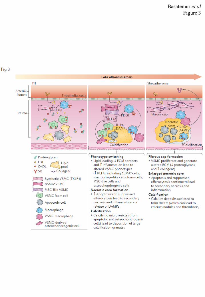
marker-positive cells in early plaques5,61. Monocytes are recruited to PITs through the 1 expression of adhesion molecules (including selectins, ICAM1, VCAM1, CD31123) and 2 chemo-attractants, including chemokines (such as CCL5, CXCL1 and CCL2120,124 , which are 3 secreted by VSMCs and ECs stimulated with inflammatory cytokines or OxLDL, in vitro125) 4 and modified lipids (such as OxLDL126). Studies in animal models have collectively revealed 5 an essential requirement for macrophages in the progression of atherosclerosis120,122,127,128, 6 which is likely to involve effects on VSMC migration, proliferation (through production of 7 factors such as PDGF129) and phenotype switching130. 8 9 10 Late atherosclerosis 11 12 PITs can progress to fibroatheromas (FIG. 3), characterised by the presence of a fibrous cap 13 and a necrotic core, the origins of which are the extra-cellular lipid pool and insufficient 14 efferocytosis (of dead VSMCs and macrophages)131–133. This phase of atherosclerosis (late 15 PIT/early fibroatheroma) is dependent on extensive accumulation of macrophages on the 16 luminal side of the lipid pool, where they phagocytose deposited lipids to become foam cells. 17 In the absence of resolution, the ensuing inflammatory reaction is self-perpetuating; 18 macrophages and VSMCs become foam cells, which die (mostly by apoptosis but potentially 19 through other mechanisms, Box 4). Since the plaque milieu suppresses efferocytosis133–136, 20 uncleared apoptotic cells subsequently undergo secondary necrosis with release of further 21 inflammatory material, such as damage-associated molecular patterns (DAMPs)137. The 22 accompanying healing response involves the formation of the fibrous cap, which, at least in 23 the early stages, is a highly cellular region, rich in VSMC-derived αSMA-positive cells22,40–24 42, amongst an altered ECM that has decreased proteoglycan expression and an increase in 25 the proportion of collagens (mostly type I and III). 26 27 In mice, the fibrous cap VSMCs are derived from medial VSMCs22,138 that have undergone 28 migration and proliferation in response to cytokines and growth factors, such as PDGF, 29 derived from macrophages and activated ECs23,129,139. This initial stage of VSMC 30 recruitment is, at least in part, Oct4 dependent21. In humans, both pre-existing intimal and 31 medial VSMCs may contribute to plaque VSMCs48. Definitive proof that VSMCs are 32 responsible for the production of the fibrous cap ECM is lacking. However, this hypothesis 33 is consistent with co-localization of collagen synthesis to VSMCs in the fibrous cap140, 34 correlation of fibrous cap thickness with VSMC phenotype in mice11,21,141, and the correlation 35 of fibrous cap stability with VSMC cell number in humans142. In addition, a recent study of 36 VSMC-specific deletion of Col15a resulted in a greater than 70% reduction in Col15a, 37 supporting VSMCs as the major source of this collagen143. Further evidence that VSMCs are 38 the major source of collagens comes from studies in vitro, including proteomic analysis of the 39 secretome of lipid-loaded VSMCs109 and induction of collagen synthesis by VSMCs in 40 culture by TGF-β, PDGF, IL-1, AngII, cholesterol, homocysteine and mechanical 41 stretch144,145. 42 43 VSMCs in the later stages of atherosclerosis have previously been thought to be entirely 44 beneficial, for example by stabilising the plaque through elaborating the fibrous cap. 45 However, lipid loading of VSMCs and altered interactions with the ECM lead to altered 46 VSMC phenotype, and increased macrophage markers59. Indeed, VSMCs contribute between 47 30-70% of the macrophage marker-positive cells11,20 and similarly to foam cells146 in mouse 48 plaques, and around 30-40% of CD68 positive cells and 50% of foam cells in humans11,60. 49 VSMC-specific deletion of the transcription factor KLF4 reduces VSMC switching to 50
10
macrophage marker-positive cells, and results in a marked increase in the thickness and 1 αSMA-positive cell content of the fibrous cap11. Although these studies have shown that 2 VSMCs can express macrophage markers, in vitro studies of the transcriptomes of VSMCs 3 and macrophage-derived foam cells indicate they are functionally distinct, and that VSMC-4 derived foam cells exhibit reduced phagocytic and efferocytic responses59. VSMCs have 5 long been known to contribute to the inflammatory milieu of the plaque through recruitment 6 of macrophages; however, these studies strongly suggest that VSMC-derived macrophage-7 like cells also directly affect plaque progression. 8 9 In early fibro-atheromas, calcification is observed as large granules in the necrotic core and 10 surrounding ECM, resulting from a number of interrelated processes, including macrophage 11 and VSMC-derived calcifying micro-vesicles147–149, release of apoptotic bodies150 or the 12 activity of osteochondrogenic cells151. As the fibro-atheroma develops, micro-calcifications 13 can coalesce into larger speckles and fragments that can form sheets or plates149 visible by 14 tomography. Fragmentation of these sheets and fibrin encapsulation can lead to the 15 formation of calcium nodules, which protrude into the vessel lumen and precipitate 16 thrombosis88. The extent of plaque calcification varies according to the vascular bed, and a 17 recent study linked this to the different propensities of the local, developmentally distinct, 18 VSMCs to undergo calcification152,153. VSMCs have long been linked to calcification150,154 19 and osteochondrogenic conversion in vitro is enhanced by plaque-like environmental cues, 20 including phenotypic conversion155, apoptotic bodies150, oxLDL156, and inflammatory 21 cytokines such as TNFα157, IL-1158 and IL-18159. Furthermore, specific genetic modulation 22 of VSMC osteochondrogenesis in vivo leads to altered calcification in models of 23 atherosclerosis160–162. Most convincingly, however, recent studies have established that most 24 of the osteochondreogenic precursors (Runx2/Cbfa1+ cells) and chondrocyte-like (type II 25 collagen+) cells of murine plaques are again VSMC-derived138. 26 27 28 Clinical sequalae 29 30 The major clinical sequelae of atherosclerosis are dependent on the anatomical site of the 31 vascular bed involved (angina and myocardial infarction in coronary arteries; stroke in 32 carotid arteries) and typically manifest as a result of thrombosis. The primary cause 33 (accounting for around 60% to 70% of cases) of thrombosis is plaque rupture163 and the 34 remaining cases are predominantly the result of plaque erosion (the latter being much more 35 frequent in young individuals, particularly women) (FIG. 4). A minority (typically around 36 5%) are due to thrombosis forming on calcified nodules. However, thrombosis and clinical 37 sequalae are not an inevitable consequence of atherosclerosis; analysis of autopsies has 38 shown that plaques often show evidence of silent (non-occlusive) thrombi which have 39 undergone repair and healing. Furthermore, the widespread uptake of clinical interventions, 40 including lipid-lowering, are changing the clinical presentation of atherosclerosis in 41 association with changes in the characteristics of the ‘vulnerable plaque’164. 42 43 As the fibroatheroma develops, so does the necrotic core; the free cholesterol content and 44 calcification increases, and there is breakdown and remodelling of the fibrous cap ECM. The 45 latter is thought to be principally due to the actions of proteases (in particular 46 metalloproteinases165), but also by sulphatases and exoglycosidases that are predominantly 47 released by macrophages166, but may also come from VSMCs167. Concomitantly, VSMCs 48 are depleted through cell death, and so the cap diminishes, whilst the growing necrotic core 49 extends outwards, leading to thinning of the fibrous cap168,169. Thin-cap fibroatheromas 50

11
(TCFA) are defined by a fibrous cap of less than 65µm, and are also known as ‘vulnerable 1 plaques’ because studies have shown that these plaques are highly susceptible to rupture. 2 The underlying mechanisms are ill-defined, but proteolytic activity166,167, mechanical stress170 3 and micro-calcification of the fibrous cap149,171 have all been linked to plaque rupture. 4 5 Plaque rupture is inversely correlated with VSMC number142, which is determined by 6 proliferation, migration and death of VSMCs. Advanced human lesions show little VSMC 7 proliferation172,173, but VSMC death, through apoptosis and necrosis (Box 4), is increased 8 compared to normal vessels174–176, and in unstable versus stable plaques177. Indeed, VSMC 9 apoptosis has been postulated to be key to plaque instability178. Seminal work showed plaque 10 VSMCs to spontaneously undergo apoptosis in vitro, with IGF-1 and PDGF acting as 11 survival factors179, and plaque VSMCs expressing less IGF-1R180. Similarly, cell to cell 12 contact via N-cadherin promotes survival181. Conversely, numerous factors that induce 13 VSMC apoptosis have been described, including cell-directed killing (by macrophages, T 14 lymphocytes and mast cells), ROS, DNA damage, anoikis and cholesterol. Studies of VSMC 15 apoptosis in vivo have utilised mice that have either alterations to apoptotic pathways or 16 systems to induce apoptosis. Early work with adenoviral p53 expression in plaques led to 17 VSMC apoptosis and cap thinning182. Similarly, VSMC-specific diphtheria toxin (DT)-18 induced apoptosis revealed short term VSMC apoptosis within established plaques to have no 19 effect on plaque size, but to result in vulnerable plaques with small fibrous caps and a paucity 20 of VSMCs and structural matrix178 – a finding subsequently corroborated many times in 21 studies that have promoted or inhibited VSMC death,167,181,183–187. Strikingly, DT induction 22 of VSMC apoptosis alongside high fat feeding during atherogenesis resulted in larger 23 plaques51, showing that the consequences of VSMC death are more than cell loss alone, and 24 in fact actively drives plaque growth - another well replicated finding167,185,187,188. A key 25 controller of VSMC apoptosis in vivo appears to be the survival kinase Akt1183,186,187; 26 conditional ablation of Akt1 during atherogenesis induces VSMC apoptosis and larger 27 plaques, and Akt1 ablation in established plaques leads to a reduced fibrous cap. The 28 contribution of VSMC death to plaque stability is complex and extends beyond direct cell 29 loss; with further consequences on the local milieu (such as initiating calcification150), and 30 wider effects in activating the immune system. The plaque environment is known to inhibit 31 phagocytosis133–136, and defective efferocytosis of apoptotic cells leading to secondary 32 necrosis and leakage of intracellular contents has been proposed to exacerbate the 33 inflammatory milieu131,132,137. Indeed, necrotic VSMCs potently drive inflammation via IL-34 1α due to a lack of IL-1R2 that normally binds and inhibits IL-1α133,189. Thus, a consensus 35 appears whereby functional VSMCs are essential to maintain the fibrous cap and thus plaque 36 stability, but death of VSMCs is a potent driver of atherogenesis. 37 38 A recent study of the VSMC transcriptome in symptomatic versus asymptomatic carotid 39 plaques has also highlighted the importance of VSMC senescence190. Unstable mature 40 plaques show low VSMC proliferation and clear evidence of VSMC senescence191. 41 Senescent VSMCs were originally thought to promote plaque instability through inaction - 42 i.e. a lack of VSMC proliferation and matrix production leads to weakening of the fibrous 43 cap. However, senescent VSMCs establish a robust IL-1α-driven SASP containing multiple 44 inflammatory cytokines, chemokines, MMPs and osteogenic factors80,192. Thus, the VSMC 45 SASP can recruit mononuclear cells, induce endothelial cell adhesion receptor expression and 46 activate adjacent normal VSMCs80, effectively amplifying the effect of a small number of 47 senescent VSMCs. Senescent VSMCs also produce less collagen and release active 48 MMP980, while BMP2 and osteoprotegerin within the SASP drive calcification192. Thus, 49

12
senescent VSMCs can have a negative impact on plaques through both loss of normal 1 function and a direct effect on the local plaque milieu. 2 3 An alternative route to thrombosis and clinical sequalae is through plaque erosion. Erosion 4 refers to the formation of a thrombus in the absence of rupture at sites of endothelial 5 denudation or disruption. The underlying plaque may be an intimal thickening or 6 fibroatheroma88,169, but VSMCs are often abundant, amidst a proteoglycan-rich ECM, 7 enriched in type III collagen, versican and hyaluronan193. Recent studies have identified an 8 important role for hyaluronan, which activates TLR2 signalling upon degradation 194 and this 9 combined with altered shear stress, leads to endothelial cell activation and apoptosis195, 10 neutrophil recruitment and thrombosis194. Thus VSMCs are implicated in the events leading 11 to plaque erosion, in particular as the major source of hyaluronan196. 12 13 14 Future perspectives 15 16 Difficulties in extrapolating studies from mice to man 17 18 Reconciling the results of studies of animal models with those of human atherosclerosis can 19 be challenging, as there are some important differences in how the disease progresses in 20 humans and animal models. This is exemplified in the case of DITs, which are absent in 21 most animal models. Another fundamental difference is that fibroatheromas rarely progress 22 to rupture in animal models, exemplified by the recently reported effects of a neutralising IL-23 1β antibody, which were deleterious on the fibrous cap in mice141, but beneficial in reducing 24 cardiovascular events in the CANTOS trial in humans197. Nonetheless, animal models have 25 been instructive in delineating important pathways and basic principles that might underlie 26 plaque development in humans. This is particularly true of the lineage tracing studies in 27 mouse models of atherosclerosis, which have unambiguously established the importance of 28 clonality and phenotype switching of VSMCs. Combinatorial genetic depletion models will 29 likely be instrumental in assessing whether biasing the phenotype of VSMC-derived cells 30 could be a potential treatment avenue. Recently developed techniques, including mass 31 cytometry (CyToF) and single-cell omics (genomics, transcriptomics and epigenomics), hold 32 great promise for high resolution, spatio-temporal analysis of plaque cells in situ, and are 33 likely to provide the conclusive human counterpart and mechanistic data for the 34 aforementioned studies. 35 36 37 38 VSMCs and genetics of atherosclerosis 39 40 Over 150 CAD loci have been identified from GWAS and other genetic association 41 studies198, many of which are associated with disease independently of other known risk 42 factors. Thus, elucidation of the underlying molecular mechanisms may reveal novel 43 pathways and hence targets for therapeutic intervention. However, identification of causal 44 variants is usually far from trivial; CAD loci are often located in non-coding regions, where 45 the causal variant is predicted to effect regulation of gene expression, which may operate 46 over large distances and be cell-type or context specific. Studies are ongoing to identify and 47 functionally characterise the causal variants responsible for each of the CAD loci, and in vitro 48 studies of VSMCs are proving an invaluable resource in this quest. Integration of 49 transcriptomic and epigenomic maps from VSMCs (and other plaque cells) with those of the 50

13
genetic architecture of CAD can be very informative for prioritising variants (and potential 1 pathways) for functional characterisation199,200. Unsurprisingly, given the key role of VSMCs 2 in atherosclerosis, a number of loci have been predicted to modulate disease risk through 3 mechanisms specific to VSMCs200. Thus, studies in cultured VSMCs, and more recently 4 VSMCs derived from stem cells49,201, are likely to be instrumental in the functional 5 characterisation of CAD variants. Recent pioneering examples of such studies include the 6 characterisation of the SMAD3 and TCF21 loci202. 7 8 9 Conclusion 10 11 The role of VSMCs in atherosclerosis extends far beyond that perceived for decades. 12 VSMCs and VSMC-derived cells comprise a (if not the) major source of plaque cells, and 13 contribute to numerous plaque cell phenotypes, including macrophage-like and foam cells, in 14 addition to cells responsible for producing the atherogenic and or athero-protective ECM 15 throughout the disease. Thus, VSMCs are implicated mechanistically at all stages of 16 atherosclerosis, and recent studies have established the extent and importance of VSMC 17 clonality and phenotype switching in plaque progression. These concepts have been around 18 for decades, but it is only very recently that technologies for genetic engineering and imaging 19 have converged with a deeper understanding of developmental processes to generate 20 conclusive data in animal models. The era of single cell omics promises to deliver the 21 evidence as to if and how these processes contribute to the disease in humans. It is clear that 22 a better understanding of the biology of VSMCs is required if we are to fulfil aspirations of 23 selectively targeting ‘culprit’ cells or manipulating cell phenotype to enhance clinical benefit 24 and/or avert processes that are detrimental in disease. 25 26 27 Key points: 28 - VSMCs and VSMC-derived cells are a major source of plaque cells and ECM at all stages 29 of atherosclerosis 30 - VSMCs contribute to many different plaque cell phenotypes, including ECM-producing 31 cells of the fibrous cap, macrophage-like cells, foam cells, mesenchymal stem cell-like and 32 osteochondrogenic cells 33 - Recently progress has been made regarding the source of plaque VSMCs and VSMC-34 derived cells, which highlights the importance of developmental origin, clonal expansion and 35 phenotype switching of VSMCs in atherosclerosis 36 37 38

14
Box 1: Historical perspective on VSMCs in atherosclerosis 1 2 The development of antibodies for ‘VSMC-specific’ function-associated markers, such as 3 smooth muscle alpha actin (αSMA)6–9, greatly facilitated immuno-histological studies of 4 VSMCs in plaques of animal models203,204 and humans98,103. These studies, alongside in vitro 5 culture models55 and models of arterial injury, such as balloon angioplasty, revealed that 6 VSMCs are capable of great phenotypic plasticity, and undergo ‘phenotypic switching’ from 7 contractile to proliferative synthetic phenotypes205–207. Phenotype switching and proliferation 8 of VSMCs in response to arterial injury and lipid infiltration were considered the main 9 pathological processes underlying plaque development207. 10 11 Studies in the 1990s characterised the role of VSMC proliferation, migration, apoptosis, and 12 phenotype switching in atherogenesis208, and revealed that VSMCs can give rise to foam 13 cells4,5,102 and osteochondrogenic cells154. However, detailed post-mortem analyses of culprit 14 plaques in sudden cardiac death established that the integrity of the fibrous cap, comprising 15 mostly αSMA-positive cells and associated extracellular matrix (ECM), is critical to stabilise 16 and protect plaques from rupture, a major cause of the clinical sequalae of 17 atherosclerosis142,163,168. These studies also highlighted the role of immune cells, particularly 18 macrophages, and inflammation as the main driver of plaque development169. Thus, the 19 prevailing model has been that VSMCs contribute to the cellularity and inflammation of the 20 developing plaque, but have a predominantly beneficial role in its stabilisation though 21 elaborating the fibrous cap209. 22 23 In the last decade, studies applying fate mapping and lineage tracing techniques have 24 revealed the limitations of relying on ‘VSMC-specific’ function-associated markers to infer 25 VSMC identity, and exposed the extent to which this can lead to false negative and false 26 positive identification of VSMCs, as well as oversimplification of VSMC heterogeneity and 27 functions in plaques11,17,18. 28 29 Text boxes (for timeline): 30 31 pre 1900s histology on morbid specimens, including by Virchow (1856) who proposed 32 atherosclerosis to result from inflammation and proliferation as a consequence of arterial 33 injury by mechanical forces 34 35 Marchand coins ‘atherosclerosis’ 36 37 Ignatowsky describes relationship between protein/lipid-rich diet and experimental 38 atherosclerosis, these studies were extended by Anichkov in 1913, who discovered the 39 importance of cholesterol 40 41 Foam cells observed in human and experimental atherosclerosis studies by light 42 microscopy210,211 43 44 Pease describes VSMC as the only cell-type in the healthy media by electron microscopy2. 45 Studies of experimental and human atherosclerosis quickly followed, revealing VSMC 46 derived cells as prominent cell type in plaques3–5 47 48

15
Wissler proposes VSMC are the primary cell type involved in atherosclerosis, assimilating 1 many studies (including Wolinsky & Glagov212) that VSMC are the contractile and ECM-2 producing cells of the media and, furthermore, contribute to plaque foam cells62 3 4 Ross further develops ‘response to injury hypothesis’, emphasizing the role of PDGF 5 mediated VSMC proliferation207 (firstly due to EC injury and platelet activation213 and later 6 updated to incorporate a role for macrophage derived PDGF129) 7 8 Benditt & Benditt propose plaque VSMC arise from clonal expansion46 9 10 Chamley-Campbell et al identify phenotype switching in cultured VSMCs55 11 12 ‘vulnerable plaque’ concept developed; studies of culprit plaques in cardiac deaths identify 13 fibrous cap integrity essential to plaque stability163,168,214 14 15 ApoE and LDLR mouse models of atherosclerosis developed203,204 16 17 ‘response to retention hypothesis’ proposed113 and supported by identification of the central 18 role of ApoB containing lipoproteins101 19 20 first lineage tracing studies11,17,18 which collectively revealed VSMC contribution much more 21 substantial than previously thought, giving rise to macrophage marker positive cells, foam 22 cells, osteochondrogenic and mesenchymal stem cell like cells 23 24 multi-colour lineage tracing studies demonstrate multiple plaque phenotypes are derived from 25 common ancestor – revealing the true extent of VSMC clonality in plaques20,22 26 27 CANTOS trial establishes causal role for inflammation in pathogenesis of atherosclerosis197 28 29 30 31 32 33 34 35 36 37 38 39

16
Box 2: Embryonic origins of VSMCs and sources of VSMC progenitors in adults 1 2 During embryonic development, medial VSMCs (and in some instances pericytes215) arise 3 from local progenitor cells, of which there are multiples distinct lineages distributed across 4 the arterial tree. In mice, more than eight distinct progenitor populations have been 5 identified44,216,217. The aortic root and outer medial layers of the ascending aorta derive from 6 the secondary heart field26,28; the inner medial layer of the ascending aorta, aortic arch, ductus 7 arteriosus, innominate and right subclavian arteries, right and left common carotid arteries 8 derive from the neural crest25; the descending aorta derives from paraxial (somatic) 9 mesoderm218; and the coronary arteries are derived from pro-epicardium, which derives from 10 lateral plate mesoderm219. 11 12 Potential VSMC progenitor populations have also been identified in the media in the adult 13 mouse, including VSMC-derived cells expressing Sca1 and other mesenchymal stem cell 14 markers11,43. These cells may be an intermediate population derived from phenotypic 15 switching, which can give rise to different VSMC-derived cell phenotypes43. Other potential 16 progenitor cells include a population of adventitial cells located close to the medial boundary 17 that express mesenchymal stem cell markers (e.g. Sca1) and are sonic hedgehog signalling-18 responsive (Gli1 positive)27,220–222, and pericytes223,224, which are VSMC-like cells of the 19 microvasculature. 20 21 Importantly, studies have shown that progenitors with distinct origins may achieve a common 22 VSMC fate with respect to expression of ‘VSMC-specific’ function-associated markers 23 (through pathways discussed in Box 3), but are nonetheless distinct with respect to other 24 functional characteristics, such as responses to growth factors. 25 26 27 28 29 30 31 32 33 34 35 36 37 38 39 40 41 42 43

17
Box 3: Molecular mechanisms underlying VSMC plasticity 1 2 Transcription factors: 3 4
Myocardin (MYOCD) family proteins drive expression of contractile genes57. 5 MYOCD is a co-factor for serum response factor (SRF), which binds CArG-box 6 elements within contractile gene promoters. Most environmental cues and signalling 7 pathways affecting VSMC function impact the expression and/or activity of 8 MYOCD225,226 9
10 KLF4 represses contractile gene expression through several mechanisms, including 11 binding to G/C repressor elements and inhibiting SRF binding to CArG-boxes. KLF4 12 inhibits proliferation; VSMC specific deletion of CHOP leads to decreased VSMC 13 proliferation through increased expression of KLF4227. Importantly, VSMC 14 phenotype switching is KLF4 dependent. KLF4 is required for induction of 15 progenitor cells prior to clonal expansion of pulmonary VSMCs in hypoxia65,66 and 16 VSMC-specific deletion of KLF4 in ApoE-/- animals results in reduced numbers of 17 VSMC-derived macrophage and mesenchymal stem cell marker positive plaque 18 cells11. 19
20 21 Extracellular stimuli: the contractile phenotype is promoted by TGF-β, whereas PDGF 22 induces KLF4 expression, VSMC proliferation and phenotypic switching. Other growth 23 factors including WNT signalling also promote proliferation and migration of VSMCs. Pro-24 inflammatory cytokines (e.g. IL-1 and TNF-α) perturb VSMC phenotype via NF-κB and AP-25 1 mediated gene regulation, including MYOCD downregulation. Cholesterol-induced 26 activation of macrophage-associated gene expression in VSMC occurs via microRNA-27 143/145, involves MYOCD and inflammatory signalling and is affected by KLF459,228. 28 29 Cell interactions: ECM proteins and heparin affect VSMC phenotype229. Notably, deletion 30 of integrin β3 results in larger lesions and affects VSMC clonality in atherosclerosis23. 31 Differences in how cells communicate with the environment may also explain the 32 documented effect of stretch and shear stress on VSMC phenotype230. 33 34 Epigenetic regulation: the reversibility of VSMC phenotypic switching indicates a cellular 35 memory of the contractile state. Indeed, contractile genes remain marked by H3K4me2 36 (generally associated with actively transcribed genes) after phenotypic switching18 and 37 manipulation of DNA methylation and histone modifying enzymes directly affect VSMC 38 behaviour in murine models of vascular injury and atherosclerosis231–233, whilst levels of 39 epigenetic markers are altered in human plaques234. Non-coding RNAs also control VSMC 40 plasticity235,236 evidenced by the effect of specific miRNAs and long non-coding RNAs on 41 VSMC biology and function237,238. 42 43 44 45 46 47 48 49 50
18
Box 4: Mechanisms of cell death 1 2 3 Apoptosis: the commonest form of programmed cell death (PCD) utilised throughout 4 development and day-to-day physiology. Executed by apoptotic caspases (e.g. 3, 7), with 5 main initiation pathways controlled via the mitochondria (via Bcl-2 family members) or 6 external death receptors (e.g. Fas, TNFR). Apoptotic cells must be phagocytosed, or 7 secondary necrosis with leakage of inflammatory contents (including DAMPs) will occur. 8 All major cell types within the plaque are witnessed to undergo apoptosis. 9 10 Autophagic cell death: a mechanism for the organised degradation and recycling of 11 intracellular components within double membraned autophagosomes that fuse with 12 lysosomes. Can be a response to stress that enables the cell to survive, but is also witnessed 13 as PCD. VSMC specific deficiency in autophagy leads to increased VSMC death and 14 enhanced features of vulnerable plaques188. 15 16 Necrosis: An un-programmed form of cell death characterized by catastrophic loss of plasma 17 membrane integrity and leakage of cell contents. Uncleared dying cells default to secondary 18 necrosis. Difficult to prove in vivo, but ultrastructural evidence suggests necrotic plaque 19 macrophages and VSMCs occur. 20 21 Necroptosis: A programmed form of necrosis allowing cell suicide when apoptosis is 22 blocked (e.g. viral caspase inhibitors). Utilises RIPK1/3 to form the ripoptosome which 23 activates MLKL that destroys the plasma membrane. Increased RIP3 and MLKL reported in 24 human plaques, but difficult to specifically detect necroptosis. 25 26 Pyroptosis: Inflammatory form of cell death that occurs in concert with inflammasome 27 activation and IL-1 production, often in response to intracellular infection. Leads to 28 activation of inflammatory caspases (e.g. 1, 4, 5, 11) that activate IL-1 and/or the pore-29 forming protein GSDMD, and subsequent membrane permeabilisation. Likely happens in 30 plaques after cholesterol crystal activation of macrophage NLRP3 inflammasomes. 31 32 Paraptosis: caspase-independent cell death leading to cytoplasmic vacuolation and eventual 33 osmotic lysis. Not currently described in atherosclerotic plaques. 34 35 36 37 38 39 40

19
Table 1: Lineage tracing studies in atherosclerosis 1 2
3 4 5

Basatemur et alFigure 1

Basatemur et alFigure 2
Basatemur et alFigure 3

Basatemur et alFigure 4

Basatemur et alBox 1 & 2

20
References 1 2 3 Highlighted references 4 Gomez 2013, Feil 2014, Shankman 2015 - these were the first lineage tracing studies of 5 VSMCs in the context of atherosclerosis 6 Chappell 2016 – this article demonstrates that different VSMC phenotypes arise from the 7 same ancestral cell in atherosclerosis 8 Misra 2018 – This article provides evidence that secreted factors affect clonality 9 Childs 2016 – This article demonstrates the impact of senescence in atherosclerosis 10 11 12 1. World Health Organisation (WHO). The top 10 causes of death. Available at: 13
https://www.who.int/news-room/fact-sheets/detail/the-top-10-causes-of-death. 14 2. Pease, D. C. & Paule, W. J. Electron microscopy of elastic arteries; the thoracic aorta 15
of the rat. J. Ultrastruct. Res. 3, 469–483 (1960). 16 3. Parker, F. An Electron Microscopic Study of Experimental Atherosclerosis. Am. J. 17
Pathol. 36, 19–53 (1960). 18 4. Geer, J. C., McGill, H. C. J. & Strong, J. P. The fine structure of human atherosclerotic 19
lesions. Am. J. Pathol. 38, 263–287 (1961). 20 5. Imai, H. et al. Atherosclerosis in rabbits. Architectural and subcellular alterations of 21
smooth muscle cells of aortas in response to hyperlipemia. Exp. Mol. Pathol. 5, 273–22 310 (1966). 23
6. Chamley, J. H., Groschel-Stewart, U., Campbell, G. R. & Burnstock, G. Distinction 24 between smooth muscle, fibroblasts and endothelial cells in culture by the use of 25 fluoresceinated antibodies against smooth muscle actin. Cell Tissue Res. 177, 445–457 26 (1977). 27
7. Gown, A. M., Vogel, A. M., Gordon, D. & Lu, P. L. A smooth muscle-specific 28 monoclonal antibody recognizes smooth muscle actin isozymes. J. Cell Biol. 100, 29 807–813 (1985). 30
8. Skalli, O. et al. A monoclonal antibody against alpha-smooth muscle actin: a new 31 probe for smooth muscle differentiation. J. Cell Biol. 103, 2787–2796 (1986). 32
9. Tsukada, T., Tippens, D., Gordon, D., Ross, R. & Gown, A. M. HHF35, a muscle-33 actin-specific monoclonal antibody. I. Immunocytochemical and biochemical 34 characterization. Am. J. Pathol. 126, 51–60 (1987). 35
10. Shanahan, C. M. & Weissberg, P. L. Smooth muscle cell heterogeneity: Patterns of 36 gene expression in vascular smooth muscle cells in vitro and in vivo. Arterioscler. 37 Thromb. Vasc. Biol. 18, 333–338 (1998). 38
11. Shankman, L. S. et al. KLF4-dependent phenotypic modulation of smooth muscle cells 39 has a key role in atherosclerotic plaque pathogenesis. Nat. Med. 21, 628–637 (2015). 40
12. Wirth, A. et al. G12-G13-LARG-mediated signaling in vascular smooth muscle is 41 required for salt-induced hypertension. Nat. Med. 14, 64–68 (2008). 42
13. Kuhbandner, S. et al. Temporally controlled somatic mutagenesis in smooth muscle. 43 Genesis 28, 15–22 (2000). 44
14. Holtwick, R. et al. Smooth muscle-selective deletion of guanylyl cyclase-A prevents 45 the acute but not chronic effects of ANP on blood pressure. Proc. Natl. Acad. Sci. U. S. 46 A. 99, 7142–7147 (2002). 47
15. Zhang, J. et al. Generation of an adult smooth muscle cell-targeted Cre recombinase 48 mouse model. Arteriosclerosis, thrombosis, and vascular biology 26, e23-4 (2006). 49
16. Raja, C. et al. Promoters to Study Vascular Smooth Muscle. Arterioscler. Thromb. 50

21
Vasc. Biol. 39, 603–612 (2019). 1 17. Feil, S. et al. Transdifferentiation of vascular smooth muscle cells to macrophage-like 2
cells during atherogenesis. Circ. Res. 115, 662–667 (2014). 3 18. Gomez, D., Shankman, L. S., Nguyen, A. T. & Owens, G. K. Detection of histone 4
modifications at specific gene loci in single cells in histological sections. Nat. Methods 5 10, 171–177 (2013). 6
19. Albarrán-Juárez, J., Kaur, H., Grimm, M., Offermanns, S. & Wettschureck, N. Lineage 7 tracing of cells involved in atherosclerosis. Atherosclerosis 251, 445–453 (2016). 8
20. Chappell, J. et al. Extensive Proliferation of a Subset of Differentiated, yet Plastic, 9 Medial Vascular Smooth Muscle Cells Contributes to Neointimal Formation in Mouse 10 Injury and Atherosclerosis Models. Circ. Res. 119, 1313–1323 (2016). 11
21. Cherepanova, O. A. et al. Activation of the pluripotency factor OCT4 in smooth 12 muscle cells is atheroprotective. Nat. Med. 22, 657–665 (2016). 13
22. Jacobsen, K. et al. Diverse cellular architecture of atherosclerotic plaque derives from 14 clonal expansion of a few medial SMCs. JCI Insight 2, (2017). 15
23. Misra, A. et al. Integrin beta3 regulates clonality and fate of smooth muscle-derived 16 atherosclerotic plaque cells. Nat. Commun. 9, 2073 (2018). 17
24. Nemenoff, R. A. et al. SDF-1alpha induction in mature smooth muscle cells by 18 inactivation of PTEN is a critical mediator of exacerbated injury-induced neointima 19 formation. Arterioscler. Thromb. Vasc. Biol. 31, 1300–1308 (2011). 20
25. Jiang, X., Rowitch, D. H., Soriano, P., McMahon, A. P. & Sucov, H. M. Fate of the 21 mammalian cardiac neural crest. Development 127, 1607–1616 (2000). 22
26. Waldo, K. L. et al. Secondary heart field contributes myocardium and smooth muscle 23 to the arterial pole of the developing heart. Dev. Biol. 281, 78–90 (2005). 24
27. Passman, J. N. et al. A sonic hedgehog signaling domain in the arterial adventitia 25 supports resident Sca1+ smooth muscle progenitor cells. Proc. Natl. Acad. Sci. U. S. A. 26 105, 9349–9354 (2008). 27
28. Sawada, H., Rateri, D. L., Moorleghen, J. J., Majesky, M. W. & Daugherty, A. Smooth 28 Muscle Cells Derived from Second Heart Field and Cardiac Neural Crest Reside in 29 Spatially Distinct Domains in the Media of the Ascending Aorta - Brief Report. 30 Arterioscler. Thromb. Vasc. Biol. 37, 1722–1726 (2017). 31
29. Chang, H. Y. Anatomic demarcation of cells: genes to patterns. Science 326, 1206–32 1207 (2009). 33
30. Pruett, N. D. et al. Changing topographic Hox expression in blood vessels results in 34 regionally distinct vessel wall remodeling. Biol. Open 1, 430–435 (2012). 35
31. Topouzis, S. & Majesky, M. W. Smooth muscle lineage diversity in the chick embryo. 36 Two types of aortic smooth muscle cell differ in growth and receptor-mediated 37 transcriptional responses to transforming growth factor-beta. Dev. Biol. 178, 430–445 38 (1996). 39
32. Xie, W.-B. B. et al. Smad2 and myocardin-related transcription factor B cooperatively 40 regulate vascular smooth muscle differentiation from neural crest cells. Circ. Res. 113, 41 76–86 (2013). 42
33. Madura, J. A. 2nd et al. Regional differences in platelet-derived growth factor 43 production by the canine aorta. J. Vasc. Res. 33, 53–61 (1996). 44
34. Oh, J., Richardson, J. A. & Olson, E. N. Requirement of myocardin-related 45 transcription factor-B for remodeling of branchial arch arteries and smooth muscle 46 differentiation. Proc. Natl. Acad. Sci. U. S. A. 102, 15122-15127 (2005). 47
35. Li, J. et al. Myocardin-related transcription factor B is required in cardiac neural crest 48 for smooth muscle differentiation and cardiovascular development. Proc. Natl. Acad. 49 Sci. U. S. A. 102, 8916–8921 (2005). 50

22
36. Trigueros-Motos, L. et al. Embryological-origin-dependent differences in homeobox 1 expression in adult aorta: role in regional phenotypic variability and regulation of NF-2 kappaB activity. Arterioscler. Thromb. Vasc. Biol. 33, 1248–1256 (2013). 3
37. Owens, A. P. 3rd et al. Angiotensin II induces a region-specific hyperplasia of the 4 ascending aorta through regulation of inhibitor of differentiation 3. Circ. Res. 106, 5 611–619 (2010). 6
38. Sata, M. et al. Hematopoietic stem cells differentiate into vascular cells that participate 7 in the pathogenesis of atherosclerosis. Nat. Med. 8, 403–409 (2002). 8
39. Caplice, N. M. et al. Smooth muscle cells in human coronary atherosclerosis can 9 originate from cells administered at marrow transplantation. Proc. Natl. Acad. Sci. U. 10 S. A. 100, 4754–4759 (2003). 11
40. Bentzon, J. F., Sondergaard, C. S., Kassem, M. & Falk, E. Smooth muscle cells 12 healing atherosclerotic plaque disruptions are of local, not blood, origin in 13 apolipoprotein E knockout mice. Circulation 116, 2053–2061 (2007). 14
41. Bentzon, J. F. et al. Smooth muscle cells in atherosclerosis originate from the local 15 vessel wall and not circulating progenitor cells in ApoE knockout mice. Arterioscler. 16 Thromb. Vasc. Biol. 26, 2696–2702 (2006). 17
42. Yu, H. et al. Bone Marrow–Derived Smooth Muscle–Like Cells Are Infrequent in 18 Advanced Primary Atherosclerotic Plaques but Promote Atherosclerosis. Arterioscler. 19 Thromb. Vasc. Biol. 31, 1291–1299 (2011). 20
43. Dobnikar, L. et al. Disease-relevant transcriptional signatures identified in individual 21 smooth muscle cells from healthy mouse vessels. Nat. Commun. 9, 4567 (2018). 22
44. Majesky, M. W. Developmental basis of vascular smooth muscle diversity. 23 Arterioscler. Thromb. Vasc. Biol. 27, 1248–1258 (2007). 24
45. Haimovici, H. The role of arterial tissue susceptibility in atherogenesis. Texas Hear. 25 Inst. J. 18, 81–83 (1991). 26
46. Benditt, E. P. & Benditt, J. M. Evidence for a monoclonal origin of human 27 atherosclerotic plaques. Proc. Natl. Acad. Sci. U. S. A. 70, 1753–1756 (1973). 28
47. Murry, C. E., Gipaya, C. T., Bartosek, T., Benditt, E. P. & Schwartz, S. M. 29 Monoclonality of smooth muscle cells in human atherosclerosis. Am. J. Pathol. 151, 30 697–705 (1997). 31
48. Chung, I. M., Schwartz, S. M. & Murry, C. E. Clonal architecture of normal and 32 atherosclerotic aorta: implications for atherogenesis and vascular development. Am. J. 33 Pathol. 152, 913–923 (1998). 34
49. Cheung, C., Bernardo, A. S., Trotter, M. W. B., Pedersen, R. A. & Sinha, S. 35 Generation of human vascular smooth muscle subtypes provides insight into 36 embryological origin–dependent disease susceptibility. Nat. Biotechnol. 30, 165–173 37 (2012). 38
50. Sinha, S. & Santoro, M. M. New models to study vascular mural cell embryonic 39 origin: implications in vascular diseases. Cardiovasc. Res. 114, 481–491 (2018). 40
51. Clarke, M. C. H. et al. Chronic apoptosis of vascular smooth muscle cells accelerates 41 atherosclerosis and promotes calcification and medial degeneration. Circ. Res. 102, 42 1529–1538 (2008). 43
52. Lee, S. H., Hungerford, J. E., Little, C. D. & Iruela-Arispe, M. L. Proliferation and 44 differentiation of smooth muscle cell precursors occurs simultaneously during the 45 development of the vessel wall. Dev. Dyn. 209, 342–352 (1997). 46
53. Poole, J. C., Cromwell, S. B. & Benditt, E. P. Behavior of smooth muscle cells and 47 formation of extracellular structures in the reaction of arterial walls to injury. Am. J. 48 Pathol. 62, 391–414 (1971). 49
54. Kocher, O. et al. Phenotypic features of smooth muscle cells during the evolution of 50

23
experimental carotid artery intimal thickening. Biochemical and morphologic studies. 1 Lab. Invest. 65, 459–470 (1991). 2
55. Chamley-Campbell, J., Campbell, G. R. & Ross, R. The smooth muscle cell in culture. 3 Physiol. Rev. 59, 1–61 (1979). 4
56. Kaur, H. et al. Single-cell profiling reveals heterogeneity and functional patterning of 5 GPCR expression in the vascular system. Nat. Commun. 8, 15700 (2017). 6
57. Pipes, G. C. T., Creemers, E. E. & Olson, E. N. The myocardin family of 7 transcriptional coactivators: versatile regulators of cell growth, migration, and 8 myogenesis. Genes Dev. 20, 1545–1556 (2006). 9
58. Rong, J. X., Shapiro, M., Trogan, E. & Fisher, E. A. Transdifferentiation of mouse 10 aortic smooth muscle cells to a macrophage-like state after cholesterol loading. Proc. 11 Natl. Acad. Sci. U. S. A. 100, 13531–13536 (2003). 12
59. Vengrenyuk, Y. et al. Cholesterol loading reprograms the microRNA-143/145-13 myocardin axis to convert aortic smooth muscle cells to a dysfunctional macrophage-14 like phenotype. Arterioscler. Thromb. Vasc. Biol. 35, 535–546 (2015). 15
60. Allahverdian, S., Chehroudi, A. C., McManus, B. M., Abraham, T. & Francis, G. A. 16 Contribution of intimal smooth muscle cells to cholesterol accumulation and 17 macrophage-like cells in human atherosclerosis. Circulation 129, 1551–1559 (2014). 18
61. Andreeva, E. R., Pugach, I. M. & Orekhov, A. N. Subendothelial smooth muscle cells 19 of human aorta express macrophage antigen in situ and in vitro. Atherosclerosis 135, 20 19–27 (1997). 21
62. Wissler, R. W. The arterial medial cell, smooth muscle, or multifunctional 22 mesenchyme? Circulation 36, 1–4 (1967). 23
63. Alves, R. D. A. M., Eijken, M., van de Peppel, J. & van Leeuwen, J. P. T. M. 24 Calcifying vascular smooth muscle cells and osteoblasts: independent cell types 25 exhibiting extracellular matrix and biomineralization-related mimicries. BMC 26 Genomics 15, 965 (2014). 27
64. Durham, A. L., Speer, M. Y., Scatena, M., Giachelli, C. M. & Shanahan, C. M. Role of 28 smooth muscle cells in vascular calcification: Implications in atherosclerosis and 29 arterial stiffness. Cardiovasc. Res. 114, 590–600 (2018). 30
65. Sheikh, A. Q., Misra, A., Rosas, I. O., Adams, R. H. & Greif, D. M. Smooth muscle 31 cell progenitors are primed to muscularize in pulmonary hypertension. Sci. Transl. 32 Med. 7, 308ra159 (2015). 33
66. Sheikh, A. Q., Saddouk, F. Z., Ntokou, A., Mazurek, R. & Greif, D. M. Cell 34 Autonomous and Non-cell Autonomous Regulation of SMC Progenitors in Pulmonary 35 Hypertension. Cell Rep. 23, 1152–1165 (2018). 36
67. Herring, B. P., Hoggatt, A. M., Burlak, C. & Offermanns, S. Previously differentiated 37 medial vascular smooth muscle cells contribute to neointima formation following 38 vascular injury. Vasc. Cell 6, 21 (2014). 39
68. Gomez, D. & Owens, G. K. Reconciling Smooth Muscle Cell Oligoclonality and 40 Proliferative Capacity in Experimental Atherosclerosis. Circ. Res. 119, 1262–1264 41 (2016). 42
69. Zhang, L. & Vijg, J. Somatic Mutagenesis in Mammals and Its Implications for 43 Human Disease and Aging. Annu. Rev. Genet. 52, 397–419 (2018). 44
70. Jaiswal, S. et al. Clonal Hematopoiesis and Risk of Atherosclerotic Cardiovascular 45 Disease. N. Engl. J. Med. 377, 111–121 (2017). 46
71. Martin, G. M. & Sprague, C. A. Clonal senescence and atherosclerosis. Lancet 47 (London, England) 2, 1370–1371 (1972). 48
72. Munoz-Espin, D. & Serrano, M. Cellular senescence: from physiology to pathology. 49 Nat. Rev. Mol. Cell Biol. 15, 482–496 (2014). 50

24
73. Campisi, J. Aging, cellular senescence, and cancer. Annu. Rev. Physiol. 75, 685–705 1 (2013). 2
74. Kuilman, T., Michaloglou, C., Mooi, W. J. & Peeper, D. S. The essence of senescence. 3 Genes Dev. 24, 2463–2479 (2010). 4
75. Grootaert, M. O. et al. Defective autophagy in vascular smooth muscle cells 5 accelerates senescence and promotes neointima formation and atherogenesis. 6 Autophagy 11, 2014–2032 (2015). 7
76. Matthews, C. et al. Vascular smooth muscle cells undergo telomere-based senescence 8 in human atherosclerosis: effects of telomerase and oxidative stress. Circ. Res. 99, 9 156–164 (2006). 10
77. Coppe, J.-P., Desprez, P.-Y., Krtolica, A. & Campisi, J. The senescence-associated 11 secretory phenotype: the dark side of tumor suppression. Annu. Rev. Pathol. 5, 99–118 12 (2010). 13
78. Coppé, J.-P. et al. Senescence-Associated Secretory Phenotypes Reveal Cell-14 Nonautonomous Functions of Oncogenic RAS and the p53 Tumor Suppressor. PLOS 15 Biol. 6, e301 (2008). 16
79. Orjalo, A. V, Bhaumik, D., Gengler, B. K., Scott, G. K. & Campisi, J. Cell surface-17 bound IL-1alpha is an upstream regulator of the senescence-associated IL-6/IL-8 18 cytokine network. Proc. Natl. Acad. Sci. U. S. A. 106, 17031–17036 (2009). 19
80. Gardner, S. E., Humphry, M., Bennett, M. R. & Clarke, M. C. H. Senescent vascular 20 smooth muscle cells drive inflammation through an interleukin-1α-dependent 21 senescence-associated secretory phenotype. Arterioscler. Thromb. Vasc. Biol. 35, 22 1963–1974 (2015). 23
81. Kang, C. et al. The DNA damage response induces inflammation and senescence by 24 inhibiting autophagy of GATA4. Science 349, aaa5612 (2015). 25
82. Laberge, R.-M. et al. MTOR regulates the pro-tumorigenic senescence-associated 26 secretory phenotype by promoting IL1A translation. Nat. Cell Biol. 17, 1049–1061 27 (2015). 28
83. Kang, T.-W. et al. Senescence surveillance of pre-malignant hepatocytes limits liver 29 cancer development. Nature 479, 547–551 (2011). 30
84. Childs, B. G. et al. Senescent intimal foam cells are deleterious at all stages of 31 atherosclerosis. Science 354, 472–477 (2016). 32
85. Coppé, J.-P. et al. A Human-Like Senescence-Associated Secretory Phenotype Is 33 Conserved in Mouse Cells Dependent on Physiological Oxygen. PLoS One 5, e9188 34 (2010). 35
86. Wang, J. et al. Vascular Smooth Muscle Cell Senescence Promotes Atherosclerosis 36 and Features of Plaque Vulnerability. Circulation 132, 1909–1919 (2015). 37
87. Shah, A. et al. Defective Base Excision Repair of Oxidative DNA Damage in Vascular 38 Smooth Muscle Cells Promotes Atherosclerosis. Circulation 138, 1446-1462 (2018). 39
88. Virmani, R., Kolodgie, F. D., Burke, A. P., Farb, A. & Schwartz, S. M. Lessons From 40 Sudden Coronary Death. Arterioscler. Thromb. Vasc. Biol. 20, 1262–1275 (2000). 41
89. Yahagi, K. et al. Pathophysiology of native coronary, vein graft, and in-stent 42 atherosclerosis. Nat. Rev. Cardiol. 13, 79–98 (2016). 43
90. Velican, C. & Velican, D. Intimal thickening in developing coronary arteries and its 44 relevance to atherosclerotic involvement. Atherosclerosis 23, 345–355 (1976). 45
91. Ikari, Y., McManus, B. M., Kenyon, J. & Schwartz, S. M. Neonatal intima formation 46 in the human coronary artery. Arterioscler. Thromb. Vasc. Biol. 19, 2036–2040 (1999). 47
92. Stary, H. C. et al. A Definition of Initial, Fatty Streak, and Intermediate Lesions of 48 Atherosclerosis. Arter. Thromb. 14, 840–857 (1994). 49
93. Velican, C. A dissecting view on the role of the fatty streak in the pathogenesis of 50

25
human atherosclerosis: culprit or bystander? Med. Interne 19, 321–337 (1981). 1 94. Armstrong, M. L., Heistad, D. D., Megan, M. B., Lopez, J. A. & Harrison, D. G. 2
Reversibility of atherosclerosis. Cardiovasc. Clin. 20, 113–126 (1990). 3 95. Strong, J. P. et al. Prevalence and extent of atherosclerosis in adolescents and young 4
adults: implications for prevention from the Pathobiological Determinants of 5 Atherosclerosis in Youth Study. JAMA 281, 727–735 (1999). 6
96. Nakashima, Y., Chen, Y.-X., Kinukawa, N. & Sueishi, K. Distributions of diffuse 7 intimal thickening in human arteries: preferential expression in atherosclerosis-prone 8 arteries from an early age. Virchows Arch. 441, 279–288 (2002). 9
97. Nakashima, Y., Wight, T. N. & Sueishi, K. Early atherosclerosis in humans: Role of 10 diffuse intimal thickening and extracellular matrix proteoglycans. Cardiovasc. Res. 79, 11 14–23 (2008). 12
98. Mosse, P. R., Campbell, G. R., Wang, Z. L. & Campbell, J. H. Smooth muscle 13 phenotypic expression in human carotid arteries. I. Comparison of cells from diffuse 14 intimal thickenings adjacent to atheromatous plaques with those of the media. Lab. 15 Invest. 53, 556–562 (1985). 16
99. Aikawa, M. et al. Human smooth muscle myosin heavy chain isoforms as molecular 17 markers for vascular development and atherosclerosis. Circ. Res. 73, 1000–1012 18 (1993). 19
100. Andreeva, E. R., Pugach, I. M. & Orekhov, A. N. Collagen-synthesizing cells in initial 20 and advanced atherosclerotic lesions of human aorta. Atherosclerosis 130, 133–142 21 (1997). 22
101. Skalen, K. et al. Subendothelial retention of atherogenic lipoproteins in early 23 atherosclerosis. Nature 417, 750–754 (2002). 24
102. Campbell, J. H., Popadynec, L., Nestel, P. J. & Campbell, G. R. Lipid accumulation in 25 arterial smooth muscle cells. Influence of phenotype. Atherosclerosis 47, 279–295 26 (1983). 27
103. Campbell, J. H., Reardon, M. F., Campbell, G. R. & Nestel, P. J. Metabolism of 28 atherogenic lipoproteins by smooth muscle cells of different phenotype in culture. 29 Arteriosclerosis 5, 318–328 (1985). 30
104. Kim, D. N., Imai, H., Schmee, J., Lee, K. T. & Thomas, W. A. Intimal cell mass-31 derived atherosclerotic lesions in the abdominal aorta of hyperlipidemic swine. Part 1. 32 Cell of origin, cell divisions and cell losses in first 90 days on diet. Atherosclerosis 56, 33 169–188 (1985). 34
105. Ang, A. H., Tachas, G., Campbell, J. H., Bateman, J. F. & Campbell, G. R. Collagen 35 synthesis by cultured rabbit aortic smooth-muscle cells. Alteration with phenotype. 36 Biochem. J. 265, 461–469 (1990). 37
106. Lee, R. T. et al. Mechanical strain induces specific changes in the synthesis and 38 organization of proteoglycans by vascular smooth muscle cells. J. Biol. Chem. 276, 39 13847–13851 (2001). 40
107. Little, P. J., Tannock, L., Olin, K. L., Chait, A. & Wight, T. N. Proteoglycans 41 synthesized by arterial smooth muscle cells in the presence of transforming growth 42 factor-beta1 exhibit increased binding to LDLs. Arterioscler. Thromb. Vasc. Biol. 22, 43 55–60 (2002). 44
108. Chang, M. Y., Potter-Perigo, S., Tsoi, C., Chait, A. & Wight, T. N. Oxidized low 45 density lipoproteins regulate synthesis of monkey aortic smooth muscle cell 46 proteoglycans that have enhanced native low density lipoprotein binding properties. J. 47 Biol. Chem. 275, 4766–4773 (2000). 48
109. S.R., L. et al. Extracellular matrix proteomics identifies molecular signature of 49 symptomatic carotid plaques. J. Clin. Invest. 127, 1546–1560 (2017). 50

26
110. Tran-Lundmark, K. et al. Heparan sulfate in perlecan promotes mouse atherosclerosis: 1 roles in lipid permeability, lipid retention, and smooth muscle cell proliferation. Circ. 2 Res. 103, 43–52 (2008). 3
111. Smith, E. B. & Slater, R. S. The microdissection of large atherosclerotic plaques to 4 give morphologically and topographically defined fractions for analysis. 1. The lipids 5 in the isolated fractions. Atherosclerosis 15, 37–56 (1972). 6
112. Tabas, I., Williams, K. J. & Borén, J. Subendothelial lipoprotein retention as the 7 initiating process in atherosclerosis: Update and therapeutic implications. Circulation 8 116, 1832–1844 (2007). 9
113. Williams, K. J. & Tabas, I. The response-to-retention hypothesis of early 10 atherogenesis. Arterioscler. Thromb. Vasc. Biol. 15, 551–561 (1995). 11
114. Thompson, J. C., Tang, T., Wilson, P. G., Yoder, M. H. & Tannock, L. R. Increased 12 atherosclerosis in mice with increased vascular biglycan content. Atherosclerosis 235, 13 71–75 (2014). 14
115. Napoli, C. et al. Fatty streak formation occurs in human fetal aortas and is greatly 15 enhanced by maternal hypercholesterolemia. Intimal accumulation of low density 16 lipoprotein and its oxidation precede monocyte recruitment into early atherosclerotic 17 lesions. J. Clin. Invest. 100, 2680–2690 (1997). 18
116. Nakagawa, K. & Nakashima, Y. Pathologic intimal thickening in human 19 atherosclerosis is formed by extracellular accumulation of plasma-derived lipids and 20 dispersion of intimal smooth muscle cells. Atherosclerosis 274, 235–242 (2018). 21
117. Kockx, M. M. et al. Apoptosis and Related Proteins in Different Stages of Human 22 Atherosclerotic Plaques. Circulation 97, 2307-2315 (1998). 23
118. Okura, Y. et al. Oxidized low-density lipoprotein is associated with apoptosis of 24 vascular smooth muscle cells in human atherosclerotic plaques. Circulation 102, 25 2680–2686 (2000). 26
119. Tulenko, T. N., Chen, M., Mason, P. E. & Mason, R. P. Physical effects of cholesterol 27 on arterial smooth muscle membranes: evidence of immiscible cholesterol domains 28 and alterations in bilayer width during atherogenesis. J. Lipid Res. 39, 947–956 (1998). 29
120. Robbins, C. S. et al. Local proliferation dominates lesional macrophage accumulation 30 in atherosclerosis. Nat. Med. 19, 1166–1172 (2013). 31
121. Ensan, S. et al. Self-renewing resident arterial macrophages arise from embryonic 32 CX3CR1(+) precursors and circulating monocytes immediately after birth. Nat. 33 Immunol. 17, 159–168 (2016). 34
122. Nahrendorf, M. Myeloid cell contributions to cardiovascular health and disease. Nat. 35 Med. 24, 711–720 (2018). 36
123. Berliner, J. A. et al. Minimally modified low density lipoprotein stimulates monocyte 37 endothelial interactions. J. Clin. Invest. 85, 1260–1266 (1990). 38
124. Nelken, N. A., Coughlin, S. R., Gordon, D. & Wilcox, J. N. Monocyte chemoattractant 39 protein-1 in human atheromatous plaques. J. Clin. Invest. 88, 1121–1127 (1991). 40
125. Cushing, S. D. et al. Minimally modified low density lipoprotein induces monocyte 41 chemotactic protein 1 in human endothelial cells and smooth muscle cells. Proc. Natl. 42 Acad. Sci. U. S. A. 87, 5134–5138 (1990). 43
126. Quinn, M. T., Parthasarathy, S., Fong, L. G. & Steinberg, D. Oxidatively modified low 44 density lipoproteins: a potential role in recruitment and retention of 45 monocyte/macrophages during atherogenesis. Proc. Natl. Acad. Sci. U. S. A. 84, 2995–46 2998 (1987). 47
127. Qiao, J. H. et al. Role of macrophage colony-stimulating factor in atherosclerosis: 48 studies of osteopetrotic mice. Am. J. Pathol. 150, 1687–1699 (1997). 49
128. Swirski, F. K. et al. Monocyte accumulation in mouse atherogenesis is progressive and 50

27
proportional to extent of disease. Proc. Natl. Acad. Sci. 103, 10340-10345 (2006). 1 129. Ross, R. et al. Localization of PDGF-B protein in macrophages in all phases of 2
atherogenesis. Science 248, 1009-1012 (1990). 3 130. Campbell, J. H., Rennick, R. E., Kalevitch, S. G. & Campbell, G. R. Heparan sulfate-4
degrading enzymes induce modulation of smooth muscle phenotype. Exp. Cell Res. 5 200, 156–167 (1992). 6
131. Ait-Oufella, H. et al. Defective mer receptor tyrosine kinase signaling in bone marrow 7 cells promotes apoptotic cell accumulation and accelerates atherosclerosis. 8 Arterioscler. Thromb. Vasc. Biol. 28, 1429–31 (2008). 9
132. Ait-Oufella, H. et al. Lactadherin deficiency leads to apoptotic cell accumulation and 10 accelerated atherosclerosis in mice. Circulation 115, 2168–77 (2007). 11
133. Clarke, M. C. H. H., Talib, S., Figg, N. L. & Bennett, M. R. Vascular smooth muscle 12 cell apoptosis induces interleukin-1-directed inflammation: Effects of hyperlipidemia-13 mediated inhibition of phagocytosis. Circ. Res. 106, 363–372 (2010). 14
134. Shaw, P. X. et al. Human-derived anti-oxidized LDL autoantibody blocks uptake of 15 oxidized LDL by macrophages and localizes to atherosclerotic lesions in vivo. 16 Arterioscler. Thromb. Vasc. Biol. 21, 1333–1339 (2001). 17
135. Schrijvers, D. M., De Meyer, G. R. Y., Kockx, M. M., Herman, A. G. & Martinet, W. 18 Phagocytosis of apoptotic cells by macrophages is impaired in atherosclerosis. 19 Arterioscler. Thromb. Vasc. Biol. 25, 1256–1261 (2005). 20
136. Li, S. et al. Defective phagocytosis of apoptotic cells by macrophages in 21 atherosclerotic lesions of ob/ob mice and reversal by a fish oil diet. Circ. Res. 105, 22 1072–1082 (2009). 23
137. Tabas, I. Macrophage death and defective inflammation resolution in atherosclerosis. 24 Nat. Rev. Immunol. 10, 36–46 (2010). 25
138. Naik, V. et al. Sources of cells that contribute to atherosclerotic intimal calcification: 26 An in vivo genetic fate mapping study. Cardiovasc. Res. 94, 545–554 (2012). 27
139. Sano, H. et al. Functional blockade of platelet-derived growth factor receptor-beta but 28 not of receptor-alpha prevents vascular smooth muscle cell accumulation in fibrous 29 cap lesions in apolipoprotein E-deficient mice. Circulation 103, 2955–2960 (2001). 30
140. Rekhter, M. D. et al. Type I collagen gene expression in human atherosclerosis. 31 Localization to specific plaque regions. Am. J. Pathol. 143, 1634–1648 (1993). 32
141. Gomez, D. et al. Interleukin-1β has atheroprotective effects in advanced 33 atherosclerotic lesions of mice. Nat. Med. 24, 1418–1429 (2018). 34
142. Davies, M. J., Richardson, P. D., Woolf, N., Katz, D. R. & Mann, J. Risk of 35 thrombosis in human atherosclerotic plaques: role of extracellular lipid, macrophage, 36 and smooth muscle cell content. Br. Heart J. 69, 377–381 (1993). 37
143. Durgin, B. G. et al. Smooth muscle cell-specific deletion of Col15a1 unexpectedly 38 leads to impaired development of advanced atherosclerotic lesions. Am. J. Physiol. - 39 Hear. Circ. Physiol. 312, H943–H958 (2017). 40
144. Amento, E. P., Ehsani, N., Palmer, H. & Libby, P. Cytokines and growth factors 41 positively and negatively regulate interstitial collagen gene expression in human 42 vascular smooth muscle cells. Arterioscler. Thromb. a J. Vasc. Biol. 11, 1223–1230 43 (1991). 44
145. Rekhter, M. D. Collagen synthesis in atherosclerosis: Too much and not enough. 45 Cardiovasc. Res. 41, 376–384 (1999). 46
146. Wang, Y. et al. Smooth Muscle Cells Contribute the Majority of Foam Cells in ApoE 47 (Apolipoprotein E)-Deficient Mouse Atherosclerosis. Arterioscler. Thromb. Vasc. 48 Biol. 39 00-00 (2019). doi:10.1161/ATVBAHA.119.312434 49
147. New, S. E. P. et al. Macrophage-derived matrix vesicles: an alternative novel 50

28
mechanism for microcalcification in atherosclerotic plaques. Circ. Res. 113, 72–77 1 (2013). 2
148. Kapustin, A. N. et al. Vascular smooth muscle cell calcification is mediated by 3 regulated exosome secretion. Circ. Res. 116, 1312–1323 (2015). 4
149. Hutcheson, J. D. et al. Genesis and growth of extracellular-vesicle-derived 5 microcalcification in atherosclerotic plaques. Nat. Mater. 15, 335–343 (2016). 6
150. Proudfoot, D. et al. Apoptosis regulates human vascular calcification in vitro: evidence 7 for initiation of vascular calcification by apoptotic bodies. Circ. Res. 87, 1055–1062 8 (2000). 9
151. Rattazzi, M. et al. Calcification of advanced atherosclerotic lesions in the innominate 10 arteries of ApoE-deficient mice: potential role of chondrocyte-like cells. Arterioscler. 11 Thromb. Vasc. Biol. 25, 1420–1425 (2005). 12
152. Leroux-Berger, M. et al. Pathologic calcification of adult vascular smooth muscle cells 13 differs on their crest or mesodermal embryonic origin. J. Bone Miner. Res. 26, 1543–14 1553 (2011). 15
153. Espitia, O. et al. Implication of molecular vascular smooth muscle cell heterogeneity 16 among arterial beds in arterial calcification. PLoS One 13, e0191976 (2018). 17
154. Proudfoot, D., Skepper, J. N., Shanahan, C. M. & Weissberg, P. L. Calcification of 18 human vascular cells in vitro is correlated with high levels of matrix Gla protein and 19 low levels of osteopontin expression. Arterioscler. Thromb. Vasc. Biol. 18, 379–388 20 (1998). 21
155. Steitz, S. A. et al. Smooth muscle cell phenotypic transition associated with 22 calcification: upregulation of Cbfa1 and downregulation of smooth muscle lineage 23 markers. Circ. Res. 89, 1147–1154 (2001). 24
156. Farrokhi, E., Samani, K. G. & Chaleshtori, M. H. Oxidized low-density lipoprotein 25 increases bone sialoprotein expression in vascular smooth muscle cells via runt-related 26 transcription factor 2. Am. J. Med. Sci. 349, 240–243 (2015). 27
157. Al-Aly, Z. et al. Aortic Msx2-Wnt calcification cascade is regulated by TNF-alpha-28 dependent signals in diabetic Ldlr-/- mice. Arterioscler. Thromb. Vasc. Biol. 27, 2589–29 2596 (2007). 30
158. Ceneri, N. et al. Rac2 Modulates Atherosclerotic Calcification by Regulating 31 Macrophage Interleukin-1β Production. Arterioscler. Thromb. Vasc. Biol. 37, 328–340 32 (2017). 33
159. Zhang, K. et al. Interleukin-18 Enhances Vascular Calcification and Osteogenic 34 Differentiation of Vascular Smooth Muscle Cells Through TRPM7 Activation. 35 Arterioscler. Thromb. Vasc. Biol. 37, 1933–1943 (2017). 36
160. Cheng, S.-L. et al. Targeted reduction of vascular Msx1 and Msx2 mitigates 37 arteriosclerotic calcification and aortic stiffness in LDLR-deficient mice fed 38 diabetogenic diets. Diabetes 63, 4326–4337 (2014). 39
161. Hofmann Bowman, M. A. et al. S100A12 in vascular smooth muscle accelerates 40 vascular calcification in apolipoprotein E-null mice by activating an osteogenic gene 41 regulatory program. Arterioscler. Thromb. Vasc. Biol. 31, 337–344 (2011). 42
162. Nakagawa, Y. et al. Paracrine osteogenic signals via bone morphogenetic protein-2 43 accelerate the atherosclerotic intimal calcification in vivo. Arterioscler. Thromb. Vasc. 44 Biol. 30, 1908–1915 (2010). 45
163. Davies, M. J. & Thomas, A. Thrombosis and acute coronary-artery lesions in sudden 46 cardiac ischemic death. N. Engl. J. Med. 310, 1137–1140 (1984). 47
164. Pasterkamp, G., Den Ruijter, H. M. & Libby, P. Temporal shifts in clinical 48 presentation and underlying mechanisms of atherosclerotic disease. Nat. Rev. Cardiol. 49 14, 21–29 (2016). 50

29
165. Newby, A. C. Dual role of matrix metalloproteinases (matrixins) in intimal thickening 1 and atherosclerotic plaque rupture. Physiol. Rev. 85, 1–31 (2005). 2
166. Sukhova, G. K. et al. Evidence for increased collagenolysis by interstitial 3 collagenases-1 and -3 in vulnerable human atheromatous plaques. Circulation 99, 4 2503–2509 (1999). 5
167. Yu, H. et al. FOXO3a (Forkhead Transcription Factor O Subfamily Member 3a) Links 6 Vascular Smooth Muscle Cell Apoptosis, Matrix Breakdown, Atherosclerosis, and 7 Vascular Remodeling Through a Novel Pathway Involving MMP13 (Matrix 8 Metalloproteinase 13). Arterioscler. Thromb. Vasc. Biol. 38, 555–565 (2018). 9
168. Falk, E. Plaque rupture with severe pre-existing stenosis precipitating coronary 10 thrombosis. Characteristics of coronary atherosclerotic plaques underlying fatal 11 occlusive thrombi. Br. Heart J. 50, 127–134 (1983). 12
169. van der Wal, A. C., Becker, A. E., van der Loos, C. M. & Das, P. K. Site of intimal 13 rupture or erosion of thrombosed coronary atherosclerotic plaques is characterized by 14 an inflammatory process irrespective of the dominant plaque morphology. Circulation 15 89, 36–44 (1994). 16
170. Gijsen, F., van der Giessen, A., van der Steen, A. & Wentzel, J. Shear stress and 17 advanced atherosclerosis in human coronary arteries. J. Biomech. 46, 240–247 (2013). 18
171. Vengrenyuk, Y. et al. A hypothesis for vulnerable plaque rupture due to stress-induced 19 debonding around cellular microcalcifications in thin fibrous caps. Proc. Natl. Acad. 20 Sci. U. S. A. 103, 14678–14683 (2006). 21
172. Gordon, D., Reidy, M. A., Benditt, E. P. & Schwartz, S. M. Cell proliferation in 22 human coronary arteries. Proc. Natl. Acad. Sci. U. S. A. 87, 4600–4604 (1990). 23
173. O’Brien, E. R. et al. Proliferation in primary and restenotic coronary atherectomy 24 tissue. Implications for antiproliferative therapy. Circ. Res. 73, 223–231 (1993). 25
174. Han, D. K. et al. Evidence for apoptosis in human atherogenesis and in a rat vascular 26 injury model. Am. J. Pathol. 147, 267–277 (1995). 27
175. Geng, Y. J. & Libby, P. Evidence for apoptosis in advanced human atheroma. 28 Colocalization with interleukin-1 beta-converting enzyme. Am. J. Pathol. 147, 251–29 266 (1995). 30
176. Isner, J. M., Kearney, M., Bortman, S. & Passeri, J. Apoptosis in human 31 atherosclerosis and restenosis. Circulation 91, 2703–2711 (1995). 32
177. Bauriedel, G. et al. Role of smooth muscle cell death in advanced coronary primary 33 lesions: implications for plaque instability. Cardiovasc. Res. 41, 480–488 (1999). 34
178. Clarke, M. C. H. et al. Apoptosis of vascular smooth muscle cells induces features of 35 plaque vulnerability in atherosclerosis. Nat. Med. 12, 1075–1080 (2006). 36
179. Bennett, M. R., Evan, G. I. & Schwartz, S. M. Apoptosis of human vascular smooth 37 muscle cells derived from normal vessels and coronary atherosclerotic plaques. J. Clin. 38 Invest. 95, 2266–2274 (1995). 39
180. Patel, V. A. et al. Defect in insulin-like growth factor-1 survival mechanism in 40 atherosclerotic plaque-derived vascular smooth muscle cells is mediated by reduced 41 surface binding and signaling. Circ. Res. 88, 895–902 (2001). 42
181. Lyon, C. A., Johnson, J. L., Williams, H., Sala-Newby, G. B. & George, S. J. Soluble 43 N-cadherin overexpression reduces features of atherosclerotic plaque instability. 44 Arterioscler. Thromb. Vasc. Biol. 29, 195–201 (2009). 45
182. von der Thusen, J. H. et al. Induction of atherosclerotic plaque rupture in 46 apolipoprotein E-/- mice after adenovirus-mediated transfer of p53. Circulation 105, 47 2064–2070 (2002). 48
183. Fernandez-Hernando, C., Jozsef, L., Jenkins, D., Di Lorenzo, A. & Sessa, W. C. 49 Absence of Akt1 reduces vascular smooth muscle cell migration and survival and 50

30
induces features of plaque vulnerability and cardiac dysfunction during 1 atherosclerosis. Arterioscler. Thromb. Vasc. Biol. 29, 2033–2040 (2009). 2
184. von der Thüsen, J. H. et al. IGF-1 has plaque-stabilizing effects in atherosclerosis by 3 altering vascular smooth muscle cell phenotype. Am. J. Pathol. 178, 924–934 (2011). 4
185. Gorenne, I. et al. Vascular smooth muscle cell sirtuin 1 protects against DNA damage 5 and inhibits atherosclerosis. Circulation 127, 386–396 (2013). 6
186. Tucka, J. et al. Akt1 regulates vascular smooth muscle cell apoptosis through FoxO3a 7 and Apaf1 and protects against arterial remodeling and atherosclerosis. Arterioscler. 8 Thromb. Vasc. Biol. 34, 2421–2428 (2014). 9
187. Rotllan, N. et al. Genetic Evidence Supports a Major Role for Akt1 in VSMCs During 10 Atherogenesis. Circ. Res. 116, 1744–1752 (2015). 11
188. Osonoi, Y. et al. Defective autophagy in vascular smooth muscle cells enhances cell 12 death and atherosclerosis. Autophagy 14, 1991–2006 (2018). 13
189. Zheng, Y., Humphry, M., Maguire, J. J., Bennett, M. R. & Clarke, M. C. H. 14 Intracellular interleukin-1 receptor 2 binding prevents cleavage and activity of 15 interleukin-1alpha, controlling necrosis-induced sterile inflammation. Immunity 38, 16 285–295 (2013). 17
190. Alloza, I. et al. RNAseq based transcriptomics study of SMCs from carotid 18 atherosclerotic plaque: BMP2 and IDs proteins are crucial regulators of plaque 19 stability. Sci. Rep. 7, 1–12 (2017). 20
191. Gorenne, I., Kavurma, M., Scott, S. & Bennett, M. Vascular smooth muscle cell 21 senescence in atherosclerosis. Cardiovasc. Res. 72, 9–17 (2006). 22
192. Liu, Y., Drozdov, I., Shroff, R., Beltran, L. E. & Shanahan, C. M. Prelamin A 23 accelerates vascular calcification via activation of the DNA damage response and 24 senescence-associated secretory phenotype in vascular smooth muscle cells. Circ. Res. 25 112, e99-109 (2013). 26
193. Kolodgie, F. D. et al. Differential accumulation of proteoglycans and hyaluronan in 27 culprit lesions: insights into plaque erosion. Arterioscler. Thromb. Vasc. Biol. 22, 28 1642–1648 (2002). 29
194. Franck, G. et al. Flow Perturbation Mediates Neutrophil Recruitment and Potentiates 30 Endothelial Injury via TLR2 in Mice: Implications for Superficial Erosion. Circ. Res. 31 121, 31–42 (2017). 32
195. Tricot, O. et al. Relation between endothelial cell apoptosis and blood flow direction 33 in human atherosclerotic plaques. Circulation 101, 2450–2453 (2000). 34
196. Papakonstantinou, E., Karakiulakis, G., Roth, M. & Block, L. H. Platelet-derived 35 growth factor stimulates the secretion of hyaluronic acid by proliferating human 36 vascular smooth muscle cells. Proc. Natl. Acad. Sci. U.S.A. 92, 9881–9885 (1995). 37
197. Ridker, P. M. et al. Antiinflammatory Therapy with Canakinumab for Atherosclerotic 38 Disease. N. Engl. J. Med. 377, 1119–1131 (2017). 39
198. van der Harst, P. & Verweij, N. Identification of 64 Novel Genetic Loci Provides an 40 Expanded View on the Genetic Architecture of Coronary Artery Disease. Circ. Res. 41 122, 433–443 (2018). 42
199. Miller, C. L. et al. Integrative functional genomics identifies regulatory mechanisms at 43 coronary artery disease loci. Nat. Commun. 7, 12092 (2016). 44
200. Liu, B. et al. Genetic Regulatory Mechanisms of Smooth Muscle Cells Map to 45 Coronary Artery Disease Risk Loci. Am. J. Hum. Genet. 103, 377–388 (2018). 46
201. Lo Sardo, V. et al. Unveiling the Role of the Most Impactful Cardiovascular Risk 47 Locus through Haplotype Editing. Cell 175, 1796–1810.e20 (2018). 48
202. Iyer, D. et al. Coronary artery disease genes SMAD3 and TCF21 promote opposing 49 interactive genetic programs that regulate smooth muscle cell differentiation and 50

31
disease risk. PLoS Genet. 14, e1007681 (2018). 1 203. Piedrahita, J. A., Zhang, S. H., Hagaman, J. R., Oliver, P. M. & Maeda, N. Generation 2
of mice carrying a mutant apolipoprotein E gene inactivated by gene targeting in 3 embryonic stem cells. Proc. Natl. Acad. Sci. U. S. A. 89, 4471–4475 (1992). 4
204. Ishibashi, S. et al. Hypercholesterolemia in low density lipoprotein receptor knockout 5 mice and its reversal by adenovirus-mediated gene delivery. J. Clin. Invest. 92, 883–6 893 (1993). 7
205. Schwartz, S. M., deBlois, D. & O’Brien, E. R. The intima. Soil for atherosclerosis and 8 restenosis. Circ. Res. 77, 445–465 (1995). 9
206. Campbell, G. R. & Campbell, J. H. Smooth muscle phenotypic changes in arterial wall 10 homeostasis: implications for the pathogenesis of atherosclerosis. Exp. Mol. Pathol. 11 42, 139–162 (1985). 12
207. Ross, R. & Glomset, J. A. Atherosclerosis and the arterial smooth muscle cell: 13 Proliferation of smooth muscle is a key event in the genesis of the lesions of 14 atherosclerosis. Science 180, 1332–1339 (1973). 15
208. Ross, R. The pathogenesis of atherosclerosis: a perspective for the 1990s. Nature 362, 16 801–809 (1993). 17
209. Schwartz, S. M., Virmani, R. & Rosenfeld, M. E. The good smooth muscle cells in 18 atherosclerosis. Curr. Atheroscler. Rep. 2, 422–429 (2000). 19
210. Movat, H. Z., Haust, M. D. & More, R. H. The morphologic elements in the early 20 lesions of arteriosclerosis. Am. J. Pathol. 35, 93–101 (1959). 21
211. Buck, R. C. The fine structure of the aortic endothelial lesions in experimental 22 cholesterol atherosclerosis of rabbits. Am. J. Pathol. 34, 897–909 (1958). 23
212. Wolinsky, H. & Glagov, S. Structural Basis for the Static Mechanical Properties of the 24 Aortic Media. Circ. Res. 14, 400–413 (1964). 25
213. Ross, R., Glomset, J. & Harker, L. Response to injury and atherogenesis. Am. J. 26 Pathol. 86, 675–684 (1977). 27
214. Davies, M. J. & Thomas, A. C. Plaque fissuring--the cause of acute myocardial 28 infarction, sudden ischaemic death, and crescendo angina. Heart 53, 363–373 (1985). 29
215. Etchevers, H. C., Vincent, C., Le Douarin, N. M. & Couly, G. F. The cephalic neural 30 crest provides pericytes and smooth muscle cells to all blood vessels of the face and 31 forebrain. Development 128, 1059–1068 (2001). 32
216. Majesky, M. W., Dong, X. R., Regan, J. N. & Hoglund, V. J. Vascular smooth muscle 33 progenitor cells: Building and repairing blood vessels. Circ. Res. 108, 365–377 (2011). 34
217. Bentzon, J. F. & Majesky, M. W. Lineage tracking of origin and fate of smooth muscle 35 cells in atherosclerosis. Cardiovasc. Res. 114, 492–500 (2018). 36
218. Wasteson, P. et al. Developmental origin of smooth muscle cells in the descending 37 aorta in mice. Development 135, 1823–1832 (2008). 38
219. Mikawa, T. & Gourdie, R. G. Pericardial mesoderm generates a population of 39 coronary smooth muscle cells migrating into the heart along with ingrowth of the 40 epicardial organ. Dev. Biol. 174, 221–232 (1996). 41
220. Hu, Y. et al. Abundant progenitor cells in the adventitia contribute to atherosclerosis 42 of vein grafts in ApoE-deficient mice. J. Clin. Invest. 113, 1258–1265 (2004). 43
221. Kramann, R. et al. Adventitial MSC-like Cells Are Progenitors of Vascular Smooth 44 Muscle Cells and Drive Vascular Calcification in Chronic Kidney Disease. Cell Stem 45 Cell 19, 628–642 (2016). 46
222. Zengin, E. et al. Vascular wall resident progenitor cells: a source for postnatal 47 vasculogenesis. Development 133, 1543–1551 (2006). 48
223. Crisan, M. et al. A Perivascular Origin for Mesenchymal Stem Cells in Multiple 49 Human Organs. Cell Stem Cell 3, 301–313 (2008). 50

32
224. Tigges, U., Komatsu, M. & Stallcup, W. B. Adventitial pericyte 1 progenitor/mesenchymal stem cells participate in the restenotic response to arterial 2 injury. J. Vasc. Res. 50, 134–144 (2013). 3
225. Parmacek, M. S. Myocardin: dominant driver of the smooth muscle cell contractile 4 phenotype. Arterioscler. Thromb. Vasc. Biol. 28, 1416–1417 (2008). 5
226. Yoshida, T., Yamashita, M., Horimai, C. & Hayashi, M. Smooth muscle-selective 6 inhibition of nuclear factor-κB attenuates smooth muscle phenotypic switching and 7 neointima formation following vascular injury. J. Am. Heart Assoc. 2, e000230–8 e000230 (2013). 9
227. Zhou, A.-X. et al. C/EBP-Homologous Protein (CHOP) in Vascular Smooth Muscle 10 Cells Regulates Their Proliferation in Aortic Explants and Atherosclerotic Lesions. 11 Circ. Res. 116, 1736–1743 (2015). 12
228. Cordes, K. R. et al. miR-145 and miR-143 regulate smooth muscle cell fate and 13 plasticity. Nature 460, 705–710 (2009). 14
229. Raines, E. W. The extracellular matrix can regulate vascular cell migration, 15 proliferation, and survival: relationships to vascular disease. Int. J. Exp. Pathol. 81, 16 173–182 (2000). 17
230. Opitz, F., Schenke-Layland, K., Cohnert, T. U. & Stock, U. A. Phenotypical plasticity 18 of vascular smooth muscle cells-effect of in vitro and in vivo shear stress for tissue 19 engineering of blood vessels. Tissue Eng. 13, 2505–2514 (2007). 20
231. Liu, R. et al. Ten-eleven translocation-2 (TET2) is a master regulator of smooth 21 muscle cell plasticity. Circulation 128, 2047–2057 (2013). 22
232. Chen, J. et al. Histone demethylase KDM3a, a novel regulator of vascular smooth 23 muscle cells, controls vascular neointimal hyperplasia in diabetic rats. Atherosclerosis 24 257, 152–163 (2017). 25
233. Zhuang, J. et al. The yin-yang dynamics of DNA methylation is the key regulator for 26 smooth muscle cell phenotype switch and vascular remodeling. Arterioscler. Thromb. 27 Vasc. Biol. 37, 84–97 (2017). 28
234. Greissel, A. et al. Alternation of histone and DNA methylation in human 29 atherosclerotic carotid plaques. Thromb. Haemost. 114, 390–402 (2015). 30
235. Lino Cardenas, C. L. et al. An HDAC9-MALAT1-BRG1 complex mediates smooth 31 muscle dysfunction in thoracic aortic aneurysm. Nat. Commun. 9, 1009 (2018). 32
236. Fiedler, J. et al. Non-coding RNAs in vascular disease - from basic science to clinical 33 applications: scientific update from the Working Group of Myocardial Function of the 34 European Society of Cardiology. Cardiovasc. Res. 114, 1281–1286 (2018). 35
237. Das, S. et al. A Novel Angiotensin II-Induced Long Noncoding RNA Giver Regulates 36 Oxidative Stress, Inflammation, and Proliferation in Vascular Smooth Muscle Cells. 37 Circ. Res. 123, 1298–1312 (2018). 38
238. Ballantyne, M. D. et al. Smooth Muscle Enriched Long Noncoding RNA (SMILR) 39 Regulates Cell Proliferation. Circulation 133, 2050–2065 (2016). 40
239. Nurnberg, S. T. et al. Coronary Artery Disease Associated Transcription Factor TCF21 41 Regulates Smooth Muscle Precursor Cells That Contribute to the Fibrous Cap. PLoS 42 Genet. 11, e1005155 (2015). 43
240. Evrard, S. M. et al. Endothelial to mesenchymal transition is common in 44 atherosclerotic lesions and is associated with plaque instability. Nat. Commun. 7, 45 11853 (2016). 46
47 48 49 50

33
1 Figure legends 2 3 Figure 1: Overview of the role of vascular smooth muscle cells (VSMCs) in 4 atherosclerosis 5 VSMCs are a major source of plaque cells and extra-cellular matrix (ECM) at all stages of 6 atherosclerosis and contribute to numerous processes throughout the disease. 7 8 Figure 2: VSMCs in early atherosclerosis 9 Summary of the role of VSMCs in early atherosclerosis (progression from diffuse intimal 10 thickening to pathological intimal thickening). VSMCs are the predominant cell type and 11 source of atherogenic, lipid (particularly LDL)-retentive extra-cellular matrix in early 12 atherosclerosis. Retained LDL is susceptible to modifications, such as oxidation (to 13 OxLDL). Uptake of OxLDL by VSMCs leads to foam cell formation and death by apoptosis. 14 Activated VSMC secrete chemokines and contribute to recruitment of monocytes, which 15 differentiate to macrophages. Progression to PITs is typically associated with decreased 16 VSMC marker positive cell content (such as smooth muscle alpha actin positive cells, 17 αSMA+) and increased macrophage marker positive cells (such as CD68+ cells), likely 18 reflecting a combination of VSMC death and VSMC phenotype switching to macrophage 19 like cells (as a consequence of decreased MYOCD and increased KLF4). 20 21 Abbreviations: ABCA1, ATP-binding cassette transporter 1 ApoB, apolipoprotein B; CCL2, CC motif 22 chemokine 2 (also known as MCP-1); CCL5, CC motif chemokine 5 (also known as RANTES); CXCL1, CXC 23 motif chemokine 1 (also known as GROα); DIT, diffuse intimal thickening, ECM, extra-cellular matrix; ECs, 24 endothelial cells; ICAM1, intercellular adhesion molecule 1; KLF4, Krüppel like factor 4; LDL, low density 25 lipoprotein; MYOCD, myocardin; PIT, pathological intimal thickening; SR, scavenger receptor; VCAM1, 26 vascular cell adhesion molecule 1; VSMCs, vascular smooth muscle cells. 27 28 29 30 Figure 3: VSMCs in late atherosclerosis 31 Summary of the role of VSMCs in late atherosclerosis (progression from pathological intimal 32 thickening to fibroatheroma). This phase of atherosclerosis is characterised by the 33 elaboration of the fibrous cap by VSMCs, and the necrotic core, which is the consequence of 34 defective efferocytosis of apoptotic cells (mostly VSMCs and macrophages). Through 35 phenotype switching, VSMCs contribute to many different plaque cell phenotypes, including 36 the extra-cellular matrix -producing cells of the fibrous cap, macrophage-like cells, foam 37 cells, mesenchymal stem cell-like and osteochondrogenic cells. VSMC also contribute to 38 calcification through a number of mechanisms, including apoptosis and osteochondrogenic 39 conversion. 40 41 Abbreviations: αSMA, smooth muscle alpha actin; DAMPs, damage associated molecular patterns; ECM, extra-42 cellular matrix; IL-1α, interleukin-1 alpha; KLF4, Krüppel like factor 4; LDL, low density lipoprotein; MSC-43 like, mesenchymal stem cell-like; MYOCD, myocardin; PDGF, platelet derived growth factor; PIT, 44 pathological intimal thickening; VSMCs, vascular smooth muscle cells. 45 46 47 Figure 4: VSMCs in clinical sequalae of atherosclerosis 48 Summary of the role of VSMCs in plaque rupture and plaque erosion, the two major 49 processes underlying thrombosis and hence the clinical sequalae of atherosclerosis. 50 51

34
Abbreviations: ECM, extra-cellular matrix; EC, endothelial cell; IL-, interleukin-; MMPs, matrix 1 metalloproteinases; SASP, senescence associated secretory phenotype; TLR, Toll like receptor; VSMCs, 2 vascular smooth muscle cells. 3 4 Table 1: Lineage tracing studies in atherosclerosis 5 6 7 Glossary 8 9 Clonal expansion – proliferation of a single or limited number of ancestral cells 10 Foam cell – lipid laden cells with a foamy appearance 11 Lineage tracing – technique of following the fate of labelled cells to enable identification of 12 progeny cells 13 Mesenchymal stem cells – multipotent stromal cells 14 Osteochondrogenic cells – cells capable of generating osteocytes and or chondrocytes 15 Phenotype switching – process by which VSMCs alter phenotype, often infered through 16 decreased expression of VSMC-specific contractile genes and or increased expression of 17 markers typical of synthetic VSMCs or other cell-types 18 Response to retention hypothesis – hypothesis that sub-endothelial retention of lipid, in the 19 form of lipoproteins, is the initial step in atherogenesis 20 Shelterin complex – multi-protein complex (including TRF2) which binds the repetitive 21 sequences of telomeric DNA, protecting against DNA damage 22 Vulnerable plaque – plaque with a phenotype associated with increased risk of rupture, also 23 known as thin-cap fibroatheromas, defined by a thin fibrous cap (of less than 65µm) and 24 large necrotic core 25 26 Abbreviations 27 AngII 28 ApoB 29 aSMA 30 CAD 31 CArG box 32 CyToF 33 DIT 34 DDR 35 DT 36 ECM 37 GAG 38 GWAS 39 MSC 40 MMPs 41 oxLDL 42 ROS 43 SASP 44 SaβG 45 Shelterin 46 X-Gal 47 48 Author contributions 49
35
G.L.B., H.F.J. and M.C.H.C. wrote the manuscript. H.F.J. and M.C.H.C. contributed equally. 1 All the authors researched data for the article, discussed its content, reviewed the manuscript 2 for important intellectual content, and edited the manuscript before submission. 3 4 5 Competing interests 6 The authors declare no competing interests. 7 8 9 10 11 12 13 14 15 16 17 18 19 20 21 22 23 24 25 26 27 28