vascular suite - usa.philips.com
TRANSCRIPT

© 2021 Koninklijke Philips N.V. All rights reserved. Specifications are subject to change without notice. Trademarks are the property of Koninklijke Philips N.V. or their respective owners.
4522 991 68941* Aug 2021
How to reach usPlease visit [email protected]
Vascular suiteRedefining the outcome of vascular treatment
1 Fowkes Fg, Rudan D, Rudan I, Aboyans V, Denenberg JO, McDermott MM, Norman PE, Sampson uK, Williams LJ, Mensah gA, Criqui MH. Comparison of global estimates of prevalence and risk factors for peripheral artery disease in 2000 and 2010: a systematic review and analysis. Lancet. 2013;382(9901):1329-40.
2 S. Jens, Henk A. Marquering , Mark J. W. Koelemay, Jim A. Reekers. Perfusion Angiography of the Foot in Patients with Critical Limb Ischemia: Description of the Technique. Cardiovasc Intervent Radiol. 2015;38(1):201-5.
3 Barshes NR, et. al. Cost-effectiveness in the contemporary management of critical limb ischemia with tissue loss. J Vasc Surg 2012;56:1015-24.
4 Norgren L, Hiatt WR, Dormandy JA, Nehler MR, Harris KA, Fowkes FgR on behalf of the TASC II Working group. Inter- Society Consensus for the Management of Peripheral Arterial Disease (TASC II). J Vasc Surg. 2007;45(1)Supplement: S5-S67.
5 Reekers JA, Koelemay MJW, Marquering A, van Bavel ET. Functional Imaging of the Foot with Perfusion Angiography in Critical Limb Ischemia.
6 Determination of treatment endpoint is the physicians conclusion on the treatment based on qualitative info (colour coded images) together with other relevant clinical data.
7 Based on a usability study with 15 participants of which 13 interventional radiologists.
8 In some cases, foot/lower leg fixation may be required for proper pre/post comparison.
9 Reekers JA et al. Functional Imaging of the Foot with Perfusion Angiography in Critical Limb Ischemia; Cardiovasc Intervent Radiol. 2016 Feb;39(2):183-9. doi: 10.1007/s00270- 015-1253-6. Epub 2015 Dec 1.
10 gutiérrez Castillo D1, San Norberto garcía EM, Fidalgo Domingos L, Fuente garrido R, Estévez Fernández I, Vaquero Puerta C.[Incidence of contrast induced nephropathy in patients who underwent an aortic endovascular repair. Rev Port Cir Cardiotorac Vasc. 2015 Apr-Jun;22(2):101-107.
11 Tacher V, et al (2013). Image guidance for Endovascular Repair of Complex Aortic Aneurysms: Comparison of Twodimensional and Three-dimensional Angiography and Image Fusion, J Vasc Interv Radiol, 24(11), 1698-1706. Doi: 10.1016/j.jvir.2013.07.016.
12 Sailer AM, et al (2014). CTA with fluoroscopy image fusion guidance in endovascular complex aortic aneurysm repair, Eur J Vasc Endovasc Surg. 2014 Apr;47(4):349-56. Doi: 10.1016/j. ejvs.2013.12.022.
13 Results are specific to the institution where they were obtained and may not reflect the results achievable at other institutions.
14 Survey Society for Vascular Surgery 2014 (uSA) of 303 survey participants.
15 Evaluated with clinical users in a simulated lab environment with a total of 17 teams consisting of a physician and a radio-tech with different levels of experience
16 Wanhainen A, et al. European Society for Vascular Surgery (ESVS) 2019 Clinical Practice guidelines on the Management of Abdominal Aorto-iliac Artery Aneurysms. European Journal of Vascular and Endovascular Surgery. (2018), https://doi.org/10.1016/j.ejvs.2018.09.020.
17 Tornqvist P, Dias N, Sonesson B, Kristmundsson T, Resch T. Intra-operative cone beam computed tomography can help avoid reinterventions and reduce CT follow up after infrarenal EVAR. Eur J Vasc Endovasc Surg. 2015;49:390e5.
Image guided therapy
Vascular suite
Azurion

Defining the future of Image Guided Therapy
Innovative solutions across the health continuumAt Philips, we look beyond technology to the experiences of patients,
providers and caregivers across the health continuum, from healthy
living to prevention, diagnosis, treatment and home care. We unlock
insights leading to meaningful innovations from hospital to home.
Our integrated solutions – packaged suites of systems, smart
devices, software and services – combine broad and deep clinical
expertise, technology and services, actionable data, consultative new
business models and partnerships. Together, with our customers,
we can transform how care is delivered and experienced, to deliver
upon the Quadruple Aim: improved patient experience, better health
outcomes, improved staff experience, and lower cost of care.
At Philips Image guided Therapy, we have played a pioneering role in
image-guided minimally invasive therapy for cardiovascular disease
since the inception of the field back in the 1950s, thanks to our
expertise in X-ray imaging systems. We aim to both improve existing
procedures and introduce new procedures so that more patients can
benefit from image-guided therapy. We also develop new business
models to cater for new care settings, such as ambulatory surgery
centers and office-based labs, and drive improved lab performance.
Today our clinical partners benefit from complete procedural solutions
to treat a wide range of diseases – from cardiovascular disease to
stroke, cancer, and spine conditions.
Clinical demands are getting more specific. So are we.During an interventional procedure you are focused on making the
best decisions you can for each patient. Each patient and each
disease has very specific challenges, complexities, and needs. As the
number of procedures and patients goes up, you can see the need
for better forms of image guidance and interventional devices for
effective treatment and decision making. At the same time, optimized
workflows are key to improving efficiency. That’s why we created
clinical suites; a flexible portfolio of integrated technologies, devices
and services for a broad range of interventional procedures.
Each of our clinical suites offers specific image guided therapy
solutions to provide more choice and flexibility for exceptional care.
So you can be confident in your performance and in the fact your
patients are receiving exceptional care. Together we aim to create the
future of image guided therapy.
Healthy living Prevention Diagnosis Treatment Home care
Coronary suite Transforming complex PCI procedures into condent care
EP suite Greater insight and condence in EP procedures
SHD suite Condence and E�ciencyin Structural Heart Interventions
CHD suite Gentle care. Powerful insights.
Vascular suite Redene outcomes for vascular treatment
Neuro suite Neuro decisions are based on what you see, so see more
Onco suite Critical insights for superior care in Interventional Oncology
Lung suite All-in-one diagnosis and treatment of lung cancer
Spine suite Perform spine surgery with condence and precision
Introducing Clinical SuitesHelping to bring across our comprehensive clinical propositions
2 3

Vascular suiteRedefining the outcome of vascular treatment
To treat the growing epidemic of peripheral artery diseases, we see a
clear need for standardization of endovascular treatment strategies.
Real-time guidance is imperative during the procedure in selecting
the correct vessel, device and pathway, but also to precisely position
devices to improve clinical outcomes and expand adoption of these
interventions. For aortic disease, radiation exposure and contrast
medium are a concern for elderly and otherwise frail patients.
These procedures are lengthy and often unpredictable. Shorter
procedures could reduce contrast medium and radiation exposure.
The Vascular suite has been designed to support diverse peripheral,
aortic, visceral, arterial, and venous procedures. From restoring vessel
patency and implanting a device to treating an aneurysm or
occlusion – Vascular suite enables clinicians to deliver fast, effective,
and simplified procedures.
Based upon the Azurion platform, Vascular suite supports increased
confidence in decision-making and deployment of devices through
dedicated interventional tools and a rich portfolio of relevant devices.
The tools provide remarkably detailed insights into anatomy,
pathology, and perfusion during each phase of procedures as you
decide, guide, treat, and confirm. Workflow innovations can support
interventional teams in dramatically reducing overall procedure time
and our technology enhances staff and patient safety by managing
radiation and contrast dose efficiently.
With the Vascular suite, you have the innovations at hand that
empower you to redefine outcomes for your vascular patients.
Key benefits
• Making therapy simpler, more informative, and less invasive to promote confident decisions
• Supports standardization and consistency of vascular lab workflow to save time, money and reduce variability
• Excellent visibility at ultra low X-ray dose levels for a comprehensive range of clinical procedures with ClarityIQ technology.
As a physician, you are confronted with an increasingly demanding and diverse landscape – inside or outside your treatment room.
54

The number of people living with diabetes continues to climb,1 bringing peripheral artery
disease (PAD) and critical limb ischemia (CLI) interventions to epidemic levels. Today
patients with PAD and CLI have more options, including endovascular interventions and
below the knee procedures. This is in part due to new devices that are designed to make
treatment more durable and facilitate retreatment – aspiring to leave nothing behind.
To standardize this fast evolving landscape, the medical community and manufacturers
are working towards the creation of evidence to answer clinical dilemma’s and
define novel guidelines. Philips participates actively to further standardization of CLI
procedures from both the imaging and device perspectives.
Our Vascular suite provides workflow options, dedicated interventional tools, and
relevant vascular devices to support high levels of standardization and redefine
outcomes for your PAD patients. They support each step of your procedure – as you
decide, guide, treat, and confirm.
Decide Guide Treat Confirm
Peripheral artery diseaseFocusing on standardization to redefine PAD outcomes
With the ever growing number of PAD patients, Azurion offers a number of workflow innovations designed to help vascular teams work efficiently and consistently, while maintaining a single-minded focus on the patient and keeping radiation dose low during peripheral vascular interventions:
Workflow options that optimize lab performance and dose management
ClarityIQ technology
Excellent visibility at
ultra low X-ray dose levels
for a comprehensive range
of clinical procedures
with ClarityIQ
technology.Zero Dose
Positioning Helps you manage dose
by positioning the system or
table on Last Image Hold so
you can prepare your next
run without using
fluoroscopy.
TSM and FlexVision Pro
CT patient information
from external source (e.g.
PACS database) readily at
hand and controllable at
table side
TSM and FlexVision Progives you full control of
all system inputs including
intravascular ultrasound (IVuS)
and CX50 vascular ultrasound
at tableside to save time and
unnecessary walking in and
out of the sterile area.
Roadmap Pro
SmartMask provides
a continuous real-time
visualization of the leg as you
navigate to the region of interest,
making efficient use
of iodinated contrast media
and radiation dose.
6 7

Peripheral artery diseaseEffective guidance in treatment and decision making
SmartPerfusion How do you know if you have done enough?SmartPerfusion enables you to obtain stable, reliable, and instant information of the foot perfusion2 while the patient is still on the table, to assess treatment effect. This image analysis software provides functional information about tissue perfusion based on a digital subtraction angiography (DSA). Compare perfusion characteristics in multiple regions of interest at different moments to quantify the effects of revascularization during and immediately after the procedure. Advanced guidance supports standardized comparisons.1
Decide Guide Treat Confirm
Intravascular ultrasound (IVUS) Identifying the correct vessel to treat is the goal during treatment planning. IVuS cross-sectional images compliment angiography and helps clinicians assess the presence and extent of disease, plaque geometry, and morphology.
3D image guidance3D Image guidance provides an intuitive and continuous 3D roadmap based on existing CTA and MRA dataset or a 3D rotational angiograhpy volume acquired in the angio suite overlayed on a live X-ray image. It provides insight into the exact position of the guide wire and catheter within the vessel during navigation. It offers a high level of precision thanks to real-time compensation for gantry, table, and small patient movements.
CX50 ultrasound system
CX50 ultrasound systemPremium image quality ultrasound at table side to support determination of device location in relation to vessel structure.
Live X-Ray guidance Live X-ray guidance IVUS IVUS
Philips IGT DevicesDuring treatment, you have to decide if it is safe to treat the lesion, and size and type of device should be used, and where to place the stent for best long term patency. Philips IgT Devices provides a portfolio of peripheral device solutions that allow you to personalize treatment decisions for each patient.
Live X-ray guidance Live X-ray guidance with ClarityIQ technology creates high definition images of vessels with exceptional vascular detail to support precise treatment strategies, navigation, and follow-up.
98

A burning clinical needWhen it comes to performing CLI procedures, there are no guidelines for the
optimal treatment approach.3 Restoring vessel patency has not been shown to
be a reliable predictor for clinical outcome –e.g., wound healing or less pain.4,5
Conversely, wound healing is known to also occur in patients that were not treated
endovascularly.5
SmartPerfusion imaging technology provides interventionalists with an objective
understanding of the impact of their treatment to help determine the outcome of
perfusion procedures. Advanced guidance supports standardized comparisons and
automated functions simplify clinical adoption.
Key benefits • Supports determination of
treatment endpoint6
• Supports physicians to assess treatment effect by providing instant perfusion parameter changes
• Seamless and automated guidance
• Standardize pre- and post comparison runs through guided positioning
SmartPerfusionInnovative perfusion technology for superior care 92% of the users
believe SmartPerfusion supports them in defining the endpoint of the treatment7
A usability study showed
SmartPerfusion angiography is a huge help in deciding when to end endovascular treatments Prof. Jae Kyu Kim, MD & Nam Yeol Yim, MD - Chonnam National University Hospital, South Korea
10 11

SmartPerfusion assists the physician in visualizing the perfusion changes beyond conventional DSA imaging.
• The total contrast distribution of a
DSA run is displayed in one color
coded image
• Easily visualize the redistribution of
arterial flow to the region of interest
through time density curve.
• Visualizes the restoration of blood
flow to multiple regions of interest
A usability study showed
93% of users agree that SmartPerfusion has all the functions and capabilities for perfusion imaging7
Guided positioning for standardization of pre-and post- comparison runs
Full table side control via TSM
Easy run selection
Efficient workflow by easy alignment of foot anatomy pre- and post-procedure (including magnification)8
Evaluate perfusion characteristics in multiple regions of interest on one single image
Facilitate clinical interpretation of the image with Dynamic Perfusion
Compare perfusion characteristics in the micro and macro circulation pre- peri- and post- intervention9
Instant overview created automatically – shows all functional parameters, pre- and post-comparison, in one screen (including graph)
Smart alignment Smart workflow Smart comparison
1312

Patient:
• 92 year old male
• Diabetes
• Non healing ulcer dig2
A usability study showed 92% of the users believe SmartPerfusion supports them in defining the endpoint of the treatment7
No significant differences between pre and post DSA runs other than slightly faster inflow on post
Significant increase in contrast passage, as demonstrated by: - larger Area under Curve (a) - higher Peak density (b) - shorter time to peak (c)
Considering the different parameters, the overall perfusion in the foot has increased, which is not clear based on the pre and post DSA’s
The color coded image also shows faster inflow in the post run
pre
post
postpost
post
prepre
pre
postpre
Stealing effect in Dorsalis Pedis Artery (DPA), based on pre and post comparison
a
a
a
b
b
post
Posterior Tibial Artery (PTA) shows more and faster flow after treatment
• Stealing effect confirmed by drop in peak density in the DPA after treatment
• Faster flow and more flow going through the PTA after treatment.
• Considering the whole forefoot, the perfusion characteristics have improved.
pre post
The forefoot is supplied with more blood after treatment
pre
Patient:
• 55 year old male
• Diabetic
• Critical Limb Ischemia
• Recent amputation of the 3rd toe, bad
healing of the wound.
• Posterior tibial artery occluded and fibular
(peroneal) artery is fragile but without
significant stenoses.
Treatment:
• Balloon Angioplasty of the distal part of
the Posterior Tibial Artery.
• Peroneal Artery is too fragile to treat
Case: Balloon Angioplasty of the distal Posterior Tibial Artery.Case: Balloon Angioplasty of the Tibioperoneal truncusTreatment:
• Balloon angioplasty of the Tibioperoneal
truncus (3 mm balloon)
Pre run at 14 secs Post run at 10 secs
14 15
time time time
den
sity
den
sity
den
sity
time
den
sity
a
c
c
ab b

See clearly, treat optimally
Philips offers advanced
visualization and specialized
therapies that enable clinicians to
tailor treatments in even the most
complex patients.
Choosing the best path forwards
starts by seeing clearly. IVuS
provides the visualization guidance
essential for assessing clinical
challenges quickly and precisely to
guide treatment decisions.
The Philips portfolio of therapeutic
devices offers the versatility
needed to treat the majority of PAD
cases, including complex lesions.
See clearly critical lesion characteristics
Treat optimally with versatility
guides device sizing to ensure
precise wall apposition, drug
delivery, and placementVessel size Crossing Cross your toughest lesions
Quick-Cross catheter
Turbo-Power laser atherectomyTurbo-Elite laser atherectomy
Phoenix atherectomy
Stellarex drug-coated balloon
Pioneer Plus catheter
AngioSculpt scoring balloon
understand plaque type and
severity to help guide proper
device selection
Plaque morphology
Vessel prep
Prepare multiple lesion
morphologies, locations and
characteristics, including CTOs, ISR,
thrombus, calcium, neo-intimal
hyperplasia, mixed morphologies
and ostial lesions
Visualize plaque burden
location for precise treatment
Plaque geometry
Confirm true lumen or sub-intimal
guidewire location
Guidewire position
Definitive treatment
Treat lesions without leaving
metal behind
1716

Pre run 10 sec Post run 8 sec The SmartPerfusion quantitative analysis shows a strong improvement in perfusion – increased time to peak (a), area under the curve (b) and peak density (c) – which was unclear from DSA, nor from the color-coded images alone.
Pre Post
No significant differences appear from the DSA runs or the 2D Perfusion images
Patient:
• 66 year old male
• Lesion in distal Anterior Tibial Artery,
Rutherford 4
Treatment:
• Atherectomy and DEB of Anterior Tibial
Artery
Case: Atherectomy and DEB of Anterior Tibial Artery.
time
den
sity
The Adept Medical Lower Leg Support provides an ideal accessory
to use during DSA, as well as in combination with SmartPerfusion. It
is designed to gently immobilize the patient’s leg during fluoroscopy
guided treatment of critical limb ischemia. The ergonomic design
optimally positions the leg for procedural requirements desired
during lower limb interventions. Immobilizing the leg can prevent
the need for additional fluoroscopy runs due to motion artifacts,
compared to imaging procedures without leg supports. By
reducing motion artifacts, the Adept Medical Lower Leg Support
also supports better comparison of the pre- and post-run with
SmartPerfusion.
Resting on top of the table mattress, the Lower Leg Support can
be firmly secured with two Table Straps that simply wrap around
the cantilevered table and mattress, ensuring device security. The
Table Straps are equipped with side release buckles allowing quick
release and tensioning. The Carbon Fibre Leg Support is fitted with a
clinically designed soft foam Leg Pad, offering pressure management
for patients who often suffer from painful ulcerations.
Adept Medical Lower Leg Support for optimized positioning and access
18 19
c
c
b
b
a
a

Endovascular treatments of aortic diseases are becoming longer and addressing more
complex anatomy. Radiation and contrast medium usage are a concern, specifically for
elderly and health-impaired patients.
Contrast-induced nephropathy (CIN), in particular, has been associated with an increase
in complications and prolonged hospital stay.10 At the same time, modular stents are
replacing expensive tailored stents to increase availability and cost-effectiveness of
suitable grafts. Integrated imaging modalities are driving higher precision in treatment
planning, guidance, and follow-up.In this dynamic area, there is a clear need for imaging
technologies which improve accuracy, efficiency, and patient safety. Our Vascular suite
offers premium workflow improvements and dedicated interventional tools to improve
procedural efficiency and redefine outcomes for your patients with aortic disease.
Aortic diseaseTargeting efficiency to redefine aortic outcomes
Decide Guide Treat Confirm
Workflow options that optimize lab performance and dose management
ClarityIQ technology
Excellent visibility at
ultra low X-ray dose levels
for a comprehensive range
of clinical procedures
with ClarityIQ
technology.
Flexible workspots
Allow team members
to access all information
from any workspot to save
time, improve consistency,
and decrease
delays.
Zero Dose Positioning
Helps you manage dose
by positioning the system or
table on Last Image Hold so
you can prepare your next
run without using
fluoroscopy.
FlexVision Pro gives you full control of
all system inputs including
intravascular ultrasound (IVuS)
and CX50 vascular ultrasound
at tableside to save time and
unnecessary walking in and
out of the sterile area.
ProcedureCards Select the EVAR
ProcedureCard and the
system is set-up the way you
want. Hospital specific protocols
and/or checklists can be added
to ProcedureCards and
displayed on monitors.
Hybrid OR solution featuring
FlexArm This innovative surgical
environment offers unmatched
procedural flexibility and ease of
use, while meeting the highest
standards for surgical
infection control and
hygiene.
With Azurion a breakthrough in workflow improvement has been realized, resulting in proven efficiency.
TSM and FlexVision Pro
CT patient information
from external source (e.g.
PACS database) readily at
hand and controllable at
table side
20 21

Aortic diseaseSuperior care in Aortic procedures
VesselNavigator The goal during aortic procedures is to place endovascular stentgrafts, quickly and precisely, while using minimal radiation and contrast. VesselNavigator provides an intuitive and continuous 3D roadmap to guide you through vasculature during the entire procedure. This reduces the need for a contrast enhanced run to create a conventional roadmap. One study showed an average of 170 ml contrast reduction during endovascular repair of complex aortic aneurysms with the use of VesselNavigator CTA image fusion guidance.11 A reduction in average procedure time from 6.3 to 5.2 (1.1) hours during FEVAR/BEVAR with VesselNavigator CTA image fusion guidance has been shown in a recent study.12
Intravascular ultrasound (IVUS)IVuS cross-sectional images compliment angiography and helps clinicians assess the presence and extent of disease, plaque geometry, and morphology.
Decide Guide Treat Confirm
Live X-ray guidance with ClarityIQ technologyEach patient has unique requirements when it comes to choosing the right device. 2D DSA with ClarityIQ technology generates high definition images of vessels with outstanding vascular detail to support precise treatment strategies, navigation, and follow-up.
CX50 ultrasound systemA realistic visualization of vasculature is required to effectively access the arterial system. Our integrated CX50 ultrasound system provides premium quality images of the radial artery and veins to support radial access interventions.
SmartCT Soft TissueWith aortic repair, the detection and management of endoleaks is important while the patient is still on the table. SmartCT Soft Tissue can detect post-EVAR complications, intraoperatively, that cannot be detected on DSA. This allows for immediate intraoperative correction of detected complications.
IVUS
VesselNavigatorPre-operative CTA or MRA imported into VesselNavigator
Live X-Ray guidance Live X-Ray guidance
2322

Key benefits
• Supports navigation through complex vessel structures, enhancing clinical outcomes
• A pre-acquired CTA or MRA reduces the need for contrast enhanced runs
• CTA Image Fusion Guidance may lead to shorter procedure times
• Intuitive and easy to use with step-by-step workflow guidance
VesselNavigatorReduce your need for contrast medium
69Y/M, Endovascular aortic aneurysm repair Contrast medium: 36 ml Air Kerma: 410 mGy Fluoro time: 11 min Procedure time: 45 min Courtesy of Prof. Dr. M. Schermerhorn
VesselNavigator provides three dimensional views of vasculature that allow you to easily define the right projection angle2 for optimal navigation and stent placement. With the use of ring markers you can easily indicate the ostia and landing zones.
“After one month of usage, we have passed the point where the clinical value of VesselNavigator outweighs the investment we made.” Prof. Dr. F. Vermassen, University Hospital Ghent.
70Y/M, Endovascular repair of juxtarenal abdominal aortic aneurysm Contrast medium: 115 ml Air Kerma: 2165 mGy Fluoro time: 57 min Procedure time: 2:14 hours Courtesy of Prof. Dr. M. Schermerhorn
71Y/M, lower left peripheral in stent restenosis Contrast medium: 40 ml Air Kerma: 86 mGy Fluoro time: 7 min Procedure time: 1:30 hours Courtesy of Prof. Dr. F. Vermassen
VesselNavigator allows image fusion of existing CTA or MRA vascular anatomical information with X-ray, to serve as a live 3D roadmap
VesselNavigator real-time navigation VesselNavigator can be used for any type of endovascular procedure. It is especially
beneficial for complex and tortuous vasculature where it is challenging to accurately
navigate and place stents or for procedures where contrast use should be minimized.
Contrast medium usage and procedure efficiency VesselNavigator’s roadmap covers the entire MR or CT volume, so you can navigate
through the entire vessel without needing to make contrast runs at each step of the
procedure.
A study of 23 patients12 has shown to reduce average contrast medium usage from
235 to 65 ml (72%) during endovascular repair of complex aortic aneurysms with the use
of Philips CTA image fusion guidance. No intraprocedural contrast agent injection was
required to create a roadmap.
Besides reducing contrast, VesselNavigator can reduce procedure time significantly.
A study of 62 patients13 showed an average reduction in procedure time from 6.3 to
5.2 hours during FEVAR/BEVAR procedures with the use of Philips CTA image fusion
guidance.
24 25

With Azurion, performance and superior care become one
The Interventional Vascular Department of St. Antonius Hospital, a leading interventional institution, has faced the
challenge of increasingly complex procedures, unpredictable demand, and growing patient waiting time. When the
time came to replace one of their existing labs, their goal was to invest in a solution that would help them improve
quality of care, maximize workflow efficiency and drive staff and patient satisfaction.
Reduction of procedure time by 17% with Philips Azurion in independently verified study with more than 770 procedures13
After installing Azurion, the interventional vascular department of St. Antonius Hospital achieved13 a:
12% reduction in patientpreparation time
28% reduction in post-procedurelab time
25% reduction in planned cases finished after normal working hours
17% reduction in procedure time
44% increase in usageof supportingsoftware tools
29% reduction in staff movement between exam and controlroom
Less movements with full tableside controlAzurion’s advanced capabilities like FlexVision Pro and TSM Pro allow full table-side control. This reduces the necessity for team members to leave the sterile exam room area during procedures. In the first Azurion lab performance study the medical team reduced their movements by 29%.13
77% usage of instant parallel working for interventional procedures
2726
Full flexibility and patient access
Our solutions are based on continuous
input and collaborations with stakeholders
across the clinical spectrum. Our most
recent survey14 of surgeons around the
globe identified their key requirements for a
Hybrid OR. The Azurion Hybrid OR with its
two unique FlexArm and FlexMove gantry
options has been developed to meet these
critical issues.
Optimal use of spaceMajor equipment is mounted on the ceiling,
the preferred location for OR equipment.
Both the FlexArm and FlexMove gantries
have a compact design, developed to
maximize use of OR space and help maintain
a clean floor.
Easy full body patient coverageTeam members can work at both sides of
the table, and the patient can be accessed
at any location from head to toe. The
imaging system can be easily moved
away from the table as needed. Azurion’s
gantry flexibility also helps to reduce and
even eliminate table pivoting or panning
which can enhance patient experience and
improve catheter control and intubation.
Positioning flexibility and clean floorImaging and surgery equipment can be easily
positioned for different teams and procedures
without touching the floor. The FlexArm
C-arm has a 270-degree range of movement
to further increase staff and equipment
positioning freedom without compromising
projection freedom.
Workflow without compromiseThe anesthesiologist can stand at the head
of the table, and other team members can
stand in their preferred working positions
for a variety of open and minimally invasive
procedures. During radial access and multiple
access cases, the transversal movement of
the gantries allows you to work in the most
ergonomic position.
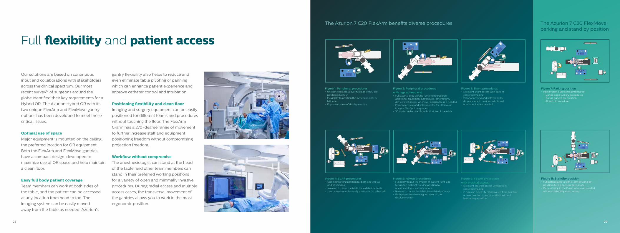
The Azurion 7 C20 FlexArm benefits diverse procedures The Azurion 7 C20 FlexMove parking and stand by position
Figure 1: Peripheral procedures• unrestricted access over full legs with C-arc
positioned at 135°• Flexibility to position the system on right or
left side• Ergonomic view of display monitor
Figure 4: EVAR procedures• Optimal working position for both anesthesia
and physicians• No need to move the table for sedated patients • Lead screens can be easily positioned at table side
Figure 2: Peripheral procedures with legs at head end • Full accessibility around foot end to position
additional equipment (ultrasound, atherectomy device, etc.) and/or whenever pedal access is needed
• Ergonomic view of display monitor for ultrasound images, FlexSpot images, etc.
• 3D tools can be used from both sides of the table
Figure 5: FEVAR procedures• Flexibility to put the system at patient right side
to support optimal working position for anesthesiologist and physicians
• No need to move the table for sedated patients • Both physicians have a good view of the
display monitor
Figure 3: Shunt procedures• Excellent shunt access with patient-
centered imaging• Ergonomic view of display monitor• Ample space to position additional
equipment when needed
Figure 6: FEVAR procedures with brachial access• Excellent brachial access with patient-
centered imaging • C-arm can be easily maneuvered from brachial
access position to aortic position without hampering workflow
Figure 7: Parking position• Park system outside treatment area:
• During open surgery procedures• During patient preparation• At end of procedure
Figure 8: Standby position• Full patient access with C-arm in stand by
position during open surgery phase• Easy to bring in the C-arm whenever needed
without disturbing room set-up
28 29

Increases clinical confidence Via the touch screen at the table, you can access clinically tailored 3D
acquisition protocols and advanced visualization and measurement
tools. These allow you to evaluate the type and extent of disease during
peripheral, aortic, visceral, arterial, and venous procedures with great
detail. Studies have shown that 3D CT-like imaging in the Angio lab can
enhance diagnostic accuracy, improve patient outcomes, and increase
procedural efficiency .
SmartCT- the next leap in simplifying and advancing 3D imaging to enhance interventional confidence
* The user level of expertise required is described in the Instructions for use as the Intended Operator Profile
82% think that the ease of using SmartCT will increase their utilization of 3D imaging in interventional procedures15
88% believe they can have more focus on their patient - thanks to full table side control with the touch screen module15
Provide superb care Increases clinical confidence for diverse
vascular procedures with advanced 3D
imaging, visualization and measurement
tools.
Optimize lab performance Easily control advanced 3D acquisition,
visualization and measurements at table side
to improve lab flexibility and efficiency.
Oustanding user experience Acquire 3D images and interact with all
SmartCT 3D features in a more natural and
effortless way.
Empowers you to easily adopt 3D imagingSmartCT allows any clinical user to perform 3D imaging with SmartCT,
regardless of their level of experience*:
• Easy room preparation to help position equipment and the Azurion
system for a 3D acquisition
• Easy protocol selection via pictorials
• Injection protocol suggestion based on literature
• Easy isocentering with visual feedback to confirm your field of view
position without using X-ray dose
• Easy 3D acquisition – you know when acquisition is completed and
you can release the push button or pedal
The Philips Image Guided Therapy clinical application software SmartCT, part of the Azurion image guided therapy platform, enriches our exceptional 3D interventional tools with clear guidance that is designed to remove the barriers to acquiring 3D images in the interventional lab.
30 31

SmartCT AngioSmartCT Angio generates a complete high-
resolution 3D visualization of cerebral,
abdominal, cardiac and peripheral
vasculature– all controlled via the touch
screen at the table. This can improve visibility
of tortuous or complex anatomy.
SmartCT RoadmapSmartCT Roadmap provides a live 3D image
overlay that can be segmented to emphasize
the targeted vessel and lesions, supporting
catheter navigation. The SmartCT Roadmap
overlays a 3D reconstruction of the vessel
tree, vessel segments, or annotations with live
fluoro images.
SmartCT Soft TissueSmartCT Soft Tissue generates a CT-like
visualization of soft tissue (cone beam CT) in
relation to other structures during procedures
– all controlled via the touch screen at
the table. You can use the CT-like images
to assess soft tissue, bone structure and
stent deployment before, during and after
interventional procedures.
Improve visibility of anatomy with clinically tailored acquisition and roadmap protocols
Benefits of CBCT during EVAR procedures
According to the 2019 guidelines for
abdominal aortic and iliac artery
aneurysms16, the use of cone beam CT
(CBCT), combined with a completion
angiogram has been shown to
be highly accurate in detecting
complications intra-operatively post
EVAR17. Intra-operative angiography
combined with cone beam CT for
completion assessment could possibly
replace the post-operative CTA17
VesselNavigator as guidance for stentgraft placement
Final check with SmartCT Soft Tissue: Deposition of contrast outside of the stentgraft, only visible on the late phase scan, showing a type 2 endoleak
First SmartCT: Early phase
Second SmartCT: Late phase
Merging the two scans clearly shows the late phase contrast filling outside the stentgraft
Patient:
• Male 80 years old
• Contrast volume SmartCT: 80 ml
(dilution 50:50)
• Flow: 12 ml/sec
• X-ray delay first run: 2 sec; Interval time
between the 2 runs: 15 sec
• DAP per SmartCT acquisition: 13 gy.cm2
Case: EVAR treatment check: Endoleak type 2 detection
Scan to viewfeature videos
32 33

System platformAzurion 3 F15, 5 C20, 7 C20,
7 C20 FlexArm
7 C20 FlexMove
ClarityIQ technology
Vascular productsSmartPerfusion
VesselNavigator
SmartCT Angio
SmartCT Roadmap
SmartCT SoftTissue
Xperguide
Vascular devicesIVuS
Visions PV
Pioneer Plus
Phoenix Atherectomy
Turbo-Power laser
Turbo-Elite laser
Turbo-Tandem catheter
Stellarex DCB
AngioSculpt
Integrated toolsCX50x Matrix ultrasound
Xper IM
IntelliSpace CV
DoseWise Portal
DoseAware
Integrated tables
Dedicated solutions to efficiently support your case mix
Azurion 7 C20 FlexArm • Staff and equipment positioning freedom• Improved workflow for multiple patient access points• Enhanced patient care due to reduced table and patient movement • Make efficient use of lab/OR space
Azurion 7 C20• Efficient workflow with Procedure Cards, Parallel Working and Checklists and Protocols• Broad portfolio of advanced workflow options like FlexVision Pro and TSM Pro• Full-body coverage• Superior imaging with ClarityIQ and MRC200+ X-ray tube
Azurion 7 C20 FlexMove • Positioning flexibility and clean floor• Easy full body patient coverage• Free Laminar Airflow field• Extended parking options
Azurion 3 F15 • Perfect fit for mixed application use in one room (cardiac/vascular) • Same user interface as the Azurion 7 series provides ease-of-use across the whole platform• Full-body coverage with table swivel• Advanced dose management with DoseWise and MRC200+ X-ray tube
Azurion 5 C20 • High performance ceiling mounted image guided therapy solution with
a 20" flat detector• Covers a wide range of cardiac and vascular procedures to offer flexibility for
multi-purpose use• Control all relevant applications via the central touch screen module at table side
Technology Maximizer
Plus
Technology Maximizer
Pro
Technology Maximizer Premium
Cardiac/Vascular
1 2 2
Learn more about Technology Maximizer
Increase value throughout your Vascular suite lifecycle
Stay clinically and operationally relevant with Technology Maximizer
To keep your Image Guided Therapy Suite state- of-art with regards to cyber security, clinical, and operational advancements, subscribe to IGT Technology Maximizer - Plus, Pro or Premium offer – for a standard duration of 4 years at point of sale.
Technology Maximizer secures all your eligible
Philips imaging equipment with the same technology
release level reducing maintenance complexity and
simplifying lifecycle management across hospital
departments. Maintain peace of mind with imaging
equipment that is always up to date, and enhance
patient care knowing you will always be first to take
advantage of technology innovations.
Standard offer Mid-level offer Premium offer
Azurion system SW version upgrade
State-of-the-art security
Latest available Operation System
Computer HW refresh to support software upgrade
New version of existing iApps
Future iApps in one clinical domain (Cardiac or Vascular)
Application training for new or enhanced functionality (days)
Future iApps in one clinical suite (Coronary, EP, SHD, Vascular, Neuro, Onco, Spine or Lung)
3534