vedic recitation and effects on brain
DESCRIPTION
An interesting research article describing the effects of systematic memorization of Vedas (Samhuta, Brahmanam etc) for a span of 8-10 years on the matter in brain. The authors find that gray matter density and cortical thickness is substantially different than a normal person. Let us see if this makes any sense.TRANSCRIPT

NeuroImage xxx (2015) xxx–xxx
YNIMG-12418; No. of pages: 12; 4C: 4, 5, 6, 7, 8
Contents lists available at ScienceDirect
NeuroImage
j ourna l homepage: www.e lsev ie r .com/ locate /yn img
Brains of verbal memory specialists show anatomical differences inlanguage, memory and visual systems
James F. Hartzell a,⁎, Ben Davis a, David Melcher a, Gabriele Miceli a, Jorge Jovicich a, Tanmay Nath b,Nandini Chatterjee Singh b, Uri Hasson a
a Center for Mind/Brain Sciences (CIMeC), University of Trento, 38060, Italyb National Brain Research Centre, Manesar, Gurgaon Dist., Haryana 122 050, India
⁎ Corresponding author at: Center for Mind/Brain Scie101, Mattarello, TN, Italy.
E-mail address: [email protected] (J.F. Hartzell).
http://dx.doi.org/10.1016/j.neuroimage.2015.07.0271053-8119/© 2015 The Authors. Published by Elsevier Inc
Please cite this article as: Hartzell, J.F., et al.,systems, NeuroImage (2015), http://dx.doi.o
a b s t r a c t
a r t i c l e i n f oArticle history:Accepted 8 July 2015Available online xxxx
Keywords:Cortical thicknessGray matter densityDiffusion tensor imagingLanguageMemoryPlasticityHippocampus
We studied a group of verbal memory specialists to determine whether intensive oral text memory isassociated with structural features of hippocampal and lateral-temporal regions implicated in languageprocessing. Professional Vedic Sanskrit Pandits in India train from childhood for around 10 years in an an-cient, formalized tradition of oral Sanskrit text memorization and recitation, mastering the exact pronunci-ation and invariant content of multiple 40,000–100,000 word oral texts. We conducted structural analysisof gray matter density, cortical thickness, local gyrification, and white matter structure, relative to matchedcontrols. We found massive gray matter density and cortical thickness increases in Pandit brains inlanguage, memory and visual systems, including i) bilateral lateral temporal cortices and ii) the anteriorcingulate cortex and the hippocampus, regions associated with long and short-term memory. Differencesin hippocampal morphometry matched those previously documented for expert spatial navigators and in-dividuals with good verbal working memory. The findings provide unique insight into the brain organiza-tion implementing formalized oral knowledge systems.
© 2015 The Authors. Published by Elsevier Inc. This is an open access article under the CC BY-NC-ND license(http://creativecommons.org/licenses/by-nc-nd/4.0/).
Introduction
A large body of research has established that acquisition of certainlong-term skill sets or knowledge is linked to plasticity in both greymatter (GM) and white matter (WM) in multiple cortical and subcorti-cal regions (May, 2011; Zatorre et al., 2004). As reviewed byMay (2011,see references within), various expert groups such as sportsmen,mathematicians, ballet dancers, and professional board-game playersall show particular morphological features that may be related tolearning and plasticity.
Our goal in the current work was to examine the potential impact ofextensive memorization and verbal recital practice on brain plasticity,as identifying brain regions implicated in these functions can elucidatethe functional capacities of both lateral and medial temporal regions,as detailed below. To investigate the potential impact of extensivememorization and verbal recital practice on brain plasticity we recruit-ed a sample group of traditional Sanskrit learners—Yajurveda SanskritPandits—whomemorize and recite one set of the most ancient Sanskrit
nces (CIMeC), Via delle Regole
. This is an open access article under
Brains of verbal memory sperg/10.1016/j.neuroimage.201
texts, the Vedas and their subsidiary texts (Vedāṅgas). The SanskritVedas are late bronze/early iron-age oral texts passed down for over3000 years in an unbroken tradition in India. They form the core ofthe ancient Sanskrit knowledge system, which developed extensiveoral and later written literature in a wide range of traditional subjectsstill taught in India's Sanskrit institutions using traditional oral memori-zation and recitationmethods (Rashtriya Sanskrit Sansthan, 2014). Pro-fessional Vedic Pandits undergo rigorous training in exactpronunciation and invariant content of these oral texts for 7 or moreyears, with 8–10 h of daily practice (totaling ~10,080 h over the courseof the initial training), starting in their childhood, and mastering multi-ple 40,000 to 100,000word oral texts (compared to ~38,000 in the bookof Genesis). The training methods strongly emphasize traditional face-to-face oral learning, and the Yajurveda recitation practice includesright hand and arm gestures to mark prosodic elements. After gradua-tion from training, professional Yajurveda Pandits work as teachers orVedic priests, with daily recitation reduced to ~3 h.
We note that while the ability of Yajurveda Pandits to performlarge-scale, precise oral memorization and recitation of VedicSanskrit texts may, prima facie, appear extraordinary or borderingon impossible, textual memorization and recitation are in fact standardpractice in traditional Sanskrit education in India (Rashtriya Sanskrit
the CC BY-NC-ND license (http://creativecommons.org/licenses/by-nc-nd/4.0/).
cialists show anatomical differences in language, memory and visual5.07.027

2 J.F. Hartzell et al. / NeuroImage xxx (2015) xxx–xxx
Sansthan, 2014).1 Thus, while the Pandit's memorization capacity mayappear unique to graduates of a Western educational system, it is oneof several memorization-related study traditions current in the Indiansubcontinent.
We had two predictions regarding brain systems possibly affectedby the intense memorization and recitation routine practiced by thePandits. First, we expected to see differences in cortical thickness orgraymatter density of lateral temporal regions. These form the core sys-tem for speech processing at the phonemic and syllabic level (Zhuanget al., 2014), with left hemisphere regions of the superior temporalplane (STP) likely sampling information at a higher rate matching thatof phonemic processing, and the right hemisphere STP sampling at alower rate matching syllable-level processing (Giraud and Poeppel,2012; Kotz and Schwartze, 2010; Morillon et al., 2012; Poeppel, 2003).Apart from their role in sublexical combinatorial processes, these re-gions also play a role in encoding sentential content tomemory. Activityin these regions predicts whether sentential content will be subse-quently remembered (a subsequent-memory effect, Hasson et al.,2007), and they show reduced activation for comprehension of repeat-ed auditory sentences (repetition suppression (RS); Dehaene-Lambertzet al., 2006; Devauchelle et al., 2009). Particularly, sentential RS effectsin these regions scale negatively with the temporal interval betweensentence repetitions (Hasson et al., 2006). Thus, extensive memoriza-tion of language content, coupled with memory for sentential contentcould affect the structure of these regions.
In addition, plasticity effects linked to memory practice have beendocumented in the human hippocampus, which is involved in boththe consolidation of prior experiences (e.g., Eichenbaum et al., 2007;Milner and Penfield, 1955; Scoville and Milner, 1957) and spatial navi-gation (e.g., Bird and Burgess, 2008; see also Eichenbaum and Cohen,2014). Hippocampal plasticity has been linked to spatial navigation ex-pertise, with greater posterior hippocampal volume and smaller anteri-or volume shown for expert urbannavigators (Maguire et al., 2000). Thehippocampus alsomediates verbalmemory (e.g., Fernandez et al., 1998;Grunwald et al., 1999), and is larger for individuals who perform betteron declarative memory tasks for verbal materials (e.g., Ashtari et al.,2011; Pohlack et al., 2014). Poppenk and Moscovitch (2011) showedthat better verbal memory for proverbs is related to greater posteriorand smaller anterior hippocampal volume, a pattern similar to thatseen for expert navigators. On the basis of this prior work we hypothe-sized that the intensivememorization demands of Pandit practicemightbe associated with changes to hippocampal volume or density.
To examine these issues, we studied a group of Pandits (N= 21) to-gether with closely matched controls. We examined cortical-level datavia voxel-based morphometry (VBM), cortical thickness (CT) and localgyrification index (LGI) analyses, and subcortical data via VBM and an-atomically defined regional measurements. We also evaluated whitematter data with diffusion tensor imaging (DTI) fractional anisotropy(FA) analysis, at a whole-brain level. The main purpose of the FA analy-sis was to evaluatewhetherWM changes would be found in the vicinityof areas linked to GM or CT differences. In particular, the frontal aslanttract (Catani et al., 2013) has been implicated in fluency and stuttering(Kronfeld-Duenias et al., 2014), as has the forceps minor in the anteriorcorpus callosum (Civier et al., 2015).
1 There are today in India around 150,000 students engaged in traditional Sanskrit stud-ies at approximately 5000 government and private institutions (Mishra, 1997; RashtriyaSanskrit Sansthan, 2010-2011; Rashtriya Sanskrit Sansthan, 2014; Pathashala, 2014).The topics (and texts memorized) at these institutions include Sanskrit literature, gram-mar, law, history, philosophy, astronomy, yoga, logic, and Vedas, subsidiary Vedic disci-plines, and Vedic commentary (Rashtriya Sanskrit Sansthan, 2014). There are in additionsome 246 registered Ayurvedic traditional medical colleges in India where some 50,000students memorize portions of Sanskrit core ("root") medical texts and subsidiary textsas part of their training (Central Council of Indian Medicine, 2014; Hartzell and Zysk,1995). Specifically for Vedic studies, there are currently an estimated 34,000Vedic Panditsin training in both government and non-government traditional Vedic schools (Shastri,2014; Pathashala, 2014; Rashtriya Sanskrit Sansthan, 2014; Mishra, 1997).
Please cite this article as: Hartzell, J.F., et al., Brains of verbal memory spesystems, NeuroImage (2015), http://dx.doi.org/10.1016/j.neuroimage.201
Methods
Participants
Forty-two male volunteers participated in the study conducted atthe National Brain Research Center in India. Twenty-one professionallyqualified Pandits were recruited from government-supported VedicPandit schools in the greater New Delhi (India) area. They underwentan extensive semi-structured interview prior to scanning to evaluatetheir extent of training, family history, current practice routines, multi-lingualism, handedness and eye dominance. Professional qualificationconstituted demonstrable mastery, i.e. complete memorization andfull recitation ability, of at least the ~40,000 word Yajurveda Saṃhitātext. All Pandits memorized part or all of one or more additional canon-ical texts (the length of these texts ranged from 1013 to 165,156 wordsbut we could not quantify precisely howmuch of these additional textswasmemorized by each Pandit). All began their training at an early age(M = 12.33, SD= 1.59, range 9–16), trained full-time for 7 years, for atotal of approximately 10,080 h, and continued training and reciting atreduced daily hours for additional years (M = 2.38, SD = 2.29, range0–8). From the interview reports, we estimated the total practicehours after competing the training (M = 11,141 h, SD = 27,196,range 2365–129,295). Note that Pandits enter training without any en-trance exams, so there is no pre-selection formemory or recital abilities,and the dropout rate from the study program is only around 5% (Shastri,2014). Thus, graduating the studies is not indicative of self-selection ei-ther prior to or during the studies themselves. Pandits had all eithergraduated from or were in the final year of professional Vedic Pandittraining, and all were self-rated as fluent in speaking, reading andwriting Sanskrit. None of the Pandits in our participant group camefrom traditional family lineages of reciters (see SI Methods). SeeSupplementary Information (SI Methods and SI Table 1) for additionalPandit demographics and practice specifics.
Twenty-one control volunteers were recruited to match the Panditpopulation in gender, age (Mpandits = 21.7; SD = 2.8 vs. Mcontrols =22.8; SD = 3.6, T-test, P = .3) and number of languages spoken(Mpandits=3.1; SD=0.8 vs.Mcontrols=3.1; SD=1.3, T-test, P=.9). Par-ticipants in the control group were members of India's National BrainResearch Centre community or students from a nearby technical col-lege. All volunteers were right-handed, right-eye dominant, with noleft-handed parent or sibling (Knecht et al., 2000). Multilingualismand handedness/eye-dominance were assessed by culturally-adaptedHindi versions of the Penn State Language History Questionnaire (v.2;Li et al., 2006), and Edinburgh Handedness questionnaire (Oldfield,1971). (Adaptations and translations by N.C.S., T.N., J.H, and a fourthnative Hindi/English speaker). The protocol was approved by India'sNational Brain Research Centre Ethics Committee and all participantsprovided written informed consent.
Image acquisition
Two T1-weighted 3D-MPRAGE sequences were acquired for eachparticipant on a Philips Achieva 3 T scanner with an 8-channel head re-ceive coil (FOV 256 × 256 × 176 mm, voxel size 1x1x1mm), TE 3.2 ms,TR 934 ms, flip angle 9°, 176 sagittal-oriented slices, acceleration 2(sense), total acquisition time 06:49.8. Image quality was evaluatedimmediately after each structural acquisition to control for motion ef-fects or other artifacts. The two structural images of each participantwere aligned using FSL's 4.1.8 FLIRT (Jenkinson et al., 2002; Jenkinsonand Smith, 2001), and averaged to increase signal-to-noise ratio.Image intensity non-uniformities were corrected in AFNI (Cox, 1996).The resulting mean structural image was used for all subsequentanalyses. Diffusion data were acquired for a subset of 15 Pandits and15 controls using single-shot EPI during the same MRI session (FOV256 × 256 × 128 mm3, voxel size 2 × 2 × 2 mm3), TE 75 ms, TR8000 ms, flip angle 90°, 64 transverse slices, slice thickness 2 mm, fat
cialists show anatomical differences in language, memory and visual5.07.027

3J.F. Hartzell et al. / NeuroImage xxx (2015) xxx–xxx
suppression, matrix 218 × 126, 60 diffusion encoding directions(bvecs), b-value = 700 mm2/s, 10 b0 volumes (saved as a single aver-aged volume), parallel imaging with acceleration factor 2 (sense),total acquisition time 10:59.6. Diffusion data was evaluated immediate-ly upon acquisition to control for motion effects or other artifacts, andre-acquired if necessary (4 scans were reacquired). The b-value of 700was chosen to be within the range of values considered optimal forhuman brain matter DTI analysis while favoring high SNR to facilitatethe detection and correction of artifacts in the diffusion weightedimages (Alexander and Barker, 2005; Ben-Amitay et al., 2012). MeanFA of the corpus callosum body (~0.52) matched values reported inthe literature (Jovicich et al., 2014; see their Fig. 6).
Voxel-based morphometry (VBM)
Structural images were analyzed using the FSL's voxel-basedmorphometry (VBM) analysis pipeline (Ashburner and Friston,2000; Good et al., 2001) with FSL-VBM tools (Douaud et al., 2007).Data consisted of the 21 aligned and averaged structural images ac-quired from Pandits and 21 from the control group. Brains were ex-tracted using FSL's brain extraction tool (BET; Smith, 2002), withmanual edits to control for extraction errors, and processed usingFSL's VBM default pipeline. Note that in the FSL VBM pipeline, thesingle-participant data prior to alignment to common space reflectsa voxel's probability of being gray matter (calculated by a combina-tion of Hidden Markov Random Field and Expectation Maximizationframework; see Zhang et al., 2001), and the final data, in commonspace, reflect an adjustment of that value by the Jacobian of the de-formation applied to the participant's data when aligning to com-mon space. Thus, this VBM implementation most closely reflectslocal volume differences. We spatially smoothed the final imagesby an isotropic Gaussian kernel (FWHM = 9.42 mm). Group-levelstatistical inference was achieved via nonparametric permutationusing the FSL tool randomise. Family-wise error was controlled forat an alpha level of P b .05 by Threshold-Free Cluster Enhancement(TFCE; Smith and Nichols, 2009), in which cluster extent isconstrained by cluster-like local spatial support. Age and wholebrain Volumewere included as covariates. References to anatomical-ly defined regions withinMNI space were established by intersectingthe group's MNI gray matter template mask with FSL's pre-definedatlases. (See SI Methods for additional information.) To evaluatethe impact of smoothing kernel, we also implemented Gaussian ker-nels of 2.35 mm, 4.71 mm, and 7.06 mm (sigma of 1, 2 and 3, respec-tively) and repeated the main analysis.
Cortical thickness analysis
Cortical thickness (CT) analysis was implemented in FreeSurfer(Dale et al., 1999), using the default processing pipeline, except formanually bypassing FreeSurfer's automatic skull stripping routinesand using instead the skull-stripped brains created in the initial stepof the VBM analysis described above. FreeSurfer's GM segmentationwas verified manually for each participant, and no manual correctionswere needed (for example participant's segmentation, see Inline Sup-plementary Figure S1). The CT estimates derived for each participantwere imported into AFNI's surface-based analysis module, SUMA(Saad et al., 2004) for further analyses. CT values were spatiallysmoothed with a conservative (Pardoe et al., 2013) 10 mm smoothingkernel on the two dimension cortical surface using an iterative HeatKernel method (Chung, 2004). The resulting CT values on the corticalsurface were interpolated to a surface mesh that maintained the samenumber of vertices for all participants, in similar locations (usingSUMA's MapIcosahedron procedure). The resulting meshes contained156,252 vertices per hemisphere. Statistical analysis of CT values onthe group level was performed using cluster-based thresholding that
Please cite this article as: Hartzell, J.F., et al., Brains of verbal memory spesystems, NeuroImage (2015), http://dx.doi.org/10.1016/j.neuroimage.201
was determined via a permutation procedure (following Nichols andHolmes, 2002; see SI Methods for details).
Inline Supplementary Fig. S1 can be found online at http://dx.doi.org/10.1016/j.neuroimage.2015.07.027.
Local gyrification index analysis
To examine potential gyrification differences between the two pop-ulations we used a method based on calculating an ‘outer surface’ (tan-gential to the folding points of the gyri), and then parcellating it intonumerous circular patches covering the entire 2D cortical surface(Schaer et al., 2008, 2012). For each patch, the local gyrification index(LGI) computes the ratio of cortex within sulcal folds to the amount ofvisible cortex (tangent to the patch). Higher values indicate that a great-er proportion of the pial matter under the patch is in sulci. The 2D sur-face maps generated by this method have, by definition, a strongdegree of spatial smoothness (FWHM of ~30 mm) that is determinedby the number of surface patches used. (Each patch has a radius of20 mm, and the computed LGI value for each patch is propagated toall surface vertices overlapping with it, necessarily yielding less local-ized results than those seen for VBM or CT analyses.) Between-groupstatistical tests of LGI patterns were based on permutation tests as forthe CT analysis. Permutation tests maintain the spatial autocorrelationof each participant's data and permit sensitivity to non-stationarychanges in LGI across cortical regions.
Diffusion tensor imaging: fractional anisotropy
The 60 diffusion-encoding direction b-vectors were corrected indi-vidually for headmotion using FSL's rot_bvecs, followed by eddy currentand subjectmotion correctionwith affine registration to the averaged b0image. Fractional Anisotropy (FA) imageswere created using FSL's Diffu-sion Toolbox (FDT) after brain-extraction using BET and manual edits toremove artifacts, then processed using FSL's Tract Based Spatial Statistics(TBSS; Smith et al., 2006) default settings. FSL's TBSS first erodes eachparticipant's FA image. For registration to common space,we used an op-tion that selects the best target image fromamong the subjects, performsa nonlinear alignment of all participants to that target, and then affineregisters the resulting aligned files to MNI152 1 mm common space.Using the mean FA calculated from the participants' files in commonspace, TBSS creates a skeletonized representation of FA-derived tractscommon to all subjects, by estimating the local surface perpendicular di-rection along the tracts and performing “non-maximum-suppression”along the perpendicular to the voxel with the highest FA value, whichmarks the center of the tract. The distance of each participant's FAvoxel to this common skeleton is then calculated, with the distance cal-culation constrained to the nearest voxels, and the participant's maxi-mum FA value in the already-calculated perpendicular to each skeletonvoxel is projected into the skeleton. The aim of this method is to reducevariance from residual misalignments of each subject's FA to commonspace (Smith et al., 2006). Voxelwise cross-participant group-level statis-tics are then performed within a thresholded mean FA skeleton mask(we used a threshold of 0.3). The threshold reduces the effects of highinter-subject variability at the outer edges of the brain. We tested be-tween group differences using 2D TFCE, controlling for family-wiseerror at an alpha level of P b .05 based on cluster extent constraints,with Age included as a covariate. References to anatomically definedwhite-matter regions withinMNI spacewere established by intersectingthe group's MNI template FA mask with FSL's predefined WM atlases(see SI Methods).
Hippocampal region-of-interest analysis
Hippocampus-optimized VBMWe also conducted a customized VBM analysis that was aimed di-
rectly at evaluating changes in the HF. This analysis consisted of the
cialists show anatomical differences in language, memory and visual5.07.027

4 J.F. Hartzell et al. / NeuroImage xxx (2015) xxx–xxx
following steps. First, the initial automatic segmentations of the HF asderived by FSL's FIRST subcortical alignment and segmentation proce-dures (Patenaude et al., 2011) were anonymized and then further man-ually evaluated andmodified by one of the authors (J.H.). Segmentationwas performed in original space, using advanced FIRST options to opti-mize the segmentation by algorithm-determined vertex numbers(modes) and internal reference to the thalamus for normalization. Inthe second step, we performed a nonlinear registration of these editedHF segmentations to MNI space (FSL's MNI152 T1 1 mm template)using high-resolution (6 mm3) nonlinear warping (FNIRT) initializedwith the affinematrix generated for each participant by FSL FIRST's sub-cortical alignment routine. After registration we multiplied the hippo-campi by their Jacobians to modulate the GM, as in the standard VBMpipeline. Note that in contrast to the whole-brain analysis (whichworks with GM probabilities), the values multiplied by the Jacobianwere the original T1 intensity values within the manually verified hip-pocampal segmentations. Steps 1 and 2 therefore provided a more pre-cise inter-participant alignment of the HF specifically. Third, to evaluatethe impact of various smoothing kernels (2.35 mm, 4.71 mm, 7.06 mmand9.42mm, sigma1, 2, 3 and 4 respectively),we smoothed onlywithintheseMNI-registered right and left HF. Steps 1, 2 and 3 ensured that ourbetween group tests focused only on the HF, thus obviating the chanceof impacting the results fromnearby regions. Thenwe performed voxel-wise tests inside the right and left hippocampal intersection masks (i.e.the hippocampal masks used in the randomize routine included onlyvoxels to which all 42 subjects contributed values). We used TFCE test-ing, and included Age and whole-brain Volume as covariates at all foursmoothing kernels. To evaluate the impact of usingmanually annotatedhippocampi, we compared the results to those obtained when applyingthe same registration and analysis pipeline but using as inputs the FIRSTautomatic hippocampi segmentations produced in Step 1 above, as wellas automatic hippocampi segmentations obtained from FreeSurfer forthese participants.
Hippocampal local-volume analysisWe conducted an additional analysis to identify whether there were
areas of the hippocampuswhose local volume differed between groups.The method was based on FSL FIRST's vertex analysis (Patenaude et al.,2011), but modified to allow incorporation of manual edits on the hip-pocampal structure (following suggestions by Jenkinson, 2014). Thisanalysis was not based on comparison of mesh-based segmentationsof the hippocampus but rather on comparisons of the outer envelopeof participants' hippocampi in common space. First, using the manually
Fig. 1. Surface projection of areaswhere Pandits showed greater graymatter density/volume (GMhere and all other analysis corrected for family-wise error at P b .05, using FSL's TFCE cluster-e
Please cite this article as: Hartzell, J.F., et al., Brains of verbal memory spesystems, NeuroImage (2015), http://dx.doi.org/10.1016/j.neuroimage.201
annotated hippocampal segmentations from FIRST, we constructed acommon core hippocampal ‘shape’ from the entire group of participantsin common space (Pandits and controls). To this end, the individual hip-pocampal shapes from original space were projected to common space(MNI152 T1 1 mm) using a rigid body alignment to maintain size andshape differences. From the group average of these MNI-registered hip-pocampal shapes we then constructed a thresholded (0.9) group aver-age boundary mask (this mask marks the outer edge of the commonHF volume, in 3D space). For each voxel in this group-level boundarymask we then calculated its distance to the nearest boundary voxel ofeach participant's binarized hippocampal mask, whether inside or out-side of the common boundary mask. This returned, for each group-level boundary-mask voxel, a vector reflecting the positive or negativedistance to each participant's boundary voxel. Group-level tests wereconducted on this ‘signed distance’ data. The result of this procedure,when applied to all participants,was a group-level statisticalmap show-ing those parts of the group-hippocampal boundary shape where(local) distances to the shape differed between the two groups. Notethat as opposed to VBM this procedure implemented a strictly “localshape” analysis that (similarly to FSL's new vertex analysis) identifiesgeometric changes, is independent of any tissue-classification step,and does not involve any smoothing of the data.
Results
Evaluation of covariates
The VBM, CT, LGI and FA analyses included whole brain analyses forthe Pandit group examining correlations of two covariates. These in-cluded Starting age of recitation training, and “Overall Practice Hourssince Completion of Training” (OPHCT). OPHCT was included since, al-though all Pandits completed the common training, therewas consider-able variance in their subsequent practice routines, and it has beenshown that even short-term cognitive and motor practice impactsneuroplasticity (e.g., Draganski et al., 2006; Driemeyer et al., 2008).None of the pair-wise correlations between Age, Start Age, and OPHCTapproached significance (Correlation tests: Start Age and Age: R =0.23, P = .39; OPHCT and Age: R = 0.06, P = .7; Start Age andOPHCT: R = 0.22, P = .32). Because age and whole brain volume arealso known to correlate with changes in GM, we included Age and Vol-ume as additional covariates in all analyses, including the between-group tests, with the exception of the CT, LGI, and FA analyses, wherewe used only Age as a covariate.
) than controls as indicated by awhole brain Voxel BasedMorphometry analysis. Analysisxtent correction (see SI Tables 2 and 3 for cluster details).
cialists show anatomical differences in language, memory and visual5.07.027
5J.F. Hartzell et al. / NeuroImage xxx (2015) xxx–xxx
Voxel-based morphometry: whole brain analysis
The whole-brain VBM analysis revealed extensive GM differences incortical, cerebellar and subcortical regions. In cortical regions Panditsdemonstrated greater GM than controls in large portions of both leftand right hemispheres (10.4% left and 12.5% right of total GM templatecortical volume). To facilitate presentation, differences found in corticalregionswere projected to an inflated cortical surface representation of abrain in MNI space (Fig. 1; see SI Tables 2 and 3 for complete cluster de-scriptives and local maxima). Differences were found bilaterally in bothauditory and visual-stream regions, including lateral temporal cortices,ventral occipital cortices, angular gyri, pre- and post-central gyri, poste-rior cingulate, lingual gyri and precuneus. Greater Pandit GM was alsofound in large bilateral areas of the anterior cingulate (ACC) and ventro-medial prefrontal cortices (vmPFC).We repeated the VBM analysis withspatial smoothing kernels of 2.35 mm, 4.71 mm, and 7.06 mm. Theresulting statistical maps were almost identical, apart from an addition-al single cluster in the base of left STG, MTG that was only found for the4.71mmsmoothing kernel (see SI Table 6 for additional smoothing ker-nel cluster specifics).
Within right lateral temporal cortex a large GM cluster was foundthat reached along the superior temporal sulcus (STS) into the STP,encompassing both the lateral transverse temporal gyrus and associa-tion cortices and extending deep into the ventral anterior temporal re-gion. Pandits' GM was also larger in the right posteromedial insula andcentral operculum, the anterior and posterior parahippocampal gyrus
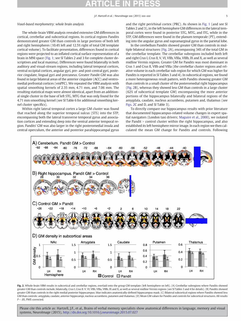
Fig. 2. Whole-brain VBM results in subcortical and cerebellar regions, overlaid onto the groupgreater GM than controls include, bilaterally, Crus I, Crus II. V, VI, VIIb, VIIIa, VIIIb, IX and X, as wgreater GM than controls in the right medial posterior hippocampus; blue indicates anatomicalGM than controls: amygdala, caudate, anterior hippocampi, nucleus accumbens, putamen and tP b .05, FWE-corrected.
Please cite this article as: Hartzell, J.F., et al., Brains of verbal memory spesystems, NeuroImage (2015), http://dx.doi.org/10.1016/j.neuroimage.201
and the right perirhinal cortex (PRC). As shown in Fig. 1 (and see SITables 2 and 3), in the left hemisphereGMdifferences in the lateral tem-poral cortex were found in posterior STG, MTG, and ITG, while in theSTP, GM differences were found in the planum temporale (PT), extend-ing into the angular gyrus and supramarginal gyrus in the parietal lobe.
In the cerebellum Pandits showed greater GM than controls in mul-tiple bilateral structures (Fig. 2A), encompassing 34% of the total GM inthe cerebellar template. The cerebellar subregions included both leftand right Crus I, Crus II, V, VI, VIIb, VIIIa, VIIIb, IX and X, aswell as severalmidline Vermis regions. Greater GM for Pandits was most dominant inCrus 1 and Crus II, VIIb and VIIIa (the cerebellar cluster regions and rel-ative volume in each cerebellar sub-region forwhich GMwas higher forPandits is reported in SI Tables 3 and 4). In subcortical regions, we founda more heterogeneous result pattern, with Pandits showing greater GMthan controls in a small cluster of the posteromedial right hippocampus(Fig. 2B), whereas they showed less GM than controls in a large cluster(62% of subcortical template GM) encompassing the more anteriorportions of the hippocampus bilaterally and bilateral regions of theamygdala, caudate, nucleus accumbens, putamen and, thalamus (seeFigs. 2C and D, and SI Table 3).
To directly compare our hippocampus results with prior literaturethat documented hippocampus-related volume changes in expert spa-tial navigators (London taxi drivers; Maguire et al., 2000), we isolatedthe Pandit N control cluster within the right hippocampus, and alsoestablished its left hemispheremirror image. In each regionwe then cal-culated the mean GM change for Pandits and controls. Following
GM template (left hemisphere on left). (A) Cerebellar subregions where Pandits showedell as several midline Vermis regions (see SI Tables 3 and 4 for details). (B) Pandits showedly-defined hippocampusmask. (C) Bilateral subcortical regions where Pandits showed lesshalamus. (D)Mean GMvalues for Pandits and controls for subcortical structures. All results
cialists show anatomical differences in language, memory and visual5.07.027

Fig. 3. Gray matter differences in hippocampi as indicated by a Whole Brain Voxel BasedMorphometry analysis. Pandits showed less gray matter than controls in bilateral anteriorhippocampal formation (left) andmore graymatter than controls in a rightmiddle-posteriorhippocampal formation cluster (right). * = P b .05, *** = P b .001.
6 J.F. Hartzell et al. / NeuroImage xxx (2015) xxx–xxx
Maguire et al (2000), mean GM was also calculated for the anterior as-pects of the hippocampus that fell within the large cluster where Pan-dits showed lower GM than controls. Fig. 3 bears out the greaterdensity for controls in the anterior hippocampus, which is markedly ab-sent, and even reversed, in the right mid-posterior hippocampus. (Notethat diverging from our analysis, Maguire et al. did not include Age ascovariate in the between-group tests, and doing the same in the currentstudy revealed even stronger similarities to their findings; see SI Discus-sion and SI Fig. 2 for visual comparisons). We then evaluated whetherthese conclusions about the hippocampus would hold up if the wholebrain VBM analysis was repeated at different smoothing kernels. Theanterior hippocampal results (Pandits b controls) survived tests at addi-tional FWHM Gaussian smoothing kernels of 2.35 mm, 4.71 mm, and7.06 mm (sigma of 1, 2 and 3, respectively), while the right posteriorhippocampus result (Pandits N controls) survived at the additionalGaussian kernel of 7.06 mm (sigma 3). We also conducted a wholebrain analysis within the Pandit group to test whether GM density cor-related with Start Age or with total post-training hours of Pandit recita-tion practice (OPHTC), both with Age and total brain Volume ascovariates. We found no significant correlations.
Hippocampus-focused analyses
Given that the hippocampal data in thewhole-brain analysismay re-flect the impact of imperfect alignment or smoothing of data from out-side the hippocampus, we implemented two additional analyses to
Fig. 4. Hippocampal region-of-interest analysis: areas within the right and left hippocampi whconducted a region-of-interest analysis using subcortex-optimized nonlinear alignment toMNI1right hippocampus, independent of spatial smoothing kernel. Greater GMdensity for Pandits waskernels. Statistical maps for both right and left hippocampus shown at 7.06 mm smoothing kern
Please cite this article as: Hartzell, J.F., et al., Brains of verbal memory spesystems, NeuroImage (2015), http://dx.doi.org/10.1016/j.neuroimage.201
better study hippocampal differences between the groups. Both analy-ses considered the hippocampus as a region of interest, and examinedVBM and local-volume changes in a more circumscribed manner. Theimplementation details of these analyses are described in the Methods.In brief, in both analyses we used accurate hippocampal segmentationsin original space, obtained from FSL's automatic subcortical segmenta-tion (FIRST; Patenaude et al., 2011), which were then further manuallyannotated. For the VBM analysis we implemented a high-resolutionalignment to common space, optimized for subcortical structures. Weused the Jacobians of the deformation to common space in order tomodulate intensity values within each person's hippocampus. For thelocal-volume analysis we implemented a procedure similar to FSLFIRST's vertex-based subcortical shape analysis. This analysis wasbased on 3 main steps: i) aligning participants' hippocampi to commonspace, ii) producing a ‘consensus shape’ of hippocampal areas whereparticipants overlapped, and iii) quantifying, for each point on the con-sensus shape's boundary, its distance to the nearest boundary of eachperson's hippocampus. (This analysis is identical to FSL FIRST's vertex-wise local distance calculations, but uses boundaries in voxel space rath-er than derived 2D meshes). Using this procedure we could determine,for each point on the consensus shape boundary, whether the twogroups differed in local volume. In contrast to VBM, this analysis is im-mune to any spatial smoothing effects, and reflects strictly local volumedifferences.
The hippocampal-optimized VBM procedure indicated a large por-tion of the posterior-middle right HF where Pandits had greater GMthan controls (see Fig. 4, and see Supplementary Table 7 for cluster spe-cifics). The volumeof this region formed between 73 and 98%of the hip-pocampal mask (depending on smoothing kernel; FWHM 2.35 mm =73%, FWHM 4.71 mm = 80%, FWHM 7.06 mm = 92%, FWHM9.42 mm = 98%; note that smoothing was implemented only withinthe hippocampal mask, thus obviating effects of nearby regions). Atlarger smoothing kernels (7.06mmand 9.42mm),we also found a clus-ter in the left posterior hippocampus where Pandits had greater GMthan controls.
The hippocampal shape analysis revealed a portion of the right mid-anterior hippocampus with greater volume for the control group (seeInline Supplementary Figure S2). We then tested, within the Panditgroup, whether hippocampal GM density or shape correlated with Pan-dit Start Age or with total post-training hours of recitation practice(OPHTC), both adjusted for Age and total brain Volume as covariates.We found no significant correlations.
Inline Supplementary Fig. S2 can be found online at http://dx.doi.org/10.1016/j.neuroimage.2015.07.027.
Cortical thickness analysis
Several brain regions differed in CT between the Pandit and controlgroup, and in all cases the Pandit group was associated with greaterCT. Differences were found in right STS, right anterior temporal pole,
ere Pandits showed greater GM than controls. In addition to the whole brain analysis, we52 T1 1mm common space. Greater GM density for Pandits was found in the mid-posteriorfound in the posterior left hippocampus, but onlywhen using 7.06 and 9.42mmsmoothingel, overlaid on MNI152-T1 1 mm template.
cialists show anatomical differences in language, memory and visual5.07.027

Fig. 6. Areas where Pandits showed less gyrification than controls.
Fig. 5.Areaswhere Pandits demonstrated greater cortical thickness than controls. (A) Singlevertex significance value set at uncorrected threshold of P b .05, corrected for family-wiseerror using cluster-extent thresholding. (B) Single vertex significance value set at uncorrect-ed threshold of P b .005, corrected for family-wise error using cluster-extent thresholding(see Methods). LH = left hemisphere; RH = right hemisphere.
7J.F. Hartzell et al. / NeuroImage xxx (2015) xxx–xxx
right occipito-temporal gyrus (OTG) and in the left rostral ACC extend-ing into dorsomedial prefrontal cortex. Fig. 5 presents these regions asidentified by two analyses, using two single voxel thresholds to identifyboth less localized clusterswhere all voxels passed the P b .05 threshold,and more highly localized clusters where all voxels passed a thresholdof P b .005. We conducted a whole brain analysis to test, within thePandit group, whether CT correlated with Start Age or total post-training hours of recitation practice (OPHTC), adjusted for Age as covari-ate. We found no significant correlations.
Differences in local gyrification
Two areas showed differences in local gyrification between the twogroups. Thesewere found in the inferior andmiddle occipital gyri on theleft and middle occipital gyrus on the right. In both cases these corticalregions showed reduced gyrification for the Pandit group (see Fig. 6).
We also examined the relationship between the LGI and CT findings.Using the regions identified by the LGI analysis as masks, we quantifiedthe mean CT within those regions per participant, and then evaluatedthese on the group level. Therewas absolutely no between-group differ-ence in mean CT within those regions. In the right hemisphere LGI clus-ter, the mean CT for Pandits and controls was 2.65 mm (SD= 0.18) vs.2.66 mm (SD = 0.21). In the left hemisphere cluster, the values were2.08 mm (SD = 0.13) vs. 2.11 mm (SD = 0.13). In short, CT valueswere almost identical across groups in areas showing LGI differences.We also tested, within the Pandit group, for correlation of LGI with the
Please cite this article as: Hartzell, J.F., et al., Brains of verbal memory spesystems, NeuroImage (2015), http://dx.doi.org/10.1016/j.neuroimage.201
Start Age or Practice (OPHTC) variables adjusted for Age as covariate.There were no significant correlations.
Differences in fractional anisotropy
Two adjacent clusters showed greater FA in Pandits compared tocontrols (see Fig. 7). No area showed the reverse pattern. The clusterswere found in close proximity to the CT and GM differences we reportfor the left vmPFC/ACC (see Fig. 7), at the intersection of the left anteriorthalamic radiation, the forceps minor, the left inferior fronto-occipitalfasciculus (IFOF), the left anterior corona radiata (ACR), the genu ofthe corpus callosum, the left cingulum bundle, and the left uncinate fas-ciculus (UF). (See SI Methods, SI Table 5 for cluster details, and SI Fig. 1for a brain map showing the location of these clusters overlaid onmeangroup FAmap.)We also tested, within the Pandit group, whether eitherFA or skeletonized FA correlated with Pandit Start Age or with totalpost-training hours of recitation practice (OPHTC), both adjusted forAge as covariate. We found no significant correlations.
Discussion
Overall, we found considerable differences in the organization of thebrains of professional Vedic Sanskrit Pandits. Specifically, they showedextensive cortical and cerebellar GM increase and subcortical GM de-crease. The hippocampal GM differences followed a differential anteri-or/posterior pattern that has been linked to expert spatial navigation(Maguire et al., 2000), and to improved memory for verbal materials(Poppenk and Moscovitch, 2011). Cortical CT increases were extensive,and overlapped closely with GM differences in right temporal regions,left medial prefrontal, and left fusiform areas. Pandits also showed sig-nificantly less gyrification in bilateral occipital regions, and significantlylarger FA in left inferior frontalWM clusters. Our findings are consistentwith the possibility that the changes to medial-temporal and medialprefrontal regions, accompanied by changes to lateral temporal regionsand cerebellum, reflect the impact of the Pandits' extensive verbalpractices.
Hippocampus and ACC/mPFC
The Pandits' pattern of hippocampal differences as evident in awhole-brain VBM analysis were similar to those reported in the studyof London taxi drivers (Maguire et al., 2000), showing a relative de-crease in bilateral anterior hippocampi, and an increase in right (butnot left)medial-posterior hippocampus. Our region-of-interest analysesidentified a local reduction in volume in the right anterior HF forPandits, accompanied by a VBM signature of increased GM in themedial-posterior right HF for this group, and an increased GM clusterin the posterior left hippocampus. Maguire et al (2000, p. 4398), whoused whole brain VBM and HF pixel counting, suggested that the in-creases in the posterior hippocampus may indicate that this regionstores a spatial representation for the environment and expands to ac-commodate this elaborated representation. A large body of subsequentresearch has shown, however, that the anterior and posterior
cialists show anatomical differences in language, memory and visual5.07.027

Fig. 7. Axial slices showing clusters (in green) where Pandits showed greater fractional anisotropy than controls, The statistical fractional anisotropy map (green) is overlaid on an MNItemplate, and shown alongside areas of themPFC/ACCwhere Pandits showed greater GM than controls in thewhole-brain analysis (red-orange), P b 0.05, FWE-corrected using ThresholdFree Cluster Enhancement. Left hemisphere shown on left.
8 J.F. Hartzell et al. / NeuroImage xxx (2015) xxx–xxx
hippocampi play differential roles in a large range of cognitive processesincluding, but not limited to novelty processing (Daselaar et al., 2006;Kohler et al., 2005; Takashima et al., 2006), encoding of ongoing and re-cent experiences (Hartzell et al., 2014), and simulation of future events(van Mulukom et al., 2013; see Fanselow and Dong, 2010; Poppenket al., 2013, and Strange et al., 2014 for reviews). Bettermemory for ver-bal materials has been associated with larger posterior and smaller an-terior hippocampal segments (Poppenk and Moscovitch, 2011). Onestudy found that the volume of the anterior hippocampus correlatespositively with verbal memory (Hackert et al., 2002), but this wasfound for an age group (60–90 y.o.a.) for which the relation may reflectvariations in the normal thinning patterns that the HF undergoes withincreasing age. Our results, taken together with these prior studies, sup-port the developing evidence that hippocampal regional changes mayoccur in various situations, beyond those necessitating memory forcomplex spatial scenes.We note that the training of London TaxiDriversdoes in fact involve rotememorization of a large volumeof preset verbalsequences: they are required to memorize street names and placenames (30,000 landmarks) in 320 set route sequences totaling~120,000 words, with part-time training over ~3–5 years(Transport.for.London, 2014). Their oral examinations necessitate pre-cise rote verbal recall of route details between the landmarks.
Greater Pandit GM/CT in anterior cingulate cortex and medial tem-poral structures is also consistentwith accommodating increasedmem-ory demands. Animal studies show long-term memory encoding in themPFC/ACC (Weible et al., 2012; Teixeira et al., 2006), with short-termencoding in the hippocampus (Takehara-Nishiuchi and McNaughton,2008), mediated by connections between perirhinal/parahippocampusand ACC (Insel and Takehara-Nishiuchi, 2013). In humans, patientswith exclusive MTL lesions perform normally on remote autobiograph-ical memory but poorly on recent memory tests (Bayley et al., 2005),while mPFC/ACC lesions conversely disrupt long-term memory, butnot short-term memory for recent experiential learning (Squire andBayley, 2007). Neuroimaging data from healthy human participantsalso suggest that recall for recent vs. remote experiences differentiallyrelies, respectively, on hippocampal vs. medial frontal cortices(Takashima et al., 2006). Taken together with these animal andhuman studies, our findings suggest that Vedic Sanskrit oral text infor-mation may be initially encoded via the hippocampus, then stored inthe mPFC/ACC regions, but a detailed longitudinal study is necessaryto examine this issue.
Lateral-temporal and parietal cortices: potential indicators of languagesystem differences
Our left and right temporal region cortical differences showeddiffer-ent topographies. The left postero-medial superior, middle, and inferiortemporal gyri GM patterns were largely confined to gyral surfaces,reaching into the antero-medial PT. Many of these regions overlap
Please cite this article as: Hartzell, J.F., et al., Brains of verbal memory spesystems, NeuroImage (2015), http://dx.doi.org/10.1016/j.neuroimage.201
with presurgical speech interference sites (Roux et al., 2012), suggestingthe observed differencesmay be related, at least in part, to recitation vo-calization. These left posterior lateral temporal regions are also implicat-ed in both lexical-phonological processing and semantic-syntacticintegration in current cortical speech processing models (seee.g., Hickok and Poeppel, 2007; Friederici, 2012), while the PT/pSTG/SMG changes reach into areas linked to speech production (DeWittand Rauschecker, 2012; Fedorenko and Thompson-Schill, 2014). Onthe right, greater GM/CT for Pandits reached into deep STS, and into lat-eral Heschl's gyrus/planum polare (HG/PP), dorsal posteromedialinsula, OP2/OP3 of posteromedial operculum, and right ventral anteriorlobe (vATL). Right HG/PP have been shown to sample acoustic informa-tion at a rate optimized for syllable-length acoustics (Kotz andSchwartze, 2010; Morillon et al., 2012; Altmann et al., 2007) andsound patterns (Altmann et al., 2007), with right STS linked to process-ing of human voices (Belin, 2006) and vocal identity (Petkov et al.,2009). The human vATL/anterior fusiform bilaterally is considered ahub for multi-modal/amodal semantic knowledge (Chan et al., 2011),linked with PRC for verbal memory construction (Bozeat et al., 2000).Greater Pandit GM in right posteromedial insula and operculummay re-flect speech-sound processing (Cloutman et al., 2012), vocalizationtuning (Remedios et al., 2009), and/or prosody detection (van Rijnet al., 2005).
The increased GM for Pandits in parietal regions suggests the possi-ble involvement of cortical resources subserving Vedic recitation ges-tures, articulation, and multilingualism. Differences in the left superiorand medial postcentral gyrus covered portions of the primary somato-sensory cortex (Ruben et al., 2001) for the right arm, wrist, hand andfingers, face, mouth and tongue regions (Kaas et al., 1979; Nakamuraet al., 1998), including areas known to be active during right hand andarm movement (Sereno and Huang, 2014). We also considered thatwhile the Pandits and controls were matched for number of languages,the Pandits are highly competent in Sanskrit due to their training, andseveral of the areas where they demonstrate greater GM have beenlinked to multilingual abilities. The differences we documented in infe-rior parietal and superior lateral temporal cortices match well withgreater GM found for bilinguals compared to monolinguals (Mechelliet al., 2004), and increased vocabulary is associated with increasedGM in left posterior SMG (Richardson et al., 2010).
Notably absent were morphological differences in grey matter orcortical thickness in bilateral inferior frontal regions that have beenlinked to higher-level language functions. The left inferior frontal regionhas been linked repeatedly to semantic and syntactic processing(e.g., Bookheimer, 2002) or control processes during language(e.g., Fedorenko et al., 2012; Fedorenko and Thompson-Schill, 2014),whereas the right has been linked to discourse related functions(e.g., Menenti et al., 2009). We also found noWM changes in these re-gions of the sort previously associated with better grammar learning(Flöel et al., 2009). The absence of differences in inferior frontal cortices
cialists show anatomical differences in language, memory and visual5.07.027

9J.F. Hartzell et al. / NeuroImage xxx (2015) xxx–xxx
could reflect the fact that the Pandits' memorization, recall and produc-tion of oral language content does not require putting ideas into wordsde novo, and so does not engage this particular use of these frontal re-gions that have been implicated in higher level language processingthrough studies typically not involving recited speech. Follow-up func-tional studies will be useful for clarifying the functional contribution ofthese temporal-parietal structural differences to the Pandits' verbal rec-itation practices.
Cerebellum
Pandit GM cerebellar differences were found in regions involved incortico-cerebellar networks subserving language and memory (Marienet al., 2014), and executive function (Stoodley, 2012), and in whichGM increases have been correlatedwith factors relevant to Vedic recita-tion: e.g. skilled hand movements with Vermis VI/VIIb (Di Paola et al.,2013) and bilingual semantic and phonemic fluency in left Crus II(Grogan et al., 2009). The large volumeof Sanskritmemorized and recit-ed by the Pandits, and their mastery of Sanskrit's complex morphology(Whitney, 1924) and semantics (Apte, 1890)may also contribute to thelarge increase in Pandit cerebellar GM (1/3rd of total cerebellar GM), afinding considerably larger than previously reported in cerebellar mor-phology analyses.
Visual system
Increased GM and CT in Pandits' visual/visual-association corticesmay relate to their traditional multi-year training regimen that consistsof close face-to-face oral instruction and repetition (including one-on-one training) and synchronized recitation gestures. Alternatively, or ad-ditionally, itmay reflect the type of cross-modal plasticity and enhancedfunction previously documented in the visually impaired, such as ultra-fast speech comprehension and exceptional spatial acoustic cue detec-tion in blind (Dietrich et al., 2013; Voss et al., 2004). One possibility,which necessitates further functional neuroimaging investigations, isthat occipital regions are recruited to aid the extensive oral language-related computations performed by Pandits; these regions have beenshown to have the potential for rapid functional plasticity even inhealthy subjects (Merabet et al., 2008).
Subcortical and gyrification differences
To our knowledge, our study is the first to document comprehensivereduction of GM in subcortical structures in a population of healthy par-ticipants. While unexpected, one potential explanation of this finding isthat it indicates a speededmaturation of these regions for Pandits. A de-velopmental study of healthy children and adolescents (Wierenga et al.,2014) showed a linear age-related reduction of GM in caudate, putamenand nucleus accumbens (regions where Pandits had lower GM thancontrols), and inverted U-shaped curves in amygdala, thalamus, hippo-campus and pallidum (the latter a region where we did not find cleardifferences between the two groups).
To our knowledge, the current work is also the first to documentlocal gyrification differences between two healthy adult groups. Corticalgyrification complexity increases up through young adulthoodwith theoccipital lobe showing both highest variability in preadolescents, andlowest complexity increase in adolescence (Blanton et al., 2001; Suet al., 2013). After adolescence, gyrification decreases steadily acrossmuch of the brain (Hogstrom et al., 2013). The Pandits in our studybegan training in late childhood or early adolescence, so their decreasedoccipital gyrification may indicate a training-related impact on the nor-mal developmental curve of brain gyrification, specifically, a relativelymore limited gyrification change attained in visual cortices.
Please cite this article as: Hartzell, J.F., et al., Brains of verbal memory spesystems, NeuroImage (2015), http://dx.doi.org/10.1016/j.neuroimage.201
WM structural differences
The WM tracts crossing through the Pandit FA clusters have beenimplicated in language processing. Increased FA in left forceps minor,genus of the corpus callosum, anterior thalamic radiation (ATR), and an-terior corona radiata has been linked to mathematical ability (Navas-Sanchez et al., 2014), while stutterers have decreased FA in the forcepsminor (Beal et al., 2013; Civier et al., 2015). Lesion studies have impli-cated left inferior frontal-occipital fasciculus (IFOF), left ATR, and leftuncinate fasciculus (UF) in semantic processing (Han et al., 2013) andfluency (Almairac et al., in press), while in healthy participants leftIFOF and UF are both prominently involved in amodal (domain general)semantic memory (de Zubicaray et al., 2011). As shown in Fig. 7, the FAclusters border the CT/GMPandit increases in themPFC/ACC, suggestingthey may also be related to those structural differences.
Convergence and divergence between morphometric measures
The different measures we used provided convergent informationregarding changes in several brain regions, but several also identifiedunique change patterns. The VBM results highlighted extensive differ-ences in bilateral temporal regions, vmPFC and lateral occipital regions,and the CT findings documented similar changes in vmPFC, the right lat-eral temporal regions and right occipito-temporal regions, though lessextensively than VBM. However, the right temporal pole areas identi-fied by the CT analysis were not identified by VBM, and conversely, oc-cipital and posterior midline regions identified by VBM were notidentified by CT. With respect to FA findings, there was a good overlapbetween the diffusion results and the mPFC/ACC cluster identified inboth the CT and VBM analysis. Finally, within the clusters showing LGIchanges, we did not find any changes in CT.
While it is interesting to find convergence in some aspects of the re-sults, it is important to note that prior work suggests that VBM, CT andLGI target at least partially different organizational aspects of structuralmorphometry. We first address the relation between VBM and CT.Whereas CT, as implemented in FreeSurfer, loads strictly on the localcortical thickness, FSL's VBM analysis, which includes modulation bythe Jacobian to account for stretching and compression, reflects (basedon GM probability metrics from the GM segmentation step) a combina-tion of thickness, surface area and differences in folding. For this reasonVBM has sometimes been interpreted as measuring “overall local vol-ume” (Hutton et al., 2009). Prior studies that have used both VBM andCT to study a single dataset show their divergent, rather than strictlyconvergent nature. Blankstein et al. (2009), Voets et al. (2008), andBermudez et al (2009) are good examples of such work. Voets et al.,who compared VBM and surface-based morphometry (SBM), conclud-ed that, “VBM-style approaches are sensitive to a combination of corticalthickness, surface area and shape measures. SBM, on the other hand,uses an explicit model of the neocortex, offering independent measuresof thickness, surface area and folding patterns. Thus, areas of significantdifference in VBM GM density may be found without a correspondingchange in SBM-derived cortical thickness” (Voets et al., p. 667).
Formal attempts at relating VBM and CT have been only moderatelysuccessful. Voets et al. (2008) tried examining the Jacobian of the warpfield, or dividing CT by change in metric distortion on the vertex wiselevel, but these did not account well for the divergence between VBMand CT. Palaniyappan and Liddle (2012) used a region of interest analysisand found that between-group differences in VBM data were only mod-erately mediated by different surface morphometry features such as CT,LGI and surface area: a large proportion of VBM-related variance (be-tween 36% and 80%) was not accounted for by these surface measures.Furthermore, depending on brain region, different surface featuresaccounted for the between-group VBM differences. VBM and surfacemeasures therefore appear to target partially different aspects of brainmorphometry; this may have to do with the fact that these measuresare related to separate genetic traits (e.g., Winkler et al., 2010).
cialists show anatomical differences in language, memory and visual5.07.027

10 J.F. Hartzell et al. / NeuroImage xxx (2015) xxx–xxx
With respect to LGI and CT, while one might expect that the twomeasureswould generally benegatively correlated, this relationship ap-pears modest, and also varies spatially. As part of their study, Hogstromet al. (2013) examined the relationship between LGI, and CT. Whilethere was a negative relation between LGI and CT in all lobes, it was rel-atively weak (−0.17 b R b−0.08), with significant correlations limitedtomedial prefrontal cortex, superior frontal gyrus, and precuneus. In all,prior work highlights the utility of using multiple measures for under-standing changes to different facets of brainmorphometry, and our cur-rentfindings are largely consistentwith the import of that body ofwork.
Potential limitations and future directions
As in any cross-sectional study, one cannot claim with absolute cer-tainty that structural differences are caused by experience-relatedchanges, rather than reflecting a genetic predisposition. However, sev-eral unique features of the Pandit selection and training very stronglyargue against explanations grounded in self-selection or genetic predis-position: there are no pre-entrance selection exams to Pandit studies sothat memorization ability is not tested as a pre-condition; the attritionrate from studies is only ~5%, arguing against self-selection during train-ing itself; and none of our specific participants came from Vedic Panditfamilies, with very few having any relatives who recite (See SI Table 1).All these are highly consistent with an experience-related explanationrather than one based on genetic predisposition (of the sort licensedfor musicians, athletes, piano tuners and other special populations).
A second apparent interpretive challenge is the absence of statisti-cally significant correlations between Pandits' practice estimate orstarting age and GM/CT/LGI/FA measures. We consider power, limitedrange, and possible ceiling effects as the reasons for this null result.First, given the sample size (N = 21), to satisfy a single-voxel criteriaof P b .005, correlations would need to exceed a level of 0.56 (Pearson'sR) in each voxel within a cluster, which is a high standard that even iffound would likely be an inaccurate documentation of the actual effectsize in the population (Yarkoni, 2009). Second, while all the Panditshad completed the basic training course, 12 of those were within1 year of graduation, and 5 otherswithin 3 years of graduation, resultingin a limited range of the post-training Practice variable (OHPTC; see SITable 1). Third, given the reported total hours of basic training of10,080 h (See SI Methods), it is also possible that the lack of correlationis due to a ceiling or plateau effect, wherein training-drivenplasticity as-ymptotes, as is seen in motor and cognitive skill acquisition studies(Macnamara et al., 2014, see references therein; Karni et al., 1998;Anderson, 1981). Further elucidation of the issue will require follow-up longitudinal studies during the training period, and/or recruitmentof a larger subject pool of qualified Pandits with a wider ranger ofpost-training practice.
We note that a recent (Kalamangalam and Ellmore, 2014) smallerscale study (Pandit N=11) examined cortical thickness differences be-tween Pandits and a control group and reported different results for thismeasure (the study reports 2 clusters limited to inferior temporal andorbito-frontal cortex, regions not typically associated with speech, lan-guage ormemory processing). That study could not examinehippocam-pal or subcortical differences due to its focus on the cortical fold, andsurprisingly, did not document differences in lateral temporal regionsimplicated in speech processing (STG, STS, STP), or regions implicatedin memory for verbal materials, concluding that those regions are notimpacted by memory training (VBM analysis was not conducted). Themarkedly divergent results in our work are probably the result of amore powerful sample and control for confounding variables.2 For
2 The study by Kalamangalam and Ellmore was conducted in Houston, Texas, with localcontrol participants, and does not report control for eye-dominance or multilingualism inthe experimental and control groups, nor Vedic lineage and assessment of Vedic compe-tence, and does not report control for Age in the analysis pathway.
Please cite this article as: Hartzell, J.F., et al., Brains of verbal memory spesystems, NeuroImage (2015), http://dx.doi.org/10.1016/j.neuroimage.201
these reasons, we cannot directly compare that particular prior workwith the current findings.
Summary
The data demonstrate that there exist extensive morphological dif-ferences in the brains of professional Vedic Sanskrit Pandits, which arein some cases identifiable by both VBM and CT measures, and in somecases only by one of these two metrics. These findings are consistentwith a role for medial temporal regions and medial prefrontal cortexin large-scale language, memory and information processing. Thesedata further suggest that inferior frontal and lateral temporal regionsplay different roles in their ability to subserve rehearsed speech. Finally,the results raise interesting questions about the potential of intensive,specialized expertise training to substantially drive plasticity in healthyadult brains, and possibly alter natural developmental curves.
Acknowledgments
We thank Prof. R.K Shastri of theMinistry of Human Resource Devel-opment, Government of India, for information regarding the currentstate of Vedic training at government-supported institutions in India.We also thank Krishna Miyapuram, India Institute of Technology, Gan-dhinagar, for helpful discussions and assistance with translation of thesurvey forms. This research has received funding from the India-TrentoProgram for Advanced Research. U.H was supported by a EuropeanCouncil Starting Grant (ERC-STG #263318).
Appendix A. Supplementary data
Supplementary data to this article can be found online at http://dx.doi.org/10.1016/j.neuroimage.2015.07.027.
References
Alexander, D.C., Barker, G.J., 2005. Optimal imaging parameters for fiber-orientation esti-mation in diffusion MRI. Neuroimage 27, 357–367. http://dx.doi.org/10.1016/j.neuroimage.2005.04.008.
Almairac, F., Herbet, G., Moritz-Gasser, S., de Champfleur, N.M., Duffau, H., 2014. The leftinferior fronto-occipital fasciculus subserves language semantics: a multilevel lesionstudy. Brain Struct. Funct. http://dx.doi.org/10.1007/s00429-014-0773-1 (in press).
Altmann, C.F., Bledowski, C., Wibral, M., Kaiser, J., 2007. Processing of location and patternchanges of natural sounds in the human auditory cortex. NeuroImage 35, 1192–1200.http://dx.doi.org/10.1016/j.neuroimage.2007.01.007.
Anderson, J.R. (Ed.), 1981. Cognitive Skills and Their AcquisitionCarnegie Mellon Sympo-sia on Cognition Series. Taylor & Francis, New York.
Apte, V., 1890. The Practical Sanskrit-English Dictionary, Containing Appendices on San-skrit Prosody and Important Literary and Geographic Names of Ancient India. Revisedand Enlarged edition. Motilal Barnasidass, Delhi.
Ashburner, J., Friston, K.J., 2000. Voxel-based morphometry—the methods. NeuroImage11, 805–821.
Ashtari, M., Avants, B., Cyckowski, L., Cervellione, K.L., Roofeh, D., Cook, P., Gee, J., Sevy, S.,Kumra, S., 2011. Medial temporal structures and memory functions in adolescentswith heavy cannabis use. J. Psychiatr. Res. 45, 1055–1066. http://dx.doi.org/10.1016/j.jpsychires.2011.01.004.
Bayley, P.J., Gold, J.J., Hopkins, R.O., Squire, L.R., 2005. The neuroanatomy of remote mem-ory. Neuron 46, 799–810. http://dx.doi.org/10.1016/j.neuron.2005.04.034.
Beal, D.S., Gracco, V.L., Brettschneider, J., Kroll, R.M., De Nil, L.F., 2013. A voxel-based mor-phometry (VBM) analysis of regional grey and white matter volume abnormalitieswithin the speech production network of children who stutter. Cortex 49,2151–2161. http://dx.doi.org/10.1016/j.cortex.2012.08.013.
Belin, P., 2006. Voice processing in human and non-human primates. Philos. Trans. R. Soc.Lond. B Biol. Sci. 361, 2091–2107. http://dx.doi.org/10.1098/rstb.2006.1933.
Ben-Amitay, S., Jones, D.K., Assaf, Y., 2012. Motion correction and registration of high b-value diffusion weighted images. Magn. Reson. Med. 67, 1694–1702. http://dx.doi.org/10.1002/mrm.23186.
Bermudez, P., Lerch, J.P., Evans, A.C., Zatorre, R.J., 2009. Neuroanatomical correlates of mu-sicianship as revealed by cortical thickness and voxel-based morphometry. Cereb.Cortex 19 (7), 1583–1596. http://dx.doi.org/10.1093/cercor/bhn196.
Bird, C.M., Burgess, N., 2008. The hippocampus and memory: insights from spatial pro-cessing. Nat. Rev. Neurosci. 9, 182–194. http://dx.doi.org/10.1038/nrn2335.
Blankstein, U., Chen, J.Y., Mincic, A.M., McGrath, P.A., Davis, K.D., 2009. The complexmindsof teenagers: neuroanatomy of personality differs between sexes. Neuropsychologia47 (2), 599–603. http://dx.doi.org/10.1016/j.neuropsychologia.2008.10.014.
Blanton, R.E., Levitt, J.G., Thompson, P.M., Narr, K.L., Capetillo-Cunliffe, L., Nobel, A.,Singerman, J.D., McCracken, J.T., Toga, A.W., 2001. Mapping cortical asymmetry and
cialists show anatomical differences in language, memory and visual5.07.027

11J.F. Hartzell et al. / NeuroImage xxx (2015) xxx–xxx
complexity patterns in normal children. Psychiatry Res. 107, 29–43. http://dx.doi.org/10.1016/S0925-4927(01)00091-9.
Bookheimer, S., 2002. Functional MRI of language: new approaches to understanding thecortical organization of semantic processing. Annu. Rev. Neurosci. 25, 151–188.http://dx.doi.org/10.1146/annurev.neuro.25.112701.142946.
Bozeat, S., Lambon Ralph,M.A., Patterson, K., Garrard, P., Hodges, J.R., 2000. Non-verbal se-mantic impairment in semantic dementia. Neuropsychologia 38, 1207–1215. http://dx.doi.org/10.1016/S0028-3932(00)00034-8.
Catani, M., Mesulam,M.M., Jakobsen, E., Malik, F., Martersteck, A., Wieneke, C., Rogalski, E.,2013. A novel frontal pathway underlies verbal fluency in primary progressiveaphasia. Brain 136 (Pt 8), 2619–2628. http://dx.doi.org/10.1093/brain/awt163.
Central Council of f Indian Medicine, 2014. http://ccimindia.org (accessed 1 October 2014).Chan, A.M., Baker, J.M., Eskandar, E., Schomer, D., Ulbert, I., Marinkovic, K., Cash, S.S.,
Halgren, E., 2011. First-pass selectivity for semantic categories in humananteroventral temporal lobe. J. Neurosci. 31, 18119–18129. http://dx.doi.org/10.1523/JNEUROSCI.3122-11.2011.
Chung, M., 2004. Heat kernel smoothing and its application to cortical manifolds. Techni-cal Report. Department of Statistics, U. W. Madison.
Civier, O., Kronfeld-Duenias, V., Amir, O., Ezrati-Vinacour, R., Ben-Shachar, M., 2015. Re-duced fractional anisotropy in the anterior corpus callosum is associated with re-duced speech fluency in persistent developmental stuttering. Brain Lang. 143,20–31. http://dx.doi.org/10.1016/j.bandl.2015.01.012.
Cloutman, L.L., Binney, R.J., Drakesmith, M., Parker, G.J., Lambon Ralph, M.A., 2012. Thevariation of function across the human insula mirrors its patterns of structural con-nectivity: evidence from in vivo probabilistic tractography. NeuroImage 59,3514–3521. http://dx.doi.org/10.1016/j.neuroimage.2011.11.016.
Cox, R.W., 1996. AFNI: software for analysis and visualization of functional magnetic res-onance neuroimages. Comput. Biomed. Res. 29, 162–173. http://dx.doi.org/10.1006/cbmr.1996.0014.
Dale, A.M., Fischl, B., Sereno, M.I., 1999. Cortical surface-based analysis. I. Segmentationand surface reconstruction. NeuroImage 9, 179–194. http://dx.doi.org/10.1006/nimg.1998.0395.
Daselaar, S.M., Fleck, M.S., Cabeza, R., 2006. Triple dissociation in the medial temporallobes: recollection, familiarity, and novelty. J. Neurophysiol. 96, 1902–1911. http://dx.doi.org/10.1152/jn.01029.2005.
de Zubicaray, G.I., Rose, S.E., McMahon, K.L., 2011. The structure and connectivity of se-mantic memory in the healthy older adult brain. NeuroImage 54, 1488–1494.http://dx.doi.org/10.1016/j.neuroimage.2010.08.058.
Dehaene-Lambertz, G., Dehaene, S., Anton, J.L., Campagne, A., Ciuciu, P., Dehaene, G.P.,Denghien, I., Jobert, A., LeBihan, D., Sigman, M., Pallier, C., Poline, J.B., 2006. Functionalsegregation of cortical language areas by sentence repetition. Hum. Brain Mapp. 27,360–371. http://dx.doi.org/10.1002/hbm.20250.
Devauchelle, A.D., Oppenheim, C., Rizzi, L., Dehaene, S., Pallier, C., 2009. Sentence syntaxand content in the human temporal lobe: an fMRI adaptation study in auditory andvisual modalities. J. Cogn. Neurosci. 21, 1000–1012. http://dx.doi.org/10.1162/jocn.2009.21070.
DeWitt, I., Rauschecker, J.P., 2012. Phoneme and word recognition in the auditory ventralstream. Proc. Natl. Acad. Sci. U. S. A. 109, E505–E514. http://dx.doi.org/10.1073/pnas.1113427109.
Di Paola, M., Caltagirone, C., Petrosini, L., 2013. Prolonged rock climbing activity inducesstructural changes in cerebellum and parietal lobe. Hum. Brain Mapp. 34,2707–2714. http://dx.doi.org/10.1002/hbm.22095.
Dietrich, S., Hertrich, I., Ackermann, H., 2013. Ultra-fast speech comprehension in blindsubjects engages primary visual cortex, fusiform gyrus, and pulvinar—a functionalmagnetic resonance imaging (fMRI) study. BMC Neurosci. 14, 74. http://dx.doi.org/10.1186/1471-2202-14-74.
Douaud, G., Smith, S., Jenkinson, M., Behrens, T., Johansen-Berg, H., Vickers, J., James, S.,Voets, N., Watkins, K., Matthews, P.M., James, A., 2007. Anatomically related greyand white matter abnormalities in adolescent-onset schizophrenia. Brain 130,2375–2386. http://dx.doi.org/10.1093/brain/awm184.
Draganski, B., Gaser, C., Kempermann, G., Kuhn, H.G., Winkler, J., Buchel, C., May, A., 2006.Temporal and spatial dynamics of brain structure changes during extensive learning.J. Neurosci. 26 (23), 6314–6317. http://dx.doi.org/10.1523/JNEUROSCI.4628-05.2006.
Driemeyer, J., Boyke, J., Gaser, C., Buchel, C., May, A., 2008. Changes in gray matter inducedby learning—revisited. PLoS One 3 (7), e2669. http://dx.doi.org/10.1371/journal.pone.0002669.
Eichenbaum, H., Cohen, N.J., 2014. Can we reconcile the declarative memory and spatialnavigation views on hippocampal function? Neuron 83, 764–770. http://dx.doi.org/10.1016/j.neuron.2014.07.032.
Eichenbaum, H., Yonelinas, A.P., Ranganath, C., 2007. Themedial temporal lobe and recog-nition memory. Annu. Rev. Neurosci. 30, 123–152.
Fanselow,M.S., Dong, H.W., 2010. Are the dorsal and ventral hippocampus functionally dis-tinct structures? Neuron 65, 7–19. http://dx.doi.org/10.1016/j.neuron.2009.11.031.
Fedorenko, E., Thompson-Schill, S.L., 2014. Reworking the language network. TrendsCogn. Sci. 18, 120–126. http://dx.doi.org/10.1016/j.tics.2013.12.006.
Fedorenko, E., Nieto-Castanon, A., Kanwisher, N., 2012. Syntactic processing in the humanbrain: what we know, what we don't know, and a suggestion for how to proceed.Brain Lang. 120, 187–207. http://dx.doi.org/10.1016/j.bandl.2011.01.001.
Fernandez, G., Weyerts, H., Schrader-Bolsche, M., Tendolkar, I., Smid, H.G.O.M.,Tempelmann, C., Hinrichs, H., Scheich, H., Elger, C.E., Mangun, G.R., Heinze, H.J.,1998. Successful verbal encoding into episodic memory engages the posterior hippo-campus: a parametrically analyzed functional magnetic resonance imaging study.J. Neurosci. 18, 1841–1847.
Flöel, A., de Vries, M.H., Scholz, J., Breitenstein, C., Johansen-Berg, H., 2009. White matterintegrity in the vicinity of Broca's area predicts grammar learning success.NeuroImage 47, 1974–1981. http://dx.doi.org/10.1016/j.neuroimage.2009.05.046.
Please cite this article as: Hartzell, J.F., et al., Brains of verbal memory spesystems, NeuroImage (2015), http://dx.doi.org/10.1016/j.neuroimage.201
Friederici, A.D., 2012. The cortical language circuit: from auditory perception to sentencecomprehension. Trends Cogn. Sci. 16, 262–268. http://dx.doi.org/10.1016/j.tics.2012.04.001.
Giraud, A.L., Poeppel, D., 2012. Cortical oscillations and speech processing: emerging com-putational principles and operations. Nat. Neurosci. 15, 511–517. http://dx.doi.org/10.1038/nn.3063.
Good, C.D., Johnsrude, I.S., Ashburner, J., Henson, R.N., Friston, K.J., Frackowiak, R.S., 2001.A voxel-based morphometric study of ageing in 465 normal adult human brains.NeuroImage 14, 21–36. http://dx.doi.org/10.1006/nimg.2001.0786.
Grogan, A., Green, D.W., Ali, N., Crinion, J.T., Price, C.J., 2009. Structural correlates of se-mantic and phonemic fluency ability in first and second languages. Cereb. Cortex19, 2690–2698. http://dx.doi.org/10.1093/cercor/bhp023.
Grunwald, T., Beck, H., Lehnertz, K., Blumcke, I., Pezer, N., Kurthen, M., Fernandez, G., VanRoost, D., Heinze, H.J., Kutas, M., Elger, C.E., 1999. Evidence relating human verbalmemory to hippocampal N-methyl-D-aspartate receptors. Proc. Natl. Acad. Sci. U. S. A.96, 12085–12089. http://dx.doi.org/10.1073/pnas.96.21.12085.
Hackert, V.H., den Heijer, T., Oudkerk, M., Koudstaal, P.J., Hofman, A., Breteler, M.M., 2002.Hippocampal head size associated with verbal memory performance innondemented elderly. NeuroImage 17 (3), 1365–1372.
Han, Z., Ma, Y., Gong, G., He, Y., Caramazza, A., Bi, Y., 2013. White matter structural con-nectivity underlying semantic processing: evidence from brain damaged patients.Brain 136, 2952–2965. http://dx.doi.org/10.1093/brain/awt205.
Hartzell, J.F., Zysk, K.G., 1995. Columbia University DharamHinduja Indie Research CenterConference: Health, Science, and the Spirit: Veda and Ayurveda in the WesternWorld. J. Altern. Complement. Med. 1, 297–301. http://dx.doi.org/10.1089/acm.1995.1.297.
Hartzell, J.F., Tobia, M.J., Davis, B., Cashdollar, N.M., Hasson, U., 2014. Differential laterali-zation of hippocampal connectivity reflects features of recent context and ongoingdemands: an examination of immediate post-task activity. Hum. Brain Mapp.http://dx.doi.org/10.1002/hbm.22644.
Hasson, U., Nusbaum, H.C., Small, S.L., 2006. Repetition suppression for spoken sentencesand the effect of task demands. J. Cogn. Neurosci. 18, 2013–2029. http://dx.doi.org/10.1162/jocn.2006.18.12.2013.
Hasson, U., Nusbaum, H.C., Small, S.L., 2007. Brain networks subserving the extraction ofsentence information and its encoding to memory. Cereb. Cortex 17, 2899–2913.http://dx.doi.org/10.1093/cercor/bhm016.
Hickok, G., Poeppel, D., 2007. The cortical organization of speech processing. Nature Re-views Neuroscience 8, 393–402. http://dx.doi.org/10.1038/nrn2113.
Hogstrom, L.J., Westlye, L.T., Walhovd, K.B., Fjell, A.M., 2013. The structure of the cerebralcortex across adult life: age-related patterns of surface area, thickness, andgyrification. Cereb. Cortex 23, 2521–2530. http://dx.doi.org/10.1093/cercor/bhs231.
Hutton, C., Draganski, B., Ashburner, J., Weiskopf, N., 2009. A comparison between voxel-based cortical thickness and voxel-basedmorphometry in normal aging. NeuroImage48 (2), 371–380. http://dx.doi.org/10.1016/j.neuroimage.2009.06.043.
Insel, N., Takehara-Nishiuchi, K., 2013. The cortical structure of consolidated memory: ahypothesis on the role of the cingulate-entorhinal cortical connection. Neurobiol.Learn. Mem. 106, 343–350. http://dx.doi.org/10.1016/j.nlm.2013.07.019.
Jenkinson, M., 2014. https://www.jiscmail.ac.uk/cgi-bin/webadmin?A2=ind1411&L=FSL&P=R16234&1=FSL&9=A&J=on&d=No+Match%3BMatch%3BMatches&z=4.
Jenkinson, M., Smith, S., 2001. A global optimisation method for robust affine registrationof brain images. Med. Image Anal. 5, 143–156. http://dx.doi.org/10.1016/S1361-8415(01)00036-6.
Jenkinson, M., Bannister, P., Brady, M., Smith, S., 2002. Improved optimization for the ro-bust and accurate linear registration and motion correction of brain images.NeuroImage 17, 825–841. http://dx.doi.org/10.1006/nimg.2002.1132.
Jovicich, J., Marizzoni, M., Bosch, B., Bartres-Faz, D., Arnold, J., Benninghoff, J., Wiltfang, J.,Roccatagliata, L., Picco, A., Nobili, F., Blin, O., Bombois, S., Lopes, R., Bordet, R.,Chanoine, V., Ranjeva, J.P., Didic, M., Gros-Dagnac, H., Payoux, P., Zoccatelli, G.,Alessandrini, F., Beltramello, A., Bargallo, N., Ferretti, A., Caulo, M., Aiello, M.,Ragucci, M., Soricelli, A., Salvadori, N., Tarducci, R., Floridi, P., Tsolaki, M.,Constantinidis, M., Drevelegas, A., Rossini, P.M., Marra, C., Otto, J., Reiss-Zimmermann, M., Hoffmann, K.T., Galluzzi, S., Frisoni, G.B., 2014. Multisite longitudi-nal reliability of tract-based spatial statistics in diffusion tensor imaging of healthy el-derly subjects. NeuroImage 101, 390–403. http://dx.doi.org/10.1016/j.neuroimage.2014.06.075.
Kaas, J.H., Nelson, R.J., Sur, M., Lin, C.S., Merzenich, M.M., 1979. Multiple representations ofthe body within the primary somatosensory cortex of primates. Science 204,521–523. http://dx.doi.org/10.1126/science.107591.
Kalamangalam, G.P., Ellmore, T.M., 2014. Focal cortical thickness correlates of exceptionalmemory training in Vedic priests. Front. Hum. Neurosci. 8, 833. http://dx.doi.org/10.3389/fnhum.2014.00833.
Karni, A., Meyer, G., Rey-Hipolito, C., Jezzard, P., Adams, M.M., Turner, R., Ungerleider, L.G.,1998. The acquisition of skilled motor performance: fast and slow experience-drivenchanges in primary motor cortex. Proc. Natl. Acad. Sci. U. S. A. 95, 861–868. http://dx.doi.org/10.1073/pnas.95.3.861.
Knecht, S., Drager, B., Deppe, M., Bobe, L., Lohmann, H., Floel, A., Ringelstein, E.B.,Henningsen, H., 2000. Handedness and hemispheric language dominance in healthyhumans. Brain 123 (Pt 12), 2512–2518. http://dx.doi.org/10.1093/brain/123.12.25122512-2518.
Kohler, S., Danckert, S., Gati, J.S., Menon, R.S., 2005. Novelty responses to relational andnon-relational information in the hippocampus and the parahippocampal region: acomparison based on event-related fMRI. Hippocampus 15, 763–774. http://dx.doi.org/10.1002/hipo.20098.
Kotz, S.A., Schwartze, M., 2010. Cortical speech processing unplugged: a timelysubcortico-cortical framework. Trends Cogn. Sci. 14, 392–399. http://dx.doi.org/10.1016/j.tics.2010.06.005.
cialists show anatomical differences in language, memory and visual5.07.027

12 J.F. Hartzell et al. / NeuroImage xxx (2015) xxx–xxx
Kronfeld-Duenias, V., Amir, O., Ezrati-Vinacour, R., Civier, O., Ben-Shachar, M., 2014. Thefrontal aslant tract underlies speech fluency in persistent developmental stuttering.Brain Struct. Funct. http://dx.doi.org/10.1007/s00429-014-0912-8.
Li, P., Sepanski, S., Zhao, X., 2006. Language history questionnaire: a web-based interfacefor bilingual research. Behav. Res. Methods 38, 202–210.
Macnamara, B.N., Hambrick, D.Z., Oswald, F.L., 2014. Deliberate practice and performancein music, games, sports, education, and professions: a meta-analysis. Psychol. Sci. 25,1608–1618. http://dx.doi.org/10.1177/0956797614535810.
Maguire, E.A., Gadian, D.G., Johnsrude, I.S., Good, C.D., Ashburner, J., Frackowiak, R.S., Frith,C.D., 2000. Navigation-related structural change in the hippocampi of taxi drivers.Proc. Natl. Acad. Sci. U. S. A. 97, 4398–4403. http://dx.doi.org/10.1073/pnas.070039597.
Marien, P., Ackermann, H., Adamaszek, M., Barwood, C.H., Beaton, A., Desmond, J., DeWitte, E., Fawcett, A.J., Hertrich, I., Kuper, M., Leggio, M., Marvel, C., Molinari, M.,Murdoch, B.E., Nicolson, R.I., Schmahmann, J.D., Stoodley, C.J., Thurling, M.,Timmann, D., Wouters, E., Ziegler, W., 2014. Consensus paper: language and the cer-ebellum: an ongoing enigma. Cerebellum 13, 386–410. http://dx.doi.org/10.1007/s12311-013-0540-5.
May, A., 2011. Experience-dependent structural plasticity in the adult human brain.Trends Cogn. Sci. 15, 475–482. http://dx.doi.org/10.1016/j.tics.2011.08.002.
Mechelli, A., Crinion, J.T., Noppeney, U., O'Doherty, J., Ashburner, J., Frackowiak, R.S., Price,C.J., 2004. Neurolinguistics: structural plasticity in the bilingual brain. Nature 431,757. http://dx.doi.org/10.1038/431757a.
Menenti, L., Petersson, K.M., Scheeringa, R., Hagoort, P., 2009. When elephants fly: differ-ential sensitivity of right and left inferior frontal gyri to discourse and world knowl-edge. J. Cogn. Neurosci. 21, 2358–2368. http://dx.doi.org/10.1162/jocn.2008.21163.
Merabet, L.B., Hamilton, R., Schlaug, G., Swisher, J.D., Kiriakopoulos, E.T., Pitskel, N.B.,Kauffman, T., Pascual-Leone, A., 2008. Rapid and reversible recruitment of early visualcortex for touch. PLoS One 3, e3046. http://dx.doi.org/10.1371/journal.pone.0003046.
Milner, B., Penfield, W., 1955. The effect of hippocampal lesions on recent memory. Trans.Am. Neurol. Assoc. 42–48.
Mishra, K.K. (Ed.), 1997. Sanskrit Studies in India: On the Occasion of 10thWorld SanskritConference, Bangalore, Jan 3–9, 1997. Rashtriya Sanskrit Sansthan, New Delhi, India.
Morillon, B., Liegeois-Chauvel, C., Arnal, L.H., Benar, C.G., Giraud, A.L., 2012. Asymmetricfunction of theta and gamma activity in syllable processing: an intra-cortical study.Front. Psychol. 3, 248. http://dx.doi.org/10.3389/fpsyg.2012.00248.
Nakamura, A., Yamada, T., Goto, A., Kato, T., Ito, K., Abe, Y., Kachi, T., Kakigi, R., 1998. So-matosensory homunculus as drawn by MEG. NeuroImage 7, 377–386. http://dx.doi.org/10.1006/nimg.1998.0332.
Navas-Sanchez, F.J., Aleman-Gomez, Y., Sanchez-Gonzalez, J., Guzman-De-Villoria, J.A.,Franco, C., Robles, O., Arango, C., Desco, M., 2014. White matter microstructure corre-lates of mathematical giftedness and intelligence quotient. Hum. Brain Mapp. 35,2619–2631. http://dx.doi.org/10.1002/hbm.22355.
Nichols, T.E., Holmes, A.P., 2002. Nonparametric permutation tests for functional neuro-imaging: a primer with examples. Hum. Brain Mapp. 15, 1–25. http://dx.doi.org/10.1002/hbm.1058.
Oldfield, R.C., 1971. The assessment and analysis of handedness: the Edinburgh inventory.Neuropsychologia 9, 97–113. http://dx.doi.org/10.1016/0028-3932(71)90067-4.
Palaniyappan, L., Liddle, P.F., 2012. Differential effects of surface area, gyrification and cor-tical thickness on voxel based morphometric deficits in schizophrenia. NeuroImage60 (1), 693–699. http://dx.doi.org/10.1016/j.neuroimage.2011.12.058.
Pardoe, H.R., Abbott, D.F., Jackson, G.D., 2013. Sample size estimates for well-poweredcross-sectional cortical thickness studies. Hum. Brain Mapp. 34, 3000–3009. http://dx.doi.org/10.1002/hbm.22120.
Patenaude, B., Smith, S.M., Kennedy, D.N., Jenkinson, M., 2011. A Bayesian model of shapeand appearance for subcortical brain segmentation. NeuroImage 56, 907–922. http://dx.doi.org/10.1016/j.neuroimage.2011.02.046.
Pathashala, Veda, 2014. Traditional Vedic Schools. http://sanskritdocuments.org(accessed October 1, 2014).
Petkov, C.I., Logothetis, N.K., Obleser, J., 2009. Where are the human speech and voice re-gions, and do other animals have anything like them? Neuroscientist 15, 419–429.http://dx.doi.org/10.1177/1073858408326430.
Poeppel, D., 2003. The analysis of speech in different temporal integration windows: ce-rebral lateralization as ‘asymmetric sampling in time’. Speech Comm. 41, 245–255.http://dx.doi.org/10.1016/S0167-6393(02)00107-3.
Pohlack, S.T., Meyer, P., Cacciaglia, R., Liebscher, C., Ridder, S., Flor, H., 2014. Bigger is bet-ter! Hippocampal volume and declarative memory performance in healthy youngmen. Brain Struct. Funct. 219, 255–267. http://dx.doi.org/10.1007/s00429-012-0497-z.
Poppenk, J., Moscovitch, M., 2011. A hippocampal marker of recollection memory abilityamong healthy young adults: contributions of posterior and anterior segments. Neu-ron 72, 931–937. http://dx.doi.org/10.1016/j.neuron.2011.10.014.
Poppenk, J., Evensmoen, H.R., Moscovitch, M., Nadel, L., 2013. Long-axis specialization ofthe human hippocampus. Trends Cogn. Sci. 17, 230–240. http://dx.doi.org/10.1016/j.tics.2013.03.005.
Rashtriya Sanskrit Sansthan, 2010–2011. Annual Report. Rashtriya Sanskrit Sansthan(Deemed University), Government of India, New Delhi.
Rashtriya Sanskrit Sansthan, 2014. http://www.sanskrit.nic.in (accessed October 1, 2014).Remedios, R., Logothetis, N.K., Kayser, C., 2009. An auditory region in the primate insular
cortex responding preferentially to vocal communication sounds. J. Neurosci. 29,1034–1045. http://dx.doi.org/10.1523/JNEUROSCI.4089-08.2009.
Richardson, F.M., Thomas, M.S., Filippi, R., Harth, H., Price, C.J., 2010. Contrasting effects ofvocabulary knowledge on temporal and parietal brain structure across lifespan.J. Cogn. Neurosci. 22, 943–954. http://dx.doi.org/10.1162/jocn.2009.21238.
Roux, F.E., Durand, J.B., Jucla, M., Rehault, E., Reddy, M., Demonet, J.F., 2012. Segregation oflexical and sub-lexical reading processes in the left perisylvian cortex. PLoS One 7,e50665. http://dx.doi.org/10.1371/journal.pone.0050665.
Please cite this article as: Hartzell, J.F., et al., Brains of verbal memory spesystems, NeuroImage (2015), http://dx.doi.org/10.1016/j.neuroimage.201
Ruben, J., Schwiemann, J., Deuchert, M., Meyer, R., Krause, T., Curio, G., Villringer, K., Kurth,R., Villringer, A., 2001. Somatotopic organization of human secondary somatosensorycortex. Cereb. Cortex 11, 463–473. http://dx.doi.org/10.1093/cercor/11.5.463.
Saad, Z.S., Reynolds, R.C., Argall, B., Japee, S., Cox, R.W., 2004. SUMA: an interface forsurface-based intra- and inter-subject analysis with AFNI. 2004 2nd IEEE Internation-al Symposium on Biomedical Imaging: Macro to Nano, pp. 1510–1513.
Schaer, M., Cuadra, M.B., Tamarit, L., Lazeyras, F., Eliez, S., Thiran, J.P., 2008. A surface-based approach to quantify local cortical gyrification. IEEE Trans. Med. Imaging 27,161–170. http://dx.doi.org/10.1109/TMI.2007.903576.
Schaer, M., Cuadra, M.B., Schmansky, N., Fischl, B., Thiran, J.P., Eliez, S., 2012. How to mea-sure cortical folding from MR images: a step-by-step tutorial to compute localgyrification index. J. Vis. Exp. e3417 http://dx.doi.org/10.3791/3417.
Scoville, W.B., Milner, B., 1957. Loss of recent memory after bilateral hippocampal lesions.J. Neurol. Neurosurg. Psychiatry 20, 11–21.
Sereno, M.I., Huang, R.S., 2014. Multisensory maps in parietal cortex. Curr. Opin.Neurobiol. 24, 39–46. http://dx.doi.org/10.1016/j.conb.2013.08.014.
Shastri, R.K., 2014, personal communication, October 15.Smith, S.M., 2002. Fast robust automated brain extraction. Hum. Brain Mapp. 17,
143–155.Smith, S.M., Nichols, T.E., 2009. Threshold-free cluster enhancement: addressing prob-
lems of smoothing, threshold dependence and localisation in cluster inference.NeuroImage 44, 83–98. http://dx.doi.org/10.1016/j.neuroimage.2008.03.061.
Smith, S.M., Jenkinson, M., Johansen-Berg, H., Rueckert, D., Nichols, T.E., Mackay, C.E.,Watkins, K.E., Ciccarelli, O., Cader, M.Z., Matthews, P.M., Behrens, T.E., 2006. Tract-based spatial statistics: voxelwise analysis of multi-subject diffusion data.NeuroImage 31, 1487–1505. http://dx.doi.org/10.1016/j.neuroimage.2006.02.024.
Squire, L.R., Bayley, P.J., 2007. The neuroscience of remote memory. Curr. Opin. Neurobiol.17, 185–196. http://dx.doi.org/10.1016/j.conb.2007.02.006.
Stoodley, C.J., 2012. The cerebellum and cognition: evidence from functional imagingstudies. Cerebellum 11, 352–365. http://dx.doi.org/10.1007/s12311-011-0260-7.
Strange, B.A., Witter, M.P., Lein, E.S., Moser, E.I., 2014. Functional organization of the hip-pocampal longitudinal axis. Nat. Rev. Neurosci. 15, 655–669. http://dx.doi.org/10.1038/nrn3785.
Su, S., White, T., Schmidt, M., Kao, C.Y., Sapiro, G., 2013. Geometric computation of humangyrification indexes from magnetic resonance images. Hum. Brain Mapp. 34,1230–1244. http://dx.doi.org/10.1002/hbm.21510.
Takashima, A., Petersson, K.M., Rutters, F., Tendolkar, I., Jensen, O., Zwarts, M.J.,McNaughton, B.L., Fernandez, G., 2006. Declarativememory consolidation in humans:a prospective functional magnetic resonance imaging study. Proc. Natl. Acad. Sci. U. S.A. 103, 756–761. http://dx.doi.org/10.1073/pnas.0507774103.
Takehara-Nishiuchi, K., McNaughton, B.L., 2008. Spontaneous changes of neocortical codefor associative memory during consolidation. Science 322, 960–963. http://dx.doi.org/10.1126/science.1161299.
Teixeira, C.M., Pomedli, S.R., Maei, H.R., Kee, N., Frankland, P.W., 2006. Involvement of theanterior cingulate cortex in the expression of remote spatial memory. J. Neurosci. 26,7555–7564. http://dx.doi.org/10.1523/JNEUROSCI.1068-06.2006.
Transport.for.London, 2014. www.tfl.gov.uk (accessed May 6, 2014).vanMulukom, V., Schacter, D.L., Corballis, M.C., Addis, D.R., 2013. Re-imagining the future:
repetition decreases hippocampal involvement in future simulation. PLoS One 8.http://dx.doi.org/10.1371/journal.pone.0069596.
van Rijn, S., Aleman, A., van Diessen, E., Berckmoes, C., Vingerhoets, G., Kahn, R.S., 2005.What is said or how it is said makes a difference: role of the right fronto-parietaloperculum in emotional prosody as revealed by repetitive TMS. Eur. J. Neurosci. 21,3195–3200. http://dx.doi.org/10.1111/j.1460-9568.2005.04130.x.
Voets, N.L., Hough, M.G., Douaud, G., Matthews, P.M., James, A., Winmill, L., Webster, P.,Smith, S., 2008. Evidence for abnormalities of cortical development in adolescent-onset schizophrenia. NeuroImage 43, 665–675. http://dx.doi.org/10.1016/j.neuroimage.2008.08.013.
Voss, P., Lassonde, M., Gougoux, F., Fortin, M., Guillemot, J.P., Lepore, F., 2004. Early- andlate-onset blind individuals show supra-normal auditory abilities in far-space. Curr.Biol. 14, 1734–1738. http://dx.doi.org/10.1016/j.c ub. 20 04. 09.0 5 1.
Weible, A.P., Rowland, D.C., Monaghan, C.K., Wolfgang, N.T., Kentros, C.G., 2012. Neuralcorrelates of long-term object memory in the mouse anterior cingulate cortex.J. Neurosci. 32, 5598–5608. http://dx.doi.org/10.1523/JNEUROSCI.5265-11.2012.
Whitney, D., 1924. Sanskrit Grammar, Including Both the Classical Language and theOlder Dialects of Veda and Brahmana. 5th ed. Breitkopf & Hartel, Leipzig.
Wierenga, L., Langen, M., Ambrosino, S., van Dijk, S., Oranje, B., Durston, S., 2014. Typicaldevelopment of basal ganglia, hippocampus, amygdala and cerebellum from age 7 to24. NeuroImage 96C, 67–72. http://dx.doi.org/10.1016/j.neuroimage.2014.03.072.
Winkler, A.M., Kochunov, P., Blangero, J., Almasy, L., Zilles, K., Fox, P.T., Duggirala, R.,Glahn, D.C., 2010. Cortical thickness or grey matter volume? The importance ofselecting the phenotype for imaging genetics studies. NeuroImage 53, 1135–1146.http://dx.doi.org/10.1016/j.neuroimage.2009.12.028.
Yarkoni, T., 2009. Big correlations in little studies: inflated fMRI correlations reflect lowstatistical power-commentary on Vul et al. (2009). Perspect. Psychol. Sci. 4,294–298. http://dx.doi.org/10.1111/j.1745-6924.2009.01127.x.
Zatorre, R.J., Bouffard, M., Belin, P., 2004. Sensitivity to auditory object features in humantemporal neocortex. J. Neurosci. 24, 3637–3642. http://dx.doi.org/10.1038/nn.3045.
Zhang, Y., Brady, M., Smith, S., 2001. Segmentation of brain MR images through a hiddenMarkov random field model and the expectation–maximization algorithm. IEEETrans. Med. Imaging 20, 45–57.
Zhuang, J., Tyler, L.K., Randall, B., Stamatakis, E.A., Marslen-Wilson, W.D., 2014. Optimallyefficient neural systems for processing spoken language. Cereb. Cortex 24, 908–918.http://dx.doi.org/10.1093/cercor/bhs366.
cialists show anatomical differences in language, memory and visual5.07.027