venous blood gases are superior arterial bloodgases assessing acid
TRANSCRIPT

Mixed Venous Blood Gases Are Superior to Arterial Blood Gases in AssessingAcid-Base Status and Oxygenation during Acute Cardiac Tamponade in DogsDavid W. Mathias, Philip S. Clifford, and H. Sidney KlopfensteinDepartments of Medicine, Anesthesiology, and Physiology Medical College of Wisconsin, Milwaukee, Wisconsin 53226;and Departments of Medicine and Physiology, The BowmanGray School of Medicine,Wake Forest University, Winston-Salem, North Carolina 27103
Abstract
Recent reports using anesthetized ventilator-dependent animalmodels, have suggested that in certain shock states, a disparityexists between arterial and mixed venous blood gases withregard to acid-base status and oxygenation. In a chronicallyinstrumented unanesthetized canine model of acute cardiactamponade breathing room air, we studied the effect of a
graded decline in cardiac output on arterial and mixed venous
pH, Pco2, and P02. Cardiac tamponade resulted in a profoundarterial respiratory alkalosis, whereas mixed venous pH,Pco2, and calculated serum bicarbonate levels remained rela-tively unchanged. As intrapericardial pressure increased andcardiac output declined, the difference between arterial andmixed venous Pco2 progressively increased. Further, whereasarterial oxygenation improved as cardiac output declined,mixed venous oxygenation steadily worsened. This disparitybegan early in cardiac tamponade (reductions in cardiac outputof 2040%) long before arterial blood pressure began to falland progressively worsened as hemodynamic deterioration andlactic acidosis developed. Our findings are consistent with thehypothesis that a reduction in blood flow, resulting in de-creased CO2delivery to the lungs, is the primary mechanismresponsible for the difference in pH and Pco2 observed be-tween arterial and mixed venous blood. In this conscious, spon-
taneously breathing animal model, mixed venous blood gases
thus are superior to arterial blood gases in assessing acid-basestatus and oxygenation, even early in acute cardiac tamponadewhen the decline in cardiac output is in the range of 20 to 40%and arterial blood pressure has not changed significantly.
Introduction
The clinical use of arterial blood gases to assess acid-base statusand oxygenation during shock states is a time-honored tradi-tion. Recent reports, using anesthetized ventilator-dependentanimal models, have suggested that in certain shock states, a
disparity exists between arterial and mixed venous (pulmonaryarterial) blood gases. In a porcine preparation of cardiac arrest,and in patients successfully resuscitated from cardiac arrest,Grundler and Weil (1, 2) demonstrated that there was a
marked paradox of venous acidemia and hypercapnia witharterial alkalemia and hypocapnia. Halmagyi et al. (3) and
Address reprint requests to Dr. H. Sidney Klopfenstein, CardiologyDivision, The Bowman Gray School of Medicine, 300 South Haw-thorne Road, Winston-Salem, NC27103.
Receivedfor publication 6 April 1987 and in revisedform I March1988.
Paluch et al. (4) have also demonstrated selective mixedvenous hypercarbia in anesthetized ventilator-dependent ca-nine models of hemorrhagic hypotension and endotoxemia.This paradox was felt to result from a decreased clearance ofcarbon dioxide from the lungs when pulmonary blood flowwas reduced. This is an important concept, because if it is trueof reduced cardiac output states in spontaneously breathingconscious individuals, our current approach to the diagnosisand treatment of disorders of acid-base balance and oxygena-tion in these clinical situations is also incorrect.
Since these past studies have been limited to a narrowspectrum of clinical shock states and have been confounded bythe use of anesthetized, ventilator-dependent models (5-9), wechose a chronically prepared unanesthetized canine model ofgraded cardiac tamponade, to test the hypothesis that, as car-diac output progressively declines, a disparity develops be-tween arterial and mixed venous blood gases with regard toacid-base status and oxygenation.
Methods
Five mongrel dogs weighing 25.6-28.7 kg were screened for parasites,fasted overnight, and anesthetized with sodium pentobarbital (30mg/kg i.v. to effect). The dogs were then intubated and ventilated by avolume respirator (Harvard Apparatus Co. Inc., The Ealing Corp., S.Natick, MA) with air enriched with oxygen (6 liter/min). The electro-cardiogram was monitored throughout the procedure and arterialblood gases were obtained hourly.
A left thoracotomy was performed in the fifth intercostal spaceusing aseptic techniques. A polyvinyl catheter (Tygon, 0.05 in i.d.;Norton Co., Akron, OH) was inserted into the left internal mammaryartery and advanced to the aortic arch. After the position of the cath-eter was manually confirmed, it was secured and used to monitorarterial blood pressure and arterial blood gases throughout the proce-dure. A similar polyvinyl catheter was inserted into the right internalmammary vein, advanced to the right atrium, filled with a heparinsolution, and sealed. A 3-4-cm incision was made in the pericardiumoverlying the proximal pulmonary artery and the left anterior descend-ing coronary artery. An electromagnetic flowprobe was placed aroundthe ascending aorta ([Howell Instrument Co., Camarillo, CA] usedwith a Narcomatic electromagnetic flowmeter, model RT-500; NarcoBio-Systems, Houston, TX). Two catheters (plastic Levin tube, 18 Fr,Davol Inc., Cranston, RI) were positioned in the pericardial spacethrough separate purse-string sutures with their tips adjacent to thediaphragmatic surface of the left ventricle. A polyvinyl catheter wasinserted into an extrapericardial branch of the left pulmonary arterythrough a purse-string suture and directed retrograde to the main pul-monary artery. After manual confirmation of its position, the catheterwas secured, filled with a heparin solution, and sealed.
The pericardium was carefully sealed with a continuous lockingsuture. A chest tube was placed and all catheters were passed individu-ally through the chest wall and tunneled subcutaneously to an areabetween the scapulae and exteriorized. The ribs were approximatedwith umbilical tape, and the muscle, subcutaneous tissue, and skinwere closed in layers to provide an airtight seal. All catheters wereflushed, filled with a heparin solution, and capped. The chest was
Mixed Venous Blood Gases during Acute Cardiac Tamponade 833
J. Clin. Invest.© The American Society for Clinical Investigation, Inc.0021-9738/88/09/0833/06 $2.00Volume 82, September 1988, 833-838
evacuated by gentle suction on the chest tube, and the dog was fittedwith a vest with a small pocket into which all catheters and transducerleads were placed. The pericardial cavity was emptied, 30 ml of sterilesaline was instilled, and the pericardial catheters were sealed. Theanimal was extubated and placed in an incubator overnight to recover.1 mg/kg i.m. meperidine was given as needed for postoperative dis-comfort. After surgery, the animals were brought to the laboratorydaily. The pericardial space was drained and 30 cm3 of normal salinewas instilled. 500 mg of cefazolin was given intravenously and allintravascular catheters were aspirated and refilled with a heparin solu-tion.
4-5 d after surgery, the conscious dog was allowed to stand com-fortably in a sling. The aortic, right atrial, pulmonary arterial, andpericardial catheters were attached directly to pressure transducers(P23Db; Statham Instrument Co., Hato Rey, PR). Respiratory ratewas measured by recording the change in electrical resistance in a smallmercury-filled silastic tube (Whitney gauge) placed around the thorax.The pericardial and pleural spaces were drained, and baseline datawere recorded when the animal was comfortable and a steady-statesituation had been achieved. When necessary, normal saline at bodytemperature was infused intravenously so that mean right atrial bloodpressure in all animals was between 0 and 4 mmHgduring the baselineperiod. Cardiac tamponade was produced by the continuous infusionof 0.9% sterile saline at 37°C into the pericardial space at a rate of 20ml/min with an infusion pump (Masterflex; Cole-Parmer InstrumentCo., Chicago, IL). The infusion was continued until decompensatedcardiac tamponade resulted, defined as a decline in mean aortic bloodpressure to 70% of the level present during the baseline period. Thehemodynamic derangement was well tolerated and could be quicklyreversed by rapid removal of the pericardial fluid.
During the infusion, hemodynamic data were continuously re-corded by an FMtape recorder (A. R. Vetter Co., Rebersburg, PA).The same data were also recorded at 2-mmHgincrements in intraper-icardial pressure on a strip chart recorder (model 2800; Gould Inc.,Cleveland, OH). At every 4-mmHg increase in intrapericardial pres-sure, 1 ml of aortic and pulmonary arterial blood was withdrawn usingheparinized tuberculin syringes (Becton, Dickinson & Co., Oxnard,CA). All air bubbles were removed from the samples, and the sampleswere sealed and placed in an ice bath. Blood gas determinations wereperformed on both aortic and pulmonary arterial samples using ablood gas analyzer (Corning model 158; Corning Medical, CorningGlass Works, Medfield, MA). The hematocrit was measured in boththe arterial and mixed venous samples using capillary tubes (LancerDiv., Sherwood Medical Industries, Foster City, CA) spun for 3 min ina micro-hematocrit centrifuge (Autocrit II; Clay Adams Div., Becton,Dickinson & Co., Parsippany, NJ). Oxygen content was measured inarterial and mixed venous blood samples (Lex-02-Con; Lexington In-struments, Waltham, MA). Percent saturation of hemoglobin was cal-culated by using the following formula: %Sat = (100) (02 content- .003 Po2)/1.39 (Hct/3) (reference 10).
Whendecompensation was reached, hemodynamic data were re-corded and blood samples were obtained. The pericardium was thenevacuated by gentle suction on the pericardial catheters. The animalwas allowed to recover for at least 1 h before recovery samples weredrawn. A maximum of two experiments were performed in 1 d on asingle animal, with sufficient time for recovery between experiments.All animals underwent four experiments over a 2-d period. Thepositions of all catheters were confirmed at autopsy, and the heartand aorta were removed with the flowprobe in place. Calibrationof the flowprobe was performed using timed collections of normalsaline (11).
In an additional group of three animals, decompensation wasmaintained for 20 min with infusions of fluid into the pericardium asneeded to maintain mean arterial blood pressure at < 70%of baselinelevels. l-ml samples of aortic and pulmonary arterial blood were col-lected at 1, 3, 5, 8, 10, 15, and 20 min after reaching a decompensatedstate, and 15 min after removal of all pericardial fluid (recovery).Besides blood gas determinations, as previously described, mixedvenous lactate levels were measured using a lactate analyzer (model23L; Yellow Springs Instrument Co., Yellow Springs, OH).
Hemodynamic data were transferred from an analogue tape to adigital computer (LSI 11/23; Digital Equipment Corp., Marlboro,MA) and sequential interactive programs were used to calculate car-diac output, all measured pressures, heart rate, and respiratory rate. Arepeated measures analysis of variance with a single factor betweensubjects (arterial or mixed venous samples) and a single factor withinsubjects (level of intrapericardial pressure) was used to analyze the data(12). The Waller-Duncan multiple comparisons technique was used tocompare individual means (13).
Results
20 episodes of acute cardiac tamponade were performed onfive dogs (two episodes of tamponade in a single animal wereeliminated due to suspected sepsis). Table I shows the meanhemodynamic data for all five dogs as cardiac tamponade pro-gressed to decompensation. Decompensation occurred at amean intrapericardial pressure of 18.4 mmHg. There was anincrease in heart rate (from 135 to 186 beats/min; P < 0.001),respiratory rate (33-42/min; NS), and mean right atrial bloodpressure (4.8-16.6 mmHg;P < 0.001) as cardiac tamponadeprogressed. Mean aortic blood pressure fell from 88 to 58mmHg(P < 0.001) and cardiac output declined from 3.0 to 1liters/min (P < 0.001) as intrapericardial pressure increased.
The effects of cardiac tamponade on mixed venous (pul-monary arterial) blood gases are illustrated in Table II. Therewas no significant change in pH as decompensation was ap-proached (7.40 to 7.39, P = 0.57), although Pco2 (36.3 to 32.0
Table I. Hemodynamic Values during Acute Cardiac Tamponade
IPP HR Respirations RA AoBP Cardiac output
mmHg beats/min breaths/min mmHg liter/min
Baseline 135±10 33±13 4.8±2.3 88±4.0 3.0±0.94 141±7 28±5 6.6±1.9 90±1.0 3.5±1.38 164±15* 30±4 8.4±1.3* 90±10 2.4±0.9*
12 175±39* 36±4 9.7±0.9* 89±12 1.8±0.4*16 196±29* 41±15 14.6±1.4* 79±8.0 1.5±0.7*Decompensation
(18.4) 186±38* 42±19 16.6±5.4* 58±5.0* 1.0±0.4*
Mean±SD; * P < 0.05 vs. baseline. Abbreviations: AoBP, aortic blood pressure; HR, heart rate; IPP, intrapericardial pressure; RA, right atrialblood pressure.
834 D. W. Mathias, P. S. Clifford, and H. S. Klopfenstein
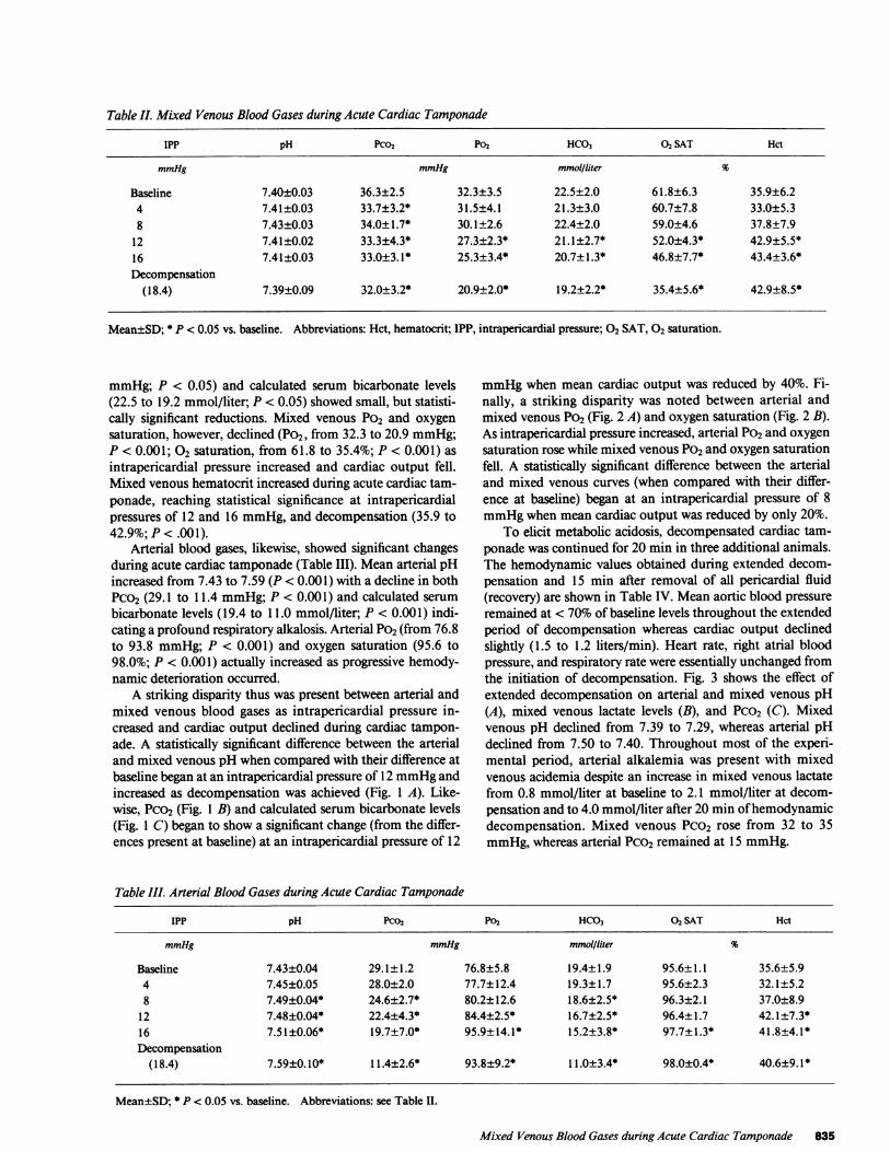
Table II. Mixed Venous Blood Gases during Acute Cardiac Tamponade
IPP pH Pco2 P02 HCO3 02 SAT Hct
mmHg mmHg mmol/liter %
Baseline 7.40±0.03 36.3±2.5 32.3±3.5 22.5±2.0 61.8±6.3 35.9±6.24 7.41±0.03 33.7±3.2* 31.5±4.1 21.3±3.0 60.7±7.8 33.0±5.38 7.43±0.03 34.0±1.7* 30.1±2.6 22.4±2.0 59.0±4.6 37.8±7.9
12 7.41±0.02 33.3±4.3* 27.3±2.3* 21.1±2.7* 52.0±4.3* 42.9±5.5*16 7.41±0.03 33.0±3.1* 25.3±3.4* 20.7±1.3* 46.8±7.7* 43.4±3.6*Decompensation
(18.4) 7.39±0.09 32.0±3.2* 20.9±2.0* 19.2±2.2* 35.4±5.6* 42.9±8.5*
Mean±SD; * P < 0.05 vs. baseline. Abbreviations: Hct, hematocrit; IPP, intrapericardial pressure; 02 SAT, 02 saturation.
mmHg; P < 0.05) and calculated serum bicarbonate levels(22.5 to 19.2 mmol/liter; P < 0.05) showed small, but statisti-cally significant reductions. Mixed venous Po2 and oxygensaturation, however, declined (P02, from 32.3 to 20.9 mmHg;P < 0.001; 02 saturation, from 61.8 to 35.4%; P < 0.001) asintrapericardial pressure increased and cardiac output fell.Mixed venous hematocrit increased during acute cardiac tam-ponade, reaching statistical significance at intrapericardialpressures of 12 and 16 mmHg, and decompensation (35.9 to42.9%; P < .001).
Arterial blood gases, likewise, showed significant changesduring acute cardiac tamponade (Table III). Mean arterial pHincreased from 7.43 to 7.59 (P < 0.001) with a decline in bothPCO2 (29.1 to 11.4 mmHg; P < 0.001) and calculated serumbicarbonate levels (19.4 to 11.0 mmol/liter; P < 0.001) indi-cating a profound respiratory alkalosis. Arterial P02 (from 76.8to 93.8 mmHg; P < 0.001) and oxygen saturation (95.6 to98.0%; P < 0.001) actually increased as progressive hemody-namic deterioration occurred.
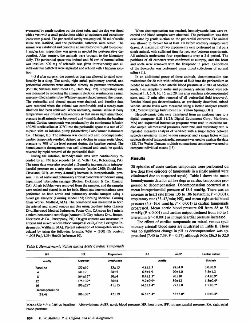
A striking disparity thus was present between arterial andmixed venous blood gases as intrapericardial pressure in-creased and cardiac output declined during cardiac tampon-ade. A statistically significant difference between the arterialand mixed venous pH when compared with their difference atbaseline began at an intrapericardial pressure of 12 mmHgandincreased as decompensation was achieved (Fig. 1 A). Like-wise, Pco2 (Fig. 1 B) and calculated serum bicarbonate levels(Fig. 1 C) began to show a significant change (from the differ-ences present at baseline) at an intrapericardial pressure of 12
mmHgwhen mean cardiac output was reduced by 40%. Fi-nally, a striking disparity was noted between arterial andmixed venous P02 (Fig. 2 A) and oxygen saturation (Fig. 2 B).As intrapericardial pressure increased, arterial P02 and oxygensaturation rose while mixed venous Po2 and oxygen saturationfell. A statistically significant difference between the arterialand mixed venous curves (when compared with their differ-ence at baseline) began at an intrapericardial pressure of 8mmHgwhen mean cardiac output was reduced by only 20%.
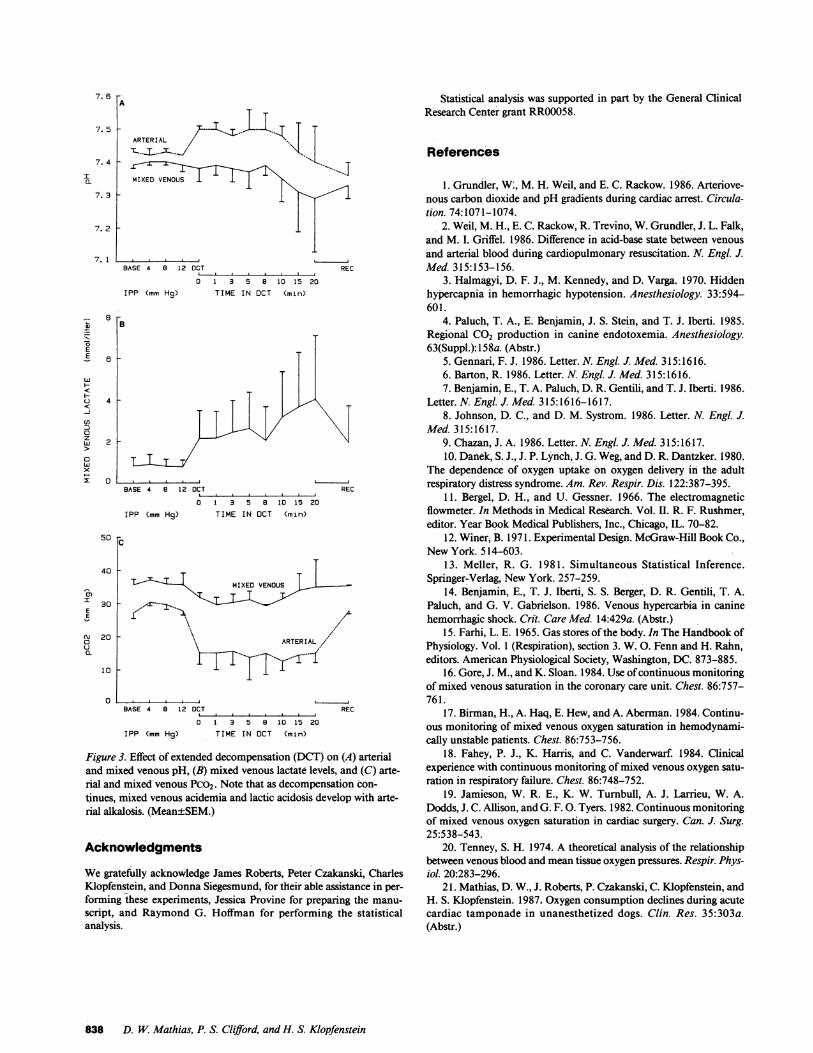
To elicit metabolic acidosis, decompensated cardiac tam-ponade was continued for 20 min in three additional animals.The hemodynamic values obtained during extended decom-pensation and 15 min after removal of all pericardial fluid(recovery) are shown in Table IV. Mean aortic blood pressureremained at < 70% of baseline levels throughout the extendedperiod of decompensation whereas cardiac output declinedslightly (1.5 to 1.2 liters/min). Heart rate, right atrial bloodpressure, and respiratory rate were essentially unchanged fromthe initiation of decompensation. Fig. 3 shows the effect ofextended decompensation on arterial and mixed venous pH(A), mixed venous lactate levels (B), and Pco2 (C). Mixedvenous pH declined from 7.39 to 7.29, whereas arterial pHdeclined from 7.50 to 7.40. Throughout most of the experi-mental period, arterial alkalemia was present with mixedvenous acidemia despite an increase in mixed venous lactatefrom 0.8 mmol/liter at baseline to 2.1 mmol/liter at decom-pensation and to 4.0 mmol/liter after 20 min of hemodynamicdecompensation. Mixed venous Pco2 rose from 32 to 35mmHg,whereas arterial Pco2 remained at 15 mmHg.
Table III. Arterial Blood Gases during Acute Cardiac Tamponade
IPP pH Pco2 Po2 HCO3 02 SAT Hct
mmHg mmHg mmol/liter %
Baseline 7.43±0.04 29.1±1.2 76.8±5.8 19.4±1.9 95.6±1.1 35.6±5.94 7.45±0.05 28.0±2.0 77.7±12.4 19.3±1.7 95.6±2.3 32.1±5.28 7.49±0.04* 24.6±2.7* 80.2±12.6 18.6±2.5* 96.3±2.1 37.0±8.9
12 7.48±0.04* 22.4±4.3* 84.4±2.5* 16.7±2.5* 96.4±1.7 42.1±7.3*16 7.51±0.06* 19.7±7.0* 95.9±14.1* 15.2±3.8* 97.7±1.3* 41.8±4.1*Decompensation
(18.4) 7.59±0.10* 11.4±2.6* 93.8±9.2* 11.0±3.4* 98.0±0.4* 40.6±9.1*
Mean±SD; * P < 0.05 vs. baseline. Abbreviations: see Table II.
Mixed Venous Blood Gases during Acute Cardiac Tamponade 835
120 [A
100 -
0)I
EE
MIXEDVENONUSN0£1.
7.3 1
0 5 10 15 20INTRAPERICARDIAL PRESSURE (mm Hg)
* P < .05- P < .005
25
MIXED VENOUS
' ARTERIAL
0 5 10 15 20INTRAPERICAROIAL PRESSURE (mm Hg)
z
m0-J0
wI
U-0
z0
I-
(1LUIr
CL
a-
25
* P < .05* P ' . 005
JTARTEI- ARTERIAL80 -
60 F
40 F
20
0
120 _
100 _
0 5 10 15 20INTRAPERICARDIAL PRESSURE (mm Hg)
* P <.05*- P < .005
L _ L_-ARTER1AL
80 F
60 _
40 F
20
00 5 10 15 20
INTRAPERICARDIAL PRESSURE (mm Hg)
30 Fc
* P .05P .005
25 F
20C
0
L-
I
*0MIXED VENOUS
15 F
10
5
0
0 5 10 15 20INTRAPERICARDIAL PRESSURE (mm Hg)
25
Figure 1. Effect of increasing intrapericardial pressure on arterial andmixed venous (A) pH, (B) Pco2, and (C) calculated serum bicarbon-
'ate. Note that a statistically significant difference between the arterialand mixed venous curves (when compared with their difference atbaseline) began at an intrapericardial pressure of 12 mmHgand in-creased as decompensation was approached. (Mean±SEM.)
Discussion
Acute cardiac tamponade results in a progressive decline incardiac output, ultimately leading to a shock state. In ourchronic unanesthetized canine model, we found that increas-ing intrapericardial pressure (and the resulting decline in car-
diac output) led to a progressive increase in the gradient be-tween mixed venous and arterial Pco2, with no evidence ofincreased CO2 production. Acute cardiac tamponade pro-duced a profound arterial respiratory alkalosis that achievedstatistical significance before the development of hypotensionwhen cardiac output had decreased by only 40% in these spon-
taneously breathing animals and progressively worsened as he-
Figure 2. Effect of increasing intrapericardial pressure on arterial andmixed venous (A) Po2 and (B) oxygen saturation. Note that once
again a statistically significant difference between the arterial andmixed venous curves (when compared with their difference at base-line) began at an interpericardial pressure of 8 mmHgand increasedas decompensation was approached. (Mean±SEM.)
modynamic deterioration ensued. Mixed venous pH, Pco2,and calculated bicarbonate levels, however, remained rela-tively constant throughout the experimental procedure.
The finding of arterial alkalemia and hypocapnia withmixed venous acidemia and hypercapnia has been reported inshock states. Grundler et al. (1), using an anesthetized ventila-tor-dependent porcine preparation exposed to cardiac arrest,demonstrated a marked venous acidemia and hypercapnia(Pco2, from 45.2 to 54.2 mmHg; pH, 7.41 to 7.31) in thepresence of arterial alkalemia and hypocapnia (Pco2, from37.4 to 20.1 mmHg;pH, 7.46 to 7.54) after successful resusci-tation. Paluch et al. (4), using an anesthetized, mechanicallyventilated canine model of endotoxemia, found a progressiveincrease in mixed venous Pco2 (from 41 to 66 mmHg)with arelatively constant arterial Pco2 (35 to 45 mmHg) 3 h afterinjection of endotoxin. Two studies of canine hemorrhagicshock (3, 14), using anesthetized ventilated dogs, have shownsevere venous hypercapnia with reduced or normal arterialPco2 levels. Wechose to use a conscious unanesthetized ca-
nine model of graded cardiac tamponade breathing room airbecause the effect of incremental decrements in cardiac out-put, such as one would expect to see clinically, may be studiedin this model before the development of and during gradedshock in the absence of the confounding influences of anesthe-sia and artificial ventilation.
The mechanisms responsible for the disparity in acid-base
836 D. W. Mathias, P. S. Clifford, and H. S. Klopfenstein
- P .05A - P ' . 005
7. 7
7. 6
7. 5
I0.
7. 4 -
7.2
50 _
40 _
30 F
NIXED VENOUS
0)I
EE
N0(L
20 F
25
10 _
0
MIXED VENOUS
25
*
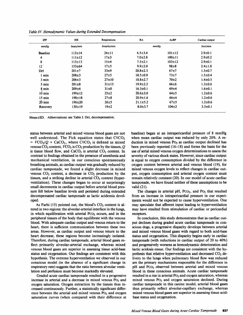
Table IV. Hemodynamic Values durihg Extended Decompensation
IPP HR Respirations RA AoBP Cardiac output
mmHg beats/min breaths/min mmHg liter/mi
Baseline 1 13±14 24±11 6.5±3.4 101±12 2.9±0.14 111±12 17±3 7.0±2.8 100±11 2.9±0.28 113±13 15±4 7.5±2.1 103±12 2.9±0.1
12 153±64 17±5 9.9±2.0 98±8 2.4±1.0Dcd 201±7 33±9 20.8±2.3 67±7 1.5±0.7
1 min 208±3 27±5 18.5±0.9 73±7 1.5±0.43 min 206±3 27±10 18.8±2.7 70+2 1.4±0.55 min 201±8 31±13 19.9±2.2 66±6 1.3±0.68 min 209±6 31±0 16.3±0.1 69±4 1.6±0.1
10 min 199±12 25±2 20.6±3.0 64±5 1.2±0.615 min 198±18 27±8 20.9±1.6 66±4 1.2±0.62b min 196±20 26±5 21.1±5.2 67±5 1.2±0.6Recovery 130±19 19±2 8.0±3.7 104±2 3.3±0.1
Mean±SD. Abbreviations: see Table I. Dct, decompensation.
status between arterial and mixed venous blood gases are notwell understood. The Fick equation states that CvCO2= VCO02Q+ CaCO2, where CvCO2 is defined as mixedvenous CO2content, VCO2is CO2production by the tissues, Qis tissue blood flow, and CaCO2 is arterial CO2 content. Incontrast to findings obtained in the presence of anesthesia andmechanical ventilation, in our conscious spontaneouslybreathing animals, as cardiac output was gradually reduced bycardiac tamponade, we found a slight decrease in mixedvenous CO2 content, a decrease in CO2 production by thetissues, and a striking decline in arterial CO2content (hyper-ventilation). These changes began to occur at surprisinglysmall decrements in cardiac output before arterial blood pres-sure fell below baseline levels and persisted during extendeddecompensated cardiac tamponade as lactic acidemia devel-oped.
As Farhi (15) pointed out, the blood's CO2 content is al-tered in two regions: the alveolar-arterial interface in the lungs,in which equilibration with arterial Pco2 occurs, and in theperipheral tissues of the body that equilibrate with the venousblood. With adequate cardiac output and venous return to theheart, there is sufficient communication between these twoareas. However, as cardiac output and venous return to theheart decrease, these regions become increasingly isolated.Therefore, during cardiac tamponade, arterial blood gases re-flect primarily alveolar-arterial exchange, whereas mixedvenous blood gases are superior in assessing tissue acid-basestatus and oxygenation. Our findings are consistent with thishypothesis. The extreme hyperventilation we observed in ourconscious model (in the absence of a significant change inrespiratory rate) suggests that the ratio between alveolar venti-lation and perfusion must become markedly elevated.
Graded acute cardiac tamponade resulted in a progressiveincrease in arterial and a decrease in mixed venous Po2 andoxygen saturation. Oxygen extraction by the tissues thus in-creased continuously. Further, a statistically significant differ-ence between the arterial and mixed venous Po2 and oxygensaturation curves (when compared with their difference at
baseline) began at an intrapericardial pressure of 8 mmHgwhen mean cardiac output was reduced by only 20%. A re-duction in mixed venous Po2 as cardiac output declined hasbeen previously reported (16-18) and forms the basis for theuse of serial mixed venous oxygen determinations to assess theseverity of various shock states. However, since cardiac outputis equal to oxygen consumption divided by the difference inoxygen content between arterial and venous blood (19), formixed venous oxygen levels to reflect changes in cardiac out-put, oxygen consumption and arterial oxygen content mustremain relatively constant (20). In our model of acute cardiactamponade, we have found neither of these assumptions to bevalid (2 1).
The changes in arterial pH, Pco2, and Po2 that resultedfrom an increase in intrapericardial pressure in our experi-ments would not be expected to cause hyperventilation. Onemay speculate that afferent input leading to hyperventilationmay have resulted from stimulation of cardiac or pericardialreceptors.
In conclusion, this study demonstrates that as cardiac out-put declines during graded acute cardiac tamponade in con-scious dogs, a progressive disparity develops between arterialand mixed venous blood gases with regard to both acid-basestatus and oxygenation. This disparity begins early in cardiactamponade (with reductions in cardiac output of 20 to 40%)and progressively worsens as hemodynamic deterioration andlactic acidosis ensue. Our findings are consistent with the hy-pothesis that relative hyperventilation and decreased CO2de-livery to the lungs when pulmonary blood flow was reducedare the primary mechanisms responsible for the difference inpH and Pco2 observed between arterial and mixed venousblood in these conscious animals. Acute cardiac tamponaderesulted in a rise in arterial P02 and oxygen saturation, whereasmixed venous Po2 and oxygen saturation declined. Duringcardiac tamponade in this canine model, arterial blood gasesthus primarily reflect alveolar-capillary exchange, whereasmixed venous blood gases are superior in assessing tissue acid-base status and oxygenation.
Mixed Venous Blood Gases during Acute Cardiac Tamponade 837
7. 6
7. 5
A
ARTERIAL
7. 4 FMIXED VENOUS
7. 2 F
7. 1
8 [B
6
4
BASE 4 8 12 OCT
0 1 3 5 8 10 15 20IPP (mm Hg) TIME IN DCT (min)
REC
2 _
0BASE 4 8 12 OCT REC
0 1 3 5 8 10 15 20IPP (mm Hg) TIME IN OCT (min)
50 Fc
40 F
30
20
10 F
0
MIXEO VENOUS
,\ ~~~~~/I1. ARTERIAL //
f-TT-T y.-BASE 4 B 12 DCT REC
0 1 3 5 8 10 15 20
IPP (mm Hg) TIME IN OCT (min)
Figure 3. Effect of extended decompensation (DCT) on (A) arterialand mixed venous pH, (B) mixed venous lactate levels, and (C) arte-rial and mixed venous Pco2. Note that as decompensation con-
tinues, mixed venous acidemia and lactic acidosis develop with arte-rial alkalosis. (Mean±SEM.)
Acknowledgments
Wegratefully acknowledge James Roberts, Peter Czakanski, CharlesKlopfenstein, and Donna Siegesmund, for their able assistance in per-
forming these experiments, Jessica Provine for preparing the manu-
script, and Raymond G. Hoffman for performing the statisticalanalysis.
Statistical analysis was supported in part by the General ClinicalResearch Center grant RR00058.
References
1. Grundler, W:, M. H. Weil, and E. C. Rackow. 1986. Arteriove-nous carbon dioxide and pH gradients during cardiac arrest. Circula-tion. 74:1071-1074.
2. Weil, M.. H., E. C. Rackow, R. Trevino, W. Grundler, J. L. Falk,and M. I. Griffel. 1986. Difference in acid-base state between venousand arterial blood during cardiopulmonary resuscitation. N. Engl. J.Med. 315:153-156.
3. Halmagyi, D. F. J., M. Kennedy, and D. Varga. 1970. Hiddenhypercapnia in hemorrhagic hypotension. Anesthesiology. 33:594-601.
4. Paluch, T. A., E.' Benjamin, J. S. Stein, and T. J. Iberti. 1985.Regional CO2 production in canine endotoxemia. Anesthesiology.63(Suppl.): 158a. (Abstr.)
5. Gennari, F. J. 1986. Letter. N. Engl. J. Med. 315:1616.6. Barton, R. 1986. Letter. N. Engl. J. Med. 315:1616.7. Benjamin, E., T. A. Paluch, D. R. Gentili, and T. J. Iberti. 1986.
Letter. N. Engl. J. Med. 315:1616-1617.8. Johnson, D. C., and D. M. Systrom. 1986. Letter. N. Engl. J.
Med. 315:1617.9. Chazan, J. A. 1986. Letter. N. Engl. J. Med. 315:1617.10. Danek, S. J., J. P. LynchJ. G. Weg, and D. R. Dantzker. 1980.
The dependence of oxygen uptake on oxygen delivery in the adultrespiratory distress syndrome. Am. Rev. Respir. Dis. 122:387-395.
11. Bergel, D. H., and U. Gessner. 1966. The electromagneticflowmeter. In Methods in Medical Research. Vol. II. R. F. Rushmer,editor. Year Book Medical Publishers, Inc., Chicago, IL. 70-82.
12. Winer; B. 1971. Experimental Design. McGraw-Hill Book Co.,NewYork. 514-603.
13. Meller, R. G. 1981. Simultaneous Statistical Inference.Springer-Verlag, NewYork. 257-259.
14. Benjamin, E., T. J. Iberti, S. S. Berger, D. R. Gentili, T. A.Paluch, and G. V. Gabrielson. 1986. Venous hypercarbia in caninehemorrhagic shock. Crit. Care Med. 14:429a. (Abstr.)
15. Farhi, L. E. 1965. Gas stores of the body. In The Handbook ofPhysiology. Vol. 1 (Respiration), section 3. W. 0. Fenn and H. Rahn,editors. American Physiological Society, Washington, DC. 873-885.
16. Gore, J. M., and K. Sloan. 1984. Use of continuous monitoringof mixed venous saturation in the coronary care unit. Chest. 86:757-761.
17. Birman, H., A. Haq, E. Hew, and A. Aberman. 1984. Continu-ous monitoring of mixed venous oxygen saturation in hemodynami-cally unstable patients. Chest. 86:753-756.
18. Fahey, P. J., K. Harris, and C. Vanderwarf. 1984. Clinicalexperience with continuous monitoring of mixed venous oxygen satu-ration in respiratory failure. Chest. 86:748-752.
19. Jamieson, W. R. E., K. W. Turnbull, A. J. Larrieu, W. A.Dodds, J. C. Allison, and G. F. 0. Tyers. 1982. Continuous monitoringof mixed venous oxygen saturation in cardiac surgery. Can. J. Surg.25:538-543.
20. Tenney, S. H. 1974. A theoretical analysis of the relationshipbetween venous blood and mean tissue oxygen pressures. Respir. Phys-iol. 20:283-296.
21. Mathias, D. W., J. Roberts, P. Czakanski, C. Klopfenstein, andH. S. Klopfenstein. 1987. Oxygen consumption declines during acutecardiac tamponade in unanesthetized dogs. Clin. Res. 35:303a.(Abstr.)
838 D. W. Mathias, P. S. Clifford, and H. S. Klopfenstein
I0.
7. 3 _
E
<w
H
-J
(nI
0zw
0wxx
IEE
N00.